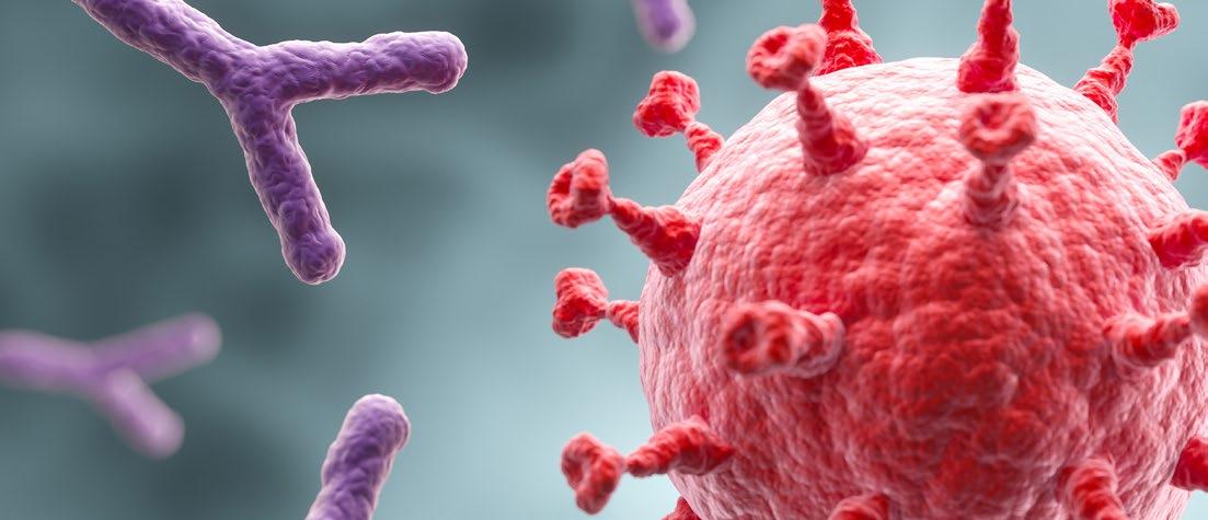
Bispecific Antibodies: A New Frontier in Haematologic Malignancies
Editor's Pick: Anti-BCMA CAR-T Cell Therapy in a HaemodialysisDependent Patient with Relapsed/ Refractory Multiple Myeloma: A First-in-India Case Report Vairamoorthy N et al.
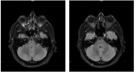
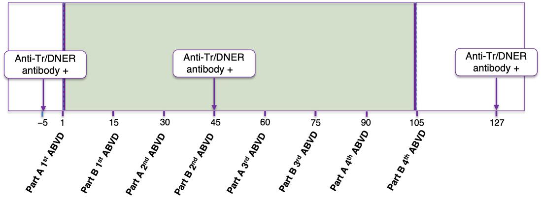
Hodgkin Lymphoma Associated with Paraneoplastic Cerebellar Degeneration with Anti-Tr (DNER) Antibodies: A Case Report López AB et al.
Editorial Board
Dimitar Efremov
Editor-in-Chief
Emanuele Angelucci
Director/Head of the Hematology and Cellular Therapies Unit, Istituto di Ricovero e Cura a Carattere Scientifico (IRCCS), Genova, Italy
Macedonian Academy of Sciences and Arts, Skopje, North Macedonia
David Gómez Almaguer
Universidad Autónoma de Nuevo León (UANL), Monterrey, México
Sabri Kemahli
Yeditepe University, Istanbul, Türkiye
Dominique Bonnet
Francis Crick Institute, London, UK
Loredana Bury
University of Perugia, Italy
Ahmet Muzaffer Demir
Trakya University, Edirne, Türkiye
Khaled Musallam
Burjeel Holdings, Abu Dhabi, United Arab Emirates
Aims and Scope
EMJ Hematology is an open access, peer-reviewed eJournal committed to publishing the highest quality medical research concerning all aspects of diseases of the blood and bone marrow to help advance the development of this field.
The journal is published annually, approximately six weeks after the European Hematology Association (EHA) Congress, and features highlights from this congress, alongside interviews with experts in the field, reviews of abstracts presented at the congress, as well as in-depth features on congress sessions. The journal also covers advances within the clinical and pharmaceutical arenas by publishing sponsored content from congress symposia, which is of high educational value for healthcare professionals. This undergoes rigorous quality control checks by independent experts and the in-house editorial team.
EMJ Hematology also publishes peer-reviewed research papers, review articles, and case reports in the field. In addition, the journal welcomes the submission of features and opinion pieces intended to create a discussion around key topics in the field and broaden readers’ professional interests. The journal is managed by a dedicated editorial team that adheres to a rigorous double-blind peer-review process, maintains high standards of copy editing, and ensures timely publication.
EMJ Hematology endeavours to increase knowledge, stimulate discussion, and contribute to a better understanding of blood disorders. Our focus is on research that is relevant to healthcare professionals in this field. We do not publish veterinary science papers or laboratory studies not linked to patient outcomes. We have a particular interest in topical studies that advance research and inform of coming trends affecting clinical practice in haematology.
Further details on coverage can be found here: www.emjreviews.com
Editorial Expertise
EMJ is supported by various levels of expertise:
• Guidance from an Editorial Board consisting of leading authorities from a wide variety of disciplines.
• Invited contributors who are recognised authorities in their respective fields.
• Peer review, which is conducted by expert reviewers who are invited by the Editorial team and appointed based on their knowledge of a specific topic.
• An experienced team of editors and technical editors.
Peer Review
On submission, all articles are assessed by the editorial team to determine their suitability for the journal and appropriateness for peer review.
Editorial staff, following consultation with a member of the Editorial Board if necessary, identify three appropriate reviewers, who are selected based on their specialist knowledge in the relevant area.
All peer review is double blind. Following review, papers are either accepted without modification, returned to the author(s) to incorporate required changes, or rejected.
Editorial staff have final discretion over any proposed amendments.
Submissions
Submissions
We welcome contributions from professionals, consultants, academics, and industry leaders on relevant and topical subjects. We seek papers with the most current, interesting, and relevant information in each therapeutic area and accept original research, review articles, case reports, and features.
We welcome contributions from professionals, consultants, academics, and industry leaders on relevant and topical subjects. We seek papers with the most current, interesting, and relevant information in each therapeutic area and accept original research, review articles, case reports, and features.
We are always keen to hear from healthcare professionals wishing to discuss potential submissions, please email: editorial.assistant@emjreviews.com
We are always keen to hear from healthcare professionals wishing to discuss potential submissions, please email: editorial.assistant@emjreviews.com
To submit a paper, use our online submission site: https://emj.kriyadocs.com/submissions/submit/emj/emj/login
To submit a paper, use our online submission site: www.editorialmanager.com/e-m-j
Submission details can be found through our website: www.emjreviews.com/contributors/authors
Submission details can be found through our website: www.emjreviews.com/contributors/authors
Reprints
Reprints
All articles included in EMJ are available as reprints (minimum order 1,000). Please contact hello@emjreviews.com if you would like to order reprints.
All articles included in EMJ are available as reprints (minimum order 1,000). Please contact hello@emjreviews.com if you would like to order reprints.
Distribution and Readership
Distribution and Readership
EMJ is distributed through controlled circulation to healthcare professionals in the relevant fields across Europe.
EMJ is distributed through controlled circulation to healthcare professionals in the relevant fields across Europe.
Indexing and Availability
Indexing and Availability
EMJ is indexed on DOAJ, the Royal Society of Medicine, and Google Scholar®
EMJ is indexed on DOAJ, the Royal Society of Medicine, and Google Scholar®
EMJ is available through the websites of our leading partners and collaborating societies. EMJ journals are all available via our website: www.emjreviews.com
EMJ is available through the websites of our leading partners and collaborating societies. EMJ journals are all available via our website: www.emjreviews.com
Open Access
Open Access
This is an open-access journal in accordance with the Creative Commons Attribution-Non Commercial 4.0 (CC BY-NC 4.0) license.
This is an open-access journal in accordance with the Creative Commons Attribution-Non Commercial 4.0 (CC BY-NC 4.0) license.
Congress Notice
Congress Notice
Staff members attend medical congresses as reporters when required.
Staff members attend medical congresses as reporters when required.
This Publication Launch Date: 2013 Frequency: Yearly Online ISSN: 2053-6631
This Publication Launch Date: 2013 Frequency: Yearly Online ISSN: 2053-6631
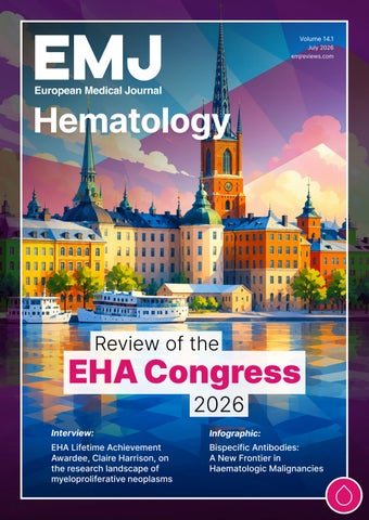
All information obtained by EMJ and each of the contributions from various sources is as current and accurate as possible. However, due to human or mechanical errors, EMJ and the contributors cannot guarantee the accuracy, adequacy, or completeness of any information, and cannot be held responsible for any errors or omissions. Although EMJ is independent of the EHA2026 review event, EMJ has received feedback on the congress interviews, congress features authored by EMJ, and certain aspects of the congress review. The use of the organisations does not constitute endorsement or media partnership in any form whatsoever. The cover photo is of Stockholm, Sweden, the location of EHA2026.
All information obtained by EMJ and each of the contributions from various sources is as current and accurate as possible. However, due to human or mechanical errors, EMJ and the contributors cannot guarantee the accuracy, adequacy, or completeness of any information, and cannot be held responsible for any errors or omissions. EMJ is completely independent of the review event (EHA2024) and the use of the organisations does not constitute endorsement or media partnership in any form whatsoever. The cover photo is of Madrid, Spain, the location of EHA2024.
Front cover and contents photograph: Digitally enhanced image Antony McAulay / stock.adobe.com
Learning through the haemoglobinopathy patient journey
Explore a wide range of educational resources related to the haemoglobinopathy patient journey
BURDEN OF DISEASE AND CLINICAL COMPLICATIONS
Explore interactive case studies on the management of clinical complications experienced by patients with sickle cell disease
Explore case studies
IDENTIFICATION FOR TRANSPLANT AND THE TRANSPLANT PROCESS
Watch a podcast of Dr Jenna Love, Natasha Lewis and Dr Nibras Naami as they share key considerations for discussing transplant with patients
Watch now
DISCHARGE AND FOLLOW UP
Watch a podcast of Dr Marco Zecca, Dr Lena Oevermann and Dr Nandini Sadasivam as they discuss long-term follow up care of patients a er potentially curative treatment
Watch now
Watch as experts, Fabio Giglio and Michelle Kenyon, reflect on key highlights from the 52nd Annual Meeting of the European Society for Blood and Marrow Transplantation (EBMT)
Watch now
Following the recent EHA meeting, look out for a similar highlights video covering key insights from the congress. Coming soon to the GlobeIN website!
Scan the QR code to access the GlobeIN website or visit
www.globeinportal.com
Editorial Director
Andrea Charles
Editor
Sean Boyle
Managing Editor
Darcy Richards
Senior Copy Editor
Noémie Fouarge
Copy Editors
Meghan Garcka, Sarah Jahncke
Editorial Leads
Helena Bradbury, Katrina Thornber, Aleksandra Zurowska
Senior Editorial Co-ordinator
Bertie Pearcey
Editorial Co-ordinators
Jess Nicholson, Alena Sofieva
Editorial Assistants
Niamh Holmes, Josh Lister, Nonyelum Okonkwo, Roli Omamuli
Marketing Director
Stephanie Corbett
Creative Director
Tim Uden
Design Manager
Stacey White
Senior Designers
Tamara Kondolomo, Owen Silcox
Designers
Shanjok Gurung, Fabio van Paris
Junior Designers
Fraser Hoey, Helena Spicer, Caleb Wylie
Senior Business Unit Lead
Nabihah Durrani
Chief Executive Officer
Justin Levett
Chief Commercial Officer
Dan Healy
Founder and Chairman
Spencer Gore
Welcome
Dear Readers,
We are delighted to welcome you to the latest issue of EMJ Hematology, featuring key highlights from the 2026 European Hematology Association (EHA) Congress, and bringing together expert perspectives on some of the most exciting developments shaping the field.
Alongside our congress coverage, we present an infographic examining the growing impact of bispecific antibodies across haematological malignancies, offering an accessible overview of one of the field’s most rapidly advancing therapeutic areas.
Be sure not to miss our exclusive interviews with 2026 EHA Lifetime Achievement Award recipient Claire Harrison, as well as Pieter Sonneveld, Simona Pagliuca, and Nicola Conran, who share their perspectives on key developments across myeloproliferative neoplasms, plasma cell disorders, transplantation, and sickle cell disease. We also speak with Young EHA Committee Chair Nuno Borges and Vice Chair Alba Maiques Diaz about supporting the next generation of haematology professionals and fostering innovation across the field.
Amongst our peer-reviewed content, this issue covers a diverse range of topics, including thrombosis, lymphoma, multiple myeloma, cellular therapies, and the growing influence of the microbiome on haematological health and disease.
This issue reflects both the scientific innovation and clinical expertise that continue to advance the field. We would like to thank our Editorial Board, authors, peer reviewers, and interviewees for their contributions to this issue. We hope you find valuable insights to support your practice and ongoing professional development.
Helena Bradbury, Editorial Lead
Editorial enquiries: editor@emjreviews.com
Sales opportunities: salesadmin@emjreviews.com
Permissions and copyright: accountsreceivable@emjreviews.com
Reprints: info@emjreviews.com
Media enquiries: marketing@emjreviews.com
Stay at the Forefront of Modern Medicine
High-level perspectives. Global experts. Essential updates.
Join us for conversations with the minds shaping healthcare's future. Gain the distilled insights you need to lead in your field and make maximum impact.
Jonathan Sackier: Non Executive Director & CMO, AiM Medical Robotics, Florida, USA
Saranya Ravindran: Paediatric Emergency Medicine Registrar, Imperial College Healthcare NHS Trust
Catherine Glass: Associate NHS GP and Senior Appraiser, NHS England
Foreword
It is a pleasure to present the 2026 edition of EMJ Hematology. This issue showcases a wide range of reviews, interviews, and highlights from the European Hematology Association (EHA) Congress 2026, capturing recent advances in research and the evolving landscape of clinical practice across haematology.
A key feature of this edition is an infographic exploring the expanding role of bispecific antibodies in haematological malignancies, offering a clear overview of one of the field's most promising therapeutic developments.
We are also delighted to include interviews with 2026 EHA Lifetime Achievement Award recipient Claire Harrison, as well as with Pieter Sonneveld, Simona Pagliuca, Nicola Conran, Nuno Borges, and Alba Maiques Diaz. Their conversations provide valuable perspectives on topics including myeloproliferative neoplasms, plasma cell disorders, transplantation, sickle cell disease, and supporting the next generation of haematology professionals.
This issue showcases a wide range of reviews, interviews, and highlights from EHA 2026, capturing recent advances in research and the evolving landscape of clinical practice across haematology
Alongside our Congress coverage, this issue features a selection of peer-reviewed case reports.
Special thanks go to our contributors and the editorial team for their dedication in bringing together this edition, as well as to the Editorial Board, authors, interviewees, and peer reviewers whose expertise and commitment have made it possible. I hope you find the content informative, thought-provoking, and valuable to your clinical practice.
Emanuele
Angelucci
Director/Head
of the Hematology and Cellular Therapies Unit, Istituto di Ricovero e Cura a Carattere Scientifico (IRCCS), Genova, Italy
EHA2026
The future of haematology is bright and filled with curiosity, innovation, and united commitment from healthcare professionals, regulators, industry, and societies to provide the best care possible for patients
Review of the European Hematology Association (EHA) 2026 Congress Congress Review
Location: Stockholm, Sweden
Date: 11th–14th June 2026
Citation:
EMJ Hematol. 2026;14[1]:10-24.
https://doi.org/10.33590/emjhematol/E62G3K61
THE EUROPEAN Hematology Association (EHA) 2026 Congress was a strong testament to the rapidly evolving field of haematology. From the implementation of AI in drug development to the approval of more treatment options for patients, it is clear that the future of haematology is bright and filled with curiosity, innovation, and united commitment from healthcare professionals, regulators, industry, and societies like EHA to provide the best care possible for patients.
More than 14,000 delegates descended on the heart of Sweden for the 2026 EHA Congress. The opening ceremony marked the beginning of an unmissable event, with a full auditorium and an unmistakable sense of anticipation in the air. After an engaging welcome from the EHA President, Konstanze Döhner, Meritxell Alberich Jordà, the Scientific Program Committee Chair, provided an insight into the coming days. This year’s event saw over 575 presentations and a record-breaking number of over 4,700 abstract submissions. Jordà made special note of the plenary sessions, showcasing the highest-scoring abstracts, and the late-breaking abstracts, capturing timely and practice-shaping data.
Dominique Bonnet, chair of the EHA Fellowships and Grants Committee and valued member of EMJ’s own haematology editorial board, subsequently announced the grant winners for this year. This comprised 13 research grants, 6 kick-off grants, 2 bilateral collaborative grants, 2 innovation grants, and 13 research mobility grants. These awards underscored EHA’s continued commitment to supporting
innovative research and fostering the next generation of leaders in haematology.
The EHA lifetime achievement is a notable, prestigious award, recognising an individual’s sustained impact on the field. This year’s award was given to Claire Harrison, Professor of Myeloproliferative Neoplasms and Clinical Director, Guy’s and St Thomas’ NHS Foundation Trust, London, UK. She reflected on her first EHA Congress in 1999, recalling how she arrived with pressing questions about patients in her care. It was at that meeting that she met future mentors, forged lasting professional relationships, and laid the foundations for collaborations that would shape her career. Returning to Guy's and St Thomas', she brought back fresh perspectives and valuable insights that helped inform the management of complex patient cases.
Harrison reflected on several notable trials that she has contributed to, such as the MAJIC-PV study, a randomised Phase II clinical trial that compared ruxolitinib with the best available therapy for patients with polycythaemia vera and resistant/intolerant
to hydroxycarbamide. Notably, the trial was the first to show that achieving a molecular response was linked to improved event-free survival, overall survival, and progressionfree survival.1 Finally, Harrison spotlighted the ‘MPN Voice Team’, a patient group she co-founded in 2006. Comprised of 19 team members, this group aimed to provide information, community, and advocacy for patients with myeloproliferative neoplasm (MPN). Since its establishment, it has grown significantly, reaching over six continents and attracting over 27,000 new users online.
To close, Harrison addressed the earlycareer haematologists in the audience, encouraging them to be courageous, build supportive communities, and keep patients at the centre of everything they do. Reflecting on the mentors, colleagues, and friendships that shaped her own career,
Since its establishment, it has grown significantly, reaching over six continents and attracting over 27,000 new users online
she urged delegates to be patient with their professional progression, celebrate the relationships they forge along the way, and remember that meaningful, lasting change is always a collective effort. EMJ had the pleasure to sit down with Harrison to delve deeper into what this award recognition meant to her and her career journey to date.
Closing the ceremony, the EHA Diversity, Equity, and Inclusion Award was given to Gianluca Gaidano, University of Eastern Piedmont, Novara, Italy; the EHA Education & Mentoring Award went to Raúl Córdoba, MD Anderson Cancer Center Madrid, Spain; and the Young EHA Award went to Nico Gagelmann, University Medical Center Hamburg-Eppendorf, Germany.
So, where does the future of haematology lie? If the opening ceremony was any indication, it rests in the hands of a vibrant and collaborative community of dedicated experts, united by a shared commitment to improving patient care and an unwavering curiosity to deepen our molecular understanding of haematological diseases.
This
year’s event saw over 575 presentations and a recordbreaking number of over
4,700 abstract submissions
Blinatumomab Replacing Intensive Chemotherapy Improves Outcomes in Paediatric High-Risk B-ALL
BLINATUMOMAB reduces toxicity and improves 4-year event-free survival (EFS) in prognostically unfavourable paediatric high-risk (HR) B cell acute lymphoblastic leukaemia (ALL). This Phase III trial, presented at EHA2026, demonstrates that replacing highly toxic chemotherapy elements with blinatumomab provides a safer treatment approach with superior antileukaemia efficacy.2
Blinatumomab is a bispecific T cell engager that targets CD19 expressed on leukaemic cells and has the potential to reduce chemotherapy while improving anti-leukaemia efficacy in newly diagnosed paediatric patients with high-risk acute lymphoblastic leukaemia (HR-ALL).
The trial’s primary objective was to determine whether two highly intensive chemotherapy treatments (HR-2’, HR-3’) could be replaced with two 28-day cycles of blinatumomab to improve EFS by 10% in paediatric patients with HR-ALL.
The trial also had two secondary objectives. The first evaluated whether the experimental blinatumomab arm (EA) could reduce mortality and treatment-related complications when compared to the chemotherapy control arm (CA). The second evaluated minimal residual disease (MRD) in both treatment arms to determine the proportion of patients with suboptimal response.
Patients were randomised to receive either the EA (n=358) or the CA (n=351) following one intensive chemotherapy course (HR-1’).
A planned interim analysis revealed that replacing intensive chemotherapy with blinatumomab increased 4-year EFS from 70.3% in the CA to 83.0% in the EA (hazard ratio: 0.51; 95% CI: 0.35–0.73; p=0.0002).
The cumulative incidence of relapse was lower in the EA compared with the CA (11.8% versus 21.4%; n=31 versus n=57 relapses). Moreover, the rate of isolated CNS relapse was also lower in the EA (0.3%; n=1) compared with the CA (2.5%; n=9).
The trial also demonstrated a more favourable toxicity profile in the EA. Clinically relevant infectious adverse reactions occurred in 22.8% of patients receiving blinatumomab compared with 69.4% in the CA (p<0.001). In the CA, 16 patients (4.7%) experienced 17 lifethreatening adverse reactions compared with one patient (0.3%) in the EA, which was also fatal.
Severe mucositis or stomatitis requiring hospitalisation occurred in 10% of patients receiving chemotherapy compared with 0.3% in the EA.
Blinatumomab reduced MRD levels in 76.9% of patients who were MRD-positive prior to randomisation, compared with 45.8% of those treated with chemotherapy (p<0.0001).
This Phase III trial demonstrates, for the first time, that highly toxic chemotherapy elements can be successfully replaced with blinatumomab in newly diagnosed ALL. It supports blinatumomab as a safer treatment
FrontMIND Phase III Study Meets Primary Endpoint in High-Risk DLBCL
FOR DIFFUSE
large B cell lymphoma (DLBCL), R-CHOP remains the standard first-line therapy, despite approximately 40% of patients being unresponsive. A new clinical trial presented at EHA2026 observed a significant reduction in the risk of disease progression or death with tafasitamab and lenalidomide combined with R-CHOP (Tafa-Len-R-CHOP), compared with R-CHOP alone.
Previously, Phase Ib First-MIND findings showed encouraging results with the combination therapy, CD19-targeted monoclonal antibody. In this new global Phase III, double-blind, placebocontrolled FRONTMIND study, patients aged 18–80 years with previously untreated DLBCL or high-grade B cell lymphoma (International Prognostic Index: adjusted International Prognostic Index: if ≤60 years) were randomised to receive Tafa-Len-R-CHOP or R-CHOP.
The primary endpoint was investigatorassessed progression-free survival (PFS).
Secondary endpoints included event-free survival, overall survival, PET-negative complete response, objective response rate, and quality of life at
Tafa-Len-R-CHOP achieved a statistically significant improvement in PFS compared with R-CHOP alone. After a median followup of 35.2 months, risk of disease progression or death by 25% (hazard ratio: 0.75; PFS rates of 71.1% versus 62.9%.
Among patients with centrally confirmed lymphoma subtypes, the benefit was greater, with a 32% reduction in risk (hazard ratio: 0.68) and 24-month PFS rates of 72.7% versus 62.2%. PFS improvement was observed across molecular subtypes, including activated B cell-like and germinal centre B cell-like disease, as well as across key clinical subgroups.
Secondary endpoints supported the primary findings, with Tafa-Len-RCHOP demonstrating improved event-free survival compared with R-CHOP. Response
time in both arms, and overall survival data remain immature. Safety findings were consistent with the known profiles of tafasitamab, lenalidomide, and R-CHOP.
The results position Tafa-Len-R-CHOP as a potential new first-line option, addressing the need for more effective treatment in high-risk, aggressive B cell lymphomas. The regimen shows clinical relevance across molecular subtypes, giving it broad applicability. Longer-term follow-up, including final overall survival analyses, will be important to fully define the durability of benefit and long-term clinical impact in this high-risk patient population.
Phase III Trial Tests Oral Mitapivat for Sickle Cell Disease
DATA from a Phase III trial presented at EHA2026 have demonstrated that oral mitapivat raises haemoglobin in patients with sickle cell disease, but does not significantly cut the rate of painful crises.4
Persistent anaemia and the ongoing destruction of red blood cells fuel acute and chronic complications in sickle cell disease and shorten lives. Mitapivat activates pyruvate kinase, raising cellular energy in the form of ATP and lowering 2,3-diphosphoglycerate, changes that could ease haemolytic anaemia and reduce the sickling of red cells that underlie the disease.
The global, double-blind, randomised, placebo-controlled Phase III RISE UP trial allocated 207 patients 2:1 to oral mitapivat 100 mg or placebo twice daily. The median age was 25 years and 57.5% were female, with similar baseline haemoglobin in the two arms (around 8.6 g/dL). The co-primary endpoints were a haemoglobin response, defined as an average rise of at least 1.0 g/ dL from Week 24 through Week 52, and the annualised rate of sickle cell pain crises.
A haemoglobin response was achieved by 40.6% of patients on mitapivat versus just 2.9% on placebo (p<0.0001). However, the annualised pain crisis rate did not differ significantly (2.62 versus 3.05;
p=0.1213). Mitapivat significantly improved haemoglobin concentration (least-squares mean difference: 0.74 g/dL; 95% CI: 0.49–1.00; p<0.0001) and indirect bilirubin (−16.91 µmol/L; 95% CI: −21.01–−12.81; p<0.0001). Among responders, haemoglobin rose by a mean of 1.64 g/dL, and they had lower crisis rates, fewer related hospitalisations, and a clinically meaningful improvement in fatigue. Serious adverse events occurred in 20.3% on mitapivat and 29.0% on placebo, with no new safety signals.
The authors concluded that mitapivat produced a significant haemoglobin response and improved haemolysis markers, with responders gaining further benefits across pain crises, hospitalisations, and fatigue. They positioned the oral drug as a convenient, potentially diseasemodifying option for sickle cell disease, while noting the trial missed its coprimary pain crisis endpoint. The post-hoc responder benefits, though encouraging, would need prospective confirmation before reshaping practice.
Mitapivat produced a significant haemoglobin response and improved haemolysis markers
Talquetamab-Based Regimens Improve Survival Outcomes in Relapsed/Refractory Multiple Myeloma
TALQUETAMAB-BASED combinations significantly improved progression-free survival and response rates compared with standard treatment in patients with relapsed or refractory multiple myeloma (RRMM), according to results from the Phase III MonumenTAL-3 trial presented at EHA2026.5
88%
More than 88% of patients achieved a response in the Tal-DP and Tal-D groups, versus 77.6% with DPd
The study enrolled 864 patients with RRMM who had received at least one prior line of therapy, including lenalidomide and a proteasome inhibitor. Participants were randomised to receive talquetamab plus daratumumab and pomalidomide (Tal-DP), talquetamab plus daratumumab (Tal-D), or daratumumab, pomalidomide, and dexamethasone (DPd).
After a median follow-up of 24.6 months, both talquetamab-containing regimens significantly reduced the risk of disease progression or death compared with DPd. Two-year progression-free survival rates were 81.3% for Tal-DP and 77.6% for Tal-D, compared with 51.2% for the standard treatment arm.
Response rates were also higher with the talquetamab combinations. More than 88% of patients achieved a response in the TalDP and Tal-D groups, versus 77.6% with DPd. Deep responses were notably more common, with complete response rates of approximately 70% in both talquetamab arms compared with 34.5% in the control
group. Minimal residual disease negativity was also substantially improved.
Importantly, both Tal-DP and Tal-D demonstrated significant overall survival benefits. At the time of analysis, approximately 70% of patients receiving talquetamab-based therapy remained on treatment, compared with 47.3% of those receiving DPd.
The safety profile was consistent with previous talquetamab studies. Cytokine release syndrome and neurotoxicity events were mostly low grade and manageable, while treatment discontinuation rates remained low across all treatment arms.
The investigators concluded that talquetamab combined with daratumumab, with or without pomalidomide, delivers clinically meaningful improvements in disease control and survival, supporting its use as a potential new standard of care for patients with RRMM as early as secondline treatment.
CD5 Inhibition May Boost T Cell Engager Efficacy Across
Tumours
A STUDY, presented at EHA2026, identified CD5 as a key intrinsic regulator limiting the activity of CD3-engaging T cell engagers (TCE), including the CD19×CD3 bispecific antibody blinatumomab. While TCEs have transformed outcomes in B cell acute lymphoblastic leukaemia (B-ALL), variable responses and relapse remain significant challenges, with mechanisms of T cell resistance incompletely understood.6
Using genome-wide and targeted CRISPR screens in primary human CD8+ T cells exposed to blinatumomab and B-ALL cells, investigators identified CD5 as a negative regulator of T cell activation. Genetic deletion or antibody-mediated blockade of CD5 enhanced TCE-driven cytotoxicity against both B-ALL cell lines and primary patient samples, improving leukaemia control and survival in xenograft models.
Mechanistically, CD5 inhibition amplified T cell activation, increasing expression of activation markers (CD25, CD69), cytokine production (interferon γ, TNFα), proliferation, and effector transcriptional programmes. Multi-omic analyses demonstrated activation of pathways associated with antitumour function, including IL-2/signal transducer and activator of transcription 5 (STAT5), TNFα/NF-κB, and mTORC1 signalling, with increased extracellular signal-regulated
Further mechanistic studies suggested that CD5 functions as an inhibitory TCRassociated signalling complex, interacting with proximal signalling proteins including LCK, ZAP70, CBLB, and UBASH3A. Antibody engagement triggered CD5 internalisation and RAB7A-dependent lysosomal degradation, supporting a model in which CD5 restrains T cell activation through modulation of TCR signalling.
Importantly, the effect was not limited to blinatumomab. CD5 inhibition enhanced the activity of multiple CD3-engaging bispecific antibodies targeting CD20, BCMA, GPRC5D, DLL3, and GD2 across haematological and solid tumour models, suggesting broad applicability.
These findings position CD5 as a potential universal immune-modulatory target to optimise TCE therapies by releasing an
In Vivo CAR-T Demonstrates Encouraging
CAR-T therapy study, presented at EHA2026, suggest that generating CAR-T cells directly within patients may offer a new treatment approach for relapsed or refractory B cell
is a group of blood cancers that develops from B CAR-T therapies have improved outcomes for patients whose disease has returned or stopped responding to treatment, their use can be limited by complex manufacturing requirements and the need for lymphodepletion before infusion. LB2501 is designed to overcome these limitations by generating
assessed the safety, pharmacokinetics, and preliminary I trial involving patients with measurable relapsed or refractory B cell non-Hodgkin lymphoma who had primary refractory disease or progression after previous lines of therapy. Twelve patients received a single intravenous infusion of LB2501 without lymphodepletion
No dose-limiting toxicities, serious adverse events, or treatmentrelated deaths were reported. Infusion-related reactions occurred in 1 or 2 and resolved within a median days without requiring tocilizumab or glucocorticoids. Cytokine release syndrome was reported in 66.7% of patients and was limited to Grade 1 or 2 events. No cases of immune effector cell-associated neurotoxicity ≥3 lentiviral vector- and CAR-Trelated adverse events were limited to decreases in lymphocyte and
The treatment also demonstrated encouraging early activity. The objective response rate across all patients was 50% (6/12), including At the higher dose level, the overall response rate reached 100% (6/6), while the complete response rate was 83.3% (5/6). All responses remained ongoing at the
CAR-T cell expansion, Researchers also reported cell specific, polyclonal, and diverse, supporting and follow-up remains limited, with a median follow-up of 2.2 months at the higher dose level. Longer-term data will be needed to assess the durability of responses and safety profile. However, these early findings suggest that in vivo CAR-T therapy could become a more accessible immunotherapy approach for patients with B cell malignancies, although further study is needed to confirm its long-term clinical potential.
Mezigdomide Improves Outcomes in Relapsed/ Refractory Multiple Myeloma
RELAPSED/REFRACTORY multiple myeloma (RRMM) remains challenging to treat, particularly in patients previously exposed to both anti-CD38 monoclonal antibodies and lenalidomide, as treatment options become increasingly limited. Researchers at EHA2026 presented new data on mezigdomide, a novel oral cereblon E3 ligase modulator (CELMoD) that promotes rapid degradation of Ikaros and Aiolos proteins, resulting in enhanced myeloma cell death and immune activation.8
The Phase III SUCCESSOR-2 trial evaluated whether the addition of mezigdomide to carfilzomib and dexamethasone (MeziKd) could improve outcomes compared with carfilzomib and dexamethasone alone (Kd). The key finding was a doubling of median progression-free survival from 8.3 to 18.0 months with MeziKd.
SUCCESSOR-2 was a randomised, twostage, inferentially seamless Phase III study enrolling adults with RRMM who had received at least one prior line of therapy, including both an anti-CD38 monoclonal antibody and lenalidomide. During Stage 1, patients were randomised to receive one of three mezigdomide dose levels combined with Kd or Kd alone, with the optimal mezigdomide dose selected for Stage 2. Mezigdomide 1.0 mg was chosen for further evaluation. The primary endpoint was progression-free survival, while secondary endpoints included overall survival, overall response rate, and safety.
A total of 479 patients were included in the analysis, comprising 288 patients receiving MeziKd and 191 receiving Kd. The median age was 68 years, with 25.1% aged 75 years or older. Most patients (92.1%) were triple-class exposed, 85.8% were refractory to anti-CD38 therapy, and 75.8% were refractory to lenalidomide. After a median follow-up of 10.6 months, MeziKd
significantly improved progression-free survival compared with Kd (18.0 versus 8.3 months; hazard ratio: 0.48; 95% CI: 0.36–0.63; p<0.0001). Overall response rates were higher with MeziKd (80.2% versus 53.4%), as were complete response rates (26.7% versus 8.9%). Grade 3–4 adverse events occurred more frequently with MeziKd (83.7% versus 56.5%), including neutropenia (61.1% versus 9.1%) and infections (34.0% versus 15.6%).
These findings demonstrate a clinically meaningful benefit of MeziKd in a heavily pretreated population with substantial unmet need. The marked improvement in progression-free survival and response rates suggests that MeziKd could become an important treatment option, potentially establishing a new standard of care for patients experiencing first or subsequent relapse. From a clinical practice perspective, the oral administration of mezigdomide and its efficacy across multiple patient subgroups are particularly encouraging. However, a higher incidence of severe adverse events, especially neutropenia and infections, highlights the need for careful monitoring and supportive care. Limitations of this study include the relatively short median follow-up and the immature overall survival data, which warrant further evaluation with longer-term follow-up.
BCMA CAR-T Therapy Shows Durable Drug-Free Remission in Refractory ITP
A FIRST-IN-HUMAN Phase I study presented at EHA2026 suggests that B cell maturation antigen (BCMA)-targeted CAR-T cell therapy may induce durable drug-free remission in patients with refractory primary immune thrombocytopenia (ITP) by resetting the immune system and eliminating disease-driving autoantibody-producing cells.9
Primary ITP is an autoimmune disorder characterised by immune-mediated platelet destruction, resulting in an increased risk of bleeding. While current therapies can temporarily increase platelet counts, many patients relapse or become refractory to treatment. Individuals with platelet-specific autoantibodies, particularly anti-GPIb/ IX antibodies, are known to have more severe disease and poorer responses to conventional therapies.
Researchers investigated whether targeting BCMA, which is expressed on plasma cells responsible for producing pathogenic antibodies, could provide a more durable therapeutic approach.
The Phase I, single-centre, open-label trial enrolled adults with refractory primary ITP who had disease for at least 6 months, platelet-specific autoantibody positivity, platelet counts below 30×10⁹ /L, and failure of multiple first- and second-line
treatments. Six patients received BCMA CAR-T cells across three dose levels, although two were excluded from efficacy analyses after one was subsequently diagnosed with myelodysplastic syndrome and another withdrew from the study. The remaining four evaluable patients had longstanding disease (2–12 years) and had previously failed corticosteroids, intravenous Ig, thrombopoietin receptor agonists, and rituximab.
This first-in-human study provides early evidence that BCMA CAR-T therapy may offer a potentially disease-modifying approach for refractory primary ITP
Three of the four patients achieved complete remission within 14 days of
Three of the four patients achieved complete remission within 14 days of treatment and remained in drug-free remission for between 6–14 months
treatment and remained in drug-free remission for between 6–14 months. The fourth patient achieved complete remission by Day 28 but relapsed after 9 months.
CAR-T cells reached peak expansion 14 days after infusion and remained detectable for approximately 3 months. Platelet-specific autoantibodies remained persistently suppressed in patients with sustained responses, while soluble BCMA levels fell rapidly following treatment.
Beyond clinical responses, extensive immune profiling suggested that BCMA CAR-T therapy induced comprehensive immune remodelling rather than transient immune depletion. Reconstituted B cells predominantly displayed immature, naïve, and non-switched memory phenotypes, while class-switched memory B cells and plasmablast-like populations remained reduced. Single-cell sequencing demonstrated reduced inflammatory, complement, platelet-associated, and
antigen-presentation pathways, alongside expansion of cytotoxic CD8-positive T cell programmes, supporting the concept of long-term immune resetting.
Treatment was well tolerated, with only Grade 1–2 cytokine release syndrome reported. No patients experienced immune effector cell-associated neurotoxicity syndrome (ICANS), Grade 3 or higher immune effector cell-associated haematotoxicity, or serious infections.
Although limited by the very small sample size and single-centre design, this firstin-human study provides early evidence that BCMA CAR-T therapy may offer a potentially disease-modifying approach for refractory primary ITP by directly targeting pathogenic plasma cells and reprogramming the immune system. Larger studies with longer follow-up are currently underway to confirm the durability, safety, and broader applicability of these findings.
Selinexor Plus Ruxolitinib Improves Spleen Volume Reduction in Myelofibrosis
PATIENTS with myelofibrosis who were treated with selinexor (S) plus ruxolitinib (R) instead of the traditional frontline therapy of using R alone, experienced deep and sustained spleen volume reductions, had similar improvements in symptoms, and had a manageable safety profile in a Phase III SENTRY trial presented at EHA2026.10
Myelofibrosis is a myeloproliferative neoplasm (MPN) associated with splenomegaly, debilitating symptoms, and reduced survival. Traditional therapy involves the use of the JAK inhibitor (JAKi), R, but only ~1/3 of R-treated patients achieved spleen volume reduction ≥35% (SVR35). S is an XPO1-mediated nuclear export inhibitor that synergises with R in MPN models and has biological activity in myelofibrosis.
In this study, 353 patients were randomised 2:1 to receive either S 60 mg once weekly plus R (n=235) or placebo plus R (n=118) to evaluate the use of S in conjunction with R in individuals with JAKi–naïve myelofibrosis. SVR35 and absolute mean change in total symptom score (AbsTSS) excluding fatigue at Week 24 were co-primary endpoints, with safety and overall survival (OS) included as secondary endpoints.
In the S+R arm, Week 24 SVR35 was achieved in 49.8% of patients compared to 28% in the R treatment arm (odds ratio: 2.58; 95% CI: 1.60–4.17; p<0.0001). Responses were rapid and sustained, with higher spleen response rates already evident at Week 12 and maintained through Week 36 (Week 12: 49.4% versus 20.3%; Week 36: 46.9% versus 23.0%; any time:
67.7% versus 44.9%). There was a greater mean spleen volume reduction in the S+R arm at Week 24 (−40.0%) when compared to R alone (−26.7%).
Symptom improvement was similar between treatment groups, with no significant difference in AbsTSS at Week 24 (adjusted mean difference: 0.97; p=0.825).
A promising OS signal was observed with S+R compared with R alone (hazard ratio: 0.43; 95% CI: 0.19–1.00; nominal p=0.022).
Landmark analysis showed that patients who achieved SVR35 demonstrated better survival outcomes, with 98% of responders alive at Week 72 compared with 88% of non-responders.
The safety profile of S+R was manageable and consistent with combination therapy. Grade ≥3 treatment-emergent adverse events occurred more frequently with S+R than with R alone (70.1% versus 50.0%), while rates of adverse events leading to death were low in both arms (0.9% versus 2.6%). Rates of leukaemic transformation were identical between groups (1.7%).
Additional analyses showed that achieving SVR35 was associated with improved OS in both the Phase III and supportive
To conclude, the encouraging OS findings and association between SVR35 and survival outcomes suggest that S+R may represent an important advance in the treatment of patients with JAKi-naïve myelofibrosis.
Phase III Trial Evaluates Gilteritinib for FLT3-Mutated
AML
GILTERITINIB combined with intensive chemotherapy did not improve overall survival compared with midostaurin in patients with newly diagnosed FLT3-mutated acute myeloid leukaemia, according to Phase III trial data, despite reducing relapse rates and prolonging event-free survival. The findings, presented at EHA2026, suggest that the observed reduction in relapse did not translate into a survival advantage, potentially because of differences in salvage treatment after relapse.11
The Phase III HOVON156/AMLSG28-18/ PASHA trial enrolled 768 adults with newly diagnosed FLT3-mutated acute myeloid leukaemia who were eligible for intensive chemotherapy. Participants were randomly assigned to receive induction and consolidation chemotherapy with either gilteritinib or midostaurin, followed by maintenance therapy with the same treatment for 1 year after consolidation or haematopoietic stem cell transplantation, where appropriate. The primary endpoint was overall survival, while secondary endpoints included event-free survival, complete remission rate, relapse-free survival, and safety.
After a median follow-up of 43.2 months, median overall survival was not reached in either treatment group, and no significant difference was observed between gilteritinib and midostaurin (hazard ratio: 1.02; 95% CI: 0.81–1.28; p=0.864). Median event-free survival was longer with gilteritinib at 51.1 months compared with 19.9 months for midostaurin, although this narrowly missed statistical significance (hazard ratio: 0.83; 95% CI: 0.68–1.02; p=0.052).
Complete remission rates following induction therapy were similar between treatment groups at 79% with gilteritinib and 83% with midostaurin. However, patients receiving gilteritinib experienced significantly fewer morphological relapses after remission, with relapse rates of 21% compared with 36% in the midostaurin group (hazard ratio: 0.68; 95% CI: 0.54–0.88; p=0.003). Rates of allogeneic haematopoietic stem cell transplantation
performed according to protocol were comparable between groups at 38% and 37%, respectively.
The data also highlighted differences after relapse. Among patients who relapsed, median overall survival was shorter in the gilteritinib group at 7.2 months compared with 10.2 months in the midostaurin group (hazard ratio: 1.54; 95% CI: 1.06–2.23; p=0.022). Investigators noted that 50% of patients initially treated with midostaurin subsequently received gilteritinib as salvage therapy, compared with 17% of those initially assigned to gilteritinib. In addition, 34% of patients in the midostaurin group underwent allogeneic haematopoietic stem cell transplantation after relapse compared with 22% in the gilteritinib group.
Treatment-related adverse events occurred in 40% of patients in both treatment groups. Grade 3 or higher treatment-
References
1. Harrison CN et al. Ruxolitinib versus best available therapy for polycythemia vera intolerant or resistant to hydroxycarbamide in a randomized trial. J Clin Oncol. 2023;41(19):3534-44.
2. Schrappe M et al. Replacement of high-dose combination chemotherapy with blinatumomab in newly diagnosed pediatric high-risk B-cell ALL improves efficacy and safety in the randomized phase 3 AIEOP-BFM ALL 2017 trial. Abstract S103. EHA Congress, 11-14 June, 2026.
3. Lenz G et al. Tafasitamab plus lenalidomide and R-CHOP for patients with previously untreated diffuse large B-cell lymphoma (DLBCL): results from the phase 3 FRONTMIND study. Abstract S101. EHA Congress, 11-14 June, 2026.
4. Andemariam B et al. Efficacy and safety of mitapivat in sickle cell disease: results from the global,
related adverse events were reported in 23% of patients receiving gilteritinib and 19% receiving midostaurin, while serious adverse events occurred in 68% and 59% of patients, respectively.
Overall, the trial did not meet its primary endpoint of improving overall survival. Although gilteritinib reduced relapse and showed evidence of longer event-free survival, these benefits were not reflected in overall survival, likely because of more frequent use of gilteritinib and allogeneic haematopoietic stem cell transplantation as salvage therapy among patients initially treated with midostaurin.
Complete remission rates following induction therapy were similar between treatment groups at 79% with gilteritinib and 83% with midostaurin
5. Voorhees P et al. Phase 3, randomized study of talquetamab (Tal) plus daratumumab (Dara) ± pomalidomide (Pom) vs dara plus pom and dexamethasone (Dpd) in relapsed/ refractory multiple myeloma (Rrmm): monumental-3. Abstract S100. EHA Congress, 11-14 June, 2026.
6. Wang W et al. CD5 inhibition as a generalizable strategy to amplify T cell-mediated anti-tumor effects of bispecific T-cell engagers. Abstract S104. EHA Congress, 11-14 June, 2026.
7. Qu X et al. First-in human trial of LB2501, an in vivo CD19/CD20 dual targeting CAR-T therapy, in relapsed/ refractory B-cell NHL. Abstract LB5006. EHA Congress, 11-14 June, 2026.
8. Dimopoulos MA et al. Mezigdomide, carfilzomib, and dexamethasone (MeziKd) vs carfilzomib and
dexamethasone (Kd) in relapsed/ refractory multiple myeloma (RRMM): results from the phase 3 successor-2 trial. Abstract LB5004. EHA Congress, 11-14 June, 2026.
9. Shu J et al. BCMA CAR-T cell therapy induces comprehensive immune resetting and durable remission in refractory primary immune thrombocytopenia. Abstract LB5003. EHA Congress, 11-14 June, 2026.
10. Harrison C et al. Selinexor plus ruxolitinib in Janus kinase inhibitor–naïve myelofibrosis: phase 3 SENTRY trial. Abstract LB5002. EHA Congress, 11-14 June, 2026.
11. Raaijmakers M et al. Gilteritinib versus midostaurin in patients with newly diagnosed flt3-mutated acute myeloid leukemia eligible for intensive therapy: results from the phase 3 HOVON156/AMLSG28-18/PASHA trial. Abstract LB5005. EHA Congress, 11-14 June, 2026.
Evolving Concepts and Therapies in Sickle Cell Anaemia and HbSC Disease
Authors: *Nicola Conran,1 David Rees2
1. Haematology and Transfusion Centre, University of Campinas (UNICAMP), Sao Paulo, Brazil
2. King’s College Hospital, London, UK
*Correspondence to conran@unicamp.br
Disclosure:
Keywords:
Conran has received grants from the Sao Paulo Research Foundation and Novartis AG; and participated in data safety monitoring or advisory boards for Novo Nordisk. Rees has participated on data safety monitoring or advisory boards for MiNA Therapeutics, Novo Nordisk, Octa Pharma, CSL Behring, Disc Therapeutics, Nuvamid, and Novartis.
THIS YEAR'S EHA Congress highlighted the expanded interest in sickle cell anaemia (SCA) and sickle cell syndromes, reflecting the diseases' growing footprint in Europe. Mapping the pathophysiology of distinct conditions like haemoglobin SC (HbSC) disease remains crucial for designing much-needed new therapies. Pipeline updates drew significant attention, alongside sessions highlighting SCA as a prime target for gene-editing breakthroughs.
TRANSLATIONAL RESEARCH GAINS MOMENTUM
It was an exciting time for championing progress in sickle cell disease (SCD) at the European Hematology Association (EHA) Congress 2026. The Congress highlighted a growing interest in translational research in the field, alongside updates on the latest therapies and advances in managing this lifelong, historically neglected, and devastating disease.
TARGETING INFLAMMASOMEDRIVEN DISEASE PATHWAYS
Understanding the pathophysiology of SCD remains fundamental to developing new therapeutic approaches. The inflammatory pathways driving many of its complications were brought into focus during a session on the ‘Inflammasome and its role in congenital anaemias’. In addition to exploring how therapeutic mRNAs might
disrupt inflammasome assembly and restore erythropoiesis, the session highlighted how haemolysis can trigger this molecular signalling platform.1 Once activated, it promotes abnormal vascular responses, haemodynamic disturbances, and impaired tissue perfusion.2 Together, these findings strengthen the rationale for therapies in SCD targeting the inflammasome machinery or its downstream cytokines, such as IL-1β.
NEW INSIGHTS INTO DISEASE COMPLICATIONS
Other translational sessions highlighted exciting advances. One oral presentation identified a novel role for ‘inflammaging’ in the development of sickle cell cardiomyopathy, demonstrating that the microtubule inhibitor, colchicine, displayed cardioprotective effects in a mouse model. Another presentation described an integrated plasma-placental
atlas of SCD pregnancy, revealing a dynamic signature of systemic vascular and inflammatory dysregulation spanning endothelial, coagulation, immune, and complement pathways. These findings could be important for identifying actionable biomarkers to improve risk stratification and mitigate adverse pregnancy outcomes.
THE NEXT GENERATION OF HbF THERAPIES
Progress in the drug pipeline was heavily featured in the session on ‘Future Treatments in Sickle Cell Disease’, which highlighted small molecules aimed at inducing fetal haemoglobin (HbF) production. These molecules broadly fall into three categories: epigenetic modulators, agents that suppress or displace γ-globin repressors, and compounds that modulate cellular signalling. Current strategies include optimising existing HbF inducers (e.g., NDec), repurposing established drugs like panobinostat, and utilising computational and structure-based drug discovery to identify novel molecules featuring new mechanisms of action, such as targeted protein degradation.
ADVANCING IN VIVO GENE EDITING AND SHOWCASING CLINICAL TRIAL DATA
The Congress also saw evolving science placing in vivo gene editing at the forefront of curative strategies, with efforts directed towards improving the delivery of these constructs to haematopoietic stem cells.3 One approach discussed was the use of lipid nanoparticles to deliver ‘Gene Writers’ directly to these target cells.
Clinical trial findings have reinforced the potential of the latest diseasemodifying therapies in development. Oral presentations included efficacy and safety data for mitapivat (from the RISE UP Phase III trial; NCT05031780), providing the first validation of pyruvate kinase activation as a therapeutic strategy in SCD, where significant improvements in haemoglobin levels and reduced transfusion requirements were reported. Key findings from the Phase II ASCENT1 study (NCT05405114) of the oral HbF inducer NDec (tetrahydrouridinedecitabine) in adults were also presented. The trial demonstrated that NDec acts as a direct inducer of HbF, even in patients with poor responses to hydroxyurea, with a twice-weekly regimen achieving significant,
sustained increases in total haemoglobin, HbF percentage, and F cells, alongside a favourable benefit–risk profile. This regimen is now progressing to Phase III evaluation. Finally, Phase Ib data for pociredir (NCT05169580) showed that a once-daily treatment for 12 weeks (at 12 or 20 mg) was generally well tolerated and resulted in clinically meaningful, dose-dependent HbF induction, supporting its further clinical development.
RECOGNISING HbSC AS A DISTINCT CONDITION
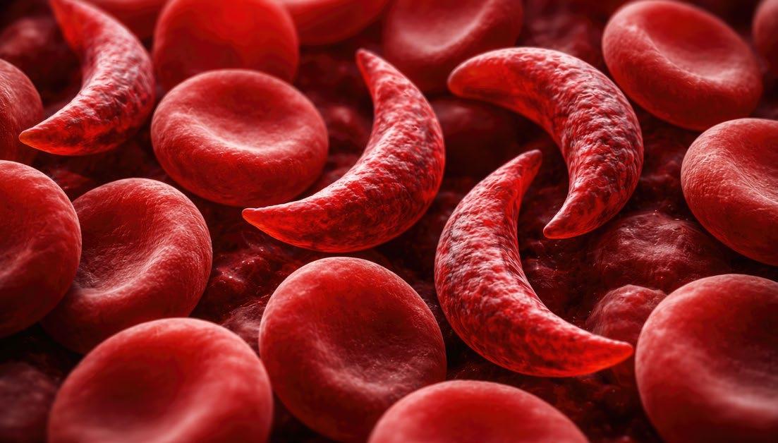
An emerging concept emphasised at the Congress, in the ‘HbSC disease’ session, is that HbSC disease should be considered a distinct disease entity rather than a mere variant of classical HbSS (sickle cell anaemia). Affecting an estimated 1–2 million people worldwide, HbSC is even more neglected than HbSS, particularly regarding clinical trial inclusion and understanding its pathophysiology, despite being associated with significant morbidity, including retinopathy and splenomegaly.
The biology of HbSC disease is unique; HbSC red blood cells sickle less readily than HbSS cells, but are highly prone to dehydration, resulting in reduced haemolysis, increased blood viscosity, and a distinct clinical phenotype.4 Representing a promising advance, new murine models that successfully recapitulate the key features of HbSC disease will provide a vital platform for future mechanistic and translational
studies.5 Overall, this growing recognition of HbSC as a biologically distinct condition highlights the need to move beyond a one-size-fits-all approach to SCD. While interest is increasing in strategies targeting the K-Cl cotransporter, oxidative stress, inflammation, and iron homeostasis, these needs are finally beginning to be addressed. This shift coincides with the evolving EHA recommendations that are currently under review, which include the selective use of hydroxyurea in children and adults with HbSC, and the use of phlebotomy, under certain circumstances, in patients aged 12 years or older.
HbSC disease should be considered a distinct disease entity rather than a mere variant of classical HbSS
CONCLUSION
Taken together, the achievements shared at EHA 2026 reflect a clear shift in the sickle cell landscape, from in vivo gene editing approaches to a more tailored view of HbSC disease, with the field shifting from generalised management towards targeted and potentially curative strategies. For the millions of people living with these conditions worldwide, these advances represent important steps forward in shaping novel therapeutic possibilities.
References
1. Tyrkalska SD et al. Inflammasome regulates hematopoiesis through cleavage of the Master Erythroid Transcription Factor GATA1. Immunity 2019;51(1):50-63.e5.
2. Brito PL et al. Caspase-1 activation drives vascular inflammatory processes and hypoperfusion in intravascular hemolysis. Am J
Physiol Heart Circ Physiol. 2025;329(4):H920-38.
3. Fontana L et al. Multiplex base editing of BCL11A regulatory elements to treat sickle cell disease. Cell Rep Med. 2025;6(10):102376.
4. Traets MJM et al. Iron deficiency in HbSC disease treated with repetitive phlebotomy is associated with
fewer sickle cell disease-related complications. Am J Hematol. 2025;100(11):2123-7.
5. Setayesh T et al. A novel mouse model of hemoglobin SC disease reveals mechanisms underlying beneficial effects of hydroxyurea. Blood. 2025;146(1):13-28.
From Microbial Ecology to Malignancy: Exploring the Gut Microbiome Across Haematological Disease
THE INFLUENCE of the gut microbiome extends far beyond the gastrointestinal tract, with growing evidence linking the gut microbiome to immune regulation, haematopoiesis, ageing, and cancer. During the European Hematology Association’s (EHA) session titled ‘The Role of the Gut Microbiome in Normal and Malignant Blood Cell Development’, experts explored how advances in microbiome research are reshaping understanding of both normal blood cell development and haematological malignancies. Presentations from Hitoshi Takizawa, International Research Center for Medical Sciences, Kumamoto University, Japan; Christina Schjellerup Eickhardt-Dalbøge, Regional Department of Clinical Microbiology at Zealand University Hospital, Denmark; and Ben Boursi, Associate Professor, Faculty of Medicine, Epidemiology & Preventive Medicine, Tel Aviv University, Israel, highlighted the microbiome as both a driver of disease and a promising therapeutic target, with implications spanning basic biology through to cancer treatment.
A DYNAMIC REGULATOR OF BLOOD CELL DEVELOPMENT
Opening the session, Takizawa described the gut microbiome as a dynamic ecosystem that evolves throughout life in response to factors including diet, environment, and ageing.1 Although the human body harbours an estimated 38 trillion bacteria, he emphasised that their importance lies not simply in their abundance, but in their constant communication with the immune and haematopoietic systems.
Recent research has begun to reveal the mechanisms underpinning this dialogue. Microbiota-derived metabolites, including lipopolysaccharides (LPS), circulate beyond the gut and stimulate cytokine pathways involving Type I interferons, TNF-α, IL-17, and thrombopoietin, ultimately influencing haematopoietic activity within the bone marrow.2,3 Despite the physical distance between the gut and bone marrow, these signalling molecules provide an important
mechanistic link between intestinal homeostasis and blood cell production.2,3
Evidence from germ-free and antibiotictreated mouse models has further demonstrated the microbiome’s importance in maintaining healthy haematopoiesis. Germ free animals exhibited reduced numbers of haematopoietic stem and progenitor cells, granulocytes, monocytes, and macrophages, resulting in impaired responses to bacterial infection.4
Takizawa highlighted studies showing that recolonisation with faecal microbiota, or even transfer of serum from specific pathogen free animals, partially restored bone marrow granulopoiesis, suggesting that circulating microbial metabolites, rather than proteins alone, are key regulators of haemopoietic activity.5
The microbiome also plays an important role during inflammation and tissue injury. Experimental disruption of the intestinal barrier allowed microbial products to enter the circulation, stimulating the expansion
of myeloid progenitors within the bone marrow before their migration to sites of inflammation, where they contributed to tissue repair. Together, these findings illustrated how microbial signals influence not only steady-state blood cell production but also the adaptive response to physiological stress.6
Microbial signals influence not only steady-state blood cell production but also the adaptive response to physiological stress
LINKING THE MICROBIOME WITH AGEING AND CLONAL HAEMATOPOIESEIS
Takizawa went on to explore the relationship between the gut, microbiome, and ageing, proposing that age-related changes in haemopoiesis may be driven, at least in part, by chronic inflammatory signalling originating in the gut. In murine models, inhibition of IL-1 receptor signalling
or depletion of microbial stimuli through antibiotic treatment partially restored lymphoid reconstitution in aged animals, suggesting that microbial-derived inflammatory signals contribute to the characteristic shift towards myeloid-biased haematopoiesis observed with ageing.7
These observations may also help explain why only a proportion of individuals with age-related clonal haematopoiesis progress to overt haematological malignancy. Takizawa presented emerging evidence identifying the bacterial metabolite ADPheptose as a potential driver of ageingrelated clonal expansion through activation of the ALPK1 signalling pathway.8 Increased concentrations of ADP-heptose was also reported in patients with inflammatory bowel disease and patients with clonal haemopoiesis, while altered ALPK1 expression has also been observed in myelodysplastic syndromes, supporting the hypothesis that microbial metabolites may directly contribute to malignant evolution.
MICROBIAL DYSBIOSIS IN MPN
Building on these mechanistic insights, Eickhardt-Dalbøge turned her attention to myeloproliferative neoplasms (MPN), describing these disorders as ‘a biological continuum’, with essential thrombocythaemia, polycythaemia vera, and primary myelofibrosis representing interconnected stages of disease progression rather than entirely discrete, unrelated disease.
Although alterations in the gut microbiome have been reported across several haematological malignancies, including acute myeloid lymphoma and chronic lymphocytic leukaemia, relatively few studies have examined MPNs specifically. Given the central role of chronic inflammation in these disorders, EickhardtDalbøge suggested that this represents an important avenue for future research.
Eickhardt-Dalbøge’s group’s analyses identified clear differences in microbial diversity between patients with MPN and healthy controls.9 Patients with essential thrombocythaemia demonstrated increased bacterial richness but reduced abundance of several beneficial taxa, indicating that greater diversity alone does not necessarily
reflect a healthier microbial ecosystem. Similar patterns were observed in polycythaemia vera.9
Perhaps most notably, microbial composition appeared to correlate more strongly with underlying mutation status than clinical diagnosis. Alterations were most pronounced in patients with JAK2 mutations, whereas differences associated CALR mutations were comparatively modest.10 Patients with a higher JAK2 allele burden also exhibited increased abundance of Akkermansia. 10 While generally considered a beneficial commensal bacterium, Eickhardt-Dalbøge noted that excessive levels may degrade the intestinal mucus layer and contribute to increased gut permeability, reinforcing the concept that microbial balance, rather than the presence of individual bacterial species, is central to maintaining intestinal health.
Taken together, the first two presentations suggested that intestinal barrier dysfunction, microbial dysbiosis, and chronic inflammation are closely intertwined. Rather than simply reflecting disease, alterations in the gut microbiome may actively shape haematopoiesis and influence the initiation and progression of haematological malignancies.
FROM MECHANISM TO MEDICINE
Concluding the session, Boursi explored how growing understanding of the gut microbiome is beginning to translate novel approaches to cancer treatment. Challenging conventional views of precision oncology, he argued that cancer should not be considered solely a genomic disease, but rather part of a broader ecological system encompassing the tumour, host, immune system, and microbiota. As Boursi remarked: “Today, when we talk about multiomics, we must remember that we are not studying just the tumour, we are studying a dynamic meta-organism.”11
Reflecting this shift in thinking, the microbiome is now recognised as an emerging hallmark of cancer, influencing tumour development, immune responses, and treatment outcomes.12 Boursi described how microbes can contribute to tumour formation by inducing DNA damage, activating oncogenic pathways, impairing DNA repair, and promoting chronic inflammation and immunosuppression.13 He also highlighted growing evidence that the microbiota influences responses to cancer therapies, either enhancing efficacy or contributing to resistance and toxicity.14-15
Much of the current research has focused on immunotherapy, where several prospective studies have demonstrated associations between gut microbial composition and response to immune checkpoint inhibitors.11 While different studies have identified different bacterial species, microbiota transfer experiments have supported a causal relationship, with similar findings also reported in CAR-T cell therapy efficacy.11 Boursi suggested that these observations have prompted increasing interest in
References
1. Johansson A et al. Microbiome and hemato-immune aging. Exp Hematol. 2025;141:104685.
2. Yan H et al. Hematopoiesis and the bacterial microbiome. Blood. 2018;132(6):559-64.
3. Sanchez JF et al. Blood and guts: how the intestinal microbiome
microbiota-directed interventions, including faecal microbiota transplantation, probiotics, prebiotics, postbiotics, and synbiotics. Early clinical evaluation of faecal microbiota transplantation has shown encouraging results, with three of 10 patients with metastatic melanoma unresponsive to at least one line of immunotherapy responding to treatment, including one durable complete response and two durable partial responses.16
The microbiome is now recognised as an emerging hallmark of cancer
CONCLUSION
Looking ahead, Boursi emphasised that understanding microbial function, rather than composition alone will be key to translating these discoveries into clinical practice. Approaches such as predictive tools for microbiota engraftment, alongside engineered bacterial therapies and strategies targeting the intestinal barrier, may further refine microbiomebased interventions. Together, the presentations highlighted the gut microbiome as a fundamental regulator of haematopoiesis, immune function, and cancer biology, suggesting that harnessing the host ecosystem could become an important component of future strategies for preventing and treating haematological malignancies.
shapes hematopoiesis and treatment of hematologic disease. Blood. 2024;143(17):1689-701.
4. Khosravi A et al. Gut microbiota promote hematopoiesis to control bacterial infection. Cell Host Microbe. 2014;15(3):374-81.
5. Balmer ML et al. Microbiotaderived compounds drive steady-
state granulopoiesis via MyD88/ TICAM signaling. J Immunol. 2014;193(10):5273-83.
6. Sezaki M et al. Hematopoietic stem and progenitor cells integrate microbial signals to promote post-inflammation gut tissue repair. 2022;41(22):e110712.
7. Kovtonyuk LV et al. IL-1 mediates microbiome-induced inflammaging
of hematopoietic stem cells in mice. Blood. 2022;139(1):44-58.
8. Agarwal P et al. Microbial metabolite drives ageing-related clonal haematopoiesis via ALPK1. Nature. 2025;642(8066):201-11.
9. Eickhardt-Dalbøge CS et al. Pronounced gut microbiota signatures in patients with JAK2V617F-positive essential thrombocythemia. Microbiol Spectr. 2023;11(5):e0066223.
10. Eickhardt-Dalbøge CS et al. JAK2V617F drives gut microbiota
differences in patients with myeloproliferative neoplasms. Eur J Haematol. 2024;112(5):776-87.
11. Boursi B. The role of the microbiota in response to cancer therapy. Presentation p115-3. EHA Congress, 11-14 June, 2026.
12. Hanahan D. Hallmarks of cancer: new dimensions. Cancer Discov. 2022;12(1):31-46.
13. Janney A et al. Host-microbiota maladaptation in colorectal cancer. Nature. 2020;585(7826):509-17.
14. Alexander JL et al. Gut microbiota modulation of chemotherapy efficacy and toxicity. Nat Rev Gastroenterol Hepatol. 2017;14(6):356-65.
15. Pernigoni N et al. Commensal bacteria promote endocrine resistance in prostate cancer through androgen biosynthesis. Science. 2021;374(6564):216-24.
These abstracts highlight some of the cutting-edge research presented at the European Hematology Association (EHA) 2026 Congress. Covering topics ranging from liquid biopsy and cardiovascular risk to pregnancy complications and rare inherited blood disorders, the findings showcase emerging advances that could influence future diagnosis, risk stratification, and patient management across haematology.
ctDNA Detects Lymphoma Years Before Diagnosis
CIRCULATING tumour DNA (ctDNA) can identify lymphoma-associated mutations years before the clinical diagnosis of diffuse large B cell lymphoma and classic Hodgkin lymphoma, according to research presented at EHA2026.1
The interval between the earliest molecular events that drive lymphoma development and the onset of symptoms remains poorly understood. Diffuse large B cell lymphoma and classic Hodgkin lymphoma both arise from germinal centre B cells and are characterised by recurrent genetic alterations that accumulate during disease evolution.
Previous observations from population studies have suggested that tumour-derived mutations may be detectable in blood before clinical diagnosis. Veltmaat et al.1 therefore investigated whether analysis of ctDNA in pre-diagnostic plasma samples could capture these early oncogenic changes and improve understanding of lymphoma development over time.
The study analysed prediagnostic plasma samples from 46 individuals in the Dutch Lifelines biobank who later developed lymphoma, including 33 patients with diffuse large B cell lymphoma and 13 patients with classic Hodgkin lymphoma. Matched tumour tissue and white blood cell samples were available for 41 cases.
Using targeted sequencing of 115 genes and selected superenhancer regions of 14 genes, investigators assessed both single- and multi-nucleotide variants, a distinctive mutational pattern generated through aberrant somatic hypermutation that frequently affects genes involved in B cell regulation.
Overall, pre-diagnostic ctDNA was detected in 67% of cases, with evidence of tumour-derived mutations identified in 31 of the 46 patients included in the analysis. The median interval between blood sampling and lymphoma diagnosis was 21 months.
Among patients with diffuse large B cell lymphoma, single-nucleotide variants were detected in 39% of cases, while multi-nucleotide variants were identified in 61%. In classic Hodgkin lymphoma, multinucleotide variants were detected in 69% of patients.
Importantly, ctDNA could be identified many years before diagnosis. The earliest detectable signal occurred 106 months before diagnosis in diffuse large B cell lymphoma and 55 months before diagnosis in classic Hodgkin lymphoma.
In patients with classic Hodgkin lymphoma, ctDNA levels showed a strong correlation with serum thymus and activation regulated chemokine concentrations, a recognised biomarker of disease activity (r=0.86; p<0.001).
Pre-diagnostic ctDNA was detected in 67% of cases, with evidence of tumourderived mutations identified in 31 of the 46 patients included in the analysis
Samples with detectable ctDNA were collected significantly closer to diagnosis than ctDNAnegative samples, and ctDNA concentrations increased as the time to diagnosis shortened. Tumour growth rates appeared similar between diffuse large B cell lymphoma and classic Hodgkin lymphoma.
The investigators concluded that ctDNA analysis, particularly approaches based on multinucleotide variants, offers a highly sensitive method for identifying early molecular changes associated with lymphoma development and may support future liquid biopsy strategies for earlier diagnosis in patients suspected of lymphoma.
New Atlas Reveals Pregnancy Sickle Cell Disease Vascular Changes
LONGITUDINAL profiling has provided new insights into the biological changes occurring during pregnancy in people with sickle cell disease (SCD), an inherited blood disorder that can cause abnormal red blood cell shape and contribute to vascular complications. The study, presented at the EHA2026, mapped changes across blood and placental pathways linked to inflammation, coagulation, and immune activity.2
Pregnancy in SCD is considered high-risk, with maternal morbidity and mortality rates reported as up to 20-fold higher than the general population. Previous evidence has linked SCD itself with endothelial injury, inflammation, and hypercoagulability, and these processes are amplified in pregnancy, potentially contributing to adverse pregnancy outcomes. The wider vascularinflammatory landscape in pregnant persons with SCD (PP-SCD) remains incompletely characterised.
Researchers aimed to create a proteotranscriptomic atlas of pregnancy in SCD and identify potential biomarkers and functional pathways that distinguish PP-SCD from healthy pregnancy and non-pregnant SCD.
The team analysed serial plasma samples collected during each trimester and postpartum from six PP-SCD, alongside eight non-pregnant SCD controls and pregnant people with normal haemoglobin. Plasma proteins were assessed using a large-scale protein analysis platform, with complement findings validated separately. Placental single-cell RNA sequencing was also performed.
Compared with the pregnant people with normal haemoglobin group, pregnancy in SCD was associated with 595 differentially abundant plasma proteins in early pregnancy and 114 in late pregnancy, including proteins involved in endothelial activation and coagulation pathways, complement activity, hypoxia, angiogenesis, and innate immune signalling. The study identified a dynamic vascular-inflammatory signature across pregnancy in SCD.
Further analyses showed changes in endothelial and complement markers over gestation, including evidence of complement activation and pregnancyrelated amplification of coagulation and inflammation pathways beyond those seen in non-pregnant SCD. Placental analysis found preserved overall cellular composition in uncomplicated pregnancies, but identified signs of endothelial hyperactivation and immune cell activation.
The study authors noted that the findings are based on a small cohort and require further validation. The integrated blood and placental atlas could support future efforts to improve risk stratification and explore biomarkers that may help guide management of SCD pregnancies.
Maternal morbidity and mortality rates reported as up to 20fold higher than the general population
Atherosclerotic Cardiovascular Disease Affects One in Six Adults with β-Thalassemia
ATHEROSCLEROTIC cardiovascular disease (ASCVD) affects a substantial proportion of adults living with β-thalassemia and appears to be driven by traditional cardiovascular risk factors rather than diseasespecific features, according to research presented at EHA2026.3
Advances in treatment have dramatically improved survival for patients with β-thalassemia, shifting attention towards age-related comorbidities. While heart disease remains the leading cause of death in this population, the burden of ASCVD has not previously been well characterised.
Traditional cardiovascular risk factors were strongly associated with ASCVD
Researchers analysed clinical, laboratory, and imaging data from 235 adults with β-thalassemia receiving care at a tertiary centre in Milan, Italy. The cohort included patients who were both transfusiondependent and non-transfusion-dependent, with a median age of 49 years.
Overall, 15.7% of patients had evidence of ASCVD. Most cases involved atherosclerotic plaques in the carotid, aortic, or femoral arteries, while only a small number of patients had experienced a previous myocardial infarction or ischaemic brain lesion. As expected, ASCVD prevalence increased with age.
Traditional cardiovascular risk factors were strongly associated with ASCVD. Alcohol consumption, a history of smoking, glucose intolerance, diabetes, and increasing blood pressure were all linked to a significantly greater likelihood of developing atherosclerotic disease.
By contrast, no associations were observed between ASCVD and haemoglobin levels, iron overload, ferritin concentrations, transfusion dependence, or markers of ineffective erythropoiesis. Lipid profiles were also unusual, with most patients demonstrating cholesterol levels within or below the normal range, regardless of ASCVD status. Similarly, endothelial activation markers did not differ between patients with and without vascular disease.
The investigators concluded that conventional cardiovascular risk factors remain important determinants of ASCVD in ageing patients with β-thalassemia. However, the atypical lipid profile seen in this population suggests that existing cardiovascular risk assessment tools may be inadequate, highlighting the need to develop thalassemia-specific approaches to cardiovascular risk prediction and prevention.
Overall, %
15.7 of patients had evidence of ASCVD
Real-World Study Confirms Benefits of Mitapivat in Pyruvate Kinase Deficiency
MITAPIVAT demonstrated high response rates and a favourable safety profile in adults with pyruvate kinase deficiency (PKD) treated in routine clinical practice, according to the first multicentre real-world study from Italy presented at EHA2026.4
PKD is a rare inherited haemolytic anaemia characterised by chronic haemolysis, ineffective erythropoiesis, and frequent long-term complications. Although mitapivat is the first disease-modifying therapy approved for PKD, evidence from real-world populations has remained limited.
Researchers retrospectively analysed outcomes in 30 adults treated with mitapivat across nine Italian haematology centres. The cohort included both transfusion-dependent (33%) and nontransfusion-dependent patients, with a median follow-up of 19 months.
Overall, 67% of patients responded to treatment. Among non-transfusiondependent patients, haemoglobin levels increased significantly from a median of 9.45 g/dL at baseline to 11.1 g/dL after treatment, with a sustained median increase of 2.2 g/dL. In the transfusiondependent group, 70% experienced a meaningful reduction in transfusion requirements, while 60% achieved complete transfusion independence.
Treatment was also associated with marked improvements in markers of haemolysis, including reductions in reticulocyte count,
lactate dehydrogenase, and indirect bilirubin, alongside increased haptoglobin levels. Most responses occurred within the first 3 months of treatment.
Response varied according to genotype, with patients carrying biallelic missense mutations demonstrating the highest response rates. In contrast, baseline pyruvate kinase enzyme activity did not predict treatment response, and previous splenectomy had no significant impact on outcomes.
Response varied according to genotype, with patients carrying biallelic missense mutations demonstrating the highest response rates
The safety profile was consistent with previous clinical trials. Increases in lowdensity lipoprotein and total cholesterol were observed in around one in five patients but were manageable, and no new safety concerns emerged.
The investigators concluded that mitapivat is highly effective in routine clinical practice, substantially improving anaemia in non-transfusion-dependent patients while enabling many transfusiondependent patients to achieve transfusion independence. The findings also highlight genotype as an important predictor of treatment response, supporting a more personalised approach to managing PKD.
Higher Mortality Rates in Haematological Malignancies Complicated by Pulmonary Embolism
MORTALITY rates among patients aged 25 years and above with haematological malignancies complicated by pulmonary embolism (PE) increased in the USA between 1999–2023. Interestingly, this coincided with a significant decline in overall mortality rates in patients with haematological malignancies over the same period according to a new study presented at EHA2026.5
PE remains a common and potentially fatal complication in patients with cancer, where an increased tendency for blood clotting contributes to a higher risk of venous thromboembolism and related mortality. Previous literature has focused on solid tumours, increasing venous thromboembolism in patients with cancer without the inclusion of haematological malignancies.
Researchers analysed ageadjusted mortality rates in individuals aged 25 years and over with haematological malignancies and haematological malignancies complicated by PE between 1999–2023 in the United States. The analysis also examined differences according to sex, age, region, urban or rural residence, and race using average annual percentage change (AAPC) and annual percentage change (APC).
Over the 24-year study period, haematological malignancies showed a steady decline with an AAPC of -1.75 (95% CI: -1.81–-1.69). The authors suggested that this decline
was achieved through improvements in diagnosis, treatment and management of haematological malignancies.
Conversely, mortality among patients with haematological malignancies complicated by PE increased over the study period. Rates rose gradually between 1999–2017 before accelerating markedly from 2017–2023 (APC: 3.02; 95% CI: 0.51–5.58).
Mortality rates for haematological malignancies with PE increased across all dimensions, notably among males, the Western region and individuals aged 75 years or over. Higher mortality rates were also observed in non-metropolitan areas and among non-Hispanic Black individuals.
Overall, this study found disparities in mortality rates between patients with haematological malignancies and those with haematological malignancies complicated by PE.
Going forward, the authors called for future research to develop
This study found disparities in mortality rates between patients with haematological malignancies and those with haematological malignancies complicated by PE
tailored intervention strategies guided by differences between subgroups to ultimately reduce the mortality rates among patients with PE and to optimise prognosis.
References
1. Veltmaat N et al. Circulating tumour DNA reveals lymphoma associated mutations long before clinical diagnosis. Abstract S256. EHA Congress, 11-14 June, 2026.
2. Boye-Doe A et al. A plasma-placental atlas of sickle cell pregnancy reveals endothelial, coagulation, and complement dysregulation. Abstract S294. EHA Congress, 11-14 June, 2026.
3. Scaramellini N et al. Atherosclerotic risk assessment in the aging beta-thalassemia population. Abstract S298. EHA Congress, 11-14 June, 2026.
4. Pedone GL et al. Real-world experience of mitapivat in pyruvate kinase deficiency. Abstract S302. EHA Congress, 11-14 June, 2026.
5. Quan X et al. Mortality trends of hematological malignancies and those complicated by pulmonary embolism: a nationwide populationbased study. Abstract: EHA-2067. EHA Congress, 11-14 June, 2026.
Congress Interviews
EMJ had the pleasure of speaking with Claire Harrison, Guy’s and St Thomas’ NHS Foundation Trust, UK, and members of the Young EHA Committee at about the evolving haematology landscape, from advances in myeloproliferative neoplasms to the priorities, opportunities, and challenges facing the next generation of clinicians and researchers.
Featuring: Claire Harrison, Nuno Borges, and Alba Maiques-Diaz
Claire Harrison Professor of
Myeloproliferative
Neoplasms and Clinical Director, Guy’s and St Thomas’, NHS Foundation Trust, UK
You’ve played a major role in advancing research into myeloproliferative neoplasms (MPN), including leading the first JAK inhibitor trial in Europe. Can you take us back to the COMFORT-II Phase 3 trial and explain why it represented such a turning point for patients with myelofibrosis?
Before the advent of JAK inhibitors, we did not have any effective therapy for patients with myelofibrosis. We could offer them a transplant, but we only started to do that in the latter part of the 1990s. And, as your readers will know, 95% of patients cannot have a transplant.
So, while the advent of JAK inhibitors was very exciting, they delivered benefits for patients that we were not expecting. We might have been expecting a sort of imatinib tyrosine kinase inhibitor drug in chronic myeloid leukaemia, but the JAK2 target is totally different. Although these inhibitors are not mutation specific, we saw that our patients were feeling better very rapidly. In the study,
we had to call them after a couple of days on drug, and, in the end, we were all fighting who was going to call them because the patients were feeling so much better.
We saw that spleens were shrinking and that symptoms were better. Problematic things like sweating a lot at night or having very itchy skin were resolving. As we further analysed the data, we saw that patients were living longer. This became very clear when we observed the patients on the control arm of the study. In the end, the differences were so stark we had to stop the patients on drug versus on the control arm coming at the same time.
We have also learned that while these drugs are very effective, they have a limited effect for patients. They are not turning the dial and modifying the disease to a significant extent. Patients are living longer and better, but, ultimately, they are still dying of disease.
Q2
For audiences less familiar with MPNs, how would you describe conditions such as essential thrombocythaemia, polycythaemia vera (PV), and myelofibrosis, and why is earlier recognition so important?
MPNs can be very confusing as entities. We typically use disease terminology like essential thrombocythemia, PV, myelofibrosis and others. However, sometimes we have difficulty to fit a patient into one of these disease states. Furthermore, these descriptions are not really based on a biological basis, as they were only described towards the end of the 19th and early 20th century.
So broadly speaking, a patient with essential thrombocythemia is a patient with a high platelet count, at risk of thrombosis, haemorrhage, and, more rarely, disease transformation. They do not have fibrosis in their marrow.
A patient with PV has polycythaemia, which means increase in all blood cell types, but predominantly red blood cells. A patient with myelofibrosis typically has fibrosis in the marrow, big
spleen, and symptom burden under worse prognosis. We could look at these as separate cases entirely, but, often, patients actually have features that overlap, or they are in transition from one form to another.
As a result, it is a bit problematic to use very old-fashioned disease terminology when we know a lot more about biology. For example, almost 100% of patients with PV have a JAK2 mutation, and 50% of patients with ET have a JAK2 mutation. There's a poster presentation at European Haematology Association (EHA) 2026, where we are right now, showing that young patients with JAK2 ET are very similar to young patients with JAK2 PV. Essentially, we need to refine how we are describing the diseases.
It's all about listening to patients, working with patients, building your team, mentoring and being mentored, as well as being very patient and determined
Q3
You lead and contribute to numerous international clinical trials. What are the most exciting therapies or treatment strategies currently emerging in the MPN space?
In terms of emerging strategies in the MPN space, there is increasing awareness that we might not need to treat some patients. Patients who have no driver mutation and a high platelet count, so-called triple negative thrombocytosis, may not need to be treated, as the treatment is associated with potential harm for patients.
I am so excited by the range of targeted therapies that are emerging. For the first time in this meeting, we're seeing data for a novel Type II JAK inhibitor that may be more mutation specific and less liable to resistance. We are also presenting data concerning INCA033989, a monoclonal antibody targeting calreticulin (CALR), which is the second most common MPN mutation. There is also a range of very exciting data with these therapies and the whole, it's a pipeline of other CALR targeting modalities coming along behind. There are also other exciting supportive care therapies that
My involvement with those communities has taught me the value of connecting patients with rare diseases, because they often won't recognise somebody else with that condition
might help with anaemia, such as elritercept, which is a TGF-β super family ligand trap, or DISC0974. All of these are super exciting. Finally, the combination study SENTRY of selinexor plus ruxolitinib delivering a survival benefit for patient.
Q4
You’ve been awarded the EHA 2026 Lifetime Achievement Award. Looking back across your career, what does this recognition mean to you, and how do you reflect on the journey that led from your early training to becoming an international leader in haematology?
It was a very overwhelming honour to receive the Lifetime Achievement Award. Although it's nominally an individual award, the thing that I would reflect most on in preparing for an acceptance speech and thinking back on my career to date, is it's all about people. It's all about listening to patients, working with patients, building your team, mentoring and being mentored, as well as being very patient and determined. And I think that's something that I've learned. Many of my friends and family might say I'm not a very
patient person, but I think at work you just have to be, you have to understand that things can take a long time. Some of the studies we've done have taken 20 years to come to fruition and that's necessary, but it was a huge honour. The other thing I would reflect on is the importance of community, which includes the pharmaceutical industry, scientists, patients, and clinicians, but also the importance of organisations like the EHA, which bring together people in a community and act as a catalyst for future collaborations. The last, but most important, reflection is the major importance of constant and steadfast support from my family.
Q5
Thrombosis and disease progression remain major concerns for patients with MPNs. What progress is being made in reducing these risks and improving long-term outcomes?
For patients with MPN, it's true to say that thrombosis, haemorrhage to a lesser extent, and disease progression are significant problems. Most of our treatments, certainly for PV and ET, have
been focused on reducing risk of thrombosis. We’ve made progress there, but we still don't have the ability to return the risk of a patient to the risk that they would have if they didn't have an MPN. We still have to go further. Where we're heading to as well as more aggressive treatment, is also a focus on lifestyle, thinking carefully about the use of anticoagulants, and other drugs such as SGLT2 inhibitors that are revolutionising diabetic care.
I think the hardest thing to traction is reducing risk of progression because it's not well defined. For patients with ET and PV, fortunately it's not common, and it happens after many years. It's hard to do a clinical trial over many years and show that, but we have made some progress. For example, we've shown that treatments that reduce the JAK2 mutant allele burden, even by 50%, and that can be achieved with agents such as interferon or ruxolitinib, mark a likely improvement in overall and progression-free survival. Where we really need to get some traction on disease progression is
in myelofibrosis. I think the data with selinexor in combination with ruxolitinib in the SENTRY trial showing a survival benefit is really important, but we need to see longer term data with that and really understand why it is doing it and how can we make that even better.
Q6
You’ve worked closely with patient organisations including MPN Voice and Blood Cancer UK. How important is the patient perspective in shaping research priorities and clinical care?
I think patients have to be front and centre of what we're doing, is the short answer to how important are patient organisations. My involvement with those communities has taught me the value of connecting patients with rare diseases, because they often won't recognise somebody else
with that condition. Sharing their collective experience back with the clinical community is very illuminating and powerful.
Also, these charities have such a major role in advocacy, so raising awareness, early diagnosis, and sometimes campaigning for adoption of new therapies, which is very important.
Q7
Looking ahead, what do you think the next decade could bring for patients living with MPNs in terms of diagnosis, treatment, and quality of life?
My hope for the next decade for my patients with MPNs is that we have truly moved from the era of Dameshek, through molecular discovery and new drugs, toward disease modification, so that we can have patients either being cured, or living with very minimal
disease. By the later, I mean we can put the disease into such a minimal state that patients have good quality of life and a degree of certainty about the future. I think that's really important and I definitely think we are on the verge of something very close to this for our patients. So, I'm really excited to see what happens in the next 10 years.
Nuno Borges,1 Alba Maiques-Diaz2
1. Haematology Registrar and Clinical Research Associate, Newcastle University, UK
2. Ramon y Cajal Group Leader, Faculty of Medicine and Health Sciences, Universitat de Barcelona, Spain
What motivated each of you to get involved with the Young EHA Committee, and how did your own career paths lead you to this point?
One of the challenges is that as the specialty grows exponentially, it's becoming a very difficult field to navigate
Maiques-Diaz: I've been an EHA member since I did my PhD, so quite a long time ago now. And then in 2018, at the Congress in Stockholm, Sweden, there was the first call for the committee to be officially launched. I had no idea what it was about, but I thought it could be interesting. So I applied to join, and it has been a fundamental piece of my career. It has opened my eyes to many things I was not aware I could do to get involved within the community. It has also given me a lot of friends and contacts of different ages, because you get to know people at all stages of their careers. Being part of the committee has really been a game changer for me.
Borges: It was similar for me. I was aware of EHA, and while I'd never been to the Congress, I used to go on the website for educational content. I saw that there was a call for applications, and I thought, why not? I was mainly trying to
be a part of the community more actively, and make some positive change, because at the time, Young EHA wasn't quite as well developed as it is now. The great thing about it, is the networking aspect. You get to meet so many different people, which is great from a career perspective, but it's also great to really connect with everyone within the community.
Q2
Borges, looking at the current generation of haematologists in training, what do you think are the biggest opportunities and pressures they are facing compared with when you started?
Borges: There are lots of opportunities, and everything feels very accessible. No matter where you are, or where you come from, there are many opportunities in terms of research funding, grants, mobility, education, training, and mentorship. Obviously, not everyone gets them all the time, but nowadays, the opportunities are much more tangible and accessible, which is a positive thing. Of course, there is still room for improvement, because there aren’t always opportunities for everyone. One of
the challenges is that haematology is expanding so much all the time, and there are constantly new developments happening. So, it's hard to stay on top of everything, and to navigate different fields within the field of haematology.
There's also a lot of pressure, especially from a clinical standpoint, to subspecialise in something within the field. And that can be hard to figure out when you're just starting out. Everyone is asking you: “What kind of haematologist are you going to be?” How do you know that when you're so early on in your career? One of the challenges is that as the specialty grows exponentially, it's becoming a very difficult field to navigate.
Q3 The Young EHA Committee plays a role in education and community building. Diaz, what initiatives have had the most impact so far, and where do you see room for growth?
Maiques-Diaz: If I have to choose one initiative that I'm very happy and proud about, it’s the Mentorship Program, which was launched last year. It is now the second edition, but it took us a while to convince the association that it was important. It is key to build a community, build peerto-peer support, and make people aware that they're part of a community that they can contribute to as mentors, or be helped by as mentees.
We had to make this accessible to anyone, so while the EHA trainees’ programmes are usually based on merit, this is completely transversal and you can apply no matter where you are. I think it's been a very great success within the Young EHA community.
Our room for improvement at the moment is how to have more people engaged and contributing. Two-thirds or even more of the association consists of young members. There is not an easy way to make these things happen, so we need to find ways to encourage people to actively and proactively participate in the things we develop, because it will be richer if we have more people.
Q4
How do you balance maintaining scientific depth in education while making content and programmes accessible for early career clinicians and researchers?
Borges: It's really important to tailor the content you are delivering to the audience. You need to ask your target audience what it is that they want, and what they need, across career stages. Obviously, someone who is early on in their career has different needs to someone who's a bit further along, but it's also true across different aspects of haematology.
There's plenty of really good quality content for clinical education within EHA, but we've had lots of feedback over the years, from researchers working in basic and translational haematology, saying that there's an area of unmet need there, and that the educational content isn't available. As a result of that, we're now putting changes in place and working with the relevant teams within the organisation to deliver that.
We're lucky that we're very well supported by the whole organisation, so when we pass that on to the office and the board, they're always very receptive, and we're able to make changes.
Q5
Borges, what are the key skills or experiences you think are essential for young haematologists who want to build a career in academic or translational medicine?
Borges: I think it's important to be open, to be receptive to opportunities, to say yes more than no, and to have an open mind, even if something doesn't perfectly fit into the plan that you had for yourself. The road to a successful, varied career with clinical work and academic work has lots of different ways to it. It's just about being receptive to different things.
Going back to what Maiques-Diaz was saying about the mentorship programme, I think it's really important to find a good mentor, which is more easily said than done. It goes a long way to find someone who can take you under their wing, recognise your strengths, and hopefully guide you a little bit more in what you're doing.
Q6 How important is international collaboration for early career researchers today, and how does the Young EHA Committee help facilitate that in practice?
Maiques-Diaz: That's something that is key, and I was thinking of this when you asked the question about challenges for the generation of researchers today. Because science is becoming very complex, you need expertise across many interconnected specialties, so you need to be able to access this network to develop your science.
EHA and Young EHA try to promote this as much as possible; we try to foster spaces where people can network, because that's very difficult when you start. Once you become more senior and more established, you already have your
contacts, so it's easier to approach someone you want to collaborate with. But when you're just starting out, you will never do it.
That's what we're trying to create with the YERM (Young Experimental Research Meeting) Extended, which is a session we organise where young researchers, PhDs, postdocs, and also junior group leaders show their science. We make a space where there is time for networking and collaboration, and we add a mixer moment afterwards to encourage people to connect.
We’ve been very cautious with choosing Young EHA committee members, and have included representatives from many countries, because we believe that if you have representation from many countries, it will expand the network that we can target with our activities. It’s a way of promoting these international interactions.
Q7
At a meeting like EHA, there is a huge concentration of science and networking. Diaz, what advice do you give to trainees to make the most of their time here?
Maiques-Diaz: I met my mentee this afternoon, and she said: “I
want to talk with this person.” I replied “yes, don't think twice.” So, as Borges was saying, just say yes.
If you see the person, just go and say hello. Because if you see this researcher you want to talk to because you want to go to his or her lab, but you let them go past, they are gone. It’s a very big congress, so you need to be open; don't wait to have the skills to do it. Just approach people and try to be as open and friendly as possible.
Someone senior once told me that for him, a good conference was a conference that he will come back from with new friends. So that's the advice I take now for conferences.
Q8
To you both, what are the biggest gaps you still see in mentorship or career support for young haematologists across Europe?
Borges: It's tricky; mentorship is so different across countries. Some countries are better than others at providing formal platforms for mentorship, and engaging with the national societies or institutes, so it's a very heterogeneous kind of situation. I think we could do with more formal mentorship platforms and training. When we were setting
up the mentorship programme, we realised that to be a mentor, you need to understand how to do it. It’s not rocket science, but it's about knowing how to fulfil your mentees' needs, wherever they might be within their career.
You need to ask your target audience what it is that they want, and what they need, across career stages
Another thing is time. Everyone is so busy that, especially from the mentor's point of view, it can be hard to find the time to really sit down and take a genuine interest in someone. Despite people's best intentions, sometimes things don’t quite work out, because people are so caught up in whatever else they're doing that from the mentee's perspective, they might feel that they're not quite as engaged as they would like them to be. But I think that's changing. People are recognising that mentorship is really important, and we're not the only organisation that has a mentorship programme, so I think it will get better.
Maiques-Diaz: If I might add, I think it's a mindset change from the mentees. When you're a trainee, you can proactively look for help. I'm a big fan of peer-topeer mentoring; I have a group of PhD friends and we meet every month. It's a peer mentoring group, so we support each other.
EHA promotes training like TRTH (Translational Research Training in Hematology), CRTH (Clinical Research Training in Hematology), CBTH (Computational Biology Training in Hematology), and these training groups can become your peer mentoring groups. It's not only about looking for support from someone more senior; you can also look for support from your peers, which may be easier to get as you are in the same stages of your careers.
Q9
How is the Young EHA Committee thinking about emerging areas such as AI, data science, and personalised medicine in shaping future training?
Borges: Precision medicine is a key priority for the whole of EHA, and there's a ‘Precision in Hematology’ specialised working group.
With regards to AI, I think people are still trying to work out if it's a good thing. It’s a grey area, and I think that because people are still feeling their way through it, it's not something that we have
prioritised as an activity as such, but again, we're very open to it.
If early career haematologists start saying that we need more haematology-based AI tools, then we would work on this, but that's not something that we've had fed back to us. I think people still have different priorities to that, and the AI subject still feels very immature. It's hard to really commit to something when you don't know where it's going.
Maiques-Diaz: It's true that we will have to decide this at some point, because it's going to have an impact on the world. Not only on the science, but probably on the clinical side as well. So that's definitely something EHA has to have a voice on, and Young EHA too. The younger generation could put it forward for next year’s Congress, so we can do a session on AI.
Q10Looking ahead, what would success look like for the Young EHA Committee over the next 5 years, both for the organisation and for the community it supports?
Maiques-Diaz: Mandy Lauw, who was a previous chair of the committee, always said that for her, success would be when there is no need for a Young EHA committee because junior members are a part of the whole
association. I'm not 100% certain I would support that, because I think we need this way of entering a community, where you start on a committee with people of a similar age. However, it is true that it would be a success to have more junior members within the different layers of the association, from the board to the different committees. That, for me, would be a success.
Borges: I agree. I think Young EHA, and other early career groups within organisations, will still be needed, even when there are lots of early career haematologists in different committees. It's not only an entry route, but it's also a single platform that can provide a voice for an entire community, with very specific needs.
If I was to think about 5–10 years' time, I think it would be great if Young EHA was still doing what we're currently doing, and have been doing well for the past 10 years or so, and still delivering what the community needs.
Hopefully those needs will continue to be addressed. And yes, we might get to the point where they won’t need a specific early career platform anymore. But I want to make sure that we continue to serve the community and deliver what it needs for as long as it needs it.
Interviews
EMJ had the pleasure of speaking with Pieter Sonneveld, Erasmus University Medical Center, Rotterdam, the Netherlands; Nicola Conran, State University of Campinas (UNICAMP), Brazil; and Simona Pagliuca, Nancy University Hospital, Vandoeuvre-lès-Nancy, France, about the latest advances in haematology, spanning multiple myeloma, sickle cell disease, bone marrow failure syndromes, and cellular therapies. They discussed how innovations in immunotherapy, gene editing, immunogenetics, immune monitoring, and minimal residual disease assessment are driving more personalised treatment approaches and improving outcomes for patients.
Featuring: Pieter Sonneveld, Nicola Conran, and Simona Pagliuca.
Professor of Hematology, Erasmus University Medical Center, Rotterdam, the Netherlands
Q1Can you tell us a little bit about your career journey, your background, and what initially inspired you to get into haematology?
I started to gain an interest in haematology during my medical studies, specifically why patients with leukaemia did so poorly on any therapy, and why other patients with other diseases, like lymphoma, did relatively better. So, I developed an interest in haematology in general, and then specifically in acute leukaemia.
at the National Cancer Institute, Bethesda, Maryland, USA, again, working on anthracyclines, like daunorubicin and Idarubicin. Then, I had to do my clinical training for internal medicine, but I also got a grant for a laboratory study on methotrexate, which was another anti-leukaemia drug, especially used in childhood leukaemia.
For the first time, we will be able to treat patients without the adverse events associated with chemotherapy, with very good and longlasting results
When I graduated, I started a PhD project on the role of doxorubicin, which was discovered a couple of years before, but at the time, there was no experience with the drug in leukaemia. I did a PhD investigation on the Pharmacokinetics of Doxorubicin at the University of Leiden, the Netherlands, and, following that, I moved to the USA for a fellowship
Later, I got a position in a haematology department in Rotterdam, where I built my career, both clinically and in research, focusing on acute leukaemia. After a couple of years, I discovered that myeloma was a disease area where the treatment results were very poor. There was nothing, in fact, for those patients, so I decided to move from leukaemia to multiple myeloma in the late 90s. Shortly after, we introduced high-dose melphalan and autologous transplant as a way to treat younger patients with multiple myeloma. This is how I
Pieter Sonneveld
got more and more interested in multiple myeloma, with the help of the late Brian Durie, Co-Founder, International Myeloma Foundation (IMF), who was at the University of London in the UK at the time.
We worked together on several research topics, and then, we developed a laboratory dedicated to multiple myeloma diagnostics and also better prognostic models in multiple myeloma. We developed clinical trials in multiple myeloma and later, with Hans Johnson from Aarhus University, Denmark; Hartmut Goldschmidt, University of Heidelberg, Germany; and Antonio Palumbo, University of Torino, Italy, we founded the European Myeloma Network (EMN), which is now a large organisation for clinical trials in Europe and abroad.
Q2As a leading expert in multiple myeloma, can you give me an overview of what the current treatment and diagnostic landscape is like for multiple myeloma? What kind of barriers do you think still persist in this area?
The FDA in the USA has approved MRD as a surrogate endpoint for PFS
I think we are in an exciting era of multiple myeloma, and that's because of the many novel drugs and treatment modalities that have been developed for this disease. I think it's one of the diseases within haematology with the most approvals for new drugs and the most successful trials. Many trials have focused on these new agents in combination with traditional treatment schedules, but also challenged some of the dogmas that we accepted in the disease for a long time, for example, high-dose melphalan and autologous transplant, which I already mentioned, was the standard in younger patients from the 1990s.
Over the past couple of years, we have started to challenge that role. Can we do better with
less side effects? One of these attempts is with CAR-T cell therapy, for example. Another one was the introduction of quadruplet therapy for induction and consolidation, combined with autologous transplant or with other approaches. The third major attempt was by improving maintenance therapy, like we have done in the PERSEUS trial, adding daratumumab to the standard maintenance of lenalidomide. From now on, I think most people will focus on the new bispecific antibodies and even trispecific antibodies that are on the horizon. That will enable us to treat patients without toxic chemotherapy, using immunologic approaches only.
This will be a great turnaround for myeloma. For the first time, we will be able to treat patients without the adverse events associated with chemotherapy, with very good and long-lasting results, maybe even leading to a cure. This is what I think we are going to see in the next 10 years or so.
Q3
What promise do you think minimal residual disease (MRD) holds? And do you think we could be using it as a decision-making tool in daily practice in the future?
There is a real need for response evaluation, which is more sensitive and earlier than the traditional progression-free survival (PFS) and overall survival. The reason is that the results of treatment have improved so much in this disease, especially in younger patients, that it may take many years before we see if PFS will be better than the traditional treatment. One such trial that clearly indicates the need for MRD is the PERSEUS trial, where daratumumab in combination with VRD (velcade, revlimid, and dexamethasone) was compared to VRD alone for induction and consolidation therapy. Then, in the maintenance phase, the treatment was daratumumab plus lenalidomide compared with lenalidomide alone in the control arm.
The trial has been published with a 4-year follow-up. Now, we are at a 6-year follow-up. We will do an updated MRD analysis later this year. We know, from calculations based on the NICE methodology used in the UK, that the predicted PFS for patients treated with daratumumab plus VRD and autologous transplants and daratumumab/lenalidomide maintenance will be up to 12–13 years.
For current and future trials, with this great efficacy of the treatments, we will not be able to evaluate the outcome of trials, the comparison with the control arm, for 10 or more years, which makes it really impossible to introduce and approve new treatments and to introduce them into daily
practice. That's where MRD comes in, because, by using MRD, we can look with great sensitivity into the marrow to see if there are still malignant cells, and this can be followed over time in the patient. We know now that MRD negativity, and especially sustained MRD negativity for more than 6–12 months, is a good predictor for PFS and for overall survival.
The FDA in the USA has approved MRD as a surrogate endpoint for PFS, which means that it can be used for earlier approval of new drugs, which is in the interest of many patients. In Europe, it is different. The European Medicines Agency (EMA) has not yet approved MRD as a surrogate endpoint, but discussions are ongoing with many experts from the field and EMA staff to see how we can reach a conclusion on this and introduce MRD in European countries. I think now is the time to get approval for that, because it’s CAR-T, the bispecific antibodies, the trispecific antibodies, other immunotherapy techniques, and other conventional drug combinations that are waiting for approval, and the lives of many patients in Europe will depend on their approval, so they can receive the best, newest treatment in time for them to have a longer survival.
Q4How do you see immunotherapies being integrated into earlier lines of treatment in the future? What promise do they hold?
They have been developed for later lines of treatment, but some of these new drugs, such as bispecifics, are already moving into first-line therapy. In the European Myeloma Network (EMN), we have just concluded the accrual of patients in a trial
evaluating CAR-T cell therapy as an alternative to autologous transplant in the first-line setting.
The CARTITUDE-6 trial included more than 700 patients. We now have to wait for the data of the first evaluation, but many patients have already received CAR-T cells as part of first-line treatment. The same is true for the bispecific antibodies, where patients receive teclistamab, elranatamab, or other bispecific antibodies in first-line, now, in the context of clinical studies, in particular for maintenance, but also, increasingly, for combining with standard treatment or even replacing standard treatments in first-line.
All this data will come in the next couple of years. The data need to mature, so we will be sure that the results observed with high MRD negativity rates and other good response criteria are durable, and that these stay considerably better than what we have seen with traditional treatment. I trust that this will be the case.
The overall response rates, the complete remission rates, and the MRD negativity rates are all so good that this most probably will translate into longer PFS and overall survival. The only way to be sure is by consistently performing MRD studies in the trials, but also observing individual patients in our daily practice, which is occurring right now.
Many clinicians already do MRD analysis in routine clinical practice for their patients. The point is that, over the next few years, we have to develop algorithms on how to use MRD data for designing further treatments, to guide patients' treatment decisions (yes or no), or maybe stopping
treatment early, if a patient is consistently MRD negative. These are important clinical questions because, by doing this, we might reduce the treatment burden on patients when they already have a deep remission.
Q5Moving on to congress, we have the European Hematology Association (EHA) Congress approaching, held in June this year. How do you feel meetings like EHA, American Society for Clinical Oncology (ASCO), and American Society of Hematology (ASH), for example, influence global treatment guidelines and generally clinical practice in haematology?
These are the three big, traditional meetings: EHA, ASCO, and ASH. More recently, we have had other meetings organised by EMN and also by the International Myeloma Society (IMS).
Therefore, there is a lot of information available, and a lot of data sharing is possible in these meetings between the investigators, and also focusing on practising clinicians. I think the guidelines are an area that should be developed in more detail. The reason is that, because there are so many new developments, the guidelines can change almost every year, or maybe more often. The EMN, together with EHA, has developed an updated guideline, which was published last year. If I compare that with the previous guideline from 2021, it's completely different.
There are hardly any similarities left in the treatment, so that's an indicator of how fast the clinical treatment landscape is changing. It also means that for the general practising physician, it is increasingly difficult to recognise what the benefits of all
these treatments are? Where do they differ? How should I make my choice for the older patient or the younger patients? There's a lot of new options, but also a lot of new choices to make, and guidelines may help the general physicians in the hospitals to make the right decision, but also to help to understand the disease, to understand why some agents are more effective, how they work, how you can measure the treatment effect, and also how to prevent adverse events, to give the right prophylaxis to prevent infections, for example, or other adverse events.
All of this is increasingly complex. The academia, the myeloma experts, and the researchers should make the biggest effort to get this information, in the right format, to both the patients and treating physicians. The guidelines were important, but they are becoming increasingly relevant. Regulatory bodies in Europe may use the guidelines to base their approval decisions on. We know this from the past, but we also know now that this is increasingly done. The regulators read the guidelines, and they include the conclusions of the guidelines in their decision-making process.
There are so many new developments, the guidelines can change almost every year
Nicola Conran
State University of Campinas (UNICAMP), Brazil
Ever since I was an undergraduate, I have been fascinated by blood cells and how they respond immediately to biological events, driving systemic responses
Your work at the State University of Campinas (UNICAMP), Brazil, has contributed significantly to the understanding of blood disorders, can you share what first drew you into this field of research?
Ever since I was an undergraduate, I have been fascinated by blood cells and how they respond immediately to biological events, driving systemic responses.
Later, when I saw how these cells were mediating and modulating blood disorders, such as sickle cell disease, I then realised how important they were for understanding these diseases and finding ways to improve patients’ lives.
Q2
Much of your research has focused on sickle cell disease and related conditions. How has the scientific understanding of these diseases evolved over the course of your career?
From a scientific point of view, haemoglobinopathies, and in particular sickle cell disease, are no longer seen solely as red blood cell disorders. Alterations in the red cell trigger a complex cascade of events that affect the entire organism through chronic vascular and inflammatory activation. This has opened up new avenues for disease-modifying approaches. We are also seeing the rapid development of curative strategies, including gene-editing therapies, which are still at an early stage, but are very promising.
Q3
What do you see as the biggest unmet needs for patients living with sickle cell disease today, and how does this vary in different regions of the world?
Sickle cell disease is still a neglected disease, and the unmet needs vary enormously across different regions around the world. Overall, we need much more research to better understand the disease’s mechanisms, develop therapies that prevent or reduce complications, and improve the efficacy, safety, and accessibility of the curative treatments that are emerging.
One of the greatest unmet needs is the ability to treat vasoocclusive episodes once they have begun. At the moment, there are no approved therapies that can reverse vaso-occlusion itself; we can only manage the consequences through pain relief, hydration, and supportive care. Developing treatments that directly halt or reverse vasoocclusion would have a major impact for patients.
Another key challenge is preventing the progressive organ damage that accumulates over a patient's lifetime. As people with sickle cell disease are living longer and healthier lives, chronic complications affecting the lungs, kidneys, brain, and cardiovascular system are becoming increasingly important. We need therapies that not only prevent acute crises, but also preserve long-term organ function and quality of life.
In low-resource settings, however, the priorities are more fundamental. Early diagnosis
through newborn screening, access to comprehensive clinical care, infection prevention, blood transfusion services, and affordable disease-modifying therapies such as hydroxyurea are still unavailable for many patients. While the new gene therapies are exciting, we still need to ensure that there is equitable access to effective standard treatments worldwide.
Q4 Your work spans both laboratory science and clinical relevance. How do you ensure that discoveries at the bench are translated into meaningful improvements for patients?
Our research often starts with a clinical question. We aim to understand the mechanisms driving sickle cell disease so we can identify new therapeutic targets that address the biggest unmet needs for patients. By combining experimental models with studies using patient samples, we try to ensure that our research remains clinically relevant and has the best chance of translating into better treatments.
Q5
What have been some of the most promising recent advances in therapies for sickle cell disease, and where do you think the field is heading next?
The development of gene therapies and gene editing approaches with the potential to cure sickle cell
disease is a huge advancement. The challenge now is to make these therapies simpler, safer, and more accessible so that they can benefit patients worldwide.
At the same time, we still need better drugs for the majority of patients. Hydroxyurea has had a great impact, but it doesn't work for everyone, and it doesn't prevent all of the complications associated with the disease. Finding new therapies that work alongside hydroxyurea, or even replace it, remains a priority.
I also think we are increasingly recognising the importance of inflammation in driving many aspects of sickle cell disease. By understanding these inflammatory pathways, we can identify new drug targets that reduce vaso-occlusion and prevent progressive organ damage and other disease complications.
Q6
Given your experience in both research and education, what do you see as the most important steps to train and support the next generation of scientists working in haemoglobin disorders?
I really enjoy working with young scientists because they bring so much energy and so many new ideas. I see my role as someone who can encourage that curiosity, but also remind them that good science takes time. Experiments
need to be well designed, carefully carried out, and reproducible. I think the best research comes from combining fresh thinking with a solid, rigorous approach.
Q7
Looking ahead, what changes would you most like to see in the diagnosis, treatment, and long-term care of patients with haemoglobin disorders?
I'd like to see us move from treating complications after they've happened to preventing them in the first place. That means earlier diagnosis, better treatments, and finding ways to stop the progressive organ damage that affects patients. I also think we need better ways of measuring whether treatments are really working. At the moment, many clinical trials focus on the frequency of vaso-occlusive crises, and while that's important, it's only one part of the disease. We need biomarkers and clinical endpoints that tell us whether a patient is getting better, with reduced inflammation, less organ damage, and better overall health. I think developing these biomarkers will be an important step in helping us evaluate new therapies and improve long-term care for patients with haemoglobin disorders.
Simona Pagliuca Professor of Immunology, University of Lorraine; Hematologist, Nancy University Hospital, Vandoeuvre-lès-Nancy, France
One of the major challenges in immunology is that the adaptive immune system is extraordinarily heterogeneous and dynamic
Q1To start with, can you take us back to how you first became interested in medicine and haematology, and what led you towards specialising in immunology and bone marrow failure syndromes?
It is actually something I have found myself reflecting on quite often recently, especially when speaking with students, residents, and younger colleagues who are trying to define their own career paths. My interest in medicine started very early, driven by a fascination with science and biology. What attracted me most was the possibility of combining scientific reasoning with patient care. I studied medicine in Italy, where admission to medical school is highly competitive. I still remember thinking that if I did not get in, I would probably pursue a career in biology or biotechnology instead. Fortunately, it worked out.
During medical school, I became increasingly drawn to haematology and oncology. Haematology felt like the specialty where everything came together: immunology, genetics, cancer biology, transplantation, and many diseases whose mechanisms were still only partially understood. It was a field that seemed to offer endless opportunities for discovery.
My interest in bone marrow failure developed progressively during my training in Naples, Paris, and later at the , Ohio, USA. These experiences, together with the mentors I had the privilege to work with, shaped both my clinical and scientific development. They allowed me to build expertise in bone marrow failure syndromes and haematopoietic stem cell
transplantation, while also opening my research interests in immunology, genetics, and clonal evolution.
What fascinated me most was that bone marrow failure is much more than a group of disorders affecting blood production. It provides a unique window into fundamental biological questions, including immune dysregulation, inherited predisposition, clonal selection, and the mechanisms that drive malignant transformation.
Today, at my current institution, I feel I have found the ideal environment to bring these different interests together. My clinical activity in transplantation and bone marrow failure remains central to my work, but it is closely connected to a highly stimulating research ecosystem spanning translational immunology, tumour biology, adaptive immunity, the bone marrow microenvironment, and cellular therapies.
My goal is to build a truly translational programme that integrates clinical haematology with computational immunology, deep immune profiling, advanced genomic and transcriptomic technologies, and AI. Ultimately, I hope this approach will help us better understand why immune responses succeed or fail in haematological diseases and translate these insights into improved patient care.
Q2Your work spans bone marrow failure syndromes, allogeneic stem cell transplantation, and cellular therapies. What key scientific questions have most shaped your research focus in immunology within haematology?
If I had to identify one scientific concept that has most shaped my research, it would be understanding how immune surveillance fails and how immune pressure subsequently shapes disease evolution.
Throughout my career, I have been fascinated by the mechanisms through which the immune system can no longer effectively control tumours, infections, or autoimmune processes. Although these conditions appear very different clinically, I believe they share common biological principles. In many ways, they all represent different manifestations of an imbalance between immune recognition, immune adaptation, and immune escape.
Since my doctoral years, I have focused on studying these mechanisms of immune failure and adaptation. What has struck me is how similar some of these processes can be across diseases. Mechanisms of immune escape that were initially described in cancer, and particularly in leukaemia relapsing after transplantation, can also be observed in non-malignant disorders such as aplastic anaemia. Likewise, viral infections can induce remarkably similar patterns of immune selection and adaptation.
This led me to become particularly interested in the concept of immunoediting: the idea that immune pressure does not simply eliminate abnormal cells, but actively shapes the composition
and evolution of tissues over time. While this concept is well established in solid tumours, it has historically received less attention in haematology.
Aplastic anaemia provides a particularly fascinating model. In this disease, autoreactive immune responses destroy haematopoietic stem cells, but at the same time, they create a powerful selective pressure that favours the emergence of immune escape mechanisms and clonal haematopoiesis. In a study published in 2023, we were among the first groups to propose that context-dependent immunoediting also operates in immune-mediated bone marrow failure. We showed that somatic alterations affecting HLA genes can be detected throughout the disease course and may represent footprints of an ongoing immune attack directed against haematopoietic stem cells.
What I find especially interesting is that the consequences of these adaptive mechanisms are highly heterogeneous and deeply influenced by each patient's genomic background. In some individuals, immune-driven clonal selection may contribute to malignant evolution, whereas in others, it may support a relatively stable and perhaps even adaptive form of clonal haematopoiesis without obvious harmful consequences.
This broader concept of immune pressure, immune escape, and context-dependent adaptation has progressively become the common thread linking many aspects of my work. It influences how I think about bone marrow failure syndromes, graftversus-leukaemia effects after transplantation, immune tolerance, and, more recently, cellular therapies.
In particular, I am fascinated by the interface between human leukocyte antigen diversity and T cell responses. I believe this interaction represents one of the fundamental determinants of immune adaptation in haematology, influencing autoimmunity, clonal selection, transplantation outcomes, and responses to immunotherapies. Understanding these mechanisms may ultimately help us predict, and perhaps even guide, the balance between effective immune control and harmful immune-mediated damage.
Q3
Your research integrates immunogenetics, T cell receptor (TCR) repertoire profiling, and computational immunology. What have these approaches revealed about immune dysregulation that earlier methods could not capture?
In many ways, these technologies have completely changed how we look at the immune system.
One of the major challenges in immunology is that the adaptive immune system is extraordinarily heterogeneous and dynamic. Traditional approaches often relied on measuring average signals across millions of cells. While these methods provided important information, they could not fully capture the diversity of immune states, clonal dynamics, or rare cellular populations that may be critical for disease progression.
The emergence of technologies such as single-cell RNA sequencing, TCR repertoire profiling, and large-scale immunogenetic analyses has transformed our ability to study immune responses. These approaches allow us to move from a population-level view of the immune system to a much more granular understanding of individual
cells, clonal populations, and their interactions. In my own research, these tools have revealed that immune dysregulation is often far more complex than we previously appreciated.
One particularly exciting aspect of TCR repertoire profiling is that it provides insight not only into the magnitude of an immune response, but also into its specificity. Increasingly, these approaches allow us to infer the direction of immune responses and distinguish T cell populations reacting against pathogens, selfantigens, tumour-associated antigens, or alloantigens. This adds an entirely new dimension to our understanding of immune dysregulation because it helps us understand not only how immune cells behave, but also what they are recognising.
I believe one of the most fascinating applications of these technologies is in cellular therapies, particularly CAR-T cells. By combining TCR sequencing, single-cell transcriptomics, and functional immune profiling, we can simultaneously characterise the specificity, immune state, and effector functions of individual T cell populations. These studies are revealing an extraordinary degree of cellular heterogeneity within CAR-T products and after infusion. Understanding this complexity may help explain why patients with apparently similar diseases can experience remarkably different outcomes in terms of efficacy, persistence, toxicity, and long-term immune recovery.
Another field where these approaches are tremendously exciting is allogeneic haematopoietic cell transplantation. One of the major unanswered questions in transplantation remains the precise identification
of alloreactive immune responses responsible for graft-versushost disease and graft-versusleukaemia effects. For decades, we have observed the clinical consequences of these responses, but our ability to dissect their antigenic targets and underlying biology has remained limited.
I believe that the integration of immunogenetics, T cell receptor repertoire analysis, single-cell technologies, and functional profiling will progressively allow us to map these alloreactive responses with unprecedented resolution. Ultimately, this could help us distinguish beneficial immune responses that mediate leukaemia control from harmful responses that damage healthy tissues.
Such knowledge could transform the way we design cellular therapies, moving the field towards a more precise and personalised form of immunotherapy, where antileukaemic immunity is preserved while toxicity is minimised.
Q4
CAR-T and other cellular therapies are expanding rapidly into haematology. Which aspects of immune monitoring do you think are still underdeveloped for predicting response and toxicity?
I think there is still an entire world to build in this field.
Despite the extraordinary progress of CAR-T cell therapies and other cellular immunotherapies, immune monitoring remains remarkably heterogeneous across centres. This is something we clearly observed in our international survey of CAR-T immune monitoring practices. While most centres perform some degree of immune assessment, there is still very limited standardisation regarding which biomarkers are measured, when they are
measured, and how the results are integrated into clinical decisionmaking. Today, most monitoring strategies focus on parameters such as blood counts, Ig levels, lymphocyte subsets, cytokines, CAR-T expansion, or B cell aplasia. These measurements are useful, but they capture only a small fraction of the biological complexity underlying treatment response and toxicity.
I believe one of the major unmet needs is a better understanding of the functional state and specificity of immune cells over time. By combining immunophenotyping, T cell receptor repertoire analysis, single-cell transcriptomics, and computational approaches, we can begin to understand not only how many immune cells are present, but what they recognise, how they behave, and how they contribute to efficacy or toxicity. This is particularly relevant for CAR-T therapies, where we are discovering an extraordinary degree of heterogeneity both within the infused products and in the post-infusion immune responses. I suspect that part of the variability in outcomes, including durable remissions, treatment failures, immune effector cell-associated neurotoxicity syndrome, cytokine release syndrome, prolonged cytopenias, and infectious complications, will ultimately be explained by biological features that remain invisible to routine monitoring.
At the same time, despite all the progress achieved, we still lack reliable biomarkers capable of predicting either response or severe toxicity at the individual patient level. Some patients develop devastating complications with very little warning, while others lose response after an initially successful treatment. In rare cases, we are also beginning
to observe long-term complications such as secondary malignancies. These events remind us that cellular therapies create dynamic immune ecosystems that continue to evolve long after infusion.
What I find particularly fascinating is that CAR-T therapies have become an unprecedented model for studying human immune surveillance in real time. Through these therapies, we are learning a tremendous amount about immune activation, adaptation, exhaustion, and tumour escape. However, although we are increasingly able to describe these processes, we are still far from being able to predict or control their trajectories.
For me, the next major challenge is therefore to move from descriptive immune monitoring towards predictive and actionable immune monitoring. The goal is not simply to understand what the immune system is doing today, but to identify early signals that allow us to anticipate toxicity, predict treatment failure, and intervene before these events become clinically apparent.
Q5In severe aplastic anaemia and other immune-mediated marrow failure syndromes, what do you think are the most promising signals emerging from immunogenetic profiling that could eventually guide treatment selection?
I think one of the most promising signals is the idea that immunogenetic profiling can help us understand not only who has immune-mediated marrow failure, but also how the immune system is shaping haematopoiesis in each patient.
In severe aplastic anaemia, immunogenetics and somatic alterations affecting HLA genes
are particularly fascinating. They may represent footprints of immune pressure on haematopoietic stem cells. In some patients, the emergence of HLA loss or specific HLA alterations suggests that haematopoietic clones are escaping an ongoing autoimmune attack. This could eventually help us identify patients in whom immune pressure is especially active and who may be more likely to respond to immunosuppressive therapy.
Another important signal is the interaction between inherited HLA background, clonal haematopoiesis, and disease evolution. Some immunogenetic profiles may be associated with stable immune escape, while others may mark a higher risk of clonal progression toward myelodysplastic syndromes or leukaemia. This distinction could become very important for choosing between continued immunosuppression, closer monitoring, or earlier transplantation.
I also think that integrating HLA profiling with T cell repertoire analysis will be extremely informative. If we can identify dominant autoreactive T cell responses, understand their antigenic direction, and follow their evolution over time, we may be able to distinguish patients with ongoing active immune destruction from those whose disease is driven by different mechanisms. This could help physicians for instance, to guide the ,discontinuation or the continuation of immune suppressive therapy.
Ultimately, I see immunogenetic profiling as a way to move beyond a one-size-fits-all approach. The goal would be to identify which patients are most likely to benefit from immunosuppression, which patients require early transplantation, and which patients
need intensified surveillance because of their risk of clonal evolution.
We are not fully there yet, but these signals are already changing the way we think about bone marrow failure. They show that aplastic anaemia is not only a disease of immune-mediated destruction, but also a dynamic process of immune selection, adaptation, and clonal evolution.
Q6 Across your work in transplant and cellular therapy, how are you currently thinking about standardising immune monitoring across centres, and what would a clinically useful 'immune signature' need to look like in practice?
I think standardisation is one of the major challenges in the field. Across transplant and cellular therapy, many centres are already performing immune monitoring, but often with different assays, time points, panels, and ways of interpreting the results. This makes it difficult to compare data across centres and, even more importantly, to translate immune monitoring into clinical decisions.
For me, the first step is to define a minimal common framework: which samples should be collected, at which time points, and which core parameters should be measured in all patients. This does not mean that every centre must perform the most advanced technologies. A clinically useful strategy should include robust and accessible markers, such as lymphocyte subsets, B cell recovery, Igs, inflammatory markers, cytokines, and, when possible, CAR-T cell expansion or donor-derived immune reconstitution.
Then, around this common backbone, more advanced
layers can be added in selected centres or prospective studies: T cell receptor repertoire profiling, single-cell analyses, deep immunophenotyping, genomics, transcriptomics, and computational modelling.
A clinically useful immune signature should be reproducible, interpretable, and actionable. It should not simply describe the immune system in a sophisticated way; it should help answer concrete clinical questions. Is this patient at risk of severe toxicity? Is immune recovery delayed? Is there a higher risk of infection? Is the patient likely to lose response? Should we adapt monitoring, prophylaxis, immunosuppression, or preemptive intervention? In practice, I imagine an immune signature as a dynamic score rather than a single biomarker. It would combine clinical data with immune cell composition, functional immune states, inflammatory signals, and, where relevant, clonal or antigen-specific information. Most importantly, it should be validated across centres and designed to work in real clinical settings, not only in highly specialised research laboratories.
Ultimately, the goal is to move from immune monitoring as a descriptive tool to immune monitoring as a clinical decision-support system: simple enough to be implemented, robust enough to be trusted, and precise enough to guide personalised interventions.
Q7What are your key takeaways from this year’s European Hematology Association (EHA) Congress, and and how do you see it impacting patient care?
One of my main takeaways from EHA 2026 is how rapidly haematology is evolving toward a more integrated understanding of disease, combining immunology, genomics, cellular therapies, and increasingly AI.
One particularly striking aspect of this year's congress was the strong presence of AI-related sessions across multiple areas of haematology.
Another major impression was the extraordinary pace of therapeutic innovation across virtually all hematologic diseases. Whether in acute myeloid leukaemia, myeloproliferative neoplasms, lymphomas, multiple myeloma, graft-versus-host disease, or bone marrow failure syndromes, we are witnessing an explosion of new molecules, cellular therapies, and targeted approaches. What is particularly encouraging is that these developments are not occurring by chance. They are the direct consequence of a much deeper understanding of disease pathophysiology, immune regulation, clonal evolution, and microenvironmental interactions. Increasingly, biological discoveries are being translated into therapeutic opportunities.
As expected, cellular therapies remained at the forefront of the Congress. The field continues to expand beyond efficacy alone, with growing attention being paid to mechanisms of resistance, long-term immune reconstitution, toxicity prediction, and optimisation of CAR-T cell products. From a transplantation perspective, one of the most stimulating discussions concerned the ongoing debate around T cell depletion and T cell repletion strategies. What I found particularly interesting is that, despite decades of progress, we are still essentially asking the same fundamental questions. To me, this debate also highlights one of the major challenges in transplantation research: the enormous heterogeneity of patients, diseases, donors, and transplant platforms and the relatively low number of prospective trials to guide many clinical decisions. As a result, much of our practice remains influenced by local experience and centrespecific approaches. Related to this point, I think the future lies not in determining whether T cell depletion or repletion is universally superior, but in understanding which strategy is best for a given patient and biological context.
Overall, EHA 2026 reinforced the idea that haematology is entering a new era in which deeper biological understanding, advanced technologies, and innovative therapies are converging to deliver increasingly personalised approaches for our patients.
Bispecific Antibodies: A New Frontier in Haematologic
A bispecific antibody is a specially engineered antibody that can recognize two different molecular targets simultaneously
Bispecific antibodies bring immune cells directly into contact with diseased cells, for example by simultaneously binding a tumour-associated antigen and CD3 on T cells by blocking two disease pathways with a single therapeutic molecule.
Unlike CAR-T cell therapy, bispecific antibodies can be administered directly and do not require T cell collection. Administration can be done subcutaneously or intravenously 1
How are Bispecific Antibodies Shaping Haematologic Cancer Care?
2017 2017 In 2017, antibody haematological
FL, MCL
Abbreviations:
B-ALL: B-cell acute lymphoblastic leukaemia; BCMA: B-cell maturation antigen; CD: cluster of differentiation; DLBCL: diffuse large B-cell lymphoma; FcRH5: Fc receptor-homolog 5; FL: follicular lymphoma; GCRP5D: G protein-coupled receptor class C group 5; ICANS: immune effector cell-associated neurotoxicity syndrome; MC: mantle cell lymphoma; RMM: relapsed/refractory multiple myeloma.
Epcoritamab and glofitamab also target CD3 and CD20 to treat diffuse large B cell lymphoma
Referances:
Referances:
1. Klein C. The present and future of bispecific antibodies 2024;23(4):301-319.
1. Klein C. The present and future of bispecific antibodies for cancer 2024;23(4):301-319.
2. Shui L et al. Mol Cancer. 2025;24(1):212.
2. Shui L et al. Mol Cancer. 2025;24(1):212.
3. Kantarjian H et al. N Engl J Med. 2017;376:836-47.
3. Kantarjian H et al. N Engl J Med. 2017;376:836-47.
Haematologic Malignancies
are Bispecific Antibodies
Shaping Haematologic Cancer Care?
blinatumomab was the first bispecific antibody to receive FDA approval for a haematological malignancy.
Future Directions
Next-generation bispecific antibodies are being engineered with extended half-lives and reduced cytokine release profiles to improve safety and dosing convenience.5
responses, including minimal residual disease negativity in selected patients.
for cancer therapy. Nat Rev Drug Discov.
cancer therapy. Nat Rev Drug Discov.
Research is ongoing to identify predictive that may help stratify patients most likely to benefit from bispecific antibody therapy.5
Benefits
Bispecifics are off-the-shelf, meaning they are pre-manufactured, stored, and immediately available for use, unlike CAR-T, where a patient’s T cells must be collected, genetically engineered, and reinfused.
burden for patients and with uneven
Bispecific antibodies can be administered to a broader patient population. The two arms increase specificity compared to monoclonal antibodies, reducing off-target toxicity.2
4. Bangolo M et al. from molecular precision to clinical practice: a comprehensive review of bispecific and trispecific antibodies in hematologic malignancies. Int J Mol. Sci. 2025;26:5319.
4. Bangolo M et al. from molecular precision to clinical practice: a comprehensive review of bispecific and trispecific antibodies in hematologic malignancies. Int J Mol. Sci. 2025;26:5319.
5. Zhou S et al. iScience. 2025;28(12):114203
5. Zhou S et al. iScience. 2025;28(12):114203
Anti-BCMA CAR-T Cell Therapy in a
Haemodialysis-Dependent
Patient with
Relapsed/Refractory
Multiple Myeloma: A First-in-India Case Report
Editor's Pick
This case report demonstrates the feasibility of CAR-T therapy in a patient with advanced multiple myeloma and severe renal failure. It also highlights two additional key issues: the importance of a multidisciplinary approach and the feasibility of a complex, high-quality therapeutic approach worldwide.
Angelucci Emanuele
Hematology and Cellular Therapy Unit, Azienda Ospedaliera Metropolitana, IRCCS Ospedale Policlinico San Martino, Genova, Italy
1. Department of Haematology/Oncology & Cellular Therapy, SunAct Cancer Institute, Tieten Medicity Hospital, Maharashtra, India
2. Department of Nephrology, Tieten Medicity Hospital, Maharashtra, India
3. Department of Haematology/Oncology, Sunrise Oncology Centre, Govind Nagar, Borivali West, Mumbai, Maharashtra, India
*Correspondence to dnvairam@gmail.com
Disclosure: The authors have declared no conflicts of interest.
Disclaimer: The data supporting this case report are not publicly available due to patient privacy considerations but are available in de-identified form from the corresponding author upon reasonable request and with appropriate ethical approval. This case report was conducted in accordance with the Declaration of Helsinki. Written informed consent was obtained from the patient for both the administration of the non-approved cellular therapy on a named-patient basis and for the publication of this case report. Ethics committee approval was not required for this single case report, as per the Institutional Ethics Committee of Tieten Medicity Hospital policy; however, written informed consent was obtained as stated above.
End-stage renal disease (ESRD) is a clinically important adverse complication of multiple myeloma, yet haemodialysis-dependent patients are routinely excluded from pivotal B cell maturation antigen (BCMA)-directed CAR-T cell trials. The principal barrier is safe lymphodepletion, particularly fludarabine, in the setting of absent renal clearance, alongside the logistical complexity of fluid management during inflammatory toxicities. A 63-yearold male patient with lambda light-chain multiple myeloma and standard-risk cytogenetics developed haemodialysis-dependent renal failure after relapse and progressed through bortezomib–lenalidomide–dexamethasone, bortezomib–cyclophosphamide–dexamethasone, and daratumumab plus carfilzomib–pomalidomide–dexamethasone. Following multidisciplinary review, the patient received bridging with melphalan–prednisolone–thalidomide and then underwent a dialysis-synchronised lymphodepletion regimen comprising dose-reduced fludarabine (15 mg/m2/day) and capped-dose cyclophosphamide (500 mg/day) on Days 5 to 3. Haemodialysis was scheduled 12 hours after each lymphodepletion dose, with an additional session the day prior to infusion. This was followed by infusion of autologous anti-BCMA CAR-T cells (4-1BB co-stimulatory domain, 1×10⁶ cells/kg). Toxicities were limited to transient cytopenias, with no cytokine release syndrome or neurotoxicity; counts recovered promptly without growth factor support. The patient achieved a Day-30 partial response with a reduction in dialysis frequency to once weekly; however, extramedullary progression occurred at 3 months and is currently responding to salvage bispecific therapy. This case demonstrates the feasibility of anti-BCMA CAR-T cell therapy in a patient with myeloma, dependent on haemodialysis, using a 12-hour, dialysis-synchronised lymphodepletion approach.
Key Points
1. Haemodialysis-dependent patients with relapsed/refractory multiple myeloma are typically excluded from pivotal B cell maturation antigen (BCMA) CAR-T cell trials, creating a practical evidence gap. The key barrier is safe delivery of lymphodepletion, particularly fludarabine, in the absence of renal clearance, alongside the complexity of dialysis and fluid management during potential inflammatory toxicities.
2. This case report describes a 63-year-old individual with lambda light-chain multiple myeloma and end-stage renal disease on maintenance haemodialysis who received anti-BCMA CAR-T cells. A protocolised dialysis-synchronised lymphodepletion regimen was used: fludarabine 15 mg/m²/day with capped cyclophosphamide 500 mg/day on Days −5 to −3, with haemodialysis scheduled 12 hours after each dose.
3. With multidisciplinary planning and dialysis-timed lymphodepletion, anti-BCMA CAR-T cell therapy was delivered without cytokine release syndrome or immune effector cell-associated neurotoxicity syndrome, and with early clinical benefit. At Day 30, the patient met International Myeloma Working Group (IMWG) criteria for partial response, with improved involved free light chains and reduced dialysis frequency, supporting dialysis dependence as a manageable logistical and pharmacokinetics issue rather than an absolute contraindication in selected cases.
INTRODUCTION
Renal impairment remains a defining and prognostically adverse complication of multiple myeloma, and up to half the patients have renal impairment at presentation, with a minority ultimately requiring dialysis.1 Patients with relapsed/refractory disease who are dialysis-dependent frequently have limited therapeutic latitude, because comorbidity,
frailty, and pharmacokinetic constraints restrict delivery of intensive regimens.1
B cell maturation antigen (BCMA)-directed CAR-T cell therapy has established substantial activity in heavily pretreated myeloma, but pivotal clinical trials have largely required preserved renal function, thereby constraining generalisability to the very population in whom unmet need is
most conspicuous.1-3 In particular, creatinine clearance thresholds (e.g., 45 mL/min in KarMMa) have systematically excluded patients with severe renal impairment, leaving clinicians to extrapolate from limited post-marketing data.1-3
The principal barrier in end-stage renal disease is not the CAR-T cell product itself, but the safe delivery of lymphodepletion, especially fludarabine, given renal clearance of its active metabolite and the attendant concern for delayed and potentially catastrophic neurotoxicity.3,4 In addition, haemodynamic fragility, heightened infection risk (including vascular-access related), and the complexity of fluid management during inflammatory toxicities can render standard supportive pathways less forgiving.1,4
Published experience in dialysis-dependent patients remains confined to small series and case reports employing heterogeneous lymphodepletion modifications and dialysis timing, including strategies that omit fludarabine altogether or schedule haemodialysis approximately 12 hours after dosing.4-8 Against this backdrop, this first report from India describes the feasibility of anti-BCMA CAR-T cell therapy in a haemodialysis-dependent patient using a dialysis-synchronised lymphodepletion approach aligned to emerging real-world practice patterns.4,8
CASE PRESENTATION
A 63-year-old male patient was referred to the authors’ centre for consideration of cellular therapy. The patient had been diagnosed 21 months before the visit with lambda light-chain multiple myeloma, with baseline standard-risk cytogenetics and staging imaging showing extensive skeletal involvement with fluorodeoxyglucose-avid rib and vertebral lesions. There was no relevant family history; psychosocial history was unremarkable.
The patient received first-line VRD (bortezomib, lenalidomide, and dexamethasone) for six cycles, achieving a very good partial response. Ten months
before visiting the authors’ centre, there was an aggressive disease relapse complicated by acute kidney injury, with creatinine rising to 911 µmol/L, necessitating the institution of twice-weekly maintenance haemodialysis. Prior to referral to the authors’ tertiary centre, the patient was managed by a community oncologist. During this period, salvage therapies were unsuccessful, including bortezomib–dexamethasone and VCD (bortezomib, cyclophosphamide, dexamethasone), followed by daratumumab–KPD (daratumumab, carfilzomib, pomalidomide, dexamethasone), which produced only a transient response before rapid progression 1 month before visiting the authors’ centre. In the context of disease refractory to the three principal drug classes (proteasome inhibitors, immunomodulatory agents, and anti-CD38 monoclonal antibody therapy), and with preserved functional status despite dialysis dependence, the patient was evaluated for anti-BCMA CAR-T cell therapy. Due to the prohibitive costs and lack of regulatory approval for imported, commercially available FDA-approved products in India, an affordable institutional-partnered anti-BCMA CAR-T cell product (4-1BB co-stimulatory domain) was utilised. This was administered under a named-patient basis following extensive patient counselling and regulatory approval. A brief therapeutic timeline is summarised in Table 1
Clinical data were collected prospectively as part of routine care; toxicities were graded according to the American Society for Transplantation and Cellular Therapy (ASTCT) criteria and response according to International Myeloma Working Group (IMWG) definitions.
Baseline Assessment and Optimisation
At referral, the dominant reported symptom was bone pain, with an Eastern Cooperative Oncology Group (ECOG) performance status one. The clinical examination revealed no focal neurological deficit or active infection, and a euvolaemic state. Pain and tenderness were noted in the left lower ribs. Dialysis dependence persisted (twice weekly), with pre-dialysis creatinine 654 µmol/L (estimated glomerular
Characteristic At diagnosis At referral
Timing
Month 0
Month 21
Age 61 years 63 years
Sex Male
Diagnosis
ECOG performance status
Cytogenetics
Multiple myeloma, lambda light chain
One (1)
One (1)
Standard risk Standard risk
Lambda light chain 3.6 g/L 29.9 g/L
R-ISS stage Stage II Stage III
Renal status
Dialysis frequency
Prior lines of therapy
Intervention planned
Serum creatinine: 100 μmol/L eGFR: 54 mL/min
CKD Stage 5 (ESRD) on maintenance haemodialysis
Serum creatinine: 654 μmol/L eGFR: 8 mL/min
Twice weekly (Monday and Friday schedule)
Three (VRD, VCD, Dara-KPD)
Anti-BCMA CAR-T cell therapy
Time to therapy (from diagnosis) 22 months
Time to therapy (from referral)
Bridging therapy
Patient snapshot at time of CAR-T therapy.
1.5 months
MPT
BCMA: B cell maturation antigen; CKD: chronic kidney disease; Dara-KPD: daratumumab, carfilzomib, pomalidomide, dexamethasone; ECOG: Eastern Cooperative Oncology Group; eGFR: estimated GFR by Cockcroft-Gault equation in adults; ESRD: end-stage renal disease; MPT: melphalan, prednisolone, thalidomide; R-ISS: Revised International Staging System; VCD: bortezomib, cyclophosphamide, dexamethasone; VRD: bortezomib, lenalidomide, dexamethasone.
filtration rate: 8 mL/min), haemoglobin 109 g/L, and platelet count 145×109 /L. Baseline diagnostic work-up included PET/ CT confirming fluorodeoxyglucose-avid osseous disease and marrow evaluation consistent with light-chain myeloma. Staging at referral was International Staging System (ISS) Stage III. Relevant baseline parameters for prognosis and eligibility included albumin 40 g/L, β2-microglobulin 46.9 mg/L, and calcium 2.94 mmol/L. Also noted was marked hypogammaglobulinaemia (IgG: 2.8 g/L).
The case was reviewed in a multidisciplinary forum incorporating haemato-oncology, cellular therapy, nephrology, and critical care, and the patient was deemed suitable to proceed, contingent upon proactive risk mitigation and coordinated renal replacement planning. In order to reduce tumour burden ahead of infusion (and thereby attenuate the risk of clinically consequential inflammatory toxicity in an anuric patient), bridging therapy with melphalan–prednisolone–thalidomide (MPT) was administered for 22 days starting
Table 1: Baseline patient characteristics.
Table 2: Modified lymphodepletion protocol synchronised with haemodialysis.
This table illustrates the specific 12-hour rule used to manage pharmacokinetics.
Fludarabine dose was reduced by 50% (standard 30 mg/m2 -> 15 mg/m2) to account for renal clearance.
Cyclophosphamide dose was capped at 500 mg.
UF: ultrafiltration volume.
from the time the patient was referred to the authors’ centre. A contemporaneous medication reconciliation was undertaken to minimise foreseeable peri-infusion complications: apixaban was discontinued in anticipation of thrombocytopenia, and prophylactic sulphonamides were withheld to facilitate marrow recovery. Given baseline hypogammaglobulinaemia, intravenous immunoglobulin 25 g was administered prior to CAR-T cell infusion as infection prophylaxis.
Dialysis-Synchronised Lymphodepletion
Lymphodepletion was recognised as the principal pharmacological challenge in end-stage renal disease, as standard protocols commonly employ fludarabine and cyclophosphamide, and fludarabine exposure is materially influenced by renal clearance. Accordingly, a dialysissynchronised regimen was implemented (as per the institutional protocol detailed in Table 2).
Fludarabine was dose-reduced to 15 mg/m2/ day (50% reduction), and cyclophosphamide was capped at 500 mg/day, with the intention of preserving lymphodepletion
intensity while mitigating risk of metabolite accumulation. Chemotherapy was administered on Days 5, 4, and 3, with haemodialysis scheduled precisely 12 hours after completion of each chemotherapy infusion, and an additional dialysis session was performed the day prior to CAR-T cell infusion. This timing was selected to permit drug distribution and intracellular uptake before mechanical clearance of circulating drug, thereby balancing pharmacodynamic effect against toxicity risk in the absence of renal excretion.
Lymphodepletion was tolerated with only mild nausea (Grade 1) and fatigue, and no immediate neurological toxicity was observed.
CAR-T Cell Infusion and Early Course
On Day 0, an autologous anti-BCMA CAR-T cell product (second-generation construct with 4-1BB co-stimulatory domain) was administered at a dose of 1×10⁶ cells/kg. Toxicities were prospectively graded using ASTCT consensus criteria for cytokine release syndrome (CRS) and immune effector cell-associated neurotoxicity syndrome (ICANS). No fever, hypotension, or hypoxia
occurred, consistent with CRS Grade 0, and tocilizumab was not required. Neurological surveillance remained unremarkable with daily ICE scores of 10/10 throughout admission, consistent with ICANS Grade 0.
Post-infusion dialysis was continued twice weekly, with conservative ultrafiltration targets (0–1.5 L) to avoid haemodynamic instability. Expected cytopenias were observed: Grade 4 neutropenia (ANC <0.5×10⁹ /L) by Day 4, followed by brisk recovery to an ANC of 1.5×10⁹ /L by Day 11, without requirement for granulocyte colonystimulating factor. Platelet recovery was similarly prompt, reaching 233×109 /L by Day 11, and thrombopoietin agonists were not required.
Response Assessment
At Day 30, the patient demonstrated a clinically meaningful response. Dialysis frequency was reduced from twice weekly to once weekly (outpatient schedule), the involved lambda free light chains fell from 29.9 g/L to 14.0 g/L with improvement in the free light chain ratio, and improvement in quality of life was reported with reduced bone pain and enhanced functional capacity. Bone marrow evaluation demonstrated a normocellular marrow without overt plasma cell infiltration. A partial response was achieved per International Myeloma Working Group criteria based on >50% reduction in involved free light chains. Regarding longterm follow-up, the patient maintained clinical stability until 4 months after initial presentation to the author’s centre. He subsequently presented with scalp swelling, and extensive investigation confirmed extramedullary disease. The patient is currently undergoing salvage therapy with daratumumab and teclistamab, yielding clinical resolution of the extramedullary lesions (with formal radiological confirmation pending). This highlights the ongoing challenge of durable control in high-risk extramedullary disease.
DISCUSSION
Dialysis dependence has historically functioned as a de facto exclusion
criterion for BCMA CAR-T cell therapy, yet accumulating real-world evidence suggests feasibility when lymphodepletion and renal replacement therapy are coordinated deliberately.3,4,8 Notably, reported dialysis-dependent experiences span (i) cyclophosphamide-only lymphodepletion with ide-cel, (ii) dose-reduced fludarabine with timed haemodialysis, and (iii) bendamustine-based alternatives during fludarabine constraints, underscoring institutional variability rather than a single accepted standard.4,5,8
Fludarabine-associated neurotoxicity is the dominant pharmacologic concern in kidney failure, with risk heightened by impaired renal clearance and cumulative exposure, and published dialysisdependent protocols therefore range from omission of fludarabine to 50% dose reduction with high-flux haemodialysis timed after administration.3,4 The “12-hour haemodialysis” strategy, used in prior dialysis-dependent CAR-T experiences, seeks to preserve sufficient distribution and intracellular pharmacodynamic effect while mitigating prolonged systemic exposure before the next dose.4,6,8
In this patient, dialysis-synchronised lymphodepletion (dose-reduced fludarabine with optimised cyclophosphamide, with haemodialysis scheduled 12 hours after chemotherapy) was followed by an uncomplicated post-infusion course without CRS or ICANS and with prompt count recovery. The absence of inflammatory toxicities contrasts with rates observed in pivotal ide-cel experience (in which CRS was common), suggesting that individual tumour burden, bridging, and host factors may substantially modulate risk and may be particularly relevant when fluid shifts are clinically consequential.2
Published dialysis-dependent anti-BCMA reports document favourable early outcomes with both ide-cel and cilta-cel, including cases with no CRS/ICANS and others with low-grade CRS, supporting the contention that dialysis dependence alone should not be considered an absolute contraindication.4,5,8 However, the Swamy et al.8 series also highlights that significant immune toxicities
(including ICANS and IEC-HS) and prolonged cytopenias can occur in this population, likely reflecting aggressive disease biology and/ or inflammatory susceptibility, mandating heightened vigilance and pre-emptive supportive planning.8
Strengths of this report include a protocolised dialysis-synchronised lymphodepletion schedule with objective toxicity grading (ASTCT) and response assessment (IMWG). Limitations include single-patient report, short follow-up, and absence of pharmacokinetic sampling, limiting generalisability and mechanistic inference regarding the absence of CRS/ICANS.
From an operational standpoint, this case supports a multidisciplinary, protocolised pathway in which nephrology and cellular therapy teams pre-specify (i) lymphodepletion dose adjustments, (ii) haemodialysis timing and prescription, and (iii) thresholds for escalation of monitoring and haemodynamic support.4,8 Prospective registries capturing dialysis timing, dialyser characteristics, cytopenia kinetics, and late neurotoxicity are now required to move practice from anecdote to reproducible guidance, particularly in settings where access is expanding but trial-based evidence remains sparse.3,4,8
Patient Perspective
The patient described the period after relapse and commencement of haemodialysis as disruptive, with persistent fatigue and concern that options were narrowing. Proceeding to CAR-T cell therapy was described as being accompanied by
References
1. Dimopoulos MA et al. Management of multiple myeloma-related renal impairment: recommendations from the International Myeloma Working Group. Lancet Oncol. 2023;24(7):e293-311.
2. Munshi NC et al. Idecabtagene vicleucel in relapsed and refractory multiple myeloma. N Engl J Med. 2021;384(8):705-16.
3. Sidana S et al. Idecabtagene vicleucel chimeric antigen receptor T-cell therapy for relapsed/ refractory multiple myeloma with
anxiety regarding risks, but also a sense of renewed direction. Following treatment, the patient perceived improvement in day-to-day functioning, particularly reduced pain and greater confidence with routine activities, and regarded the reduction in dialysis frequency as a meaningful practical benefit. Following the subsequent presentation of extramedullary disease, the patient reported understanding the necessity of ongoing salvage therapy and remains compliant with the daratumumab and teclistamab regimen.
CONCLUSION
This case demonstrates that anti-BCMA CAR-T cell therapy can be delivered safely in a haemodialysis-dependent patient with relapsed/refractory multiple myeloma when lymphodepletion is deliberately individualised, and dialysis is operationally co-ordinated. A dialysis-synchronised approach, here employing dose-reduced fludarabine with timed haemodialysis 12 hours after each lymphodepletion dose, was associated with absence of CRS/ICANS, rapid haematological recovery, and early objective disease response with reduced dialysis requirements. In settings where trial evidence remains limited due to systematic exclusion of severe renal impairment, such protocolised multidisciplinary pathways may broaden access while maintaining safety, but prospective registry capture of dosing, dialysis prescription, and late toxicity remains essential to refine reproducible guidance.
4. Pak WLW et al. Chimeric antigen receptor (CAR) T-cell therapy use in patients with multiple myeloma and kidney failure on maintenance hemodialysis: a report of 2 cases. Kidney Med. 2024;6(8):100856.
5. Wäsch R et al. Safe and successful CAR-T cell therapy targeting BCMA in a multiple myeloma patient requiring hemodialysis. Ann Hematol. 2023;102(5):1269-70.
6. Hunter BD et al. Successful administration of chimeric antigen
7. Wood AC et al. Outcomes of CD19-targeted chimeric antigen receptor T cell therapy for patients with reduced renal function including dialysis. Transplant Cell Ther. 2022;28(12):829.e1-829.e8.
8. Swamy V et al. Chimeric antigen receptor T-cell therapy in dialysis dependent patients with multiple myeloma: insights from a five-case series. Kidney Med. 2025;7(12):101141.
Hodgkin Lymphoma Associated with Paraneoplastic Cerebellar Degeneration with
Anti-Tr
(DNER) Antibodies: A Case Report
Authors: *Ane Brieva López,1 Juan Sastre Ortega,1 Guillermo González-Ortega,2 Mariana Cerrato Salas,1 Pavel-Manuel Acevedo Pérez,1 Keina Susana Quiroz Cervantes,1 Esther Jaro Arias,1 Alicia Lorenzo Jambrina,1 Mª Ángeles Andreu Costa,1 María Ordóñez García1
1. Department of Hematology and Hemotherapy, Hospital Universitario de Móstoles, Madrid, Spain
2. Department of Neurology, Hospital Universitario de Móstoles, Madrid, Spain
*Correspondence to brievalopezane@gmail.com
Disclosure: The authors have declared no conflicts of interest.
Acknowledgements: The authors wish to thank the staff of the ICU for their dedication to patient care and their contribution to the clinical management of the cases presented in this study. Written informed consent was obtained from the patient for the publication of this case report and any accompanying images and clinical data.
Erratum: This article was originally published online on 16th July 2026. An erratum has since been issued and can be viewed here
Abstract
Introduction: Paraneoplastic cerebellar degeneration (PCD) is a rare immune-mediated neurological syndrome associated with Hodgkin lymphoma (HL). It typically presents with subacute cerebellar dysfunction and is strongly linked to anti-Tr/delta/notch-like epidermal growth factor-related receptor (DNER) antibodies directed against cerebellar Purkinje cells.
Objective: To describe a case of HL presenting with PCD and anti-Tr/DNER antibodies, highlighting the clinical, radiological, and immunological features, and to contextualise the findings within the existing literature.
Case report: This article reports the case of a woman in her early 50s who presented with rapidly progressive cerebellar syndrome characterised by gait ataxia, severe dysarthria, and cerebellar signs. Cerebrospinal fluid analysis showed mononuclear pleocytosis with normal protein and glucose levels. Brain MRI demonstrated subtle bilateral cerebellar cortical hyperintensities. Anti-Tr/DNER antibodies were detected in serum and cerebrospinal fluid. Subsequent imaging revealed widespread lymphadenopathy, and lymph node biopsy confirmed classical HL (Ann Arbor Stage IIA). The patient received
four cycles of ABVD (doxorubicin, bleomycin, vinblastine, and dacarbazine) chemotherapy alongside immunomodulatory treatment for PCD, including corticosteroids, plasmapheresis, and rituximab.
Results: Complete oncological remission was achieved; however, anti-Tr antibodies persisted and no significant neurological recovery occurred, with ongoing ataxia, dysarthria, and oculovestibular symptoms.
Conclusion: This case underscores the role of anti-Tr/DNER-associated PCD as an early indicator of HL and highlights the striking dissociation between excellent oncological outcomes and poor neurological prognosis. Early recognition and prompt treatment are essential, as irreversible cerebellar damage may occur despite successful tumour control.
Key Points
1. Anti-Tr/delta/notch-like epidermal growth factor-related receptor-associated paraneoplastic cerebellar degeneration often precedes Hodgkin lymphoma diagnosis, frequently leading to detection at an early stage.
2. Although oncological prognosis is generally favourable, neurological improvement remains limited in the majority of reported patients.
3. Anti-Tr/delta/notch-like epidermal growth factor-related receptor antibody titre fluctuations do not reliably parallel neurological clinical evolution.
INTRODUCTION
Paraneoplastic cerebellar degeneration (PCD) is a rare neurological syndrome that is frequently associated with underlying malignancies, such as Hodgkin lymphoma (HL). It is characterised by a subacute cerebellar dysfunction, immunologically mediated by anti-Tr/delta/notch-like epidermal growth factor-related receptor (DNER) antibodies directed against Purkinje cells.1
Objective
To present a clinical case of HL with neurological onset in the form of PCD and positive anti-Tr/DNER antibodies, outlining the clinical, radiological, and immunological course, and comparing it with data reported in the literature. This case is notable for the presence of early cerebellar cortical hyperintensity on MRI, persistence of anti-Tr antibodies despite complete oncological remission, and the presence of atypical extracerebellar neurological manifestations.
CASE DESCRIPTION
This report describes the case of a female patient in her early 50s who was admitted with a subacute neurological syndrome characterised by gait instability, severe dysarthria, and cerebellar signs. Cerebrospinal fluid analysis revealed mononuclear pleocytosis (150 cells), with normal protein and glucose levels. Brain MRI, fluid-attenuated inversion recovery, and T2 sequences (Figure 1) showed bilateral cerebellar cortical hyperintensity without diffusion restriction or contrast enhancement, and there was no evidence of cerebellar atrophy at presentation. Anti-Tr (DNER) antibodies were identified in both serum and cerebrospinal fluid. A thoracoabdominopelvic CT scan revealed lymphadenopathy in the axillary, retropectoral, supraclavicular, and mediastinal regions. Lymph node biopsy confirmed a diagnosis of classical HL. Alternative causes of cerebellar syndrome were excluded, including infectious, metabolic, toxic, and structural aetiologies. PET/CT staging could not be performed due to progressive neurological deterioration
Axial brain MRI slices on diffusion-weighted imaging show a questionable subtle cortical signal hyperintensity in both cerebellar hemispheres, diffusely distributed, without a clear decrease in apparent diffusion coefficient values, which may suggest mild cortical oedema, probably related to paraneoplastic cerebellar degeneration.
(anarthria, bradyphrenia, heteroaggression); however, based on the CT findings, the disease was consistent with Ann Arbor Stage IIA. Treatment for PCD was initiated early with corticosteroid pulses and five sessions of plasmapheresis due to rapid neurological deterioration. Rituximab was subsequently administered. Following confirmation of HL, the patient received four cycles of ABVD (doxorubicin, bleomycin, vinblastine, and dacarbazine) chemotherapy.
RESULTS
Following ABVD, complete remission was achieved on CT. However, anti-Tr antibodies remained positive, and no significant neurological improvement was observed, with persistent symptoms of nausea, dizziness, dysarthria, bradylalia, and ataxia (Figure 2).
Figure 2: Timeline of treatment and serum anti-Tr/DNER antibody titre measurements.
PCD has been described in gynaecological cancers such as ovarian and breast cancer, as well as in small-cell lung cancer and HL.2 The presence of anti-Tr/DNER antibodies is strongly associated with HL.3 Rare associations with other tumours have been reported, including Warthin tumour,4 as well as limbic encephalitis with anaplastic large cell lymphoma.5 Pathophysiologically, the condition involves the loss of Purkinje cells in the cerebellum, resulting in subacute cerebellar dysfunction. The target antigen of these antibodies is DNER, a transmembrane protein expressed in Purkinje cells dendrites.1
As summarised in Table 1, previously published cases of anti-Tr/DNERassociated PCD indicate that this condition may act as an early warning signal within the body. As noted by Campana and Silva1 in their systematic review, HL is typically detected at an early stage (I or II) in three out of four cases associated with these antibodies. This observation is consistent with the case series by Bernal et al.,3 in which 76% of patients had localised-stage disease at the time of diagnosis. From a
pathophysiological perspective, Campana and Silva1 suggest that the paraneoplastic syndrome may be linked to an enhanced immune response against tumour cells, facilitating early detection before the cancer has disseminated. In this context, the onset of ataxia allows the identification of a neoplasm that would otherwise remain occult. According to the reviewed literature, ataxia precedes the diagnosis of HL in approximately 80% of cases. For the patient described in this article, the onset of the neurological condition and subsequent detection of anti-Tr antibodies were the key factors that prompted oncological screening, which revealed localised-stage HL.
A marked disparity exists in the reviewed literature between the success of oncological treatment and functional recovery. According to the systematic review by Campana and Silva,1 which analysed 85 cases of patients with antiTr/DNER antibodies detected in serum or plasma (of whom over 90% had an underlying tumour), while the oncological response was excellent (with complete responses approaching 90% among treated cases), the neurological prognosis
remained poor. Although their review reports improvement in 41% of cases, larger historical series analysed by Bernal et al.3 and Graus et al.6 agree that the rate of recovery or significant improvement is only around 14%. However, there are case reports, such as those by Fiorelli et al.,7 Christensen et al.,8 Gungor et al.,9 and Peltola et al.,10 in which the neurological course following treatment of the underlying HL was associated with improvement, or even resolution, of neurological symptoms. In contrast, in published cases, such as the recent report by Kadubandi et al.,11 as well as those by Arratibel et al.,12 Avramova et al.,13 Ypma et al.,14 and the series by Graus et al.,2 Bernal et al.,3 and Hammack et al.,15 neurological symptoms have been reported to persist without improvement or to become established despite remission of the underlying HL.
Regarding radiological findings, in this case the cortical hyperintensity observed on the initial MRI does not match the majority of published cases. Most of the consulted case reports and series indicate that neuroimaging is typically normal in the early stages or may reveal late cerebellar atrophy.3-12,14-17 However, Campana and Silva1 report that only 6% of patients present with MRI hyperintensity, which suggests a phase of acute inflammatory cerebellitis. Similar cases reported by Suri et al.18 and Avramova et al.13 show that these hyperintensities may resolve following treatment, although they often precede irreversible atrophy.
The presence of anarthria, bradyphrenia, and heteroaggression as presenting symptoms is unusual when compared with the literature. The involvement is typically limited to the cerebellum, as the DNER antigen is expressed almost exclusively in Purkinje cells in humans. Extracerebellar signs, such as encephalopathy, occur in only 8% of patients, according to the review by Campana and Silva.1
Immunotherapy strategies (high-dose corticosteroids, intravenous Igs, plasma exchange, and occasionally B cell-depleting therapies) have been used in anti-Tr/ DNER PCD; however, reported neurological responses are overall limited and frequently incomplete.1 Consistently, reviews of paraneoplastic neurological syndromes in lymphoma emphasise that achieving tumour remission is the cornerstone of management, whereas immunotherapy adds variable and often modest benefit in classic onconeural antibody-associated syndromes.2
The persistence of anti-Tr antibody positivity after treatment, along with minimal neurological improvement, reveals some interesting findings. Bernal et al.3 documented that anti-Tr antibodies disappeared in 100% (10 out of 10) of patients who achieved tumour remission. However, this seronegativity does not usually translate into clinical improvement. As explained by Campana and Silva,1 and reinforced in the analysis by Arratibel et al.,12 the lack of recovery is attributed to the massive and irreversible destruction of Purkinje cells during the acute phase of the immune attack. A notable exception is the case reported by Peltola et al.,10 in which a tenfold reduction in antibody titres correlated with near-complete neurological recovery, emphasising the critical need for early intervention before irreversible neuronal damage occurs. The persistence of positive titres in the patient described in this report, despite complete oncological remission confirmed by CT, is a rarely described phenomenon. However, Geromin et al.17 observed that an increase in titres may precede nodal relapse, which requires close oncological follow-up in such cases.
This case reflects the tragic dichotomy described by Graus et al.2 in their 2014 review and in the case series by Hammack
Table 1: Summary of published reports on anti-Tr/DNER PCD associated with HL reviewed in the literature.1-3,6-19
NR Bilateral hyperintensities; regression afterwards
NR Subtle leptomeningeal enhancement; no parenchymal lesions
Oncologic treatment
Oncologic treatment
Partial improvement
Stabilisation
14% recovery
Marked improvement
Good recovery
Moderate improvement
Partial improvement
Disappeared in CSF and tenfold fall in serum titres
Remained positive after initial immunosuppressive treatment
after treatment
Negative after treatment
Negative after treatment
Negative after treatment
Negative after treatment
Negative after treatment
Negative after successful treatment
Negative after treatment
remain stable or worse despite antibody negativity
remained disabled despite titre reduction
Gradual improvement after removal of the antigenic stimulus
Mild symptoms persisted after tumour treatment
Plasmapheresis/IVIg contributed to improvement
Exceptional case where antibody decline correlated with recovery
Persistent severe symptoms
Permanent symptoms after antibody seronegativity
Ataxia persisted despite paediatric age and antibody clearance
Irreversible damage due to diagnostic delay
Most had permanent disability (wheelchair bound/bedridden)
DFPP removed antibodies and improved quality of life
PCD and HL remission at 10 years
Stabilisation after the 3rd chemotherapy cycle
Immune response may be monophasic and transient
et al.:15 HL associated with anti-Tr antibodies is highly curable (with remission achieved in approximately 90% of cases), but neurological recovery remains exceptional. Only 14–41% of patients show significant improvement. As noted in the literature, this is due to the massive and irreversible destruction of Purkinje cells during the acute phase. Although ABVD treatment successfully eliminated the tumour’s immunogenic stimulus, the structural damage in the current patient’s cerebellum was already permanent.
In conclusion, this case highlights that anti-Tr/DNER PCD should be regarded as a neurological emergency in which time to diagnosis is critically important.
2. Graus F. Paraneoplastic neurological syndromes in Hodgkin and nonHodgkin lymphomas. Blood. 2014;123(21):3230-8.
3. Bernal F et al. Anti-Tr antibodies as markers of paraneoplastic cerebellar degeneration and Hodgkin’s disease. Neurology. 2003;60(2):230-4.
4. Huang S et al. Antibody- associated paraneoplastic neurological syndrome complicated with Warthin tumor: a case report and literature review. Cerebellum. 2025;24:187.
5. Fujii S et al. Anti-Tr/DNER antibodyassociated paraneoplastic neurological syndrome presenting limbic encephalitis with anaplastic large cell lymphoma. Intern Med. 2025;64(13):2057-61.
6. Graus F et al. Immunological characterization of a neuronal antibody (anti-Tr) associated with paraneoplastic cerebellar degeneration and Hodgkin’s disease. J Neuroimmunol. 1997;74(1-2):55-61.
7. Fiorelli N et al. Hodgkin lymphoma with diplopia and nystagmus: a paraneoplastic cerebellar degeneration with ectopic expression of DNER
CONCLUSION
The diagnosis of subacute PCD often precedes the diagnosis of HL, which is also typically detected at a localised stage. This case highlights the importance of recognising atypical radiological and clinical features as potential indicators of paraneoplastic neurological syndromes. When treatment of the lymphoma results in complete remission, anti-Tr antibodies in serum usually become undetectable. However, among those who seroconvert, only a small percentage achieve neurological improvement, according to the literature. In this case, despite achieving complete remission on CT following chemotherapy for HL, anti-Tr antibodies remained positive and there was no neurological improvement.
antigen on Reed-Sternberg cells. Clin Lymphoma Myeloma Leuk. 2022;22(2):e124-7.
8. Christensen PB et al. Anti-Tr/DNER antibody paraneoplastic cerebellar degeneration preceding a very late relapse of Hodgkin lymphoma after 12 years. Cerebellum Ataxias. 2021;8(1):14.
9. Gungor S et al. Hodgkin’s lymphoma associated with paraneoplastic cerebellar degeneration in children: a case report and review of the literature. Childs Nerv Syst. 2017;33(3):509-12.
10. Peltola J et al. A reversible neuronal antibody (anti-Tr) associated paraneoplastic cerebellar degeneration in Hodgkin’s disease. Acta Neurol Scand. 1998;98(5):360-3.
11. Kadubandi A et al. Hodgkin lymphoma presenting as paraneoplastic cerebellar degeneration: a case report. SAGE Open Med Case Rep. 2025;13:2050313X251328391.
12. Arratibel N et al. A case of paraneoplastic cerebellar degeneration that preceded the diagnosis of classical Hodgkin’s lymphoma by 16 months. Am J Case Rep. 2020;21:e922342-1–5.
13. Avramova BE et al. Cerebellar degeneration as a rare paraneoplastic syndrome in a child with hodgkin lymphoma. J Pediatr Hematol Oncol. 2016;38(6):470-2.
14. Ypma PF et al. Paraneoplastic cerebellar degeneration preceding the diagnosis of Hodgkin’s lymphoma. 2006;64(7):243-7.
15. Hammack J et al. Paraneoplastic cerebellar degeneration. II. Clinical and immunologic findings in 21 patients with Hodgkin's disease. Neurology. 1992;42(10):1938-43.
16. Taniguchi Y et al. A case report of plasmapheresis in paraneoplastic cerebellar ataxia associated with anti‐Tr antibody. Ther Apher Dial. 2006;10(1):90-3.
17. Geromin A et al. Paraneoplastic cerebellar degeneration associated with anti-neuronal anti-Tr antibodies in a patient with Hodgkin’s disease. Leuk Lymphoma. 2006;47(9):1960-3.
18. Suri V et al. Paraneoplastic cerebellar degeneration in Hodgkin′s lymphoma. Ann Indian Acad Neurol. 2012;15(3):205-7.
19. Greene M et al. Antibodies to delta/ notch-like epidermal growth factor–related receptor in patients with anti-Tr, paraneoplastic cerebellar degeneration, and Hodgkin lymphoma. JAMA Neurol. 2014;71(8):1003-8.