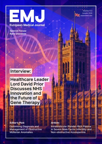
Volume 11.3 Autumn 2026 emjreviews.com
Special Focus: Rare Diseases
Interview: Healthcare Leader Lord David Prior Discusses NHS Innovation and the Future of Gene Therapy
Editor’s Pick:
Article:
Optimising Diagnosis and Management of Obstructive Müllerian Anomalies
Intratesticular Platelet-Rich Plasma in Severe Male Factor Infertility and Non-obstructive Azoospermia
Contents Editorial Board 04
Congress Features 10 Beyond Weight Loss: Redefining MASLD Management Through Obesity Pharmacotherapy
Welcome 07
Ravaioli F
16 Bridging Basic Science, Biomarkers, and AI in Allergy and Immunology Shamji MH
Foreword 09
Symposium Review 19 Mimicking Breastmilk Lipids:
Programming Long-Term Health
Congress Interview 28 Ana Lleo Interviews 32 Location Matters: The Outsize
Prof Markus PeckRadosavljevic
Quality of Life Burden of Psoriasis in High-Impact Areas
Professor of Medicine and Chairman, Department of Gastroenterology and Hepatology, Endocrinology, Rheumatology and Nephrology, Klinikum Klagenfurt am Wörthersee, Austria
37 From Lubricity to Surface Integrity: Expert Perspectives on PolyvinylpyrrolidoneCoated Intermittent Catheters
50 Lord David Prior 53 William Gahl 58 Helen Lachmann
2
Flagship
●
September 2026
●
Copyright © 2026 EMJ
●
CC BY-NC 4.0 Licence
Find out more about our Flagship content
64 Ari Zimran 71 David Lynch 74 Derek Gilroy Infographic Dermatitis with Hand Involvement 80 Atopic When Inflammation Becomes Visible: Understanding its Burden
Features 82 Optimising Diagnosis and Management of Obstructive Müllerian Anomalies
Platelet-Rich Plasma 107 Intratesticular in Severe Male Factor Infertility and Non-obstructive Azoospermia: A Narrative Review
Feinstein J and Fei YF
87 The Changing Landscape of Danon Disease
Khachatryan M and Fanarjyan D
Sertich AP III and Taylor M
Rare Occurrence of Carotid Artery 118 ADissection Caused by Hypermobile
Articles
Ehlers-Danlos Syndrome and Vomiting Leading to Anterior and Posterior Circulation Infarction
94 Anaphylaxis from Oral Sex Linked to Drug-
Kulendran A et al.
Contaminated Semen: A Case Report Bahna SL et al.
99
Assessment of Some Inflammatory Biomarkers Among Individuals with Sickle Cell Disease Attending Murtala Muhammad Specialist Hospital in Kano, Nigeria
Microbiome and Venous 125 Gut Thromboembolism: Emerging
Insights and Diagnostic Potential Augustus A et al.
Garba N et al.
CC BY-NC 4.0 Licence
●
Copyright © 2026 EMJ
●
September 2026
●
Flagship
3
Editorial Board Editor-in-Chief Markus Peck-Radosavljevic Klinikum Klagenfurt am Wörthersee, Austria Current Chairman and Head of the Department of Gastroenterology and Hepatology, Endocrinology, Rheumatology and Nephrology at Klinikum Klagenfurt am Wörthersee, with expertise in portal hypertension, hepatocellular carcinoma, and HIV-HCV coinfection.
Dr Pierfrancesco Agostoni
Dr Ahmad Awada
Ziekenhuis Aan de Stroom (ZAS) Middelheim, Belgium
Chirec Hospital Group, Belgium
Dr Fernando Alfonso Hospital Universitario de La Princesa, Spain
Prof Sorin T. Barbu
Dr Emanuele Angelucci
“Iuliu Hațieganu” University of Medicine and Pharmacy, Romania
IRCCS Ospedale Policlinico San Martino, Italy
Dr George Anifandis University of Thessaly, Greece
Prof Riccardo Autorino Prof Abdullah Erdem Canda Glickman Urological Institute, USA Koç University Hospital, Türkiye
Dr Mátyás Benyó University of Debrecen, Hungary
Prof Andrew Bush Prof Ian Chikanza
Imperial College London, UK
Harley Street Clinic, UK
Dr Hassan Galadari United Arab Emirates University, United Arab Emirates
Dr Amir Hamzah Abdul Latiff Pantai Hospital, Malaysia
Prof Lászlo Vécsei University of Szeged, Hungary
Dr Lorenz Räber Bern University Hospital, Switzerland
4
Flagship
●
September 2026
●
Copyright © 2026 EMJ
●
CC BY-NC 4.0 Licence
Aims and Scope EMJ, the flagship journal of the EMJ portfolio, is an openaccess, peer-reviewed eJournal, committed to elevating the quality of healthcare globally by publishing high-quality medical content across the 18 clinical areas covered in our portfolio. The journal is published quarterly and showcases the latest developments across these clinical areas. EMJ publishes peer-reviewed research papers, review articles, and case reports across all therapy areas of the EMJ portfolio. In addition, the journal publishes features and opinion pieces create a discussion around key topics in the field and broaden readers’ professional interests. The journal also features interviews with leading experts in various clinical disciplines. The journal covers advances within the pharmaceutical arena by publishing sponsored content from congress symposia, which is of high educational value for healthcare professionals. This undergoes rigorous quality control checks by independent experts and the in-house editorial team. EMJ endeavours to increase knowledge, stimulate discussion, and contribute to the delivery of world-class updates in the clinical realm. We do not publish veterinary science papers or laboratory studies that are not linked to patient outcomes. Further details on coverage can be found here: www.emjreviews.com Editorial Expertise EMJ is supported by various levels of expertise: • • • • •
Guidance from an Editorial Board consisting of leading authorities from a wide variety of disciplines. Invited contributors who are recognised authorities in their respective fields. Peer review, which is conducted by expert reviewers who are invited by the Editorial team and appointed based on their knowledge of a specific topic. An experienced team of editors and technical editors. A team of internal and independent medical writers.
Peer Review Every review article, case report, feature, and research article published in EMJ undergoes peer review by at least two independent experts. On submission, all manuscripts are assessed and undergo a technical check by the EMJ Editorial staff to determine their suitability for the journal and appropriateness for peer review. Editorial staff identify appropriate reviewers who are selected based on their specialist knowledge in the relevant area. All peer review is double-blind. Following review, manuscripts are either accepted without modification, returned to the author(s) to incorporate required changes, or rejected. Editorial staff are responsible for ensuring that necessary amendments to the manuscript have been made, with input from our Editorial Board or the original reviewers where necessary. The Editor of EMJ has final discretion over any proposed amendments. Manuscripts authored by members of the Editorial Board are subjected to the same double-blind process. Short opinion pieces are published following internal review and publication is at the discretion of the Editor. Congress-associated content authored by the EMJ Editorial staff undergoes internal quality control checks. Congress-related content sponsored or funded by our industry partners undergoes quality control checks independently. Industry-supported content that falls into any of the categories that are eligible for peer review, undergoes the same peer review process.
CC BY-NC 4.0 Licence
●
Submissions We welcome contributions from professionals, consultants, academics, and industry leaders on relevant and topical subjects. We seek papers with the most current, interesting, and relevant information in each therapeutic area and accept original research, review articles, case reports, and features. We are always keen to hear from healthcare professionals wishing to discuss potential submissions, please email: editorial.assistant@emjreviews.com To submit a paper, use our online submission site: https://emj.kriyadocs.com/submissions/submit/emj/emj/login Submission details can be found through our website: www.emjreviews.com/contributors/authors Reprints All articles included in EMJ are available as reprints (minimum order 1,000). Please contact hello@emjreviews.com if you would like to order reprints. Distribution and Readership EMJ is distributed through controlled circulation to healthcare professionals in the relevant fields globally. Indexing and Availability EMJ is indexed on DOAJ, the Royal Society of Medicine, and Google Scholar®. EMJ is available through the websites of our leading partners and collaborating societies. EMJ journals are all available via our website: www.emjreviews.com Open Access This is an open-access journal in accordance with the Creative Commons Attribution-Non Commercial 4.0 (CC BY-NC 4.0) license. Congress Notice Staff members attend medical congresses as reporters when required. This Publication Launch Date: 2016 Frequency: Quarterly Online ISSN: 2397-6764 All information obtained by EMJ and each of the contributions from various sources is as current and accurate as possible. However, due to human or mechanical errors, EMJ and the contributors cannot guarantee the accuracy, adequacy, or completeness of any information, and cannot be held responsible for any errors or omissions. EMJ is completely independent of any event reviews in this issue and the use of the organisations does not constitute endorsement or media partnership in any form whatsoever. Front cover photograph: Digitally enhanced image © Mihai Zaharia / stock.adobe.com
Copyright © 2026 EMJ
●
September 2026
●
Flagship
5
Stay at the Forefront of Modern Medicine High-level perspectives. Global experts. Essential updates. Join us for conversations with the minds shaping healthcare's future. Gain the distilled insights you need to lead in your field and make maximum impact.
Jonathan Sackier:
Non Executive Director & CMO, AiM Medical Robotics, Florida, USA
6
Saranya Ravindran:
Paediatric Emergency Medicine Registrar, Imperial College Healthcare NHS Trust
Catherine Glass: Associate NHS GP and Senior Appraiser, NHS England
Listen Now Flagship
●
September 2026
●
Copyright © 2026 EMJ
●
CC BY-NC 4.0 Licence
Welcome Editorial Director Andrea Charles
Dear Readers,
Editor Sean Boyle
It is my pleasure to welcome you to issue 11.3 of EMJ flagship. This edition focuses on rare diseases, and explores the impact of uncommon conditions on patient wellbeing, treatment pathways, and the provision of healthcare.
Managing Editor Darcy Richards Associate Editor Helena Bradbury Senior Copy Editor Noémie Fouarge Copy Editors Meghan Garcka, Lizzie Green, Sarah Jahncke Editorial Leads Katrina Thornber, Aleksandra Zurowska Senior Editorial Co-ordinator Bertie Pearcey Editorial Co-ordinators Jess Nicholson, Alena Sofieva Editorial Assistants Niamh Holmes, Josh Lister, Nonyelum Okonkwo Creative Director Tim Uden Design Manager Stacey White Senior Designers Tamara Kondolomo, Owen Silcox Designers Shanjok Gurung, Fabio van Paris
This issue investigates carotid artery dissection in relation to Ehlers-Danlos syndrome, anaphylaxis in relation to drugcontaminated semen, intratesticular platelet-rich plasma in severe male factor infertility, and chronic kidney disease. Expert-led features from this year’s European congresses include an exploration of obesity and MASLD in population groups, and how AI is being optimised to help treat conditions across allergy and immunology. The theme of rare diseases is expanded across a range of interviews with key industry leaders. Helen Lachmann offers insights on amyloidosis, David Lynch discusses the latest developments in Friedreich's ataxia, and William Gahl highlights treatment pathways for people living with cystinosis. As ever, EMJ is proud to present a range of original research. This issue includes an assessment of gut microbiome and venous thromboembolism, covering emerging insights in the field, as well as its real-world diagnostic potential. Another article focuses on the use of inflammatory biomarkers in the treatment of individuals living with sickle cells, and what new developments are most relevant to European practice. I would like to extend my sincere thanks to our Editorial Board, authors, and peer reviewers for their continued dedication and expertise, as well as Roli Omamuli (Editorial Assistant, EMJ, London, UK) for her extensive contributions to this issue. I hope you find this issue both insightful and relevant to your clinical practice.
Junior Designers Molly Edwards, Fraser Hoey, Cameron Levett, Helena Spicer, Caleb Wylie
Sean Boyle, Editor
Marketing Director Stephanie Corbett
Contact us
Chief Executive Officer Justin Levett Chief Commercial Officer Dan Healy Founder and Chairman Spencer Gore
Editorial enquiries: editor@emjreviews.com Sales opportunities: salesadmin@emjreviews.com Permissions and copyright: accountsreceivable@emjreviews.com Reprints: info@emjreviews.com Media enquiries: marketing@emjreviews.com
CC BY-NC 4.0 Licence
●
Copyright © 2026 EMJ
●
September 2026
●
Flagship
7
Join the No.1 open access destination for HCPs & Pharma globally Free newsletters & alerts in your therapy area
Get the latest updates on all our upcoming journals and receive first-class insights into ground-breaking news and advancements in medicine across multiple therapeutic areas.
Join Today 8
Flagship
●
September 2026
●
Copyright © 2026 EMJ
●
CC BY-NC 4.0 Licence
Foreword Dear Colleagues, Welcome to the third EMJ flagship issue of 2026. This edition is dedicated to rare diseases, a theme that extends across every speciality of medicine, yet remains too often overlooked. Rare diseases encompass a diverse range of conditions, from inherited metabolic disorders to haematological, reproductive, and systemic diseases. Their complexity and often limited evidence base can create significant challenges in diagnosis and treatment, reinforcing the importance of continued research, multidisciplinary collaboration, and patient-centred care. In this issue, we explore several important dimensions of rare disease research and management. Our feature articles and interviews examine conditions including amyloidosis, cystinosis and rare Müllerian anomalies. We also consider inflammatory biomarkers in sickle cell disease and emerging approaches to diagnosis and treatment, including novel strategies in reproductive medicine. Collectively, these contributions highlight the diversity of rare disease and the continued need for improved understanding and care.
Beyond rare diseases, this issue explores developments shaping the wider future of medicine, from AI in allergy and immunology to advances in inflammation, obesity, MASLD, and autoimmune liver disease. Our interview with Lord David Prior further considers the future of healthcare, technology, and system reform.
Their complexity and often limited evidence base can create significant challenges in diagnosis and treatment I am grateful to our authors, interviewees, reviewers, and Editorial Board for their expertise and continued commitment. I hope this issue encourages thoughtful engagement with the research and innovation advancing the diagnosis, treatment, and understanding of rare diseases.
Prof Markus Peck-Radosavljevic Professor of Medicine and Chairman, Department of Gastroenterology and Hepatology, Endocrinology, Rheumatology and Nephrology, Klinikum Klagenfurt am Wörthersee, Austria
CC BY-NC 4.0 Licence
●
Copyright © 2026 EMJ
●
September 2026
●
Flagship
9
Congress Feature
●
EASL 2026
Beyond Weight Loss: Redefining MASLD Management Through Obesity Pharmacotherapy Author:
*Federico Ravaioli1,2 1.
Department of Medical and Surgical Sciences (DIMEC), University of Bologna, Italy 2. Division of Internal Medicine, Hepatobiliary and Immunoallergic Diseases, IRCCS Azienda Ospedaliero-Universitaria di Bologna, Italy *Correspondence to f.ravaioli@unibo.it Disclosure:
Ravaioli has received payment or honoraria for lectures, presentations, speakers bureaus, manuscript writing, or educational events from Madrigal, Eli Lilly, Novo Nordisk, and Errekappa Pharma. Ravaioli has also received support for attending meetings and/or travel from Novo Nordisk, and has participated on a data safety monitoring board or advisory board for Madrigal.
Keywords:
BMI, glucagon-like peptide-1 (GLP-1), metabolic dysfunction-associated steatotic liver disease (MASLD), obesity, weight gain, weight loss.
Citation:
EMJ. 2026;11[3]:10-15. https://doi.org/10.33590/emj/1463768R
THE MANAGEMENT of metabolic dysfunction-associated steatotic liver disease (MASLD) is undergoing a profound transformation. Once viewed primarily through the lens of lifestyle modification and gradual weight reduction, obesity is now increasingly recognised as a chronic neuroendocrine disease requiring long-term pharmacological management. During the European Association for the Study of the Liver (EASL) Congress 2026, a joint EASL/European Association for the Study of Obesity (EASO) session outlined how modern obesity therapies are reshaping the treatment landscape for MASLD, moving the field beyond weight loss alone towards disease modification, cardiometabolic risk reduction, and durable improvements in overall health.1-4
OBESITY TREATMENT ENTERS THE MASLD ERA The session brought together obesity specialists, endocrinologists, and hepatologists to address a central question facing clinical practice today: how should clinicians integrate highly effective obesity-management medications into the care of patients with MASLD? Through discussions spanning obesity pathophysiology, personalised treatment goals, management of adverse events, and preservation of musculoskeletal health, a consistent message emerged: obesity 10
Flagship
●
September 2026
●
pharmacotherapy should no longer be considered simply a tool for reducing body weight, but rather a cornerstone of chronic disease management.1-4
OBESITY AS A CHRONIC DISEASE: UNDERSTANDING WHY WEIGHT LOSS IS SO DIFFICULT Opening the session, Luca Busetto, University of Padova, Italy, challenged the enduring misconception that obesity is merely the result of excessive caloric intake or insufficient willpower. Instead,
Copyright © 2026 EMJ
●
CC BY-NC 4.0 Licence
EASL 2026
he described obesity as a chronic disease driven by dysfunction of the biological systems regulating energy balance.1 Drawing parallels with hypertension and Type 2 diabetes, Busetto argued that obesity deserves disease status because it arises from alterations in physiological regulatory mechanisms. Genetic predisposition, environmental exposures, developmental factors, sleep disruption, psychological influences, and modern food environments can all disrupt the neuroendocrine networks governing appetite and energy expenditure. The resulting symptoms include diminished satiety, increased hunger, and heightened desire for food. Importantly, Busetto stressed that these are consequences of the disease process rather than its causes.1
Three Layers of Appetite Regulation Busetto presented obesity as a disorder affecting three interconnected systems within the brain: •
Homeostatic eating, which regulates energy needs and hunger signals
•
Hedonic eating, which governs food reward and pleasure
•
Executive function, which influences decision-making and behavioural control
Evidence presented during the lecture showed that obesity is associated with abnormalities across all three domains. Studies have demonstrated reduced postprandial secretion of satiety hormones such as glucagon-like peptide-1 (GLP-1) and peptide YY, alongside altered neural responses to nutrient intake and foodrelated stimuli. Functional neuroimaging findings suggest that individuals with obesity continue to experience food reward signals even after nutrient ingestion, potentially perpetuating eating behaviour despite adequate energy intake.1
CC BY-NC 4.0 Licence
●
●
Congress Feature
The Biology of Weight Regain
Perhaps the most clinically relevant aspect of Busetto’s presentation concerned the biological resistance to sustained weight loss. Following caloric restriction, the body activates powerful compensatory mechanisms designed to restore lost weight. Ghrelin concentrations rise, anorexigenic hormones decline, leptin levels fall, and energy expenditure decreases. Together, these adaptations create a physiological environment favouring weight regain.1,5 Stopping obesity medication is associated in most cases with weight regain, and this is considered normal if obesity is a chronic disease.1 This concept has important implications for treatment duration. Just as antihypertensive therapy is not expected to cure hypertension permanently, obesity medications should not be viewed as shortterm interventions. Rather, they may need to be continued long-term to counteract persistent biological drivers of weight gain.1,5
Stopping obesity medication is associated in most cases with weight regain, and this is considered normal if obesity is a chronic disease
FROM WEIGHT TARGETS TO HEALTH OUTCOMES If Busetto explained why obesity treatment is necessary, Lidia Graur, University of Medicine and Pharmacy, Iași, Romania, addressed what clinicians should aim to achieve. Her presentation reflected a growing shift away from a purely weight-centred approach toward outcome-based management.2 A recurring theme from recent obesity congresses, she noted, has been: “See Obesity, Think Liver, Treat Hard.” The slogan encapsulates the growing recognition that obesity management should extend beyond weight reduction to address obesity-related complications directly. Graur highlighted MASLD as arguably one of the most common complications of obesity, occurring in the majority of affected individuals.
Copyright © 2026 EMJ
●
September 2026
●
Flagship
11
Congress Feature
●
EASL 2026
Indeed, she suggested that encountering a patient with obesity who does not have steatotic liver disease is becoming increasingly unusual in clinical practice.2
Not All Adipose Tissue Is Equal
Central to Graur’s argument was the concept of adiposopathy, or dysfunctional adipose tissue. While subcutaneous fat expansion may occur relatively safely, visceral and ectopic fat deposition drives inflammation, insulin resistance, and cardiometabolic dysfunction. As a result, two patients with identical BMI values may have vastly different metabolic risk profiles.2 This heterogeneity helps explain why BMI alone is an inadequate measure of disease burden. Graur instead advocated for broader clinical assessment incorporating metabolic, mechanical, and psychological complications. She highlighted the Edmonton Obesity Staging System (EOSS) as a useful framework for risk stratification because it correlates more closely with morbidity and mortality than BMI categories alone.2 Importantly, she argued that treatment goals should be determined not simply by the amount of weight lost, but by the specific obesity-related complications present in each patient. This approach reflects a move towards personalised obesity medicine, in which therapeutic success is defined by improvements in clinically meaningful outcomes rather than changes on the scale alone.2
12
Flagship
●
September 2026
●
A New Therapeutic Landscape
The emergence of highly effective obesity-management medications has transformed what clinicians can realistically expect to achieve. Historically, lifestyle interventions produced weight reductions of approximately 5–7%, often insufficient to substantially affect liver disease progression. Contemporary incretin-based therapies, however, routinely achieve weight losses of 15–20%, approaching outcomes previously attainable only through bariatric surgery.2,6-8 This degree of weight reduction matters because different obesity-related complications appear to require different thresholds of weight loss for meaningful improvement. Evidence suggests that a 5–10% reduction in body weight may improve glycaemic parameters and cardiometabolic risk factors, whereas greater weight loss is generally required to induce remission of steatohepatitis, improve fibrosis, and alleviate mechanical complications such as obstructive sleep apnoea.2,9 The arrival of GLP-1 receptor agonists and dual incretin agonists has fundamentally altered the therapeutic landscape. Cardiovascular outcome studies have demonstrated reductions in major adverse cardiovascular events, while emerging MASLD-specific studies suggest improvements in steatohepatitis resolution and fibrosis endpoints. Graur highlighted that clinicians now have an opportunity not only to reduce weight, but also to
Copyright © 2026 EMJ
●
CC BY-NC 4.0 Licence
EASL 2026
●
Congress Feature
target specific complications through therapies with increasingly differentiated efficacy profiles.2,9,10
a slightly higher incidence of vomiting, which may contribute to marginally higher discontinuation rates.8
These findings support the notion that obesity treatment should be regarded as disease-modifying therapy rather than cosmetic intervention. As pharmacological options continue to expand, comprehensive baseline assessment, including waist circumference, cardiometabolic risk evaluation, diabetes screening, and systematic evaluation for MASLD, will become increasingly important in guiding treatment selection and defining realistic therapeutic goals.2
To minimise treatment interruption, Klobucar proposed a pragmatic framework based on the “Three Es”:3
IMPROVING PERSISTENCE: WHY MANAGING SIDE EFFECTS MATTERS For Sanja Klobucar, Clinical Hospital Centre Zagreb, Croatia, the remarkable efficacy of modern obesity-management medications can only translate into meaningful clinical outcomes if patients remain on treatment. Despite the transformative weight loss effects observed with semaglutide and tirzepatide, real-world persistence remains a major challenge, with more than half of patients discontinuing therapy within the first year. Gastrointestinal adverse events have emerged as one of the principal drivers of early treatment cessation.3,8 Nausea, vomiting, diarrhoea, constipation, bloating, and abdominal discomfort are among the most frequently reported adverse events, affecting between 60–80% of participants across major clinical trials. These symptoms are largely attributable to the pharmacological mechanisms underlying incretin-based therapies, including delayed gastric emptying and central GLP-1 receptor activation. Reassuringly, most events are mild-to-moderate, occur predominantly during treatment initiation and dose escalation, and tend to diminish over time.3,6-8 Discussing data from the head-to-head SURMOUNT-5 study, Klobucar noted that semaglutide and tirzepatide exhibit broadly similar gastrointestinal safety profiles, although semaglutide was associated with CC BY-NC 4.0 Licence
●
•
Educate patients before treatment initiation about expected effects and potential adverse events
•
Eat differently, encouraging behavioural and dietary adaptations that improve tolerability
•
Escalate slowly, tailoring dose titration according to individual patient tolerance rather than adhering rigidly to standard schedules
A substantial portion of the presentation focused on practical nutritional counselling. Patients should be encouraged to eat slowly, consume smaller and more frequent meals, stop eating when comfortably satisfied rather than full, and avoid highfat, highly processed, excessively sweet, or spicy foods. Hydration was repeatedly highlighted as a simple but highly effective intervention, particularly for managing constipation and diarrhoea. Foods such as yogurt, crackers, apples, gingercontaining beverages, rice, and clear broths were suggested as useful strategies for alleviating gastrointestinal symptoms.3 Importantly, Klobucar argued that dose escalation should remain flexible. Extending titration intervals, delaying dose increases during active symptoms, temporarily returning to a previously tolerated dose, or even maintaining patients on lower-thanlabel maintenance doses may substantially improve long-term adherence. In selected individuals, achieving persistence on a lower dose may be preferable to treatment discontinuation altogether.3 Her concluding message was clear: successful obesity pharmacotherapy requires active clinical support. Regular follow-up, early recognition of adverse events, personalised titration strategies, and ongoing patient education are essential
Copyright © 2026 EMJ
●
September 2026
●
Flagship
13
Congress Feature
●
EASL 2026
not only for maintaining weight loss, but also for preserving the broader metabolic, cardiovascular, and hepatic benefits that these therapies can deliver.3
LOOKING BEYOND THE SCALE The final presentation, delivered by Liisa Tolvanen, Karolinska Institute, Stockholm, Sweden, explored a question that is becoming increasingly relevant as weightloss therapies become more potent: what happens to muscle and bone during treatment? Framing her talk within the ongoing ‘paradigm shift’ in obesity care, Tolvanen argued that clinicians must move beyond weight loss as the sole marker of success and instead focus on preserving functional health while achieving sustained reductions in adiposity.4 All effective obesity treatments reduce both fat mass and lean mass. This occurs with lifestyle interventions, pharmacotherapy, and bariatric surgery alike. However, she stressed that the interpretation of these changes is often more complex than commonly assumed. Fat-free mass includes water, connective tissue, organs, and bone, in addition to skeletal muscle.4 Recent evidence suggests that reductions in fat-free mass do not necessarily translate into clinically meaningful deterioration in muscle health. Indeed, muscle quality may improve as intramuscular fat infiltration decreases during weight loss. Nevertheless, maintaining physical function remains a key objective, particularly among older adults and patients with chronic disease. Tolvanen noted that the clinical significance of body-composition changes depends heavily on baseline patient characteristics, including age, comorbidity burden, and pre-existing frailty risk.4
The Importance of Exercise
with structured physical activity helps preserve lean mass and optimise functional outcomes. Exercise should therefore be viewed not as an optional adjunct, but as an integral component of comprehensive obesity management, particularly in patients achieving substantial weight loss with incretin-based therapies.4
Monitoring More Than Weight
Tolvanen also highlighted the limitations of relying exclusively on body weight and BMI when evaluating treatment success. Waist circumference, waist-to-height ratio, body composition assessments, and measures of physical function can provide a more comprehensive understanding of therapeutic effects. While body weight remains an important and accessible measure, it cannot distinguish between changes in adipose tissue, muscle, and bone, nor does it adequately capture improvements in functional status.4 Bone health deserves particular attention. Significant weight loss may affect bone mineral density, although the long-term clinical implications remain uncertain. Current evidence suggests that substantial weight reduction may influence bone metabolism, but the clinical relevance of these findings remains incompletely understood. Until more data become available, Tolvanen advocated an individualised approach to risk assessment, with particular attention to older adults and those with pre-existing osteoporosis or other skeletal vulnerabilities.4 Ultimately, her message echoed a recurring theme throughout the session: successful obesity treatment should not be defined solely by kilograms lost. Rather, clinicians should aim to optimise body composition, preserve muscle function, maintain skeletal health, and improve long-term quality of life while reducing obesityrelated complications.4
One of the clearest messages from Tolvanen’s presentation was that exercise should accompany pharmacological treatment whenever possible. Data suggest that combining obesity medications 14
Flagship
●
September 2026
●
Copyright © 2026 EMJ
●
CC BY-NC 4.0 Licence
EASL 2026
A NEW PARADIGM FOR HEPATOLOGISTS The EASL/EASO session reflected a broader transformation occurring across obesity medicine and hepatology. The discussion repeatedly returned to a central theme: obesity should be recognised and managed as a chronic disease with complex biological underpinnings, rather than as a lifestyle choice or behavioural failure. From altered neuroendocrine pathways regulating appetite and energy balance to the development of obesityrelated complications such as MASLD, the speakers consistently emphasised the need for a biologically informed, long-term treatment strategy.1-4 For hepatologists, the implications are substantial. As obesity-management medications continue to demonstrate benefits extending beyond weight reduction, including improvements in cardiovascular outcomes, metabolic dysfunction, and liver disease, these agents are becoming increasingly relevant to routine MASLD care. The traditional separation between obesity management and hepatology is rapidly disappearing, as accumulating evidence suggests that effective treatment of obesity may become
●
Congress Feature
one of the most powerful tools available to modify liver-related outcomes.2,9,10 With the advent of highly effective incretinbased therapies, achieving clinically meaningful weight loss is no longer the primary obstacle. Instead, clinicians must learn how to personalise therapy, maximise adherence, preserve musculoskeletal health, and maintain benefits over the long term. Equally important will be identifying which patients are most likely to benefit from specific therapeutic approaches and defining treatment success according to meaningful clinical outcomes rather than weight alone.2-4 As highlighted throughout the session, the future of obesity medicine lies not in chasing numbers on the scale but in improving health outcomes across multiple organ systems. In its place is emerging a more ambitious goal: using obesity pharmacotherapy to improve liver health, reduce cardiometabolic risk, and alter the natural history of chronic disease. For patients with MASLD, this shift may ultimately redefine the role of obesity treatment, from a supportive intervention to a central component of disease management itself.1-4
For patients with MASLD, this shift may ultimately redefine the role of obesity treatment, from a supportive intervention to a central component of disease management itself
References 1.
2.
3.
4.
Busetto L. What are the biological mechanisms behind weight loss. Presentation. EASL Congress, 27-30 May, 2026. Graur L. How do you set up individualized goals (weight loss, cardiovascular health, liver health). Presentation. EASL Congress, 27-30 May, 2026. Klobucar S. How do you manage side effects and increase patient adherence? Presentation. EASL Congress, 27-30 May, 2026.
5.
6.
CC BY-NC 4.0 Licence
Tolvanen L. How do you evaluate the consequences on muscle and bone health after obesity treatment? Presentation. EASL Congress, 27-30 May, 2026. Rubino DM et al. Effect of continued weekly subcutaneous semaglutide vs placebo on weight loss maintenance in adults with overweight or obesity: the STEP 4 randomized clinical trial. JAMA. 2021;325(14):1414-25. Wilding JPH et al. Once-weekly semaglutide in adults with overweight or obesity. N Engl J Med. 2021;384(11):989-1002.
●
Copyright © 2026 EMJ
●
7.
Jastreboff AM et al. Tirzepatide once weekly for the treatment of obesity. N Engl J Med. 2022;387(3):205-16.
8.
Aronne LJ et al. Tirzepatide as compared with semaglutide for the treatment of obesity. N Engl J Med. 2025;393(1):26-36.
9.
Sanyal AJ et al. ESSENCE study group. Phase 3 trial of semaglutide in metabolic dysfunction-associated steatohepatitis. N Engl J Med. 2025;392(21):2089-99.
10. Lincoff AM et al. Semaglutide and cardiovascular outcomes in obesity without diabetes. N Engl J Med. 2023;389(24):2221-32.
September 2026
●
Flagship
15
Congress Feature
●
EAACI 2026
Bridging Basic Science, Biomarkers, and AI in Allergy and Immunology Author:
*Mohamed H. Shamji1 1.
Immunomodulation and Tolerance Group (ITG) Allergy & Clinical Immunology, National Heart & Lung Institute Victor Phillip Dahdaleh (VPD) Building, Imperial College London, UK *Correspondence to m.shamji99@imperial.ac.uk Disclosure:
Shamji reports research grants from the Immune Tolerance Network, Medical Research Council, Allergy Therapeutics, LETI Laboratorios, and Rovolo Biotherapeutics; and lecture fees from Allergy Therapeutics and LETI Laboratorios, all outside the submitted work.
Keywords:
AI, allergen immunotherapy, allergy and immunology, angioedema, asthma, biomarkers, machine learning, precision medicine, responsible implementation.
Citation:
EMJ. 2026;11[3]:16-18. https://doi.org/10.33590/emj/19151690
AI HAS progressed from a theoretical concept into a practical tool with growing relevance across healthcare. In allergy and immunology, its potential is particularly clear: the field generates large and diverse datasets spanning clinical phenotypes, biomarkers, omics platforms, and patient-reported outcomes. As AI continues to be integrated into medicine, the development of these systems has been accompanied by an increasing emphasis on responsible implementation. This article summarises a talk given at the European Academy of Allergy & Clinical Immunology (EAACI) Congress 2026 held in Istanbul, Türkiye.
FROM ETHICAL AI TO CLINICAL UTILITY Ethical AI delivery depends on key pillars including fairness, transparency, interpretability, auditability, explainability, and responsibility, all of which are essential if these technologies are to be translated safely into clinical practice. More recent discussions have also emphasised the importance of accountability, privacy, and human oversight, particularly in healthcare contexts where algorithmic decisions may influence diagnosis or treatment access.1 The clinical appeal of AI lies in its ability to support decision-making, streamline workflows, and reduce the burden of data interpretation. In healthcare settings, AI is increasingly being used to improve documentation, automate electronic health records, support shared decision-making, 16
Flagship
●
September 2026
●
and simplify prior authorisation processes. At the same time, patients are interacting more frequently with large language models and other digital health tools, reinforcing the need for appropriate guidance and oversight. Within allergy and immunology, AI is emerging as a powerful adjunct to traditional approaches because it can integrate complex and multidimensional data in ways that are difficult to achieve through conventional analysis alone. That includes not only laboratory and omics information, but also symptom patterns, treatment history, challenge outcomes, and exposure data, which together can improve the precision of disease modelling.1-3 AI is also increasingly being applied across a range of medical specialities. Dermatology, pathology, radiology, cardiology, and surgery have all begun to incorporate AI-driven tools into diagnosis,
Copyright © 2026 EMJ
●
CC BY-NC 4.0 Licence
EAACI 2026
image interpretation, risk prediction, and procedural support. Allergy and immunology are now following a similar trajectory, with applications extending from biomarker discovery to disease endotyping and treatment prediction. This explanation reflects both the complexity of immune-mediated disease and the growing recognition that more sophisticated analytical approaches are required to unlock clinically useful insights.
AI FOR DISEASE ENDOTYPING AND DIAGNOSIS In asthma, machine learning has been used to identify biologically distinct endotypes from proteomic and transcriptomic data. In one study, unsupervised clustering of nasal protein signatures, followed by feature selection and validation in an independent cohort, enabled the separation of severe asthma into meaningful subgroups.4 This work is particularly valuable because it uses non-invasive nasal fluid sampling to infer molecular phenotypes in the lower airway, offering a practical route to precision endotyping.
CC BY-NC 4.0 Licence
●
●
Congress Feature
The model identified two severe asthma endotypes that were replicated in nasal transcriptomic data from an independent cohort, strengthening confidence in the robustness of the approach. Notably, the clusters were associated with clinically relevant features such as airway obstruction and small airway disease, suggesting that AI can link molecular signatures to disease severity and physiology. Shapley-based analysis identified cluster-defining proteins and pathways associated with Th1, Th2, neutrophils, and JAK-STAT, underscoring the ability of machine learning not only to classify disease, but also to reveal the biological mechanisms underlying heterogeneity. Such work illustrates the value of AI not simply as a classificatory tool, but as a means of revealing the underlying biology of heterogeneous disease.
In asthma, machine learning has been used to identify biologically distinct endotypes from proteomic and transcriptomic data
Copyright © 2026 EMJ
●
September 2026
●
Flagship
17
Congress Feature
●
EAACI 2026
Another important example is recurrent angioedema, where diagnosis is often complicated by overlapping clinical features. Machine learning was applied to a large multicentre cohort of 482 patients across several classification frameworks, including mast cell-mediated, bradykinin-mediated, drug-induced, chronic spontaneous urticaria-associated, and hereditary forms of angioedema. The models demonstrated strong performance, particularly in simpler classification tasks, and remained robust even when diagnosis-defining variables were excluded. Notably, clinical features alone provided substantial discriminatory power, suggesting that AI can capture genuine patterns in disease presentation and may support diagnostic decisionmaking in settings where specialist testing is limited.2
BIOMARKERS AND PERSONALISED IMMUNOTHERAPY In allergen immunotherapy, AI offers a different type of value. Rather than replacing clinical judgement, it can assist in identifying response patterns and biomarkers that predict outcome. The GRASS study has shown that more than 70 immunological, functional, and clinical variables can be assessed to identify biomarkers of response and nonresponse.5 Machine learning approaches are well suited to integrating cellular, serological, and functional readouts, highlighting the importance of composite
References 1.
2.
18
Khoury P et al. A framework for augmented intelligence in allergy and immunology practice and research—a work group report of the AAAAI health informatics, technology, and education committee. J Allergy Clin Immunol Pract. 2022;10(5):1178-88. Van Breugel M et al. Artificial intelligence in allergy and immunology: recent develop-ments, implementation challenges, and the road toward clinical impact. J Allergy Clin Immunol. 2025;157(2):295-306.
Flagship
●
biomarker strategies in a field where no single marker is likely to be sufficient on its own.6,7 This is particularly relevant for allergen immunotherapy, where response is influenced by complex interactions between humoral and cellular immune pathways. The central message is clear: AI is most effective when it is applied to well-defined clinical questions and supported by robust biological understanding. In allergy and immunology, the technology holds promise because of the field’s multidimensional nature and the need to interpret complex data in a clinically meaningful way. However, the same complexity that makes AI attractive also demands caution. Validation, interpretability, and awareness of bias remain essential if these tools are to improve rather than complicate care. As recent reviews have emphasised, trustworthiness in healthcare AI depends not only on algorithmic performance, but also on explainability, equitable data practices, and reliable governance. As AI continues to evolve, allergy and immunology are well positioned to benefit from its integration. From asthma endotyping to angioedema classification and immunotherapy biomarker discovery, the field is moving towards more personalised, data-driven care. The challenge now is to ensure that these advances are implemented responsibly, with ethical safeguards and clinical utility at the forefront.
3.
Ray A et al. Determining asthma endotypes and outcomes: Complementing existing clinical practice with modern machine learning. Cell Rep Med. 2022;3(12):100857.
4.
Agache I et al. Multidimensional endotyping using nasal proteomics predicts molecular phenotypes in the asthmatic airways. J Allergy Clin Immunol. 2023;151(1):128-37.
5.
response to allergen challenge at 3 years among patients with moderate to severe seasonal allergic rhinitis: the GRASS randomized clinical trial. JAMA. 2017;317(6):615-25.
Scadding GW et al. Effect of 2 years of treatment with sublingual grass pollen immuno-therapy on nasal
September 2026
●
Copyright © 2026 EMJ
●
6.
Layhadi JA et al. Mechanisms and predictive biomarkers of allergen immunotherapy in the clinic. J Allergy Clin Immunol Pract. 2024;12(1):59-66.
7.
Durham SR, Shamji MH. Allergen immunotherapy: past, present and future. Nat Rev Immunol. 2023;23(5):317-28.
CC BY-NC 4.0 Licence
Symposium Review
Mimicking Breastmilk Lipids: Programming Long-Term Health This symposium took place on 25th June 2026 as part of the 58th Annual Meeting of the European Society for Paediatric Gastroenterology, Hepatology and Nutrition (ESPGHAN) Congress held in Lille, France Funding:
The publication of this article was funded by Danone Research & Innovation, Utrecht, the Netherlands, who were involved in its creation and development.
Speakers:
Yvan Vandenplas,1 Meng Mao,2 Ingrid Renes,3 Jonathan Swann4 1. ‘KidZ Health Castle’, UZ Brussel, Belgium 2. West China Second University Hospital of Sichuan University, Chengdu, China 3. Danone Research & Innovation, Utrecht, the Netherlands 4. University of Southampton, UK
Disclosure:
Vandenplas has participated as a clinical investigator, advisory board member, consultant, and/or speaker for Abbott Nutrition, Alba Health, Arla, BioGaia, Danone, Nestlé Nutrition and Health, Nutricia, Mead Johnson Nutrition, and United Pharmaceuticals (Novalac). Mao has participated as a consultant and/or speaker for Danone. Renes is an employee of Danone Research & Innovation. Swann has participated as a consultant and/or speaker for Danone, Yakult, Nestlé Health Science, and Epetome.
Acknowledgements:
Medical writing assistance was provided by Allison Kirsop, Scientific Writers Ltd, Aberdeenshire, UK.
Keywords:
Breastmilk, cognition, growth, infant development, lipid droplet, lipids, metabolic programming, milk fat globule (MFG), milk fat globule membrane (MFGM).
Citation:
EMJ. 2026;11[3]:19-27. https://doi.org/10.33590/emj/C6S80308
Y
US IND TR
R
TN
This symposium review examines whether mimicking breastmilk lipid globule architecture in infant formula may influence health outcomes in early life. It summarises a scientific presentation and expert panel discussion focused on mimicking the size, structure, and composition of human milk fat globules (MFG) in concept infant formula, compared with standard formulas that contain smaller, protein-coated lipid droplets without a phospholipid membrane.
I
P
PA
Meeting Summary
ERSH
The article outlines how breastmilk lipids provide approximately half of an infant’s energy needs while also serving as structural components and signalling molecules essential for organ and brain development. It explains how lipids in human milk are naturally organised into large globules surrounded by a bioactive membrane rich in phospholipids and other MFG membrane (MFGM) components, while conventional processing of infant formula yields smaller droplets with different interfacial properties.
CC BY-NC 4.0 Licence
●
Copyright © 2026 EMJ
●
September 2026
●
Flagship
19
Symposium Review
According to the in vitro digestion studies presented during the symposium, larger and more complex lipid droplets have been shown to remain longer in the stomach, resulting in slower gastric emptying and more gradual lipid bioaccessibility than smaller lipid droplets. Clinical findings from four studies indicate that the concept formula is safe and well tolerated, with the Mercurius trial and its 5-year follow-up supporting the hypothesis that mimicking MFG architecture may contribute to nutritional programming for growth (BMI), metabolic health (blood pressure), and cognition. The panel discussion explored plausible mechanisms, including altered lipid utilisation, effects on body composition and neurodevelopment, and shifts in the gut microbiome and bile-acid profiles. Throughout, the speakers emphasised that the longer-term data are still emerging and should be interpreted with appropriate caution, underlining the need for replication and extended follow-up to confirm these early signals.
Introduction The symposium opened with the premise that breastmilk remains the preferred source of infant nutrition and the reference standard against which formula innovations should be considered. Within that context, the session focused on milk lipids, emphasising that they provide a substantial share of infant energy intake, yet have historically received less attention than proteins or oligosaccharides in formula development, despite their physiological importance. A recurring theme was earlylife nutritional programming, the idea that nutritional exposures in the first months of life can shape later trajectories for growth, metabolic health, and neurocognitive development, and that lipid quality and droplet architecture may be one such programming factor. This article centres on the concept of early-life programming, whereby nutritional exposures during infancy may influence health trajectories that become apparent later in childhood.
Why Mimic Breastmilk Lipid Architecture? Ingrid Renes, Danone Research & Innovation, Utrecht, the Netherlands, began by outlining the biological importance of lipids in breastmilk. As presented during this introductory session, breastmilk lipids 20
Flagship
●
September 2026
●
contribute almost 50% of an infant’s energy intake and are especially important for growth and development.1 Human milk fat is naturally organised into large MFGs with diameters in the micrometre range, typically around 4 µm, each consisting of a triglyceride core surrounded by a complex triple-layer membrane. This membrane contains phospholipids and other MFGM components with bioactive functions, and the lipid globule’s size, structure, and composition arise from mammary gland secretion processes.1-3 Against this biological backdrop, the symposium introduced a concept infant formula designed to mimic the architecture of human MFGs compared to that of standard formula. Conventional formula manufacturing produces small lipid droplets, largely due to high pressure processing and spray drying, and these droplets are coated mainly with milk proteins. Even when MFGM components are added to standard formula, the lipid droplets remain small and lack a continuous phospholipid membrane. By contrast, the patented Nuturis® process (Danone Research & Innovation, Utrecht, the Netherlands), uses a gentler, low-pressure approach to generate larger lipid droplets with an average diameter close to that of breastmilk fat globules, coated with MFGM components (Figure 1).4
Copyright © 2026 EMJ
●
CC BY-NC 4.0 Licence
Symposium Review
Figure 1: Schematic overview of lipid droplets in human milk, conventional infant formula, and concept infant formula.
Human milk fat globules
Control formula lipid droplets
Nuturis®-derived lipid droplets
Concept infant formula with lipid droplets derived via the patented Nuturis® process, closer in size to human milk fat globules. Nuturis®: Danone Research & Innovation, Utrecht, the Netherlands.
Renes also stressed that the rationale for this design was not limited to structural resemblance. Rather, the goal was to test whether a formula that more closely mimics breastmilk lipid globule architecture could be digested in a more breastmilk-like manner and, in turn, might support health outcomes that are closer to those seen in breastfed infants.5-7 The symposium repeatedly framed these relationships within an early-life nutritional programming model. As shown in Figure 2, the proposed sequence links lipid droplet architecture to digestion and absorption kinetics, downstream lipid utilisation, and the exploratory longer-term outcomes discussed later in this review.
Digestion and Bioaccessibility A major theme of the presentation was that lipid droplet size and architecture may influence how lipids are processed in the gastrointestinal tract. The in vitro and preclinical work discussed in the symposium suggested that larger and more complex lipid droplets behave differently from smaller droplets during gastric digestion. According to the evidence presented, larger and more complex lipid droplets have been shown to remain longer in the stomach,6 by forming a lipid layer above the gastric contents, and are released more slowly CC BY-NC 4.0 Licence
●
than smaller lipid droplets. In addition, larger lipid droplets are broken down more slowly by digestive enzymes because they provide less surface area for lipase action. The speakers argued that, collectively, this results in more gradual lipid digestion and prolonged bioaccessibility, which may support more efficient lipid utilisation both as an energy source and as building blocks for developing tissues during periods of rapid infant growth.8,9 This point was important to the overall early-life programming hypothesis proposed during the symposium. The speakers framed these kinetic differences as a mechanistic basis for early-life nutritional programming, proposing that the timing and pattern of lipid delivery to tissues could help set long-term pathways for energy balance, body composition, and organ development. The panel repeatedly returned to the idea that the biological relevance may lie not simply in the total amount of lipid eventually absorbed, but in the rate and pattern of digestion and bioavailability early in life.4-6,10,11 During the audience discussion, this mechanistic argument was examined directly. In response to questions about why digestion kinetics matter if total lipid absorption is ultimately similar between formulas, Renes stated that the biologically relevant differences may arise early in digestion and indigestion kinetics. More
Copyright © 2026 EMJ
●
September 2026
●
Flagship
21
Symposium Review
Figure 2: Hypothesised pathway linking infant formula lipid droplet architecture to early-life nutritional programming.
Gradual/slower lipid digestion
Lipids stay longer in the stomach (lipid layering)
Breastmilk lipid globule
More gradual bioavailability of lipids
More efficient utilisation of lipids as fuel and building blocks for optimal growth and development
Compared to: Smaller lipid droplet Potential for long-term programming of metabolic and cognitive health outcomes
Digestion
Utilisation Absorption
Compared to smaller lipid droplets, larger complex lipid droplets are digested and absorbed more gradually, impacting the use of lipids as energy and developmental building blocks rather than storage, with potential long-term effects on growth, metabolic health, and neurocognitive outcomes.5-9
gradual and prolonged lipid bioavailability at this stage could influence how lipids are used. At the same time, stool data showing lower residual fatty acids and palmitate in the concept formula were presented as supportive of improved absorption. The panel also emphasised that mimicking lipid droplet architecture should not be reduced to the presence or absence of membrane components alone. Instead, the proposed effect was described as depending on the combination of larger lipid droplet size and phospholipid-rich surface composition. In vitro studies suggested that adding membrane components to smaller lipid droplets does not reproduce the same digestion kinetics as observed with the larger droplets coated with MFGM components.
Clinical Evidence The symposium positioned the clinical evidence as supportive of the concept while making it clear that not all studies carry the same weight. Four clinical studies have shown the concept formula to be safe and 22
Flagship
●
September 2026
●
well tolerated. The Mercurius study and its follow-up formed the primary focus of the symposium because they provide the longest follow-up currently available.12,13 The Mercurius study was a randomised, controlled, double-blind trial in healthy term infants that compared infants receiving a concept formula (n=115) with those receiving a standard formula (n=108), and a breastfed reference group (n=88). The concept infant formula contained large lipid droplets coated with MFGM components created through the patented Nuturis manufacturing process, as previously described and shown in Figure 1. The subsequent follow-up assessed BMI and cognitive performance up to 5 years of age, and was presented during the symposium as the main clinical dataset informing the possible early-life programming hypothesis (Figure 3A).8,13,14 As discussed in the presentation, children in the concept formula group showed consistently lower BMI values than the control group, with the largest differences observed at 1 year of age. BMI values up to 5 years were also described as being closer to those of the breastfed reference group.13
Copyright © 2026 EMJ
●
CC BY-NC 4.0 Licence
Symposium Review
This pattern was interpreted as being consistent with an early-life nutritional programming effect, and was linked to preclinical work in which early exposure to the concept formula was associated with lower adiposity under a later high-fat diet challenge (Table 1).19,28 The symposium also addressed neurocognitive development as part of the longer-term health narrative. According to the data presented, concept formula-fed infants had a lower erythrocyte omega-6/ omega-3 long-chain polyunsaturated fatty acid ratio at 13 weeks than the control group, with values described as being closer to the breastfed reference group.
This finding was discussed as being consistent with improved omega-3 bioavailability, particularly of docosahexaenoic acid, during a period of rapid brain development. The speaker linked this possibility to neurocognitive testing at 5 years of age. In that follow-up, concept formula-fed children reportedly scored higher than the control group on the Dimensional Change Card Sort (DCCS) test, with results described as closer to those of breastfed children,14 and consistent with early-life programming effects seen in preclinical and proof of concept studies (Figure 3B).15,16,19,28 Importantly, the symposium framing remained measured. These data were described as exploratory and as
Table 1: (Pre-) Clinical evidence on short- and long-term health impact of the concept infant formula.
Investigation
Area of focus
Key publications
Discovery and concept
Studying lipid structure, size, and membrane composition
Gallier S et al.2 Gallier S et al.15 Zhao P et al.4
Lipid handling and absorption closer to breastmilk
Van den Braak C et al.16 Abrahamse M et al.13 Thomassen G et al.6 Wei W et al.5 Zhao P et al.4 Thomassen G et al.17 Thomassen G et al.10 Wei W et al.11
Nutritional programming
Beneficial programming effects on metabolic health and cognitive function in later life from experimental research
Oosting A et al.18 Oosting A et al.19 Baars A et al.20 Schipper L et al.21 Kodde A et al.22 Ronda O et al.23 Ronda O et al.24 Abbink M et al.25 Ronda O et al.26 Van Heijningen S et al.27 Oosting A et al.28 Jelenik T et al.29
Proof of concept study
Altered postprandial responses and lipid handling in adults
Baumgartner S et al.30 Smolinska A et al.31
Safety and tolerance
Supporting adequate infant growth, welltolerated and safe, closer to breastfed stool characteristics
Breij L et al.12 Teoh O et al.32 Van de Heijning B et al.33 Kakaroukas A et al.34 Dorrepaal D et al.35
Paediatric clinical evidence
Closer to breastfed childhood growth, metabolic health, and cognitive function; modulation of the infant gut microbiota and metabolome
Schipper L et al.14 Abrahamse-Berkeveld M et al.13 Kakaroukas A et al.34 Zuffa S et al.8 Dorrepaal D et al.35
In vitro digestion and absorption
CC BY-NC 4.0 Licence
●
Copyright © 2026 EMJ
●
September 2026
●
Flagship
23
Symposium Review
Figure 3: Mercurius follow-up: breastfed-like growth and cognitive function up to 5 years of age. A Breastfed-like growth
A
Intervention period 18
Control IF
Concept IF
BF reference
17
p=0.001
a
BMI (kg/m2)
16
p<0.001
b
p=0.018
b
15
p=0.005
b
p=0.005
b
14
c
p=0.005
13
12 Start N=311
B
1 yr N=176
4 mo N=237
3 yr N=151
4 yr N=155
Lower n-6/n-3 LCPUFA ratio RBC at 3 months of age
Higher cognitive performance at 5 years of age
8 p=0.025
6
6 5 4 3
5 4 3
2
2
1
1
0
p=0.021
7
DCCS computed score
Erythrocyte omega-6/omega-3 ratio
8 7
5 yr N=147
Control IF
Concept IF
Breastfed
0
Control IF
Concept IF
Breastfed
oncept versus control. C BF versus control. c BF versus formula groups. a
b
These are exploratory results. (A) BMI development over time per study group redrafted from study data.13 Infants who received the concept formula in early life showed appropriate growth. The longer‑term follow‑up showed that BMI up to 5 years remained closer to breastfed compared to the control group. This figure represents the LS Mean (SE) BMI over time per study group in the ITT population. Groups were compared using a longitudinal mixed effects model analysis with visit, sex, age at study entry, continent, birth weight and group as covariates, and for BF group comparisons also maternal smoking, BMI, and education. (B) Erythrocyte and fatty acid concentrations, and cognitive performance test results adapted from published data.14 BF: breastfed; DCCS: Dimensional Change Card Sort; IF: infant formula; ITT: intention to treat; LCPUFA: long-chain polyunsaturated fatty acids; LS: least squares; RBC: red blood cell; SE: standard error; yr: years.
24
Flagship
●
September 2026
●
Copyright © 2026 EMJ
●
CC BY-NC 4.0 Licence
Symposium Review
suggesting that early-life exposure to the concept formula may possibly influence neurocognitive outcomes through early-life nutritional programming, rather than proving a definitive causal effect. The panel discussion reinforced why these findings are of interest to clinicians. The speakers noted that lipids are not simply energy dense; they also provide membrane components for developing cells and contribute to brain development through structural and signalling functions, making cognitive outcomes a biologically plausible area for future investigation.
Panel Perspectives The panel broadened the discussion from the introductory presentation into a more clinical and translational perspective. Meng Mao, West China University Hospital of Sichuan University, Chengdu, China, emphasised that paediatricians observe different growth trajectories between breastfed and formula-fed infants. She suggested that early growth patterns may have implications for later BMI and metabolic disease risk.
shifts in microbiome composition. These included lower abundance of taxa described as potential pathobionts, higher levels of butyrate-producing bacteria, and bile-acid profiles that moved somewhat closer to those observed in breastfed infants.8 These observations were presented as relevant because bile acids have roles beyond digestion, including signalling effects related to glucose and lipid metabolism, inflammation, immunity, and possibly cognition. The panel consequently proposed that in addition to digestion kinetics, altered lipid structure may also influence host physiology through downstream microbiome and metabolome interactions. Changes in lipid droplet structure may first reshape the microbiome, resulting in a different pattern of metabolites that may subsequently influence the infant’s physiology and development. At the same time, several speakers highlighted important limits to current knowledge. Renes stated that the precise post-absorptive mechanisms remain unclear, and while digestion kinetics differ, the pathway linking those early differences to later health outcomes remains something of a ‘black box’.
Yvan Vandenplas, ‘KidZ Health Castle’, UZ Brussel, Belgium, argued that lipids have historically been more difficult to mimic in formula than proteins or oligosaccharides, despite being a major component of human milk. He suggested that earlier technical efforts tended to modify only isolated aspects of the lipid ‘puzzle’, for example, adjusting formula mainly for lipid content and distribution, whereas the current concept formula seeks to reproduce several key structural features of human MFGs simultaneously, in a way that is more physiologically relevant.
This caution was echoed in the discussion of subgroup findings. Vandenplas noted that beneficial effects on BMI, blood pressure, and cognitive function were seen in the overall cohort. These effects appeared larger in children born to mothers who were overweight, suggesting that maternal metabolic context might influence responsiveness and highlighting the need for further study and replication.
Jonathan Swann, University of Southampton, UK, expanded the mechanistic discussion by describing how metabolomics can provide a systemslevel view of nutritional effects on infant metabolism and the microbiome. In the work discussed during the symposium, larger lipid droplets were associated with
For general paediatricians, neonatologists, and other HCPs, the symposium message was not that breastmilk can be replaced or matched. Rather, the panel consistently positioned breastfeeding as the gold standard and framed formula innovation as an approach that aimed to bring outcomes
CC BY-NC 4.0 Licence
●
Relevance for Practice
Copyright © 2026 EMJ
●
September 2026
●
Flagship
25
Symposium Review
closer to breastfed reference outcomes when breastfeeding is not possible. A practical implication of the session was that clinicians may need to think about lipids in infant feeding not only in compositional terms, such as which fatty acids are present, but also in structural terms, including lipid droplet size, interfacial composition, digestion kinetics, and downstream bioavailability. Meng presented this as an area where HCP awareness may still be evolving. The discussion also highlighted the importance of long-term follow-up. Several speakers highlighted as clinically relevant the possibility that a nutritional intervention in the first months of life might still be associated with measurable differences years later. At the same time, they emphasised that additional randomised trials and extended follow-up are required before firmer conclusions can be drawn.
Conclusion The symposium presented a coherent case that the size, structure, and composition of milk lipid droplets are important determinants of how lipids are digested, absorbed, and utilised in early life. By aiming to mimic the architecture of human MFGs,
References 1.
2.
3.
Brink LR, Lönnerdal B. Milk fat globule membrane: the role of its various components in infant health and development. J Nutr Biochem. 2020;85:108465.
4.
Zhao P et al. In vitro lipid digestion of milk formula with different lipid droplets: a study on the gastric digestion emulsion structure and lipid release pattern. J Agric Food Chem. 2024;72(44):24736-48.
5.
Wei W et al. Structural changes and triacylglycerol lipolysis products of
26
Flagship
●
Within the evidence reviewed, the strongest emphasis was placed on the Mercurius study and its 5-year follow-up, which were presented as demonstrating that the concept formula was safe and well tolerated, together with exploratory signals for BMI and cognitive outcomes that more closely resembled those seen in breastfed children than infants receiving standard formula. The panel linked these findings to a broader early-life programming framework, positioning lipid droplet architecture as one potential contributor within the overall framework while noting that the underlying mechanisms are still only partly understood, and that the longer-term findings need to be confirmed in future studies. Overall, the principal clinical message was that lipid quality in infant feeding should be considered in terms of droplet structure as well as fatty acid composition. For HCPs, the symposium suggested that mimicking breastmilk lipid structure is a promising approach that should be viewed as emerging evidence, requiring thoughtful interpretation, replication, and longer-term follow-up.
milk formula with large phospholipidcoated lipid droplets during in vitro digestion: comparison with human milk and commercial standard formulas. Food Hydrocoll. 2024;151:109831.
Demmelmair H, Koletzko B. Lipids in human milk. Best Pract Res Clin Endocrinol Metab. 2018;32(1):57-68. Gallier S et al. A novel infant milk formula concept: mimicking the human milk fat globule structure. Colloids Surf B Biointerfaces. 2015;136:329-39.
the concept infant formula discussed during the session was presented as one approach towards making formula-fed infants more comparable to breastfed infants in terms of gastrointestinal handling of lipids and selected longer-term outcomes.
6.
Thomassen GGM et al. In vitro gastrointestinal lipid handling and bioaccessibility rate of infant formula with large phospholipid-coated lipid droplets are different from those of standard formula and closer to human milk. Food Hydrocoll. 2024;156:110336.
7.
Sun Y et al. Changes in interfacial composition and structure of milk fat globules are crucial regulating lipid digestion in simulated in-vitro infant gastrointestinal digestion. Food Hydrocoll. 2023;134:108003.
8.
Zuffa S et al. Milk phospholipidcoated lipid droplets modulate the infant gut microbiota and metabolome
September 2026
●
Copyright © 2026 EMJ
●
influencing weight gain. Microbiome. 2025;13(1):120. 9.
Bourlieu C, Michalski MC. Structurefunction relationship of the milk fat globule. Curr Opin Clin Nutr Metab Care. 2015;18(2):118-27.
10. Thomassen G et al. Lipid droplet structure modules in vitro digestion and absorption kinetics, and influences lipid partitioning and oxidative capacity. Abstract RF225. ESPGHAN Annual Meeting, 24-27 June, 2026. 11. Wei W et al. Large lipid droplets coated with MFGM components in infant formula enhances lipid and vitamin absorption: evidence from an in vitro digestion and absorption model. Abstract EPP216. ESPGHAN Annual Meeting, 24-27 June, 2026. 12. Breij LM et al. An infant formula with large, milk phospholipid-coated lipid
CC BY-NC 4.0 Licence
Symposium Review
droplets containing a mixture of dairy and vegetable lipids supports adequate growth and is well tolerated in healthy, term infants. Am J Clin Nutr. 2019;109(3):586-96. 13. Abrahamse-Berkeveld M et al. Infant milk formula with large, milk phospholipid-coated lipid droplets enriched in dairy lipids affects body mass index trajectories and blood pressure at school age: follow-up of a randomized controlled trial. Am J Clin Nutr. 2024;119(1):87-99. 14. Schipper L et al. Infant formula containing large, milk phospholipidcoated lipid droplets and dairy lipids affects cognitive performance at school age. Front Nutr. 2023;10:1215199. 15. Gallier S et al. Natural and processed milk and oil body emulsions: bioavailability, bioaccessibility and functionality. Food Struct. 2017;13:13-23. 16. Van den Braak C et al. A concept infant formula combining specialized lipid structure and protein hydrolysate exhibits digestive kinematics and upper gastrointestinal transit trajectories similar to breast milk. J Pediatr Gastroenterol Nutr. 2015;60(S1):740. 17. Thomassen G et al. Dietary lipid droplet size and phospholipids modulate lipid digestion and absorption in early life nutrition, an in vitro study. Food Chem. 2026;503:147742. 18. Oosting A et al. Size and phospholipid coating of lipid droplets in the diet of young mice modify body fat accumulation in adulthood. Pediatr Res. 2012;72(4):362-9. 19. Oosting A et al. Effect of dietary lipid structure in early postnatal life on mouse adipose tissue development and function in adulthood. Br J Nutr. 2014;111(2):215-26.
20. Baars A et al. Effects of early-life nutrition with a complex fat structure on long-term metabolic health in mice. Br J Nutr. 2016;115(11):1930-7. 21. Schipper L et al. A postnatal diet containing large, phospholipid-coated lipid droplets restores modulated white adipose tissue accumulation and brain myelination in a mouse model of early-life nutrient restitution. J Nutr. 2016;146(6):1155-61. 22. Kodde A et al. Supramolecular structure of dietary fat in early life modulates expression of markers for mitochondrial content and capacity in adipose tissue of adult mice. Nutr Metab (Lond). 2017;14:37. 23. Ronda O et al. Programming effects of an early life diet containing large phospholipid-coated lipid globules are transient under continuous exposure to a high-fat diet. Br J Nutr. 2019;122(12):1321-8. 24. Ronda O et al. Effects of an early life diet containing large phospholipidcoated lipid globules on hepatic lipid metabolism in mice. Sci Rep. 2020;10:16128. 25. Abbink M et al. The effects of early life stress, postnatal diet modulation, and long-term western-style diet on laterlife metabolic and cognitive outcomes. Nutrients. 2020;12(2):570. 26. Ronda O et al. An early-life diet containing large phospholipid-coated lipid globules programmes later-life postabsorptive lipid trafficking in highfat diet- but not in low-fat diet-fed mice. Br J Nutr. 2021;125(9):961-71. 27. Van Heijningen S et al. Early life exposure to a diet with a supramolecular lipid structure close to that of mammalian milk improves early life growth, skeletal development, and later life neurocognitive function in individually and socially housed male c57bl/6j mice. Front Neurosci. 2022;16:838711.
28. Oosting A et al. Beyond ingredients: supramolecular structure of lipid droplets in infant formula affects metabolic and brain function in mouse models. PLOS One. 2023;18(8):e0282816. Erratum in: PLOS One. 2025;20(4):e0321667. 29. Jelenik T et al. Dietary lipid droplet structure in postnatal life improves hepatic energy and lipid metabolism in a mouse model for postnatal programming. Pharmacol Res. 2022;179:106193. 30. Baumgartner S et al. Infant milk fat droplet size and coating affect postprandial responses in healthy adult men: a proof-of-concept study. Eur J Clin Nutr. 2017;71(9):1108-13. 31. Smolinska A et al. Comparing patterns of volatile organic compounds exhaled in breath after consumption of two infant formulae with a different lipid structure: a randomized trial. Sci Rep. 2019;9(1):554. 32. Teoh O et al. An infant formula with large, milk phospholipid-coated lipid droplets supports adequate growth and is well-tolerated in healthy, term asian infants: a randomized, controlled double-blind clinical trial. Nutrients. 2022;14(3):634. 33. Van de Heijning B et al. A concept formula with large, milk phospholipidcoated lipid droplets enriched with milk fat decreases palmitic acid and calcium levels in stools of healthy, term infants. Eur J Lipid Sci & Technol. 2022;124:2200018. 34. Kakaroukas A et al. Formula with large, milk phospholipid-coated lipid droplets in late-moderate preterm infants: a double-blind RCT. Pediatr Res. 2025;97(2):639-46. 35. Dorrepaal D et al. Body composition trajectories of healthy, term infants receiving milk formula with large, milk phospholipid-coated lipid droplets enriched in dairy lipids. Clin Nutr. 2026;60:106596.
FOR REPRINT QUERIES PLEASE CONTACT: INFO@EMJREVIEWS.COM
CC BY-NC 4.0 Licence
●
Copyright © 2026 EMJ
●
September 2026
●
Flagship
27
Congress Interview
●
EASL 2026
Congress Interview For Ana Lleo, a career in hepatology began almost by chance and grew into a passion for understanding some of the liver’s most complex and overlooked diseases. In this interview from the European Association for the Study of the Liver (EASL) Congress 2026, she talks about the challenges facing patients with autoimmune and biliary liver diseases, why earlier diagnosis matters, and where she sees the biggest opportunities for progress. From the need for better, safer treatments to the potential of immunology and new biomarkers, Lleo offers an insightful look at what could shape the future of hepatology.
Citation:
EMJ. 2026;11[3]:28-31. https://doi.org/10.33590/emj/RE9QA6H6
Author:
Roli Omamuli
Q1 Ana Lleo Professor of Internal Medicine, Humanitas University; Consultant Hepatologist and Head of the Center for Autoimmune Liver Diseases, Division of Internal Medicine and Hepatology, IRCCS Humanitas Research Hospital; Head of the Hepatobiliary Immunopathology Laboratory, Humanitas Research Hospital, Milan, Italy
28
Flagship
●
Your work has focused heavily on autoimmune and biliary liver diseases, particularly primary biliary cholangitis. I know you trained in the division of Clinical Immunology and Rheumatology, but what first drew you to this area of hepatology, and what still motivates you today? I must say that, initially, it was random. I was unsure what I wanted to do, but I loved internal medicine. When I did my rotation in hepatology, I really loved it, because the liver involves many processes in the body and I found it very intriguing. I also realised that, in the beginning, liver diseases are mostly silent, so, in the majority of cases, people are often diagnosed too late. That was something that attracted me to liver diseases, and I started there. I had been passionate about immunology for a long time, and my first interest was clinical immunology, so the two of them matched together perfectly in autoimmune liver diseases and
September 2026
●
Copyright © 2026 EMJ
●
cholestasis. I wanted to do a PhD, and the opportunity I found was in that area, and I became very passionate about it. I also have to say that the community was amazing; I made good friends. All of this combined meant that I enjoyed my job very much and it became a passion. What motivates me today is that the more patients I treat, the more I realise how many patients I can help, because their diseases are less common. There are many unmet needs, and I feel the responsibility to help as much as I can. I feel like a lot of the focus is on MASLD and metabolic dysfunction-associated steatohepatitis, but not as much on the biliary side of liver diseases. When I started, focus was largely placed on viral hepatitis. It was a huge unmet need, because, at the time, people were dying or having transplants because of
CC BY-NC 4.0 Licence
EASL 2026
hepatitis B and C infection. Now, MASLD is a main area of interest because of its frequency.
Q2
You’ve spent much of your career trying to translate discoveries from the laboratory into clinical practice. I recently read an interesting paper on immunosuppressants and recompensation in patients with autoimmune hepatitis with cirrhosis. Where do you think we are seeing the biggest breakthroughs right now in autoimmune liver disease research? The article you mentioned was a collaborative effort. One of the fundamental requirements in rare diseases is collaboration and a strong network. You need to put together the data and the expertise from many parts of the world to have meaningful research, results, and data that can help you to validate the best way to treat
patients. In autoimmune liver diseases, mostly in autoimmune hepatitis, I believe we still have a primary unmet need: we use very old drugs, like steroids or azathioprine, that have lots of side effects, and people really have a hard time dealing with that. I believe that's one of the things we need to work towards improving in the upcoming years, so that we can offer patients better treatment with less side effects that don't have that huge of an impact on quality of life. A chronic disease diagnosis is never easy, because you deal with it your whole life, and you are afraid, questioning: ‘Is it going to get worse?’, ‘Am I going to die from this?’, ‘Am I going to have complications?’, ‘Are my children going to suffer from it?’ There are so many questions we don't have answers to. On top of that, there is the possibility of side effects from the drugs, which can impact quality of life greatly.
●
Congress Interview
Q3
Autoimmune liver diseases clearly show a strong female predominance. In your experience, how well do we understand the biological and hormonal or immunological drivers behind these sex differences? We still know very little. There are studies that have been done on the X chromosome, and hormonal replacement. There are some data, but we still don't know the exact pathogenesis or the exact reason why females are more affected, and that's common to the majority of autoimmune diseases in general. One of the most important things here is that we shouldn't underestimate the prevalence of these diseases in men, or the fact that, often, men have a more aggressive course of disease. Sometimes, if you are not an expert, you may exclude the disease because the patient is not a female, and that's a huge risk. We have seen huge delays in diagnosis in men, because the
We shouldn't underestimate the prevalence of these diseases in men, or the fact that, often, men have a more aggressive course of disease
CC BY-NC 4.0 Licence
●
Copyright © 2026 EMJ
●
September 2026
●
Flagship
29
Congress Interview
●
EASL 2026
disease is not suspected in them. So that's something we need really to disseminate awareness of if we want to see men diagnosed and treated as early as possible.
Q4
Biliary disorders can sometimes receive less public attention than other major liver diseases. There’s also a broader discussion in the field about visibility of hepatology research within general gastroenterology literature. Do you feel these conditions receive enough attention in research, publications, and funding, or is there still a gap that needs to be addressed? I think hepatology in general is a little bit mistreated, because it doesn't really exist as a discipline. There is no specific training in hepatology, and, in many European countries, there are no fellowships or programmes that really allow you to qualify as a hepatologist. Then, if you dig deeper into hepatology, of course, there are some diseases that have more attention than others. Biliary diseases are a little bit forgotten because they are less common. Some of them, such as primary sclerosing cholangitis, have a very aggressive course in younger patients. Even if patients have
30
Flagship
●
a transplant, often the disease reoccurs after transplantation. So, that's a huge unmet need. On the other side, the fact that we now have some therapies available has helped a lot, because when you have a therapy available, you need to have a better understanding of who is the best patient for it, and who's going to truly benefit from that treatment. I think funds from the EU, charities, or the ministries of health in different countries are still very limited for these diseases. The majority of funds are for cancer, and it is hard to really have research funds for these patients. So I think there is an unmet need, because the less common a disease is, the less attention it attracts. It's understandable, but it is still something we have a hard time with. If you think about diseases like autoimmune hepatitis or primary sclerosing cholangitis, there is still very limited funding available. There are other biliary genetic diseases that are often diagnosed at paediatric age, but that may have also a later onset. That's something that we need to be aware of and that we need to get to know better, and for that we really need funding.
September 2026
●
Copyright © 2026 EMJ
●
It's quite sad because, from my time here at Congress, I've seen that a lot of biliary liver diseases can increase risk of cholangiocarcinomas, and then with alcohol there’s the risk of hepatocellular carcinoma, but the funding only begins when it reaches that point, instead of treating the issue. That’s a really important point. We need to avoid reaching a stage that is very difficult to reverse. When you have already developed cancer, when you have already developed advanced cirrhosis, this is not where treatment should start. The treatment should avoid people getting to this stage.
Q5
Looking at cholangiocyte biology and the liver’s innate immune pathways, how do you see immunology reshaping the future of hepatology over the next decade? That's a very difficult question! There is so much we have learned over the past 2 decades in immunology. For instance, cancer treatment is now greatly shaped by immunotherapy. We have learned how the immune system interacts with the tumour or the other way around. Now, we have ways to potentiate that
CC BY-NC 4.0 Licence
EASL 2026
Awareness in the medical community in general is another thing that's missing, as well as funding
immune system that sometimes gets modulated in the opposite way. We would like the immune system to attack the tumour that has become invisible to the immune system, so we use immunotherapy to reshape those mechanisms. For some diseases, like autoimmune hepatitis, which is a truly immune-mediated disease where the modulation of the immune system is key, we need to learn more, and the more we learn, the more likely we will be able to treat that disease. In some other diseases, we know that the immune system has an impact, but we still don't know if it's the main driver of the development of the disease. I'm thinking here about primary biliary cholangitis or primary sclerosing cholangitis, in which we know that the immune system is somehow affected or has an influence in the progression, but it's probably not the main driver. The main driver is probably within the cholangiocyte for primary biliary cholangitis, and in the interaction with the gut in primary sclerosing cholangitis, but there are many things we still don't know. We also need to know not only how to modulate the immune system, but what are the pathways in that specific disease that are altered, and that can then be used to the advantage of the patient. If I think of how that will influence the way we treat patients in the next decade, I think for autoimmune hepatitis, it will be a game changer. For other diseases, it’s probably not what's going to truly change the fate of patients.
Q6
As both a clinician and a researcher, what do you see as the biggest unmet need today for patients living with autoimmune or biliary liver diseases? Awareness is one thing which I think is missing; awareness in the community to really give the attention to these diseases and to the patients that they deserve, but also awareness in general practitioners when it comes to identifying those ‘alarm signs’. For instance, if you see an elevation on the liver test, don't under evaluate the relevance that can have. Dig deeper, because it is very important to rule out liver disease. In the majority of cases, it’s the general practitioner who should refer the patient to the hepatologist as soon as possible. Very often, the patients are told: ‘you need to lose weight, you need to stop drinking, and everything will get better’. In some cases that's true, but in others, that could lead to a delay in diagnosis that changes the prognosis of the patient. So, I think we need to work closer with general practitioners. For rare diseases, it's very difficult, because if you put yourself in the shoes of the general practitioner, it's not like they can know everything. It's understandable that sometimes you may miss cases, mostly because the patients are not symptomatic; they may feel perfectly well and it's just a matter of biochemistry. Then, it’s the job of the specialist to tell you how relevant that is. So, I think awareness in the medical community in general is
CC BY-NC 4.0 Licence
●
Copyright © 2026 EMJ
●
●
Congress Interview
another thing that's missing, as well as funding. We need funding for research, and it's very limited here. I think it's an interesting point that so many people are asymptomatic until it's very severe, because I read a paper from the north of Mexico, where more than half of the patients who were diagnosed with liver disease were already in decompensation. Absolutely. If you think about autoimmune hepatitis, a third of patients who are diagnosed are already cirrhotic, when they never knew they had the disease to begin with.
Q7
As the European Association for the Study of the Liver (EASL) Congress 2026 ends, are there any sessions, themes, or latebreaking presentations that have particularly stood out to you this year, especially in autoimmune or biliary liver disease research? I'm happy to see how prolific the community has become and how much new data we have available. We are digging deeper into the prognostic factors, for instance, for autoimmune hepatitis, and the definitions of the disease. We have new treatments for pruritus, as well as a lot of data on realworld evidence on how the new peroxisome proliferator-activated receptors (PAAR) agonists in primary biliary cholangitis are being used, and the results in a real-life setting besides the clinical trials. So these are very exciting times; we also have new biomarkers for cholangiocarcinoma in patients with primary sclerosing cholangitis, which is another unmet need. I truly believe that there are a lot of data to study after the congress.
September 2026
●
Flagship
31
Interview
Location Matters: The Outsize Quality of Life Burden of Psoriasis in High-Impact Areas Interviewee:
Maria Politou1 1.
1st University Clinic of Dermatology and Venereal Diseases, Medical School, National and Kapodistrian University of Athens, Andreas Syggros Hospital for Skin and Venereal Diseases, Greece
Funding Statement:
The publication of this article was funded by LEO Pharma.
Disclosure:
Politou has received funding, either as an investigator, consultant, or speaker, from LEO Pharma, AbbVie, UCB, Genesis pharma, JanssenCilag, Novartis, Lilly, Sanofi, Mylan, Menarini, Relife, Pfizer, Calderma, Faran, and Avene (Pierre Fabre Group); is a board member of the Hellenic Dermatological and Venereological Society (EDAE); and is a paid consultant for LEO Pharma.
Acknowledgements:
Medical writing assistance was provided by Amanda Barrell, freelance medical writer, Brighton, UK.
Disclaimer:
The views and opinions expressed in this article are those of the individual speaker and do not necessarily reflect those of LEO Pharma or EMJ. This interview is a promotional communication intended for healthcare professionals only and is sponsored by and conducted in collaboration with LEO Pharma. LEO Pharma is the Marketing Authorisation Holder for Enstilar® (calcipotriol and betamethasone dipropionate) foam, which is indicated for the topical treatment of psoriasis vulgaris in adults; the Summary of Product Characteristics for the UK can be found here, and for Greece here. LEO Pharma is the Marketing Authorisation Holder for Kyntheum® (brodalumab), which is indicated for the treatment of moderate-to-severe plaque psoriasis in adult patients who are candidates for systemic therapy; the summary of product characteristics can be found here for the UK and here for the EU. No information is intended to support the use of Enstilar or Kyntheum outside their approved indications. Please refer to the locally approved Summary of Product Characteristics and report adverse events according to local requirements. It is important to consult the product contraindications, special warnings, and precautions for use prior to any treatment. For the avoidance of doubt around high impact areas discussed in this article, one of the key warnings for Enstilar is that it should not be used on the skin of the face and genitals due to the potent corticosteroids. Availability may vary by country. UK prescribing information for Kyntheum (brodalumab) can be found here. EMA prescribing information for Kyntheum (brodalumab) can be found here. UK prescribing information for Enstilar (calcipotriol and betamethasone dipropionate) foam can be found here. Greek prescribing information for Enstilar (calcipotriol and betamethasone dipropionate) foam can be found here. Adverse events reporting information can be found at the end of this article.
32
Flagship
●
September 2026
●
Copyright © 2026 EMJ
●
CC BY-NC 4.0 Licence
Interview
Keywords:
Body surface area (BSA), Dermatology Life Quality Index (DLQI), hard-totreat psoriasis, high-impact locations, psoriasis, psoriasis comorbidities, psoriasis special sites, quality of life (QoL), systemic psoriasis therapy, topical psoriasis therapy.
Citation:
EMJ. 2026;11[3]:32-36. https://doi.org/10.33590/emj/3O47BJU9
PA
RT
Interview Summary
IP
ARMA PH
N ERSH
When patients are living with psoriasis in high-impact body areas, defining disease severity by objective measures of body surface extent affected does not provide the full picture. Scalp, palmo-plantar, nail, genital, and flexural involvement can all place a significant burden on quality of life (QoL) in people with psoriasis. In recognition of this, current consensus guidance recommends moving beyond a purely severity-based classification to a more holistic, patient-centred approach that incorporates disease location, symptom burden, and treatment response. The International Psoriasis Council (IPC) consensus recommends classifying patients as candidates for topical or systemic therapy, with systemic therapy candidacy informed by body surface area (BSA) >10%, involvement of special areas, or failure of topical therapy. Here, Maria Politou, senior attending dermatologist/venereologist and medical researcher at the Andreas Syggros Hospital in Athens, Greece, and board member of the Hellenic Dermatological and Venereological Society (EDAE), discusses the outsize QoL impact of psoriasis in high-impact areas, explains why location matters just as much as extent, and shares her own approach to holistic assessment and management of psoriasis, including comorbidity and treatment safety considerations.
INTRODUCTION Psoriasis, a chronic, immune-mediated inflammatory skin disease, affects around 60 million people worldwide.1 The various psoriasis subtypes include plaque (which accounts for up to 90% of cases), flexural, guttate, pustular, or erythrodermic.1,2 Psoriasis severity is often classified and tracked using objective methods such as BSA, or the percentage of a patient’s skin affected, with moderate-to-severe disease often defined as ≥10% BSA.2,3 Politou described psoriasis in highimpact areas as the involvement of anatomical sites where the disease has a disproportionately large burden on the person’s QoL, regardless of BSA.1,4 “This is because the locations are visible, function critical, and particularly sensitive,” Politou explained. “We are talking about the scalp, the palms and soles (palmo-plantar), the nails, the genitals, and the flexural areas.”1,4 High-impact area involvement is common in psoriasis.5,6 A Danish cross-sectional CC BY-NC 4.0 Licence
●
study of more than 4,000 adults with plaque psoriasis found that around 65% had involvement in at least one high-impact area, 42% had involvement of more than two, and 22% more than three.5 Similarly, a Chinese nationwide population-based study of more than 7,000 patients found that 71%, 37%, and 16% had involvement of ≥1, ≥2, or ≥3 hard-to-treat areas, respectively.6 Among those with mild-tomoderate disease, defined as <10% BSA, 65% experienced psoriasis in at least one high-impact area, with a significant impact on QoL.6 Both studies found the most commonly affected sites to be the scalp, the face, and the nails.5,6
QUALITY OF LIFE IMPACT OF PSORIASIS IN HIGH-IMPACT AREAS Psoriasis in high-impact areas can have an “enormous, profound, and often devastating impact” on QoL, said Politou, explaining that it is visible, can impede function, and often carries stigma. The impact is
Copyright © 2026 EMJ
●
September 2026
●
Flagship
33
Interview
wide ranging, with patients frequently reporting embarrassment, shame, low self-esteem, and depression.7,8 Visible areas of psoriasis, such as on the face and scalp, can increase feelings of social stigma and self-consciousness.7 Psoriasis on the hands, scalp, or face may interfere with work, social interactions, and daily activities, with patients potentially avoiding public settings or certain clothing and hairstyles, for example.4,8 Genital psoriasis has been linked with impaired sexual function, intimacy avoidance, pain during intercourse, and a loss of libido, and many patients report significant emotional distress and reduced confidence.7,9 Hand and/or feet involvement can cause pain, cracking, and sensitivity that may impair walking, standing, manual tasks, and exercise, while nail involvement can affect dexterity and fine motor activities.10,11 In addition, the pain, irritation, and itch of psoriasis can disturb sleep, potentially reducing overall wellbeing.12 “It affects people psychologically, with high rates of depression, social isolation, and stigma,” said Politou. “It affects them physically, with itching, pain, burning, and restricted mobility. It affects their sexual life, it affects their work life, their social life, and their sleep.”7-12 All of this contributes to poor QoL, and Dermatology Life Quality Index (DLQI) scores in patients living with psoriasis in high-impact areas tend to be high. The UPLIFT survey was a large population and web-based survey conducted in North America, Europe, and Japan between March–June 2020, including 3,806 adults with self-reported HCP-diagnosed psoriasis and/or psoriatic arthritis (PsA), alongside 473 dermatologists.13 Among patients with psoriasis, limited skin involvement (BSA ≤3%), and the involvement of at least one special area, the mean DLQI score was 9.4, and 56.4% had a DLQI score >5.13 Such data highlight, Politou explained, that “location matters as much as extent.”
ASSESSMENT OF PSORIASIS IN HIGH-IMPACT AREAS Traditionally, psoriasis severity, a leading consideration in eligibility for systemic therapies, has been classified as mild, 34
Flagship
●
September 2026
●
moderate, or severe, as guided by objective measurements such as BSA, Physician’s Global Assessment (PGA), and the Psoriasis Area and Severity Index (PASI).3 However, this approach may underestimate severity where high impact areas are involved, and, therefore, exclude patients from systemic therapy access.3,8 As such, the IPC recommends moving away from the traditional “mild,” “moderate,” and “severe” classification of psoriasis.3 In a 2020 international Delphi consensus statement, the IPC proposed classifying patients as either candidates for topical therapy or candidates for systemic therapy, with systemic therapy candidacy defined by at least one of the following criteria: BSA >10%, disease involving special areas, or failure of topical therapy.3 “Assessment should be comprehensive and patient centred,” Politou said, before going on to describe her own approach. It starts with clinical examination to evaluate extent, morphology, induration, and erythema, as well as PASI and BSA.3,8,14 “This measurement is crucial, but it is not sufficient alone,” Politou added. The next stage is documenting specific site involvement and using patient-reported outcome measures (PROM) to understand the impact of the condition on the individual patients.8,14 “PROMs are very important,” Politou said. “I note the subjective burden of the condition on the patient, not just what I can see.” Politou’s PROM recommendations include Numerical Rating Scales (NRS) for itch, ease of use, touch avoidance, and sleep disturbance, and the DLQI. In cases of genital involvement, Politou also utilises the Genital Psoriasis Sexual Impact Scale (GPSIS). Factors such as frequency of flares, previous treatment response, and the person’s known comorbidities are documented, and Politou also screens for unknown comorbidities that are common in psoriasis. These include metabolic syndrome, PsA, cardiovascular risk, depression, and inflammatory bowel disease (IBD).8,15,16 Comorbidity information is critical to guiding patient-centred management decisions, Politou explained.1 Psoriasis in high-impact areas, particularly the nails, is associated with a higher likelihood of PsA, meaning
Copyright © 2026 EMJ
●
CC BY-NC 4.0 Licence
Interview
they may require additional monitoring.1,17 Obesity is associated with reduced efficacy of psoriasis therapies, particularly biologics, and may decrease efficacy or potentiate side effects of conventional oral systemic therapies.18 Psoriasis is also associated with systemic inflammation and comorbidities including depression, cardiovascular disease, non-alcoholic fatty liver disease, and IBD.16,18,19 In addition, high DLQI scores, which are typical in patients living with psoriasis in high-impact areas, are linked to depression, Politou added.20
MANAGEMENT OF PSORIASIS IN HIGH-IMPACT AREAS The goal of psoriasis treatment should be clear or almost clear skin “with minimal symptom burden and maximum QoL,” Politou believes.14 Politou described her approach to the management as “individualised, step wise, and patient centred.” First-line treatment, she went on, would be an appropriate topical therapy, and phototherapy is considered where appropriate and practical for the patient. “High-impact disease often requires more aggressive therapy earlier, because of its outsize impact on QoL,” Politou noted.3,7 “Most importantly, management of psoriasis must be a shared decision-making process. We, the dermatologists, must keep in mind that this is our patients’ treatment plan, so their priorities and concerns must guide our choices.” Asked about safety considerations when selecting and recommending therapy, Politou said there were several. Politou reiterated the importance of screening for comorbidities that may impact choice and response to treatment, such as obesity, PsA, IBD, and skin atrophy, and highlighted the increased infection risk among patients using immunosuppressive systemic therapies.1,14,18 “Screening for latent tuberculosis and hepatitis is essential,” Politou said. “We also need to assess malignancy risk, cardiovascular safety, mental health impact, organ-specific toxicity, and pregnancy and fertility.”14
CC BY-NC 4.0 Licence
●
The overall principle, Politou went on, should be risk stratification at baseline and choosing the most appropriate therapy “for each patient you have in front of you.” Regular monitoring is important, both in terms of evaluating response to treatment and identifying and addressing any emerging comorbidities.1,14 “Our approach should be patient-centred and holistic,” said Politou. “We must remember that, as dermatologists, we are not only here to treat the skin, we are here to treat the patient.” At each 3- to 6-monthly visit, Politou monitors treatment response and disease activity, with clinical examination and PASI and BSA assessment, as well as impact, using DLQI and PROMs. It is worth noting that a five-point reduction in DLQI has been shown to correlate with the minimum clinically meaningful change in health-related QoL.14 In addition, Politou screens for PsA and treatment tolerability, as well as, importantly, lifestyle. “This is something we should be aware of, and we should do at each visit,” Politou said, adding that weight reduction may help reduce psoriasis severity and improve quality of life, and may also support treatment response when used alongside psoriasis therapy.21,22 Every 6–12 months, depending on patientspecific risk factors, Politou screens for metabolic comorbidities, cardiovascular risk, and mental health. “We should always offer psychological support when the DLQI is high,” Politou emphasised.
CONCLUSION Summing up, Politou had three main messages for fellow dermatologists. “First of all, extent matters, but high-impact areas matter as well,” Politou said, reiterating the disproportionately high affect this form of the disease can have on QoL.3-10 “In these patients, we should consider early systemic treatment, and never forget to approach the patient holistically.”1,11,13 The goal is not to prescribe the medicine; it is to treat the person living with psoriasis in high-impact areas, Politou concluded.
Copyright © 2026 EMJ
●
September 2026
●
Flagship
35
Interview
Adverse events should be reported. Reporting forms and information can be found at: www. mhra.gov.uk/yellowcard or search for MHRA Yellow Card in the Google Play or Apple App Store. Adverse events should also be reported to Drug Safety at LEO Pharma by calling +44 (0)1844 347333 or e-mail medical-info.uk@leo-pharma.com.
References
9.
1.
Raharja A et al. Psoriasis: a brief overview. Clin Med (London). 2021;21(3):170-3.
2.
Kimmel GW, Lebwohl M. Psoriasis: overview and diagnosis. Evidence-Based Psoriasis. 2018;DOI:10.1007/978-3-31990107-7_1.
3.
Strober B et al. Recategorization of psoriasis severity: Delphi consensus from the International Psoriasis Council. J Am Acad Dermatol. 2020;82(1):117-22.
4.
Butacu AI et al. Updates on psoriasis in special areas. J Clin Med. 2024;13(24):7549.
5.
6.
7.
8.
Egeberg A et al. Epidemiology of psoriasis in hard-to-treat body locations: data from the Danish skin cohort. BMC Dermatol. 2020;20(1):3. Lu L et al. Epidemiology and clinical features of psoriasis in hard-to-treat body locations: a Chinese nationwide population-based study. BMC Immunol. 2025;26(1):45. Dopytalska K et al. Psoriasis in special localizations. Reumatologia. 2018;56(6):392-8. Blauvelt A et al. Psoriasis involving special areas is associated with worse quality of life, depression, and limitations in the ability to participate in social roles and activities. J Psoriasis and Psoriatic Arthritis. 2023;8(3):100-6.
Ryan C et al. Genital psoriasis is associated with significant impairment in quality of life and sexual functioning. J Am Acad Dermatol. 2015; 72(6):978-83.
17. Wilson FC et al. Incidence and clinical predictors of psoriatic arthritis in patients with psoriasis: a population‐ based study. Arthritis Care Res. 2009;61(2):233-9.
10. Elinkichari D et al. Palmoplantar psoriasis: epidemiological and clinical features and impact on quality of life. Dermatol Pract Concept. 2024;14(3):e2024191.
18. Burshtein J et al. The association between obesity and efficacy of psoriasis therapies: an expert consensus panel. J Am Acad Dermatol. 2025;92(4):807-15.
11. Hilton R. Management of psoriasis affecting high-impact sites. Dermatol Nurs. 2016;15(4):23-7.
19. Hedemann TL et al. Associations between psoriasis and mental illness: an update for clinicians. Gen Hosp Psychiatry. 2022;75:30-7.
12. Halioua B et al. Sleep disorders and psoriasis: an update. Acta Derm Venereol. 2022;102:adv00699. 13. Lebwohl M et al. Evolution of patient perceptions of psoriatic disease: results from the Understanding Psoriatic Disease Leveraging Insights for Treatment (UPLIFT) survey. Dermatol Ther. 2022;12(1):61-78. 14. Nast A et al. EUROGUIDERM guideline for the systemic treatment of psoriasis vulgaris. Available at: https://www. guidelines.edf.one/guidelines/ psoriasis-guideline. Last accessed: 12 May 2026.
20. Bakar RS et al. Depression and anxiety among patients with psoriasis: a correlation with quality of life and associated factors. J Taibah Univ Med Sci. 2021;16(4):491-6. 21. Morrow S et al. Impact of weight‐loss interventions on psoriasis severity: a systematic review and meta‐analysis. J Eur Acad Dermatol Venereol. 2026;40(6):980-93. 22. Alotaibi HA. Effects of weight loss on psoriasis: a review of clinical trials. Cureus. 2018;10(10):e3491.
15. Lønnberg AS, Skov L. Co-morbidity in psoriasis: mechanisms and implications for treatment. Expert Rev Clin Immunol. 2017;13(1):27-34. 16. Korman NJ. Management of psoriasis as a systemic disease: what is the evidence? Br J Dermatol. 2020;182(4):840-8.
MAT-96449 | August 2026 36
Flagship
●
September 2026
●
Copyright © 2026 EMJ
●
CC BY-NC 4.0 Licence
Interview
From Lubricity to Surface Integrity: Expert Perspectives on Polyvinylpyrrolidone-Coated Intermittent Catheters Interviewees:
Michael J. Kennelly,1 Xavier Gamé,2 Stefania Musco,3 Todd Linsenmeyer,4,5 Andrei Krassioukov,6 Angie Rantell,7,8 Diane K. Newman,9 Piet Eelen,10 Ased Ali11,12 1.
Atrium Health Wake Forest School of Medicine, Carolinas Medical Center, Charlotte, North Carolina, USA
2. Department of Urology, Kidney Transplantation, and Andrology, Rangueil Hospital, Toulouse University Hospital, France 3. Careggi University Hospital, Florence, Italy 4. Kessler Institute for Rehabilitation, West Orange, New Jersey, USA 5. Rutgers New Jersey Medical School, Newark, New Jersey, USA 6. International Collaboration on Repair Discoveries (ICORD) and Division of Physical Medicine and Rehabilitation, Faculty of Medicine, University of British Columbia, Vancouver, Canada 7.
King's College Hospital NHS Foundation Trust, London, UK
8. Brunel University, London, UK 9. University of Pennsylvania, Philadelphia, USA 10. National Multiple Sclerosis Center Melsbroek, Steenokkerzeel, Belgium 11. Convatec Ltd., Flintshire, Wales, UK 12. Mid Yorkshire Teaching NHS Trust, Yorkshire, UK Funding Statment:
The publication of this article was supported by Convatec.
Disclosure:
Kennelly has served as a consultant for Convatec, Coloplast, and Becton Dickinson; and has received research grants from Coloplast and Becton Dickinson. Gamé has served as a consultant and speaker for B. Braun, Coloplast, Convatec, Pierre Fabre, and Viatris; as a speaker for Abbvie, Hollister, ISBA, Ipsen, and Laborie; and as the President of the National Professional Council of Urology (CNPU); and as administrator of the French Urological Association (AFU) and of the Société Interdisciplinaire Francophone d’Urodynamique et de Pelvi-Périnéologie (SIFUD-PP). Musco has served as an honorary speaker for Wellspect, Coloplast, Convatec, Pierre Fabre, Teleflex, B. Braun, and Ecupharma; as an advisory board member for Convatec; as a scientific editorial board member for Pierre Fabre; has received a study grant from Hollister; has served as a panel member for the European Association of Urology (EAU) neurourology guidelines; a member of the International Consultation of Incontinence (ICI) Committee for Neurourology chapter; a scientific committee member for the International Continence Society (ICS) Annual Meeting 2026; secretary of the International Neurourology Society (INUS); and secretary of the Società Italiana di Urodinamica (SIUD). Linsenmeyer has served as an advisory board member for Convatec. Krassioukov has served as a consultant for Convatec, Coloplast, and Wellspect; and has received research grants from Convatec and Coloplast. Rantell has served as a consultant or speaker for Convatec, Coloplast, Hollister, Laborie, Essity, AbbVie, Mediplus, and Clinimed; and is Chair of the Executive Committee for the Association for Continence Professionals. Newman has served as a consultant for Convatec; and received a research grant from
CC BY-NC 4.0 Licence
●
Copyright © 2026 EMJ
●
September 2026
●
Flagship
37
Interview
Hollister. Eelen has participated in Advisory Boards and collaborations with Bayer, Biogen, BMS, Coloplast, Convatec, Janssen, Merck, Novartis, Roche, Sandoz, Sanofi, Teva, and Viatris. Ali is an employee of Convatec Ltd. Acknowledgements:
Medical writing assistance was provided by Samantha Stanbury, PhD, Stockport, UK.
Disclaimer:
The opinions expressed in this article belong solely to the named interviewees.
Keywords:
Catheter, hydrophilic, integrated amphiphilic surfactant (IAS), intermittent catheterisation (IC), lubricity, polyvinylpyrrolidone (PVP), porcine urethral model, sperm motility, urethral microtrauma, urinary tract infection (UTI).
Citation:
EMJ. 2026;11[3]:37-49. https://doi.org/10.33590/emj/A4OZ25DI
PA
RT
Interview Summary Intermittent catheterisation (IC) is practised by patients with a variety of conditions that can affect bladder function, including spinal cord injury (SCI) and multiple sclerosis (MS). EMJ interviewed a multidisciplinary panel of HCPs involved in the care of patients using IC, including functional urologists, SCI experts, and continence nurses, to discuss benefits and limitations of polyvinylpyrrolidone (PVP) catheter coatings, which are widely used to reduce friction during catheterisation. Interviewees highlighted the value of hydrophilicity as an important catheter property conferred by PVP coatings, but also acknowledged concerns about adhesion of catheter coatings to the urethral mucosa, resulting in damage to the urothelium and deposition of PVP residues. The panel also discussed evolving catheter technology and alternative approaches to conferring hydrophilic surface properties without requiring application of coating material. Expert perspectives, based on extensive collective clinical experience combined with evaluation of scientific evidence from preclinical studies investigating the performance of PVP catheter coatings in vitro and ex vivo, were offered by Michael Kennelly, Atrium Health Wake Forest School of Medicine, Charlotte, North Carolina, USA; Xavier Gamé, Toulouse University Hospital, France; Stefania Musco, Careggi University Hospital, Florence, Italy; Todd Linsenmeyer, Kessler Institute for Rehabilitation, West Orange, New Jersey, USA; Andrei Krassioukov, University of British Columbia, Vancouver, Canada; Angie Rantell, King's College Hospital NHS Foundation Trust and Brunel University, London, UK; Diane Newman, University of Pennsylvania, Philadelphia, USA; Piet Eelen, National Multiple Sclerosis Center Melsbroek, Steenokkerzeel, Belgium; and Ased Ali, Mid Yorkshire Teaching NHS Trust, Yorkshire and Convatec Ltd., Flintshire, Wales, UK.
EVOLUTION AND CURRENT ROLE OF HYDROPHILIC CATHETERS IN IC The experts interviewed for this article manage patients with bladder emptying dysfunction of various aetiologies, including neurogenic lower urinary tract dysfunction resulting from SCI or MS, obstruction of the bladder outlet, or idiopathic voiding dysfunction. Bladder management is 38
Flagship
●
September 2026
●
a prominent aspect of care for many patients with such conditions; Krassioukov estimated that as many as 60% of patient interactions in his SCI rehabilitation clinics involve discussions about managing bladder issues. IC is an important tool to enable patients to manage their urinary function independently. Catheterisation is performed multiple times daily; patients’ experience of IC therefore has a considerable impact on
Copyright © 2026 EMJ
●
CC BY-NC 4.0 Licence
IP
ARMA PH
N ERSH
Interview
their daily lives, and so ease, comfort, and safety are paramount. Interviewees outlined developments in improving catheter technology over time, with some describing the addition of a hydrophilic coating of PVP to improve lubricity as “a revolution.” First-generation plastic catheters are lubricated by applying gel, which can be messy and may not provide uniform lubrication of the whole catheter surface. Catheters with hydrophilic coatings become lubricious when wet and are often supplied ‘ready-to-use’ in wetting solution. Meta-analyses of multiple studies have shown that, compared with uncoated catheters, hydrophilic-coated catheters are associated with reduced rates of urethral microtrauma, urethral stricture, and urinary tract infections (UTI).1-4 PVPcoated catheters have been the mainstay of intermittent catheter technology since they were introduced in the 1980s, and their hydrophilic properties bring clear benefits over uncoated catheters. However, Kennelly commented, “coating composition and integrity meaningfully influence catheter performance and patient experience” and matter more than many HCPs realise. PVP polymer coatings are widely used across medical devices to reduce friction during insertion and navigation. In other device settings, including vascular and endourological procedures, coating damage, shedding, or delamination of PVPcoated guidewires, stents, and catheters has been recognised as a potential problem, particularly when devices are exposed to mechanical stress.5-7 Although the clinical context differs from IC, these examples illustrate that coating integrity is a relevant design and safety consideration for medical devices. Ali highlighted that IC differs from many procedural uses because exposure is repeated several times daily over many years. This makes the durability and behaviour of the hydrophilic surface during catheter insertion, dwell time, and withdrawal clinically relevant, even when PVP exposure associated with each individual catheterisation appears minor.
CC BY-NC 4.0 Licence
●
LIMITATIONS OF PVP‑BASED HYDROPHILIC CATHETER COATINGS Impact on Patients’ Experience of IC
Although hydrophilic-coated catheters have improved the overall catheterisation experience for many patients, some patients report ‘stickiness’ of PVP-coated catheters leading to difficulty or discomfort on removal of the catheter after voiding.8 In a prospective randomised study comparing patient-reported performance of different PVP-coated catheters, Fader et al.8 demonstrated that sticking on withdrawal occurs with hydrophilic-coated catheters and varies by product, although the long-term clinical implications were not established in that study. A separate study found urethral epithelial cells on the surface of the catheters following removal with all brands tested,9 including those with lower rates of patient-reported ‘stickiness’,8 consistent with microtrauma to the urothelium. Patient-reported outcome measures evaluating catheter performance have traditionally focused on ease of insertion,10,11 Ali pointed out, whereas the randomised study by Fader et al.8 used a bespoke questionnaire with several items relating to removal of catheters. The withdrawal phase may be particularly relevant when evaluating PVP-coated catheters, which need to be wetted before use to ‘activate’ the hydrophilic coating. During catheterisation, however, the hydrated PVP coating may lose surface water. As hydration decreases over time, some PVP-coated surfaces can become more adhesive, which, Ali explained, makes decreasing lubricity particularly relevant during catheter removal. Rantell confirmed that patients describe pulling or tugging to remove catheters, and this sometimes causes pain and/or bleeding. Musco added that patients sometimes feel a burning sensation after removing their catheter, while Newman recounted patients reporting a sensation “like pins and needles” or that it “feels like I am tearing something” when withdrawing their catheter.
Copyright © 2026 EMJ
●
September 2026
●
Flagship
39
Interview
This adhesive quality of PVP begins to develop in as little as 2 minutes with some catheters, according to data from multiple in vitro studies.12-15 Catheterisation can take considerably longer than this for many patients who use IC, particularly those with impaired hand function/dexterity, for example due to high cervical spine injury or disability progression in MS. It typically takes about 6–8 minutes to drain urine from the bladder via the catheter (voiding time) and Linsenmeyer indicated that the overall process of catheterisation (insertion, voiding, and removal) can, for some patients, take as long as 15–20 minutes from removing the catheter from the moistened environment of its packaging, creating considerable opportunity for PVP coatings to dehydrate and become sticky. Case reports have described instances in which patients with prolonged dwell times (taking up to 10 minutes to empty an overdistended bladder) required undue force to remove their catheter, resulting in urethral trauma and bleeding,16 and an association between the length of time patients took to catheterise and self-reported ‘sticking’ on removal has been reported.8
urethral trauma and associated UTI risk remain a problem. Krassioukov added that repeated urethral trauma can also lead to urethral stricture due to scar tissue formation.18 Linsenmeyer described how difficult or painful catheterisation can exacerbate risk of autonomic dysreflexia, a phenomenon that can affect people with cervical and high thoracic SCI whereby blood pressure rises uncontrollably when the bladder is distended.19
Patients using IC for different reasons have varying degrees of sensation, ranging from hyperalgesia, for example in some patients with MS, to total loss of sensation in SCI patients with complete spinal cord lesions. Patients’ experience in terms of discomfort during or after catheterisation therefore varies considerably. However, the clinical impact of catheter adhesion may extend beyond discomfort. Krassioukov pointed out that pulling the catheter out can be challenging for individuals with SCI whose grip is affected, if it sticks rather than sliding out easily. Furthermore, urethral trauma, even if not painful, is a risk factor for UTI.17
Adhesion of PVP to the urothelium, as well as causing urethral trauma and associated consequences (e.g., pain, bleeding, UTI risk) as discussed above, can also lead to detachment of PVP from the catheter surface (delamination), leaving PVP residues inside the urethra. It is difficult to quantify residual PVP in vivo, but given that IC is performed multiple times per day and the urethra is not ‘flushed out’ by unassisted urination between catheterisations, there is potential for considerable accumulation of PVP residues during long-term IC. The experts interviewed for this article agreed that, while the presence and persistence of PVP residues in clinical practice have not been fully characterised, the potential biological effects of repeated exposure warrant consideration.
Risk of UTI is a concern for all patients using IC, regardless of catheter type. UTI risk is multifactorial17 and, as such, it can be difficult to pinpoint causes in individual cases, but urethral trauma is a recognised risk factor.17 Some studies suggest a trend for reduced incidence of UTIs in patients using hydrophilic PVP-coated catheters compared with uncoated catheters,2-4 but 40
Flagship
●
September 2026
●
PVP-coated catheters have provided clear benefits over uncoated catheters, particularly in terms of reduced friction on insertion, and Rantell and Eelen noted that some people practise IC using PVPcoated catheters for many years without encountering problems. However, some IC users report issues including sticking, discomfort, pain, and bleeding, which often appear to be associated with catheter removal. Discomfort and difficulty with removal can affect patients’ overall opinion and acceptance of their catheter.8
Biological Implications of PVP Residue Deposition
Several interviewees raised concerns about possible effects of PVP exposure on sperm motility. In reproductive medicine, PVP is used to immobilise spermatozoa for intracytoplasmic sperm injection (ICSI). Prolonged exposure to PVP is known to
Copyright © 2026 EMJ
●
CC BY-NC 4.0 Licence
Interview
affect sperm viability and morphology and increase DNA fragmentation,20,21 and it is therefore recommended that PVP concentration and exposure time are minimised as far as possible during ICSI.21 Most men with SCI wishing to conceive with their partners require assisted reproductive technologies such as ICSI, due to effects of SCI on erectile and ejaculatory function and sperm viability.22 For men practising IC using PVP-coated catheters, there is therefore a plausible mechanism for additional, uncontrolled exposure of sperm to residual PVP within the urethra, although the clinical significance of this exposure has not yet been established. Given the known risks, the impact on sperm of exposure to PVP from catheter coatings has been studied in preclinical models (Table 1).12-15,23-26 An in vitro study in which porcine spermatozoa were subjected to 15 minutes of exposure to PVP (incubated in solutions prepared using PVP extracted from commercially-available catheters and diluted to concentrations consistent with predicted PVP accumulation following regular IC for 1 day or 1 week) found morphological abnormalities and reduced sperm motility.24 In another set of tests, porcine urethral segments were catheterised with PVP-coated catheters for 2 minutes, followed by simulated ejaculation whereby porcine semen was passed through the same urethra segments. Even when sperm passed straight through the urethra, motility was reduced; this appeared to be due to immobilisation of sperm within fragments of catheter coating debris that were expelled along with the ‘ejaculate’.23 Interaction of sperm with PVP during more prolonged exposure was associated with morphological changes including irregularities in spermatozoa tails, consistent with reduced propulsive efficiency and, thus, reduced motility.24 Newman noted the need for further clinical research to determine whether similar effects occur in vivo and, if so, what implications this has for reproductive health of people using IC. Musco highlighted another possible risk in the potential for foreign bodies such as PVP fragments to induce an inflammatory CC BY-NC 4.0 Licence
●
response. Inflammatory responses have been observed in the cardiovascular setting following hydrophilic polymer embolism27 and in a human cell-based in vitro urethral model.25,26
INNOVATIONS IN HYDROPHILIC SURFACE DESIGN Most hydrophilic catheters comprise a plastic tube with PVP bonded to the external surface. However, as several experts pointed out, PVP-coated catheters are not a single entity. There is some variation in the properties and performance of different products within the PVPcoated catheter class. Following the initial evidence-based approval of proprietary PVP-coating technology, Kennelly explained, many manufacturers have developed generic alternatives that may use different PVP formulations, for example, polymers with different molecular weights, or different bonding techniques, but do not require separate testing and approval. There is a paucity of evidence around how different manufacturing processes affect performance of the coating, for example, how quickly different catheters ‘dry out’ and become sticky, or whether some have a greater propensity for delamination than others. However, preclinical studies have shown some variation in properties and performance among commercially available hydrophilic catheters under simulated use conditions,12-15 and rates of patient-reported sticking on removal vary between brands.8 Evolving materials science has led to the exploration of alternatives to PVP coating, including an innovative approach of integrating an amphiphilic surfactant material into the catheter tube instead of applying a coating to achieve a lubricious surface. A catheter with intrinsic hydrophilic properties eliminates the need to apply a hydrophilic coating, and can thus confer the benefits of hydrophilicity without exposing patients to potential risks such as coating delamination and adhesion to the urethral epithelium.28 Integrated amphiphilic surfactant (IAS) technology uses polymers with both hydrophilic and hydrophobic regions (amphiphilic molecules). When
Copyright © 2026 EMJ
●
September 2026
●
Flagship
41
Interview
Table 1: Overview of evidence from in vitro and ex vivo models used to assess properties of different types of hydrophilic catheter.12-15,23-26
Model
Tests
Key findings
Studies of physical properties of catheters and effects on urethral tissue • Hydrated and dyed catheter segments were lowered into vials of agar for 2 minutes then withdrawn.
Agar model12
•
• Hydrated catheter segments were drawn across a urothelial cell monolayer seeded on a silicone sheet, after 0 or 2 minutes resting on the monolayer (to mimic removal after 2 minutes’ dwell time). Damage to the cell monolayer was examined using light microscopy.
Biomimetic cell culture model13
•
•
Simulated catheterisation of ex vivo porcine urethral tissue, subsequently examined by scanning electron microscopy.
Ex vivo mucoadhesion test in porcine urethra14
•
•
•
Porcine urethral segments were suspended in vials, supported in place by agar around the proximal and distal ends of the segments. Catheters were inserted, held in place for 2 minutes, and withdrawn while measuring force and work done.
360° ex vivo porcine model15
•
•
• Catheter pieces pressed onto the surface of the cell-based 3D-UHU model for 2 minutes.
3D-UHU model25
42
Flagship
●
September 2026
●
Copyright © 2026 EMJ
•
●
Greater withdrawal forces were required to remove three out of four types of PVP-coated catheter compared with IAS catheters. Residual dye was visible in agar following withdrawal of all types of PVP coated catheters, indicating shedding of dyed PVP, whereas there was minimal evidence of staining following withdrawal of IAS catheters. Removal of cells from the monolayer was evident after 2 minutes ‘indwelling’ with most types of PVP-coated catheter tested (significantly reduced percentage area coverage of the silicone sheet), mimicking microtrauma to the urethral epithelium. Only slight damage to the cell monolayer (non-significant reduction in percentage coverage) was observed after exposure to IAS catheter segments for 0 and 2 minutes. Coating residue was visible on the silicone sheet after removal of three out of four types of PVP-coated catheter, but not on those exposed to IAS catheters. Examination of urethral tissue revealed damage to the apical cell layer after contact with PVPcoated but not IAS catheters. Examination of the catheters showed transfer of GAGs and DNA from urethral tissue to the surface of PVP-coated catheters. Quantitative analysis found significantly lower amounts of GAGs and DNA present on IAS than PVP-coated catheters after contact with the porcine urethra. These results are consistent with adhesion of PVP-coated catheters to the urethral mucosa. Significantly greater withdrawal force was required to remove the majority (three out of four types) of PVP-coated catheters than that required for IAS catheters. Post-catheterisation examination of the urethras revealed changes consistent with damage to the uroepithelium, which correlated with degree of withdrawal force required for different catheter types. Coating residue was present within the lumens of porcine urethral segments after single catheterisation for 2 minutes with PVP-coated catheters (Figure 1). The extent of coating delamination varied between different brands of PVP-coated catheter; no residue was left by IAS catheters. Mucoadhesion (adherence of cell material to catheter pieces following removal) was greater with PVP-coated than with IAS catheters. This was associated with greater disruption to barrier function and increased production of inflammatory markers in models exposed to PVP-coated catheters.
CC BY-NC 4.0 Licence
Interview
Table 1: Overview of evidence from in vitro and ex vivo models used to assess properties of different types of hydrophilic catheter.12-15,23-26 (Continued.)
•
3D-UHU model; infection challenge26
Catheter pieces pressed onto the surface of the cell-based 3D-UHU model for 2 minutes. Model tissue was then exposed to bacterial strains associated with UTI, and cytotoxicity was measured using a LDH assay.
•
•
Two of three types of PVP-coated catheter tested were associated with increases in cytotoxicity compared with uncatheterised control and IAS catheters, even in the absence of infection. Following infection, all model samples exhibited increased cytotoxicity, but this was significantly greater with the same two PVP-coated catheter brands than with IAS catheters. Greater mucoadhesion and microtrauma observed in previous tests25 may predispose urothelial tissue to bacterial infection.
Studies of effects of PVP on sperm quality •
Sperm motility assessment
23
In vitro investigation of effects of PVP residue exposure on sperm quality24
Single or repeated (x5) catheterisations (insertion and withdrawal) of porcine urethral segments (360° model15) followed by simulated ejaculation, whereby porcine semen diluted to match typical human sperm count was passed through the same urethral segments.
PVP residue deposition was quantified in the 360° urethral model15 and PVP solutions containing concentrations representative of single and repeated catheterisations (x5 or x35, representing typical daily and weekly IC frequencies) were prepared. Porcine sperm samples were incubated in PVP solutions for 5–30 minutes.
•
• •
• • •
Sperm motility was significantly reduced in semen samples that had passed through urethras that had undergone single or multiple catheterisations with 2 brands of PVP-coated catheters, compared with uncatheterised porcine urethra. Microscopic inspection revealed that spermatozoa were immobilised within residual catheter coating debris that was expelled along with the ‘ejaculate’. No effect on sperm motility was seen after catheterisation with IAS catheters. No morphologic changes were observed following brief exposure during simulated ejaculation through pre-catheterised urethras. Morphological abnormalities were observed in the heads, midpieces, and tails of spermatozoa exposed to PVP at all concentrations studied. Sperm motility and viability declined in a concentration-related manner following exposure to various concentrations of PVP. Morphological changes and associated impairment of motility appeared to result from physical interaction with PVP; for example, restriction of flagellar movement of tails in viscous PVP solutions.
GAG: glycosaminoglycan; IAS: integrated amphiphilic surfactant; IC: intermittent catheterisation; LDH: lactose dehydrogenase; PVP: polyvinylpyrrolidone; UHU: urine-tolerant human urothelial; UTI: urinary tract infection.
water is applied to the catheter, the hydrophobic ‘tails’ are repelled towards the interior of the tubing material and polymers become oriented such that the hydrophilic ‘heads’ create a uniform hydrophilic surface.28 Temporary hydrogen bonds between the hydrophilic surface and water molecules do not alter surface chemistry,12 so IAS catheters do not develop adhesive properties as hydration levels drop. Several experts described the evolution of catheter technology, progressing from uncoated catheters that required application of gel to lubricate them, to hydrophilicCC BY-NC 4.0 Licence
●
coated catheters, which significantly improved ease of insertion by reducing friction when well hydrated, but do have some drawbacks relating to PVP coating material (as discussed above), to the recent innovation of IAS, conferring intrinsic hydrophilic properties. Kennelly emphasised the importance of scientific evidence to drive new innovation, pointing to a comprehensive battery of preclinical research that has been conducted to support the development of IAS catheters, as outlined in the next section and in Table 1.12-15,23-26
Copyright © 2026 EMJ
●
September 2026
●
Flagship
43
Interview
PRECLINICAL EVALUATION OF HYDROPHILIC CATHETER TECHNOLOGIES Tests performed in vitro demonstrated that the coefficient of friction for IAS catheters is similar to that for multiple brands of PVP-coated catheters, and significantly lower than that of uncoated catheters, confirming that IAS technology provides comparable lubricity to the current gold standard in hydrophilic catheters.13 Further testing in more physiologically-relevant models has revealed differences in various aspects of catheter performance including adhesion, urothelial damage, and deposition of PVP residues. The range of preclinical models used to provide a comprehensive assessment of the different types of hydrophilic catheters is shown in Table 1,12-15,23-26 along with key findings from studies using each model. Ali highlighted that studies using multiple different models consistently demonstrated evidence of mucoadhesion, microtrauma, and delamination with PVP-coated catheters, providing confidence that these findings are not an artefact of a particular model. Importantly, a human cell-based urothelial model (3-dimensional urine-tolerant human urothelial [3D-UHU] model),29 designed to provide a physiologically-relevant model to bridge the gap between animal studies (in porcine tissue) and clinical investigation, yielded results that were consistent with those obtained using porcine ex vivo models. Preclinical models were sensitive to variation in performance between different brands of PVP-coated catheter, and revealed clear differences between IAS and most PVP-coated catheters. Krassioukov and Ali both noted that all these studies used a conservative simulated indwelling time of 2 minutes, and signs of mucoadhesion and microtrauma were detected within that timeframe with the majority of PVP-coated catheters tested. There was no evidence of mucoadhesion with IAS catheters in a 2-minute timeframe in any of the preclinical models.
44
Flagship
●
September 2026
●
The experts interviewed for this article highlighted several aspects of the preclinical evidence summarised in Table 112-15,23-26 that stood out to them. Linsenmeyer commented on an ex vivo study that demonstrated transfer of glycosaminoglycans, which are present in the protective mucosal lining of the urethra and bladder, to catheter surfaces14 suggested ‘scraping’ that could leave sub-mucosal urothelial layers exposed and susceptible to infection. Kennelly noted that electron microscopy provided further evidence of damage to the apical cell layer after contact with PVP-coated catheters, in the same study,14 and Rantell concurred that the damage to urethral tissue following exposure to PVP-coated catheters, clearly visible on scanning electron microscopy, was particularly striking. Ali added that tests in a 360° porcine urethral model revealed visible residue left behind inside the urethral lumen after removal of PVPcoated catheters (Figure 1).15 Kennelly additionally highlighted studies in the 3D-UHU model that showed inflammatory responses and immunogenicity in models subject to damage from PVP-coated catheter samples.25,26 Musco pointed to the difference in cytotoxicity induced by pathogens associated with UTI in 3D-UHU model samples exposed to PVP-coated versus IAS catheters.26 Together, these findings support a biologically plausible hypothesis that PVP-coated catheters can cause damage that increases risk of UTI, to a greater extent than IAS catheters, although all interviewees noted that UTI risk associated with different catheter types has not been compared in clinical studies. Musco acknowledged the challenges of investigating UTIs as an outcome in clinical trials, particularly in a patient population with frequent comorbidities, as UTI risk is multifactorial and it is difficult to determine the impact of a single potential contributory factor or variable such as catheter type. Krassioukov and others commented on the studies showing impacts of PVP residues on sperm. This is particularly relevant to patients using IC to manage voiding dysfunction resulting from SCI. SCI disproportionately affects men in their
Copyright © 2026 EMJ
●
CC BY-NC 4.0 Licence
Interview
Figure 1: Deposition of PVP coating residues in the urethral lumen in a porcine ex vivo model.15
A
B
C
D
E
F
Dyed catheter tubes from different brands of PVP-coated catheter (A–D), IAS catheter (E), or no catheter (control, F) were inserted into porcine urethral segments, left for 2 minutes to mimic short-term indwelling, then removed. Urethral segments were cut open and the internal surface examined using light microscopy. Scale bars are 1 mm. Adapted from Burns et al.15 IAS: integrated amphiphilic surfactant; PVP: polyvinylpyrrolidone.
peak reproductive years (for example, Krassioukov’s patients include military veterans with SCI) and causes fertility issues due to effects on erectile and ejaculatory function and sperm quality.22 Further negative effects on sperm viability are therefore particularly undesirable for this group of IC users, who already face significantly impacted reproductive health. One study evaluating sperm motility compared effects of PVP-coated catheters versus IAS catheters in a porcine urethral model and found that, while significant impairment to motility was seen in sperm samples that had passed through urethra segments following insertion of PVP-coated CC BY-NC 4.0 Licence
●
catheters, exposure to IAS catheters did not lead to any reduction in sperm motility compared with control (uncatheterised urethra segments).23 Newman noted that preclinical findings raise important questions regarding the biological interactions between coating materials and urethral tissues, and emphasised the need to establish the clinical significance of such observations in patients performing long-term IC.
Copyright © 2026 EMJ
●
September 2026
●
Flagship
45
Interview
EARLY CLINICAL EXPERIENCE WITH IAS CATHETERS
possible causes of those problems, armed with the ability to offer potential solutions.
The in vitro and ex vivo evidence outlined above provides a clear scientific basis to hypothesise that IAS catheters should perform differently to PVP-coated catheters. How this translates in the clinical realm has not been formally investigated in controlled studies to date. Ali outlined challenges of assessing urethral trauma in patients, explaining that it is not possible to obtain samples for microscopic inspection of urothelial damage or PVP residue deposition. However, Kennelly observed that “initial patient feedback in clinical practice aligns with expectations derived from preclinical findings, though systematic clinical evaluation remains necessary.” He recounted feedback from patients in his clinic who have used IAS catheters and reported very good comfort levels, with easy insertion and removal, while stickiness has not been reported. Linsenmeyer also observed that patients in his clinic had reported a positive experience of using IAS catheters, saying the catheters “slid in [and out] easily.”
Kennelly highlighted clinical priorities of IC, including “minimising irritation, trauma, and infection risk while optimising patient comfort and adherence.” Linsenmeyer outlined how negative experiences of IC can lead to aversion to self-catheterisation and ultimately lead to discontinuation, explaining that if patients find inserting or withdrawing the catheter uncomfortable or painful, they’re inclined to catheterise less frequently, leading to bladder overfilling and distension, causing the sphincter to contract, which in turn makes catheterisation more difficult and uncomfortable when the patient can no longer put it off, creating a vicious cycle. Patients who discontinue IC require indwelling catheters, which are associated with more long-term complications than IC. Linsenmeyer emphasised the importance of removing barriers to successful IC, and highlighted teaching correct technique to patients initiating IC as an important factor, as well as improving catheter performance to minimise discomfort and difficulty.
CONSIDERATIONS FOR SELECTING HYDROPHILIC CATHETERS IN PRACTICE
Even when successful, IC can still create considerable burden in patients’ daily lives, especially for those with impaired hand function for whom catheterisation can be a time-consuming process. Krassioukov described some patients spending up to 4–5 hours a day in the bathroom, if they perform IC five or six times per day taking 30–40 minutes each time to complete the process (including preparation, wetting the catheter or applying lubricant, insertion, voiding, and removal). This underscores the importance of optimising ease of use at every stage, including easy-to-open packaging and convenient pre-hydrated catheters, as well as ease of insertion and withdrawal.
When considering benefits of PVP-coated versus uncoated catheters, Ali commented that the focus has always been on insertion, acknowledging that “we’d almost accepted that some discomfort when [the catheter is] removed is largely inevitable.” However, he expressed hope that IAS catheters could do for withdrawal what introduction of PVP-coated catheters did for insertion, in terms of improving the patient experience. Linsenmeyer concurred that patients and HCPs often perceive that a certain amount of discomfort is to be expected, and simply accept it, while Gamé observed that patients don’t tend to spontaneously complain about discomfort, but often acknowledge difficulties when questioned. Greater awareness of differing properties of different types of hydrophilic catheter, and potential problems associated with hydrophilic coatings, may encourage HCPs to proactively seek to identify problems and 46
Flagship
●
September 2026
●
Many different attributes determine what catheter is the best choice for an individual patient. Rantell and Eelen outlined characteristics including size/length of the catheter; how easy it is to grip; whether it’s pre-hydrated or needs to be wetted or lubricant gel applied; whether a urine collection bag is attached; and packaging, which needs to be compact and discreet, so
Copyright © 2026 EMJ
●
CC BY-NC 4.0 Licence
Interview
patients feel comfortable carrying catheters with them to catheterise while they’re out of the home, as well as easy to open. All these factors, Rantell explained, contribute to patients’ overall perception of a product. Eelen emphasised that being able to choose a catheter type that suits them is an important aspect of patients’ acceptance of IC. Gamé noted the need to consider catheter choice from the point-of-view of both usability and risk of complications. On initial selection of a catheter, hydrophilic coating may be considered simply in terms of coated or uncoated, with PVPcoated catheters being the default choice for most patients and choice of specific brand influenced largely by other product characteristics. However, coating properties or the nature of the hydrophilic surface may become a more important consideration for patients who experience problems with their catheter. For example, if a patient presents with pain, bleeding, or recurrent UTIs, Rantell suggested that the possibility that PVP coating could be contributing to those problems should be considered when troubleshooting such problems, especially in light of the preclinical evidence that provides a potential explanation.12-15,26 Indeed, Kennelly stated, “emerging preclinical evidence indicates that coating properties can affect urothelial interaction and catheter behaviour.” The quality and properties of PVP coating varies, as discussed above. Ali further pointed out that “not all hydrophilic technologies are equivalent.” As well as variation between different PVP-coated catheter brands, IAS catheters, where available, now provide an alternative catheter type. Gamé and Linsenmeyer both confirmed that when they see patients who are experiencing problems with IC using PVP-coated catheters in their practices, they switch them to IAS catheters. While plausible concerns about PVP coating material have been raised, these should not be a cause for alarm among patients with a history of long-term IC using PVP-coated catheters, Ali reassured. Indeed, as Rantell and Eelen noted, some patients have been using PVP-coated catheters for many years without encountering problems. However, since problems do persist for some patients, CC BY-NC 4.0 Licence
●
and given the growing evidence suggesting that PVP coating can contribute to those problems, availability of alternative options could be beneficial. Experience to-date in patients who have tried IAS catheters supports this. The experts interviewed for this article advocated consideration of the nature of the hydrophilic surface as a catheter characteristic that might influence new patients’ choice of catheter from initiation of IC, particularly in certain patient populations. Gamé identified men of reproductive age with fertility problems resulting from SCI as a group for whom PVP-free catheters would be a rational choice from the outset of IC. Newman noted that patients who require extended catheterisation time, for example because of impaired dexterity or limited hand function, may experience challenges associated with reduced surface hydration during catheterisation. She suggested that catheter technologies designed to provide intrinsic hydrophilicity may offer potential advantages for these individuals, although comparative clinical evidence remains limited. Several interviewees emphasised the value of awareness of scientific data demonstrating the effects of PVP on urethral tissue, to enable HCPs to counsel their patients to make informed decisions on choice of catheter, with a clear, evidencebased rationale for their recommendations. Gamé recognised a need for education among urology specialists, while Newman and Rantell emphasised the need for clear information to be accessible to nurses. Specialist nurses are usually patients’ primary point of contact and have a crucial role in teaching self-catheterisation techniques, advising patients on catheter selection, and troubleshooting catheterrelated problems.
KEY TAKEAWAYS AND CLINICAL IMPLICATIONS Hydrophilic-coated catheters represented a major advance in IC, reducing friction during insertion and improving the catheterisation experience compared with uncoated catheters that require external
Copyright © 2026 EMJ
●
September 2026
●
Flagship
47
Interview
lubrication.1-3 The expert discussion did not challenge the value of hydrophilicity as a catheter property. Rather, it highlighted a more specific and increasingly relevant question: whether all hydrophilic catheter surfaces have equivalent properties and comparable performance throughout the full catheterisation episode, including removal after a period of urethral dwell time.
injury. However, for patients who report sticking, difficult withdrawal, discomfort, bleeding, or recurrent UTI when using PVP-coated catheters, those who require prolonged catheterisation time, or those with concerns relating to fertility, the nature of the hydrophilic surface may be clinically relevant and should be considered during catheter troubleshooting and selection.
The evidence reviewed in this article suggests that PVP-coated catheters may, in some circumstances, develop adhesive surface properties during use. This is consistent with patient reports of sticking, pulling, discomfort, pain, or bleeding on withdrawal, and is supported by a range of preclinical models showing increased withdrawal force, mucoadhesion, urothelial disruption, and coating residue deposition with several PVP-coated catheters. Importantly, these findings have been observed across different experimental systems, including physical testing, ex vivo porcine urethral tissue, and human urothelial cell-based models. Ali commented that the consistency of the mechanistic evidence means these findings should not be dismissed as merely speculative.
A key message from the interviewees was therefore not that patients should revert to uncoated catheters to avoid PVP exposure, nor that PVP-coated catheters are unsuitable for all patients. The points raised do, however, highlight the importance of recognising potential limitations of PVP coatings, and support a view that differences in surface properties of different types of hydrophilic catheter are clinically relevant. Surface design, coating integrity, withdrawal comfort, residue formation, and patient-specific needs may all influence catheter performance. Noncoated intrinsic hydrophilic technologies, such as IAS catheters, offer a plausible route to preserving the benefits of hydrophilicity while avoiding some coating-related limitations.
The clinical significance of PVP-associated mucoadhesion and residue is likely to vary between patients. Many people use PVPcoated catheters successfully for years without significant problems, and catheterrelated symptoms or complications are rarely attributable to a single factor. UTI, pain, bleeding, urethral trauma, adherence to IC, and long-term urethral complications are all multifactorial and influenced by anatomy, sensation, dexterity, technique, catheterisation frequency, dwell time, comorbidities, and previous urethral References 1.
2.
48
Liao X et al. Effects of hydrophilic coated catheters on urethral trauma, microtrauma and adverse events with intermittent catheterization in patients with bladder dysfunction: a systematic review and meta-analysis. Int Urol Nephrol. 2022;54(7):1461-70. Rognoni C, Tarricone R. Intermittent catheterisation with hydrophilic and non-hydrophilic urinary catheters:
Flagship
●
Further clinical evaluation will be important, but early clinical experience supports a substantial body of preclinical evidence indicating that IAS catheters do not develop adhesive surface properties, so carry lower risk of urethral microtrauma than some types of PVP-coated catheters, and that IAS catheters do not leave residual material in the urethra. Together, these observations suggest a need for greater awareness of catheter surface behaviour when counselling patients and individualising catheter choice.
systematic literature review and meta-analyses. BMC Urol. 2017;17(1):4. 3.
Plata M et al. Hydrophilic versus non-hydrophilic catheters for clean intermittent catheterization: a metaanalysis to determine their capacity in reducing urinary tract infections. World J Urol. 2023;41(2):491-9.
4.
Ali S et al. Hydrophilic catheters for intermittent catheterization and occurrence of urinary tract infections.
September 2026
●
Copyright © 2026 EMJ
●
A retrospective comparative study in patients with spinal cord injury. BMC Urol. 2024;24:122. 5.
Qian L et al. Preparation and performance optimization of PVP/PVA coatings with high adhesion and high hydrophilicity for coronary guidewires. Prog Org Coat. 2025;209:109600.
6.
Müller SJ et al. Hydrophilic polymer coating delamination during neurointerventional treatment after
CC BY-NC 4.0 Licence
Interview
microcatheter withdrawal: particulate identification through attenuated total reflection Fourier-transform infrared spectroscopy. Front Neurol. 2025;15:1479375. 7.
8.
9.
Orecchia L et al. Are hydrophilic guidewires safe to pass through a metallic needle during PCNL? Results from a laboratory stress test. J Urol. 2025;213(5S):e433. Fader M et al. Coated catheters for intermittent catheterization: smooth or sticky? BJU Int. 2001;88(4):373-7. Biering-Sørensen F et al. Urethral epithelial cells on the surface on hydrophilic catheters after intermittent catheterization: cross-over study with two catheters. Spinal Cord. 1999;37(4):299-300.
10. Pinder B et al. Development and psychometric validation of the intermittent self-catheterization questionnaire. Clin Ther. 2012;34(12):2302-13. 11. Guinet-Lacoste A et al. Intermittent catheterization difficulty questionnaire (ICDQ): a new tool for the evaluation of patient difficulties with clean intermittent self-catheterization. Neurourol Urodyn. 2016;35(1):85-9. 12. Pollard D et al. Evaluation of an integrated amphiphilic surfactant as an alternative to traditional polyvinylpyrrolidone coatings for hydrophilic intermittent urinary catheters. Biotribology. 2022;32:100223. 13. Burns J et al. Comparing an integrated amphiphilic surfactant to traditional hydrophilic coatings for the reduction of catheter-associated urethral microtrauma. ACS Omega. 2024;9(20):22410-22.
14. Barbieri L et al. Intermittent catheters with integrated amphiphilic surfactant reduce urethral microtrauma in an ex vivo model compared with polyvinylpyrrolidone-coated intermittent catheters. J Funct Biomater. 2025;16:256. 15. Burns J et al. An ex vivo porcine urethral model for investigating intermittent catheter-associated urethral microtrauma. Mater Des. 2025;259:114727. 16. Vaidyanathan S et al. Unusual complications of intermittent self-catheterisation in spinal cord injury patients. Spinal Cord. 1996;34(12):745-7. 17. Kennelly M et al. Adult neurogenic lower urinary tract dysfunction and intermittent catheterisation in a community setting: risk factors model for urinary tract infections. Adv Urol. 2019;2019:2757862. 18. Walter M et al. Prevalence of selfreported complications associated with intermittent catheterization in wheelchair athletes with spinal cord injury. Spinal Cord. 2021;59(9):1018-25. 19. Krassioukov A et al. Evaluation and management of autonomic dysreflexia and other autonomic dysfunctions: preventing the highs and lows: management of blood pressure, sweating, and temperature dysfunction. Top Spinal Cord Inj Rehabil. 2021;27(2):225-90. 20. Sabour M et al. Prolonged exposure of human spermatozoa in polyvinylpyrrolidone has detrimental effects on sperm biological characteristics. Andrologia. 2022;54(6):e14402.
22. Sinha V et al. Reproductive health of men with spinal cord injury. Top Spinal Cord Inj Rehabil. 2017;23(1):31-41. 23. Irwin RN et al. Intermittent catheter coating residues compromising spermatozoa motility: a novel fertility consideration for spinal cord injury patients. Abstract 648. ICS-EUS, 17-20 September, 2025. 24. Irwin RN et al. Investigating the effect of polyvinylpyrrolidone-based urinary catheter coatings on spermatozoa quality. PP. 2026;5(1):rqag003. 25. Jafari NV, Rohn JL. Modeling catheterassociated bladder mucosal adhesion and microtrauma using a human urothelial microtissue model. Adv Nanobiomed Res. 2026;DOI:10.1002/ anbr.202500254. 26. Murray B et al. A catheter-induced urinary tract infection microtissue model to study the role of mucoadhesion and microtrauma. Abstract P0972. EAU Annual Congress, 13-16 March, 2026. 27. Mehta RI, Mehta RI. Hydrophilic polymer embolism: implications for manufacturing, regulation, and postmarket surveillance of coated intravascular medical devices. J Patient Saf. 2021;17(8):e1069-79. 28. Carson L, Wylie M. Guide to intermittent catheterisation technology. Br J Nurs. 2022;31(Sup 19):1–7. 29. Jafari NV, Rohn JL. An immunoresponsive three-dimensional urine-tolerant human urothelial model to study urinary tract infection. Front Cell Infect Microbiol. 2023;13:1128132.
21. Wang M et al. Prolonged exposure to polyvinylpyrrolidone heightens DNA breaks in human sperm. Sci Rep. 2026;16(1):5337.
CC BY-NC 4.0 Licence
●
Copyright © 2026 EMJ
●
September 2026
●
Flagship
49
Interview
Interviews This collection of interviews brings together leading voices from across rare disease research, clinical practice, and healthcare leadership to explore the advances and challenges shaping the field today. The conversations span a diverse range of conditions and perspectives, from improving diagnosis and developing targeted therapies to delivering innovative treatments and strengthening multidisciplinary care. Across the interviews, common themes emerge around precision medicine, gene therapy, translational research, patient-centred care and the importance of collaboration. Together, they offer a timely insight into the progress being made in rare diseases and the priorities that will shape the next generation of research and treatment. Featuring: Lord David Prior, William Gahl, Helen Lachmann, Ari Zimran, David Lynch, and Derek Gilroy
EMJ. 2026;11[3]:50-52. https://doi.org/10.33590/emj/401MWVB
Citation:
Q1
You've had a remarkably diverse career spanning healthcare, government, and the private sector. Looking back, what experiences have most shaped your approach to leadership and healthcare reform?
Lord David Gifford Leathes Prior Deputy Chairman and Global Senior Advisor, Lazard, London, UK; Chairman of the Board, bit.bio, Cambridge, UK
I think the future is going to be much more about consumer-driven healthcare
50
Flagship
●
My time at British Steel, North Lincolnshire, UK, which was back in the 1980s, has most shaped my leadership style. At that time, British Steel were going through a period of restructuring with large numbers of tragic redundancies in parts of the country where it was going to be very difficult for employment to return to, such as South Wales, Scotland, Teesside, Scunthorpe, and Sheffield. As these areas had a heavy dependence on steel making, coal mining, shipbuilding, and heavy industry, making significant
September 2026
●
Copyright © 2026 EMJ
●
redundancies in those areas was tough. During the process, the experience of learning to be empathetic to people while delivering really bad news has stayed with me.
Q2
You have served in several influential NHS leadership roles, including Chair of NHS England. What are you most proud of from your time working within the NHS, and what lessons have stayed with you? That's a difficult question. The NHS has often been compared to a national religion in the UK, which makes change difficult, particularly at a time in which technology is changing in nature and application.
CC BY-NC 4.0 Licence
Interview
I aimed to make the NHS more open to innovation, with limited success. It is very difficult for a state-controlled monopoly to ever be open to real change because of all the political pressures that it must deal with. I would have most liked to change the NHS from being a sick care service to being a health care service. Essentially, keeping people healthy rather than treating them when they are very sick. Yes, you must treat people when they're very sick, but would it not be nicer to get ahead of the curve and keep people healthier for longer? The introduction of glucagonlike peptide-1, for example, is a case where some countries are going to get ahead of the game by prescribing it on a much wider basis than the NHS. In the UK, people are going private. I think the future is going to be much more about consumer-driven healthcare. Now, with the NHS funding treatment in part for those who cannot afford it themselves, I think you are going to see a much more mixed system, and that will be difficult for the NHS because it is so heavily hospital-dominated. So how do you get a hospitaldominated provider to switch to consumer healthcare? It is going to be very difficult.
If we can cure genetic diseases and indeed cure some cancers through cell and gene therapies, it will make a remarkable change in healthcare
The issue really is, can we reduce the cost, and can we have a different reimbursement system so that the reimbursement is spread over time based on outcomes, rather than an upfront payment? Because these are one-off cures that we are talking about, these do not treat chronic conditions over time. As a result, the reimbursement method must be very different from how we reimburse medication for chronic disease. For example, if you take sickle cell disease, which can be treated with CRISPR-based therapies, treatment can cost between 2–3 million USD per person, which is unaffordable on any system at a population scale. If you think of the hundreds of millions of people living with sickle cell disease, reducing the cost of treatment to, say, 10% of its current level and spreading the cost over 10 years on an outcomes-based basis would make it mainstream medicine. But we've got to take the cost out of it.
Q3
Q4
If we can cure genetic diseases and indeed cure some cancers through cell and gene therapies, it will make a remarkable change in healthcare. The problem is that they are very expensive and the NHS does not have the resources to fund that kind of medicine.
I am the chairman of bit.bio, Cambridge, UK, which is an innovative discoverer and manufacturer of synthetic human cells. Along with AI, these will be the features that will completely change preclinical drug development, particularly when combined with new
Cell and gene therapies have the potential to transform outcomes for patients with serious diseases. How well positioned is the NHS to deliver these advanced therapies at scale?
We are at the Human Cell Forum 2026 event. Can you walk our readers through what this event entails and how you got involved?
CC BY-NC 4.0 Licence
●
Copyright © 2026 EMJ
●
approach methodologies. This could massively change the cost structure of drug development in the UK, Europe, and the wider world. And if we could do it in Europe, rather than seeing it go to China, would that not be fantastic?
Q5
What have been your key takeaways from the discussions and presentations you have heard over the course of the event? What struck me is the growing consensus that animal models are not sufficiently predictive to deliver successful clinical trials. As a result, there is a clear need to find alternatives, which are now beginning to emerge. The regulators, such as the FDA, EMA and the MHRA, are open to this and are providing clear guidance that they are willing to consider preclinical evidence that does not rely on animal models. I have been struck by the level of enthusiasm for this change, and I believe it will fundamentally change the drug discovery paradigm, making important medicines more widely available. This is what we all believe in.
Q6
Looking ahead, what developments in cell and gene therapy are you most excited about, and where do you believe the UK has the greatest opportunity to lead globally? The UK has a long history as a leader in genomics, which I am optimistic will continue. The
September 2026
●
Flagship
51
Interview
double helix was discovered at the Laboratory of Molecular Biology (LMB), the 100,000-genome project was done in the UK, and we have some new genomesequencing technology coming out of Cambridge University, UK. The UK also have one of the finest polygenic risk-all companies in the world in Oxford. In terms of both diagnostics and therapeutics, the UK is extremely well positioned in science. The next big project is getting
52
Flagship
●
that science into delivery within the NHS. There is a growing momentum across the life sciences industry towards reducing reliance on animal models and instead increasing the use of models that are predictive of human biology in drug development. In terms of bit.bio’s own work, we recently announced the launch of a new human iPSC-derived hepatocyte, with the hopes of addressing
September 2026
●
Copyright © 2026 EMJ
●
some of the limitations in liver toxicity testing and preclinical drug development.
In terms of both diagnostics and therapeutics, the UK is extremely well positioned in science
CC BY-NC 4.0 Licence
Interview
Citation:
EMJ. 2026;11[3]:53-57. https://doi.org/10.33590/emj/6G9L8777
Q1 William A. Gahl Senior Investigator, Human Biochemical Genetics Section, Medical Genetics Branch, National Human Genome Research Institute, National Institutes of Health (NIH), Bethesda, Maryland, USA
Your career has spanned a remarkable transformation in rare disease medicine, from understanding fundamental disease mechanisms to developing targeted therapies for patients. What first drew you to cystinosis and inherited metabolic disorders, and which milestones have been most significant in shaping your career? At the Massachusetts Institute of Technology (MIT), Cambridge, USA, I was fascinated by the fact that the biochemical pathways we studied were functional in humans and that defective biochemical reactions caused disease. I took all the undergraduate and graduate biochemistry and biophysical chemistry courses and worked in the lab of John Stanbury, an endocrinologist at Harvard University, Cambridge, Massachusetts, USA, who wrote the first editions of the ‘Metabolic Basis of Inherited Disease’.1 In my paediatric residency at the University of Wisconsin, Madison, USA, I saw one or two patients with cystinosis before there was any treatment directed towards the cystine accumulation and before the basic defect was known. When I went to the National Institutes of Health (NIH) for a genetics fellowship, I worked in the lab of Joseph Schulman, a cystinosis expert at the National Institute of Child Health and Human Development (NICHD), who put together a team to determine the cause of cystinosis. A superb sulphur chemist, Frank Tietze, National Institute of Diabetes and Digestive and Kidney Diseases (NIDDK), was instrumental in developing techniques to measure
CC BY-NC 4.0 Licence
●
Copyright © 2026 EMJ
●
cystine efflux from lysosomes. Working with the NIH team allowed me to establish a career in the field of cystinosis and, more broadly, metabolic diseases. The NIH Clinical Center also offered me the opportunity to evaluate more than 300 patients with cystinosis over the years.
Q2
Cystinosis was once a devastating multisystem disorder with limited treatment options. Through your research, the underlying defect in cystinosis was characterised and therapeutic approaches were developed that changed the outlook for patients. How has our understanding of cystinosis evolved over the past decades, and what discoveries have been the most important in transforming patient care? Over the past 5 decades, we have come to understand cystinosis as a multisystemic disease due to the cellular damage caused by lysosomal cystine accumulation in various tissues. Nephropathic cystinosis was previously considered largely a kidney disease, with renal Fanconi syndrome occurring in the first year of life and end-stage kidney disease at 9–10 years of age. Renal transplantation, which was first performed on a patient with cystinosis around 1968, allowed for patients to survive to adulthood, revealing the late, nonrenal complications of the disease. Clinical studies also revealed the variable severity of cystinosis, ranging from classical infantile nephropathic cystinosis, to juvenile or adolescent cystinosis, to adult or ocular cystinosis; the spectrum
September 2026
●
Flagship
53
Interview
We also learned from the genetics of cystinosis that cells have much more lysosomal transport capacity than they need
represents a continuum rather than discrete subtypes and reflects the amount of residual cystinetransporting capacity provided by the lysosomal carrier protein, cystinosin. Several critical issues advanced patient care, including early recognition of the disease to allow for symptomatic and directed treatments as well as cystinedepleting therapy with cysteamine. Your work helped establish the role of impaired lysosomal cystine transport in cystinosis and demonstrated how understanding disease mechanisms can guide treatment development. What have we learned from cystinosis about the relationship between genetic defects, cellular pathways, and disease manifestations?
pursuing this hypothesis did not yield positive results. However, transport studies in polymorphonuclear leukocytes showed that, normally, cystine could exit lysosomes by a carriermediated process that was lacking in cystinosis cells. This created an entire field of lysosomal disorders due to transport defects (rather than enzyme defects) and revealed the importance of the lysosome in moving small molecules produced by lysosomal hydrolysis into the cytosol. In some cases, the lysosomal transport function prevents harmful accumulation in the lysosome and sometimes it salvages small molecules for re-use in the cytoplasm. In the case of cystine, the disulfide is reduced to cysteine once it enters the cytoplasm.
It was known as early as the 1960s–70s that cystinosis involved accumulation of the disulfide amino acid cystine within lysosomes, but the mechanism was not understood. Since all lysosomal storage disorders at the time were enzyme defects resulting in the accumulation of (large) substrates, it was considered that cystinosis could be due to a defective cystine-reducing enzyme within the lysosome. Experiments
We also learned from the genetics of cystinosis that cells have much more lysosomal transport capacity than they need, just as, in general, cells have much more enzyme activity than they need; cystinosis heterozygotes, with 50% of the normal amount of cystine transporting capacity, are entirely normal. Finally, we began to understand that the various lysosomal storage diseases have different clinical manifestations, depending upon the substance
Q3
54
Flagship
●
September 2026
●
Copyright © 2026 EMJ
●
that is stored. For example, the gangliosidoses manifest with neurological complications because gangliosides make up so much of the neuronal membranes, while cystinosis affects the kidney due to the relatively greater protein degradation that yields cysteine and cystine.
Q4
The development of cysteamine represented a landmark achievement in rare disease medicine, transforming cystinosis from a condition associated with severe early complications into a chronic disease that can be managed over a lifetime. What were the key challenges in translating scientific discoveries into an effective therapy, and what lessons does this experience offer for researchers developing treatments for other rare diseases? Understanding the mechanism of action of a treatment is important, and early transport studies enabled by Tietze revealed that cysteamine interacts with lysosomal cystine in a disulfide interchange reaction to produce cysteine and cysteine-cysteamine mixed disulfide, both of which can exit the cystinosis lysosome without requiring the defective cystine carrier. An early study
CC BY-NC 4.0 Licence
Interview
performed by Jess Thoene, now at University of Michigan, Ann Arbor, USA, in the lab of Jerry Schneider, University of California, San Diego, USA, showed that cysteamine depleted cystinotic cultured fibroblasts of cystine; this led to a first-in-human trial of intravenous cysteamine in a single patient. Proving safety and efficacy for the disease in general was very challenging. First, the rarity of the disease meant that recruitment could be difficult, especially if only a few clinical sites were involved. Second, feasible and meaningful outcome measures needed to be chosen. One might not expect the renal tubular damage to be reversed, and glomerular filtration function deteriorated over the course of years, so the study would need to be fairly long. Enrolled patients needed to have enough kidney function for there to be noticeable preservation. In fact, one early article treating patients near renal failure concluded that cysteamine was not beneficial, and this publication influenced many nephrologists to avoid treating with cysteamine therapy for a time. In addition, new drug development takes considerable resources. Although the NIH funded long-term clinical trials of different cysteamine preparations, FDA approval required a pharmaceutical company sponsor, and that took significant effort. After mechanistic work in the lab of Schulman, he and his NIH team, along with Schneider and Thoene, spearheaded international clinical trials that began in 1978 and resulted in FDA approval for a cysteamine bitartrate-based drug in 1994. While this drug is given every 6 hours and is absorbed in the stomach, a delayed-release cysteamine preparation, taken every 12 hours, was approved by
the FDA in 2013; it is about 50 times more expensive. Cysteamine eyedrops were first shown to dissolve corneal cystine crystals in 1986 and were approved by the FDA in 2012. Lessons emanating from these experiences include the recognition that outcome measures must be appropriate, compliance is critical and can be fostered by frequent followup visits, and government and industry support are needed to advance rare disease treatment.
Q5
Despite major advances in treatment, cystinosis remains a lifelong multisystem disorder, requiring ongoing monitoring for complications affecting organs beyond the kidney, including the eyes, endocrine system, muscles, and nervous system. How has the approach to long-term care evolved, and what unmet needs remain for patients living with cystinosis? The availability of kidney transplantation and the subsequent recognition of late, non-renal complications of cystinosis required many physicians to address unfamiliar issues. Cystinosis has become a team enterprise involving specialists in transplantation, nephrology, ophthalmology, neurology, endocrinology, gastroenterology, physical therapy, social work, and counselling, among others. Usually, coordination of overall care is led by the team member most invested in the individual patient. Current needs of the cystinosis community include transitioning of care between paediatrics and internal medicine, comprehensive centres that can provide several specialists working in concert, and improved cysteamine formulations that are more palatable.
CC BY-NC 4.0 Licence
●
Copyright © 2026 EMJ
●
Cysteamine tastes and smells awful, so the development of more acceptable preparations would constitute a great advance
Q6
Research into cystinosis has continued to expand beyond established therapies, with approaches including improved drug delivery strategies, novel molecular therapies, and potential gene-based treatments being explored. Which emerging developments do you believe hold the greatest promise for further improving outcomes for people living with cystinosis? Cysteamine tastes and smells awful, so the development of more acceptable preparations, promoted by Donald Cairns, Robert Gordon University, Aberdeen, UK, and others, would constitute a great advance. Gene therapy directed at the tissues most affected, such as the kidney and muscle, could be transformational; a target could be the 57-kb deletion present in about 50% of European and North American patients. Of huge importance for the community is newborn screening, which would allow for diagnosis and treatment with cysteamine in the first 2 weeks of life; the current mean age of diagnosis is approximately 14 months, by which time significant renal glomerular damage has already occurred. Katharina Hohenfellner, Johannes Gutenberg University, Mainz, Germany, has championed molecular newborn screening for cystinosis, demonstrating its feasibility and showing prevention of both tubular and glomerular damage with early cysteamine treatment.
September 2026
●
Flagship
55
Interview
Q7
Through your leadership of the NIH Undiagnosed Diseases Program (UDP), you have helped transform the diagnosis of patients with previously unexplained conditions and contributed to the discovery of new genetic diseases. How has your experience with undiagnosed patients changed the way we think about rare disease discovery? Some of the lessons emphasised by the NIH UDP, which expanded in 2014 to a national Undiagnosed Diseases Network (UDN), were already largely recognised. The UDP experience has illustrated the importance of federated support (i.e., combined efforts of government and medical institutions), linking basic and clinical research towards a unified goal, and sharing genetic information via platforms such as Matchmaker Exchange. The UDP and UDN further expanded upon those themes, developing resources for advanced sequencing and analyses of patient and family DNA, creating model organism centres to evaluate potential disease-causing variants, providing a secure patient database and a repository for patient specimens, and performing functional studies to address mechanisms of disease.
Q8
The UDP has demonstrated the power of combining clinical expertise, genomic sequencing, and functional studies to identify the causes of rare disorders. What lessons from this approach could be applied more broadly to improve diagnosis for patients with rare diseases worldwide? The success of the UDP and UDN illustrates not only the value of sharing information and resources, but the importance of
56
Flagship
●
September 2026
●
Copyright © 2026 EMJ
●
referring patients to centres of expertise at appropriate times. In fact, the international community has recognised these points by engaging in the Undiagnosed Diseases Network International (UDNI), founded in 2014 by the Wilhelm Foundation (an undiagnosed diseases advocacy group centred in Sweden) and the UDP.
Q9
Cystinosis has become an example of how rare disease research can progress from understanding a molecular defect to developing effective treatments. What factors have enabled this success, and what lessons could other rare disease communities take from the experience of cystinosis? Several assets were required to achieve effective therapy for rare diseases. For cystinosis, we needed to know the mechanism of the disease, and basic science was absolutely critical for that. One specific example involved loading normal lysosomes with large amounts of cystine in order to compare the rate of lysosomal cystine egress in normal and cystinosis leukocytes. This was achieved only by an awareness of previous basic science work showing that the methyl esters of amino acids are rapidly converted into the amino acids themselves by acidic hydrolases in the lysosome. Hence, cystine dimethylester was used to load normal leukocyte lysosomes to cystinotic levels, and transport out of the lysosomes, which follows Michaelis-Menten kinetics, could be measured. Another example involved the ability to measure small amounts of cystine; this was achieved using a bacterial cystine binding protein with a Michaelis constant in the nanomolar range. Leukocyte cystine measurements,
CC BY-NC 4.0 Licence
Interview
now performed using mass spectrometry, not only make the diagnosis of cystinosis in most cases, but also allow for adjustment of cysteamine dosing. Certainly, the discovery of the cystinosis gene in 1998 allowed for molecular diagnostics and will be the basis for future molecular newborn screening. The cystinosis story also illustrates the value of clinical expertise, support for clinical trials, and the importance of engagement by patients and advocacy groups such as the Cystinosis Foundation, the Cystinosis Research Network (CRN), Cystinosis Ireland, Cystinosis United®, and the Cystinosis Research Foundation (CRF).
Q10
Despite advances in diagnosis, treatment, and research, significant challenges remain across the rare disease field. What do you see as the greatest unmet needs today, and where should future efforts be focused to improve outcomes for patients? Gene therapy, emphasising targeting of treatments to specific organs, offers enormous promise for rare diseases. Attempts are underway to employ delivery vectors whose cargo can be modified for specific genetic diseases. These types of pursuits require the combined efforts and resources of government, advocacy groups, philanthropists, and industry. For rare diseases that already have treatments, molecular newborn screening can be transformational.
Gene therapy, emphasising targeting of treatments to specific organs, offers enormous promise for rare diseases
Q11
Looking ahead, what developments in cystinosis research and rare disease medicine are you most hopeful about, and what advice would you give to the next generation of clinicians and researchers entering the field? Choosing a field with an unmet need is important not only for the rare disease community, but for the advancement and career satisfaction of a young physician– scientist. Rare diseases provide enormous rewards to investigators because affected individuals appreciate the specific expertise provided by that disorder’s specialists. Another worthwhile practice is sharing of information, which provides synergistic benefits and expanded returns on investment. Enhancing awareness at the community and government levels yields increased support and better outcomes. Investigators should keep their eye on the prize: treatment. Fertile pursuits in the field of rare diseases include genetic treatments such as gene editing, allele-specific oligonucleotide therapies, and molecular-based newborn screening.
References 1.
Stanbury JB, Metabolic Basis of Inherited Disease (1983) 5th edition, New York: McGraw-Hill.
CC BY-NC 4.0 Licence
●
Copyright © 2026 EMJ
●
September 2026
●
Flagship
57
Interview
EMJ. 2026;11[3]:58-63. https://doi.org/10.33590/emj/IC6RV098
Citation:
Q1 Helen J. Lachmann Professor of Medicine and Honorary Consultant in Amyloidosis, University College London, UK
The way modern healthcare is structured is not always helpful in recognising multisystem disease
Your career has spanned a period of remarkable progress in the diagnosis and treatment of amyloidosis. What inspired you to specialise in this field, and which milestones stand out as having most fundamentally changed the way clinicians care for patients today?
routine genetic testing in the diagnosis of amyloidosis.
I went into amyloidosis research in 1999 as a very naïve nephrologist who wanted to do clinical-based research in multisystem diseases. I was very lucky to coincide with a period of huge advances spanning the understanding of pathogenesis to innovations in treatment. Amyloidosis moved from a disease that very few had heard of, and even fewer could spell, to a high-profile disease with a number of innovative specific treatments, and hopefully more on the way.
2005: First description of the characteristic cardiovascular MRI appearance of cardiac amyloidosis, establishing a diagnostic pattern.4
This was such a productive period that I have struggled to keep the milestones down, but I believe they are as follows (since 1999): 1999: The UK National Amyloidosis Centre (NAC) was founded by Philip Hawkins and Mark Pepys, University College London, UK. The centre pioneered routine serum amyloid P component scintigraphy for quantifying wholebody amyloid burden. 2001: Serum free light chain assays are validated,1 transforming diagnosis and monitoring of amyloid light chain (AL) amyloidosis. 2002: Genetic screening shows that hereditary amyloidosis is commonly misdiagnosed,2 establishing the importance of
58
Flagship
●
September 2026
●
Copyright © 2026 EMJ
●
2004: The Mayo cardiac biomarker staging for AL amyloidosis is published and becomes the global prognostic standard, which has subsequently been updated but not replaced.3
2005: First major description of ⁹⁹ᵐTc-DPD bone scintigraphy in transthyretin (TTR) cardiac amyloidosis,5 the basis of the ‘Perugini grade’ later formalised by Gillmore et al.6 in 2016 into a nonbiopsy diagnostic pathway for TTR amyloid cardiomyopathy. 2007: In amyloid AA amyloidosis, normalising circulating serum amyloid A protein levels is defined as the treatment target,7 confirming that fibril precursor supply predicts outcome. 2008: Leukocyte chemotactic factor 2 amyloidosis (ALECT2) is identified,8 a new systemic amyloid type. 2009: Mass spectrometrybased amyloid typing from routine biopsies is found to be more reliable for typing than immunohistochemistry.9 2012: Tafamidis is approved for hereditary TTR amyloidosis (ATTR) polyneuropathy,10 this is the first specific agent for hereditary amyloidosis and the first precursor protein stabiliser.
CC BY-NC 4.0 Licence
Interview
2015: Wild-type ATTR is recognised as a common cause of heart failure with preserved ejection fraction.11 2015: Trial of birtamimab, an anti-fibril monoclonal antibody targeting misfolded light chain deposits, in AL amyloidosis;12 Phase 3 VITAL trial later suggested survival benefit confined to Mayo Stage IV patients.13 2018: The first gene silencers are approved for hereditary ATTR polyneuropathy: patisiran, the first small interfering RNA therapeutic,14 and inotersen, the first antisense oligonucleotide.15 2019: Tafamidis is approved for ATTR cardiomyopathy (ATTRCM);16 first treatment for ATTR-CM. 2021: The combination drug treatment of daratumumab, cyclophosphamide, bortezomib, and dexamethasone is approved for AL amyloidosis,17 and is the first monoclonal antibody regimen in AL amyloid. 2021: First-in-human systemic CRISPR-Cas9 gene editing study in hereditary ATTR.18
2022–3: Second-generation gene silencers approved for hereditary ATTR polyneuropathy.19,20
but wrong explanation, and that leaves the patient trying to connect the pieces.
2024: Acoramidis is approved as second-generation oral TTR stabiliser for ATTR-CM.21
The best solution should not ask any one clinician to become an expert in all rare multisystem diseases. There are real, systematic opportunities to support diagnosis. Examples of this include routine Congo red staining of carpal tunnel tissue in older adults, AI-assisted recognition of unusual echo strain patterns, or collation of a paraprotein alongside biomarkers of renal or cardiac disease. None of it should need anyone to be cleverer, just a supporting system that helps physicians spot the red flags for rapid onward referral.
Q2
Despite growing awareness and improved diagnostic tools, many patients with amyloidosis still experience significant delays before receiving a diagnosis. What do you see as the main barriers to earlier recognition, and how can clinicians across different specialties improve diagnostic pathways? For patients, delayed diagnosis is a huge issue. Behind the ‘average 2.7 years to diagnosis’ statistic is an individual who felt something was very wrong long before; the patient surveys are devastating on this point.22 The way modern healthcare is structured is not always helpful in recognising multisystem disease. Tingling hands go to the neurologist or surgeon, breathlessness to the cardiologist, and fatigue gets blamed on getting older. All too often, each specialist offers a reasonable
Q3
Your research has focused extensively on the phenotypic characterisation of amyloidosis. How has a better understanding of disease heterogeneity changed the way clinicians diagnose, monitor, and manage patients, and what important questions remain unanswered? The most important message is that amyloidosis is not a complete diagnosis. It is vital to recognise that, although all types of amyloid share physiochemical properties and many share a broadly similar clinical picture, current management relies on precise typing and treatment targeting the fibril precursor protein. Consequently, expert centres that can provide definitive typing have become the recognised standard of care. There is a lot we still do not really understand. The biggest issues, in my mind, include a better understanding of the determinants of organ trophism, and whether it will become possible to predict organ involvement for individual
CC BY-NC 4.0 Licence
●
Copyright © 2026 EMJ
●
September 2026
●
Flagship
59
Interview
patients; understanding the mechanisms underlying variable penetrance and expressivity in hereditary amyloidosis; and, most importantly, understanding how amyloid is cleared. We know from clinical observation that some patients appear able to clear more than half their amyloid load within a year, whereas others with apparently equivalent suppression of precursor protein show no detectable clearance. The hope is that better understanding of how aberrantly folded protein deposits are handled might result in novel therapies enhancing clearance mechanisms.
Q4
Amyloidosis often requires input from multiple specialties, including cardiology, nephrology, neurology, haematology, and rheumatology. What are the key ingredients of an effective multidisciplinary approach, and how can we improve coordination of care for these patients? A multidisciplinary team only really works when it meets regularly with shared time and space. The team has to be able to see and discuss patients together. Coordinated care works best if there is a single referral pathway and a clear definition of responsibilities and #of the individual taking overall responsibility for care. 60
Flagship
●
In my view, healthcare systems should cover designing pathways specifically for a multisystem disease, including shared clinics, shared access to clinical records and investigations, and wellattended regular multidisciplinary meetings. It should also ensure a single point of contact for patients, and enough system resources to allow rapid reassessment and additional investigations when required.
Q5
The therapeutic landscape for amyloidosis has evolved rapidly in recent years, with advances ranging from gene-silencing therapies and TTR stabilisers to emerging amyloid-targeting approaches. Which recent advances do you believe have been the most transformative, and which research directions hold the greatest promise for further improving patient outcomes over the next decade? If you'd asked this question 15 years ago, the honest answer would have been "not much." Now it's almost an embarrassment of riches, which is a much better problem to have. The single most transformative change, I think, has been simply having anything at all approved for TTR amyloid. Previously,
September 2026
●
Copyright © 2026 EMJ
●
a diagnosis of ATTR-CM was essentially treated supportively, with management of cardiac failure and arrhythmias. The recognition of highly suggestive features on cardiac imaging, combined with specific treatments, has changed the disease landscape and enormously increased awareness and diagnosis. Close behind that is genesilencing: patisiran, then inotersen, then their successors that are dosed every few months instead of every week. Turning off the liver's production of the TTR protein at its source, rather than just mopping up after it, was a genuinely different way of thinking about treatment, and is proving effective. And for AL amyloidosis, adding daratumumab to standard chemotherapy has been transformative in a less glamorous way: not a new mechanism exactly, but deeper, faster clonal responses that translate into organ recovery, which is what patients care about. Looking ahead, the thing I find most exciting is the shift from stabilising or slowing amyloid deposition to clearing it away via therapeutic antibodies that target the fibrils already in the heart or nerves. If that works reliably, it changes the conversation from "let's try to stop this getting
CC BY-NC 4.0 Licence
Interview
worse" to "let's see some of that damage recover," which is a lovely conversation to be able to have with a patient. The other direction worth watching is gene editing as a single treatment. It's early days, but it looks promising as a route to long term cure in hereditary TTR amyloidosis. If I had predict what will matter most over the next decade, it's probably not a single new drug, it's working out how to combine these tools sensibly, and catching the disease early enough that organ function and quality of life is protected.
Q6
The development of effective therapies for rare diseases depends not only on scientific advances, but also on well-designed clinical trials and appropriate regulatory pathways. Drawing on your experience with the Medicines and Healthcare products Regulatory Agency (MHRA), what do you see as the biggest challenges and opportunities when it comes to translating promising amyloidosis research into routine clinical practice? Rare disease drug development is a reminder that science solving a problem is less than half the battle. The other half is convincing a regulator, with a disease too rare to run the trials that medicine normally insists on, that you can demonstrate an effect.
The central headache is simply numbers. A trial big enough to prove a drug saves lives needs patients that amyloidosis, mercifully, doesn't have in abundance. So, the field has had to get rather inventive, using organ response and biomarkers as early markers of benefit rather than waiting years for a mortality signal, borrowing statistical strength from natural history registries instead of insisting on a fresh placebo arm for every trial, and running master protocols that test several ideas across several amyloid subtypes under one umbrella rather than starting from scratch each time. That last point raises an ethical issue that's rather particular to this field: now that effective treatments exist, putting a newly diagnosed patient on placebo starts to feel genuinely uncomfortable, which pushes trials towards active comparators or "add-on" designs, which are trickier to run and analyse. The regulatory side has been good to this field lately. Accelerated pathways (e.g., orphan designations, rolling review, early scientific advice) mean a promising therapy doesn't have to queue behind the ordinary process, and having UK, European, and American regulators each running their own path means more than one door to knock on, if occasionally more paperwork behind each one.
AA amyloidosis has changed from being a relatively frequent finding to becoming a genuinely rare cause of renal amyloid
CC BY-NC 4.0 Licence
●
Copyright © 2026 EMJ
●
But honestly, the part that trips things up the most in practice isn't the licencing, it's what happens after. A drug be approved and still take years to reach a patient because the health economics case for an ultra-rare, high-cost therapy is hard to build. That gap between ‘licensed’ and ‘actually prescribable’ is, in my view, the bit that deserves far more attention than it currently gets. The nicest opportunity, I think, is international collaboration by pooling patients, sharing registries, and treating a rare disease trial as a genuinely global project.
Q7
Many clinicians associate AA amyloidosis with a historical disease burden, yet advances in biologic therapies have dramatically changed outcomes for patients with systemic autoinflammatory disorders. How has the management of AA amyloidosis evolved during your career, and what lessons from this field might inform the treatment of other forms of amyloidosis? Over my career, AA amyloidosis has changed from being a relatively frequent finding to becoming a genuinely rare cause of renal amyloid. This reflects advances in the control of chronic inflammation in general, particularly the widespread introduction of highly effective biologic therapies into rheumatology practice. As a result, we now see very few cases of AA amyloidosis complicating inflammatory arthritis or autoinflammatory disease. There has been a shift in epidemiology so that the major underlying causes are now long-term substance use disorder in people who inject drugs, chronic inflammation of unknown aetiology, and metabolic inflammation related to obesity.
September 2026
●
Flagship
61
Interview
The lessons from this are both highly applicable to other types of amyloid and specific to AA amyloidosis. The broadly applicable finding is that, if fibril precursor proteins are suppressed from the outset, amyloidosis does not develop. The more specific observation is that, as a complication of chronic inflammation, the treatment advances that have dramatically changed the epidemiology of AA amyloidosis have been driven by advances in the treatment of the underlying disorders. Because these conditions require treatment in their own right, there have not been the same challenges regarding the detection of subclinical amyloid and the timing of interventions that we are now facing, particularly in ATTR.
Q8
Are there important sex differences in the presentation, diagnosis, or progression of amyloidosis that clinicians should be more aware of? Do women face particular challenges in recognition or access to diagnosis, and what further research is needed to better understand these differences? That ‘more common in men’ pattern holds in almost all types of amyloidosis, but ATTR takes it to an extreme: wild-type cardiac ATTR cohorts are approximately 90% male. The underlying mechanisms for this are not yet clear, although increased mechanical stress and hormonal effects have both been considered. We know that in hereditary ATTR, penetrance can be affected by sex, and in some cohorts of those with the V30M mutation, men seem to be affected earlier and more severely than their female relatives. In addition, in some populations, maternal inheritance 62
Flagship
●
A third of patients with AL amyloidosis wait over a year and see five or more physicians
has been associated with higher penetrance and earlier onset.
encourages translational and reverse-translational research.
What's much better documented is that when women do get wildtype ATTR, they tend to be older at diagnosis and have more advanced disease, which raises concerns around ascertainment bias and systematic under-referral and under-investigation of women. This has already translated into bias in the treatment data, as the pivotal ATTR drug trials were approximately 90% male.
The mixed-specialty setup is essential for a multisystem disease, as is the weekly multidisciplinary team. Planned visits with all investigations done at a single time in one site support prompt decision-making, and the current development of a national networked hub-and-spoke model will improve local care provision and reduce unnecessary patient travel for follow-up visits.
Sex-stratified re-analyses of existing registry and trial data, and of referral patterns for ⁹⁹ᵐTcDPD scanning and cardiac MRI, may help us understand how much of this is cultural rather than biological.
The lessons for other rare diseases: concentrate expertise for the whole disease pathway in a specialised and well-funded centre, commit to following patients for decades, and support a network with shared experience, research, and training opportunities.
Q9
As clinical lead of the NAC, you have helped shape one of the world's leading specialist services. What lessons from the centre's model of care could be applied more broadly to improve outcomes for patients with amyloidosis and other rare diseases? The NAC model has benefited from central funding by NHS England Highly Specialised Services, and over the last 25 years this has supported both clinical care and research, with innovations in diagnostics and treatments, the building of largescale cohorts with long-term follow-up, and specialist training. Co-location of the NAC laboratory facilities for diagnosis and typing of amyloid with university space
September 2026
●
Copyright © 2026 EMJ
●
Q10
Beyond scientific advances, what do you see as the greatest unmet needs for people living with amyloidosis today, and where should future research and healthcare policy focus to achieve the greatest impact? Earlier diagnosis tops every patient survey; a third of patients with AL amyloidosis wait over a year and see five or more physicians before they are diagnosed, and nearly two-thirds say the diagnostic process needs serious improvement. Access without prolonged travel is a real need. Concentrating expertise in a handful of national
CC BY-NC 4.0 Licence
Interview
centres makes rigorous diagnosis possible, but it also means patients face a long round trip for every review. In the UK, we are addressing this with the setup of a network with designated regional centres with central funding. Recognition of the burden amyloidosis inflicts on patients and families deserves more attention. This is a lifelong, lifeshortening diagnosis, and a caregiver survey found people spending 6 hours a day on practical care and another 4 hours on emotional support, with impact on their own quality of life. We measure organ function pretty well, but we measure what the diagnosis does to the person sitting next to the patient in clinic very badly.
References 1.
Finally, what advice would you give to clinicians and researchers entering the field of amyloidosis, and what discoveries or developments do you hope to see over the next decade? If I can borrow rather than invent the best advice, my father gave an interview more than 20 years ago, and two things he said have stuck with me. The first he got from his own father: be sceptical, especially of the received wisdom of the rich and powerful; coming with a fresh eye and questioning existing dogma is immensely valuable. The second: it matters what you discover, not where you publish it.
9.
Bradwell AR et al. Highly sensitive, automated immunoassay for immunoglobulin free light chains in serum and urine. Clin Chem. 2001;47(4):673-80.
2.
Lachmann HJ et al. Misdiagnosis of hereditary amyloidosis as AL (primary) amyloidosis. N Engl J Med. 2002;346(23):1786-91.
3.
Dispenzieri A et al. Serum cardiac troponins and N-terminal pro-brain natriuretic peptide: a staging system for primary systemic amyloidosis. J Clin Oncol. 2004;22(18):3751-7.
4.
Maceira AM et al. Cardiovascular magnetic resonance in cardiac amyloidosis. Circulation. 2005;111(2):186-93.
5.
Perugini E et al. Noninvasive etiologic diagnosis of cardiac amyloidosis using 99mTc-3,3-diphosphono-1,2propanodicarboxylic acid scintigraphy. J Am Coll Cardiol. 2005;46(6):1076-84.
6.
Q11
Gillmore JD et al. Nonbiopsy diagnosis of cardiac transthyretin amyloidosis. Circulation. 2016;133(24):2404-12.
7.
Lachmann HJ et al. Natural history and outcome in systemic AA amyloidosis. N Engl J Med. 2007;356(23):2361-71.
8.
Benson MD et al. Leukocyte chemotactic factor 2: a novel renal amyloid protein. Kidney Int. 2008;74(2):218-22.
Vrana JA et al. Classification of amyloidosis by laser microdissection and mass spectrometry-based proteomic analysis in clinical biopsy specimens. Blood. 2009;114(24):4957-9.
10. Coelho T et al. Tafamidis for transthyretin familial amyloid polyneuropathy: a randomized, controlled trial. Neurology. 2012;79(8):785-92. 11. López-Sainz Á et al. Prevalence of cardiac amyloidosis among elderly patients with systolic heart failure or conduction disorders. Amyloid. 2019;26(3):156-63. 12. Gertz MA et al. Organ response in patients with AL amyloidosis treated with NEOD001, an amyloid-directed monoclonal antibody. Am J Hematol. 2016;91(12):E506-8. 13. Gertz MA et al. Birtamimab plus standard of care in light-chain amyloidosis: the phase 3 randomized placebo-controlled VITAL trial. Blood. 2023;142(14):1208-18. 14. Adams D et al. Patisiran, an RNAi therapeutic, for hereditary transthyretin amyloidosis. N Engl J Med. 2018;379(1):11-21. 15. Benson MD et al. Inotersen treatment for patients with hereditary transthyretin amyloidosis. N Engl J Med. 2018;379(1):22-31.
CC BY-NC 4.0 Licence
●
Copyright © 2026 EMJ
●
Don't, meanwhile, let the recent wave of approved treatments make it feel like the interesting questions all have answers. If anything, they highlight the harder ones: we still struggle with why the misfolded protein deposits in one organ over another, how identical mutations produce different outcomes in the same family, and how amyloid regression occurs and how this can translate into reversal of organ damage. As for the next decade, I'd hope that we get a more sophisticated understanding of the risk factors for amyloidosis, that diagnostic delay becomes a historical embarrassment, and that we move into an age of individually tailored treatment, combining different treatment strategies with hope of long-term cure.
16. Maurer MS et al.; ATTR-ACT Study Investigators. Tafamidis treatment for patients with transthyretin amyloid cardiomyopathy. N Engl J Med. 2018;379(11):1007-16. 17. Kastritis E et al.; ANDROMEDA Trial Investigators. Daratumumab-based treatment for immunoglobulin lightchain amyloidosis. N Engl J Med. 2021;385(1):46-58. 18. Gillmore JD et al. CRISPR-Cas9 in vivo gene editing for transthyretin amyloidosis. N Engl J Med. 2021;385(6):493-502. 19. Adams D et al.; HELIOS-A Collaborators. Efficacy and safety of vutrisiran for patients with hereditary transthyretin-mediated amyloidosis with polyneuropathy: a randomized clinical trial. Amyloid. 2023;30(1):1-9. 20. Coelho T et al.; NEURO-TTRansform Investigators. Eplontersen for hereditary transthyretin amyloidosis with polyneuropathy. JAMA. 2023;330(15):1448-58. 21. Gillmore JD et al.; ATTRibute-CM Investigators. Efficacy and safety of acoramidis in transthyretin amyloid cardiomyopathy. N Engl J Med. 2024;390(2):132-42. 22. Lousada I et al. Light chain amyloidosis: patient experience survey from the Amyloidosis Research Consortium. Adv Ther. 2015;32(10):920-8.
September 2026
●
Flagship
63
Interview
EMJ. 2026;11[3]:64-70. https://doi.org/10.33590/emj/J4VU0PG0
Citation:
Q1
Your career has spanned four decades of remarkable progress in Gaucher disease (GD), from an era with no therapeutic options to one with multiple targeted therapies. What first drew you to this field?
Ari Zimran The Gaucher Unit, The Eisenberg R&D Authority, Shaare Zedek Medical Center, Jerusalem; Faculty of Medicine, Hebrew University of Jerusalem, Israel
Unexplained thrombocytopenia, particularly with splenomegaly, should immediately raise suspicion
64
Flagship
It is indeed remarkable to realise that it is now exactly 40 years since I joined the late Ernest Beutler as a research fellow at the Scripps Research Institute, La Jolla, California, USA. Beutler was a legendary haematologist and scientist whose seminal discoveries transformed our understanding and treatment of numerous haematologic disorders. As a mentor, he taught me that the essence of being a physicianscientist is to think creatively, challenge established dogmas, and never accept consensus simply because everyone else does. These principles have guided me throughout my entire career. My original intention was to deepen my knowledge and skills in molecular biology. I was assigned to the group working on GD shortly after they had cloned, independently and in parallel with Ed Ginns at the National Institutes of Health (NIH), the complementary DNA encoding glucocerebrosidase, the lysosomal enzyme whose inherited deficiency causes this disease. This discovery opened the molecular era of GD. This was the third major milestone in the history of GD; the first was the original description of the disease by Philippe Gaucher in 1882, and the second was Roscoe Brady's identification in 1965 of the glucocerebrosidase deficiency as the aetiology of GD.
●
September 2026
●
Copyright © 2026 EMJ
●
For me, choosing GD was also quite practical. I knew that its relatively high prevalence among Ashkenazi Jews would allow me to continue studying the disease upon my return to Israel. Importantly, when I entered the field in 1986, there was no specific treatment for GD. Patients received symptomatic treatment for pain and complications, and many severely affected patients underwent splenectomy, orthopaedic procedures, or even bone marrow transplantation. I could hardly have imagined then how dramatically this situation, and the lives of our patients, would change over the following few years.
Q2
Looking back over this time period, which milestones have had the greatest impact on the care of people living with GD? Two key developments during my fellowship stand out, both representing milestones not only for GD but for medicine at large. The first was PCR, the revolutionary technique developed by Nobel Laureate Kary Mullis, Cetus Corporation, Berkeley, California, USA, that made it possible to amplify tiny amounts of DNA into billions of copies. In GD, PCR enabled precise molecular diagnosis and facilitated genotype–phenotype correlations, accurate prenatal diagnosis, large-scale screening, and many other advances. The second was the development of safe and effective enzyme replacement therapy (ERT). The first clinical trial was underway at the NIH, and toward the end of my fellowship in 1989, I
CC BY-NC 4.0 Licence
Interview
received an early indication of its success when a representative of Genzyme, Cambridge, Massachusetts, USA, approached Beutler for permission to use the glucocerebrosidase complementary DNA to develop recombinant ERT. This ultimately led to the approval of imiglucerase in 1994, 3 years after the approval of the placental-derived enzyme, and it remains the most widely used treatment for GD worldwide. Importantly, ERT’s impact extended far beyond GD: its remarkable clinical and commercial success opened the door to orphan-drug development for numerous other rare diseases, benefiting patients worldwide. Returning to Israel knowing that an effective treatment was imminent allowed me to advise patients to avoid irreversible interventions such as splenectomy or bone marrow transplantation and instead await disease-specific therapy. This contributed to the rapid expansion of our Gaucher Clinic, which within a few years became the world’s largest GD centre. Looking back, the combination of accurate molecular diagnosis and safe, effective, disease-specific therapy fundamentally transformed the lives of people with GD.
Q3
Although GD is one of the better understood lysosomal storage disorders, delays in diagnosis remain common. What are the main challenges to earlier recognition, and what should clinicians across different specialties be looking for when assessing patients with unexplained symptoms? As with many rare diseases, the major challenge is indeed delayed diagnosis due to lack of awareness. Diagnosing GD is relatively easy once the physician
remembers to consider it: from a simple dried blood spot, we can perform the whole gene sequence, measure the highly sensitive and specific biomarker glucosylsphingosine (Lyso-Gb1), and assess enzymatic activity. GD is highly heterogeneous and multisystemic, and its individual manifestations are often non-specific. Patients may therefore present to haematologists, paediatricians, gastroenterologists, orthopaedists, rheumatologists, or other specialists and undergo extensive, sometimes invasive investigations before the correct diagnosis is made. Remarkably, one study showed that even when experienced haematologists were presented with the classic combination of anaemia, thrombocytopenia, hepatosplenomegaly, and bone pain, only 20% considered GD in the differential diagnosis. My message to clinicians is therefore simple: think Gaucher. Unexplained thrombocytopenia, particularly with splenomegaly, should immediately raise suspicion. Other clues include anaemia, hepatomegaly, bleeding, bone pain, fractures or osteopenia, fatigue, and growth retardation in children. Hyperferritinaemia, low high-density lipoprotein (HDL), monoclonal gammopathy, or a family history of Parkinson’s disease (PD) may provide additional clues. Although Ashkenazi Jewish ancestry increases suspicion, GD is pan-ethnic. Diagnostic delays may last years, often leading to irreversible complications such as avascular necrosis of large joints, and causing pain and disability. It may also expose the patients to unnecessary invasive diagnostic procedures, such as liver
CC BY-NC 4.0 Licence
●
Copyright © 2026 EMJ
●
biopsies, or even to unnecessary splenectomy. Another preventable tragedy is the birth of another affected sibling before the diagnosis is finally made, before even considering several of the preventive reproductive options available today.
Q4
Throughout your career, you have studied the relationship between genotype and clinical phenotype in GD. How has our understanding of this variability evolved, and what important questions about disease expression remain unanswered? Predicting the clinical phenotype of patients with GD from mutations identified at the DNA level was a major focus of my fellowship and the subject of my GD-related publication in 1989.1 Its clinical importance lies primarily in distinguishing non-neuronopathic GD (Type 1), for which safe and effective therapies are available, from the neuronopathic forms (Types 2 and 3), the neurological manifestations of which there are still no approved treatment for, and which may cause severe morbidity and, particularly in Type 2, early mortality. In those early days, we had only the first two identified mutations: N370S, a ‘mild’ mutation, and L444P, a ‘severe’ one. Remarkably, these already allowed meaningful predictions: two mild mutations generally resulted in mild Type 1 disease; two severe mutations could lead to neuronopathic GD; and one of each typically resulted in more severe Type 1 disease, demonstrating that a single N370S allele provides protection against neurological involvement. I sometimes joke that with today’s sequencing technology, my 3-year fellowship might have been completed in a week!
September 2026
●
Flagship
65
Interview
Today, more than 800 GBA1 variants have been identified, creating a very different challenge. Increasingly, genetic testing identifies previously uncharacterised variants whose clinical significance is uncertain. When two such rare variants are detected, one in each parentto-be, predicting whether they are disease-causing and what phenotype they might produce can remain difficult, limiting our ability to provide patients and families with truly informative genetic counselling.
in most GD trials conducted to date, with notable exceptions being the seminal successful trial of placental-derived ERT and an early trial of isofagomine, the first pharmacological chaperone, which ultimately failed.
Q5
You have led clinical trials evaluating enzyme replacement therapy, substrate reduction therapy (SRT), pharmacological chaperones, and other novel treatments. Looking back, which therapeutic advances have most fundamentally changed outcomes for people living with Gaucher disease? Having a large number of patients at a single referral centre provides a major advantage for drug development, particularly in rare diseases, where access to eligible patients is often critical to successful clinical trials. We have been privileged to participate 66
Flagship
●
Our large patient population has even enabled us to conduct FDAguided, single-centre trials. These included the Phase I/II study of the first gene-activated recombinant ERT produced in a human cell line by TKT, velaglucerase alfa, and an earlier Phase II maintenance study of miglustat, the first oral SRT. The rationale was simple: even if miglustat was less effective than intravenous ERT in reversing disease manifestations, perhaps it could maintain the improvements already achieved with ERT. As to which therapeutic advance most fundamentally changed outcomes, the answer is unequivocal: the original ERT and its recombinant successors. Remarkably, unlike many areas of medicine where successive generations of therapies steadily improve outcomes, no subsequent GD therapy has proven more effective or safer than the original placental-
September 2026
●
Copyright © 2026 EMJ
●
derived ERT introduced in 1991. Moreover, what ERT could not address remains a major unmet need despite newer ERTs, oral SRTs, and ongoing development of novel approaches, including gene therapy.
Q6
How have treatment goals evolved as new options have become available? First, I would like to put the concept of therapeutic goals in GD into perspective. Formal therapeutic goals are not defined for most diseases; rather, clinical guidelines generally establish specific treatment targets, such as glycaemic control in diabetes or blood-pressure targets in hypertension. GD is somewhat unusual in having formally defined therapeutic goals for haematologic, visceral, skeletal, and quality-of-life outcomes. In my view, these goals are better regarded as expected responses to ERT rather than as universal definitions of optimal treatment outcomes. Fortunately, to the best of my knowledge, these formal therapeutic goals are not routinely used in any of the major GD referral centres.
CC BY-NC 4.0 Licence
Interview
Ultimately, our ambitions for patients should extend well beyond meeting predefined numerical targets. We should strive for normalisation, or the greatest possible improvement, of disease manifestations; prevention of irreversible complications and GD-associated comorbidities; and, above all, the best possible longterm health and quality of life.
Q7
Research is now moving beyond established therapies towards gene therapy, oral enzyme replacement, and other innovative approaches. Which emerging treatments do you believe have the greatest potential to transform the future management of GD? Gene therapy might well become the ultimate therapeutic modality for patients with GD: a single administration that could provide a lifelong supply of normal glucocerebrosidase from the patient’s own cells. However, this extraordinary promise must be balanced against uncertainties regarding long-term safety, durability, and efficacy. In GD, particularly Type 1, we already have highly effective and remarkably safe treatments. It may therefore be more appropriate for gene therapy to demonstrate its transformative potential first in severe, life-threatening genetic diseases for which no effective treatment currently exists. As experience accumulates and the technology becomes safer, more predictable, and demonstrably durable, gene therapy could assume an increasingly important role in GD and perhaps ultimately transform its management, fulfilling the long-held aspiration of replacing lifelong therapy with a single therapeutic intervention. That said, our Gaucher Unit at Shaare Zedek Medical Center,
Jerusalem, Israel, is participating in a Phase III gene-therapy trial and recruiting carefully selected, highly motivated patients. One rationale for participation is that regulatory approval, if ultimately achieved, does not necessarily guarantee reimbursement or timely access in every country. For some patients, participation in an advanced Phase III trial therefore represents an opportunity to gain early access to a potentially transformative therapy, while contributing to the evidence needed to establish its long-term safety, efficacy, and durability.
Q8
One of the most significant discoveries in the field has been the association between GBA variants and PD. How has this changed our understanding of GD, and what opportunities does it present for improving the diagnosis, monitoring, and potential prevention of PD? Of the more than 370 papers I have published, I consider our 1996 report2 describing the association between GD and PD my most important contribution. Our original six patients shared a distinctive phenotype: earlieronset, more severe PD, relatively mild GD, and prominent cognitive impairment, features now well recognised in GBA1-associated PD. GBA1 variants are, in fact, the most important genetic risk factor for PD, and GBA1PD is increasingly recognised as a distinct clinical and pathobiological entity. We prefer the name Sidransky syndrome, honouring Ellen Sidransky, whose pioneering work established this remarkable connection and helped open an entirely new field of research and drug development.
CC BY-NC 4.0 Licence
●
Copyright © 2026 EMJ
●
I believe this association has taught us more about PD than about GD. In my view, PD is a comorbidity of GD rather than another manifestation of its metabolic defect. Importantly, I believe the key mechanism is not simply loss of glucocerebrosidase activity (loss of function), but the consequences of mutant glucocerebrosidase misfolding, endoplasmic reticulum stress, and impaired proteostasis, ultimately promoting α-synuclein accumulation and dopaminergic neurodegeneration (gain of function). This distinction is not merely academic; it determines how we design therapies. Our concern about substrate reduction as a strategy was raised years ago, before its subsequent clinical failure. Ultimately, if a safe diseasemodifying therapy becomes available, identifying at-risk GBA1 carriers during the prodromal, pre-motor phase could offer the extraordinary possibility of preventing PD before it develops.
Q9
As founder of one of the world's largest GD referral centres, you have cared for hundreds of patients over many years. What have you learned about the value of long-term follow-up and multidisciplinary care, and how can these lessons be applied to other rare diseases? We are living in an era when there is considerable discussion about whether AI may replace some of what doctors currently do. This may be true for specific tasks, such as interpreting imaging or pathology, summarising medical records, suggesting differential diagnoses, or checking drug interactions. However, AI is unlikely to replace the comprehensive,
September 2026
●
Flagship
67
Interview
AI is unlikely to replace the comprehensive, longitudinal care required for patients with rare diseases
longitudinal care required for patients with rare diseases. GD is an excellent example. It is a multisystem genetic disorder with extraordinary phenotypic heterogeneity. At one end of the spectrum are asymptomatic or very mildly affected individuals who may require little more than periodic follow-up; at the other are infants with severe manifestations, including hydrops fetalis, for whom specific therapy may offer little benefit and where genetic counselling and prevention of recurrence become particularly important. Long-term follow-up allows us to understand the natural history of the individual patient and, importantly, to distinguish Gaucher-related manifestations from unrelated comorbidities or treatment-related complications. An unexpected change in a patient's clinical course or response to treatment should prompt us to look for another explanation. This is also why multidisciplinary care in an experienced referral centre is so valuable. Specialists in orthopaedics, haematology, neurology, hepatology, gynaecology, genetics, and other disciplines develop specific expertise in managing these patients throughout their lives. With the increasing number of therapeutic options, individualised clinical judgement becomes even more important. GD therefore provides a model for other rare diseases: centres of 68
Flagship
●
excellence should combine longterm follow-up, multidisciplinary expertise, genetic counselling, and individualised treatment. AI will undoubtedly become an increasingly valuable tool, but it should complement rather than replace the experienced physician who knows both the disease and the patient.
Q10
GD has often been cited as a model for successful rare disease drug development. What factors enabled the field to progress so rapidly, and what lessons could researchers working on other rare diseases take from this experience?
GD is indeed a remarkable success story in rare disease drug development. Several factors contributed to this progress. Early identification of the underlying enzymatic defect provided a clear therapeutic target and ultimately led to ERT, transforming GD from a potentially debilitating disorder into a treatable condition, accompanied, as mentioned earlier, by remarkable commercial success. Close collaboration between basic scientists, clinicians, patients, and industry was equally important. Although GD is rare and highly heterogeneous, its ethnic predilection provided a unique advantage. Among Ashkenazi Jews, GD is relatively common (~1:800), and the concentration of a large number of patients in several academic centres facilitated both basic research and clinical development. Moreover, most patients have Type 1 GD, without primary neurological involvement or severe deformities, and may have a near-normal lifespan even without treatment, making the disease particularly suitable for long-term therapeutic development.
September 2026
●
Copyright © 2026 EMJ
●
Another advantage is the availability of objective, easily measurable outcome parameters, including spleen and liver size, blood counts, and biochemical parameters, complemented by sensitive disease-specific biomarkers. Success stimulated further innovation, from several ERTs to oral SRT, pharmacological chaperones, and experimental gene therapies. The broader lesson is that successful rare disease drug development requires more than a promising drug: it requires an understanding of disease biology, well-characterised patient cohorts, reliable biomarkers, centres of excellence, longterm registries, and international collaboration. Nevertheless, important unmet needs remain, particularly neuronopathic GD, access to therapy in poorer countries, and the challenges of comorbidities such as certain malignancies and PD.
Q11
Over the course of your career, you have been involved in the development and evaluation of ERTs, SRTs, and pharmacological chaperones. As treatment options continue to expand, how do you approach selecting the most appropriate therapy for different patients, and is the field moving towards truly personalised treatment strategies? The increasing number of therapeutic options in GD is certainly good news, but it also makes treatment decisions more complex. Treatment should be tailored to the individual patient rather than simply to the diagnosis. For Type 1 GD, I still consider ERT the standard first-line therapy, based on its efficacy, safety, and more than three decades of experience. Although available
CC BY-NC 4.0 Licence
Interview
ERTs are similar, differences in immunogenicity, hypersensitivity reactions, administration, and cost may influence individual choice. Oral SRT may be attractive, particularly for patients unwilling or unable to receive intravenous therapy, but age, comorbidities, concomitant medications, drug interactions, and compliance must be considered. Personalisation also means recognising that not every patient requires treatment. Mildly affected patients may need only careful follow-up, whereas severe systemic disease requires early intervention. Neurological involvement presents a particular challenge because conventional ERT does not cross the blood–brain barrier (BBB).
Investigational approaches such as the pharmacological chaperone high-dose ambroxol and brainpenetrant SRTs such as venglustat are therefore of particular interest. True personalised medicine should integrate genotype, phenotype, biomarkers, comorbidities, previous treatment response, and, importantly, patients’ preferences and lifestyle. In reality, however, GD remains rare, making head-tohead trials of different therapeutic modalities unlikely. Moreover, cost and reimbursement remain major considerations in many countries. Consequently, treatment decisions are often influenced not only by what is optimal for the individual patient, but also by where the patient lives and the restrictions imposed by local healthcare
systems. Nevertheless, the expanding therapeutic toolbox should increasingly allow us to select the right treatment, or sometimes no treatment, for the right patient at the right time.
Q12
Despite major therapeutic advances, what do you consider to be the greatest unmet needs for people living with GD today, and where should future research efforts be focused? Despite the remarkable success of ERT and SRT, major unmet needs remain. The greatest is undoubtedly neuronopathic GD. ERT has dramatically improved the systemic manifestations of Type 3 GD, but it does not cross the BBB and therefore does not prevent progressive neurological disease. Developing safe therapies that reach the brain should therefore remain a major research priority. A second challenge is preventing long-term complications that are not adequately addressed by current therapies. These include PD and haematological malignancies, particularly multiple myeloma. Understanding why these complications occur, identifying patients at the greatest risk, and developing preventive strategies are important goals for the next generation of research. Indeed, current ERTs and SRTs have not been shown to eliminate these GD-related comorbidities.
GD is indeed a remarkable success story in rare disease drug development
CC BY-NC 4.0 Licence
●
Copyright © 2026 EMJ
●
September 2026
●
Flagship
69
Interview
We also need better biomarkers and predictors of disease progression at the presymptomatic stage. Earlier diagnosis is important because delayed treatment can result in irreversible, particularly skeletal, complications; conversely, increasingly early diagnosis through genetic or newborn screening creates the problem of determining who actually needs treatment and when to start it. Finally, perhaps the most troubling unmet need is global access. We have highly effective but extremely expensive treatments that remain unavailable to many patients in poorer countries. Scientific progress should therefore be measured not only by developing better therapies, but also by ensuring that effective treatments reach the patients who need them.
Q13
Finally, looking ahead, what developments in GD research are you most hopeful about, and what advice would you give to clinicians and researchers entering the field? Looking ahead, I try to combine optimism with realism. Gene therapy is enormously exciting and has attracted considerable scientific and financial expectations, but we are still at a very early stage. Important questions remain regarding safety, durability of gene expression, and, ultimately, whether these approaches will provide meaningful lifelong clinical benefit. I therefore believe that a more immediate and realistic hope is the development of an effective pharmacological chaperone that combines the safety and efficacy we have achieved with ERT with the ability to cross the BBB. Such a therapy could address the major unmet need of neuronopathic GD and, potentially, reduce the risk of GBA1-associated PD. High-dose ambroxol provides encouraging proof of concept, although definitive controlled trials are still required, and a second generation
with an even better BBB penetrance and possibly a single lower dose should be developed. My advice to young clinicians and researchers is first to acquire deep knowledge of the disease. AI will become an extraordinary tool, but you need sufficient expertise to recognise whether the answers it provides are correct. For clinicians, nothing replaces listening carefully to patients and learning from them. I would also encourage young colleagues to visit centres in different countries and populations. GD in Egypt may look different from that in the UK, and patients in Pakistan may differ from those in Japan. International collaboration, including telemedicine and focused working groups, can teach us an enormous amount. Finally, remain independent and intellectually honest. Question consensus statements, understand who produced them, and avoid allowing relationships with industry to influence scientific judgement. Above all, remain curious, dedicated, and enthusiastic.
References 1.
70
Zimran A et al. Prediction of severity of Gaucher's disease by identification of mutations at DNA level. Lancet. 1989;2(8659):349-52.
Flagship
●
2.
Neudorfer O et al. Occurrence of Parkinson's syndrome in type I Gaucher disease. QJM. 1996;89(9):691-4.
September 2026
●
Copyright © 2026 EMJ
●
CC BY-NC 4.0 Licence
Interview
Citation:
EMJ. 2026;11[3]:71-73. https://doi.org/10.33590/emj/D3Q0R578
Q1
Your career has spanned a period of major transformation in our understanding of Friedreich’s ataxia (FRDA), from defining the underlying disease mechanisms to developing targeted therapies. David R. Lynch What first drew you to this field, Department of Neurology, Perelman and which milestones have been School of Medicine, University of most significant in changing how Pennsylvania; Friedreich's Ataxia clinicians understand and manage Program, Children's Hospital of this condition? Philadelphia; Children's Hospital of Philadelphia Research Institute, A series of coincidences led me Philadelphia, Pennsylvania, USA to FRDA. Initially, Rob Wilson, University of Pennsylvania, USA, who had a small project on ferritin levels that he needed a clinician for and I was ‘volunteered’ to recruit 10 patients. Wilson also had another small study to assess mitochondrial disease in exercising patients that I became involved FRDA is not a neverin, but what attached me to FRDA to-be-treated was the collaborative nature of disease but is now a the patient population and the presence of the strong advocacy disease for which real group Friedreich’s Ataxia Research treatments are slowly Alliance (FARA), which was in its coming to fruition infancy then. I think through this collaboration and advocacy, the perspective has changed such that FRDA is not a never-to-betreated disease but is now a disease for which real treatments are slowly coming to fruition.
Q2
FRDA is a rare, multi-system neurodegenerative disorder that can affect neurological, cardiac, metabolic, and other aspects of health. What are the key challenges in recognising and diagnosing FRDA, and how canclinicians improve earlier identification of affected individuals? The biggest diagnostic challenges are those common to all slowly
CC BY-NC 4.0 Licence
●
Copyright © 2026 EMJ
●
progressive disorders: separating an individual with a progressive disease from one whose difficulties represent variants of normal and are not destined to progress, an extremely difficult distinction. Earlier diagnosis (necessary because FRDA has now become a partially treatable disease) will require that clinicians err on the side of testing sooner and, thus, test more people who do not have the disease. This is counter to the way we are traditionally taught, particularly in paediatrics, in which progression over time is used to differentiate pathologic symptoms from benign ones.
Q3
Your research has contributed significantly to understanding the metabolic dysfunction caused by frataxin deficiency. How has our understanding of the molecular and cellular mechanisms underlying FRDA evolved over recent decades, and what important biological questions remain unanswered? When the mutation was first identified, and the protein frataxin discovered, the emphasis was on FRDA as a form of mitochondrial disease and on frataxin as a regulator of mitochondrial function. Now, with the identification of the role of frataxin in iron-sulfur cluster synthesis in diverse proteins outside the traditional mitochondrial enzymes of energy production, FRDA must be viewed as a disease with many distinct features, though some are more important than others. The key question remaining is why some tissues are affected in FRDA and others are spared, even though the loss of frataxin and
September 2026
●
Flagship
71
Interview
iron-sulfur clusters is relatively ubiquitous. Within these cells, is the pathological process the same in all cells? This remains to be determined.
Q4
FRDA is a multisystem disorder, with neurological symptoms occurring alongside important cardiac and metabolic complications, including cardiomyopathy and diabetes. How has understanding and management of these nonneurological manifestations evolved, and what should clinicians be aware of when caring for patients with FRDA? The non-neurological features of FRDA are complex, and one part has become clear: cardiomyopathy, diabetes, scoliosis, and vision loss are most severe in the earliest onset individuals. The risk is lower for people presenting as teenagers, and essentially nonexistent for those presenting as adults. However, such nonneurological issues vary among those with early onset. Different people with FRDA all handle things differently within the high-risk group, and it is hard to differentiate what will become
a problem from what will remain abnormal but not problematic. The diastolic dysfunction can become problematic anytime, and the unusual fatigue of FRDA makes many infections more sustained. Due to such variability, non-neurologist clinicians must become knowledgeable about all aspects of the disease.
The key question remaining is why some tissues are affected in FRDA and others are spared
Q5
The approval of omaveloxolone represented a landmark moment for the FRDA community as the first approved therapy targeting disease progression. How has this changed the treatment landscape, and what have we learned from its introduction into clinical practice? The most important aspect is hope, to some degree, in the identification of a helpful drug, but also simply in the realisation
that new treatments can come. This makes people willing to work for further agents. Its value for slowing progression is modest and clearly will require other agents to create an ideal treatment. Still, it pushes people to seek better synergistic treatments.
Q6
Beyond currently available treatments, several therapeutic approaches are being explored, including metabolic therapies, novel small molecules, and gene-based strategies. Which emerging approaches do you believe hold the greatest promise for further transforming outcomes for people living with FRDA? Ideal therapy goes to the root cause: loss of frataxin. The ideal therapy restores frataxin levels at all relevant sites to a sufficient degree without significant side effects. Many of the new agents satisfy some of these, but I’m not sure any meet them all. Some of them have severe side effects. Others fail to enter the brain, making their effects on ataxia very limited. Others are, at present, insufficiently tested. We have to find out systematically which ones work.
Q7
Clinical trials in rare neurological diseases face unique challenges, including small patient populations, variable disease progression, and the need for sensitive outcome measures. What lessons have been learned from FRDA trials, and how can these insights improve future studies in rare diseases more broadly? The key aspect is collaboration at all levels: basic and clinical science, patients, investigators, and clinicians. Only by working together can things be sufficiently efficient to work in rare disease.
72
Flagship
●
September 2026
●
Copyright © 2026 EMJ
●
CC BY-NC 4.0 Licence
Interview
On these matters, patients become well informed more reliable subjects, trials recruit quickly, and results become consistent, ensuring the results of trials have applicability to the real world. The collaboration of all groups in FRDA has made this possible.
in the context of the meaning of benefit, as well as the safety of the intervention. Biomarkers can reflect cause of disease, but they can also reflect homeostatic responses. In other cases, they are epiphenomena whose relationship to disease is neither causal nor protective. Care must be used in future biomarker-based trials to ensure that results are meaningful.
Q9
Rare disease research increasingly depends on close collaboration between researchers, clinicians, patients, and advocacy organisations. How has engagement with the FRDA community shaped research priorities and accelerated progress towards new treatments?
Q8
Biomarkers are increasingly important in rare disease research, both for understanding disease progression and evaluating treatment response. What progress has been made in identifying meaningful biomarkers for FRDA and how might these tools support future clinical trials and personalised treatment approaches? More and more biomarkers are emerging, both imaging (e.g., optimal coherence tomographs of the retina and brain MRIs) and biochemical (e.g., frataxin and neurofilament light chain levels). It should be possible to design trials that look for alterations in biomarkers rather than less sensitive clinical measures. However, one has to remember that the results from biomarker studies require interpretation
For shaping priorities, regulatory authorities are constantly asking what is clinically important to patients. Thus, engagement with the patient community is essential for understanding this. In FRDA, the strong relationship with the patient community and advocacy organisations has directly aided FRDA research through recruitment and support. Trials in FRDA always recruit fast, and we always get enough samples for biomarkers studies. That wouldn’t be possible without the support of FARA and other advocacy groups.
Q10
Despite recent advances, significant unmet needs remain for people living with FRDA. Beyond the development of new therapies, what areas of patient care, access, and support require greater attention? While new pharmacology is important, does it improve people’s lives? While we think so, it is difficult to measure. Individuals’ lives differ greatly so the effects of biological advance in treatment will vary. If new
CC BY-NC 4.0 Licence
●
Copyright © 2026 EMJ
●
therapies are indeed helpful, we will have more individuals going to college, developing new careers, and having a greater satisfaction with their lives. In particular, I think access in the workplace is a crucial aspect of the future improvement in the lives of people with FRDA. Adults, in some ways, are defined by their jobs, but many patients with FRDA cannot find jobs. Some of this reflects economic developments, but it is still hard to find jobs with appropriate accommodations. This needs to be addressed in the upcoming years.
Q11
Looking ahead, what developments in FRDA research and care are you most hopeful about, and what message would you give to clinicians and researchers entering the field? The message is that this is a period of evolution and improvement, but also a period of hard work. There are many new developments right now in research but translating them into clinical care takes effort in the form of clinical trials, as well as studies, to understand the true benefit of new agents. A single treatment does not make a cure; optimum long-term treatment takes an iterative approach in which the goal of each new study is to improve on the previous. That takes time. The collaborative nature of the FRDA field should serve well during this period.
I think access in the workplace is a crucial aspect of the future improvement in the lives of people with FRDA
September 2026
●
Flagship
73
Interview
EMJ. 2026;11[3]:74-79. https://doi.org/10.33590/emj/P5D58UMJ
Citation:
Q1
Your research has focused on understanding how inflammation resolves rather than simply how it starts. What first drew you to the concept that resolution is an active biological process rather than a passive one?
Derek Gilroy Head of Department of Experimental & Translational Medicine, University College London, UK
By using human skin as a window into the immune system, we can safely induce a controlled inflammatory response
During my PhD, we came across a serendipitous observation using rat pleuritis. If I remember correctly, we made an error and created an extra group of rats which were left to run for a couple of days. We were normally interested in the acute inflammatory phase: how the heat, redness, oedema, and pain occur. This normally occurs within the first 24 hours. It was serendipity! A group of rats were euthanised and we examined their pleural cavity. Despite expecting that everything would have gone away, there was still immune activity, that, we now know, is associated with clearing up debris and helping to switch off inflammation. That made us curious, thinking “we know a lot about the heat, redness, oedema, and pain that occurs during inflammation. How does that switch off? Does it just passively switch off or are there other things that actively help the pain to go away, vascular redness to reverse, oedema to disappear, immune cells to die and be eaten up and cleared in an antiinflammatory manner?” It was in the late 1990s when we began to ask these questions, considering that there must be more happening at the site of inflammation, but logically there must also be things able to help reverse that process. That was the catalyst.
74
Flagship
●
September 2026
●
Copyright © 2026 EMJ
●
Q2
You've argued that many chronic inflammatory diseases may result from failures in the body's natural resolution pathways. How has our understanding of these pathways changed over the course of your career, and what do you think are the biggest unanswered questions today? When I first entered the field, our view of inflammation was relatively straightforward. Immune cells arrived at the site of infection or injury, eliminated the threat, and then simply disappeared. We knew they had to die, but we didn't really appreciate that how they died was just as important as what they had done while they were alive. Over the past 25–30 years, we've learnt that resolution is an extraordinarily active and tightly regulated process. Immune cells don't just disappear, they undergo a carefully orchestrated form of cell death and are then cleared by other immune cells in a way that actively suppresses further inflammation and promotes tissue repair. At the same time, tissues produce specialised mediators and activate intracellular signalling pathways that restore normal function and establish immune tolerance. Remarkably, all of this happens almost silently, without us ever noticing. One of the biggest advances has been recognising that resolution isn't a single universal programme. Different organs resolve inflammation differently. The pathways operating in the skin may be quite distinct from those in the lung, liver, or brain. Resolution also varies depending on the trigger, whether it's an infection, trauma,
CC BY-NC 4.0 Licence
Interview
One of the great advantages we've had in our laboratory is being able to study inflammation directly in humans. By using human skin as a window into the immune system, we can safely induce a controlled inflammatory response, follow the development of heat, redness, swelling, and pain over time, and then watch the entire process resolve. Importantly, we can repeatedly sample the tissue as this happens. That gives us an opportunity that very few models can offer, namely to see how immune cells, blood vessels, stromal cells, and, potentially, sensory nerves interact in real time in human tissue.
or autoimmune disease, and is influenced by age, sex, and an individual's overall health. In other words, there isn't one resolution programme; there are many. The really exciting challenge now is understanding what goes wrong in disease. In chronic inflammatory conditions, these natural healing pathways become dysregulated or fail altogether. Instead of restoring tissue health, inflammation persists, causing progressive damage. We now need to identify precisely why resolution fails in one disease, one organ, or one group of patients, and why those mechanisms differ from another. I think that's where the future lies. If we can understand the specific pathways that fail in individual diseases, we can move beyond simply suppressing inflammation. Instead, we could develop therapies that actively restart the body's own healing programmes, restoring health rather than just dampening the inflammatory response. I think that's one of the most exciting opportunities in medicine today.
Q3
Your answer has made me think about how we typically explain chronic pain through mechanisms like peripheral and central sensitisation, as well as persistent inflammation. However, we don't often discuss the role of immune cells interacting directly with sensory nerves. Do you think that understanding those neuroimmune interactions, particularly during the resolution of inflammation, could fundamentally change the way we think about chronic pain and why it persists in some people? Absolutely. In fact, I think this is one of the most exciting frontiers in inflammation research. Historically, we've tended to think about chronic pain in terms of changes within the nervous system; peripheral sensitisation, central sensitisation, and persistent inflammation. But we've paid surprisingly little attention to how immune cells and sensory nerves communicate directly, particularly during the phase when inflammation is naturally resolving.
CC BY-NC 4.0 Licence
●
Copyright © 2026 EMJ
●
The reality is that we still know remarkably little about what happens around sensory nerves during the resolution phase. We have hundreds of archived tissue samples in the laboratory that contain this information, but we've barely begun to explore it. The obvious next question is: at the peak of pain, which immune cells are sitting alongside sensory nerve endings, what signals are they exchanging, and how do those interactions change as pain resolves? I suspect those answers could fundamentally change how we think about chronic pain. Rather than viewing pain simply as a consequence of inflammation, we may discover that the failure to restore healthy communication between immune cells and sensory nerves is one of the reasons pain persists long after the original injury or inflammation should have resolved. Ultimately, we'd like to take those discoveries into chronic inflammatory and autoimmune diseases. If we can identify the cellular conversations that normally switch pain off, we may
September 2026
●
Flagship
75
Interview
be able to design therapies that don't simply block pain signals but instead restore the body's own natural mechanisms for resolving pain. I think that's an exciting prospect, and to be honest, we've only just begun to scratch the surface.
Q4
A recurring theme in your work is the idea of developing therapies that actively drive tissues back towards resolution of inflammation. What are the biggest challenges in translating pro-resolution biology into treatments that can benefit patients? I think the biggest challenge is recognising that there isn't a single ‘resolution pathway’. Biology simply isn't that straightforward. For many years we've searched for universal anti-inflammatory drugs, but resolution biology is much more nuanced. The mechanisms that restore tissue health after a bacterial infection are likely to be very different from those that resolve autoimmune inflammation in lupus or rheumatoid arthritis, or fibrosis in the lung. Even within the same disease, different biological pathways can be driving pathology in different patients. Take lupus as an example. We increasingly recognise that lupus isn't one disease, it's probably four or five distinct biological endotypes that happen to produce similar clinical symptoms. That helps explain why some patients respond remarkably well to a particular treatment, while others with an apparently identical diagnosis derive little or no benefit. The same principle applies to many inflammatory diseases, including sepsis.
76
Flagship
●
There are also important biological differences between people. Autoimmune diseases are far more common in women than in men, suggesting that both the mechanisms driving disease and those responsible for restoring immune balance are likely to differ. Age, ethnicity, genetics, and the affected organ all add further layers of complexity. So, I would always be cautious when someone claims they have a single pro-resolution therapy that could treat every inflammatory disease. I simply don't think biology works that way. Resolution pathways are likely to be disease-, organ-, and patient-specific. To me, the future lies in precision resolution medicine. Rather than asking, "How do we treat inflammation?", we should first ask, "Why has this particular patient's inflammation failed to resolve?" Once we understand the specific pathways that have become impaired in that disease, or even that endotype of disease, we can develop therapies that reactivate the body's own healing mechanisms. That's a much more rational approach, and I think it's where the field is heading over the next decade.
Once we understand the specific pathways that have become impaired in that disease, we can develop therapies that reactivate the body's own healing mechanisms
September 2026
●
Copyright © 2026 EMJ
●
Q5
In your recent work you've highlighted intermediate monocytes as potentially important drivers of tissue damage in diseases such as lupus and leishmaniasis. What makes these cells so interesting, and how close are we to being able to therapeutically target them? Serendipity is a very important word in science. It isn't quite the same as luck. Luck simply lands in your lap; serendipity usually follows hard work, careful observation, and being prepared to notice something unexpected. As the saying goes, the harder you work, the luckier you get. Our interest in intermediate monocytes began in exactly that way. We were studying an enzyme called soluble epoxide hydrolase, or sEH, which regulates a group of bioactive lipid mediators. Our initial experiments in mice suggested that blocking this pathway could alter inflammation, and we were then fortunate to have access to a clinically tested sEH inhibitor that could be studied in our human skin inflammation model. We began the study with a clear hypothesis about what the drug would do. In fact, it produced almost the opposite result. But when we looked carefully through the data, we found something much more interesting than we had originally expected: blocking sEH had a profound effect on the appearance of intermediate monocytes during inflammation. Human blood monocytes are commonly divided into three populations: classical, intermediate, and non-classical monocytes. Classical monocytes make up the great majority, whereas intermediate monocytes represent only a small proportion of the cells circulating in healthy blood.
CC BY-NC 4.0 Licence
Interview
However, during inflammation, the intermediate population can expand substantially. What we discovered was that the sEH pathway appears to regulate the transition from classical to intermediate monocytes. Importantly, this process begins in the circulation. Following infection or injury, intermediate monocytes increase in the blood and, although they remain relatively few in absolute terms, they are disproportionately represented among the monocytes that subsequently enter inflamed tissues. That is what makes them so interesting. When we isolate these cells and compare them with other monocyte populations, they have a remarkable capacity to release inflammatory mediators, degrade extracellular matrix, and damage surrounding cells and tissues. They may be relatively rare in the blood, but once recruited into tissue they can have an outsized biological effect. This is particularly relevant to diseases characterised by repeated episodes of flare and remission, such as lupus and rheumatoid arthritis. Each flare can bring another wave of inflammatory cells into the skin, joints, kidneys, or other organs. Over time, that repeated and often insidious process contributes to cumulative tissue damage. Intermediate monocytes are also persistently expanded in a range of chronic inflammatory, infectious, and metabolic diseases, which suggests that they may represent a common pathogenic component in at least some patient groups. So, we think these cells may be less like passive biomarkers of inflammation and more like active drivers of tissue injury.
Sex isn't simply another variable to record, it's a biological determinant of how inflammation starts, resolves, and leaves tissues protected afterwards
In terms of therapy, we are at an encouraging but still relatively early stage. We already have a drug capable of modulating the sEH pathway, and it has been given safely to humans. That gives us a major advantage. The next step is to design carefully targeted clinical studies to determine whether altering this pathway can reduce the generation or tissue recruitment of intermediate monocytes and, crucially, whether that translates into less tissue damage.
redness and swelling, fewer immune cells infiltrate the tissue, and they generally report less inflammatory pain than males.
The opportunity is certainly there, but we still need to identify the diseases and patient endotypes in which these cells are truly pathogenic. If we get that right, intermediate monocytes could become both a useful biomarker and a tractable therapeutic target.
We've also become increasingly interested in what happens after inflammation appears to have resolved. Although the tissue looks normal again, there's still a tremendous amount of immune activity taking place beneath the surface. We believe these processes are crucial for maintaining tissue integrity, restoring immune tolerance, and preventing future tissue damage. Our unpublished work suggests that these postresolution programmes are fundamentally different between males and females.
Q6
When we spoke previously, I highlighted the importance of sexdisaggregated data in allergy and immunology research. Do you believe sex stratification of data remains underutilised, and what opportunities are being missed when researchers fail to incorporate it into study design? Our own work illustrates this very clearly. In our human skin inflammation model, we challenge the forearm skin of healthy volunteers with bacteria and follow the inflammatory response over several days. What we consistently find is that females mount a much more restrained inflammatory response. They develop less
CC BY-NC 4.0 Licence
●
Copyright © 2026 EMJ
●
What's fascinating, however, is that this doesn't represent a weaker immune response. Quite the opposite. Women clear the bacterial challenge more rapidly, and the inflammatory response resolves significantly faster. In other words, they achieve a better outcome with a smaller inflammatory response.
To me, this reinforces the idea that sex isn't simply another variable to record, it's a biological determinant of how inflammation starts, resolves, and leaves tissues protected afterwards. The challenge, of course, is practical. Properly powering studies to detect sex-specific biology usually means recruiting larger cohorts or studying male
September 2026
●
Flagship
77
Interview
and female animals separately. That inevitably increases costs, time, and experimental complexity. Funding agencies such as the Medical Research Council (MRC) and UK Research and Innovation (UKRI) quite rightly encourage researchers to incorporate both sexes into their study designs, but the resources available don't always fully reflect the additional work required. Nevertheless, I think it's a challenge we have to overcome. If we continue to average male and female data together, we risk overlooking fundamental biology and, potentially, missing opportunities to develop more effective, personalised therapies. Rather than viewing sex as a confounding variable, we should be embracing it as one of the keys to understanding why inflammatory diseases develop differently and why patients respond differently to treatment.
Q7
You've worked with large multidisciplinary collaborations throughout your career. Looking back, what have you learned about building successful research teams and creating environments where younger scientists can thrive? The first thing I'd say is: employ people who are smarter than you. And, just as importantly, employ people who aren't afraid 78
Flagship
●
Everyone is encouraged to question, challenge, and contribute ideas. Nobody has a monopoly on good ideas
to disagree with you. Don't be intimidated by that, embrace it. If everyone in the room thinks the same way as you do, you're probably not asking the right questions. The second lesson is to create an environment where everyone has a voice. Yes, as Head of Department I may write the grants or set the broad scientific direction, but I've learnt that my presence can sometimes unintentionally inhibit younger scientists. Hierarchy can be surprisingly powerful in academia. Junior researchers may hesitate to challenge an idea simply because it comes from someone more senior. I think that's one of the biggest barriers to innovation. I've always tried to run my laboratory as a cooperative rather than a hierarchy. Everyone has a voice. Everyone is encouraged to question, challenge, and contribute ideas. Nobody has a monopoly on good ideas, and everyone is equally capable
September 2026
●
Copyright © 2026 EMJ
●
of being right or wrong. Some of our best discoveries have come from conversations where someone junior has questioned an assumption that everyone else had accepted. I also think it's important to give young scientists what I call ‘the freedom to fail’. Science is built on experiments that don't work, hypotheses that turn out to be wrong, and unexpected observations that lead you in entirely new directions. If people become afraid of making mistakes, they stop taking risks, and that's when creativity disappears. Ultimately, my job isn't to produce clones of myself. It's to help young scientists become independent thinkers with the confidence to develop their own ideas and challenge existing dogma. I see my role as providing the vision, resources, and support, then stepping back and allowing talented people to flourish.
Q8
Throughout your career you've received numerous awards and recognitions, published extensively, and helped shape the field of inflammation research. Looking back, which achievement are you personally most proud of and why? There are probably two things I'm most proud of.
CC BY-NC 4.0 Licence
Interview
The first is helping to establish the idea that the resolution of inflammation is an active biological process worthy of study in its own right. When I began my career, almost all the emphasis was on understanding how inflammation starts and how to suppress it. Over the years, our work has contributed to shifting that perspective by showing that the body possesses its own highly regulated pathways that actively restore tissue health. I'm also proud that we've translated much of that work from animal models into carefully controlled studies in humans, allowing us to study inflammation and its resolution where it matters most: in people. But if I'm completely honest, the achievement that gives me the greatest satisfaction has very little to do with papers, grants, or awards. Over 20 years ago, my good friend Jane Mitchell from Imperial College London, UK, gave me some advice that has stayed with me ever since. She said: "You can never guarantee that you'll cure a disease, discover a receptor, or develop a successful drug. But you can guarantee that you'll help develop the careers of other scientists. That's something you can control." I've never forgotten those words. Watching PhD students and postdoctoral researchers grow into independent scientists, establish their own laboratories, and mentor the next generation is probably the most rewarding part of my career. Science is a relay race, not an individual sprint. Each of us makes a small contribution before passing the baton to those who follow.
Scientists often begin their careers dreaming of curing cancer or winning a Nobel Prize. The reality is rather different. Progress usually comes through thousands of small, incremental advances made by people working together over many years. If I've helped move the field forward a little, and at the same time helped others build successful scientific careers, then I'd consider that a career well spent.
Q9
If we were having this conversation 10 years from now, what discovery or breakthrough in inflammation research would you most like to see happen? I think it all comes back to precision immunology. For too long we've tended to think of inflammatory diseases as single entities. I believe the future lies in recognising that every disease is made up of multiple biological endotypes, each driven by different pathways. Rather than trying to develop one treatment for everyone, we'll identify the specific pathway that's gone wrong in an individual patient and target that. My hope is that, 10 years from now, we'll have moved beyond simply suppressing inflammation. Instead, we'll be able to harness the body's own resolution programmes to actively reset the immune system and restore tissue health. The ultimate goal would be something even more profound. In many chronic inflammatory and autoimmune diseases, the immune system appears to acquire a maladaptive memory that continually drives tissue damage. I'd love to see us understand how to erase or reprogramme that pathological memory, allowing the immune system to return to
CC BY-NC 4.0 Licence
●
Copyright © 2026 EMJ
●
the healthy state it was always designed to achieve. That's a huge scientific challenge, and we're certainly not there yet. But if we can learn how to restore immune balance, rather than simply dampen inflammation, I think it would fundamentally change the way we treat chronic inflammatory diseases. For me, that would be the dream. Not just controlling disease, but truly resetting immunity and allowing the body to heal itself.
September 2026
●
Flagship
79
Atopic Dermatitis with Hand Involvement When Inflammation Becomes Visible: Understan What is AD with Hand Involvement?
The Burden of AD with Hand
AD is a common, chronic, immune-mediated, inflammatory skin disease.1,2 Hand involvement is present in up to 56% of patients with moderate-to-severe AD, often located on the wrists and back of hands where environmental exposures are frequent.3,4
A cross-sectional study in patients with mode AD (n=541)3 found that 56% of the cohort ha involvement. Of these, around 44% of adults with hand involvement had a very large effec
Adults with AD report lower QoL scores in life satisfaction, as well as physical and mental health, compared to individuals without AD, especially among those with moderate-to-severe AD. Patients frequently report head and neck, and hand involvement as the most burdensome aspects of their AD.5-7
Patients with moderate-to-severe AD involvin were 2.7-times more likely to experience gr severity compared to those without hand inv higher scores on scales for itch impact on m
Adequate and timely management of AD is im particularly with hand involvement, where is highly visible and may interfere with QoL, d functioning, and work.3,8
AD with hand involvement imposes a significant burden on ability to work.8 AD lesions, particularly in sensitive or visible areas, may lead to patient discomfort, social isolation, loss of confidence, and difficulty finding work, resulting in negatively impacted QoL.3,7,8
58% of patients with hand inv fingers as the most bother Patients with CHE may present with multiple subtypes, each with a distinct immune signature.9-13,a Other CHE subtypes exist including vesicular dermatitis/ dyshidrotic eczema, hyperkeratotic hand eczema, nummular hand eczema, pulpitis, and protein contact dermatitis.
a
53%
14,b
of CHE cases
22% Upper extremities
AD Th2/Th22 profile9-12
Cases of CHE can have multiple subtypes
Irritant contact dermatitis Th1/Th17 profile9-12
Allergic contact dermatitis Th1/Th17 profile9-12 Th2/Th22 profile9-12
32%
36%
14,b
14,b
of CHE cases
of CHE cases
Percentages are from an online survey that included 982 patients in the USA with selfreported, physician-diagnosed CHE.11
b
58% Hands/fingers
c Based on a USA survey of physicians (N=150) and adult patients with AD (N=599).5 d Multiple areas included in term: head and neck (skin around eyes and other areas of the face) and front (chest, abdomen, pelvis, and genitals).5
Adapted from Lio PA et al.5
References: 1. 2. 3. 4.
Calabrese L et al.; Omcci Group. Acta Derm Venereol. 2025;105:adv42275. AAAAI/ACAAI JTF Atopic Dermatitis Guideline Panel; Chu DK et al. Ann Allergy Asthma Immunol. 2024;132(3):274-312. Silverberg JI et al.; TARGET-DERM Investigators. J Am Acad Dermatol. 2023;89(3):519-28. Halling-Overgaard AS et al. Dermatol Clin. 2017;35(3):365-72.
5. Lio PA et al. J Drugs Dermatol. 2020;19(10):943-8. 6. Lio PA et al. J Drugs Dermatol. 2023;22(2):119-31. 7. Silverberg JI et al. Ann Allergy Asthma Immunol. 2018;121(3):340-7. 8. Richard MA et al. Ann Dermatol Venereol. 2023;150(2):95-100. 9. Apfelbacher C et al. Acta Derm Venereol. 2014;94(2):163-7. 10. Dubin C et al. Ther Clin Risk Manag. 2020;16:1319-32. Erratum in: Ther Clin Risk Manag. 2021;17:233.
11. Diepgen TL et al. J Dtsch Dermatol Ges. 2009;7(Suppl 3):S1-16. 12. Thyssen JP et al. Contact Dermatitis. 2022;86(5):357-78. 13. Di Domizio J et al. J Eur Acad Dermatol Venereol. 2025;40(3):440-5. 14. Simpson E et al. J of Skin. 2025;9(6):s674. 15. Pesqué D et al. Contact Dermatitis. 2025;92(6):421-35. 16. Ho JSS, Molin S. J Cutan Med Surg. 2023;27(5):493-503. 17. Port LR, Brunner PM. Dermatol Clin. 2024;42(4):619-23.
1 1 2
2
The publication of this infographic was funded and developed by LEO pharma A/S and is intended for healthcare professionals.
nding its Burden
EMJ. 2026;11[3]:80-81 https://doi.org/10.33590/emj/J536S52X MAT-95914 July 2026
d Involvement
Treatment for AD with Hand Involvement
erate-to-severe ad AD with hand s reported that AD ct on their life.3
Challenges to the effective management of hand involvement in moderate-to-severe AD. 1. Accurate diagnosis: Moderate-to-severe AD with hand involvement may be mimicked by a number ofother conditions.15
ng the hands reater disease volvement, with mood and sleep.3
2. Multiple triggers and exposure to contact allergens: Hands are often exposed to irritants, making trigger avoidance difficult.3,16,17 3. Limited topical treatment effectiveness: Thick palmar skin may limit topical medication absorption, and frequent hand washing may reduce medication contact time, necessitating repeated application and contributing to poor treatment adherence.3
mportant, disease daily
volvement rated their hands/ rsome aspect of their AD5,c
Clinical presentation can vary, and some patients may benefit from early specialist evaluation and individualised management planning.8 Appropriate management should be guided by individual patient needs, clinical assessment, and relevant clinical guidelines.19
68%
While more progress is needed, guidelines and expert opinions reflect a shift toward accounting for patient burden, including the impact of AD in sensitive/visible areas, in treatment targets and decisions, mainly for sensitive visible high burden areas significantly impacting patient QoL.18-20
Head and neckd
Conclusion/Key Learnings Patients with AD in high-burden areas, such as the hands, experience particularly significant disease burden, with a negative impact on patient QoL and functional impairment.3,7
30% Frontd
17% Back
There is a need to further evaluate and understand why head, neck, and hand involvement in atopic dermatitis is particularly burdensome to patients compared with other body regions, focusing on the psychological and occupational impact of these high-burden areas.21
16% Lower extremities
The need for tailored/personalised treatment should be guided by disease severity and patient profile.21
17%
Decisions should be guided by patient demographics, comorbidities, and individual patient factors to support informed decision-making and clinical outcomes.21
Feet/toes
18. Werfel T et al. J Dtsch Dermatol Ges. 2024;22(2):307-20. 19. Sigg N et al. J Eur Acad Dermatol Venereol. 2026;40(1):e52-6. 20. Silverberg JI et al. J Eur Acad Dermatol Venereol. 2024;38(11):2139-48. 21. Scholl IM et al. J Allergy Clin Immunol Pract. 2026;14(2):344-59.
Abbreviations:
AAAAI: American Academy of Allergy, Asthma & Immunology; ACAAI: American College of Allergy, Asthma & Immunology; AD: atopic dermatitis; CHE: chronic hand eczema; IGA: Investigator Global Assessment; JAK: Janus kinase; QoL: quality of life; TCS: topical corticosteroid; Th: T-helper.
Feature
Optimising Diagnosis and Management of Obstructive Müllerian Anomalies Editor's Pick
Obstructive Müllerian anomalies can present with significant pain and reproductive morbidity, yet their diagnosis and management remain complex, particularly in adolescents. This insightful feature brings together the embryological basis of these anomalies with practical guidance on recognising key presenting symptoms, choosing appropriate imaging, and navigating treatment. Particularly valuable is the emphasis that Müllerian obstruction is not necessarily a surgical emergency: menstrual suppression can provide effective symptom control while allowing time for accurate diagnosis, careful surgical planning, and referral to specialist centres. The article also looks beyond current practice to some of the most exciting areas for future development, including advances in neovaginal graft materials, fertility-preserving approaches to cervical agenesis, and more personalised strategies for menstrual suppression. By combining practical clinical guidance with a thoughtful discussion of where the field needs to go next, this feature offers an engaging and useful perspective on improving care for patients with these challenging and often under-recognised conditions. Markus Peck-Radosavljevic Klinikum Klagenfurt am Wörthersee, Austria
Authors:
Julia Feinstein,1 *Y. Frances Fei1,2 1. Department of Obstetrics and Gynecology, The Ohio State University College of Medicine, Columbus, USA 2. Division of Pediatric and Adolescent Gynecology, Nationwide Children’s Hospital, Columbus, Ohio, USA *Correspondence to Frances.Fei@nationwidechildrens.org
Conflicts of interest:
The authors declare there are no conflicts of interest.
Gen AI use:
Generative AI was not used in the preparation of this work.
Peer review:
This article was accepted following double-blind peer review.
Received:
15.07.26
Accepted:
20.08.26
Keywords:
Cervical agenesis, diagnosis, menstrual suppression, Müllerian anomalies, neovaginal reconstruction, obstructive anomalies, reproductive tract malformations, surgical management.
Citation:
EMJ. 2026;11[3]:82-86. https://doi.org/10.33590/emj/13R2U275
Abstract Müllerian anomalies are congenital reproductive tract malformations affecting 5–7% of individuals with female reproductive organs, and they can be associated with significant gynaecologic and obstetric morbidity. This review summarises the embryology, classification, diagnosis, and management of obstructive Müllerian anomalies. Obstructive anomalies often present with primary amenorrhoea, cyclic pelvic pain, or dysmenorrhoea, requiring
82
Flagship
●
September 2026
●
Copyright © 2026 EMJ
●
CC BY-NC 4.0 Licence
Feature timely evaluation with physical examination, ultrasound, and MRI. Definitive treatment is surgical reconstruction, while menstrual suppression provides effective symptom control and facilitates appropriate surgical planning. Emerging areas of research include optimisation of neovaginal graft materials, fertility-preserving management of cervical agenesis, and personalised approaches to menstrual suppression.
Key Points 1. Müllerian anomalies are congenital reproductive tract malformations affecting 5–7% of individuals with female reproductive organs and can be associated with significant gynaecologic and obstetric morbidity. 2. Obstructive anomalies often present with primary amenorrhoea, cyclic pelvic pain, or dysmenorrhoea, requiring timely evaluation with physical examination, ultrasound, and magnetic resonance imaging. 3. Definitive treatment of obstructive Müllerian anomalies is typically surgical reconstruction, but initiation of menstrual suppression provides effective symptom control and facilitates appropriate surgical planning.
INTRODUCTION Müllerian anomalies are a group of anomalies of the reproductive tract resulting from incomplete or atypical development of the Müllerian ducts during embryologic development. Müllerian anomalies affect 5–7% of people born with female reproductive organs and can increase the risk of a range of obstetric and gynaecologic problems, including endometriosis, pelvic inflammatory disease, infertility, ectopic pregnancy, preterm birth, and fetal malpresentation.1,2
EMBRYOLOGY Embryologically, a fetus at 5–6 weeks old has both mesonephric and paramesonephric ducts, otherwise known as Wolffian and Müllerian ducts. In XY fetuses, the SRY gene on the Y chromosome produces the sex-determining region Y (SRY) protein, which creates antiMüllerian hormone, leading to resorption of the Müllerian ducts. In XX fetuses, the Müllerian ducts migrate towards the pelvis and form the female reproductive tract. The superior portions become the fallopian tubes, while the inferior portions fuse at around 10 weeks of gestation to give way to the uterus and the proximal one-third of the vagina. The septum connecting the previously separate ducts resorbs by 20 CC BY-NC 4.0 Licence
●
weeks of gestation. The distal two-thirds of the vagina originate from the sinovaginal bulbs, which are derived from the urogenital sinus. This then fuses with the proximal vagina and canalises to form a single vaginal canal.1 Failures in the formation, fusion, and resorption contribute to the development of Müllerian anomalies. For example, if the Müllerian ducts do not fuse, a uterine didelphys or a bicornuate uterus could result. Failure of resorption of the septum could lead to a septate uterus. If the vagina fails to canalise, a transverse vaginal septum or distal vaginal agenesis may result. Complete absence of development of the paramesonephric ducts leads to uterovaginal agenesis or Mayer-RokitanskyKüster-Hauser syndrome.2 Notably, the renal and anorectal systems are closely related to the development of the Müllerian structures, both in timing and geography, and therefore patients with renal anomalies or anorectal malformations more frequently have concomitant Müllerian anomalies and should undergo early screening.1,3,4
OBSTRUCTIVE VERSUS NON-OBSTRUCTIVE MÜLLERIAN ANOMALIES There are several classification systems for Müllerian anomalies. Currently, the
Copyright © 2026 EMJ
●
September 2026
●
Flagship
83
Feature
most common classification systems used are the 2013 European Society of Human Reproduction and Embryology (ESHRE), the European Society for Gynaecological Endoscopy (ESGE), and the 2021 American Society for Reproductive Medicine (ASRM) classification systems.5,6 Müllerian anomalies can be divided into two broad categories: obstructive and non-obstructive. Obstructive anomalies are defined as anomalies that prevent menstrual egress. Within this category, the anomalies can either be complete or partial. Patients with partial obstruction may have a fenestration that allows for incomplete menstrual egress, or a duplicated system where only one side is able to menstruate. Non-obstructive anomalies are often asymptomatic in early reproductive years and may be an incidental finding on imaging or diagnosed during infertility evaluations. The hymen originates from the urogenital sinus, and hymenal abnormalities are usually not categorised as Müllerian anomalies. Obstructive anomalies are typically diagnosed earlier than non-obstructive anomalies due to the presence of symptoms, including primary amenorrhoea, severe dysmenorrhoea, cyclic abdominal pain without menstruation, prolonged menstrual periods, or foul-smelling menstrual discharge.2 Delayed diagnosis can increase the risk of endometriosis (possibly related to retrograde menstruation), chronic pelvic pain, pelvic inflammatory disease, and infertility.3,4 Examples of obstructive anomalies include cervicovaginal agenesis, obstructed uterine remnant, distal vaginal agenesis, transverse vaginal septum, and obstructed hemivagina and ipsilateral renal anomaly (OHVIRA).2
DIAGNOSIS OF OBSTRUCTIVE ANOMALIES For patients presenting with symptoms that suggest an obstructive Müllerian anomaly, an age-appropriate physical exam is an important first step in diagnosis. An abdominal examination assesses areas of tenderness and can identify an abnormal uterine mass. An examination for sexual maturity can be helpful to rule out pre84
Flagship
●
September 2026
●
pubertal aetiologies. As patients are often young and not yet mature, it is important to take any pelvic examination slowly. To proceed with a genital exam, providers can start with traction on the labia to visualise the vaginal introitus, which could reveal an imperforate hymen with haematocolpos or distal vaginal atresia. If the patient is able to tolerate it, a single digit examination of the vagina can be helpful in identifying the level of the obstruction. For example, a vaginal dimple or shortened vagina may be consistent with distal vaginal agenesis or transverse vaginal septum. A tender bulge along one wall of the vagina and a single cervix palpated at the vaginal apex can be diagnostic of OHVIRA. Some patients may be more comfortable with a rectal exam than with a vaginal exam. A rectal exam can similarly allow for palpation of haematocolpos and assessment of the uterine body.2 Ultrasound is recommended as the first step in diagnostic imaging. Abdominal ultrasound might be better tolerated than transvaginal ultrasound in young patients. Ultrasound, especially 3D ultrasound, can evaluate the uterine contour and can assess for evidence of haematometra or haematocolpos. Discussing the concern for Müllerian obstruction with radiology prior to imaging can allow for improved identification and diagnosis. For example, uterine remnants may be in an extra-pelvic location and missed on pelvic ultrasound if not specifically considered. MRI is currently the gold standard in diagnosis. It allows for assessment of the presence of a cervix as well as the presence of endometrium in uterine remnants, both of which might be hard to evaluate with other imaging modalities.1 Recent studies have shown increased sensitivity and specificity with 3D ultrasound, which may be able to replace MRI for diagnosis in experienced hands.7
MANAGEMENT OF OBSTRUCTIVE ANOMALIES Surgery offers definitive treatment of symptomatic obstructive anomalies. However, not every patient is initially ready for surgery. Depending on the diagnosis,
Copyright © 2026 EMJ
●
CC BY-NC 4.0 Licence
Feature
pre- and/or post-operative vaginal dilation may be recommended to avoid the risk of post-operative vaginal stenosis, which might be challenging for a young or immature patient. Inadequate postoperative dilation may necessitate future reconstructive surgeries, which may be made more difficult by vaginal scarring. It is important to note that Müllerian anomalies are not surgical emergencies. Symptoms can be managed, in the short or long term, with menstrual suppression. This allows time for accurate diagnosis, appropriate surgical planning, and transfer to specialty centres, if needed. Menstrual suppression can be achieved with hormonal contraceptives, noncontraceptive hormonal suppression (i.e., norethindrone acetate), or gonadotropinreleasing hormone agonists. The goal of any surgical procedure should be definitive reconstruction, and interim measures, such as drainage of a haematocolpos, should be avoided, as these can lead to infection and scarring. The possibility of endometriosis or pelvic adhesions should also be considered in any surgical planning.1,2 For patients who are diagnosed with a Müllerian obstruction but are asymptomatic, they should be thoroughly counselled regarding associated symptoms to allow for timely management in the future, but there is no need for urgent surgery at the time of diagnosis.1 In some patients, hydrocolpos due to Müllerian obstruction may be diagnosed in the neonatal period due to residual maternal oestrogenisation. In the majority of cases, families can be counselled that the hydrocolpos will most likely resorb spontaneously and surgery should be delayed until puberty. Patients should then be closely monitored during the peri-pubertal years for pubertal progression and any signs of developing obstruction. Pelvic ultrasound should be performed around the age of expected menarche, or with any concerning symptoms to allow for timely management.1 Surgical options vary across obstructive Müllerian anomalies. Patients with vaginal septa (including transverse vaginal septum CC BY-NC 4.0 Licence
●
and OHVIRA) are often treated with septum resection, though this can be complicated by vaginal stenosis and the need for definitive management with hysterectomy. Patients with a thicker transverse vaginal septum or distal vaginal agenesis may need vaginoplasty with or without interposition graft. Patients with obstructed uterine remnants or cervical agenesis frequently undergo hysterectomy.1,2
FUTURE DIRECTIONS While surgical and medical management are the mainstays of treatment for anomalies, there is room for improvement. Limited data exist regarding the optimal timing and method of surgical intervention, as well as surgical complications. For example, many options exist for management of transverse vaginal septum or distal vaginal agenesis. Many experts recommend pre-operative vaginal dilation, which can thin the septum or shorten the atretic portion of the vagina. This can shorten the distance from obstruction to vaginal introitus, which may avoid the need for interposition graft. If needed, there are a variety of different options for graft tissue that can be utilised for the creation of neovaginas, each with different advantages and disadvantages. For example, skin grafts are common, offer fast recovery, and can create good vaginal length with stretch. However, skin grafts can also lead to additional scarring and might have hair growth.8 Other options include abdominal grafting, using intestinal or peritoneal mucosa. In terms of graft texture and moisture, these options are more similar to a vagina. However, these options also require abdominal surgery, which might be more complicated, risk damaging surrounding structures, and have longer recovery.8 Bowel neovaginas also suffer from copious discharge, neovaginal prolapse, and potential for the development of inflammatory bowel disease or malignancy.9 Buccal or oral mucosa can also offer texture and moisture similar to the vagina, but involve a potentially painful oral procedure to harvest the tissue.10,11 Autologous vaginal grafts are another
Copyright © 2026 EMJ
●
September 2026
●
Flagship
85
Feature
option that are associated with increased cost. Tilapia fish skin can be used to create stratified squamous epithelium to create a similar texture and length to a vagina.12 Human amnion grafts offer a more readily available option at a more limited cost, offer another good texture, and also provide antifibrotic activity.8 Cervical agenesis is a rare diagnosis that would benefit from increased data and longterm follow-up. At most centres, cervical agenesis is treated with hysterectomy. There are small studies and case reports of fertility-sparing surgical management that may hold promise for the future. Several approaches have been described, including neocervix creation with vaginal anastomosis, uterovaginal anastomosis, and uterus or cervical canalisation. However, resulting pregnancy rates are low and may result in morbidity or mortality related to ascending infection and stenosis.13 Additional research in this area and shared outcome data could create opportunity for fertility for the patients who present with these rare anomalies. Finally, another path for improvement is in menstrual suppression. Menstrual References 1.
2.
5.
Hare K, Childress KJ. Obstructive and non-obstructive müllerian anomalies. Semin Pediatr Surg. 2025;DOI:10.1016/j. sempedsurg.2025.151544. American College of Obstetricians & Gynecologists (ACOG). Management of acute obstructive uterovaginal anomalies: ACOG committee opinion, number 779. Obstet Gynecol. 2019;133(6):e363-71.
3.
Fei YF et al. Should we screen for Müllerian anomalies following diagnosis of a congenital renal anomaly? J Pediatr Urol. 2022;18(5):676.e1-e7.
4.
Friedman MA et al. Screening for Mullerian anomalies in patients with unilateral renal agenesis: leveraging early detection to prevent complications. J Pediatr Urol. 2018;14(2):144-9.
86
Flagship
CONCLUSION Overall, it is important for clinicians to be able to recognise signs and symptoms of Müllerian anomalies and their implications on obstetrical and gynaecologic outcomes. Clinicians should be aware of the primary methods and timing of treatment options. Further research should investigate the best methods for neovaginal grafts, neocervical creation for fertility, and how personalised medicine might allow for better personalisation of treatment.
Grimbizis GF et al. The ESHRE/ESGE consensus on the classification of female genital tract congenital anomalies. Hum Reprod. 2013;28(8):2032-44.
6.
Pfeifer SM et al. ASRM müllerian anomalies classification 2021. Fertil steril. 2021;116(5):1238-52.
7.
Graupera B et al. Accuracy of threedimensional ultrasound compared with magnetic resonance imaging in diagnosis of Müllerian duct anomalies using ESHRE-ESGE consensus on the classification of congenital anomalies of the female genital tract. Ultrasound Obstet Gynecol. 2015;46(5):616-22.
8.
●
management is often a trial-and-error process to find the method that works best for the patient. Different methods have different rates of amenorrhoea and breakthrough bleeding that might differ from patient to patient. Complete amenorrhoea is often difficult to achieve regardless of method, which can lead to breakthrough bleeding or pain. Improved understanding of the causes of inadequate suppression and/or personalised genomic medicine might allow for earlier identification of optimal menstrual suppression for each patient.
9.
Vatsa R et al. Evaluation of amnion in creation of neovagina in women with Mayer-Rokitansky-KusterHauser syndrome. Fertil steril. 2017;108(2):341-5. Griffin KL et al. Complications and long-term outcomes of patients with
September 2026
●
Copyright © 2026 EMJ
●
cloacal malformation after bowel neovagina creation. J Pediatr Surg. 2025;60(9):162396. 10. Macedo A et al. Buccal mucosa graft vaginoplasty: a viable option demonstrated step-by-step. J pediatr urol. 2023;19(4):485-6. 11. Oakes MB et al. Augmentation vaginoplasty of colonic neovagina stricture using oral mucosa graft. J Pediatr Adolesc Gynecol. 2010;23(1):e39-42. 12. Dias MTPM et al. Neovaginoplasty using nile tilapia fish skin as a new biologic graft in patients with Mayer-Rokitansky-Küster-Hauser Syndrome. J Minim Invasive Gynecol. 2020;27(4):966-72. 13. Mikos T et al. Current knowledge about the management of congenital cervical malformations: a literature review. Fertil steril. 2020;113(4):723-32.
CC BY-NC 4.0 Licence
Feature
The Changing Landscape of Danon Disease Authors:
*Anthony “Trey” Patrick Sertich III,1 Matthew Taylor1 1. University of Colorado Anschutz Medical Campus, Aurora, Colorado, USA *Correspondence to trey.sertich@cuanschutz.edu
Conflicts of interest:
Sertich has declared no conflicts of interest. Taylor has received a research grant from Rocket Pharmaceuticals and consulting fees from AskBio.
Funding Statement:
The authors declare they received no funding for this feature.
Author contributions:
The original draft was written by Sertich with critical review and editing by Taylor. Sertich read and approved the final version of the manuscript.
Gen AI use:
No generative AI was used in the writing of this paper.
Peer review:
This article was accepted following double-blind peer review.
Received:
27.07.26
Accepted:
25.08.26
Keywords:
Cardiac transplantation, Danon disease (DD), dilated cardiomyopathy, gene therapy, hypertrophic cardiomyopathy (HCM), LAMP2, lysosomal storage disease.
Citation:
EMJ. 2026;11[3]:87-93. https://doi.org/10.33590/emj/6D00XWGT
Abstract Danon disease (DD) is a rare, highly penetrant, X-linked pleiotropic disorder characterised by massive ventricular hypertrophy, progressive heart failure, and need for cardiac transplantation in many who are affected. It is a partial phenocopy of sarcomeric hypertrophic cardiomyopathy and is definitively diagnosed using genetic testing of the LAMP2 gene. The loss of function variants in LAMP2 causes DD and induces impaired cellular autophagy, cardiomyocyte vacuolisation, and fibrosis that mediate cardiac dysfunction. DD can be accompanied by mild intellectual disability and skeletal myopathy in males, and retinopathy in both males and females. Females favour a cardiac-predominant manifestation and may develop hypertrophic or dilated cardiomyopathy. Therapies have largely centred on treating heart failure medically, but cardiac transplantation remains the mainstay of definitive management in Danon cardiomyopathy. Gene therapy is a burgeoning treatment with encouraging Phase I data and an ongoing Phase II trial. Herein, the authors provide a broad overview of DD, including mechanisms, sequelae, natural history, and current and evolving therapies.
Key Points 1. DD is one of the most severe of the inherited cardiomyopathies, and cardiac transplantation is the only definitive treatment pathway to prolong life of affected patients, though gene therapy presents promise for disease-modifying therapy.
CC BY-NC 4.0 Licence
●
Copyright © 2026 EMJ
●
September 2026
●
Flagship
87
Feature
2. DD is defined by massive left ventricular hypertrophy that can evolve into a dilated cardiomyopathy, pre-excitation, high arrhythmia burden, and eventual pump failure. 3. Its multisystem nature warrants multidisciplinary care that may include genetics, cardiology, ophthalmology, neurology, speech language pathology, and physical and occupational therapies.
INTRODUCTION
DISEASE OVERVIEW
A 15-year-old boy presented to Mount Sinai Hospital, New York, USA, in 1936 complaining of progressive exertional dyspnoea over the preceding 2 years. He was admitted, evaluated, and largely palliated for 6 weeks before he died in his sleep. William Antopol, Beth Israel Medical Center, Boston, Massachusetts, USA, a pathologist, completed the patient’s autopsy and published the results of massive cardiomegaly coupled with “the end stages of glycogenesis.”1 Forty years later, Morris Danon, New Orleans VA Medical Center, Louisiana, USA, described similarly affected boys afflicted by massive cardiomegaly, heart failure, myopathy, and developmental delay that resembled some aspects of Pompe disease. As acid maltase activity was normal, he termed the condition glycogen storage disease Type IIb.2 Subsequently, the condition adopted the eponymous naming of Danon disease (DD). However, it was not until the turn of the millennium that Ichizo Nishino, National Centre of Neurology and Psychiatry, Tokyo, Japan, and colleagues, causally linked DD to the LAMP2 gene located on the X chromosome.3 It would take almost another 25 years for the first gene therapy trial targeting LAMP2 and DD to be published and show clinical benefit.4
DD is a highly penetrant X-linked pleiotropic disease primarily defined by massive concentric left ventricular hypertrophy (LVH) accompanied by mild cognitive dysfunction, proximal skeletal myopathy, and retinopathy (Figure 1).5,6 There is no classic appearance that can be identified upon physical examination. It is rare, without a declared prevalence, although it is suggested to make up 1–4% of diagnosed HCM.7 This condition results from a deficiency in lysosomeassociated membrane protein 2 (LAMP2) protein synthesis due to pathogenic variants in the LAMP2 gene causing loss-of-function. These include nonsense, frameshift, insertion/deletion, and splice site alterations. At the present time, data are limited on genotype-to-phenotype correlations beyond the observation that loss-of-function variants affecting all the LAMP2 isoforms are causative for DD. A small number of case reports linking variants affecting only the LAMP2-B protein isoform with potentially milder phenotypes exist, although more data are needed before drawing firm conclusions. Unlike other lysosomal enzyme diseases (e.g., Pompe or Fabry), DD primarily results from disruption of a lysosomal structural protein, the most integral in DD suspected to be the LAMP-2B isoform, which is more heavily expressed within cardiac tissue compared to its other isoforms.8 This difference is relevant for emerging therapy for DD, which, until recently, has been limited. The other LAMP2 isoforms include LAMP-2A and LAMP-2C, the former of which participates in cellular-mediated autophagy and the latter in nucleic acid lysosomal degradation.9 Impairment in LAMP2 proteins reduces cellular autophagy, which results in the progressive accumulation and vacuolisation of predominantly glycogen.10 This intracellular accumulation results in cardiomyocyte hypertrophy, progressive
This feature provides a broad overview of DD, including mechanisms, sequelae, natural history, and current and evolving therapies. The authors also dissect clinical challenges and hopeful future directions of this troubling condition. A literature review utilising a search of PubMed using the terms “Danon disease,” “LAMP2,” and “Danon hypertrophic cardiomyopathy (HCM)” was performed, coupled with known work cowritten by one of the coauthors of this paper (Taylor). 88
Flagship
●
September 2026
●
Copyright © 2026 EMJ
●
CC BY-NC 4.0 Licence
Feature
Figure 1: Multisystem manifestations of Danon disease.5,6
DCM: dilated cardiomyopathy; HCM: hypertrophic cardiomyopathy; LVH: left ventricular hypertrophy.
cellular dysfunction, replacement fibrosis, and eventual death. Clinically, without transplantation, this results in worsening cardiac dysfunction, heart failure, and death.
DIAGNOSIS The diagnosis of DD (Table 1) is largely based on genetic testing after clinical history and multimodal imaging suggest the DD phenotype.7 This genetic testing will identify a pathogenic or likely pathogenic variant in LAMP2, whereas other conditions that appear akin to DD will have their own distinct molecular and genetic diagnoses. Specifically, monogenic sarcomeric HCM will in most cases involve MYH7 and MYBPC3, both coding for sarcomeric contractile proteins.11,12 However, not all sarcomeric HCM will not have an identifiable genetic basis. A close partnership with providers trained in genetic testing and genetic counselling is paramount, and variant screening of the identified LAMP2 variant should be performed within the proband’s family (cascade screening). CC BY-NC 4.0 Licence
●
Though it may appear on echocardiography akin to sarcomeric HCM, DD is an HCM phenocopy: a condition masquerading as sarcomeric HCM. However, it is mechanistically, clinically, therapeutically, and prognostically distinct. DD is a storage cardiomyopathy characterised by extreme LVH, progressive systolic dysfunction, and eventual dilatation prior to death from pump failure or a lethal ventricular arrhythmia. Other features that distinguish it from sarcomeric HCM include predisposition to pre-excitation, or premature bypassing of the normal electrical pathways of the heart. This is attributed to accessory conduction pathways that arise after disruption of the annulus fibrosus (i.e., the fibrous skeleton of the heart). As myocyte dysfunction progresses along with fibrosis, atrial and ventricular arrhythmia burden increase accordingly. It is the combination of pump failure and lethal arrhythmia that contribute the bulk of mortality risk in patients with DD.5,7,13,14 A differential diagnosis for DD is included in Table 2.
Copyright © 2026 EMJ
●
September 2026
●
Flagship
89
Feature
Table 1: Diagnostics for Danon disease.
System
Cardiac
Test/modality
Findings
ECG
Ventricular hypertrophy, atrial and/or ventricular ectopy, and pre-excitation
Echocardiography
Massive LVH or DCM; outflow obstruction may or may not be present
Cardiac MRI
Confirms ventricular hypertrophy with late gadolinium enhancement Pathogenic/likely pathogenic variant(s) in the LAMP2 gene
Genetic
Single-gene, multigene panel, or whole exome testing
Laboratory
AST/ALT and CK
Elevated 2–5× the upper limit of normal
Neuromuscular
Exam and electromyography
Proximal muscle weakness
Neuropsychiatric
Neurocognitive and psychiatric assessment
Mild intellectual disability and comorbid mood disorders
Visual field testing
Reduced acuity, impaired colour vision, and central scotomata
Fundoscopic exam
Retinal pigment loss and bull's eye maculopathy
A variant of uncertain significance without supportive phenotype is insufficient for diagnosis
Ophthalmologic
ALT: alanine aminotransferase; AST: aspartate aminotransferase; CK: creatine kinase; DCM: dilated cardiomyopathy; HCM: hypertrophic cardiomyopathy; LVH: left ventricular hypertrophy.
The X-linked nature of DD leads, in general, to more severe and earlier expression in males with a broader, later-presenting, and occasionally absent phenotype in most females.5,7,13,14 Males will typically have cardiac onset in late adolescence, whereas females will typically present in adulthood on average 10–15 years later.5 Both typically present complaining of heart failure symptoms, such as exertional dyspnoea, orthopnoea, palpitations, swelling, and reduced functional status. Males more often show multisystemic involvement, including proximal muscle weakness, delayed paediatric motor milestones, mild cognitive impairment, and pigmentary retinopathy manifesting as progressive visual dysfunction and severe colour vision impairment.7,15 Their reported ages of cardiac transplantation and death are in the second decade of life.9 Females favour an isolated cardiac presentation 90
Flagship
●
September 2026
●
more often with a dilated phenotype and slower progression towards heart failure.6 Under one-fifth of affected women require eventual transplantation, which on average occurs in the fourth decade of life. Despite these differences, analysis of case reports suggests the same lifetime composite risk of death, transplantation, or ventricular assist device of 37%.6 Other epidemiologic data suggest two cohorts of females with DD: a fulminant phenotype, and the majority with a more delayed presentation. The former’s course mimics that of males in terms of severity and rapidity of deterioration, and this is attributed to X-inactivation (lyonization) of the normal X chromosome, leaving the affected female effectively hemizygous for the pathogenic LAMP2 alteration. Nonetheless, the combination of rarity, later presentation, and more often single-system involvement challenge prompt diagnosis in women, and accordingly,
Copyright © 2026 EMJ
●
CC BY-NC 4.0 Licence
Feature
Table 2: Differential diagnosis for Danon disease.
Differential diagnosis Cardiac amyloidosis Dilated cardiomyopathy Fabry disease Friedreich ataxia Hypertensive heart disease Mitochondrial cardiomyopathies Noonan syndrome Pompe disease PRKAG2 cardiomyopathy RASopathies (Noonan syndromes) Sarcomeric hypertrophic cardiomyopathy VMA21 disease
the risk of sudden cardiac death rather than pump failure may be higher in this population.16 Thus, the presence of LVH and pre-excitation in a young woman should prompt consideration of genetic testing with a multigene panel that includes LAMP2. Traditional markers of cardiac congestion and myocardial death are elevated in most males, being N-terminal pro-B-type natriuretic peptide and high-sensitivity troponin assays, respectively, along with liver enzymes, creatine kinase, and lactate dehydrogenase. Electrocardiography may reveal pre-excitation, atrial and/or ventricular ectopy, frank arrhythmia, or conduction disturbance, including sinus node disease, atrioventricular nodal block, and bundle branch blocks. Accordingly, external heart rhythm monitors or implantable loop recorders are often used for surveillance. Multimodal imaging, including cardiac MRI, structural cardiac CT, and echocardiography will reveal concentric LVH as the predominant feature, but late gadolinium enhancement (LGE), representing scar and fibrosis, will often be found on cardiac MRI.7 Some evidence suggests that women will CC BY-NC 4.0 Licence
●
develop LGE and myocardial fibrosis without an established phenotype, thus cardiac MRI may offer the clinician a useful surveillance tool in those who are genotype positive and phenotype negative.16 Otherwise, LGE is nearly universally present across patients with expressed DD. The pattern suggested in small retrospective case series data is fibrosis favouring the apical and free wall regions with subendocardial involvement and basal septal sparing.15 Prospective and validation are lacking comparing this to other characteristic cardiomyopathy LGE patterns, however. More recent data examining a small cohort of patients with DD further support sex-based differences in phenotype, with females favouring a dilated cardiomyopathy and males an HCM morphology.17
TREATMENT The progressive nature of the disease and its high penetrance have challenged clinicians to find adequate therapies, and the only definitive treatment that exists if medical therapy fails is cardiac transplantation or ventricular assist device implantation.
Copyright © 2026 EMJ
●
September 2026
●
Flagship
91
Feature
This is necessary in around one-third of all patients with DD,6 although perhaps all patients will progress to end-stage disease at some point. Otherwise, patient guidance centred on lifestyle modification to limit exposure-mediated amplification of disease (e.g., substance use, sedentarism, poor sleep, pollution exposure), strict comorbidity control, and symptomatic management of congestion coupled with guidelinedirected medical therapies generalised from the heart failure literature form standard medical therapy.11 Implantable cardioverterdefibrillators (ICD) have been used in those with a history of sustained ventricular arrhythmia, but also those perceived to be at greater risk attributed to high burden of non-sustained ventricular tachycardia or unexplained or recurrent syncope. Unfortunately, the severe LVH of DD may limit ICD efficacy by raising thresholds to defibrillate above what can be delivered, resulting in a lethal arrhythmia despite ICD implantation.14 Defibrillation testing is recommended at the time of implantation accordingly, but the progressive nature of DD may render what is initially sufficient inadequate later as hypertrophy worsens. Physical therapy may be helpful to mitigate skeletal myopathy issues, as are visual aids for retinal sequelae. A combination of developmental and language assistance programs can and should be utilised depending upon the severity of the intellectual disability.7 Partners in care include specialists within and outside of cardiology, neurology, ophthalmology, speech language pathology, and physical and occupational therapies. Recent advances in genetic therapies have begun to offer novel options for patients with DD. These hope to offer disease modification rather than palliation with an express goal to prevent premature mortality and need for cardiac transplantation. Unlike many classic storage disorders resulting from an enzymatic deficiency, DD stems from an abnormality in the structural protein, LAMP2, necessary for normal lysosome functioning. This has excluded enzyme replacement therapy from patients with DD as infeasible as there is no enzyme to replace. However, the use of transgene delivery using an 92
Flagship
●
September 2026
●
adeno-associated viral vector (AAV) has shown encouraging preliminary safety and efficacy signals.4 Briefly, gene delivery involves the delivery of a normal copy of a gene to replace the function of a missing or damaged gene. Several delivery strategies are under investigation, with the use of AAV capsids being currently favoured for cardiac-directed gene therapy. Rationales for using AAV include that AAV are relatively non-pathogenic, trigger manageable immune reactions, have specific tissue tropisms, are relatively efficient in delivering their payloads, and deliver persistent episomes of the desired genetic material with minimal DNA integration, reducing the risk of mutagenesis. The persistence of episomal material accounts for the observed durability of treatment. Balancing tissue tropism, adequate delivery concentration of genetic material, and an attenuated immune response are key barriers in genetic and precision medicine. Practically, this has manifested as a need for immunosuppression, observing and dealing with sequelae of its use, and patients predominantly suffering from hepatotoxicity. A Phase I trial delivered a single infusion of an AAV containing a LAMP2-B transgene in seven male patients,4 all of whom required immunosuppression and were alive without persistent side effects by the conclusion of follow-up (up to 54 months). However, serious complications, such as prednisonemediated exacerbation of DD skeletal myopathy and acute kidney injury due to thrombotic microangiopathy were noted. A Phase II trial is currently underway and experienced a temporarily clinical hold due to the death of one subject who succumbed to a capillary leak syndrome.18 Nearly 100 years since that 15-year-old boy presented to Mount Sinai Hospital and succumbed to a yet-described illness, 60 years since Danon echoed Antopol’s preliminary observations, and 25 years since Nishino established the genetic cause of DD, gene therapy presents the first hope for a disease modifying therapy for the condition. Ongoing investigations into the efficacy and safety of gene therapy are needed. However, cardiac transplantation remains the mainstay, definitive therapy for the most serious sequela of DD, cardiomyopathy.
Copyright © 2026 EMJ
●
CC BY-NC 4.0 Licence
Feature
References 1.
2.
3.
4.
8.
Danon MJ et al. Lysosomal glycogen storage disease with normal acid maltase. Neurology. 1981;31(1):51-7.
9.
Nishino I et al. Primary LAMP2 deficiency causes X-linked vacuolar cardiomyopathy and myopathy (Danon disease). Nature. 2000;406(6798):906-10. Greenberg B et al. Phase 1 Study of AAV9.LAMP2B gene therapy in Danon Disease. N Engl J Med. 2025;392(10):972-83.
5.
Boucek D et al. Natural history of Danon disease. Genet Med. 2011;13(6):563-8.
6.
Brambatti M et al. Danon disease: gender differences in presentation and outcomes. Int J Cardiol. 2019;286:92-8.
7.
ncbi.nlm.nih.gov/books/NBK554742/. Last accessed: 24 July 2026.
Antopol W et al. Cardiac hypertrophy caused by glycogen storage disease in a fifteen-year-old boy. Am Heart J. 1940;20(5):546-56.
Taylor MRG, Adler ED, “Danon disease”. Adam MP et al. (eds.) GeneReviews® [Internet]. (2020) Seattle: University of Washington. Available at: https://www.
Endo Y et al. Danon disease: a phenotypic expression of LAMP2 deficiency. Acta Neuropathol. 2015;129(3):391-8. Qiao L et al. LAMP2A, LAMP2B and LAMP2C: similar structures, divergent roles. Autophagy. 2023;19(11):2837-52.
10. Rowland TJ et al. Danon disease - dysregulation of autophagy in a multisystem disorder with cardiomyopathy. J Cell Sci. 2016;129(11):2135-43. 11. Cirino AL et al., “Nonsyndromic hypertrophic cardiomyopathy overview” Adam MP et al. (eds.) GeneReviews® [Internet]. (2020) Seattle: University of Washington. Available at: https://www.ncbi.nlm. nih.gov/books/NBK554742/. Last accessed: 24 July 2026. 12. Arbelo E et al.; ESC Scientific Document Group. 2023 ESC Guidelines for the management of cardiomyopathies. Eur Heart J. 2023;44(37):3503-26.
CC BY-NC 4.0 Licence
●
Copyright © 2026 EMJ
●
13. D'Souza RS et al. Danon disease: clinical features, evaluation, and management. Circ Heart Fail. 2014;7(5):843-9. 14. Hong KN et al. International consensus on differential diagnosis and management of patients with Danon disease: JACC state-of-the-art review. J Am Coll Cardiol. 2023;82(16):1628-47. 15. Prall FR et al. Ophthalmic manifestations of Danon disease. Ophthalmology. 2006;113(6):1010-3. 16. Miani D et al. Sudden death associated with Danon disease in women. Am J Cardiol. 2012;109(3):406-11. 17. Liu N et al. Sex-specific cardiac magnetic resonance phenotypes in danon disease: a retrospective cohort study. J Magn Reson Imaging. 2026;63(2):403-14. 18. Rocket Pharmaceuticals Inc. A gene therapy study of RP-A501 in male patients with Danon disease. NCT06092034. https://clinicaltrials. gov/study/NCT06092034?tab=study.
September 2026
●
Flagship
93
Case Report
Anaphylaxis from Oral Sex Linked to DrugContaminated Semen: A Case Report Authors:
*Sami L. Bahna,1 Adil Khan,1 Antony Ham-Pong2 1. Allergy and Immunology Section, Louisiana State University Health Sciences Center, Shreveport, USA 2. Smyth Medical Center, Ottawa, Canada *Correspondence to samibahna@gmail.com
Conflicts of interest:
The authors have declared no conflicts of interest.
Data sharing:
Data are available from the authors on reasonable request.
Funding statement:
The authors have declared they received no funding for this study.
Author contributions:
Bahna interviewed the patient, outlined the evaluation plan, and shared in preparation of the manuscript. Khan shared in the literature search and manuscript preparation. Ham-Pong performed the allergy evaluation, obtained the subjects’ consent and institutional approval, and shared in manuscript preparation.
Gen AI use:
No Gen AI was used.
Informed consent:
Written informed consent for evaluation and publication was obtained from the patient and her husband.
Acknowledgements:
The authors thank Frank Chan (Department of Microbiology, Children’s Hospital of East Ontario, Ottawa, Canada) for performing the cephalexin assay. This report would not have been possible without the couple’s remarkable cooperation and consent for the clinical investigation and publication of this report.
Peer review:
This article was accepted following double-blind peer review.
Received:
26.04.26
Accepted:
06.08.26
Keywords:
Anaphylaxis, cephalexin allergy, drug allergy, intimate behaviour, oral sex, semen allergy.
Citation:
EMJ. 2026;11[3]:94-98. https://doi.org/10.33590/emj/10S1PE36
Abstract Allergic reactions associated with sexual activity are under-recognised and have been primarily attributed to vaginal contact with seminal glycoprotein or a contaminating drug or food. The authors describe a case of anaphylaxis in a woman following oral contact with her husband’s semen, despite having previously tolerated such exposure without adverse reactions. This incident occurred while the husband was receiving cephalexin therapy. She has a history of respiratory allergy and urticaria to penicillin. The evaluation included skin testing with penicillin reagents, cefazolin solution, and her husband’s seminal plasma. The couple consented to further investigation by having the husband taking cephalexin for 2 days, and the drug level was measured in his serum, urine, and seminal plasma before and after administration. The wife’s skin test results were positive for penicillin but negative to cefazolin and her husband’s seminal plasma. Cephalexin was undetectable in the husband’s serum, urine, and seminal plasma samples obtained before the drug administration. Post-
94
Flagship
●
September 2026
●
Copyright © 2026 EMJ
●
CC BY-NC 4.0 Licence
Case Report
administration specimens showed high concentrations in the urine, followed by a lower concentration in the serum. However, the drug was below the detectable level in the seminal plasma (the assay’s sensitivity was 6.25 µg/mL). The patient was counselled regarding preventive measures whenever her husband takes penicillin or first-generation cephalosporins. To the best of the authors’ knowledge, this seems to be the first reported case of allergy in a woman with oral exposure to semen contaminated with cephalexin. Healthcare providers should be aware of allergic reactions associated with oral sex, and that the causative allergen may not be the seminal plasma glycoprotein but a contaminating drug or food. Other potential causes include environmental substances or factors related to intimate interaction.
Key Points 1. Allergic reactions associated with intimate behaviours are markedly underdiagnosed and rarely reported, such that patients may not reveal or suspect a relationship between their allergic reactions to sexual activity. 2. The anaphylaxis reaction presented here was believed to be associated with oral exposure to semen from a partner receiving cephalexin. 3. Whenever the cause of an acute allergic reaction is not obvious, the differential diagnosis should include the potential route of oral sex that warrants greater clinical awareness.
INTRODUCTION Hypersensitivity reactions associated with intimate sexual activity are probably underdiagnosed and frequently go undiagnosed. Reported cases have most commonly occurred following vaginal intercourse, with the causative allergen typically identified as the seminal plasma glycoprotein and less frequently as a contaminating drug or food excreted in semen.1,2 Allergic reactions may also occur through kissing by drug or food allergens in the saliva of a non-allergic partner.3 A particularly unrecognised route of exposure is fellatio, as illustrated by the present case. Compared with other common allergic disorders, hypersensitivity reactions related to intimate sexual activity may have substantial psychosocial consequences, adversely affecting quality of life and interpersonal relationships.4
CASE PRESENTATION A 37-year-old woman developed a systemic allergic reaction following oral exposure to her husband’s semen. Approximately 10 minutes after semen ingestion, she CC BY-NC 4.0 Licence
●
developed oral pruritus, wheezing, anxiety, nausea, and flushing of the face and upper chest. She took oral diphenhydramine 50 mg; however, symptoms continued to worsen, peaking at 50 minutes, then gradually subsided over an hour. Previously, the couple had fellatio, with semen oral contact, without any adverse reactions. However, in this incident, the husband was taking cephalexin (a first-generation cephalosporin) tablet 500 mg four times a day for the preceding 2 days. The medication was discontinued and the patient avoided further exposure to semen. The patient had a history of allergic rhinoconjunctivitis and mild intermittent asthma that was well-controlled. More than 15 years earlier, the patient had an episode of acute generalised urticaria following oral penicillin administration at that time. Penicillin allergy skin testing performed at that time was positive and remained positive when tested 4 years later. Since then, she had not been re-exposed to penicillin reagents or first-generation cephalosporins. The family history was notable for allergic rhinoconjunctivitis in the patient’s mother, sister, and brother.
Copyright © 2026 EMJ
●
September 2026
●
Flagship
95
Case Report
Table 1: Cephalexin level in the husband’s serum, urine, and seminal plasma before and after taking the drug for 2 days.
Specimen and time obtained after last dose
Cephalexin level
Serum Pre-cephalexin Post-cephalexin by 135 min
Undetectable 12 µg/mL
Urine Pre-cephalexin Post-cephalexin by 120 min
Undetectable 2,400 µg/mLa
Seminal plasma Pre-cephalexin Post-cephalexin by 90 min
Undetectable Undetectableb
1 mL = 20 tiny drops; 1 drop contains 120 µg.
a
ᵇThe assay’s sensitivity was only 6.25 µg/mL.
EVALUATION Both the patient and her husband shared in the authors’ interest for further investigation of the reaction and consented to the authors’ proposed plan that was submitted to the Institutional Ethics Review Board and was granted an exemption as a clinical case investigation. The patient underwent skin testing with penicillin reagents, cefazolin (first-generation cephalosporin injectable solution), and her husband’s seminal plasma while he was not taking any medications. The penicillin allergy skin testing followed a standard procedure of using penicillin reagents in graded concentrations of penicillin G, benzylpenicilloyl polylysine, and a mixture of minor determinants prepared by alkaline hydrolysis of benzyl penicillin G. Skin prick tests were negative to the three reagents. Intradermal testing was positive to benzylpenicilloyl polylysine (6x10⁻⁵ mol), but not to penicillin G or the minor determinants mixture. Because cephalexin is available only in tablet or capsule forms, skin testing was done with cefazolin (another firstgeneration cephalosporin) sterile injectable solution. Testing was negative to cefazolin 10 mg/mL by skin prick testing and to 96
Flagship
●
September 2026
●
3 mg/mL intradermally. She also had a negative skin prick test to her husband’s seminal plasma. The husband was prescribed cephalexin tablets 500 mg four times daily for 2 days, replicating the regimen he had before the reaction. He provided blood, urine, and semen samples before and after cephalexin. A standard microbiological assay5 was used to measure cephalexin level in the six specimens in duplicate, and the mean of the two measurements was recorded. The cephalexin concentration was measured in the husband’s serum, urine, and seminal plasma before and after 2 days of drug administration. The drug was not detected in any of the samples obtained before the drug intake. Following cephalexin administration, the drug concentration was highest in the urine (2,400 µg/mL) followed by the serum (12 µg/mL), but below the limit of detection in the seminal plasma; the assay’s sensitivity was down to only 6.25 µg/mL (Table 1).
DISCUSSION This report describes an acute systemic allergic reaction in a woman after oral exposure to the semen of her husband
Copyright © 2026 EMJ
●
CC BY-NC 4.0 Licence
Case Report
who was taking cephalexin. The patient is atopic and had a history of allergy to penicillin, which she has been avoiding but appeared to remain sensitised to. Per the manufacturer (Pragma Pharmaceuticals, LLC, Locust Valley, New York, USA), cephalexin has up to 10% cross-reactivity with β-lactam antibacterials and is contraindicated in patients with a penicillin allergy.6 Cephalexin’s low cross-reactivity with penicillin and its presumed very low concentration in semen might be behind the limited severity of the reaction that occurred in this patient. The patient’s negative skin reactivity to cefazolin does not rule out allergy to cephalexin. Cross-reactivity between penicillins and cephalosporins varies widely (2–16%), because it is side-chain specific.7,8 Cefazolin in particular has negligible cross-reactivity with penicillins because of its unique side chain.9 The patient was counselled regarding preventive measures. Whenever her husband is taking penicillin or a firstgeneration cephalosporin, the couple should avoid fellatio and use barrier protection for vaginal intercourse upon initiation of the antibiotic and continuing for a few days after its discontinuation. The half-life of penicillins ranges between 1.0–1.5 hours, whereas cephalexin's is approximately 0.8 hours.10 The couple supported the authors’ interest in investigating the reaction further and both consented to the authors’ plan that would not expose the wife to risk. Following the husband’s intake of cephalexin, the drug was detected in his serum at 12 µg/mL, which falls within the previously reported range of 0.17–16.55 µg/mL.10 The assay did not detect the drug in the seminal plasma; however, this may have been due to the limited sensitivity of the assay. Previous studies reported very low concentrations in prostatic secretion (0.09–3.00 µg/mL).10 Another possible explanation is that the semen involved in the reaction may have been contaminated with trace amounts of urine. Cephalexin concentration in the urine was 2,400 µg/mL; assuming the volume of 1 mL has 20 small drops, a single drop would contain approximately 120 µg of the drug. Additionally, the low concentration CC BY-NC 4.0 Licence
●
of cephalexin in semen may be related to physicochemical properties. Cephalexin is acid-stable and has low lipid solubility, whereas semen is a lipid-rich alkaline fluid with a mean pH of 8.2±0.3.11 The prevalence of allergic reactions associated with fellatio is unknown, but most likely is higher than currently recognised, as patients are unlikely to suspect or volunteer this information. Also, the limited awareness among healthcare providers of the fellatio route may contribute to its underdiagnosis and consequently to the paucity of reported cases. The authors’ thorough literature search did not identify any case reports similar to the present case. Two case reports may be somewhat relevant. The first was from Croatia in 1997, involving a man with fixed drug eruption to trimethoprim-sulphamethoxazole.12 His lesions recurred at the same anatomical site after sexual intercourse with his wife whenever she was taking the drug orally. The second case report was from Canada in 2021, with fatal anaphylaxis attributed to exposure to a peanut allergen through oral sex between two adolescent males.13 Subject A, who had well-controlled asthma and a peanut allergy, received fellatio by Subject B, who had consumed peanut butter just before performing oral sex on Subject A. The latter developed acute severe bronchospasm that did not respond to a self-administered inhaled bronchodilator and was transferred to the emergency department. He subsequently died despite aggressive treatment.13 The surviving partner denied passionate kissing before the reaction, suggesting that the peanut allergen was transferred from his mouth to the victim’s urethral mucosa.13 In both cases, the exposure to the allergen was through an unconventional route involving sexual oral contact.
CONCLUSION To the best of the authors’ knowledge, this case may represent the first reported drug systemic allergic reaction by fellatio in a woman. It should alert healthcare providers, particularly allergists, gynaecologists, and
Copyright © 2026 EMJ
●
September 2026
●
Flagship
97
Case Report
emergency medicine physicians, to this substantially under-recognised route of allergen exposure. Affected subjects are unlikely to suspect this route or to voluntarily disclose it during medical evaluation. Given the diversity of intimate or sexual practices, for allergic reactions of unclear aetiology, the medical history should include a tactful inquiry about any preceding sexual or intimate activity. Recognition of a potential association may facilitate appropriate evaluation and management. If the affected subject has a history of drug or food allergies, the partner should be questioned regarding recent exposure to or ingestion of the suspected allergen during the preceding few days. Our literature search did not identify any reports on allergy to seminal plasma glycoprotein through oral or anal exposure, though both are conceivably possible. Compared to other common hypersensitivities, allergic reactions associated with intimate behaviours can have a substantial psychosocial impact on quality of life and interpersonal relationships. The differential diagnosis should include allergic reactions to exogenous substances associated with intimate activity such
References 1.
Lavery WJ et al. An overview of seminal plasma hypersensitivity and approach to treatment. J Allergy Clin Immunol Pract. 2020;8(9):2937-42.
2.
Schacher K et al. Seminal plasma hypersensitivity: a systematic review of clinical presentation, diagnostics, and management options. Am J Reprod Immunol. 2024;91(5):e13865.
3.
Liccardi G et al. Anaphylaxis and intimate behaviour. Curr Opin Allergy Clin Immunol. 2017;17(5):350-5.
4.
Caminati M et al. Allergy and sexual behaviours: an update. Clin Rev Allergy Immunol. 2019;56(3):269-77.
5.
Latte S et al., "Applications, significance of, and methods for measurement of antimicrobial concentrations in human fluids," Amsterdam D (ed.), Antibiotics in Laboratory Medicine (2014) 6th edition, Philadelphia: Wolters Kluwer Health, pp.319-407.
98
Flagship
●
as latex, lubricants, spermicides, lotions, and cosmetics. Non-allergenic aetiologies should be also considered, such as cholinergic urticaria (emotional or physical exertion), exercise-induced bronchospasm, or exposure to inhaled irritants (perfumes, scented candles, or tobacco smoke), particularly in patients with asthma.
LIMITATIONS This case report may be considered to have two limitations. First, though the most suspected cause of anaphylaxis in this patient is cephalexin-contaminated semen, it remains speculative. For safety reasons, replication of the incident was only partial. A definitive proof of a causal relationship would require subjecting the patient to oral challenge with the semen of her husband after his intake of cephalexin. This could carry a high risk that would not be acceptable to the authors, the patient, or the institutional Ethics Review Board, particularly since it is not a part of the standard of care in routine clinical practice. Second, the cephalexin assay had low sensitivity and could not detect the drug in the seminal fluid that was possibly in a concentration below 6.25 µg/mL.
6.
US Food and Drug Administration (FDA). Cephalexin capsules, USP. Available at: https://www. accessdata.fda.gov/drugsatfda_docs/ label/2018/050405s107lbl.pdf. Last accessed: 14 March 2026.
7.
Picard M et al. Cross-reactivity to cephalosporins and carbapenems in penicillin-allergic patients: two systematic reviews and metaanalyses. J Allergy Clin Immunol Pract. 2019;7(8):2722-38.e5.
8.
Stevenson B et al. Cephalosporin allergy: R1 side-chain and penicillin cross-reactivity patterns in an Australian cohort. J Allergy Clin Immunol Glob. 2025;5(1):100583.
9.
Sousa-Pinto B et al. Assessment of the frequency of dual allergy to penicillins and cefazolin: a systematic review and meta-analysis. JAMA Surg. 2021;156(4):e210021.
September 2026
●
Copyright © 2026 EMJ
●
10. Bamberger DM et al., Extravascular antimicrobial distribution and the respective blood and urine concentrations in humans, Amsterdam D (ed.), Antibiotics in Laboratory Medicine (2014) 6th edition, Philadelphia: Wolters Kluwer Health, pp.628-780. 11. Harraway C et al. Semen pH in patients with normal versus abnormal sperm characteristics. Am J Obstet Gynecol. 2000;182(5):1045-7. 12. Gruber F et al. Postcoital fixed drug eruption in a man sensitive to trimethoprim-sulphamethoxazole. Clin Exp Dermatol. 1997;22(3):144-5. 13. McKibbin LR et al. Fatal anaphylaxis due to peanut exposure from oral intercourse. Allergy Asthma Clin Immunol. 2021;17(1):110.
CC BY-NC 4.0 Licence
Research
Assessment of Some Inflammatory Biomarkers Among Individuals with Sickle Cell Disease Attending Murtala Muhammad Specialist Hospital in Kano, Nigeria Authors:
Nura Garba,1 *Jibril Adamu,1 Danladi S. Bala,1 Hayatu Saidu,1 Kabiru Adamu,2 Oluseun Ogunkoya,3 Maryam Musa4 1.
Department of Medical Laboratory Science, Faculty of Allied Health Sciences, College of Health Sciences, Bayero University, Kano, Nigeria 2. Department of Medical Laboratory Science, School of Health Sciences, Maryam Abacha American University of Nigeria, Kano, Nigeria 3. Department of Medical Laboratory Science, Faculty of Allied Health Sciences, Baze University, Abuja, Nigeria 4. Medical Laboratory Department, Federal Medical Centre, Daura, Nigeria *Correspondence to messagejibril@gmail.com Ethical approval:
Ethical approval was obtained from the Ministry of Health, Kano State, Nigeria with approval number: NHREC/2024/5737. All ethical issues involving the identity, compensation, and management of the participants was followed based on the approved guidelines. Participants’ consent was sought before inclusion into the study.
Conflicts of interest:
The authors have declared no conflicts of interest.
Data sharing:
The datasets used and/or analysed during the current study are available from the corresponding author on reasonable request.
Funding statement:
The authors have not received funding for this work.
Author contributions:
Adamu participated in conceptualisation of the research, design, data collection, analysis, interpretation, decision to publish, and preparation of the manuscript. Garba participated in the research conceptualisation, design, interpretation, and review of the manuscript. Bala and Saidu participated in the design, interpretation, and review of the manuscript. Adamu, Ogunkoya, and Musa participated in the review of the manuscript. All authors read and approved the final manuscript.
Gen AI use:
No generative AI was used throughout the writing of the manuscript.
Peer review:
This article was accepted following double-blind peer review.
Received:
06.04.26
Accepted:
28.07.26
Keywords:
Anaemia, biomarkers, C-reactive protein (CRP), fibrinogen, inflammatory, sickle cell disease (SCD).
Citation:
EMJ. 2026;11[3]:99-106. https://doi.org/10.33590/emj/31M33N35
CC BY-NC 4.0 Licence
●
Copyright © 2026 EMJ
●
September 2026
●
Flagship
99
Research
Abstract Sickle cell anaemia (SCA) results from the inheritance of two defective haemoglobin genes from both parents, where glutamic acid is substituted for valine at the sixth position. This leads to sickled red blood cells that occlude the blood vessels, resulting in haemolysis and low oxygen levels that are among the triggers of inflammation. The body’s normal physiological processes become disrupted, which affects the wellbeing of the affected individuals. This study assesses some inflammatory biomarkers in individuals with SCA attending Murtala Muhammad Specialist Hospital, Kano, Nigeria, to appraise the use of these biomarkers in disease management. A total of 66 participants, both male and female, were recruited into the study, with a mean±SD age of 6.55±3.58 years, constituting case and control groups. The case group (n=41) were individuals with SCA registered in the sickle cell clinic of the area of study, while the control group (n=25) were apparently healthy individuals in the same age bracket. Haemoglobin electrophoresis was used to confirm the haemoglobin electrophoretic pattern of the participants. Three inflammatory biomarkers were analysed: the erythrocyte sedimentation rate (ESR), C-reactive protein (CRP), and fibrinogen. CRP and fibrinogen were analysed by the enzyme linked immunosorbent assay method, while ESR was analysed by the Westergren method. There was a significant increase (p<0.05) in the assessed biomarkers in the test group compared to the controls in both the general group and among the genders. However, no significant difference (p>0.05) was observed among the different age groups. There was very weak positive correlation between the parameters (ESR, CRP, and fibrinogen), except the correlation between CRP and fibrinogen that indicated a significant weak positive correlation (r=0.313; p=0.01) with higher linearity (R2=0.098) than between ESR and other parameters. The significant differences observed in this study indicated that these inflammatory biomarkers, especially CRP and fibrinogen, could serve as promising monitoring and evaluation tools in the management of sickle cell disease in determining the intensity and risk of crisis.
Key Points 1. Most individuals with sickle cell face crises accompanied with pain (which is one of the signs of inflammation). 2. This retrospective study compares the inflammatory status of individuals with sickle cell disease with those of healthy controls. 3. C-reactive protein and fibrinogen may serve as good inflammatory biomarkers for monitoring inflammation due to crises in sickle cell disease.
INTRODUCTION Sickle cell disease (SCD) is a genetic disease that is due to single nucleotide polymorphism of the β-haemoglobin gene in the first exon resulted from point mutation.1 It affects only a single amino acid without altering the general β-globin amino acid sequence. This leads to substitution of the 100
Flagship
●
September 2026
●
amino acid glutamic acid with valine for haemoglobin S (HbS), thereby affecting the functionality of the haemoglobin.2 SCD is therefore a term used to describe a group of disease conditions caused by the inheritance of gene coding for HbS. The most severe and prevalent form is the homozygous haemoglobin SS (HbSS), inherited from both parents with the S
Copyright © 2026 EMJ
●
CC BY-NC 4.0 Licence
Research
allele in the β-globin gene.3 SCD and its variants are the most common genetic human blood disorders, with millions of individuals affected worldwide. Statistical reports of the prevalence differ globally, but approximately 4.4 million individuals were reported to have SCD globally, with approximately 300,000 infants born each year with this disorder.4 SCD signs and symptoms usually start manifesting in early childhood, when there is the switch from fetal haemoglobin to adult haemoglobin. The characteristic features associated with this disorder include: anaemia, periodic pain episodes, and repeated infections. The signs and symptoms in different individuals vary in severity. While some people tend to have mild health issues, some are faced with severe complications that result in frequent crises.5 Infants with SCD tend to be asymptomatic during the first few months of life, before the development of complications that last for a lifetime. This worsens as the individual ages, resulting into reoccurring pain, mostly involving bones and joints as a result of intermittent vaso-occlusive crises.4 The occurrence of complications associated with SCD is due to haemolysis caused by red cell injury, vaso-occlusion, and inflammation. These result from a number of triggers, including ischaemia-reperfusion injury, intravascular haemolysis,6 and priapism, a condition causing persistent painful penile erection with or without sexual arousal.7 The haemolysed sickled red blood cells interact with various types of blood cell and endothelial cells, leading to endothelial injury, microcirculation, oxidative stress, and chronic inflammation. These mechanisms together cause the painful vaso-occlusive crisis, and even cumulative organ damage.8 Individuals are also faced with other complications, such as haematuria as a result necrosis of renal papillary, chronic kidney disease, carcinoma of renal medullary, asymptomatic bacteriuria (in females), splenic infarction, and even sudden death after exertion.9
CC BY-NC 4.0 Licence
●
Inflammation is found to play a major role in the disease pathophysiology, but not all inflammatory pathways involved in the complications of SCD are known. To optimise management, the clinician therefore needs some parameters to predict the severity of the crisis and make the necessary approach towards effective management.10 Due to the harsh weather conditions in Kano, Nigeria, limited data availability in the area, and lifestyle and nutritional variations, the study therefore assesses some inflammatory biomarkers in individuals with SCD attending Murtala Muhammad Specialist Hospital, Kano, Nigeria, to complement existing national data.
MATERIALS AND METHODS Research Design
The study is a descriptive, cross-sectional, retrospective study.
Study Location
The study was carried out at Murtala Muhammad Specialist Hospital, Kano, Nigeria.11
Study Subjects/Population
The participants for the study were individuals attending the sickle cell clinic with sickle cell anaemia (SCA) in a steady state who were on hydroxyurea. The control participants were apparently healthy individuals from the same geographical area.
Inclusion and Exclusion Criteria
All individuals diagnosed with SCA who were in a steady state without any other underlying illness and who had consented to participate in the study were recruited. Individuals with other underlying illnesses, individuals with sickle cell trait, and those that declined to participate were excluded from the study.
Sample Size Determination
The study sample size was calculated from the formula described by Fox et al.,12 where:
Copyright © 2026 EMJ
●
September 2026
●
Flagship
101
Research
• n=the required sample size; • SD=the standard deviation; and • SE=the standard error of the mean. Using a C-reactive protein (CRP) SD of 4.5 μg/mL from the study of Ugwu et al.,13 a marginal error of two, and a CI of 99%, a total sample size of 41, with a 20% attrition rate, and 25 controls were recruited.
Sample Collection and Storage
Samples were collected during the winter season with environmental temperatures ranging between 27–29 °C. Three millilitres of blood were collected from each participant into 3 mL bottles containing ethylenediaminetetraacetic acid by venipuncture, transported immediately under cold chain to an analysis laboratory. Plasma samples were separated and stored at −20 °C until ready for analysis.
Sample Analysis
Fibrinogen was determined using an enzyme linked immunosorbent assay test kit in accordance with the manufacturer’s instruction (Shanghai IdealMedical Technology Co., LTD., China), while erythrocyte sedimentation rate was carried out using the Westergren tube method.14
Statistical Analysis
Data obtained were presented in table formats and analysed statistically with Statistical Package for Social Sciences (SPSS®; IBM Corporation, Armonk, New York, USA) version 27.0 software. Student t-test and one-way analysis of variance were used to analyse the statistical differences in categorical variables. While Pearson correlation was used to determine correlation between the variables, a p value of <0.05 was considered to be statistically significant at 95% CI.
RESULTS A total of 66 samples were recruited for this study, constituting 41 individuals with SCA and 25 control individuals. The participants were both male and female, with a mean±SD age of 6.55±3.58 years grouped according to Munich Age Classification System (MACS) into the age brackets of 1–2 years, 3–5 years, and 6–14 years (Table 1). The inflammatory biomarkers
Samples were analysed according to standard methods. CRP was determined using an enzyme linked immunosorbent assay test kit in accordance with the manufacturer’s instruction (PerkinElmer Health Sciences Inc., Shelton, Connecticut, USA).
Table 1: Demographic data of SCA participants.
Categories
Age brackets (years)
Gender
1–2
3–5
6–14
Male
Female
SCA
9
9
23
13
28
Controls
3
6
16
11
14
Total
12
15
39
24
42
SCA: sickle cell anaemia.
102
Flagship
●
September 2026
●
Copyright © 2026 EMJ
●
CC BY-NC 4.0 Licence
Research
were significantly higher than those of the controls (p=0.001) in the general population (Table 2), while erythrocyte sedimentation rate (ESR) and fibrinogen were significantly higher in males than females (p=0.012 and p=0.003, respectively), and CRP level was significantly lower (p=0.001) in males than in females. CRP and fibrinogen were significantly higher (p<0.05) in the different age groups compared with the controls, but not significant in ESR levels. Correlations between the parameters were presented in Table 3, which shows non-significant (p>0.05) weak positive correlation, except in fibrinogen, where significant (p=0.010) weak positive correlation was observed.
DISCUSSION Inflammation is one way the body responds to injury. Individuals with SCA are faced with recurrent tissue injury due to vasoocclusive crises leading to stimulation of IL-6, which initiates inflammatory response.15 Response to inflammation by the body results in secretion of acute phase proteins, which can be elevated in the blood plasma. In this study, the assessed inflammatory biomarkers, ESR, CRP, and fibrinogen, in individuals with SCA were significantly (p<0.05) higher than those of the controls. The CRP plasma level of SCA (3.55±0.11 mg/L) was about 2.5 times higher than levels in the control group (1.48±0.12 mg/L). This was in agreement with similar studies by Okocha et al.,16 who also found a significant rise in CRP in SCA than the control subjects. However, Akpan et al.17 reported no significant
Table 2: Some inflammatory biomarkers levels in SCA participants and controls at different strata.
ESR (mm/hr)
p value
CRP (mg/L)
p value
Fibrinogen (g/L)
p value
SCA
37.25±6.13
0.001
3.55±0.11
0.001
2.35±0.41
0.001
Control
12.96±1.12
1.48±0.12
2.01±0.03
t-value
−3.894
−11.961
−4.766
Category
General population
Variable
SCA Gender Controls
SCA Age groups (years) Controls
Male
26.46±6.00a,b
0.012
Female
42.28±8.43ᵃ
3.64±0.64ᵃ
2.34±0.08a,b
Male
11.09±1.58b
1.49±0.16b
2.01±0.04b
Female
14.50±1.55a,b
1.48±0.19b
2.01±0.04b
1–2
54.11±13.97ᵇ
3–5
22.44±5.44ᵇ
3.44±0.24ᵃ
2.48±0.20ᵃ
6–14
36.47±8.99ᵇ
3.68±0.13ᵃ
2.28±0.07a,b
1–2
10.66±4.17ᵇ
1.76±0.55ᵇ
1.91±0.05b
3–5
14.39±1.10ᵇ
1.43±0.11ᵇ
2.00±0.03a,b
6–14
14.51±1.72ᵇ
1.45±0.17b
2.030±0.043a,b
0.022
3.36±0.22ᵃ
3.33±0.28ᵃ
0.001
2.36±0.09ᵃ
0.001
2.41±0.10a,b
0.003
0.006
ᵃValues with this superscript are not significantly different (p>0.05). ᵇValues with this superscript are not significantly different (p>0.05). Values are mean±standard error of mean. CRP: C-reactive protein; ESR: erythrocyte sedimentation rate; SCA: sickle cell anaemia.
CC BY-NC 4.0 Licence
●
Copyright © 2026 EMJ
●
September 2026
●
Flagship
103
Research
Table 3: Correlation between ESR, CRP, and fibrinogen levels in sickle cell anaemia participants.
Pearson correlation coefficient (r)
p value
N
ESR vs CRP
0.232
0.061
66
ESR vs fibrinogen
0.061
0.627
66
CRP vs fibrinogen
0.313ᵃ
0.010
66
ᵃCorrelation is significant at the 0.05 level (2-tailed). CRP: C-reactive Protein; ESR: erythrocyte sedimentation rate; vs: versus.
increase in the level of CRP compared to the control subjects. The increase in CRP level can be due to bodily responses to constant challenge to the body physiological processes. The mean value obtained of 3.55 mg/L has entered a high-risk state in reference to the risk value classification from the study of Hebib et al.,18 who classify risk status values of ≤3.0 mg/L as low-risk, whereas values from 3.1–10.0 mg/L are classified as high-risk. CRP functions physiologically in response to inflammation, tissue injury, bacteria, and fungi and yeast infections, rising from the basal level about 1,000fold in around 48 hours. It eliminates pathogens and damaged cells through binding to chromatin, histones, glycans, and small ribonucleoprotein through calcium-dependent processes.19 When the pentameric form dissociates into monomeric, CRP can induce monocyte chemotaxis and promotion of neutrophil survival that may lead to inflammation amplification.20 A significant increase in the CRP levels was attributed to acute vaso-occlusive crises, but in steady-state is seen not to be significantly different from those of the controls.21 Significant increases in this study can also be attributed to the reduced atmospheric temperature (27–29 °C) due to the season when the samples were collected, compared to non-winter temperatures that are usually above 35 °C. 104
Flagship
●
September 2026
●
ESR is the rate at which the red cells sediment, which can be affected by several factors, including inflammation. During inflammation, the production of acute phase proteins can raise the value of ESR, which increases with age due to an increase in acute-phase protein production, and is higher in females than in males.21 This trend is in agreement with this study, where ESR values increased with age and were higher in females than males. The age effect on ESR was seen in both participants with SCA and control subjects, except in the age bracket of 1–2 years, which had an elevated value compared to the higher ages. This may be due to a switch from the fetal haemoglobin to sickle haemoglobin in this period of growth, resulting in the development of crises that call for inflammatory response. Fibrinogen is majorly synthesised by the liver hepatocytes and forms a key plasma glycoprotein circulating at concentrations of 1.5–4.0 g/L in the bloodstream. It is a major component of fibrin, having a half-life of 3–5 days. During coagulation, fibrinogen is cleaved into a fibrin clot, mediated by thrombin to form a mesh network that stabilises blood clotting and halts bleeding, thereby enhancing wound healing.22 This study shows a significant (p=0.001) increase in the plasma level of fibrinogen in the group with SCA compared to the non-SCA controls, in line with the report from Kandji et al.23 The increase was more
Copyright © 2026 EMJ
●
CC BY-NC 4.0 Licence
Research
significant in the male participants than in the females, and in the early ages of 1–5 years than the late childhood ages of 6–14 years. This increase may be due to the cold environmental influence when the samples were taken, contributing to the rate of development of crisis, and also the constant replacement of fetal haemoglobin with adult haemoglobin at this age. Qualitative and quantitative changes in fibrinogen as a multifaceted protein are said to be influenced by genetic and environmental factors,24 which may account for the changes observed in this study. Vascular injury during vaso-occlusive crises can lead to platelet activation and aggregation to initiate coagulation mechanism. Fibrinogen serves as a key factor in platelet aggregation mediation,24 which can therefore contribute to elevation of the blood level of fibrinogen. Elevated levels of fibrinogen in the study by Manfouo et al.25 serve as a risk factor for thrombosis, which can help in anticipating plasma hypercoagulopathy and viscosity. This is due to fibrinogen serving a role as a coagulation factor that promotes endothelial repair. Also, as fibrinogen has C3 complement function,26 increased fibrinogen concentration in the blood plasma may be seen in individuals with SCD who are infected with pathogens, especially in females. Weak positive correlation existed between the three biomarkers (ESR, CRP, and fibrinogen) assessed, with the correlation more between CRP and fibrinogen. Gulhar et al.26 stated that fibrinogen correlates with ESR. This is inconsistent with the present study, where weak positive correlation
References 1.
2.
Tozatto-Maio K et al. Polymorphisms in inflammatory genes modulate clinical complications in patients with sickle cell disease. Front Immunol. 2020;DOI:10.3389/fimmu.2020.02041. Inusa BP et al. Sickle cell diseasegenetics, pathophysiology, clinical presentation and treatment. Int J of Neonatal Screen. 2019;5(2):20.
(r=0.061; p=0.627) existed between ESR and fibrinogen. This correlation was partially linear (R2=0.004), indicating that an increase in fibrinogen cannot produce a relative increase in ESR. However, the weak positive correlation may be due to the molecular weight of fibrinogen (340 kDa). Fibrinogen being a soluble glycoprotein in the blood may be a contributing factor to the sedimentation rate of the red blood cells.22 The correlation between ESR and CRP (r=0.232; p=0.061) was more prominent than correlation between ESR and fibrinogen. This may be the result of the production of CRP in high amounts during the acute phase, which can also serve as an acute inflammatory response. CRP correlation with fibrinogen produces a significant correlation (r=0.313; p=0.01), with higher linearity (R2=0.098) than the other two correlations (ESR versus fibrinogen, and ESR versus CRP). This coefficient of determination (R2) implies a 9.8% likelihood of one of these two biomarkers (CRP versus fibrinogen) to change proportionally with respect to the other, indicating that they can serve as a good index for inflammatory assessment.
CONCLUSION The assessed inflammatory biomarkers in individuals with SCD were significantly increased compared to the control individuals without SCD. Correlation between these markers showed weak positive correlation, with a significant correlation between CRP and fibrinogen. CRP and fibrinogen levels can serve as potential biomarkers in determining the severity of inflammation and give a clue on the development of crises in affected individuals.
3.
Houwing ME et al. Sickle cell disease: clinical presentation and management of a global health challenge. Blood Rev. 2019;37:100580.
6.
Ansari J, Gavins FNE. Ischemiareperfusion injury in sickle cell disease: from basics to therapeutics. Am J Pathol. 2019;189(4):706-18.
4.
Tebbi CK. Sickle cell disease, a review. Hemato. 2022;3(2):341-66.
7.
5.
National Library of Medicine (NLM). Sickle cell disease. 2024. Available at: https://medlineplus.gov/download/ genetics/condition/sickle-celldisease.pdf. Last accessed: 23 September 2024.
Ya'u NA et al. Sickle cell anaemia (SCA) related priapism in Kano, NorthWestern, Nigeria; reemphasizing the important role of haemolysis. J Med. 2023;9(4):84-7.
8.
de Ligt LA et al. Neutrophils in sickle cell disease: exploring their potential role as a therapeutic target. Am J Hematol. 2024;99(6):1119-28.
CC BY-NC 4.0 Licence
●
Copyright © 2026 EMJ
●
September 2026
●
Flagship
105
Research
9.
Ashorobi D et al., Sickle cell trait [Internet] (2024) Treasure Island: StatPearls. Available at: https:// www.ncbi.nlm.nih.gov/books/ NBK537130/#:~:text=Sickle%20 cell%20trait%20is%20caused,acid%20 to%20valine%20amino%20acid. Last accessed: 23 September 2024.
10. Alkan G et al. Hematological parameters and inflammatory markers in children with multisystem inflammatory syndrome. Genel Tip Derg. 2022;32(4):415-24.
(Westergren technique) (2006) 2nd edition, New York: Cambridge University Press, pp. 329-1. 15. Nyienakuna BP et al. Serum levels of immunoglobulin M, interleukin-10 and C-reactive protein in adults with sickle cell disorder in Nigerian population. Int J Health Sci Res. 2024;14(1):18-29. 16. Okocha CE et al. C-reactive protein and disease outcome in Nigerian sickle cell disease patients. Ann Med Health Sci Res. 2014;4(5):701-5.
11. Oladele OV et al. Prevalence of malaria infection among patients attending Murtala Muhammed specialist hospital Kano, Nigeria. 2018;DOI:10.4314/ ajcem.v19i3.9.
17. Akpan RP et al. C-Reactive protein levels in adults with sickle cell disease visiting the University College Hospital, Ibadan, Nigeria. Biomed Sci Clin Res. 2023;2(3):331-4
12. Fox N et al. Sampling and sample size calculation. 2009. Available at: https://www.semanticscholar.org/ paper/Sampling-and-Sample-SizeCalculation-Fox-Hunn. Last accessed: 6 November 2024.
18. Hebib L et al. Life’s essential 8 is inversely associated with high sensitivity C-reactive protein. Sci Rep. 2024;14(1):15024.
13. Ugwu NI et al. Study of C - reactive protein levels and haematological parameters in individuals with and without sickle cell anaemia in Abakaliki, Nigeria. Af J Health Sci. 2022;35(5):620-7. 14. Cheesbrough M. District laboratory practice in tropical countries: part 2. Erythrocyte sedimentation rate
106
Flagship
●
19. Simeon CA et al. Significance of inflammatory biomarkers in clinical diagnostics: erythrocyte sedimentation rate versus other inflammatory biomarkers: a review. Int J Sci Res Arch. 2024;12(2):1980-95. 20. Zhou HH et al. C-reactive protein: structure, function, regulation, and role in clinical diseases. Front Immunol. 2024;DOI:10.3389/ fimmu.2024.1425168.
September 2026
●
Copyright © 2026 EMJ
●
21. Festus A. Comparative analysis of fibrinogen degradation products and C-reactive protein in sickle cell disease and non-sickle cell individuals in University College Hospital, Ibadan, Oyo State. Int J Res Rep in Hematol. 2024;7(2):194-201. 22. Khalil RH, Al-Humadi N. Types of acute phase reactants and their importance in vaccination. Biomed Rep. 2020;12(4):143-52. 23. Kandji PM et al. Biochemical and hematological profiles in sickle cell patients with aseptic osteonecrosis. Int J Adv Biochem Res. 2024;8(12):1099-103. 24. Nencini F et al. Fibrinogen oxidation and thrombosis: shaping structure and function. Antioxidants (Basel). 2025;14(4):390. 25. De Manfouo RT et al. Hematological and hemostasis profile, thrombotic risk in sickle cell patient in Cameroon: an analytical crosssectional study. J Hematol Disord. 2024;DOI:10.58489/2836-3582/011. 26. Gulhar R et al., Physiology, acute phase reactants [Internet] (2023) Treasure Island: StatPearls. Available at: https://www.ncbi.nlm.nih.gov/ books/NBK519570/. Last accessed: 13 November 2024.
CC BY-NC 4.0 Licence
Literature Review
Intratesticular Platelet-Rich Plasma in Severe Male Factor Infertility and Non-obstructive Azoospermia: A Narrative Review Authors:
*Mariam Khachatryan,1 Davit Fanarjyan2 1. Endocrinology Service, Fanarjyan Clinic, Yerevan, Armenia 2. Urology Service, Fanarjyan Clinic, Yerevan, Armenia *Correspondence to mdkhachatryan@gmail.com
Conflicts of interest:
The authors declare there are no conflicts of interest.
Funding statement:
The authors declare they received no funding for this study.
Author contributions:
Khachatryan conceived the review, performed the literature search, extracted and analysed the data, and drafted the manuscript. Fanarjyan provided critical clinical input on urological and surgical aspects and revised the manuscript. Both authors approved the final version.
Gen AI use:
During the preparation of this work, the authors used Grammarly to refine grammar and improve language clarity. After using this tool, the authors carefully reviewed and edited the content as needed and take full responsibility for the content of the publication.
Peer review:
This article was accepted following double-blind peer review.
Received:
28.03.26
Accepted:
12.08.26
Keywords:
Azoospermia, intratesticular injection, male infertility, non-obstructive azoospermia (NOA), oligoasthenoteratozoospermia, platelet-rich plasma (PRP), spermatogenesis, testicular sperm extraction.
Citation:
EMJ. 2026;11[3]:107-117. https://doi.org/10.33590/emj/KAZF9FGD
Abstract Non-obstructive azoospermia and severe oligoasthenoteratozoospermia are among the most challenging forms of male factor infertility, with few disease-modifying options. Intratesticular injection of autologous platelet-rich plasma (PRP) has recently emerged as a regenerative strategy to modulate the testicular microenvironment, support spermatogenesis, and improve sperm retrieval. PRP is an autologous blood product enriched with growth factors such as platelet-derived growth factor, vascular endothelial growth factor, transforming growth factor-β, and insulin-like growth factor-1, which promote angiogenesis, cell proliferation, and tissue repair. A search of PubMed/MEDLINE, Scopus, Web of Science, and ClinicalTrials.gov (January 2000–February 2026) identified 21 relevant items, including clinical studies, reviews, registrations, and case reports. Uncontrolled prospective series report sperm retrieval in a proportion of patients with non-obstructive azoospermia after failed microsurgical sperm retrieval from the testicle following intratesticular PRP, although the absence of control arms means a causal contribution of PRP cannot be established; a single randomised trial in severe oligoasthenoteratozoospermia provides controlled evidence of improved sperm parameters. Overall, current evidence is limited by small, mainly single-arm studies, heterogeneous protocols, and short follow-up. RCTs with standardised PRP preparation are needed before routine clinical use can be recommended.
CC BY-NC 4.0 Licence
●
Copyright © 2026 EMJ
●
September 2026
●
Flagship
107
Literature Review
Key Points 1. Non-obstructive azoospermia and severe spermatogenic impairment remain major causes of male infertility, with limited options for improving endogenous sperm production before surgical sperm retrieval. 2. This review examines the biological rationale, available clinical evidence, and practical considerations for intratesticular platelet-rich plasma administration in men with impaired spermatogenesis. 3. Intratesticular platelet-rich plasma is an investigational approach; larger well-designed controlled studies are needed to establish efficacy, safety, optimal protocols, and its role alongside testicular sperm extraction.
INTRODUCTION Male factor infertility contributes to approximately half of all infertility cases, affecting one in six couples worldwide.1 Azoospermia, defined as the complete absence of spermatozoa in the ejaculate, represents the most severe manifestation of male infertility, diagnosed in approximately 1% of all men and 10–15% of those presenting for fertility evaluation.2,3 Non-obstructive azoospermia (NOA), which accounts for the majority of men with azoospermia, results from impaired spermatogenesis due to primary testicular failure, genetic abnormalities, or hypothalamic–pituitary dysfunction, rather than from ductal obstruction.3-5 The histological landscape of NOA is heterogeneous, encompassing Sertoli cellonly syndrome, maturation arrest at various stages, and hypospermatogenesis.5 These patterns arise from diverse aetiologies, including Y-chromosome microdeletions, Klinefelter syndrome, cryptorchidism, gonadotoxic exposures, and a substantial proportion of idiopathic cases.3-5 Severe oligoasthenoteratozoospermia (OAT), while not strictly azoospermia, represents another clinically challenging phenotype, in which sperm concentration, motility, and morphology are simultaneously impaired, often accompanied by elevated oxidative stress and DNA fragmentation. Current management of NOA centres on microdissection testicular sperm extraction (mTESE), which yields positive sperm retrieval in approximately 40–60% of cases, depending on the underlying histology.4 Varicocelectomy in the presence of clinical 108
Flagship
●
September 2026
●
varicocele and hormonal optimisation (gonadotropin therapy for hypogonadotropic hypogonadism, aromatase inhibitors, or selective oestrogen receptor modulators) may enhance outcomes in selected patients.5 However, when mTESE fails, few evidence-based options exist, and repeat mTESE success rates decline with each subsequent attempt.6-8 In this context, platelet-rich plasma (PRP) has emerged as a regenerative adjunct aiming to modulate the testicular microenvironment and stimulate residual spermatogenesis.5,6 Intratesticular PRP injection has been proposed specifically as a salvage strategy for NOA patients with prior failed mTESE and as an adjunctive therapy for severe OAT.7-9 This narrative review synthesises the current evidence on intratesticular PRP in severe male factor infertility and NOA, evaluating biological rationale, preclinical data, clinical outcomes, safety, and research gaps, with the aim of informing future clinical practice and trial design.
METHODS Search Strategy
A comprehensive literature search was performed using PubMed/MEDLINE, Scopus, Web of Science, and ClinicalTrials. gov databases from January 2000–February 2026. The following keywords and their combinations were used: “plateletrich plasma,” “PRP,” “intratesticular,” “testicular injection,” “non-obstructive azoospermia,” “azoospermia,” “severe oligoasthenoteratozoospermia,” “male
Copyright © 2026 EMJ
●
CC BY-NC 4.0 Licence
Literature Review
infertility,” “sperm retrieval,” and “spermatogenesis.” Reference lists of identified articles were manually screened to capture additional relevant publications.
remodelling.5,10 The principal growth factors and their proposed testicular actions are summarised in Table 1.
Mechanism of Action in Testicular Tissue
BIOLOGICAL RATIONALE FOR INTRATESTICULAR PRP PRP Composition and Growth Factors
PRP is an autologous, centrifuged blood product with a platelet concentration 3–7 times above physiological levels.10,11 Once activated, platelets release the contents of their α-granules, including platelet-derived growth factor (PDGF), vascular endothelial growth factor (VEGF), transforming growth factor-β (TGF-β), epidermal growth factor (EGF), fibroblast growth factor (FGF), and insulin-like growth factor-1 (IGF-1), along with cytokines, chemokines, and adhesive proteins such as fibrin, fibronectin, and vitronectin. Together, these factors drive angiogenesis, cell migration, proliferation, differentiation, and extracellular matrix
Within the testicular microenvironment, PRP-derived growth factors may exert their effects through several complementary mechanisms. VEGF promotes neovascularisation and enhances perfusion of the seminiferous tubules, thereby facilitating nutrient and oxygen delivery to the germinal epithelium.5,9 TGF-β and IGF-1 modulate Sertoli cell function and promote germ cell survival via activation of anti-apoptotic pathways.12 PDGF stimulates Leydig cell activity and may augment testosterone biosynthesis.5 FGF enhances flagellar phosphorylation and activates the protein kinase B signalling cascade, thereby increasing sperm motility. In addition, PRP demonstrates antioxidant activity through upregulation of superoxide dismutase and attenuation of reactive oxygen species
Table 1: PRP growth factors and proposed mechanisms in testicular tissue.
Growth factor/component
Source
Proposed testicular action
VEGF5,9
Platelet α-granules
Neovascularisation; improved seminiferous tubule perfusion and O₂/nutrient delivery
IGF-112
Platelet α-granules
Modulates Sertoli cell function; promotes germ cell survival via anti-apoptotic pathways
TGF-β12
Platelet α-granules
Modulates Sertoli cell function; germ cell survival
PDGF5
Platelet α-granules
Stimulates Leydig cell activity; may augment testosterone biosynthesis
FGF5,10
Platelet α-granules
Enhances flagellar phosphorylation; activates PKB cascade → increased motility
EGF5,10
Platelet α-granules
Cell proliferation, migration, differentiation; matrix remodelling
Antioxidant activity (↑ Zn/Cu-SOD, ↓ ROS)13
PRP milieu
Mitigates oxidative damage to spermatogenic cells
Adhesive proteins (fibrin, fibronectin, vitronectin)5,10
Platelet α-granules
Scaffold for cell migration and niche reconstitution
EGF: epidermal growth factor; FGF: fibroblast growth factor; IGF-1: insulin-like growth factor-1; PDGF: plateletderived growth factor; PKB: protein kinase B; PRP: platelet-rich plasma; ROS: reactive oxygen species; SOD: superoxide dismutase; TGF-β: transforming growth factor- β; VEGF: vascular endothelial growth factor; Zn/Cu-SOD: zinc/ copper-containing superoxide dismutase.
CC BY-NC 4.0 Licence
●
Copyright © 2026 EMJ
●
September 2026
●
Flagship
109
Literature Review
generation, mitigating oxidative damage to spermatogenic cells.13 PRP has further been shown to support the self-renewal and proliferation of spermatogonial stem cells (SSC) in both 2D and 3D culture systems derived from human testicular tissue.13,14 PRP-based scaffolds reconstitute a permissive niche for SSC proliferation, with functionality confirmed by xenotransplantation into azoospermic mice. Collectively, these findings provide a mechanistic basis for the hypothesis that intratesticular PRP may stimulate residual spermatogonial progenitors in men with NOA.
Ex Vivo Evidence on Human Testicular Tissue
Demyashkin et al.12 investigated the effect of leukocyte-poor PRP (LP-PRP) on archival testicular biopsies from patients with NOA. LP-PRP administration led to an exponential release of growth factors from platelet α-granules, including TGF-β, IGF-1, and VEGF-A, resulting in increased expression of proliferation markers (Ki67) and the anti-apoptotic protein Bcl-2, alongside decreased levels of pro-apoptotic markers caspase-3 and p53. These data indicate that LP-PRP can restore the proliferative–apoptotic balance in the germinal epithelium, providing a potential cellular basis for improved spermatogenesis following intratesticular PRP injection.
INTRATESTICULAR PRP IN NONOBSTRUCTIVE AZOOSPERMIA: CLINICAL EVIDENCE The clinical studies of intratesticular PRP in NOA and severe OAT are summarised in Table 2.
Acibadem Prospective Series
The largest published clinical experience with intratesticular PRP in NOA comes from the Acibadem Maslak Hospital group in Istanbul, Türkiye.7 Gudelci et al.7 reported a prospective case series of 177 men with NOA, of whom 135 met eligibility criteria, who underwent intratesticular 110
Flagship
●
September 2026
●
autologous PRP injection prior to salvage mTESE. PRP was prepared from autologous blood and injected percutaneously into the seminiferous tubules or interstitial tissue under ultrasound guidance, with approximately 3 mL per testis and without tension on the tunica albuginea.5,7 Positive sperm retrieval rates were 27.5% after a single prior failed mTESE and 16.4% after two or more failures. Among those with successful retrieval, ICSI fertilisation rates were 86.4% and 100%, and clinical pregnancy rates 36.8% and 22.2% per embryo transfer, respectively. These data suggest that PRP may offer a meaningful chance of sperm recovery even in the salvage setting, though the absence of a control arm precludes causal inference. The series’ principal strengths are its sample size, the largest to date, and its realworld salvage context. Its interpretation is nonetheless limited by the single-arm, non-randomised design and the lack of histological stratification of responders.
Acibadem Clinical Trial (NCT04237779)
The Acibadem group also registered a single-arm interventional trial (NCT04237779) evaluating intratesticular PRP in men with NOA or cryptozoospermia.15 Autologous PRP (approximately 3 mL per testis) was injected under local or sedation anaesthesia, and participants were reassessed at 8 weeks (semen analysis, testicular volume, follicle-stimulating hormone, testosterone), followed by salvage mTESE at 3 months post-injection.15 This trial, listed as completed, represents the foundational registered protocol for the clinical series published by Gudelci et al.7,15 The narrative review by Cakiroglu et al.6 synthesised the rationale, animal data, and clinical outcomes from this programme, concluding that intratesticular PRP is a promising, but investigational approach warranting controlled trials.6,15
Stanford Inception Cohort
Basran et al.8 at Stanford University, California, USA, reported an inception cohort of 29 men with NOA and at least one prior
Copyright © 2026 EMJ
●
CC BY-NC 4.0 Licence
Literature Review
Study
Prospective single-arm case series
Design
NOA, prior failed mTESE
Indication
135 (of 177)
N (analysed) Autologous PRP ~3 mL/testis, US-guided, into seminiferous tubules/interstitium
PRP protocol
Salvage mTESE after injection
Timing to mTESE/ reassessment
Sperm retrieval 27.5% (1 prior failure), 16.4% (≥2); ICSI fertilisation 86.4–100%; clinical pregnancy 36.8/22.2% per ET
Key outcomes 36.8/22.2% per ET
No control arm; no histological stratification; non-randomised
Main limitations
Table 2: Clinical studies of intratesticular PRP in NOA and severe OAT.
Gudelci et al.⁷
mTESE ≥90 days later
Cohort of Basran et al.8 nested within this trial
Retrieval 16.0% overall (15.0% with 1 prior failure; 33.3% with 2; 0% with 3); testosterone unchanged; higher baseline BMI in retrieval-positive (30.1 versus 27.1; p=0.011)
Foundational registered protocol underpinning Gudelci et al.;7 outcomes reported in the series
Single-arm; small planned enrolment
Small sample; no comparator; BMI association likely chance; 13.8% withdrawal
Single-arm; registry record
Reassessed 8 wk; salvage mTESE at 3 mo
Arteriocyte Magellan® kit, ≤1.5 mL/ testis, after spermatic cord block
Salvage mTESE
Autologous PRP ~3 mL/testis, local/ sedation anaesthesia
25 (of 29)
Autologous intratesticular PRP
Not reported (completed)
NOA, ≥1 prior failed extraction ~10 (est. enrolment)
NOA or cryptozoospermia
Prospective inception cohort (single-arm) NOA, adjunct to salvage mTESE
Acibadem trial (NCT04237779)¹⁵
Basran et al.⁸
Ongoing single-arm trial
3–4 months
Registered single-arm interventional trial
Stanford trial (NCT05479474)¹⁶
Intratesticular PRP injection
Abstract only (ESHRE 2021); not randomised; co-interventions
No control group; incomplete reporting; non-indexed journal 50
Concentration and motility improved in 18/33 (54.5%) versus no change in controls
Single centre; 3-mo follow-up; no live-birth/ pregnancy outcome; OAT not NOA
4 months
3 months
NOA
88 (PRP 44/ control 44)
Prospective, uncontrolled
Severe OAT
Intratesticular PRP 0.5 mL/testicle, within hormonal + antioxidant framework
PRP versus control: concentration 16.06 versus 11.32 (p=0.030); progressive motility 11.97% versus 8.86% (p=0.014); DFI 17.23% versus 25.62% (p<0.001); morphology/volume NS
Farhan et al.¹⁷
RCT
Severe OAT
68 (PRP 33/ control 35)
2 cc activated autologous PRP/ testicle; dual-spin; CaCl₂ activation
Therapeutic benefit in 15 (30%); significant change in FSH and LH (p=0.001); baseline spermatocytes on FNA predicted lower persistent azoospermia
Fazli et al.⁹
Somova et al.¹⁸
Prospective comparative (conference abstract)
ET: embryo transfer; Magellan®: Arteriocyte (now Isto Biologics), Hopkinton, Massachusetts, USA; mTESE: microdissection testicular sperm extraction; mo: month; NOA: non-obstructive azoospermia; OAT: oligoasthenoteratozoospermia; PRP: platelet-rich plasma; US: ultrasound; Wk: week.
111
Flagship
●
September 2026
●
Copyright © 2026 EMJ
●
CC BY-NC 4.0 Licence
Literature Review
failed surgical sperm extraction, enrolled between January 2023–February 2025.8 PRP was prepared using a Magellan® kit (Arteriocyte, now Isto Biologics, Hopkinton, Massachusetts, USA), and up to 1.5 mL was injected into each testicle following a spermatic cord block; microscopic-assisted testicular sperm extraction was performed at least 90 days later.8 Of 25 patients who completed the protocol, four (16.0%) achieved positive sperm retrieval: three out of 20 (15.0%) with one prior failed procedure, one of three (33.3%) with two prior failed procedures, and zero of two with three prior failed procedures. Total testosterone was unchanged after treatment, and those with successful retrieval had a higher baseline BMI (30.1 versus 27.1; p=0.011). Four patients (13.8%) withdrew. The authors concluded that autologous intratesticular PRP is feasible and well tolerated, with a low complication rate and rapid recovery, but that current evidence is insufficient to establish efficacy. Its limitations are the small sample, the absence of a comparator arm, and the counterintuitive BMI-retrieval association, which more likely reflects chance in a small dataset than a true predictor.
INTRATESTICULAR PRP IN SEVERE OLIGOASTHENOTERATOZOOSPERMIA IRCT20220317054318N2 Clinical Trial Fazli et al.9 conducted an RCT in 88 men with severe OAT (sperm count ≤4x10⁶ / mL, progressive motility ≤30%, normal morphology ≤1%) at Fatemieh Hospital, Hamedan, Iran (IRCT20220317054318N2). Participants were allocated to control (n=44) or PRP intervention (n=44) groups, the latter receiving 2 cc of activated autologous PRP per testicle, prepared by dual-spin centrifugation and activated with calcium chloride.9
This cohort is nested within the ongoing single-arm Platelet Rich Plasma Testis Treatment for Infertile Men trial (NCT05479474) at Stanford University, which has an estimated enrolment of 10 participants and investigates PRP as an adjunct to salvage mTESE in NOA.16
At 3 months, the PRP group showed significantly higher sperm concentration (16.06±15.16 versus 11.32±8.44; p=0.030) and progressive motility (11.97±11.82% versus 8.86±7.79%; p=0.014), and a significantly lower DNA fragmentation index (17.23±9.15% versus 25.62±12.84%; p<0.001) than controls. No significant differences were observed in morphology (p=0.628) or semen volume (p=0.663). These results are shown in Figure 1.
Al-Nahrain University, Iraq
2021 Prospective Study
An earlier study by Farhan et al.17 examined 50 patients with NOA who underwent intratesticular PRP injection. Therapeutic benefits were observed in 15 patients (30%) within 3–4 months after PRP injection.17 Results showed a highly significant difference in folliclestimulating and luteinising hormonal levels following PRP injection (p=0.001). Patients with spermatocytes identified on initial fine needle aspiration had a lower percentage of persistent azoospermia compared to those without pre-existing spermatocyte evidence, suggesting that 112
baseline histological status may predict PRP response.17 This study is limited by the absence of a control group, incomplete methodological reporting, and publication in a non-indexed regional journal, so its findings should be regarded as hypothesisgenerating rather than confirmatory.
Flagship
●
September 2026
●
Somova et al.18 compared sperm parameters in 33 men with severe OAT who received intratesticular PRP (0.5 mL per testicle) with 35 controls who did not receive PRP, all within a 6-month treatment framework that included hormonal and antioxidant therapy.18 After 4 months, sperm concentration and motility improved in 18 of 33 men (54.5%) in the PRP group compared with their baseline, while no changes were observed in the control group. While published only as a conference abstract, this study provided early clinical signals for the potential of intratesticular PRP in OAT.
Copyright © 2026 EMJ
●
CC BY-NC 4.0 Licence
Literature Review
Figure 1: Randomised trial of intratesticular PRP in severe OAT: sperm parameters at 3 months.9
Sperm concentration (x106 /mL)
DNA fragmentation index (%)
Progressive motility (%) 50
Mean±SD at 3 months
40
p<0.001
30
p=0.030
40
p=0.014
25
30
30
20 15
20
20
10
10 11.3 Control
16.1
5
12.0
8.9 Control
PRP
25.6
10
17.2
PRP
Control
PRP
PRP n=44, control n=44. Lower DNA fragmentation index indicates improvement. Only controlled (randomised) datasets in this review. OAT: oligoasthenoteratozoospermia; PRP: platelet-rich plasma.
IN VITRO AND CRYOPRESERVATION APPLICATIONS RELEVANT TO INTRATESTICULAR PRP PRP and Oxidative Stress Protection
Bader et al.13 treated semen samples from 30 healthy men with increasing PRP concentrations (2%, 5%, 10%) in the presence or absence of hydrogen peroxideinduced oxidative stress.13 The 2% PRP concentration yielded optimal results, significantly improving progressive and total motility while reducing reactive oxygen species-positive cells, DNA fragmentation, vacuolisation, and cell death in both stressed and non-stressed spermatozoa. These findings demonstrate that PRP can directly protect spermatozoa from oxidative damage, a mechanism relevant to the intratesticular microenvironment in NOA and OAT where oxidative stress is elevated.
CC BY-NC 4.0 Licence
●
PRP in Spermatogonial Stem Cell Culture
Khadivi et al.14 used PRP as a scaffold for SSC culture from donors who were brain-dead (ages 17–26 years), demonstrating maintained self-renewal markers and functionality confirmed by xenotransplantation into azoospermic mice.14
PRP as a Cryoprotectant
Lorian et al.19 evaluated autologous PRP as an additive to cryopreservation media for OAT semen samples at three platelet concentrations (1×10⁵ /μL, 0.5×10⁵ /μL, and 0.25×10⁵ /μL). Adding PRP at 1×10⁵ /μL was shown to reduce the adverse effects of cryopreservation on sperm parameters, including DNA fragmentation and protamine deficiency.19 Here, the concentration is reported as platelet count per microlitre of PRP rather than as a percentage volume fraction, reflecting a different reporting convention across studies.
Copyright © 2026 EMJ
●
September 2026
●
Flagship
113
Literature Review
SYSTEMATIC REVIEWS Moradian et al.20 conducted a PRISMAcompliant, PROSPERO-registered systematic review and it concluded that PRP supplementation of culture media may enhance in vitro sperm generation; its incorporation into incubation and cryopreservation protocols is associated with improved motility, viability, and structural integrity; in vivo intratesticular injection may improve sperm parameters, hormonal balance, and testicular tissue restoration; and PRP may reduce testicular toxicity and ischaemiareperfusion injury through antioxidative and tissue-reparative mechanisms.20 The authors emphasised the need for standardised protocols, long-term efficacy and safety evaluation, and elucidation of molecular mechanisms before clinical translation. Pang21 conducted the first systematic review focusing exclusively on human studies of PRP in male factor infertility, following the PRISMA 2020 statement with risk-of-bias assessment using JBI checklists. Of 119 articles retrieved, 10 met pre-defined population, intervention, comparison, and outcome criteria. PRP appeared to improve semen parameters, reduce DNA fragmentation, enhance recovery of cryopreserved sperm, and increase surgical sperm retrieval rates.21 The review noted substantial heterogeneity in inclusion criteria and outcomes that limited quantitative synthesis, concluding that early data are promising but that further well-designed clinical studies are needed.
SAFETY, TECHNIQUE, AND PRACTICAL CONSIDERATIONS Injection Technique
Across published protocols, intratesticular PRP is an outpatient procedure performed under local anaesthesia (spermatic cord block) or sedation, typically with ultrasound guidance.7,8 PRP is injected percutaneously into the seminiferous tubules or interstitial space using a fine-gauge (21G) or butterfly needle, avoiding tension on the tunica albuginea.5,7 Injection volumes range from 114
Flagship
●
September 2026
●
0.5–3 mL per testis, and the interval to subsequent mTESE or semen re-evaluation varies from 8 weeks to 3–4 months.7-9,17,18
PRP Preparation Variability
A critical limitation is the lack of standardised preparation, with protocols differing in centrifugation (single- versus dual-spin), platelet concentration, leukocyte content, activation method, and injectable volume.8,9
Safety Profile
No serious adverse events have been reported across published human series. Intratesticular PRP is described as feasible and well tolerated, with low rates of haematoma, infection, or significant pain, and no documented testicular atrophy on short-term follow-up. However, long-term endocrine and oncologic safety remain unknown, as no study has followed patients beyond the immediate assisted reproductive technology (ART) outcome period.20,21
DISCUSSION The evidence on intratesticular PRP in male infertility is encouraging, but it is still early. A few themes stand out. Taken as a whole, the studies point in the same direction despite using very different methods. Ex vivo works on NOA biopsies by Demyashkin et al.12 and in vitro studies of oxidative stress and cryopreservation by Bader et al.13 and Lorian et al.19 show that PRP can protect sperm directly. SSC culture models by Khadivi et al.14 show that testicular cells respond to PRP.14 Uncontrolled NOA series by Gudelci,7 Basran,8 Farhan,17 and their colleagues report sperm retrieval in patients who previously had no options left.7,8,17 The one randomised OAT trial by Fazli et al.9 offers controlled evidence that semen parameters improve.9 The two systematic reviews by Moradian et al.20 and Pang21 arrive at the same overall conclusion on their own. What matters most is that the quality of evidence runs opposite to disease severity. The controlled data come from OAT, while every NOA study lacks a control group. It is this
Copyright © 2026 EMJ
●
CC BY-NC 4.0 Licence
Literature Review
pattern, not the result of any one paper, that shapes how the rest of the findings should be read.
Promise of Intratesticular PRP as a Salvage Strategy
For patients with NOA who have exhausted conventional options, including failed mTESE, the prospect of a minimally invasive autologous intervention that may enable subsequent sperm retrieval is clinically significant. Sperm retrieval rates of 16–27.5% were observed by Gudelci et al.7 and Basran et al.8 in this previously ‘no-option’ population.7,8 These rates are notable, but because both series were uncontrolled, it cannot be determined how many of these retrievals would have occurred at repeat mTESE without PRP; the figures should therefore be read as outcomes associated with, rather than caused by, the intervention. Similarly, the improvements in semen parameters and DNA fragmentation reported by Fazli et al.9 in severe OAT suggest that PRP may modulate testicular function beyond the NOA population.9
Biological Plausibility
The ex vivo data from Demyashkin et al.,12 demonstrating LP-PRP-mediated restoration of the proliferative–apoptotic balance in the germinal epithelium through upregulation of Ki-67 and Bcl-2 and downregulation of caspase-3 and p53, provide a plausible cellular mechanism. Combined with evidence from SSC culture studies and oxidative-stress models, these data establish a coherent mechanistic rationale.12-14 It must be emphasised, however, that biological plausibility and demonstration of cellular effects ex vivo or in vitro do not by themselves establish clinical efficacy. Mechanistic findings indicate that PRP can act on spermatogenic cells under controlled conditions. Whether this translates into clinically meaningful sperm retrieval or live birth in vivo remains an open question that only controlled trials can answer.
CC BY-NC 4.0 Licence
●
Critical Limitations
Despite these encouraging signals, the evidence base has substantial limitations. No RCT has compared intratesticular PRP with sham injection or standard-of-care (observation or repeat mTESE without PRP) in patients with NOA. All prospective NOA series are single-arm and lack blinding, introducing the possibility that observed sperm retrieval could reflect natural heterogeneity or improved surgical technique rather than a PRP effect.7,8 The only controlled, randomised data come from the OAT setting, permitting cautious causal inference there,9 whereas all NOA evidence is observational and singlearm, supporting associations only. In uncontrolled series, improvements observed after PRP cannot be causally attributed to PRP, because regression to the mean, sampling variability between surgical attempts, and refinements in microsurgical technique are uncontrolled alternative explanations. PRP preparation protocols vary substantially across studies, making dose–response relationships and optimal formulations impossible to determine.20,21 Follow-up is limited to sperm retrieval and early ART outcomes; no study reports live birth rates or long-term safety endpoints. Patient selection criteria are inconsistent, and sub-group analyses by histological pattern (Sertoli cell-only versus maturation arrest versus hypospermatogenesis) are largely absent. When these limitations are weighted collectively, the evidence base rests disproportionately on small, single-arm, methodologically heterogeneous studies whose protocols are not directly comparable. Consequently, the apparent consistency of positive signals across reports should not be mistaken for cumulative confirmation, since shared design weaknesses, lack of controls, variable PRP preparation, and short follow-up affect most studies in the same direction and could bias the literature toward favourable outcomes.
FUTURE DIRECTIONS The most pressing need is a multicentre RCT comparing salvage mTESE with and without intratesticular PRP, with a sham injection arm and blinded sperm retrieval
Copyright © 2026 EMJ
●
September 2026
●
Flagship
115
Literature Review
assessment. PRP preparation must also be standardised; centrifugation protocol, platelet concentration, leukocyte content, and injection volume currently vary too much between studies to permit meaningful comparisons. Future trials should stratify by testicular histology and genetic background to identify which patients actually respond, and should report live birth as the primary endpoint rather than sperm retrieval alone. Long-term safety data are missing entirely: no published series has tracked endocrine function, testicular volume, or oncologic outcomes beyond the immediate ART cycle. These gaps must be closed before intratesticular PRP can move from an experimental salvage attempt to a defined treatment option.
References 1.
Agarwal A et al. A unique view on male infertility around the globe. Reprod Biol Endocrinol. 2015;DOI:10.1186/s12958015-0032-1.
2.
Schlegel PN. Nonobstructive azoospermia: a revolutionary surgical approach and results. Semin Reprod Med. 2009;27(2):165-70.
3.
Wosnitzer M et al. Review of azoospermia. Spermatogenesis. 2014;4(1):e28218.
4.
Corona G et al. Sperm recovery and ICSI outcomes in men with nonobstructive azoospermia: a systematic review and meta-analysis. Hum Reprod Update. 2019;25(6):733-57.
5.
6.
7.
116
9.
Alharbi B. Role of platelet-rich plasma in the management of nonobstructive azoospermia. Cureus. 2024;16(9):e69387. Cakiroglu Y et al. Intratesticular autologous platelet-rich plasma injection in couples with nonobstructive azoospermia: a narrative review. Fertil Steril. 2025;124(3):417-25. Gudelci T et al. The effect of intratesticular autologous platelet-rich plasma injection on sperm retrieval rates and in vitro fertilization outcomes in couples with non-obstructive
Flagship
8.
●
CONCLUSION The case for intratesticular PRP differs by indication. In severe OAT, one randomised trial shows measurable improvement in sperm concentration, motility, and DNA fragmentation, which is controlled evidence, but from a single study. In NOA, the picture is more complicated. Several prospective series report sperm retrievals in patients who had previously failed mTESE, which matters clinically, but none included a control arm, so it is impossible to determine how many retrievals would have occurred regardless. Ex vivo data support the mechanism, where PRP shifts the germinal epithelium toward proliferation and away from apoptosis, but mechanistic plausibility does not substitute for a randomised trial. Preparation methods, patient selection, and follow-up periods vary too much across studies to pool results reliably. Until multicentre RCTs report live-birth outcomes using standardised protocols, intratesticular PRP should remain confined to clinical trials rather than routine clinical practice.
azoospermia. J Obstet Gynaecol Res. 2024;50(10):1977-84.
human spermatozoa. Andrology. 2020;8(1):191-200.
Basran SS et al. The effect of intratesticular platelet-rich plasma injection on sperm retrieval rates in non-obstructive azoospermia male after failed testicular sperm extraction: an inception cohort. World J Mens Health. 2026;DOI:10.5534/ wjmh.250281.
14. Khadivi F et al. Application of plateletrich plasma (PRP) improves selfrenewal of human spermatogonial stem cells in two-dimensional and threedimensional culture systems. Acta Histochem. 2020;122(8):151627.
Fazli F et al. Effects of testicular platelet-rich plasma (PRP) injection on sperm parameters in men with severe oligoasthenoteratozoospermia (OAT): a clinical evaluation. Int J Fertil Steril. 2024;18(Suppl 1):71-6
10. Marx RE. Platelet-rich plasma: evidence to support its use. J Oral Maxillofac Surg. 2004;62(4):489-96. 11. Dhurat R, Sukesh M. Principles and methods of preparation of plateletrich plasma: a review and author’s perspective. J Cutan Aesthet Surg. 2014;7(4):189-97. 12. Demyashkin G et al. Non-obstructive azoospermia: influence of PRP on proliferation, apoptosis, and growth factors of male germ cells. Medicina (Kaunas). 2025;61(8):1450. 13. Bader R et al. In vitro effect of autologous platelet‐rich plasma on H2O2‐induced oxidative stress in
September 2026
●
Copyright © 2026 EMJ
●
15. Acibadem University. The effects of intratesticular PRP injection in men with azoospermia or cryptozoospermia. NCT04237779. https://clinicaltrials.gov/study/ NCT04237779. 16. Stanford University. Platelet rich plasma testis treatment for infertile men. NCT05479474. https://clinicaltrials.gov/ study/NCT05479474. 17. Farhan RH et al. Effects of PRP intratesticular injection in nonobstructive azoospermic infertile men spermatogenesis and reproductive hormonal levels (FSH and LH). Iraq J Embryos Infert Res. 2022;12(1):86-93. 18. Somova O et al. The effectiveness of the platelet-rich plasma treatment of men with severe oligoasthenoteratozoospermia. Abstract P-050. ESHRE Annual Meeting, 26 June-1 July, 2021.
CC BY-NC 4.0 Licence
Literature Review
19. Lorian K et al. Application of autologous platelet-rich plasma exerts cryoprotective effects on biological characteristics of human oligoasthenoteratospermia samples after freezing and thawing procedures. Urol J. 2024;21(5):340-7.
20. Moradian SA et al. Platelet-rich plasma (PRP) and the future of male fertility: a path forward for personalized and regenerative therapies. Stem Cell Res Ther. 2025;16(1):486.
CC BY-NC 4.0 Licence
●
Copyright © 2026 EMJ
●
21. Pang KH. The role and implication of platelet-rich plasma in male factor infertility: a systematic review of human studies. Andrology. 2026;14(1):284-93.
September 2026
●
Flagship
117
Case Report
A Rare Occurrence of Carotid Artery Dissection Caused by Hypermobile EhlersDanlos Syndrome and Vomiting Leading to Anterior and Posterior Circulation Infarction Authors:
*Angela Kulendran,1 Shweta Awatramani,1 Taslima Patel,1 Phoebe Latham,1 Timothy Hampton,2 Mehool Patel,1 Udayaraj Umasankar,1 Yan Thant1 1. University Hospital Lewisham, London, UK 2. Queen Elizabeth Hospital, London, UK *Correspondence to a.kulendran@nhs.net
Conflicts of interest:
The authors declare there are no conflicts of interest.
Funding Statement:
The authors declare they received no funding for this study.
Gen AI use:
During the preparation of this work, the authors did not use AI to write or edit this publication, apart from Google for occasional sentences to ensure readability.
Informed consent:
Written informed consent for publication of their clinical details and clinical images was obtained from the patient.
Peer review:
This article was accepted following double-blind peer review.
Received:
18.05.26
Accepted:
20.08.26
Keywords:
Ehlers-Danlos syndrome (EDS), infarction, posterior communicating artery (PComA), spontaneous carotid artery dissection (CAD), stroke.
Citation:
EMJ. 2026;11[3]:118-124. https://doi.org/10.33590/emj/QK4E3VXE
Abstract Spontaneous carotid artery dissection in young patients can be caused by vascular structural abnormalities secondary to connective tissue disorders, or minor or no trauma. This is a rare case of a 37-year-old woman who had carotid artery dissection from the minor trauma of vomiting, and also had anterior and posterior cerebral infarcts via embolisation from the carotid artery dissection. She presented with multiple generalised and focal neurological symptoms; therefore, various neurological disorders were considered, which were then excluded. She was subsequently diagnosed with ischaemic stroke, which led to the diagnosis of carotid artery dissection, and managed with antiplatelet therapy. A further detailed history revealed an underlying diagnosis of Ehlers-Danlos syndrome. Consideration of spontaneous carotid artery dissection and ischaemic stroke or transient ischaemic attack in patients presenting with neurological symptoms, with a past medical history or family history of a connective tissue disorder or other condition predisposing vascular fragility, can lead to timely diagnosis and treatment. Young patients with spontaneous carotid artery dissection, and no significant medical or family history or trauma to indicate a cause, should be screened for connective tissue disorders and considered for genetic testing.
118
Flagship
●
September 2026
●
Copyright © 2026 EMJ
●
CC BY-NC 4.0 Licence
Case Report
Key Points 1. Spontaneous carotid artery dissection is a very rare complication of hypermobile Ehlers-Danlos syndrome, caused by vascular structural abnormalities secondary to connective tissue disorders or by minor trauma such as vomiting. 2. Young patients with spontaneous carotid artery dissection and no vascular risk factors should be screened for connective tissue disorders and considered for genetic testing. A detailed past medical history and family history should also be obtained to try and identify an underlying cause. 3. Carotid artery dissection is a recognised cause of anterior circulation ischaemic stroke or transient ischaemic attack. It can also, rarely, cause posterior circulation infarction or ischaemia due to an anatomical variant of a fetal posterior cerebral artery, or by embolisation from the dissection via a dominant posterior communicating artery.
INTRODUCTION Carotid artery dissection (CAD) can arise from traumatic, spontaneous, and iatrogenic causes. Spontaneous CAD is generally rare, but a relatively common cause of ischaemic stroke in people under the age of 50 years.1 Certain connective tissue disorders (Ehlers-Danlos syndrome [EDS], Marfan syndrome, fibromuscular dysplasia, and osteogenesis imperfecta), and infectious and inflammatory disease, all increase the risk of spontaneous CAD due to associated vascular structural abnormalities.1 Spontaneous CAD can also be caused by minor trauma. EDS is a rare, genetic, and heterogenous group of connective tissue disorders characterised by tissue fragility and hypermobility.2 Vascular EDS is commonly associated with significant arterial wall weakness, therefore predisposing individuals to CAD.1,3 However, spontaneous CAD is a rare complication of the other subtypes, such as hypermobile EDS.4 Due to vessel fragility in patients with EDS, dissections are liable to occur spontaneously or after minor trauma.1,5 CAD is a recognised cause of anterior circulation infarction. However, in rare cases, CAD can lead to posterior circulation infarction. This case report illustrates multiple rare medical occurrences in one individual. Spontaneous CAD occurred in a woman with hypermobile EDS, and caused anterior and posterior circulation infarction. It CC BY-NC 4.0 Licence
●
highlights the importance of obtaining a detailed history of symptoms, particularly past medical history and family history, to make a timely and accurate diagnosis. Additionally, it prompts consideration of screening for connective tissue disorders and genetic testing for young patients with no significant medical history who present with spontaneous arterial dissection.
CASE PRESENTATION A 37-year-old woman presented to the emergency department with a 24-hour history of vomiting, abdominal pain, and myalgia after travelling abroad. She was diagnosed with gastroenteritis and discharged. She presented again 3 days later with confusion, agitation, headache, visual hallucinations, right visual field loss, photosensitivity, and transient rightsided weakness with persistent altered sensation. Her past medical history included depression and irritable bowel syndrome. She smoked marijuana, but no tobacco, and was teetotal. A clinical examination was unremarkable.
Diagnosis
On initial presentation to the emergency department, the patient was diagnosed with gastroenteritis, having just returned from the Caribbean. Three days later, she presented with multiple neurological symptoms, non-specific and focal. Inflammatory markers were mildly raised (white cell count: 12.3x10⁹ /L;
Copyright © 2026 EMJ
●
September 2026
●
Flagship
119
Case Report
C-reactive protein: 42 mg/L). A CT brain scan was normal. Differential diagnoses included migraine with aura, infectious or autoimmune encephalitis, multiple sclerosis, and functional neurological disorder. Based on her symptoms, recent travel, and raised inflammatory markers, she was treated for infectious encephalitis. A lumbar puncture was performed. Cerebrospinal fluid (CSF) analysis revealed a white cell count of 3 cells/mm3, a glucose level of 3.1 mmol/L (no serum glucose for comparison), and a protein level of 263 mg/L, which were normal. Bacterial culture, virology PCR, and Cryptococcus antigen were all negative, excluding infectious encephalitis. Oligoclonal bands and N-methyl-d-aspartate (NMDA) receptor antibodies were also negative, excluding multiple sclerosis and autoimmune encephalitis, respectively. The CSF result was therefore entirely normal. MRI of the brain was subsequently carried out, which showed acute ischaemia in the left hippocampus and left high parietal and occipital regions (Figure 1A). CT angiography showed a left cervical CAD with significant stenosis and a small dissecting aneurysm (Figure 1B). There was no vertebral artery dissection. The patient had no preceding trauma. Her only vascular risk factors were smoking marijuana and her grandmother having a stroke in older age.
Initial Management Plan and Outcome
The patient was admitted following her second presentation to the emergency department with neurological symptoms. She was commenced on intravenous antimicrobial therapy (ceftriaxone and aciclovir) for suspected infectious encephalitis. Once this diagnosis was excluded, further investigations were performed, which led to the diagnosis of ischaemic stroke and CAD. The patient was started on high-dose aspirin (300 mg once daily) for 14 days and then clopidogrel 75 mg once daily thereafter. She was referred to the stroke team. The patient was discharged 7 days after admission.
120
Flagship
●
September 2026
●
Case History
During assessment by a stroke specialist, a history of EDS was identified. The patient was diagnosed with Type III (hypermobile) EDS aged 18 years after having recurrent knee subluxations and dislocations without trauma from the age of 13 years, as well as skin hyperextensibility. Her mother also had EDS, with unspecified cardiac complications, so she was not offered genetic testing but had a skin biopsy. Management of her EDS was conservative, which included supportive exercises and swimming. Surgery was not recommended due to the increased risk of developing osteoarthritis at a young age. As she had skin hyperextensibility, she had surgery for the removal of excess skin after significant weight gain followed by weight loss as an adult. The underlying connective disorder with minor trauma from vomiting was thought to cause the CAD. Posterior circulation ischaemia was an unexpected consequence of embolisation from the CAD via a dominant posterior communicating artery (PComA; Figure 1C). An interval CT angiogram 5 months later showed near complete remodelling of the previously dissected left cervical internal carotid artery. The dissecting aneurysm had resolved with now normal arterial calibre. Although the cervical internal carotid artery was normal on a CT angiogram at 9 months, given the EDS rendering vascular susceptibility and risk of further arterial dissection, clopidogrel was continued.
DISCUSSION CAD is rare and contributes to 2% of ischaemic strokes overall, but is a common cause of ischaemic stroke in young people under the age of 50 years, accounting for 20–25% of cases.3,5 Spontaneous CAD is even less common, and can occur as a result of minor trauma, such as neck manipulation, coughing, sneezing, and vomiting.1,5 Certain connective tissue disorders, as well as infectious and inflammatory diseases, can cause spontaneous CAD by compromising the structural integrity of arterial walls.1 The combination of vascular fragility secondary
Copyright © 2026 EMJ
●
CC BY-NC 4.0 Licence
Case Report
Figure 1: Imaging demonstrating cerebral ischaemia, arterial dissection, and dominant PComA.
A
B
C
A) MRI diffusion-weighted restriction image demonstrating faint diffusion restriction in the left hippocampal body/tail and left occipital cortex. B) CT angiogram axial imaging demonstrating a non-occlusive dissection of the left cervical carotid artery with a small, distal dissecting aneurysm. C) CT angiogram MIP demonstrating a small left P1 posterior cerebral artery segment as a result of a dominant PComA, and surface rendered model of the circle of Willis (viewed from above and posterior) demonstrating congenitally absent left A1 and P1 segments with the left internal carotid artery supplying the left posterior cerebral artery. MIP: maximum intensity projection; PComA: posterior communicating artery.
CC BY-NC 4.0 Licence
●
Copyright © 2026 EMJ
●
September 2026
●
Flagship
121
Case Report
to EDS and minor trauma from vomiting were the reasons for CAD leading to strokes in this case. Patients with CAD often present with nonspecific symptoms, such as headache, neck pain, and dizziness.2 More typical symptoms of CAD are ipsilateral cervical pain (with headache), ipsilateral Horner’s syndrome, transient ischaemic attack (TIA), or ischaemic stroke.5 In this case, the patient presented with multiple generalised and focal neurological symptoms. Furthermore, she had no major vascular risk factors, and the past medical history of EDS was not identified during her initial assessment. Various neurological diagnoses were considered, but not CAD or ischaemic stroke. Based on the patient’s symptoms and initial investigation results, encephalitis was thought to be the more likely diagnosis, which is why the lumbar puncture was performed before a brain MRI. If the history of EDS was elicited on admission, this may have prompted consideration of the patient’s typical and focal symptoms, which could have led to earlier diagnosis of CAD and ischaemic stroke, and an invasive lumbar puncture could have been avoided. The gold standard investigation for diagnosing CAD is digital subtraction angiography; however, CT angiography is primarily used due to its high specificity and sensitivity, availability, speed, and because it is non-invasive.1,3 In this case, as soon as the diagnosis of ischaemic stroke was made from the brain MRI, CT angiography was carried out, which revealed CAD. Antithrombotic treatment with either antiplatelet or anticoagulant medication remains the mainstay of treatment for CAD.3 The duration of treatment varies between individuals. It is usually given for 3–6 months, but may be longer depending on vascular risk factors and the extent of remodelling of the dissection.3 For patients with CAD with recurrent TIAs or ischaemic stroke despite medical therapy, haemodynamic cerebral hypoperfusion, or expanding pseudoaneurysm (associated with CAD), endovascular treatment should be considered, such as carotid stenting or coil embolisation of a pseudoaneurysm.1,3 122
Flagship
●
September 2026
●
EDS is a rare, genetic, and heterogenous group of connective tissue disorders characterised by tissue fragility and joint hypermobility, of which there are 13 subtypes.2 Specific subtypes, notably vascular EDS (previously known as Type IV EDS), are commonly associated with significant arterial wall weakness, which predisposes individuals to arterial dissection spontaneously or after minor trauma.1,2 Spontaneous CAD is a frequent complication of vascular EDS,2,6 and so this EDS subtype is more commonly associated with ischaemic stroke or TIA. Young patients with spontaneous CAD and ischaemic stroke or TIA with few or no vascular risk factors should be screened for connective tissue disorders and considered for genetic testing.3 Vascular EDS can cause other arterial complications, which include rupture of medium and large-sized arteries, aneurysm formation, and arteriovenous fistulas.2,6 It is also associated with non-vascular complications, such as hiatus hernia, organ perforation (gastrointestinal), and rectal and uterine prolapse.7 Most patients with vascular EDS tend to develop arterial complications by the age of 40 years.2 This case highlights that the occurrence of arterial complications at a young age is also applicable to other EDS subtypes, such as hypermobile EDS. Vascular EDS is diagnosed by genetic testing, where mutations in the COL3A1 gene yield abnormal Type III collagen protein.2,6 As Type III collagen is also found in the skin and joints, patients with vascular EDS present with thin, translucent skin, easy bruising, and hypermobility of small joints.2 If the patient in this case did not have an established diagnosis of EDS, it would be prudent to carry out a further assessment and genetic testing for connective tissue disorders to identify the cause of spontaneous CAD. By making a diagnosis of a connective tissue disorder, appropriate management and surveillance can then be initiated.7 Hypermobile EDS is the most common subtype of EDS, which was diagnosed in the patient at the age of 18 years. Although it is
Copyright © 2026 EMJ
●
CC BY-NC 4.0 Licence
Case Report
an inherited disorder, it is the only subtype of EDS that cannot be diagnosed via genetic testing, as no genetic mutations have been identified.8 It predominantly occurs in females. The diagnosis of hypermobile EDS relies on clinical examination for evidence of a systemic connective tissue disorder; identifying generalised joint hypermobility of specific joints, such as knees and elbows, using the Beighton scale; and a family history of hypermobility and EDS.4,8 In this case, the patient’s history of recurrent knee subluxations and dislocations and family history led to her diagnosis. She had a skin biopsy, which can show ultrastructural skin connective tissue abnormalities,9 and this method can be used to support the diagnosis of hypermobile EDS. In this case, the patient inherited EDS from her mother, who also had associated cardiac complications, which were unspecified. Hypermobile EDS can be associated with aortic root dilatation and mitral valve prolapse.4,8 Whilst hypermobile EDS is frequently characterised by joint hypermobility and skin hyperextensibility, patients with hypermobile EDS may, rarely, have extra-aortic complications, with cases of spontaneous coronary artery dissection and CAD being reported,4 as evidenced by this case. The patient had recurrent joint hypermobility issues during adolescence, as well as a positive family history, which led to her diagnosis of EDS. She had no prior history of vascular or other extra-arterial complications until the spontaneous CAD at the age of 37 years. There is no definitive treatment for vascular EDS. Management is centred on symptomatic and preventative treatment, and genetic counselling.2 Preventative treatment can include lifestyle modification, such as avoiding certain physical activities to minimise injury and therefore reduce risk of arterial dissection and organ rupture,7 and control of vascular risk factors, especially hypertension.2,7 Patients with vascular EDS who have an ischaemic stroke or TIA are usually treated with antithrombotic therapy, either antiplatelet or anticoagulant, for 3–6 months.2 Antithrombotic therapy can be stopped if there is complete resolution of the CAD, CC BY-NC 4.0 Licence
●
but it can be continued to prevent new or recurrent ischaemic stroke.2 In this case, clopidogrel was continued because of concerns about the high risk of recurrence of ischaemic stroke or TIA due to the underlying EDS, despite the hypermobile subtype, and spontaneous CAD. Endovascular treatment or surgery for CAD and any other arterial dissection in patients with vascular EDS is not recommended due to the high risk of complications from existing vascular fragility, such as dissection or ischaemic stroke,2,10 unless the event is life-threatening. Stenting is a treatment option for recurrent CAD and recurrent ischaemic stroke or TIA, but this would require careful consideration. CAD is a recognised cause of anterior circulation infarction, usually affecting the middle and anterior cerebral artery territories, while vertebral artery dissection commonly causes posterior circulation infarction. However, there are rare occurrences of posterior circulation infarction secondary to CAD. This can be due to underlying anatomical variants. This includes fetal origin of the posterior cerebral artery (PCA), whereby the PCA originates directly from the internal carotid artery, so that the posterior circulation is provided by the PComA.11 However, there are cases where PCA territory infarcts can be caused by embolisation from a CAD via a patent or dominant PComA.11 The main limitation of this case is that the diagnosis of EDS was made several years ago and before the wider availability of electronic medical records. Until the patient had CAD and ischaemic stroke, the EDS did not cause her significant problems in adulthood, only during adolescence. Therefore, the history of diagnosis and management of EDS was solely obtained from the patient’s recall of events 20 years ago, as her medical records from that period could not be accessed for specific information about clinical assessments, investigations, and management.
Copyright © 2026 EMJ
●
September 2026
●
Flagship
123
Case Report
CONCLUSION There were multiple rare medical occurrences in one individual resulting in an extremely rare case. Spontaneous CAD is rare, but commonly causes ischaemic stroke in patients under the age of 50 years. CAD can have non-specific presentations, which can lead to delayed diagnosis. A detailed history, particularly past medical history and family history, is vital for guiding appropriate investigations in making a correct diagnosis. Young patients diagnosed with ischaemic stroke or TIA with few or no vascular risk factors should
have CT angiography for possible arterial dissection. Connective tissue disorders, such as EDS, are rare and can cause vascular structural abnormalities that can result in spontaneous CAD, especially with no or minor trauma, as demonstrated in this case. Young patients with spontaneous CAD and no relevant medical history or vascular risk factors should be screened for connective tissue disorders and considered for genetic testing. CAD has the potential to cause posterior circulation infarction in rare circumstances, either by embolisation from the dissection via a dominant PComA or an underlying vascular anatomical variant.
References
5.
1.
Umemoto T et al. Endovascular treatment of spontaneous carotid artery dissection. EMJ Int Card. 2016;4(1):82-90.
Clarke R et al. Three cases of vomiting-associated cervical artery dissection. J Radiol Med Imaging. 2018;1:1004.
6.
2.
Pużyńska W et al. Ischaemic stroke caused by spontaneous unilateral carotid artery dissection in patient with connective tissue disorder. Aktualin Neurol. 2021;21(1):41-6.
Adham S et al. Spontaneous cervical artery dissection in vascular EhlersDanlos syndrome: a cohort study. Stroke. 2021;52(5):1628-35.
7.
Byers PH et al. Diagnosis, natural history, and management in vascular Ehlers–Danlos syndrome. Am J Med Genet C Semin Med Genet. 2017;175:40-7.
3.
4.
124
Yaghi S et al. Treatment and outcomes of cervical artery dissection in adults: a scientific statement from the American Heart Association. Stroke. 2024;55(3):e91-e106. Rashed ER et al. Cardiovascular manifestations of hypermobile Ehlers– Danlos syndrome and hypermobility spectrum disorders. Vasc Med. 2022;27(3):283-9.
Flagship
●
8.
Rozen TD et al. Prevalence of intracranial and cervical artery abnormalities in patients with hypermobile Ehlers–Danlos syndrome and hypermobility spectrum disorders presenting to an academic headache clinic. Neurol Int. 2026;18(2):33.
September 2026
●
Copyright © 2026 EMJ
●
9.
Debette S et al. CADISP-plus consortium. Familial occurrence and heritable connective tissue disorders in cervical artery dissection. Neurology. 2014;83(22):2023-31.
10. Ikenouchi H et al. Bilateral carotid artery dissection due to Eagle syndrome in a patient with vascular Ehlers-Danlos syndrome: a case report. BMC Neurol. 2020;20:285. 11. Libman RB, Lustrin ES. Posterior cerebral artery infarction associated with carotid dissection. J Stroke Cerebrovasc Dis. 1998;7(2):157-60.
CC BY-NC 4.0 Licence
Narrative Review
Gut Microbiome and Venous Thromboembolism: Emerging Insights and Diagnostic Potential Authors:
Akil Augustus,1 *Geran Maule,2,3 Ogbeide Marvellous Aghahowa,4 Ahmad Alomari,2,3 Qusai Alqudah2,3 1. Milton Cato Memorial Hospital, Kingstown, Saint Vincent and the Grenadines 2. University of Central Florida College of Medicine, University of Central Florida, Orlando, USA 3. HCA Florida North Florida Hospital, Graduate Medical Education Internal Medicine Residency Program, Gainesville, USA 4. School of Medicine, College of Medical Sciences, University of Benin, Benin City, Nigeria *Correspondence to geran.maule@ucf.edu
Conflicts of interest:
The authors have declared no conflicts of interest. This work was supported by HCA Healthcare and/or an HCA Healthcare affiliated entity. The views expressed in this publication represent those of the author(s) and do not necessarily represent the official views of HCA Healthcare or any of its affiliated entities.
Funding Statement:
The authors declare they received no funding for this work.
Gen AI use:
No generative AI tools were used in the preparation of this manuscript.
Peer review:
This article was accepted following double-blind peer review.
Received:
11.10.25
Accepted:
01.07.26
Keywords:
Biomarkers, diagnostics, gut microbiome, thrombosis, venous thromboembolism (VTE).
Citation:
EMJ. 2026;11[3]:125-134. https://doi.org/10.33590/emj/433LMJXG
Abstract The role of the gut microbiome in modulating systemic health has become a focal point in recent research, particularly in the context of venous thromboembolism (VTE), which includes deep vein thrombosis and pulmonary embolism. This review explores the potential of microbiome analysis in VTE diagnostics, highlighting recent insights into microbiome profiles associated with thrombotic risk, mechanisms linking gut microbial alterations to clot formation, and emerging diagnostic applications. Microbiome-based diagnostics, through tools such as metabolite profiling and microbial risk scoring, represent a promising, non-invasive approach to identifying individuals at high risk for VTE. However, challenges such as inter-individual variability and the influence of external factors on the microbiome need to be addressed. Advancing our understanding of the microbiome’s influence on thrombotic risk could pave the way for innovative, personalised diagnostics and preventive strategies. This review proposes a framework for integrating microbiome-derived biomarkers into existing VTE diagnostic algorithms, highlighting opportunities for precision medicine.
CC BY-NC 4.0 Licence
●
Copyright © 2026 EMJ
●
September 2026
●
Flagship
125
Narrative Review
Key Points 1. Venous thromboembolism remains a leading cause of cardiovascular morbidity and mortality worldwide, yet current diagnostic strategies have limitations, highlighting the need for novel biomarkers that improve risk stratification and early detection. 2. This narrative review examined evidence linking the gut microbiome to venous thromboembolism, summarising microbial signatures, mechanistic pathways, and emerging microbiome-based diagnostic approaches identified through literature published to June 2025. 3. Gut microbial dysbiosis and metabolites such as trimethylamine-N-oxide show promise as biomarkers of thrombotic risk, although standardisation, prospective validation, and integration with established clinical risk models are required before clinical implementation.
INTRODUCTION Venous thromboembolism (VTE), encompassing both deep vein thrombosis (DVT) and pulmonary embolism (PE), is a major cause of cardiovascular morbidity and mortality worldwide, with an estimated 10 million cases annually.1 The condition imposes a substantial economic burden on healthcare systems globally.2-4 Hospitalised patients are at increased risk for VTE, accounting for approximately one-half to two-thirds of VTE incidence worldwide.5 Other risk factors include old age, male sex, obesity, smoking, Factor V Leiden, long-haul flight, pregnancy, surgery, antiphospholipid syndrome, and cancer.4,6,7 While the mortality rates of VTE have been relatively stable since 2008, with slight increments recorded during the COVID-19 pandemic,8 the systemic underuse of prophylactic treatment in hospitalised patients and high risk of recurrence continue to fuel the burden of this condition.4 The clinical signs and symptoms of DVT usually include lower extremity (calf) pain, swelling, erythema, and tenderness.1,9,10 PE typically presents with dyspnoea, pleuritic chest pain, haemoptysis, tachycardia, or, in severe cases, haemodynamic instability.11-14 This non-specificity in symptomatology accounts for the limited accuracy of VTE diagnosis using clinical signs and symptoms, with only half of patients with VTE reporting symptoms or positive findings on physical examination.15
126
Flagship
●
September 2026
●
The current diagnostic paradigm for VTE includes a combination of clinical pretest probability, D-dimer testing, and imaging.14 The Wells score and revised Geneva score are commonly used to estimate the likelihood of a DVT and PE diagnosis, respectively.16,17 Imaging modalities, such as compression ultrasonography for DVT and CT pulmonary angiography for PE, are commonly used following assessment of clinical pretest probability and D-dimer testing.18 Although effective, these approaches may result in diagnostic delays, increased imaging utilisation, and reduced diagnostic accuracy in certain high-risk populations, including patients with cancer.14,19,20 Increasing evidence suggests that the gut microbiome plays an active role in thrombosis. Experimental studies have demonstrated that the absence of gut microbiota (GM) is associated with reduced von Willebrand factor (VWF) expression and impaired thrombus formation, indicating a direct influence of microbial signals on haemostasis.21,22 Human studies further show that gut microbial dysbiosis and increased production of metabolites such as trimethylamine-N-oxide (TMAO) are associated with enhanced platelet reactivity and an elevated risk of cardiovascular and thrombotic events, including VTE related outcomes.23-26 Given the established role of the gut microbiome in regulating immune, metabolic, and inflammatory pathways,27-29 these findings suggest a plausible mechanistic link between gut microbial composition and thrombotic risk.30
Copyright © 2026 EMJ
●
CC BY-NC 4.0 Licence
Narrative Review
This review examines current evidence linking the gut microbiome to VTE, explores the underlying biological mechanisms, and evaluates the potential role of microbiome based diagnostic approaches.
METHODS Study Design
This is a narrative review examining evidence linking the gut microbiome to VTE and exploring microbiome-informed diagnostic strategies. A narrative review style was chosen due to heterogeneity of the available data.
Literature Search
The authors conducted a focused literature search using PubMed and Google Scholar from inception through June 2025, combining terms related to “venous thromboembolism,” “gut microbiome,” and “diagnostics.” Additional keywords were adapted as necessary to capture relevant variations. Studies were screened by title and abstract to identify relevant human studies, reviews, and mechanistic articles. Inclusion criteria were: publications in English, human-based or translational studies with relevance to VTE and the gut microbiome, and review articles providing mechanistic or diagnostic insights. Exclusion criteria were: non-English publications, animal-only studies without translational relevance, and articles unrelated to the research question. Reference lists of included studies were also reviewed to identify additional relevant sources. Given the narrative nature of the review, no formal risk-of-bias tool was applied and the inclusion was at the authors’ discretion to support the narrative synthesis.
MICROBIOME PROFILES LINKED TO VTE RISK The GM of healthy adults is predominantly composed of obligate anaerobic bacteria, with over 95% of microbial species belonging to the phyla Firmicutes and Bacteroidetes.31 Emerging research has CC BY-NC 4.0 Licence
●
identified specific microbiome signatures that may correlate with an increased risk of VTE.32 Alterations in the diversity and composition of gut bacteria are commonly observed in individuals with cardiovascular diseases and inflammatory conditions, both of which are established risk factors for thromboembolic events. Dysbiosis, characterised by imbalances in gut microbial populations, frequently occurs in these high-risk populations.33,34 Reduced microbial diversity and an imbalance in the abundance of specific bacterial taxa, such as Firmicutes and Bacteroidetes, have been associated with metabolic dysfunction and inflammation. For instance, individuals with a higher prevalence of Prevotella and a relative paucity of Bacteroides are more likely to develop metabolic syndrome, a condition that increases VTE risk.35 Additionally, dysbiosis observed in those with cardiovascular disease often overlaps with the microbiome profiles found in individuals predisposed to thrombosis.36,37 Certain high-risk populations, including individuals with autoimmune diseases, obesity, and metabolic syndrome, exhibit distinct microbiome patterns. These patterns may serve as potential biomarkers for VTE risk, highlighting the need to identify specific microbial signatures in groups prone to thrombotic events.38
MECHANISMS CONNECTING THE MICROBIOME TO THROMBOTIC RISK The principal pathways through which gut microbial dysbiosis may contribute to venous thrombosis are summarised in Figure 1. The relationship between GM composition and thrombotic risk is mediated through several interconnected mechanisms, including the production of metabolites, inflammatory modulation, and endothelial function regulation.21,23 Several diseases with an increased risk of VTE are associated with an imbalance in the gut microbiome, characterised by a decrease in commensal anaerobic bacteria and an increase in the abundance of pathogenic
Copyright © 2026 EMJ
●
September 2026
●
Flagship
127
Narrative Review
Figure 1: Proposed mechanisms linking gut microbiome dysbiosis to venous thromboembolism.
TMAO: trimethylamine-N-oxide; SCFA: short-chain fatty acid; VTE: venous thromboembolism.
bacteria, of which the most common is the gram-negative Enterobacteriaceae. 23 Key pathways include lipopolysaccharidemediated immunothrombosis, trimethylamine-N-oxide (TMAO) associated platelet activation, toll-like receptor signalling, inflammatory cytokine release, dysbiosis associated endotoxaemia, and the altered production of microbiota derived metabolites such as short chained fatty acids and tryptophan metabolites. The main microbiome-related mechanisms implicated in thrombosis are summarised in Table 1.
Lipopolysaccharides
Bacterial lipopolysaccharides (LPS), the glycolipids found on the outer membrane of gram-negative bacteria, is one of the links between the microbiome and hypercoagulability. Support for this mechanistic relationship has also been demonstrated in germ-free and microbiotadepleted mouse models. In a stenosis induced deep vein thrombosis model, depletion of microbiota significantly reduced circulating LPS levels, thrombus formation, and inflammatory activation, 128
Flagship
●
September 2026
●
whereas restoration of microbial associated LPS signalling enhanced thrombotic susceptibility.57 These findings provide casual experimental evidence that gut microbiota derived endotoxins contribute directly to venous thrombogenesis. LPS binds to toll-like receptors to activate endothelial cells and platelets, leading to activation of the coagulation cascade.23 In the intrinsic pathway, LPS triggers the kallikrein-kinin system, leading to the activation of factor XII and subsequent coagulation and inflammation.39 High molecular weight kininogen, a key LPS carrier, is essential for this process, as its absence reduces LPS-induced mortality.39 Factor FXII activation by LPS also amplifies cytokine release, such as IL-1, IL-6, and IL-23, linking coagulation with inflammatory responses. In the extrinsic pathway, LPS enhances tissue factor activity through the externalisation of phosphatidylserine, mediated by caspase-11 (Casp11).40 This promotes thrombin generation and platelet aggregation, which are key drivers of
Copyright © 2026 EMJ
●
CC BY-NC 4.0 Licence
Narrative Review
Table 1: Proposed mechanisms linking the gut microbiome to thrombotic risk and venous thromboembolism.
Mechanism
Microbial component/ metabolite
Pathophysiologic pathway
Impact on thrombosis
Key references
LPS
Gram-negative bacteria (e.g., Enterobacteriaceae)
Binds TLR → activates endothelial cells and platelets; triggers kallikrein-kinin system → FXII activation; enhances TF activity via caspase-11
Coagulation cascade activation, platelet hyperreactivity, immunothrombosis
21,23,39,40
Microbial metabolism of choline, carnitine, phosphatidylcholine
Increases platelet aggregation and thrombus formation; U-shaped mortality association in VTE
TMAO
Modulates thrombosis risk; potential therapeutic target via microbiome modulation
23-26
PAMPs
Activates platelet and endothelial responses; increases VWF expression
Promotes platelet aggregation and thrombus growth
21,22,41-43
Inflammatory cytokines
Various gut bacteria
Release of IL-6, TNF-α→ endothelial damage, ↓ nitric oxide→ impaired vasodilation
Endothelial dysfunction → increased clotting tendency
44,45
Dysbiosis
Overgrowth of Enterobacteriaceae
↑ LPS production + gut barrier permeability
Systemic endotoxemia → hypercoagulability
23,46,47
Gut–liver axis and intestinal inflammation
Gut-derived endotoxins, inflammatory cytokines, microbial metabolites
Increased intestinal permeability and portal translocation of microbial products alter hepatic inflammatory signalling and coagulation factor synthesis
Promotes systemic hypercoagulability, endothelial dysfunction, and thromboinflammatory activation associated with VTE
23,48-52
Tryptophan metabolites
Indole derivatives, kynurenine pathway metabolites
Activates AhR; modulates immune and endothelial responses
Dysregulated signalling may promote inflammation and thrombotic susceptibility
53,54
Short-chain fatty acids
Butyrate, acetate, propionate
Maintains gut barrier integrity; modulates inflammation and endothelial function; regulates platelet activity
Reduced inflammation and endothelial dysfunction may protect against thrombosis
55,56
TLR signalling
AhR: aryl hydrocarbon receptor; FXII: factor XII; LPS: lipopolysaccharide; PAMP: pathogen-associated molecular patterns; TLR: toll-like receptor; TMAO: trimethylamine-N-oxide; VTE: venous thromboembolism; VWF: von Willebrand factor.
hypercoagulability.40 This convergence of coagulation activation, platelet hyperreactivity, and inflammatory signalling supports the role of immunothrombosis in VTE and identifies potential therapeutic targets, such as Casp11 or tissue factor, for managing LPSdriven hypercoagulable states.
Trimethylamine-N-oxide
Western diets are rich in trimethylaminecontaining nutrients such as choline, CC BY-NC 4.0 Licence
●
carnitine, and phosphatidylcholine.24 The gut microbiota converts these nutrients from the host’s diet into metabolites such as TMAO.23-25 Elevated TMAO levels have been associated with increased platelet aggregation and thrombus formation.23-25 Interestingly, a U-shaped association has been observed between TMAO levels and mortality in elderly patients with acute VTE, with the lowest mortality risk occurring at moderate TMAO levels around 4 μmol/L.26 This suggests that both deficient and
Copyright © 2026 EMJ
●
September 2026
●
Flagship
129
Narrative Review
excessive TMAO levels could have adverse effects, underscoring the complexity of TMAO’s role in thrombosis and systemic outcomes.26 Modulating the gut microbiome to target balanced TMAO levels may be an innovative approach for decreasing the risk of thrombosis.23
overgrowth of Enterobacteriaceae and a reduction in microbial diversity.23,46,47 This overgrowth increases LPS production in the gut lumen, which can pass through a compromised gut epithelial barrier into the portal and systemic circulation.23,46,47 The mechanism of LPS is outlined above.
Toll-Like Receptor Signalling
Short-Chain Fatty Acids
The microbiota produces pathogenassociated molecular patterns, such as peptidoglycans, which leak into circulation and activate toll-like receptor (TLR) signalling. This not only influences immune function but also regulates haemostatic processes in endothelial cells and platelets. TLR stimulation directly evokes platelet responses,22,41-43 mediates endothelial responses to pathogen-associated molecular patterns, and can promote coagulation by endothelial cells. TLR engagement also leads to enhanced VWF expression in the liver, facilitating platelet aggregation and thrombus growth.21 Reduced VWF levels in germ-free and TLR2-deficient mice correlate with impaired thrombus growth, suggesting a critical regulatory role for microbial patterns in maintaining normal VWF levels.21
Inflammatory Cytokines
Some gut bacteria may promote the release of inflammatory cytokines like IL-6 and TNF-α, which have been linked to thrombotic processes. Chronic inflammation can damage endothelial cells, leading to dysfunction and increased clotting risk. Gutderived metabolites and pro-inflammatory cytokines can compromise endothelial cell integrity, impairing vascular function.44,45 The microbiome’s influence on nitric oxide production, critical for vasodilation, can thus impact vascular tone and reactivity, fostering conditions conducive to thrombus formation. As endothelial dysfunction is a precursor to thrombosis, the microbiome’s role in affecting vascular health directly ties to its potential influence on VTE risk.
Dysbiosis
Genetic and environmental factors can cause dysbiosis, characterised by an 130
Flagship
●
September 2026
●
Short-chain fatty acids (SCFA), primarily acetate, propionate, and butyrate, are produced through bacterial fermentation of dietary fibre in the colon.55,56 These metabolites play an important role in maintaining intestinal barrier integrity, regulating immune responses, and preserving endothelial function.55,56,58 Reduced SCFA production has been associated with systemic inflammation and endothelial dysfunction, both of which contribute to thrombotic susceptibility.55,58 Among SCFAs, butyrate has demonstrated anti-inflammatory and antithrombotic properties through inhibition of proinflammatory cytokine production and modulation of platelet activation pathways.55 SCFAs also help maintain gut epithelial integrity, thereby limiting translocation of prothrombotic bacterial products such as lipopolysaccharides into the systemic circulation.56,58 Experimental studies suggest that depletion of SCFA producing bacteria may contribute to a prothrombotic state through enhanced inflammation and vascular dysfunction, although direct evidence specifically linking SCFAs to VTE remains limited.
Tryptophan Metabolites
The GM also influences host tryptophan metabolism, generating bioactive metabolites including indole derivatives and kynurenine pathway products.53,54 These metabolites interact with host immune and vascular pathways, particularly through activation of the aryl hydrocarbon receptor, an important regulator of inflammation and endothelial homeostasis.53,54,59 Altered tryptophan metabolism has been implicated in chronic inflammatory and cardiovascular disease associated
Copyright © 2026 EMJ
●
CC BY-NC 4.0 Licence
Narrative Review
with increased thrombotic risk.53,60 Dysregulation of microbiota derived tryptophan metabolites may promote endothelial dysfunction, oxidative stress, and inflammatory activation, thereby creating a vascular environment conducive to thrombosis.54,59,60 Although the relationship between tryptophan metabolites and VTE remains incompletely understood, emerging evidence suggests that these pathways may contribute to thrombo-inflammatory processes and represent potential targets for future diagnostic and therapeutic investigations.
Intestinal Inflammation, Infection, and the Gut–Liver Axis
microbial metabolites and endotoxins directly influence hepatic immune signalling and coagulation factor synthesis. Dysbiosisassociated endotoxemia may stimulate hepatic inflammatory responses and alter production of prothrombotic mediators including fibrinogen, VWF, and coagulation factors.51,52 Given the liver’s central role in regulating haemostasis, perturbations within the gut–liver axis may substantially influence thrombotic susceptibility. Emerging evidence suggests that this bidirectional interaction between the gut microbiota and hepatic coagulation pathways may represent an important contributor to VTE development and progression.
Clinical conditions characterised by intestinal inflammation or infection provide additional evidence supporting a relationship between the gut microbiome and VTE. Patients with inflammatory bowel disease (IBD), including Crohn’s disease and ulcerative colitis, exhibit a significantly increased risk of VTE compared with the general population, particularly during periods of active disease and hospitalisation.48-50 Chronic intestinal inflammation in IBD promotes endothelial dysfunction, platelet activation, and systemic hypercoagulability through sustained cytokine release and disruption of the intestinal epithelial barrier.49,50
POTENTIAL FOR DIAGNOSTIC APPLICATIONS
Similarly, intestinal infections may contribute to thrombotic risk through microbiota disruption, endotoxemia, and activation of inflammatory and coagulation pathways. Increased intestinal permeability during enteric infections facilitates translocation of microbial products such as lipopolysaccharides into the systemic circulation, promoting immunothrombosis and endothelial activation.23,39 These observations further support the concept that intestinal barrier dysfunction and microbial dysbiosis may contribute to systemic thrombo-inflammatory states relevant to VTE pathogenesis.
In the context of VTE, emerging evidence suggests a dynamic interplay between GM dysbiosis and thrombotic processes. Among the metabolites of interest, TMAO has also attracted attention for its potential diagnostic value. Rather than reiterating its mechanistic role, recent evidence suggests that TMAO may hold potential as a qualitative biomarker for risk stratification. In a crosssectional study of 859 patients with acute VTE, elevated TMAO levels were associated with arterial thrombotic events, including myocardial infarction and stroke, as well as increased mortality.26 Notably, the risk of recurrent VTE did not significantly differ across patients with low, medium, or high TMAO levels,26 suggesting that while absolute concentrations may not predict recurrence, the presence of elevated TMAO could still indicate a broader prothrombotic state.
The gut–liver axis also represents an important mechanistic link between the intestinal microbiome and thrombosis. Through the portal circulation, gut-derived CC BY-NC 4.0 Licence
●
Microbiome analysis has emerged as a valuable tool in the diagnosis and management of various gastrointestinal disorders. For example, in IBD, gut microbiome profiling reveals significant alterations in microbial composition. Studies have demonstrated that patients with IBD often have reduced levels of beneficial bacteria such as Faecalibacterium prausnitzii, a species known for its antiinflammatory properties. This reduction correlates with heightened disease activity and an increased risk of relapse.61
Copyright © 2026 EMJ
●
September 2026
●
Flagship
131
Narrative Review
This raises the possibility that TMAO measurement could be explored as part of a qualitative screening panel, flagging patients with a heightened thrombotic phenotype, rather than as a standalone quantitative predictor of VTE recurrence. Supporting this concept, two prospective clinical studies have demonstrated strong association between elevated plasma TMAO levels and an increased risk of major adverse cardiovascular events, emphasising the potential relevance of these metabolites in integrated thrombotic risk assessment models.62 Advanced sequencing technologies, including next-generation sequencing and 16S rRNA gene sequencing, have provided significant insights into the role of the gut microbiome in VTE pathogenesis. These methods enable the identification of microbial taxa and metabolites associated with thrombotic risk, offering promising avenues for innovative diagnostic applications. For example, Mendelian randomisation analyses have been employed to investigate the causal role of gut microbiota composition and plasma metabolite levels in VTE development. By utilising single nucleotide polymorphisms as instrumental variables, these studies have identified microbial taxa with protective roles against VTE, while highlighting others that may contribute to its pathogenesis.63 The potential of microbiome profiling as a diagnostic tool for VTE risk assessment is a developing but promising field. Microbiomebased diagnostics could offer non-invasive methods to assess VTE risk. Integrating microbiome data with traditional risk factors such as age, BMI, and comorbid conditions can enable the creation of more accurate risk scoring models. Such scoring systems could provide a personalised assessment of VTE risk, improving the precision of diagnostic processes. The development of predictive models through machine learning and bioinformatics could further refine microbiome-based diagnostics.
132
Flagship
●
September 2026
●
CHALLENGES AND FUTURE DIRECTIONS Despite the promising potential of microbiome-based diagnostics in VTE, significant challenges remain that must be addressed to facilitate widespread clinical implementation. Firstly, there is currently no standardised definition of a healthy gut microbiome. There are no standard cut-offs for effect sizes.64,65 This lack of standardisation leads to inconsistent findings across studies. Secondly, many microbiome-based prediction models lack rigorous external validation and remain susceptible to overfitting. In a recent systematic review quantitatively analysing human gut microbiome classification studies (n=102), only 12% (n=12) of included studies reported a bona fide test set area under the receiver operating curve, casting serious doubt on the diagnostic potential of gut microbiome.66 Thirdly, microbiome composition is strongly influenced by factors such as diet, medications (particularly antibiotics), and lifestyle, making diagnostic interpretation challenging.67 In addition, the high cost of omics technologies, including metatranscriptomics, metabolomics, and metaproteomics, may limit widespread adoption, particularly in resource limited settings. Beyond these technical challenges, important knowledge gaps remain. Large scale prospective studies are needed to establish temporal and causal relationships between specific microbiome signatures and VTE. Similarly, interventional studies evaluating dietary modification, probiotic or prebiotic therapies, and targeted microbial metabolite modulation remain limited and have not yet demonstrated clear clinical benefit in VTE prevention or diagnosis. Furthermore, few studies have examined how microbiome profiles interact with established VTE risk factors, such as immobility, surgery, and cancer, or how they may be incorporated into existing risk prediction models. The absence of standardised protocols for
Copyright © 2026 EMJ
●
CC BY-NC 4.0 Licence
Narrative Review
sample collection, sequencing, and data analysis further limited reproducibility across studies. Future research should focus on adequately powered longitudinal studies to identify microbial taxa and metabolites associated with incident VTE, while interventional trials should evaluate whether microbiome targeted therapies can reduce thrombotic risk or improve diagnostic accuracy. Equally important is the development of cost-effective, standardised, and reproducible microbiome assays suitable for routine clinical practice. Finally, integrating microbiome derived biomarkers with traditional clinical risk factors through advanced analytical approaches, including machine learning, may facilitate more personalised VTE risk prediction and prevention strategies.
References
9.
1.
Khan F et al. Venous thromboembolism. Lancet. 2021;398:64-77.
2.
Grosse SD et al. The economic burden of incident venous thromboembolism in the United States: a review of estimated attributable healthcare costs. Thromb Res. 2016;137:3-10.
3.
4.
Barco S et al. European Union-28: an annualised cost-of-illness model for venous thromboembolism. Thromb Haemost. 2016;115(4):800-8. Wendelboe A, Weitz JI. Global health burden of venous thromboembolism. Arterioscler Thromb Vasc Biol. 2024;44(5):1007-11.
5.
Heit JA et al. Incidence of venous thromboembolism in hospitalized patients vs community residents. Mayo Clin Proc. 2001;76(11):1102-10.
6.
Neeman E et al. Trends and risk factors for venous thromboembolism among hospitalized medical patients. JAMA Netw Open. 2022;5(11):e2240373.
7.
Pastori D et al. A comprehensive review of risk factors for venous thromboembolism: from epidemiology to pathophysiology. Int J Mol Sci. 2023;24(4):3169.
8.
Knight R et al. Association of COVID-19 with major arterial and venous thrombotic diseases: a population-wide cohort study of 48 million adults in England and Wales. Circulation. 2022;146(12):892-906.
CONCLUSION The emerging evidence linking the gut microbiome to VTE risk highlights the potential of microbiome profiling as a diagnostic tool. While the field remains in its early stages, microbiome-based diagnostics could offer a non-invasive, cost-effective approach to identifying individuals at high risk for thrombotic events. As research progresses, addressing challenges related to variability, standardisation, and causality will be crucial in refining these diagnostic tools. Integrating microbiome-based diagnostics with traditional risk assessment methods could ultimately lead to more personalised and effective approaches to VTE prevention and management, contributing to better patient outcomes in thrombosis care.
Anand SS et al. Does this patient have deep vein thrombosis? JAMA. 1998;279(14):1094.-9.
10. Haeger K. Problems of acute deep venous thrombosis: I. the interpretation of signs and symptoms. Angiology. 1969;20(4):219-23. 11. Anderson FA et al. A population-based perspective of the hospital incidence and case-fatality rates of deep vein thrombosis and pulmonary embolism: the Worcester DVT study. Arch Intern Med. 1991;151(5):933-8. 12. Stein PD et al. Silent pulmonary embolism in patients with deep venous thrombosis: a systematic review. Am J Med. 2010;123(5):426-31. 13. Douma RA et al. Acute pulmonary embolism. Part 1: epidemiology and diagnosis. Nat Rev Cardiol. 2010;7(10):585-96. 14. Patel H et al. Advances in the diagnosis of venous thromboembolism: a literature review. Diagnostics (Basel). 2020;10(6):365. 15. Cranley JJ et al. The diagnosis of deep venous thrombosis: fallibility of clinical symptoms and signs. Arch Surg. 1976;111(1):34-6. 16. Wells PS et al. Evaluation of d-dimer in the diagnosis of suspected deepvein thrombosis. N Engl J Med. 2003;349(13):1227-35. 17. Klok FA et al. Simplification of the revised Geneva score for assessing clinical probability of pulmonary embolism. Arch Intern Med. 2008;168(19):2131-6.
CC BY-NC 4.0 Licence
●
Copyright © 2026 EMJ
●
18. Lim W et al. American Society of Hematology 2018 guidelines for management of venous thromboembolism: diagnosis of venous thromboembolism. Blood Adv. 2018;2(22):3226-56. 19. Liederman Z et al. Current challenges in diagnosis of venous thromboembolism. J Clin Med. 2020;9(11):3509. 20. Brotman DJ et al. Limitations of d-dimer testing in unselected inpatients with suspected venous thromboembolism. Am J Med. 2003;114(4):276-82. 21. Jäckel S et al. Gut microbiota regulate hepatic von Willebrand factor synthesis and arterial thrombus formation via toll-like receptor-2. Blood. 2017;130(4):542-53. 22. Semeraro F et al. Extracellular histones promote thrombin generation through plateletdependent mechanisms: involvement of platelet TLR2 and TLR4. Blood. 2011;118(7):1952-61. 23. Hasan RA et al. The gut microbiome and thromboembolism. Thromb Res. 2020;189:77-87. 24. Wang Z et al. Gut flora metabolism of phosphatidylcholine promotes cardiovascular disease. Nature. 2011;472(7341):57-63. 25. Peng J et al. Interaction between gut microbiome and cardiovascular disease. Life Sci. 2018;214:153-7.
September 2026
●
Flagship
133
Narrative Review
26. Reiner MF et al. Gut microbiotadependent trimethylamine-N-oxide (TMAO) shows a U-shaped association with mortality but not with recurrent venous thromboembolism. Thromb Res. 2019;174:40-7.
41. Rivadeneyra L et al. Regulation of platelet responses triggered by toll-like receptor 2 and 4 ligands is another non-genomic role of nuclear factor-kappaB. Thromb Res. 2014;133(2):235-43.
27. Haase S et al. Impacts of microbiome metabolites on immune regulation and autoimmunity. Immunology. 2018;154(2):230-8.
42. Rex S et al. Immune versus thrombotic stimulation of platelets differentially regulates signalling pathways, intracellular proteinprotein interactions, and alphagranule release. Thromb Haemost. 2009;102(1):97-110.
28. Martin AM et al. The influence of the gut microbiome on host metabolism through the regulation of gut hormone release. Front Physiol. 2019;10:428. 29. Clemente JC et al. The role of the gut microbiome in systemic inflammatory disease. BMJ. 2018;360:j5145. 30. Yoshida N et al. Gut microbiome and cardiovascular diseases. Diseases. 2018;6(3):56. 31. Rowland I et al. Gut microbiota functions: metabolism of nutrients and other food components. Eur J Nutr. 2018;57(1):1-24. 32. Zhernakova A et al. Populationbased metagenomics analysis reveals markers for gut microbiome composition and diversity. Science. 2016;352(6285):565-9.
45. Najem MY et al. Cytokine and chemokine regulation of venous thromboembolism. J Thromb Haemost. 2020;18(5):1009-19.
49. Grainge MJ et al. Venous thromboembolism during active disease and remission in inflammatory bowel disease: a cohort study. Lancet. 2010;375(9715):657-63.
36. Le Chatelier E et al. Richness of human gut microbiome correlates with metabolic markers. Nature. 2013;500(7464):541-6. 37. Jie Z et al. The gut microbiome in atherosclerotic cardiovascular disease. Nat Commun. 2017;8(1):845. 38. Wen L, Duffy A. Factors influencing the gut microbiota, inflammation, and type 2 diabetes. J Nutr. 2017;147(7):1468S-75S. 39. Wu Y. Contact pathway of coagulation and inflammation. Thromb J. 2015;DOI:10.1186/s12959-015-0048-y. 40. Yang X et al. Bacterial endotoxin activates the coagulation cascade through gasdermin d-dependent phosphatidylserine exposure. Immunity. 2019;51(6):983-996.e6.
Flagship
44. Saghazadeh A, Rezaei N. Inflammation as a cause of venous thromboembolism. Crit Rev Oncol Hematol. 2016;99:272-85.
48. Nguyen GC, Sam J. Rising prevalence of venous thromboembolism and its impact on mortality among hospitalized inflammatory bowel disease patients. Am J Gastroenterol. 2008;103(9):2272-80.
35. Ambrosetti M et al. Metabolic syndrome as a risk factor for deep vein thrombosis after acute cardiac conditions. Thromb Res. 2007;120(6):815-8.
134
43. Blair P et al. Stimulation of toll-like receptor 2 in human platelets induces a thromboinflammatory response through activation of phosphoinositide 3-kinase. Circ Res. 2009;104(3):346-54.
47. McDonald D et al. Extreme dysbiosis of the microbiome in critical illness. mSphere. 2016;1(4):e00199-16.
34. Carding S et al. Dysbiosis of the gut microbiota in disease. Microb Ecol Health Dis. 2015;26:26191.
50. Danese S et al. Inflammation and coagulation in inflammatory bowel disease: the clot thickens. Am J Gastroenterol. 2007;102(1):174-86. 51. Tripathi A et al. The gut-liver axis and the intersection with the microbiome. Nat Rev Gastroenterol Hepatol. 2018;15(7):397-411. 52. Wiest R et al. Pathological bacterial translocation in liver cirrhosis. J Hepatol. 2014;60(1):197-209. 53. Agus A et al. Gut microbiota regulation of tryptophan metabolism in health and disease. Cell Host Microbe. 2018;23(6):716-24.
●
September 2026
●
Copyright © 2026 EMJ
●
55. Vinolo MAR et al. Regulation of inflammation by short chain fatty acids. Nutrients. 2011;3(10):858-76. 56. Dalile B et al. The role of short-chain fatty acids in microbiota-gut-brain communication. Nat Rev Gastroenterol Hepatol. 2019;16(8):461-78.
46. Piper HG et al. Severe gut microbiota dysbiosis is associated with poor growth in patients with short bowel syndrome. J Parenter Enter Nutr. 2017;41(7):1202-12.
33. Tang WHW et al. Gut microbiota in cardiovascular health and disease. Circ Res. 2017;120(7):1183-96.
54. Zelante T et al. Tryptophan catabolites from microbiota engage aryl hydrocarbon receptor and balance mucosal reactivity via interleukin-22. Immunity. 2013;39(2):372-85.
57. Liu C et al. Circulating LPS from gut microbiota leverages stenosis-induced deep vein thrombosis in mice. Thromb J. 2023;21(1):71. 58. Koh A et al. From dietary fiber to host physiology: short-chain fatty acids as key bacterial metabolites. Cell. 2016;165(6):1332-45. 59. Lamas B et al. Aryl hydrocarbon receptor and intestinal immunity. Mucosal Immunol. 2018;11(4):1024-38. 60. Gao J, Xu K, Liu H, Liu G, Bai M, Peng C, et al. Impact of the gut microbiota on intestinal immunity mediated by tryptophan metabolism. Front Cell Infect Microbiol. 2018;8:13. 61. Cao Y et al. Association between Faecalibacterium prausnitzii reduction and inflammatory bowel disease: a meta-analysis and systematic review of the literature. Gastroenterol Res Pract. 2014;2014:872725. 62. Tang WHW et al. Intestinal microbial metabolism of phosphatidylcholine and cardiovascular risk. N Engl J Med. 2013;368(17):1575-84. 63. Cheng P et al. Systematic Mendelian randomization study of the effect of gut microbiome and plasma metabolome on venous thromboembolism. Res Sq. 2023;DOI:10.21203/rs.3.rs-3432073/v1. 64. Acharjee A et al. The diagnostic potential and barriers of microbiome based therapeutics. Diagnosis (Berl). 2022;9(4):411-20. 65. Gerasimova Y et al. Challenges for pathologists in implementing clinical microbiome diagnostic testing. J Pathol Clin Res. 2024;10(5):e70002. 66. Quinn TP. Stool studies don’t pass the sniff test: a systematic review of human gut microbiome research suggests widespread misuse of machine learning. arXiv. 2021;DOI:10.48550/ARXIV.2107.03611. 67. Damhorst GL et al. Current capabilities of gut microbiome-based diagnostics and the promise of clinical application. J Infect Dis. 2021;223(12 Suppl 2):S270-5.
CC BY-NC 4.0 Licence
If you like it,why not
share it...
Share with a colleague today
CC BY-NC 4.0 Licence
●
Copyright © 2026 EMJ
●
September 2026
●
Flagship
135