





the American Thoracic Society (ATS) International Conference, May 15–20, 2026



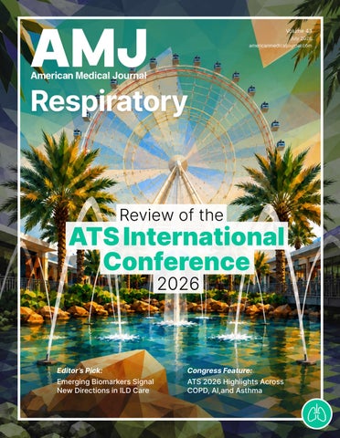
the American Thoracic Society (ATS) International Conference, May 15–20, 2026




Dr Kelly Pennington
Mayo Clinic, Minnesota, USA


Prof Laren Tan
Loma Linda University School of Medicine, California, USA


Dr Rohan Thompson
Indiana University, Indiana, USA
Dr Jacques Bouchard Université Laval, Canada
Dr Jezreel Pantaleón García
The University of Texas MD Anderson Cancer Center, Texas, USA
Prof Michal Senitko
University of Mississippi Medical Centre, Mississippi, USA
AMJ Respiratory is an open-access, peer-reviewed eJournal committed to helping elevate the quality of healthcare in respiratory medicine by publishing high quality content on all aspects of lung function and respiratory diseases.
The journal is published annually, 6 weeks after the American Thoracic Society (ATS) International Conference, and features highlights from this congress, alongside interviews with experts in the field, reviews of abstracts presented at the congress, as well as in-depth features on congress sessions. Additionally, this journal covers advances within the clinical and pharmaceutical arenas by publishing sponsored content from congress symposia, which is of high educational value for healthcare professionals. This undergoes rigorous quality control checks by independent experts and the in-house editorial team.
AMJ Respiratory also publishes peer-reviewed research papers, review articles, and case reports in the field. In addition, the journal welcomes the submission of features and opinion pieces intended to create a discussion around key topics in the field and broaden readers’ professional interests. AMJ Respiratory is managed by a dedicated editorial team that adheres to a rigorous double-blind peer-review process, maintains high standards of copy editing, and ensures timely publication.
Our focus is on research that is relevant to all healthcare professionals in pulmonary medicine. We do not publish veterinary science papers or laboratory studies not linked to patient outcomes. We have a particular interest in topical studies that advance research and inform of coming trends affecting clinical practice in the respiratory field.
Editorial Expertise
AMJ is supported by various levels of expertise:
• Guidance from an Editorial Board consisting of leading authorities from a wide variety of disciplines.
• Invited contributors are recognised authorities from their respective fields.
• Peer review, which is conducted by AMJ’s Peer Review Panel as well as other experts appointed due to their knowledge of a specific topic.
• An experienced team of editors and technical editors.
Peer Review
On submission, all articles are assessed by the editorial team to determine their suitability for the journal and appropriateness for peer review.
Editorial staff, following consultation with either a member of the Editorial Board or the author(s) if necessary, identify three appropriate reviewers, who are selected based on their specialist knowledge in the relevant area. All peer review is double blind.
Following review, papers are either accepted without modification, returned to the author(s) to incorporate required changes, or rejected. Editorial staff have final discretion over any proposed amendments.
Submissions
We welcome contributions from professionals, consultants, academics, and industry leaders on relevant and topical subjects.
We seek papers with the most current, interesting, and relevant information in each therapeutic area and accept original research, review articles, case reports, and features.
We are always keen to hear from healthcare professionals wishing to discuss potential submissions, please email: editorial@americanmedicaljournal.com
To submit a paper, use our online submission site: https://emj.kriyadocs.com/submissions/submit/emj/emj/login
Submission details can be found through our website: www.emjreviews.com/contributors/authors
All articles included in AMJ are available as reprints (minimum order 1,000). Please contact hello@emjreviews.com if you would like to order reprints.
Distribution and Readership
AMJ is distributed through controlled circulation to healthcare professionals in the relevant fields globally.
Open Access
This is an open-access journal in accordance with the Creative Commons Attribution-Non Commercial 4.0 (CC BY-NC 4.0) license.
Congress Notice
Staff members attend medical congresses as reporters when required.
This Publication Launch Date: 2023 Frequency: Yearly Online ISSN: 2976-7873
AMJ Respiratory is published once a year. For subscription details please visit: www.emjreviews.com
All information obtained by AMJ and each of the contributions from various sources is as current and accurate as possible. However, due to human or mechanical errors, AMJ and the contributors cannot guarantee the accuracy, adequacy, or completeness of any information, and cannot be held responsible for any errors or omissions. AMJ is completely independent of the review event (ATS 2026 International Conference) and the use of the organisations does not constitute endorsement or media partnership in any form whatsoever.
Front cover and contents photograph: Digitally Enhanced Image © aiisha / stock.adobe.com



High-level perspectives. Global experts. Essential updates.
Join us for conversations with the minds shaping healthcare's future. Gain the distilled insights you need to lead in your field and make maximum impact.

Jonathan Sackier: Non Executive Director & CMO, AiM Medical Robotics, Florida, USA



Saranya Ravindran: Paediatric Emergency Medicine Registrar, Imperial College Healthcare NHS Trust

Catherine Glass: Associate NHS GP and Senior Appraiser, NHS England
Editorial Director
Andrea Charles
Vice President of Content
Anaya Malik
Copy Editors
Noémie Fouarge, Meghan Garcka, Sarah Jahncke
Editorial Helena Bradbury, Katrina Thornber, Aleksandra Zurowska, Bertie Pearcey, Jess Nicholson, Alena Sofieva, Niamh Holmes, Nonyelum Okonkwo, Roli Omamuli, Josh Warren Lister
Creative Director
Tim Uden
Design Manager
Stacey White
Design
Tamara Kondolomo, Owen Silcox, Shanjok Gurung, Fabio van Paris, Fraser Hoey, Helena Spicer, Caleb Wylie
Head of Marketing
Stephanie Corbett
Vice President of Customer Success
Alexander Skedd
Vice President of Business Development
Robert Hancox
Chief Executive Officer
Justin Levett
Chief Commercial Officer
Dan Healy
Founder and Chairman
Spencer Gore
Dear Readers,
Welcome to this issue of AMJ Respiratory. I’m very proud to bring your our annual issue of peer-reviewed articles, expert-led congress coverage, and abstract reviews from the American Thoracic Society (ATS) International Conference 2026.
ATS 2026, held in Orlando, Florida, USA, was a fantastic showcase of research, providing immediate take-home messages for attendees and translating groundbreaking scientific discovery into practice. We thoroughly enjoyed our time at the meeting and at industry dinners exploring the cutting-edge data to support forced vital capacity decline in idiopathic pulmonary fibrosis, with exciting FDA approvals in this area.
This issue is designed to celebrate just that. Our congress review is introduced by Ali El Mokahal, accompanied by expert-led features exploring occupational lung disease, interventional pulmonology, and COPD. The abstract reviews, authored by meeting presenters, represent the wide scope of subspecialties within pulmonary medicine. This issue also includes a fantastic selection of articles addressing some of the most relevant questions in clinical respiratory practice. Our Editor’s Pick, ‘Current and Emerging Biomarkers in Interstitial Lung Disease’, provides a timely overview of the evolving biomarker landscape in interstitial lung disease. I am also pleased to feature ‘Advancements in Pediatric Lung Transplantation’, which explores progress in a complex and highly specialized area of respiratory care.
We are honored to support the fantastic work of the ATS through our carefully curated Congress Review, providing healthcare professionals with succinct, digestible information to inform daily clinical practice, and supporting patient outcomes globally.
Thank you to Kelly Pennington and our Editorial Board, authors, reviewers, editorial team, and readers, for your continued support as we work to make AMJ Respiratory the go-to place for healthcare professionals.
Best wishes,


Editorial
Anaya Malik Vice President of Content

Dear Colleagues,
The 2026 AMJ Respiratory issue brings together a fantastic collection of the American Thoracic Society (ATS) International Conference coverage, including broad-spectrum reviews and original abstract summaries that reflect the array of topics presented at the congress.
Across the meeting, several themes stood out, including the need for more individualized risk prediction, and the continued importance of multidisciplinary care, rehabilitation, and critical care in achieving measurable improvements for patients.
The Congress Review, introduced by Ali El Mokahal, provides a timely overview of key ATS 2026 developments, with focused, expert-led features on occupational lung disease, interventional pulmonology, and COPD. These are valuable assets with comprehensive coverage of data from the meeting, designed to directly influence your clinical decision-making and improve patient outcomes in your daily practice.
The abstract reviews included in this publication highlight several important clinical questions. Research on the burden of fungal infection after lung transplantation emphasizes the need for surveillance, prophylaxis, and individualized management. Other work focuses on pulmonary rehabilitation in interstitial lung disease, virtual maintenance rehabilitation with lung conditions, and invasive mechanical ventilation.


Across the meeting, several themes stood out, including the need for more individualized risk prediction, and the continued importance of multidisciplinary care
For my Editor’s Pick, I selected 'Current and Emerging Biomarkers in Interstitial Lung Disease'. This review provides a thoughtful and clinically relevant overview of the rapidly evolving biomarker landscape in interstitial lung disease, spanning physiologic measures, AI-assisted imaging, molecular profiling, and multidimensional risk models. Its emphasis on precision medicine and individualized risk prediction highlights an exciting and highly translational direction for the future of respiratory care.
I hope readers find this issue clinically practical and intellectually stimulating as we continue to advance patient-centered respiratory medicine.
Best wishes,

Kelly Pennington Division of Pulmonary and Critical Care Medicine, Mayo Clinic, Rochester,
Minnesota, USA


PRINT® Technology produces uniform ~1 µm particles designed to enhance deep-lung delivery and improve tolerability.2-4
YUTREPIA® (treprostinil) is a prostacyclin mimetic indicated for the treatment of:
• Pulmonary arterial hypertension (PAH; WHO Group 1) to improve exercise ability. Studies establishing effectiveness predominately included patients with NYHA Functional Class III symptoms and etiologies of idiopathic or heritable PAH (56%) or PAH associated with connective tissue diseases (33%).
• Pulmonary hypertension associated with interstitial lung disease (PH-ILD; WHO Group 3) to improve exercise ability. The study establishing effectiveness predominately included patients with etiologies of idiopathic interstitial pneumonia (IIP) (45%) inclusive of idiopathic pulmonary fibrosis (IPF), combined pulmonary fibrosis and emphysema (CPFE) (25%), and WHO Group 3 connective tissue disease (22%).
SELECT IMPORTANT SAFETY INFORMATION
WARNINGS AND PRECAUTIONS
• Treprostinil is a pulmonary and systemic vasodilator. In patients with low systemic arterial pressure, treatment with treprostinil may produce symptomatic hypotension.
• Treprostinil inhibits platelet aggregation and increases the risk of bleeding.
• Co-administration of a cytochrome P450 (CYP) 2C8 enzyme inhibitor (e.g., gemfibrozil) may increase exposure (both Cmax and AUC) to treprostinil. Co-administration of a CYP2C8 enzyme inducer (e.g., rifampin) may decrease exposure to treprostinil. Increased exposure is likely to increase adverse events associated with treprostinil administration, whereas decreased exposure is likely to reduce clinical effectiveness.
Please see Brief Summary of Prescribing Information on following page. For more information about YUTREPIA, including Full Prescribing Information and Instructions for Use, please visit www.YUTREPIAhcp.com.
References: 1. YUTREPIA. Prescribing information. Liquidia Technologies, Inc; 2025. 2. Hill NS, Feldman JP, Sahay S, et al; INSPIRE study investigators. INSPIRE: safety and tolerability of inhaled Yutrepia (treprostinil) in pulmonary arterial hypertension (PAH). Pulm Circ. 2022;12(3):e12119. doi:10.1002/pul2.12119 3. Garcia A, Mack P, Williams S, Fromen C, et al. Microfabricated engineered particle systems for respiratory drug delivery and other pharmaceutical applications. J Drug Deliv. 2012;2012:941243. doi:10.1155/2012/941243 4. Roscigno RF, Vaughn T, Parsley E, Hunt T, et al. Comparative bioavailability of inhaled treprostinil administered as LIQ861 and Tyvaso® in healthy subjects. Vascul Pharmacol. 2021;138:106840. doi:10.1016/j.vph.2021.106840
© 2026 Liquidia Technologies, Inc. YUTREPIA and PRINT are registered trademarks of Liquidia Technologies, Inc. All other trademarks are the property of their respective owners. All rights reserved. US-YUTR-2600013 04/26
BRIEF SUMMARY OF PRESCRIBING INFORMATION
FOR YUTREPIA® (treprostinil) inhalation powder, for oral inhalation
SEE PACKAGE INSERT FOR FULL PRESCRIBING INFORMATION
INDICATIONS AND USAGE
YUTREPIA® is a prostacyclin mimetic indicated for the treatment of:
• Pulmonary arterial hypertension (PAH; WHO Group 1) to improve exercise ability. Studies establishing effectiveness predominately included patients with NYHA Functional Class III symptoms and etiologies of idiopathic or heritable PAH (56%) or PAH associated with connective tissue diseases (33%).
The effects diminish over the minimum recommended dosing interval of 4 hours; treatment timing can be adjusted for planned activities.
While there are long-term data on use of treprostinil by other routes of administration, nearly all controlled clinical experience with inhaled treprostinil has been on a background of bosentan (an endothelin receptor antagonist) or sildenafil (a phosphodiesterase type 5 inhibitor).
The controlled clinical experience was limited to 12 weeks in duration.
• Pulmonary hypertension associated with interstitial lung disease (PH-ILD; WHO Group 3) to improve exercise ability. The study establishing effectiveness predominately included patients with etiologies of idiopathic interstitial pneumonia (IIP) (45%) inclusive of idiopathic pulmonary fibrosis (IPF), combined pulmonary fibrosis and emphysema (CPFE) (25%), and WHO Group 3 connective tissue disease (22%).
DOSAGE AND ADMINISTRATION
Please see Full Prescribing Information for complete information. CONTRAINDICATIONS
None.
Risk of Symptomatic Hypotension: Treprostinil is a pulmonary and systemic vasodilator. In patients with low systemic arterial pressure, treatment with treprostinil may produce symptomatic hypotension.
Risk of Bleeding: Treprostinil inhibits platelet aggregation and increases the risk of bleeding.
Effect of Other Drugs on Treprostinil: Co-administration of a cytochrome P450 (CYP) 2C8 enzyme inhibitor (e.g., gemfibrozil) may increase exposure (both Cmax and AUC) to treprostinil. Co-administration of a CYP2C8 enzyme inducer (e.g., rifampin) may decrease exposure to treprostinil. Increased exposure is likely to increase adverse events associated with treprostinil administration, whereas decreased exposure is likely to reduce clinical effectiveness. Bronchospasm: Like other inhaled prostaglandins, YUTREPIA may cause acute bronchospasm. Patients with asthma or chronic obstructive pulmonary disease (COPD), or other bronchial hyperreactivity, are at increased risk for bronchospasm. Ensure that such patients are treated optimally for reactive airway disease prior to and during treatment with YUTREPIA.
ADVERSE REACTIONS
Clinical Trials Experience: The safety and tolerability of YUTREPIA was evaluated in an open label study (INSPIRE) of 121 patients with PAH (WHO Group 1 and NYHA Functional Class II [80 patients] and Class III [41 patients]) followed for up to 2 months.
Adverse Reactions Occurring in ≥4% of Patients in the INSPIRE Study
Adverse Reaction Transition*; n=55 Add-On†; n=66 n (%) n (%)
Cough 15 (27)
Headache 14 (25) 18 (27)
Throat Irritation 5 (9) 14 (21)
Dizziness 6 (11) 7 (11)
Diarrhea
*Transition: Patients were on stable doses of treprostinil inhalation solution for at least 3 months prior to enrollment in the study and transitioned to treatment with YUTREPIA.
†Add-on: Patients were prostacyclin-naïve and were taking no more than 2 approved oral PAH therapies for at least 3 months at time of enrollment and addition of treatment with YUTREPIA.
The adverse reactions in the INSPIRE study were consistent with those observed in previous studies of inhaled treprostinil.
Adverse Reactions Identified in Post-Marketing Experience: Angioedema has been identified during the post-approval use of treprostinil inhalation solution.
Effect of Cytochrome P450 Inhibitors and Inducers: Co-administration of an oral formulation of treprostinil (treprostinil diolamine) with the CYP2C8 enzyme inhibitor gemfibrozil increases exposure (both Cmax and AUC) to treprostinil. Co-administration of the CYP2C8 enzyme inducer rifampin decreases exposure to treprostinil. It is unclear if the safety and efficacy of treprostinil by the inhalation route are altered by inhibitors or inducers of CYP2C8.
Effect of Other Drugs on Treprostinil: Co-administration of treprostinil (oral or subcutaneous) with acetaminophen (4 g/day), warfarin (25 mg/day), or fluconazole (200 mg/day) did not affect the pharmacokinetics of treprostinil. Treprostinil did not affect the pharmacokinetics or pharmacodynamics of warfarin.
Pregnancy – Risk Summary: Limited case reports of treprostinil use in pregnant women are insufficient to inform a drug-associated risk of adverse developmental outcomes. However, there are risks to the mother and the fetus associated with pulmonary arterial hypertension. In animal studies, no adverse reproductive and developmental effects were seen for treprostinil at >9 and >145 times the human exposure when based on Cmax and AUC, respectively, following a single YUTREPIA dose of 79.5 mcg. The estimated background risk of major birth defects and miscarriage for the indicated populations is unknown.
Lactation – Risk Summary: There are no data on the presence of treprostinil in human milk, the effects on the breastfed infant, or the effects on milk production.
Pediatric Use: Safety and effectiveness in pediatric patients have not been established.
Geriatric Use: Placebo-controlled clinical studies of treprostinil inhalation solution did not include sufficient numbers of patients aged 65 years and over to determine whether they respond differently from younger patients. The open-label INSPIRE study in PAH patients included 28 patients aged 65 and over in which no age-related differences were noted. In general, dose selection for an elderly patient should be cautious, reflecting the greater frequency of hepatic, renal, or cardiac dysfunction, and of concomitant diseases or other drug therapy.
Patients with Hepatic Insufficiency: Plasma clearance of treprostinil, delivered subcutaneously, was reduced up to 80% in subjects with mild-to-moderate hepatic insufficiency. Uptitrate slowly when treating patients with hepatic insufficiency because of the risk of an increase in systemic exposure which may lead to an increase in dose-dependent adverse effects. Treprostinil has not been studied in patients with severe hepatic insufficiency.
Patients with Renal Insufficiency: No dose adjustments are required in patients with renal impairment. Treprostinil is not cleared by dialysis.
In general, symptoms of overdose with treprostinil include flushing, headache, hypotension, nausea, vomiting, and diarrhea. Provide general supportive care until the symptoms of overdose have resolved.
Advise the patient to read the FDA-approved patient labeling (Instructions for Use). Train patients in the administration process for YUTREPIA, including dosing, inhaler preparation, administration, cleaning, and maintenance, according to the instructions for use. To avoid potential interruptions in drug delivery because of equipment malfunction, patients should have access to a back-up. In the event that a scheduled dose is missed, take another dose as soon as possible.
1
Mepolizumab helped more patients with eosinophilic COPD achieve disease stability, with benefits seen across GOLD severity categories
2
4
Home-based pulmonary rehabilitation delivered functional gains comparable to center-based programs and improved completion rates
5
Twice-yearly depemokimab maintained asthma and reduced exacerbations over 2 years in patients Type 2 asthma
Cumulative air pollution exposure was linked mortality and longer in acute respiratory

depemokimab asthma control exacerbations patients with pollution linked to higher longer ventilation respiratory failure

3
Brensocatib provides clinically meaningful benefits across multiple dimensions of disease burden, including respiratory symptoms and exacerbation control
Home-based programs delivered comparable functional improvements to center-based rehabilitation and helped more patients complete treatment
6
Electronic health record data identified distinct ARDS subphenotypes associated with sharply different clinical outcomes
Location: Orlando, Florida, USA
Date: May 15–20, 2026
Citation: Respir AMJ. 2026;4[1]:12-25. https://doi.org/10.33590/respiramj/R12O7F5B
THE American Thoracic Society (ATS) International Conference 2026, held in Orlando, Florida, USA, May 15–20, united the pulmonary medicine community for a week of discussion on the latest data, advances, and challenges in respiratory diseases. The meeting themes included the need for more individualized risk prediction, multidisciplinary care, and rehabilitation, which are explored in the following Congress Review.

ACCORDING to new research presented at ATS 2026, disease stability may be an achievable treatment goal for a broad range of patients with COPD receiving mepolizumab, even among those who remain poorly controlled despite triple therapy.1
COPD is a progressive respiratory disease characterized by worsening symptoms, declining lung function, and recurrent exacerbations that accelerate disease progression. While reducing exacerbations has long been a key treatment objective, disease stability, defined as sustained low disease activity following optimization of treatment, is emerging as a more ambitious target. Researchers sought to determine whether mepolizumab, an approved add-on maintenance therapy for adults with eosinophilic COPD, could help more patients achieve this outcome.
The analysis pooled data from three Phase III RCTs: METREX, METREO, and MATINEE. A total of 1,146 patients with COPD, blood eosinophil counts of at least 300 cells/µL, a history of exacerbations, and ongoing triple therapy were included. Investigators assessed disease stability at Week 52 using a composite endpoint that required patients to experience no moderate or severe exacerbations, maintain or improve their COPD Assessment Test (CAT) score, and preserve or improve lung function compared with baseline.


Importantly, the benefits of mepolizumab were observed across all Global Initiative for Chronic Obstructive Lung Disease (GOLD) severity categories. Disease stability was achieved by 26% of patients with GOLD 2 disease receiving mepolizumab compared with 23% receiving placebo. In patients with GOLD 3 disease, rates were 11% versus 9%, while among those with the most severe GOLD 4 disease, disease stability was achieved in 16% of patients receiving mepolizumab compared with just 6% of those receiving placebo.
The findings suggest that disease stability is not only measurable in COPD but may also be attainable across a wide spectrum of disease severity
Results showed that 18% of patients receiving mepolizumab achieved disease stability after 52 weeks, compared with 15% of those receiving placebo. The greatest contributor to this difference was a reduction in exacerbations. More than half of patients treated with mepolizumab met the exacerbation criterion for disease stability (52% versus 42% with placebo), while rates for maintaining health status and lung function were broadly similar between the groups.
Results showed that of patients receiving mepolizumab achieved disease stability after 52 weeks, compared with 15% of those receiving placebo
18%
The findings suggest that disease stability is not only measurable in COPD but may also be attainable across a wide spectrum of disease severity. For patients with eosinophilic COPD who continue to experience symptoms and exacerbations despite triple therapy, mepolizumab may offer an opportunity to achieve more sustained disease control.
While the analysis was conducted post hoc and further prospective validation is needed, the results support the growing view that disease stability could become an important therapeutic target in COPD management.
NEW data presented at ATS 2026 showed that depemokimab, the first ultra-long-acting biologic for Type 2 asthma, delivered sustained reductions in exacerbations and maintained improvements in asthma control and quality of life over a 2-year treatment period.2
Depemokimab is designed to target IL-5, a key driver of eosinophilic inflammation in Type 2 asthma. Its extended half-life enables dosing just twice yearly, offering a potentially convenient treatment option for patients with inadequately controlled disease. While previous Phase III studies demonstrated efficacy over 52 weeks, the latest analysis assessed whether these benefits could be maintained over the longer term.
Researchers conducted an integrated post-hoc analysis of the Phase III SWIFT-1 and SWIFT-2 trials and the AGILE open-label extension study. In the original SWIFT studies, 762 patients with Type 2 asthma characterized by elevated blood eosinophil counts were randomized to receive either subcutaneous depemokimab 100 mg or placebo every 26 weeks for 1 year. Patients completing the trials were eligible to enter AGILE, where they either continued depemokimab treatment or switched from placebo to depemokimab, allowing assessment of outcomes across a total of 104 weeks.
The results demonstrated a substantial and durable reduction in asthma exacerbations. During the first year of treatment, depemokimab reduced the annualized
exacerbation rate by 54% compared with placebo, with rates of 0.51 versus 1.11, respectively. Among patients who received depemokimab continuously, this benefit was maintained throughout the full 2-year period, with an annualized exacerbation rate of 0.52.
Patient-reported outcomes also remained consistently improved. At Week 52, participants receiving depemokimab experienced a marked improvement in health-related quality of life, reflected by a 13.92-point reduction in St George’s Respiratory Questionnaire score from baseline. This improvement was sustained through Week 104, reaching a 16.29-point reduction. Similarly, improvements in asthma control, measured using the Asthma Control Questionnaire-5 (ACQ-5), were maintained over the 2-year follow-up period, with scores improving from a reduction of 0.81 points at Week 52 to 0.89 points at Week 104.
Investigators also reported persistent suppression of Type 2 inflammation throughout treatment. Blood eosinophil counts remained consistently reduced over the 2 years among patients receiving continuous depemokimab therapy, indicating ongoing biological control of the inflammatory processes underlying disease activity.
Importantly, patients who switched from placebo to depemokimab after the first year experienced rapid and clinically meaningful improvements in clinical outcomes during their second year of treatment. These gains were sustained through the end of the study.
During the first year of treatment, depemokimab reduced the annualized exacerbation rate by 54% compared with placebo, with rates of 0.51 versus 1.11, respectively
The findings suggest that depemokimab provides durable clinical benefits and longterm disease control in patients with Type 2 asthma, while supporting the potential value of a twice-yearly dosing schedule. Although the analysis was conducted post hoc and the extension study was open-label, the results provide encouraging evidence that the efficacy observed during the initial Phase III trials can be maintained over an extended treatment period.
Brensocatib treatment resulted in improvements across all respiratory symptom domains compared with placebo

is a first-in-class oral, reversible dipeptidyl peptidase-1 inhibitor that targets neutrophil-driven inflammation in bronchiectasis. This post-hoc analysis of the Phase III ASPEN trial evaluated the impact of brensocatib on individual respiratory symptoms using the Quality of Life-Bronchiectasis Respiratory Symptoms Score (QOL-B RSS), as well as symptom outcomes according to on-study pulmonary exacerbation status.3
In ASPEN, adults with bronchiectasis and at least two pulmonary exacerbations in the preceding year were randomized to receive brensocatib 10 mg, brensocatib 25 mg, or placebo once daily for 52 weeks. QOL-B assessments were completed every 2 weeks throughout the study. The analysis focused on nine respiratory symptom items, including cough, congestion, sputum production and color, dyspnea, wheezing, chest pain, and nocturnal cough.
Overall, brensocatib treatment resulted in improvements across all respiratory symptom domains compared with placebo, with the greatest and most consistent benefits observed in the 25 mg treatment arm. At Week 52, the largest treatment differences versus placebo were seen for cough, congestion, dyspnea, and sputum color. Improvement in sputum color is of particular interest, as this parameter is a validated marker associated with neutrophilic airway inflammation.
Patients who experienced pulmonary exacerbations during the study reported a greater symptom burden than those who remained exacerbation-free. Nevertheless, symptom improvements with brensocatib were observed irrespective of whether patients experienced on-study exacerbations, including severe exacerbations, suggesting a treatment benefit beyond exacerbation prevention alone.
These findings complement the primary ASPEN results, in which brensocatib significantly reduced exacerbation frequency, while the 25 mg dose also slowed lung function decline and demonstrated improvements in patientreported outcomes. Collectively, the data indicate that brensocatib provides clinically meaningful benefits across multiple dimensions of disease burden, including respiratory symptoms and exacerbation control. Further real-world studies are warranted to confirm the durability and generalizability of these symptom improvements in broader bronchiectasis populations.
At Week 52, the largest treatment differences versus placebo were seen for cough, congestion, dyspnea, and sputum color
NEW integrated data from the WILLOW and ASPEN clinical trials suggest that higher systemic exposure to brensocatib is associated with meaningful improvements in lung function and pulmonary exacerbation outcomes in patients with non-cystic fibrosis bronchiectasis, while maintaining a generally favorable safety profile.
4
reversible inhibitor of dipeptidyl peptidase 1, serine proteases and dampen neutrophildriven airway inflammation. It is approved for use in patients aged 12 years and older with bronchiectasis.
The exposure–response analysis included 1,431 participants drawn from two pivotal studies: the Phase II WILLOW trial (NCT03218917) and the Phase III ASPEN trial (NCT04594369). Researchers evaluated relationships between steady-state drug exposure (AUCτ) and key efficacy outcomes, including annualized pulmonary exacerbation rate, time to first exacerbation, and postbronchodilator forced expiratory volume in 1 second (FEV1), as well as safety events of special interest such as hyperkeratosis, periodontal disease, and pneumonia.

When exposure was analyzed as a continuous variable, no statistically significant relationship was observed for exacerbation rate or time to first exacerbation. However, threshold-based modelling identified clinically meaningful improvements at AUCτ levels above 1,100 ng·h/mL. At this exposure, most patients receiving 25 mg (100%) and a majority receiving 10 mg (69%) achieved benefit in exacerbation-related outcomes.
A stronger relationship was observed for lung function. An AUCτ threshold of 1,531 ng·h/mL was associated with preservation of FEV1 over the treatment period. Above this threshold, nearly all patients on 25 mg (>99%) and almost half on 10 mg (46%) achieved exposures linked to stabilized lung function. In contrast, patients below this level or receiving placebo experienced comparable declines in FEV1.
No exposure-response relationship was identified for pneumonia or periodontal disease. A modest exposure-dependent increase in hyperkeratosis was observed, although events were generally mild or moderate and absolute rates remained low, with modelled probabilities of approximately 1.5-3.4% on active treatment versus 0.5% with placebo.
Overall, the findings support the use of 10 mg and 25 mg brensocatib doses in bronchiectasis, demonstrating exposuredependent efficacy across key clinical endpoints while maintaining an acceptable safety profile.
RESEARCH presented at ATS 2026 explored whether home-based pulmonary rehabilitation (PR) can provide comparable benefits while improving program completion. Investigators conducted a meta-analysis of RCTs directly comparing home-based and center-based approaches.5
Three randomized trials were included in the analysis, evaluating a total of 396 participants with chronic respiratory diseases. The primary outcome was change in 6-minute walk distance (6MWD), a widely used measure of functional exercise capacity. Using a predefined equivalence margin of ±30 m, the pooled analysis demonstrated virtually no difference between home-based and center-based PR, with a mean difference of −0.42 m. Statistical testing confirmed equivalence under the standard randomeffects model, indicating that home-based programs produced functional improvements comparable to those achieved in supervised center-based settings.
A more conservative sensitivity analysis using the Hartung-Knapp method widened the confidence intervals, resulting in an inconclusive equivalence assessment. Nevertheless, the findings did not suggest that home-based rehabilitation was inferior, highlighting the robustness of the overall functional outcomes despite some uncertainty.
Program completion emerged as a notable advantage of home-based rehabilitation. Across the included studies, participants assigned to home-based PR were

approximately 45% more likely to complete their program than those enrolled in centerbased services. Based on pooled estimates, home-based delivery increased completion rates by nearly 27%, corresponding to a number needed to treat of approximately four patients for one additional program completion. Although substantial heterogeneity was observed between studies, the overall trend consistently favored home-based approaches.
Program completion emerged as a notable advantage of home-based rehabilitation
These findings suggest that structured home-based PR, including programs supported by telehealth or remote coaching, may offer a practical solution for expanding access to rehabilitation without sacrificing functional benefit. Given the ongoing challenges surrounding PR uptake worldwide, home-based pathways could help address longstanding barriers while maintaining clinical effectiveness. Future research should focus on standardizing definitions of program completion, evaluating responder outcomes, and identifying which implementation components, such as coaching intensity, digital support, or remote monitoring, most strongly influence adherence and long-term success.
RESEARCH presented at ATS 2026 evaluated a novel smartphone application-based rehabilitation program combining music-guided exercise and singing training in patients with moderate-to-severe COPD.6
In this multicenter RCT conducted across five centers in China, 70 patients with stable COPD were assigned to one of three groups: multicomponent training (MT), rhythmguided walking (RW), or usual care (UC). The MT program combined tempo-guided walking, singing exercises, and educational content delivered through a dedicated smartphone application, while the RW group received only the walking component. Participants in the intervention arms completed a 12-week home-based program with individualized progression.
The primary outcome was exercise capacity, assessed using the Incremental Shuttle Walking Test (ISWT). At 12 weeks, patients in the MT group achieved significantly greater improvements in walking distance compared with those receiving usual care, with a mean increase of 56.35 m. This improvement exceeded the threshold generally considered clinically meaningful, demonstrating the potential of digitally delivered, musicsupported rehabilitation to enhance physical performance in COPD.
The benefits of the multicomponent intervention extended beyond exercise capacity. Compared with usual care, participants in the MT group reported significant reductions in breathlessness, as measured by the modified Medical Research Council scale, alongside improvements in health status assessed by the COPD Assessment Test. Anxiety symptoms also decreased significantly, while inspiratory capacity showed meaningful physiological improvement. Notably, the multicomponent program outperformed rhythm-guided walking alone in reducing dyspnea, suggesting that the addition of singing
training may provide benefits beyond those achieved through exercise alone.
In contrast, rhythm-guided walking by itself did not produce significant differences compared with usual care across the measured outcomes, highlighting the importance of the program’s combined approach. The findings suggest that integrating music, breathing control, vocal training, and exercise into a single digital platform may create a more engaging and effective rehabilitation experience.
This study demonstrates that a musicfacilitated, smartphone-based pulmonary rehabilitation program can deliver clinically meaningful improvements in exercise capacity, symptoms, and psychological wellbeing in patients with COPD. As healthcare systems increasingly explore remote and hybrid models of care, such digitally enabled interventions may offer an innovative strategy for expanding access to pulmonary rehabilitation while enhancing patient engagement and adherence. Further studies involving larger populations and longer follow-up periods will help determine the sustainability of these benefits and identify the key components driving treatment success.

At 12 weeks, patients in the MT group achieved significantly greater improvements in walking distance compared with those receiving usual care
ACUTE respiratory failure outcomes were significantly worse among critically ill patients with higher cumulative air pollution exposure, according to a large multicenter study, presented at ATS 2026.7

Researchers conducted a retrospective cohort study across seven academic medical centers in the USA, participating in the Common Longitudinal ICU Format (CLIF) consortium between 2018–2024. The analysis included 128,808 adults admitted to intensive care who met clinical criteria for acute respiratory failure. Residential county data were linked to annual mean satellite derived estimates of fine particulate matter (PM2.5) and nitrogen dioxide (NO2) exposure prior to admission. Census tract sociodemographic variables were also incorporated into the analysis.
The cohort had a mean age of 62±15 years and was 59% male. Non-Hispanic Black patients accounted for 32% of the study population. In hospital mortality was 16.9±1.6%, while the mean duration of mechanical ventilation was 53.4±156.5 hours. Average annual exposure levels were 7.6±1.5 µg/m³ for PM2.5 and 6.0±2.9 ppb for NO2, with notable regional variation.
The findings demonstrated a consistent association between greater air pollution exposure and poorer clinical outcomes. Each 10 ppb increase in cumulative NO2 exposure was associated with a 5% higher risk of in hospital death: (95% CI: 1.03–1.08). It was also associated with a 2% longer duration of invasive ventilation (95% CI: 1.00–1.04).
Similarly, each 10 µg/m³ increase in cumulative PM2.5 exposure was associated with a 9% higher risk of in hospital death (95% CI: 1.03–1.16) and a 9% higher risk of death within 30 days (95% CI: 1.02–1.17).
Further analysis using competing risk models suggested that air pollution exposure may affect the likelihood of recovery relative to death. Greater cumulative NO2 exposure was associated with a 51% higher subdistribution hazard ratio (SHR) of death relative to recovery (SHR: 1.51; 95% CI: 1.07–2.13). Higher PM2.5 exposure was linked to a 2.45-fold higher SHR of death relative to recovery (SHR: 2.45; 95% CI: 1.03–5.79).
The authors concluded that cumulative air pollution exposure was consistently associated with increased time on invasive ventilation and higher mortality among critically ill patients with acute respiratory failure. These findings suggest that longterm exposure to PM2.5 and NO2 may hinder recovery even within controlled intensive care environments and could be relevant to clinical decision making.
Higher PM2.5 exposure was linked to a 2.45fold higher SHR of death relative to recovery (SHR: 2.45; 95% CI: 1.03–5.79)

A NEW analysis of two Phase III clinical trials has shown that AD109 significantly reduced snoring frequency compared with placebo in adults with obstructive sleep apnea (OSA), offering a potential new approach to addressing one of the condition’s most common and socially disruptive symptoms.8 The findings suggest that, alongside improving airway obstruction, oxygenation, and disease severity, AD109 may also provide meaningful benefits for patients affected by chronic snoring.
OSA is characterized by repeated upper airway collapse during sleep, leading to disrupted breathing, reduced oxygen levels, and fragmented sleep. Snoring is one of its most recognizable symptoms and can have a substantial impact on quality of life, affecting both patients and their bed partners. Despite its prevalence, snoring is often assessed subjectively, and objective measures of treatment response remain limited.
Researchers evaluated the effect of AD109, a once-daily oral combination of aroxybutynin, a novel antimuscarinic, and atomoxetine, a selective norepinephrine reuptake inhibitor. The analysis included data from the randomized, placebo-controlled SynAIRgy and LunAIRo Phase III trials. Snoring was measured objectively during overnight polysomnography using a tracheal piezoelectric snore sensor, with snoring defined as breathing sounds exceeding 20 dB above background breathing noise.
Among participants with available baseline and Week 26 sleep study data, 189 individuals receiving AD109 and 281 receiving placebo met the predefined criteria for inclusion. Baseline snoring frequency was similar between groups, with snoring present
during approximately 42–45% of breaths during sleep.
By Week 26, AD109 reduced the proportion of breaths associated with snoring by 22.3% (95% CI: 19.1–25.5), compared with a 10.0% reduction in the placebo group (95% CI: 7.4–12.6). This represented a significant treatment difference of −12.3% (95% CI: −16.4–−8.2; p<0.001). In addition, 60.8% of participants receiving AD109 achieved at least a 50% reduction in snoring frequency, compared with 32.0% of those receiving placebo (p<0.001).
Sensitivity analyses using stricter definitions of snoring, based on sound thresholds exceeding 25 dB and 30 dB above background breathing noise, produced similar findings, supporting the robustness of the results.
These findings indicate that AD109 may address an important symptom burden associated with OSA while complementing its previously demonstrated benefits on airway obstruction, oxygenation, and overall disease severity. Further research may help determine how reductions in snoring translate into improvements in patient-reported outcomes and quality of life.
By Week 26, AD109 reduced the proportion of breaths associated with snoring by 22.3% (95% CI: 19.1–25.5)
BREAKTHROUGH treatment evidence is emerging for adolescents with Type 2 asthma, according to an abstract presented at ATS 2026.9 Asthma is a chronic inflammatory airway disease with variable airflow obstruction and symptoms including wheeze and breathlessness. Severe Type 2 asthma is associated with eosinophilic inflammation and increased exacerbation risk. Depemokimab is an ultra-long-acting monoclonal antibody targeting IL-5 with high binding affinity and an extended half-life, enabling 26-week dosing.
In Phase III SWIFT-1/-2 trials, patients with Type 2 asthma characterized by elevated blood eosinophil counts and ≥2 exacerbations in the previous year were included. Participants were then randomized 2:1 to depemokimab 100 mg subcutaneously or placebo every 26 weeks in addition to standard therapy. This prespecified analysis assessed adolescents aged 12–17 years. The primary endpoint was set as the annualized clinically significant exacerbation (CSE) rate over 52 weeks. A Bayesian dynamic borrowing approach incorporated adult data due to low adolescent numbers.
The pooled SWIFT-1/-2 population included 30 adolescents, 15 of whom received depemokimab, and 732 adults. Annualized CSE rate reductions were generally consistent between adolescents and adults. In adolescents, depemokimab reduced annualized CSEs by 43% versus placebo, compared with 54% in adults, showing that reduction in CSEs was consistent across adolescent and adult populations. Bayesian analysis indicated a minimum prior weight of 0.6 to achieve statistical equivalence, with consistent point estimates across borrowing scenarios. This approach incorporated a proportion of adult data to improve estimate precision given the limited adolescent sample size. Adverse event incidence was similar between adolescents and adults (73% versus 72%).
Among adolescents, two treatment-related adverse events were reported and one serious adverse event (abdominal pain),
In adolescents, depemokimab reduced annualized CSEs by 43% versus placebo, compared with 54% in adults
which resolved and was not considered treatment-related. No deaths or treatmentrelated discontinuations were reported. Overall, adverse event rates were comparable between adolescents and adults with no study withdrawal. Findings are limited by small adolescent participants, although adult data supported extrapolation using prespecified statistical methodology. Overall, results support a favorable benefit–risk profile of depemokimab in adolescents with Type 2 asthma.
No deaths or treatment-related discontinuations were reported

IDENTIFYING two distinct acute respiratory distress syndrome (ARDS) subphenotypes from routine electronic health record data could support phenotype-guided risk stratification, according to research presented at ATS 2026.10

ARDS is a life-threatening condition where fluid builds up in the lungs, impairing oxygen exchange and leading to severe respiratory failure. As a heterogeneous condition, latent phenotypes identified in clinical trial populations have rarely been reproduced in electronic health record cohorts.
Researchers analyzed data from MIMIC-IV, a freely accessible electronic health record database, to determine whether latent profile analysis could identify clinically meaningful ARDS subphenotypes and whether these groups were associated with different outcomes. The retrospective study included 24,363 ARDS admissions and used 25 routinely collected clinical variables, including demographics, vital signs, laboratory results, ventilator parameters, and key diagnoses.
The analysis identified two ARDS subphenotypes: a hypoinflammatory group (61.9%) and a hyperinflammatory group (38.1%). Patients in the hyperinflammatory group were generally younger and were more likely to be women or Black patients. They also had poorer oxygenation and greater physiologic derangement at baseline.
Patients with the hyperinflammatory phenotype experienced substantially worse outcomes. Ninety-day mortality
reached 40.8%, compared with 19.1% in the hypoinflammatory group, and mean survival time over 90 days was 11.9 days shorter in the hyperinflammatory group.
Furthermore, the hyperinflammatory phenotype was associated with greater use of advanced organ support, with patients more likely to require invasive mechanical ventilation, vasopressor therapy, renal replacement therapy, and tracheostomy.
As a retrospective analysis of electronic health record data, the study cannot determine whether the identified phenotypes directly cause differences in outcomes. However, the findings demonstrate that clinically distinct ARDS subgroups can be reproduced at scale using routinely collected data.
The researchers suggest that phenotypeguided risk stratification could eventually be incorporated into clinical decision-support systems and used to identify candidates for future phenotype-directed clinical trials. They also noted that observed differences across sex and racial groups highlight the importance of equity safeguards as such approaches are developed. Future research could potentially explore how these subphenotypes perform prospectively and whether they can help guide treatment strategies in clinical practice.
The analysis identified two ARDS subphenotypes: 61.9% 38.1%
A hypoinflammatory group and a hyperinflammatory group
References
1. Singh D et al. Disease stability is achievable in a wide spectrum of patients with chronic obstructive pulmonary disease receiving mepolizumab: pooled results from phase III randomized controlled trials. Poster Board P1506. ATS International Conference, May 15-20, 2026.
2. Pavord ID et al. Depemokimab demonstrates sustained long-term efficacy and consistent patient-reported outcomes over 2 years in patients with type 2 asthma: an integrated analysis of the SWIFT-1/-2 and AGILE studies. Poster Board P1396. ATS International Conference, May 15-20, 2026.
3. Flume PA et al. Effect of Brensocatib on patient-reported symptoms in patients with non-cystic fibrosis bronchiectasis: a post hoc analysis of QOL-B RSS individual items from the ASPEN phase 3 trial. Poster P1549. ATS International Conference, May 15-20, 2026.
4. Usansky H et al. Exposure-response relationships of brensocatib in adult and adolescent patients with non-cystic fibrosis bronchiectasis. P9745. ATS International Conference, May 15-20, 2026.
5. Patel R et al. Home vs center pulmonary rehabilitation: do we see functional equivalence and higher completion rates? A meta-analysis. ATS International Conference, May 15-20, 2026.
6. Shi M et al. Music-facilitated homebased pulmonary rehabilitation with a smartphone application for patients with chronic obstructive pulmonary disease: randomized controlled trial. ATS International Conference, May 15-20, 2026.
7. Graffy P et al. Cumulative air pollution exposure increases the severity of acute respiratory failure: a U.S. multicenter study. Poster. ATS International Conference, May 15-20, 2026.
8. Jewell L et al. AD109 significantly decreases snoring in patients with obstructive sleep apnoea. Presentation. ATS International Conference, May 15-20, 2026.
9. Jackson DJ et al. Efficacy and safety of twice-yearly depemokimab in adolescent patients with asthma: subgroup analysis of the SWIFT-1/-2 studies. Abstract P1388. ATS International Conference, May 15-20, 2026.
10. Krishna NS et al. Subphenotypes in acute respiratory distress syndrome: EHR-based latent profile analysis and clinical outcomes in 24,363 ICU admissions. Poster Board P1018. ATS International Conference, May 15-20, 2026.
Author: *Ali El Mokahal1
1. Mayo Clinic, Rochester, Minnesota, USA
*Correspondence to elmokahal.ali@mayo.edu
Disclosure: The author declares no conflicts of interest.
Keywords:
AI, American Thoracic Society (ATS) 2026, bronchiectasis, idiopathic pulmonary fibrosis (IPF), Mycobacterium avium complex (MAC), obstructive sleep apnea, pulmonary arterial hypertension (PAH).
Citation: Respir AMJ. 2026;4[1]:26-29. https://doi.org/10.33590/respiramj/NO08144K
THE American Thoracic Society (ATS) International Conference, held May 15–20, 2026, in Orlando, Florida, USA, brought together more than 14,000 clinicians, researchers, educators, trainees, and industry leaders from more than 100 countries. Through 6 days of scientific sessions, clinical trials, keynote addresses, and educational programming, ATS 2026 focused on a field in transition, increasingly shaped by AI and precision therapeutics, while maintaining an emphasis on patient-centered care, equity, and professional resilience.
The meeting opened with a keynote address from Robert M. Wachter, from the University of California, San Francisco, USA, and author of the national bestseller ‘A Giant Leap: How AI is Transforming Healthcare and What That Means for Our Future’ 1 The presentation, ‘How AI is Transforming Health Care’, set the tone for a conference in which AI emerged as a defining force poised to reshape medicine. Wachter highlighted the exhilarating progress of ambient documentation platforms, clinical decision-support systems, and AI assisted chart summarization tools, while also acknowledging important challenges including misinformation, privacy concerns, and potential clinician de-skilling. Nonetheless, his overall message was one of informed optimism, emphasizing that AI does not need to be perfect to improve healthcare, only better than the current status quo. He encouraged clinicians to actively engage
with and help shape the integration of AI into medical practice and education.
This emphasis on engagement with emerging technologies was reinforced through the ATS 2026 AI Lab, a new initiative dedicated to exploring applications of AI in pulmonary, critical care, and sleep medicine. Educational sessions focused on a wide range of topics, including AI literacy, model development, and clinical applications of large language models, while interactive activities allowed attendees opportunities to explore practical applications of these technologies firsthand. The AI Lab reflected ATS’s broader effort to prepare clinicians for the rapidly evolving digital landscape of medicine.
Despite the conference’s strong emphasis on technological innovation, ATS 2026 also emphasized deeply human themes. The
plenary session featured Rana L. Awdish from Henry Ford Health, Detroit, Michigan, USA, author of medical memoir ‘In Shock: My Journey from Death to Recovery and the Redemptive Power of Hope’.2 Her presentation chronicled her personal experience surviving catastrophic critical illness and her subsequent reflections as both physician and patient. Drawing from her journey of recovery, Awdish discussed the profound impact of communication, empathy, and language during moments of vulnerability and suffering, emphasizing how clinician interactions can shape patients’ experiences long after hospitalization. The plenary resonated strongly with attendees and served as a reminder that medicine remains fundamentally grounded in human relationships, compassion, and hope, even amid extraordinary scientific and technological progress.
During the conference, multiple practice changing clinical trials were introduced on a variety of topics in pulmonary and critical care medicine. Below, the author reviews some of the highlights
One of the major pulmonary vascular highlights of ATS 2026 was the growing sense that pulmonary arterial hypertension (PAH) entered a new therapeutic era. This was emphasized during the President’s symposium, ‘Celebrating Science: Pulmonary Hypertension from Basic Discoveries to Translational Breakthroughs, to Clinical Applications’, which tracked the evolution of PAH therapy from early patho-biologic discoveries to modern precision therapeutics.
Against this backdrop, Vallerie McLaughlin, University of Michigan, Ann Arbor, USA, presented data from the global Phase III randomized, double-blind, placebocontrolled ADVANCE OUTCOMES trial evaluating ralinepag, an oral selective prostacyclin receptor agonist in patients with PAH receiving background therapy.
With 687 Group 1 patients with PAH enrolled, the study demonstrated a statistically significant 55% reduction in the risk of first clinical worsening when compared with placebo. This benefit was consistent across major subgroups, including time since diagnosis, baseline 6-minute walk test (6MWD), WHO functional class, N-terminal pro-B-type natriuretic peptide (NT-ProBNP) levels, background therapies, and etiology. Statistically significant improvements relative to placebo were observed in multiple secondary outcomes, including 6MWD and NT-proBNP. This trial highlighted the continued evolution of the therapeutic armamentarium in PAH.
Mycobacterium avium complex (MAC) is a difficult disease to treat, requiring prolonged multidrug therapy, with a current 60% treatment success rate. Amikacin liposome inhalation suspension (ALIS) was previously for refractory MAC disease.3
Charles L. Daley, from National Jewish Health, Denver, Colorado, USA, presented results from the ENCORE trial, a Phase IIIb randomized, double-blind, multinational trial evaluating the efficacy of ALIS as a firstline treatment in MAC. Symptomatic adults with positive cultures were randomized to ALIS with azithromycin and ethambutol or placebo with azithromycin and ethambutol. Unique among MAC trials, ENCORE utilized the validated Quality of Life Bronchiectasis Respiratory Symptom Scale (QOL-B RSS) patient-reported outcome score as its primary endpoint. Among 425 patients randomized, 80% were treatment naïve.
Patients receiving ALIS had a 17.77-point improvement from baseline compared with 14.66 points in the placebo group (p=0.0299).
Culture conversion by Month 6 occurred in 87.8% of patients receiving ALIS compared with 57% in the placebo arm, and benefits
persisted through Month 12 with durable culture conversion through Month 15 of 76.2% compared to 47.6% in the placebo arm. These findings suggest that upfront incorporation of ALIS may improve symptoms and microbiologic eradication rates in MAC lung disease.
Interest in inhaled treprostinil as a potential therapy for idiopathic pulmonary fibrosis (IPF) emerged following the INCREASE trial, a study evaluating inhaled treprostinil in patients with pulmonary hypertension associated with interstitial lung disease. The INCREASE trial unexpectedly demonstrated improvements in forced vital capacity (FVC),4 raising the possibility of antifibrotic effects independent of pulmonary vasodilatory effects. These findings provided the rationale for the TETON clinical trial program.
Steven D. Nathan, from Inova Fairfax Hospital, Woodburn, Virginia, USA, presented results from the TETON-1 trial. This was a Phase III randomized, double-blind, placebocontrolled clinical done in Canada and the US evaluating inhaled treprostinil in patients with IPF. Patients with or without background antifibrotic therapy were randomized to receive inhaled treprostinil or placebo over a 52-week treatment period. The primary endpoint was change in absolute FVC.
TETON-1 enrolled 598 patients and met its primary endpoint, with inhaled treprostinil demonstrating a statistically significant reduction in FVC decline compared with placebo. Median change in FVC at Week 52 was −43.3 mL in the inhaled treprostinil group compared with −196.2 mL in the placebo arm, corresponding to a treatment difference of 130.1 mL (p<0.001). In participants not on background therapy, on nintedanib, or on pirfenidone, the mean differences were 98.7 mL, 123.4 mL, and 168 mL, respectively. An important secondary outcome was that inhaled treprostinil also reduced the risk of
clinical worsening by 33%, defined as death, respiratory hospitalization, or a ≥10% relative decline in FVC.
Clinically, the TETON-1 trial is important because it represents one of the first Phase III studies to demonstrate a meaningful reduction in FVC decline with a novel inhaled therapy in IPF. If incorporated into future treatment paradigms, inhaled treprostinil could substantially expand the currently limited therapeutic landscape for patients with IPF.
Sanjay R. Patel and Patrick J. Strollo, from the University of Pittsburgh, Pennsylvania, USA, presented pooled findings from the SynAIRgy and LunAIRo Phase III trials evaluating aroxybutynin and atomexetine (AD109), an investigational oral therapy for obstructive sleep apnea.
AD109 combines aroxybutynin, an antimuscarinic agent, and atomoxetine, a selective norepinephrine reuptake inhibitor, with the goal of improving upper airway dilator muscle tone and reducing upper airway collapse during sleep. SynAIRgy was a 26-week randomized, double-blind, placebo-controlled trial enrolling 646 participants across the US and Canada, while LunAIRo was a 51-week study enrolling 660 participants from 64 US sites. Eligible participants had OSA defined by a 4% ApneaHypopnea Index (AHI4) greater than five events/hour, PROMIS fatigue raw score ≥17, and had failed or declined positive airway pressure therapy.
In the pooled treatment policy estimand, which included all randomized participants receiving at least one dose of study drug, AD109 reduced AHI4 by a least-squares mean difference of −4.0 events/hour versus placebo (p<0.0001), corresponding to a 39.3% reduction from baseline compared with 12.6% for placebo. In the supportive on-treatment analysis, AD109 achieved a 51.6% reduction
from baseline compared with 13.3% for placebo. Secondary endpoints also improved, including hypoxic burden, daytime sleepiness, and patient-reported fatigue and sleep impairment. Disease control, defined as AHI4 <5 events/hour, was achieved in 22.6% of treated participants versus 8.3% with placebo.
Collectively, these studies represent an exciting new oral alternative for the treatment of obstructive sleep apnea for patients unable or unwilling to use positive airway pressure therapy.
ATS 2026 also featured the annual BEAR Cage competition, a ‘Shark Tank’-style competition designed to fund innovative translational ideas in pulmonary and critical care medicine. This year’s winner, Emily Mitchell from the University of California San Diego, USA, presented ‘Breaking the Silence: Eye-Tracking for Restoring Patient Agency and Communication in the ICU’, a software platform utilizing eye-tracking technology to help intubated ICU patients communicate
References
1. Wachter R., A Giant Leap: How AI is Transforming Healthcare and What That Means for Our Future (2026), New York: Portfolio.
2. Awdish R., In Shock: My Journey from Death to Recovery and the Redemptive Power of Hope (2017), New York: St. Martin’s Press.
and regain autonomy. For winning the competition, Mitchell received the 10,000 USD grand prize, which she plans to reinvest into further development and hospital testing of the platform later this year.
Collectively, the highlighted sessions reflect a specialty entering a period of rapid therapeutic advancement. Novel therapeutic modalities in the fields of pulmonary hypertension, interstitial lung disease, nontuberculous mycobacterial infections, and obstructive sleep apnea present exciting opportunities to improve outcomes in diseases that have historically carried substantial morbidity and limited treatment options. More broadly, the conference highlighted the growing role of AI and digital innovation in modern medicine, while reaffirming the importance of compassionate, patient-centered care. Overall, ATS 2026 reflected a field poised to enter an exciting new era of respiratory medicine, one defined by both innovation and compassion.
3. Griffith DE et al. Amikacin liposome inhalation suspension for treatmentrefractory lung disease caused by mycobacterium avium complex (CONVERT). A prospective, open-label, randomized study. Am J Respir Crit Care Med. 2018;198(12):1559-69.
4. Nathan SD et al. Inhaled treprostinil and forced vital capacity in patients with interstitial lung disease and associated pulmonary hypertension: a post-hoc analysis of the INCREASE study. Lancet Respir Med. 2021;9(11):1266-74.
Authors: *Rakeem Levy,1 Carlos Gracidas2
1. Caribbean Medical University, Willemstad, Curaçao 2. Universidad Marista de Merida, Mexico *Correspondence to Rakeemlevymed@gmail.com
Disclosure: The authors have declared no conflicts of interest.
Keywords: AI, COPD, CT phenotyping, digital health technologies, eosinophilic COPD, mepolizumab, precision medicine.
Citation: Respir AMJ. 2026;4[1]:30-33. https://doi.org/10.33590/respiramj/5785Y50A
AS THE American Thoracic Society (ATS) International Conference returned to Orlando, Florida, USA, it focused on important advances in COPD, reflecting the increase in personalized and technology-driven care. Across clinical sessions and research presentations, investigators highlighted biologic therapies, AI-assisted imaging, and digital respiratory tools designed to improve symptom control, reduce exacerbations, and better characterize COPD phenotypes.1 At the same time, several presentations emphasized the ongoing real-world challenges in COPD management, including barriers to treatment access and implementation in everyday clinical practice.
The biologics picture for COPD took center stage at ATS 2026, with three key datasets advancing our understanding of targeted therapy in specific inflammatory pathways in this population. Building on the FDA approval of mepolizumab for eosinophilic COPD, Criner et al.2 presented a pooled analysis of the Phase III METREX, METREO, and MATINEE trials, demonstrating that mepolizumab added to inhaled triple therapy significantly reduced exacerbations requiring emergency department visits and/or hospitalizations by 23% versus placebo in patients with a blood eosinophil count >150 cells/μL. A complementary pooled analysis by Singh et al.3 extended this concept beyond
exacerbation reduction, reporting that disease stability at Week 52 was achieved in a considerably larger number of patients receiving mepolizumab than in those receiving placebo, with fewer exacerbations in patients with the biologic. These findings support the use of blood eosinophils as a relevant biomarker for identifying patients with COPD who may benefit from targeted anti-IL-5 therapy.3
Across clinical sessions and research presentations, investigators highlighted biologic therapies, AI-assisted imaging, and digital respiratory tools
As the discussion of COPD therapeutics expanded beyond biologics, ensifentrine emerged as a non-biologic add-on strategy for patients who remain symptomatic despite standard inhaled maintenance therapy. A first-in-class dual phosphodiesterase (PDE)3/ PDE4 inhibitor with bronchodilator and antiinflammatory effects represents a distinct therapeutic approach in this population. In the RELIEF study, an observational 6-month longitudinal analysis of patients initiating ensifentrine, most participants were already receiving stable triple therapy; among patients with a baseline COPD Assessment Test (CAT; GSK, London, UK) score >10, the mean CAT score improved from 24.6 at baseline to 20.3 at 1 month and 19.9 at 3 months, with 65% meeting the CAT responder threshold at both time points.4 Another real-world analysis of 2,372 patients receiving ensifentrine with concomitant dual or triple therapy showed an 11% reduction in annualized severe exacerbations and a 17% reduction in COPD emergency room visits after treatment initiation.5 Together, these
findings suggest that ensifentrine may offer a non-biologic add-on alternative for patients with persistent symptoms or exacerbationrisk events on maintenance therapy, although longer-term data will be needed to better define its role in routine COPD management.
ATS 2026 highlighted a parallel shift toward precision phenotyping in COPD. The session 'COPD Biomarkers and Phenotyping: MultiOmic Insights, AI-Driven Discovery, and Clinical Implications' framed COPD as a heterogeneous disease that may require more refined tools than spirometry. This concept was exemplified by analyses of the SOURCE and COPDGene cohorts, in which CT-based assessment of mucus plugging helped identify structural changes associated with disease activity and prognosis. In COPDGene, an AI-based algorithm was used for the automated detection of CT airway mucus plugs in 8,971 participants. Greater mucus plug burden was associated with lower post-

bronchodilator forced expiratory volume in 1 second, increased air trapping, worse quality-of-life scores, reduced 6-minute walk distance, and an independently higher risk of mortality and exacerbations.6 A complementary analysis from the SOURCE early COPD progression cohort applied a deep learning pipeline to baseline CT scans from 657 younger participants with smoking history, identifying mucus plugs in 7% of participants. Although uncommon, mucus plugs were associated with emphysema, CTdefined functional small-airway disease, and more frequent exacerbations.7 These findings suggest that an AI-assisted CT analysis may help transform mucus plugging from a visually recognized imaging feature into a quantifiable biomarker with potential value for COPD phenotyping, risk stratification, and future phenotype-directed interventions.
Accessible technologies also emerged as a promising strategy to support earlier COPD detection and longitudinal monitoring. In a multicenter prospective study, Wang et al.8 evaluated Cough Search, a multimodal AI-based cough analysis software for the auxiliary diagnosis of COPD. Among 378 participants, Cough Search demonstrated 92% sensitivity and 89% specificity as a standalone tool, and, when combined with physician assessment, achieved 93% sensitivity and 94% specificity, showing higher sensitivity than portable spirometry-assisted physician diagnosis while maintaining non-inferior specificity.8 A separate but prospective study evaluated smartphone-based spirometry using machine learning and showed strong agreement with conventional spirometry for forced expiratory volume in 1 second/forced vital capacity, excellent twice-daily reproducibility, and the ability to capture diurnal variation and deviations from baseline.9 Together, these studies suggest the availability of an innovative respiratory tool that may help extend COPD care beyond clinic-based
testing, supporting earlier identification of high-risk patients and more continuous monitoring of disease instability.
Improving outcomes in COPD requires more than identifying and administering effective pharmacologic therapies. The access, treatment implementation, and social factors that shape real-world care were emphasized during the Conference. The US DUALITY disparities study, which included 338,947 patients with COPD who had experienced one severe or two moderate exacerbations, found that only 6.9% of patients initiated triple therapy within 12 months, with a variation depending on age, sex, race/ ethnicity, payer coverage, geography, and symptom profile, and with higher initiation among patients with documented symptoms.10 A separate study focused on food insecurity in COPD demonstrated that 34.5% of patients with COPD self-reported food and/or financial insecurity.11 Together, these findings suggest that future COPD care will need to go beyond effective medication prescriptions to address implementation gaps, recognize social needs, and ensure that they are addressed as part of routine COPD care.
The COPD updates presented at ATS 2026 reflected a field in constant flux toward a uniform treatment model of individualized, clinically practical care. Biologic therapies highlighted the importance of identifying treatable inflammatory traits, while ensifentrine underscored the need for additional non-biologic options for patients who remain symptomatic despite maintenance therapy. At the same time, AI-assisted imaging and digital respiratory tools showed that COPD assessment may extend beyond spirometry-based diagnosis and stratification, supporting precise
phenotyping, earlier detection, and closer monitoring. However, the Conference also addressed the need to translate innovation into real-world care. Persistent gaps in guideline-directed therapy initiation and the under-recognition of social needs, such as food insecurity, remind healthcare workers that improvements in outcomes will depend not only on new therapies and technologies,
1. ATS Conference News. ATS 2026 Orlando sessions examine recent insights and unique challenges for treating COPD. 2026. Available at: https://www.atsconferencenews.org/ ats-2026-orlando-sessions-examinerecent-insights-and-unique-challengesfor-treating-copd/. Last accessed: May 31, 2026.
2. Criner GJ et al. A34-11 Mepolizumab reduces the burden of chronic obstructive pulmonary disease exacerbations in a wide spectrum of patients: pooled results from phase III randomized controlled trials. Am J Respir Crit Care Med. 2026;212 (Suppl 1):aamag162.1629.
3. Singh D et al. A34-21 Disease stability is achievable in a wide spectrum of patients with chronic obstructive pulmonary disease receiving mepolizumab: pooled results from phase III randomized controlled trials. Am J Respir Crit Care Med. 2026;212(Suppl 1):aamag162.1639.
but also on an equitable healthcare systems and patient-centered support. Together, these findings show that the future of COPD care will require matching personalized interventions with innovation to ensure that it reaches those most likely to benefit.
4. Washko GR et al. B24-20 Realworld COPD Assessment Test (CAT) outcomes of patients initiating ensifentrine: 3 month interim analysis of the RELIEF study. Am J Respir Crit Care Med. 2026;212(Suppl 1):aamag162.1779.
5. Mahler DA et al. B24-07 The effect of ensifentrine on symptoms in highly symptomatic patients at baseline according to two definitions: a posthoc pooled analysis of the ENHANCE trials. Am J Respir Crit Care Med. 2026;212(Suppl 1):aamag162.1766.
6. Oyer JM et al. D96-01 AI-based detection of CT airway mucus plugs and clinical correlates in COPDGene. Am J Respir Crit Care Med. 2026;212 (Suppl 1):aamag162.2066.
7. Ram S et al. A16-04 Significance of mucus plugs in early COPD: an analysis of the SOURCE cohort. Am J Respir Crit Care Med. 2026;212 (Suppl 1):aamag162.1588.
8. Wang Q et al. B24-17 Auxiliary diagnosis of COPD with cough search and portable spirometry: a multicenter, prospective, comparative validation study. Am J Respir Crit Care Med. 2026;212(Suppl 1):aamag162.1776.
9. Boyle AD. B24-05 Smartphone spirometry for monitoring respiratory function - a prospective observational study. Am J Respir Crit Care Med. 2026;212(Suppl 1):aamag162.1764.
10. Chima-Melton C et al. A23-01 Clinical and social drivers of triple therapy initiation amongst patients with COPD exacerbations: results from the US DUALITY disparities study. Am J Respir Crit Care Med. 2026;212(Suppl 1):aamag162.1601.
11. Hart JL et al. A23-12 Starving for attention: a mixed-methods study of food insecurity in chronic obstructive pulmonary disease. Am J Respir Crit Care Med. 2026;212 (Suppl 1):aamag162.1612.
Author: *Samvel Gaboyan1,2
1. Division of Pulmonary, Critical Care, and Sleep Medicine & Physiology, Department of Medicine, University of California, San Diego, USA
2. Pulmonary & Critical Care Section, VA San Diego Healthcare System, California, USA
*Correspondence to samvel.s.gaboyan@gmail.com
Disclosure: The author has declared no conflicts of interest.
Keywords:
Citation:
Countertops, end-stage lung disease, engineered stone, lung transplantation, occupational lung disease, silicosis, surveillance.
Respir AMJ. 2026;4[1]:34-37.
https://doi.org/10.33590/respiramj/2QFDAP13
AT THE 2026 American Thoracic Society (ATS) International Conference, held in Orlando, Florida, USA, numerous presentations highlighted advances in research on silicosis. The ongoing resurgence of rapidly progressive silicosis, particularly among engineered stone countertop fabricators, with no existing treatments outside of lung transplantation, underscores the urgent need for continued investigation into this preventable yet fatal occupational lung disease. The wide range of work environments and exposure patterns complicate efforts to characterize disease pathogenesis, evaluate potential therapies, and develop effective prevention and screening strategies. Collectively, the studies presented at ATS 2026 demonstrated the continuum of silicosis research, spanning the identification of disease mechanisms, characterization of clinical manifestations and disease outcomes, and the enhancement of surveillance and screening approaches aimed at reducing disease burden.
TRANSLATIONAL STUDIES REVEAL POTENTIAL THERAPEUTIC TARGETS FOR DELAYING SILICOSIS DISEASE PROGRESSION
Studies in both humans and rodents identified molecular pathways and novel cell populations and characterized their roles in the pathogenesis of silicosis. Miao et al.1 demonstrated that the expression and activity of histone deacetylase 6 (HDAC6), a cytoplasmic regulator of TANK-binding
kinase 1 and interferon regulatory factor 3 (TBK1-IRF3), were increased in human and mouse silicosis lung tissue; thus, the expression of downstream proinflammatory and profibrotic genes were also increased. In addition, silica-exposed HDAC6 knockout mice had decreased activity of alveolar macrophages, cytokine production, and collagen deposition. In normal human precision cut lung slices (PCLS) cultured with silica particles, Liebler et al.2 found an increase in the expression of mesenchymal

markers associated with lung injury and collagen deposition, including α-smooth muscle actin (α-SMA), vimentin, fibroblast specific protein 1 (FSP1 or S100A4), ER stress marker activated transcription factor 6 (CI-ATF6), mTOR effector eIF4E-BP1 phosphorylation, C/EBP homologous protein (CHOP), keratin 5 (KRT5), and KRT8. PCLS is evidently an emerging ex vivo model that can be used to study the different stages of silicosis pathogenesis. For example, Papagianis et al.3 developed a novel PCLS model for studying mechanisms that drive macrophage uptake in early silicosis in both human and lung tissue.
Furthermore, using single-nuclei mRNA sequencing and spatial transcriptomics methods, Hussein et al.4 identified different macrophage phenotypes, including osteoclast-like macrophages, associated with different stages of granuloma and fibrotic progression in lung tissue from patients with silicosis who fabricate engineered stone countertops. Through bulk RNA sequencing, Gaboyan et al.5 identified an increased expression of genes associated with plasma cell differentiation and immunoglobulin secretion, including marginal zone B and B1 cell specific (Mzb1), joining (J) chain, and
immunoglobulin heavy constant mu (Ighm) among many others, in silica-exposed rats from 6–18 months post-exposure. Emerging data provide new insights on potential pharmacologic targets.
The wide range of work environments and exposure patterns complicate efforts to characterize disease pathogenesis
Several case reports showcased silicosis presenting concomitantly with infection, as demonstrated by Castillo-Morales et al.,6 who described a case of silicosis with relapsing tuberculosis and aspergillosis, and by Moyer et al.,7 who described a rare case of a stone fabricator with silicosis and concomitant cryptococcosis and rheumatoid arthritis. While silicosis has been well documented to be associated with tuberculous and nontuberculous mycobacteria infections, the
connection between silicosis and fungal infections is less well characterized. Moyer et al.7 theorized that increased susceptibility to fungal infection may be due to the impaired function of alveolar macrophages from exposure to silica dust. These cases further demonstrate the importance of routine and comprehensive monitoring with microbiologic testing for patients with silicosis to provide adequate treatment and avoid relapse of opportunistic infections.
Additionally, while parenchymal nodules are the hallmark radiographic finding of silicosis, a subset of patients present with predominant mediastinal and hilar lymph node involvement, as described in a case by Karakaya et al.8 Such presentations may closely resemble sarcoidosis, lymphoma, or tuberculosis, and thus, obtaining occupational exposure history is a crucial step to avoid misclassification.
Moreover, few studies have examined the outcomes of patients with silicosis and concomitant pulmonary hypertension (PH). Among a cohort of 16 engineered stone countertop fabricators with silicosis who
were referred for lung transplantation at the University of California Los Angeles, USA, Saggar et al.9 reported a greater prevalence of precapillary PH (100%) and severe Group 3 PH (38%) when compared to patients with idiopathic pulmonary fibrosis (50% and 24%, respectively). These observations underscore the importance for further investigation, given the shortage of data on PH in silicosis and the potential for complications during lung transplantation.
Furthermore, among a cohort of 63 cassiterite miners in the Kayonza District of Rwanda, Surya et al.10 uncovered that the nine miners with silicosis (chest radiograph profusion score cut-off of 1/1) had significantly decreased absolute forced vital capacity (-447 mL; 95% CI: -806–-89) over 8 months after adjusting for age. There was no significant change estimated in age-adjusted forced expiratory volume in one second (-125 mL; 95% CI: -313–63). Additional longitudinal cohort studies are warranted to further assess the effect of occupational exposure to silica dust on lung function.

While vulnerable workers continue to be exposed to alarmingly high levels of respirable crystalline silica dust, ongoing efforts to enhance surveillance and screening measures led by national experts in occupational respiratory health are encouraging.
Laura Reynolds, National Institute for Occupational Safety and Health, West Virginia, USA, emphasized the importance of standardized chest radiograph interpretation through the International Labor Organization (ILO) classification system and certified B readers, which serve as foundational components of screening and surveillance programs for pneumoconiosis, including silicosis, allowing for earlier disease detection and more consistent case identification.
David J. Blackley, National Institute for Occupational Safety and Health, highlighted
References
1. Miao J et al. Alveolar macrophage histone deacetylase 6 promotes silicainduced lung inflammation and fibrosis by deacetylating and phosphorylating tank-binding kinase 1. Am J Respir Crit Care Med. 2026;162:2219.
2. Liebler JM et al. Implications of ER Stress-mTOR signalling in epithelial cell injury and lung fibrosis in human silicosis. Am J Respir Crit Care Med. 2026;212(Suppl 1):1873.
3. Papagianis P et al. Modeling acute silica-induced inflammation using precision-cut lung slices. Am J Respir Crit Care Med. 2026;162:2776.
the growing potential of AI in chest radiograph screening for pneumoconiosis to enhance surveillance efficiency and accessibility.
The studies and presentations highlighted above represent only a portion of the large body of translational, clinical, and surveillance work focused on silicosis and occupational lung disease presented at ATS 2026. Silicosis remains a devastating yet preventable disease that continues to disproportionately affect vulnerable workers worldwide. Lessons learned from both unique clinical presentations and cohortlevel investigations can help to improve the management and risk stratification of silicosis. Continued research is essential to better understand the mechanism driving disease progression and perpetuation, identify novel therapeutic targets, and improve outcomes for affected individuals.
4. Hussein NA et al. Spatial transcriptomics reveal several distinct profibrotic macrophage phenotypes in human endstage silicosis. Am J Respir Crit Care Med. 2026;162:2890.
5. Gaboyan SS et al. Transcriptomic signature of plasma cells in lungs of silica-exposed rats. Am J Respir Crit Care Med. 2026;162:5283.
6. Castillo-Morales LM et al. Relapsing tuberculosis and aspergillosis in a patient with silicosis: a case report. Am J Respir Crit Care Med. 2026;162:3159.
7. Moyer M et al. A rare case of silicosis with overlying cryptococcosis. Am J Respir Crit Care Med. 2026;162:4312.
8. Karakaya SC et al. Lymph nodedominant occupational pneumoconiosis (silicosis): a rare presentation mimicking sarcoidosis and malignancy. Am J Respir Crit Care Med. 2026;162:5257.
9. Saggar A et al. Silicosis and lung transplantation: a single center experience. Am J Respir Crit Care Med. 2026;162:6553.
10. Surya SL et al. Change in lung function in miners with silicosis in Kayonza district, Rwanda. Am J Respir Crit Care Med. 2026;162:5253.
Author: *William W. Mangin1
1. Mayo Clinic, Rochester, Minnesota, USA
*Correspondence to drwmangin@gmail.com
Disclosure: The author has declared no conflicts of interest.
Keywords:
Bioimpedance, bronchoscopy, cryobiopsy (CB), interventional pulmonology, lung biopsy, prolonged breath hold.
Citation: Respir AMJ. 2026;4[1]:38-41.
https://doi.org/10.33590/respiramj/F480KUSJ
THERE WERE many incredible cases and exciting trials presented at this year’s American Thoracic Society (ATS) International Conference, work that is helping to push the boundaries of the field. Key areas of focus included the use of the small 1.1 mm cryoprobe, which allows for biopsies to be taken through the bronchoscope working channel, as well as several trials involving bronchoscopic biopsy of peripheral lung lesions. Highlighted here is some of the fantastic work people are doing in this rapidly growing field.
Presented as a late-breaking abstract, the FROSTBITE-2 trial presented results of their multicenter prospective study comparing diagnostic yield of the 1.1 mm cryoprobe to 2.0 mm forceps for transbronchial biopsies.1,2 Thiboutot et al.1,2 impressively showed that biopsies using the cryoprobe had a significantly higher diagnostic yield, with larger and higher-quality biopsy specimens without significant bleeding, post-procedural pneumothorax, or post-procedural respiratory failure. The study, which was conducted at nine US academic medical centers, randomized 490 patients to either forceps (n=245) or cryoprobe (n=245). Randomization was stratified by indication (lung transplant, lung nodule/mass, and diffuse parenchymal lung disease). All biopsy samples were sent for
centralized review, with pathologists blinded to intervention. The cryoprobe group had a significantly increased overall diagnostic yield (89% versus 79%; p=0.003). Subgroup analysis showed improvements of diagnostic yield in both the lung transplant (96% versus 89%; p=0.03) and lung nodule/mass (83% versus 70%; p=0.04) groups, but not in the parenchymal lung disease subgroup (72% versus 63%; p=0.55).
In a multicenter prospective trial at four institutions in Japan, Takashima et al.3 evaluated that the addition of cryobiopsy (CB) to forceps biopsy (FB) improves diagnostic yield of small peripheral lesions compared to FB alone. Three hundred and thirty patients with peripheral pulmonary lesions (≤30 mm) were randomized to either FB alone or FB plus CB (FB/CB). Similarly to the FROSTBITE-2 trial, the 1.1 mm cryoprobe was also utilized. The diagnostic
yield was significantly higher in the combined FB/CB group compared to the FB alone group (73.2% versus 56.1%; p=0.001). Bleeding complications were higher in the FB/CB group (Grade 2: 42.1% versus 6.1%; Grade 3: 0.6% versus 0%), but significant bleeding was rare.
Gershman et al.4 investigated the diagnostic yield of endobronchial ultrasound (EBUS)guided CB compared to EBUS-guided transbronchial needle aspiration (EBUSTBNA) in patients with suspected mediastinal lymphoproliferative disorders. Fifty patients underwent EBUS-TBNA followed by EBUSguided CB from the same lymph node. diagnostic in 88% of cases, while diagnosis via TBNA occurred in only 60% of cases. In those with non-diagnostic TBNA biopsy, 80% were diagnostic by CB (odds ratio: 4.89; CI: 1.76–13.60). No major procedural complications were reported.
Stübler et al.5 similarly evaluated the addition of EBUS-guided CB to standard EBUSTBNA for the evaluation of mediastinal/hilar lymphadenopathy. Of 28 patients with a nondiagnostic rapid onsite exam following TBNA, diagnosis was made via CB in 19 patients (67.9%; p<0.001), with only mild oozing or self-limited bleeding reported with either diagnostic modality.
Alternatively, Kumar et al.6 retrospectively reviewed 82 patients who underwent EBUS-TBNA of 290 mediastinal lymph node stations. EBUS-TBNA alone was compared to TBNA plus 1.2 mm intra-nodal FB (IFB). The addition of IFB led to a significant reduction in inadequate sampling compared to TBNA alone (4.83% versus 14.14%; p<0.001).
TBNA plus IFB significantly increased overall diagnostic yield (38.28% versus 26.55%; p=0.003) and diagnostic yield in benign disease (49% versus 19%; p<0.001).
Improvement in malignant diagnosis was not statistically significant.
In a single-center retrospective cohort study, Ghodrati et al.7 set out to characterize the effects of prolonged discrete breath holds (BH) during robot-assisted bronchoscopy. The group retrospectively analyzed 615 bronchoscopy procedures, 321 with prolonged BH ≥5 minutes (BH group) and 294 contemporaneous nonBH controls (non-BH group). Procedures with BHs had a higher number of conebeam CT spins (3 versus 2; p<0.001), which resulted in higher radiation doses (132 mGy versus 105 mGy; p=0.005).
Prolonged BHs were well tolerated, with no difference in hypotension and significantly less hypoxia events in the BH group (6.9% versus 12.9%; p=0.014). Median lesion size in the BH group was smaller compared to the non-BH group (17 mm versus 19
mm; p=0.029). There was no difference in diagnostic yield between the groups, with 84.1% and 81.6% for BH and non-BH groups, respectively (p=0.41). Looking at only cases with BHs ≥15 minutes, the same group found 137 cases, with 154 unique prolonged BHs. The duration of each apnea episode was up to 42 minutes (median: 19 minutes). Peak post-BH end-tidal CO2 was 106.4 mmHg (mean: 63.3 mmHg; median: 60.3 mmHg). Despite significant hypercarbia, prolonged BHs were well tolerated.
Results were presented from the MULTIBRANCH study, a multicenter retrospective observational study of patients with multifocal pulmonary nodules that were biopsied during the same procedure.8 The data from six US medical centers included demographic, radiologic, procedural, pathologic, and safety data that were collected in a central registry. In cases of nodule pairs having the same histology, each center submitted biopsy samples for review by an independent molecular pathologist. Of the 191 patients, 177 had biopsies of two and 14 had biopsies of three nodules. Overall diagnostic yield was 72.2% and remained high irrespective of the biopsy count. One hundred and twenty-two (63.9%) patients had two diagnostic nodules. Eighty-five (44.5%) patients had two nodules positive for malignancy. Of these patients, 44 (51.8%) were found to have intrapulmonary metastasis, with the remainder having either histopathologic heterogeneity (17.6%) or molecular heterogeneity (30.6%). Of the 167 patients (87.4%) who underwent lymph node sampling, 18 (10.8%) were positive for nodal metastases. Seventeen (94%) of those patients had at least one fully solid nodule. Sufficient tissue for next-generation sequencing was collected in 91% of malignant nodules. Although there were slightly abovestandard procedure complication rates, these procedures were overall well tolerated, with complications occurring in 20 patients (10.5%).
In the first-in-human INSPECT trial, Hanna et al.9 evaluated the use of an impedance-
based sensor built into a biopsy needle. This in situ information on lung lesions would complement the spatial navigation provided by cone-beam CT. The team previously designed and validated a prediction model from an ex vivo dataset. In this study, impedance data was taken from 26 patients just prior to lung biopsy, and their prediction model was used to differentiate healthy lung tissue and lesions. In differentiating healthy tissue from lesional tissue, the model had an accuracy of 80%, a sensitivity of 88.5%, and a specificity of 71.4%. Additionally, the model was able to differentiate cancer from all other tissues, including necrosis, with an accuracy of 78.7%, a sensitivity of 78.3%, and a specificity of 79.2%.
Their initial results show that the prediction model is in a linear growth phase, suggesting continued improvement in performance with increasing numbers in their training set. Although in its infancy, these results show the potential for tissue impedance to be an additional tool to help confirm tool-in-lesion during bronchoscopic lung biopsy.
The studies mentioned above highlight some new technologies and techniques and offer a glimpse into where the field of interventional pulmonology is headed. On the whole, CB continues to show impressive results for biopsy of both peripheral lesions and lymph nodes, without sacrificing safety. However, its use for biopsy in diffuse interstitial lung disease remains uncertain. Other studies offer insights into innovative ways of improving diagnostic accuracy, both in early-stage development and with some already being utilized in clinical practice. As noted by the genetic diversity of nodules in the MULTIBRANCH study, clinical significance of nextgeneration sequencing will only continue to increase. Future studies are needed to determine which biopsy modality is best for obtaining sufficient tissue for testing.
References
1. Thiboutot J et al. A randomized controlled trial of cryobiopsy versus forceps for transbronchial lung biopsy. Abstract. ATS International Conference, May 15-20, 2026.
2. Thiboutot J et al. Cryobiopsy vs forceps for bronchoscopic lung biopsy: the FROSTBITE-2 randomized clinical trial. JAMA. 2026;DOI:10.1001/ jama.2026.7908
3. Takashima Y et al. A21-07 Utility of adding cryobiopsy to forceps biopsy during ultrathin bronchoscopy for diagnosing small peripheral pulmonary lesions: a randomized trial. Am J Respir Crit Care Med; 2026;212(Suppl 1):9aamag162.3394.
4. Gershman E et al. Diagnostic yield of EBUS cryobiopsy vs TBNA in suspected lymphoproliferative disorders: a prospective multicenter study. Poster Board P461. ATS International Conference, May 15-20, 2026.
5. Stübler J et al. C110-03 EBUS-guided transbronchial intranodal cryobiopsy (TBINCB) following non-diagnostic transbronchial needle aspiration (TBNA) in patients with enlarged mediastinal/ hilar lymphnodes - a single center experience. Am J Respir Crit Care Med. 2026;212(Suppl 1):aamag162.3573.
6. Kumar K et al. A21-08 Diagnostic yield of EBUS-TBNA enhanced by 1.2 mm intranodal forceps biopsy: retrospective analysis from a tertiary center. Am J Respir Crit Care Med. 2026;212 (Suppl 1):aamag162.3395.
7. Ghodrati S et al. B80-4-16 Stop the wind to hit the mark: prolonged breath hold in robotic-guided lung biopsy. Am J Respir Crit Care Med. 2026;212 (Suppl 1):aamag162.3452.
8. Bhargava R et al.; Interventional Pulmonary Outcomes Group (IPOG). A21-05 Multicenter bronchoscopic assessment of multifocal peripheral pulmonary nodules: the MULTIBRANCH study. Am J Respir Crit Care Med. 2026;212(Suppl 1):aamag162.3392.
9. Hanna A et al. C31-05 In situ lung tissue characterization using bioimpedance for tool-in-lesion confirmation during bronchoscopic biopsy of central and peripheral lesions: results from the first-in-human study INSPECT. Am J Respir Crit Care Med. 2026;212(Suppl 1):aamag162.3487.
These presentations took place between May 15–20, 2026, as part of the American Thoracic Society (ATS) International Conference held in Orlando, Florida, USA
Support: The publication of this article was supported by Sanofi and Regeneron.
Presenters: Simon Couillard,1 Surya Bhatt,2 Klaus Rabe,3 Fernando Martinez,4 Guy Brusselle,5 Mona Al-Ahmad6
1. University of Sherbrooke, Canada
2. University of Alabama at Birmingham, USA
3. Christian-Albrechts University, Kiel, Germany
4. University of Massachusetts Chan Medical School and Memorial Hospital, Worcester, USA
5. Ghent University Hospital, Belgium
6. Kuwait University, Kuwait City, Kuwait
Disclosure:
Couillard declares non-restricted research grants, speaker honoraria/ sponsorship, and/or consultancy fees from AstraZeneca, bioMérieux, FirstThought, GSK, National Institute for Health and Care Research Oxford Biomedical Research Centre, Quebec Respiratory Health Research Network, Sanofi-Regeneron Pharmaceuticals Inc, and Valeo Pharma; is Research Chair in respiratory medicine at the Association Pulmonaire du Québec; and is an advisory board member and has stock options in Biometry Inc. Bhatt declares advisory boards, consultancy, honoraria, and/or funds paid to the institute for research from Apreo, AstraZeneca, Boehringer Ingelheim, Chiesi, Connect Biopharma, COPD Foundation, Genentech, GSK, Horizon CME, illuminate.health, Integritas Communications, Integrity CE, Kymera, Medscape, Merck, Nuvaira, Polarean, Regeneron Pharmaceuticals Inc, Sanofi, Uniquity, and Verona; and is supported by NIH grants R01HL151421 and UH3HL155806. Rabe declares consultant, speaker fees, and advisory board membership from AstraZeneca, Boehringer Ingelheim, Chiesi, Gilead, GSK, Novartis, Pearl Pharmaceuticals, Sanofi, and Teva; and is a cofounder of rnatics. Martinez declares steering committee, advisory board, and consultant roles with Afferent/Merck, AstraZeneca, Bayer, BioScale/ProTerrix Bio, BMS, BridgeBio Therapeutics, Boehringer Ingelheim, Chiesi, CSL Behring, Gala Therapeutics, Genentech, Gilead, GSK, Nitto BioPharma, Novartis, Patara Pharma/Respivant Sciences, Pearl Pharmaceuticals, Physicians' Education Resource, Promedior/Roche, ProMetic Life Sciences, Stromedix/ Biogen, Sunovion, Teva, twoXR, Veracyte, and Zambon; medical education presentation support for Canadian Respiratory Network, Chiesi, CME Outfitters, Dartmouth University, France Foundation, Inova Fairfax, MD Magazine, Methodist Hospital, Miller Communications, National Association for Continuing Education/Haymarket, New York University, PeerView, PRIME Education, Rare Diseases Healthcare Communication, Rockpointe, University of Alabama at Birmingham, UpToDate, Vindico Pharmaceuticals, WebMD/MedScape, and Zambon; and data and safety monitoring boards for Boehringer Ingelheim and GSK. Brusselle declares consultant and speaker fees from AstraZeneca, Boehringer Ingelheim, Chiesi, GSK, Novartis, Sanofi, and Teva. Al-Ahmad declares speaker and advisory board honoraria from AstraZeneca, GSK, Novartis, and Sanofi.
Acknowledgements: Writing assistance was provided by Helen Boreham, HB Medical (UK) Ltd, Wetherby, UK.
Keywords: AEOLUS, asthma, BOREAS, COPD, dupilumab, itepekimab, NOTUS, QUEST, TRAVERSE, Type 2 inflammation.
Citation: Respir AMJ. 2026;4[1]:42-53. https://doi.org/10.33590/respiramj/A3973834
Biologics represent important treatment options for COPD and asthma, targeting inflammatory cytokines involved in the underlying disease pathophysiology. Dupilumab is a fully human monoclonal antibody (mAb) that blocks the shared receptor component for IL-4 and IL-13, key and central drivers of Type 2 inflammation, and is approved in the USA for the treatment of COPD and asthma as well as atopic dermatitis, chronic rhinosinusitis with nasal polyps, eosinophilic esophagitis, prurigo nodularis, chronic spontaneous urticaria, bullous pemphigoid, and allergic fungal rhinosinusitis. Itepekimab, an investigational mAb targeting IL-33, is undergoing clinical evaluation as a potential treatment for COPD and other respiratory diseases. This article summarizes data from a selection of poster and oral presentations on dupilumab and itepekimab in COPD and asthma disclosed during the 2026 American Thoracic Society (ATS) International Conference.
In COPD, Simon Couillard from the University of Sherbrooke, Canada, presented a posthoc analysis of the pivotal BOREAS and NOTUS trials, showing that dupilumab reduced exacerbations and improved lung function versus placebo, irrespective of the degree of airway damage at baseline, with a greater magnitude of exacerbation reduction and greater lung function improvement among patients with less airway damage. This suggests additional clinical benefits with earlier dupilumab treatment when lung function impairment is less pronounced and lung damage remains more reversible. Findings from a causal mediation analysis, disclosed by Surya Bhatt from the University of Alabama at Birmingham, USA, demonstrated that patient-reported outcome (PRO) improvements with dupilumab were mediated by a combination of reduction in exacerbation frequency, improvement in lung function, and declines in fractional exhaled nitric oxide (FeNO) levels. Bhatt also presented study design details for the ongoing multinational Phase IV AEOLUS trial in patients with COPD and Type 2 inflammation, which will use lung imaging to evaluate the effect of dupilumab on airway inflammation and remodeling, including mucus plugging. Within the wider biologic space, Klaus Rabe from Christian-Albrechts University in Kiel, Germany, and Fernando Martinez from the University of Massachusetts, USA, discussed data on the efficacy and safety of itepekimab in former smokers with COPD from the pivotal Phase III AERIFY-1 and AERIFY-2 trials.
In the asthma arena, Guy Brusselle from Ghent University Hospital, Belgium, presented results from a post-hoc analysis showing that patients who achieved clinical remission with dupilumab in the pivotal QUEST trial were more likely to remain in remission in the long-term TRAVERSE extension study. Finally, Mona Al-Ahmad from Kuwait University, Kuwait, showcased data from an analysis of QUEST and TRAVERSE, demonstrating the long-term efficacy of dupilumab in patients with asthma and coexisting Type 2 inflammatory conditions.
COPD is characterized by progressive damage to the lungs and airways, leading to airflow obstruction, chronic respiratory symptoms, and an increased risk of exacerbations.1 In the Phase III BOREAS and NOTUS trials, add-on dupilumab significantly reduced moderate or severe exacerbations and improved lung function, symptoms, and quality of life (QoL) versus placebo in patients with COPD and Type 2 inflammation.2-5
BOREAS (NCT03930732) and NOTUS (NCT04456673) were both Phase III, randomized, double-blind, placebo-controlled trials that enrolled patients aged 40–85 years with COPD and Type 2 inflammation (screening blood eosinophil counts ≥300 cells/μL) on inhaled triple therapy. Patients received dupilumab 300 mg every 2 weeks (q2w) or matching placebo for a total treatment period of 52 weeks.2-5
Impaired lung function in patients with COPD is known to be associated with a higher risk for disease exacerbations. The opportunity for treatment is therefore greatest before advanced structural lung damage arises, particularly when inflammatory activity is high.6-8 Early identification and intervention in COPD were also key themes in the updated 2026 Global Initiative for Chronic Obstructive Lung Disease (GOLD) guidelines, which emphasize modifiable biological activity and encourage clinicians to treat early to slow disease progression beyond symptom control.9,10
Against this backdrop, post-hoc analysis of the BOREAS and NOTUS trials was undertaken to assess whether airway damage (estimated by baseline post-bronchodilator percent predicted forced expiratory volume in 1 second [ppFEV1]) and disease activity (indicated by Type 2 biomarkers) interact to predict future exacerbation risks and lung function decline in dupilumabtreated patients.11
Annualized moderate or severe exacerbation rates and change from baseline to Week 52 in pre- and post-bronchodilator FEV1 were measured in patients with baseline postbronchodilator ppFEV1 ≤60% and >60%. In patients with baseline biomarker data, continuous spline regression (stratified by baseline post-bronchodilator ppFEV1 ≤40%, >40% to ≤60%, and >60%) was also used to assess:
• Adjusted annualized exacerbation rates by baseline FeNO and blood eosinophil count
• Change from baseline in postbronchodilator FEV1 at Week 52 by baseline FeNO
In total, 1,726 patients were included in this post-hoc analysis, 861 treated with dupilumab and 865 in the placebo arm.11
Results showed that dupilumab treatment was associated with greater reductions in moderate or severe exacerbation rates and lung function improvement compared to placebo, regardless of airway damage at baseline. However, patients receiving dupilumab who had less airway damage at baseline experienced a greater magnitude of exacerbation reduction during the trial, as well as lung function improvement at Week 52 versus placebo (Figure 1). In both arms, patients with baseline post-bronchodilator ppFEV1 ≤40% experienced more exacerbations than those with ppFEV1 of 40–60% or >60%.11
In terms of biomarker interactions, reductions in moderate or severe exacerbations occurred in dupilumab-treated patients versus placebo, with a greater magnitude of reduction observed in patients with higher baseline FeNO or eosinophil count. Continuous spline regression also indicated that dupilumab versus placebo improved post-bronchodilator FEV1 at Week 52, with significantly greater lung function improvements seen in patients with higher baseline FeNO and eosinophil levels (interaction p=0.049).11
Figure 1: Patients receiving dupilumab versus placebo, and who had less airway damage at baseline, had a greater magnitude of exacerbation reduction during the trial as well as lung function improvement at Week 52.11
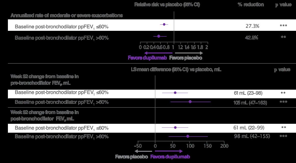
***p<0.001
**p<0.01 vs placebo
ppFEV1 <60%: dupilumab, n=720; placebo, n=693. ppFEV1 >60%: dupilumab, n=215; placebo, n=242.
LS: least squares; ppFEV1: percent predicted forced expiratory volume in 1 second; vs: versus.
Overall, findings from this post-hoc analysis showed that dupilumab reduced exacerbations and improved lung function, regardless of the degree of airway damage at baseline. However, patients with better preserved lung function at the start of the treatment period had greater improvements compared with patients with advanced airway damage at baseline, as did patients with higher 2-type biomarker levels.11
Starting treatment with dupilumab earlier in the disease course, when lung function impairment is smaller and lung damage has a greater reversible component, may therefore result in better therapeutic outcomes than
initiating treatment later, especially in patients with more active disease.11 This approach is aligned with updated GOLD guidelines in COPD, which emphasize the importance of early targeted intervention.9,10
Dupilumab significantly improved PROs in patients with COPD and Type 2 inflammation in the BOREAS and NOTUS studies; however, the mechanisms underlying this improvement, including QoL and symptom burden, are not well understood.2-4
Causal mediation analysis is a statistical method used to understand the mechanisms through which exposure to a drug affects an outcome by partitioning the total effect of the drug on the outcome into direct and indirect effects based on different mediators. The total effect denotes the overall effect of the treatment on a clinical outcome, while indirect and direct effects estimate the impact with and without mediator(s), respectively.12
To explore the underlying mechanisms by which dupilumab improves PROs, causal mediation analysis was performed on patients with COPD and Type 2 inflammation in the BOREAS and NOTUS clinical trials. The endpoints evaluated were the change from baseline to Week 52 in St George’s Respiratory Questionnaire (SGRQ) and Evaluating Respiratory Symptoms in COPD (E-RS:COPD) total score in the pooled intention-to-treat population. These outcomes measure QoL and symptom burden, respectively. Four key mediators were considered in the analysis: change in the number of moderate or severe exacerbations, change in pre-bronchodilator FEV1, change in post-bronchodilator FEV1, and change in FeNO levels. The analysis was controlled for potential confounding factors, including age, sex, smoking status, region, and the number of moderate or severe exacerbations in the year prior to the study.13
Results of the causal mediation analysis showed that dupilumab’s effect on QoL, as measured by the SGRQ score, was partially mediated by fewer exacerbations, improved lung function, and lower FeNO levels. The total effect of dupilumab versus placebo for change in SGRQ total score was estimated as −3.6, −3.6, −3.5, and −3.6, with change in exacerbation frequency, change in pre- and post-bronchodilator FEV1, and change in FeNO as mediators, respectively.13
Change in exacerbation frequency, change in pre- and post-bronchodilator FEV1, and change in FeNO indirectly affected dupilumab’s total effect on SGRQ by estimates of −0.5, −0.9, −0.8, and −0.5, respectively, mediating 13.3% (95% CI: 3.8–22.8; p=0.006),
25.7% (95% CI: 10.7–40.6; p<0.001), 23.6% (95% CI: 9.4–37.8; p=0.001), and 13.3% (95% CI: 2.5–24.1; p=0.016) of the overall QoL benefit, respectively.13
Similarly, dupilumab’s effect on symptom burden reduction, as measured by the E-RS:COPD, was also found to be partially mediated by reduced exacerbations, improved lung function, and lower FeNO levels. Dupilumab’s total effect on change in E-RS:COPD versus placebo was estimated as −0.9, as mediated by change in exacerbation frequency and change in pre- and postbronchodilator FEV1, and −0.1, as mediated by change in FeNO.13
Change in exacerbation frequency, change in pre- and post-bronchodilator FEV1, and change in FeNO indirectly affected dupilumab’s total effect on E-RS:COPD by an estimated −0.1–−0.2 points. These mediators were therefore responsible for an estimated 9.8% (95% CI: 0.5–19.1; p=0.039), 24.9% (95% CI: 6.2–43.6; p=0.009), 23.5% (95% CI: 5.0–42.0; p=0.013), and 13.6% (95% CI: −0.7–27.8; p=0.062), respectively, of the total dupilumab benefit on symptom burden.13
Overall, results from this causal mediation analysis of the BOREAS and NOTUS studies indicate that PRO improvements with dupilumab in patients with COPD are partially mediated by changes in exacerbation frequency, lung function, and FeNO. Improvement in lung function, in particular, plays a role in QoL and symptom improvement. This supports the interplay between Type 2 inflammatory biomarkers, clinical events, and PROs in patients with COPD treated with dupilumab.13
Phase IV AEOLUS Study of Dupilumab in Airway Remodeling and Mucus Plugging IL-4 and IL-13, key and central drivers of Type 2 inflammation, contribute to mucus hypersecretion and mucus plug formation via promotion of goblet cell differentiation, excess mucin (MUC5AC) production, and associated mucociliary disruption, leading to poorer
disease outcomes in patients with COPD.14-18
The VESTIGE trial showed that dupilumab can reduce mucus plugs and improve small airway function in patients with moderate-to-severe asthma. However, currently, there are limited data on the effect of dupilumab on mucus plugging in patients with COPD.19
Replicating the approach taken in asthma, the multinational AEOLUS study (NCT07053423) has therefore been designed to evaluate the effect of dupilumab on airway resistance and remodeling, including airway inflammation and mucus plugging, in patients with COPD and Type 2 inflammation. Associated improvements in exacerbations, lung function, symptoms, and QoL in dupilumab-treated patients will also be assessed.20
AEOLUS is a Phase IV, randomized, doubleblind, placebo-controlled, parallel-group trial that will enroll patients aged 40–85 years with moderate or severe COPD (Figure 2). In total, approximately 218 patients from 23 countries across Asia, Europe, Latin America, and North America will be randomized (2:1) to receive add-on dupilumab (300 mg subcutaneous) or matched placebo every 2 weeks for a total treatment period of
24 weeks. Participating countries include Argentina, Brazil, Canada, China, the EU, Japan, Saudi Arabia, Singapore, South Korea, Switzerland, Taiwan, the United Arab Emirates, the UK, and the USA.20
Key patient eligibility criteria for the AEOLUS study are:20
• Former or current smokers
• Physician-diagnosed COPD for ≥1 year with post-bronchodilator FEV1/forced vital capacity ratio ≤0.70 and postbronchodilator ppFEV1 ≥30% and <80%
• On inhaled triple therapy for ≥3 months before randomization: inhaled corticosteroids, long-acting β2-agonist, and long-acting muscarinic antagonists, or dual therapy if inhaled corticosteroids are contraindicated
• History of high exacerbation risk, defined as ≥2 moderate or ≥1 severe exacerbations in the prior year
• Medical Research Council (MRC) Dyspnea Scale Grade ≥2 or COPD Assessment Test (CAT) score ≥10 at screening
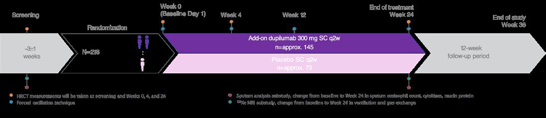
AEOLUS will enroll patients (40–85 years) with COPD, moderate-to-severe airflow limitation (post-bronchodilator FEV1/FVC <0.7 and post-bronchodilator ppFEV1 of 30% to <80%), and Type 2 inflammation (blood eosinophil count ≥300 cells/µL at screening or ≥150 cells/µL at screening with a blood eosinophil count ≥300 cells/µL within the past year).
FVC: forced vital capacity; HRCT: high-resolution CT; ppFEV1: percent predicted forced expiratory volume in 1 second; q2w: every 2 weeks; SC: subcutaneous.
• CT mucus plug score ≥3 at screening
Patients participating in AEOLUS also require evidence of Type 2 inflammation, defined as blood eosinophils ≥300 cells/µL at screening or blood eosinophils ≥150 cells/µL at screening and a history of blood eosinophils ≥300 cells/µL in the previous year. This is slightly more relaxed than the corresponding inclusion criteria for the BOREAS/NOTUS trials, which required blood eosinophil levels to be ≥300 cells/µL at the screening visit.2-4
AEOLUS study exclusion criteria include a prior or current diagnosis of asthma or other significant pulmonary disease; acute exacerbation or respiratory tract infection ≤4 weeks prior to screening; history of clinically significant laboratory findings or abnormal ECG; α-1 anti-trypsin diagnosis; previous use of dupilumab or other biologics/immunosuppressives; controller therapy compliance <80% during screening; recent live attenuated vaccination; oxygen treatment; and treatment with antiarrhythmics, antidiuretics, macrolides, roflumilast, ensifentrine, or mucolytics (unless on a stable dose for >6 months).20
The primary endpoint of the study is the change from baseline to Week 24 in lung mucus score. Scores range from 0–18, with higher scores indicating greater mucus burden and mucus plugging. AEOLUS will also use innovative lung imaging parameters such as high-resolution CT (HRCT), forced oscillation technique, and 129Xe MRI techniques to evaluate key secondary endpoints related to airway remodeling and mucus plugging. These include change from baseline to Week 24 in global mucus volume and trimmed distal airway wall thickness at total lung capacity, measured by HRCT; and the difference in airway resistance from R5 to R20 and reactance area measured by forced oscillation technique. R5 to R20 denotes the difference in airway resistance at 5 Hz and 20 Hz. Treatment-emergent adverse events (TEAE), serious adverse events (SAE), and adverse events of special interest will also be assessed as secondary outcomes, alongside
potentially clinically significant abnormalities in hematology, biochemistry, and vital signs.20
AEOLUS will additionally evaluate several exploratory outcomes, including acute exacerbations, lung inflammation (FeNO), oscillometry, healthcare resource utilization, remote monitoring, HRCT imaging, spirometry, PROs, and correlation of mucus score with spirometry and PROs. Substudy analyses will look at the change from baseline to Week 24 in key sputum components: eosinophil counts, cytokines, and mucin protein. 129Xe MRI imaging will also be used to evaluate the change from baseline to Week 24 in ventilation (as measured by ventilation defect percent) and gas exchange.20
As of May 2026, the AEOLUS study was underway and recruiting patients. Study completion is estimated for 2029.21
The efficacy of dupilumab in the treatment of asthma has been demonstrated in multiple clinical trials, including the pivotal Phase III QUEST trial (NCT02414854) and its open-label extension study, TRAVERSE (NCT02134028).22,23 QUEST was a randomized, double-blind, placebo-controlled study in which patients with moderate-tosevere asthma received add-on dupilumab (200 mg or 300 mg) q2w for 52 weeks. In the QUEST study, treatment with dupilumab significantly reduced exacerbation rates and improved lung function, asthma control, and QoL. Safety was consistent with the known dupilumab safety profile.22
TRAVERSE was a large, multicenter, open-label extension study that set out to establish the long-term safety and efficacy of dupilumab over a 3-year period. Patients who received dupilumab in the QUEST trial continued treatment (dupilumab–dupilumab), while those who previously received placebo switched to dupilumab (placebo–dupilumab).23
Clinical remission is a recently recognized therapeutic goal in the management of moderate-to-severe asthma.24 In the pivotal QUEST study, 37% of dupilumabtreated patients achieved on-treatment clinical remission at Week 52, where clinical remission was defined as the achievement of four key criteria: 1) no severe exacerbations; 2) no oral corticosteroid use during the study period; 3) stable or improved prebronchodilator FEV1 (defined as ≤5% decline from QUEST baseline); and 4) 5-item Asthma Control Questionnaire (ACQ-5) score <1.5. These therapeutic effects were sustained through the TRAVERSE extension study, with more than 70% of patients with Type 2 asthma remaining in clinical remission for up to 2 years.22,25
This post-hoc analysis evaluated whether achieving on-treatment clinical remission in QUEST, as defined above, at Week 52 was associated with sustained clinical benefits through Week 48 of TRAVERSE. It included patients with Type 2 asthma (baseline blood eosinophil count ≥150 cells/μL or FeNO ≥25 ppb) who completed QUEST and enrolled in TRAVERSE. Patients were stratified according to whether they met this composite clinical remission endpoint at Week 52 of QUEST.26
The following endpoints were then assessed at Week 48 of TRAVERSE:
• Proportion of patients in remission
• Unadjusted annualized severe asthma exacerbation rates
• Mean change in ACQ-5 score from QUEST baseline
• Percentage change in pre-bronchodilator FEV1 from QUEST baseline
Baseline demographics and disease characteristics were characterized at QUEST
baseline and were broadly similar in patients who achieved clinical remission at Week 52 compared to those who did not. Mean patient age ranged from 46–49 years, and over 50% were female.26
Results from the post-hoc analysis showed that patients who achieved clinical remission at Week 52 of QUEST were more likely to remain in remission at Week 48 of TRAVERSE. In TRAVERSE, 76.9% (placebo–dupilumab) and 74.2% (dupilumab–dupilumab) of patients who achieved remission in QUEST stayed in remission, compared with 44.8% (placebo–dupilumab) and 37.8% (dupilumab–dupilumab) of patients not in remission in QUEST who achieved remission at TRAVERSE Week 48 (Figure 3).26
Exacerbation rates in TRAVERSE were lower among patients who achieved clinical remission in QUEST at Week 52. The unadjusted annualized rate of severe exacerbations was 0.13 (placebo–dupilumab) and 0.10 (dupilumab–dupilumab) in patients who attained clinical remission, compared to 0.41 and 0.43, respectively, in those who did not.26
Improvements in lung function and ACQ-5 scores during TRAVERSE were also greater in patients who achieved clinical remission in QUEST. Mean increases in pre-bronchodilator FEV₁ from QUEST baseline to TRAVERSE Week 48 were 28.9% (placebo–dupilumab) and 34.1% (dupilumab–dupilumab) in patients who achieved remission in QUEST versus 24.2% and 20.7%, respectively, in patients who did not achieve remission. ACQ-5 scores in TRAVERSE improved by −2.02–−2.09 in patients who attained clinical remission in QUEST, compared to changes of −1.55–−1.58 in those who did not.26
Overall, this post-hoc analysis showed that patients who achieved clinical remission with dupilumab in QUEST were more likely to be in remission and experienced greater sustained clinical effects with longterm dupilumab treatment in TRAVERSE. However, substantial clinical benefits were
Figure 3: Patients who achieved clinical remission at (A) Week 52 of QUEST were more likely to meet remission criteria at (B) Week 48 of TRAVERSE.26
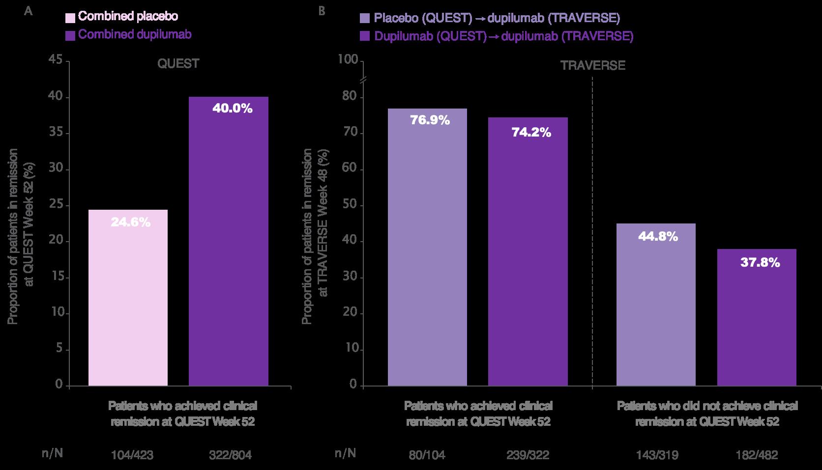
also observed among patients who did not achieve remission during QUEST.26
These findings support clinical remission as a durable and relevant treatment goal in Type 2 asthma and reinforce the long-term benefits of achieving early disease control with dupilumab.26
The presence of coexisting Type 2 inflammatory conditions can complicate asthma management.27-29 This analysis of the QUEST and TRAVERSE studies therefore set out to evaluate the long-term efficacy of dupilumab in patients with Type 2 asthma and ongoing coexisting allergic rhinitis
(n=859), atopic dermatitis (n=142), or chronic rhinosinusitis with nasal polyposis or nasal polyposis (CRSwNP/NP; n=329). Type 2 asthma was defined as baseline blood eosinophil count ≥150 cells/μL or FeNO ≥25 ppb. Patients received dupilumab treatment or placebo q2w for 52 weeks in QUEST, followed by dupilumab 300 mg q2w in TRAVERSE for up to 96 weeks.30
The study assessments included in this analysis were unadjusted annualized severe exacerbation rates and change from the QUEST baseline in FEV1 and ACQ-5 score over time. These endpoints were analyzed in patients stratified by their coexisting Type 2 inflammatory condition. QUEST baseline demographics, lung function, and disease control were broadly similar for patients in the combined placebo and combined
dupilumab groups across the three different Type 2 comorbidities. The mean patient age ranged from 42–52 years, and over 50% were female.30
The results of this analysis showed that dupilumab reduced asthma exacerbations compared to placebo during QUEST, and maintained these reductions through TRAVERSE in patients with ongoing Type 2 coexisting conditions. In QUEST, dupilumab versus placebo reduced exacerbation rates in patients with coexisting allergic rhinitis (0.53 versus 1.10), atopic dermatitis (0.66 versus 1.01), and CRSwNP/NP (0.56 versus 1.52). In TRAVERSE, patients in the placebo–dupilumab group experienced markedly reduced exacerbation rates: 0.33, 0.31, and 0.36 for allergic rhinitis, atopic dermatitis, and CRSwNP/NP, respectively. Patients in the dupilumab–dupilumab group showed further reduced exacerbation rates: 0.32, 0.43, and 0.33, respectively.30
Similarly, dupilumab improved lung function during QUEST and maintained improvements through TRAVERSE in patients with ongoing Type 2 coexisting conditions. Compared to placebo, dupilumab improved prebronchodilator FEV1 at Week 52 of QUEST in patients with allergic rhinitis (0.36 versus 0.17), atopic dermatitis (0.41 versus 0.27), and CRSwNP/NP (0.42 versus 0.14). At Week 48 in TRAVERSE, dupilumab treatment improved pre-bronchodilator FEV1 in the placebo–dupilumab group, with changes of 0.38, 0.41, and 0.40 for patients with coexisting allergic rhinitis, atopic dermatitis, and CRSwNP/NP, respectively. Dupilumab also sustained QUEST pre-bronchodilator FEV1 improvements in the dupilumab–dupilumab group: 0.40, 0.42, and 0.43, respectively.30
In patients with ongoing Type 2 coexisting conditions, dupilumab also improved asthma control during QUEST and sustained these improvements during TRAVERSE. Dupilumab reduced ACQ-5 scores compared to placebo by 1.5–1.7 across patients with each of the three different inflammatory comorbidities by Week 52 of QUEST. At Week 48 of TRAVERSE, asthma control improved in both the placebo–
dupilumab and dupilumab–dupilumab groups in patients with coexisting allergic rhinitis, atopic dermatitis, and CRSwNP/NP.30
Overall, this analysis demonstrated that dupilumab reduced exacerbation rates and improved lung function and asthma control for up to 2 years in patients with moderate-tosevere Type 2 asthma, despite the presence of coexisting Type 2 inflammatory conditions.30
Itepekimab in COPD: Results from the AERIFY-1 and -2 Trials
Itepekimab is a fully human IgG4 monoclonal antibody that binds with high affinity to IL-33 to block IL-33-mediated signaling. IL-33 is a cytokine triggered by airway insults that has emerged as a potential component in COPD pathogenesis and a promising therapeutic target for treating inflammatory lung disease.31-33 In a previous clinical study, itepekimab was shown to reduce the rate of moderate or severe exacerbations in patients with COPD by 19% and in a pre-specified subgroup of former smokers by 42% (nominally significant).34
The AERIFY-1 and -2 trials were randomized, double-blind, placebocontrolled, international Phase III studies that investigated the efficacy and safety of two doses of itepekimab in former smokers with diagnosed COPD.35 Patients eligible for the study were aged 40–85 years, with moderate-to-severe airflow limitation (post-bronchodilator ppFEV1 30–80%), on stable inhaled triple or dual therapy, and had experienced ≥2 moderate or ≥1 severe exacerbations in the previous year. Patients were required to be former smokers, with cessation ≥6 months before screening.35 A secondary population of current smokers was also included in AERIFY-2 but is not discussed here.35
After a 4-week screening period, patients in both studies were randomized 1:1:1 to itepekimab 300 mg every 4 weeks (q4w), itepekimab 300 mg q2w, or placebo q2w for
52 weeks. In total, 1,127 former smokers with COPD were randomized in AERIFY-1 and 953 in AERIFY-2. The primary endpoint in both studies was annualized rate of moderate or severe exacerbations in former smokers. Change from baseline to Week 24 in prebronchodilator FEV1 in former smokers was a key secondary endpoint. Other endpoints included severe exacerbation rate in former smokers and AEs.35
Baseline demographics, disease characteristics, and biomarkers were wellbalanced across the treatment arms. The mean patient age was approximately 68 years and the average time since smoking cessation was 9–10 years.35
Looking at the primary endpoint, itepekimab q4w and q2w reduced the rate of moderate or severe exacerbations by 20.5% (p=0.021) and 27.1% (p=0.002) versus placebo in AERIFY-1. The estimated annualized moderate or severe exacerbation rate was 0.71, 0.65, and 0.89 for the three treatment arms, respectively. In AERIFY-2, itepekimab q4w and q2w reduced the rate of moderate or severe exacerbations by 12.4% and 1.6% compared to placebo; however, these differences were not statistically significant. Itepekimab treatment was also associated with numerical reductions in the cumulative mean number of moderate or severe exacerbations in former smokers in the AERIFY-1 and -2 trials.35
In terms of secondary endpoints, itepekimab q4w and q2w significantly reduced the rate of annualized severe exacerbations by 34.1% (nominal p=0.057) and 44.4% (p=0.010) compared to placebo in AERIFY-1. The estimated annualized severe exacerbation rate was 0.09, 0.08, and 0.14 for the two itepekimab arms and placebo, respectively. In AERIFY-2, itepekimab q4w reduced the rate of severe exacerbations by 8.6% compared to placebo, while there was a 7.5% increase compared to placebo in the q2w arm (both p>0.05). In the pooled AERIFY-1 and -2 study populations, the reductions were 21.1% and 22.6% with itepekimab q4w and q2w, respectively, albeit not statistically significant.35
For change in pre-bronchodilator FEV1, itepekimab q4w and q2w demonstrated least squares mean differences versus placebo of 10 mL and 6 mL in AERIFY-1, which were not statistically significant. The corresponding changes in AERIFY-2 were 75 mL and 38 mL, with nominal p-values of <0.001 and 0.031, respectively.35
The safety profile of itepekimab was comparable across both dose groups, and rates of TEAEs, severe TEAEs, treatmentemergent SAEs, and TEAEs leading to discontinuation were broadly similar to placebo. Any TEAEs occurred in 69.5% (n=472) of patients treated with itepekimab q4w, 65.8% (n=460) who received itepekimab q2w, and 66.5% (n=462) on placebo. Rates of treatment-emergent SAEs were 21.5% (n=146), 20.3% (n=142), and 22.2% (n=154), respectively.35
In summary, in former smokers with COPD, itepekimab significantly reduced the rate of moderate or severe exacerbations versus placebo in the AERIFY-1 study but not in AERIFY-2. Similarly, itepekimab showed clinically meaningful reductions in severe exacerbation rates in AERIFY-1, which were not seen in AERIFY-2 or the pooled population. These discordant findings warrant further investigation to evaluate the potential causative. Itepekimab demonstrated an acceptable safety profile across both trials.35
Collectively, these new data presented at ATS 2026 provide further evidence supporting the efficacy profile of the dual IL-4 and IL-13 inhibitor, dupilumab, in treating asthma and COPD in patients with Type 2 inflammation. In particular, post-hoc analyses of pivotal Phase III trials support the potential use of dupilumab earlier in the disease pathway for both COPD and asthma, in order to maximize treatment outcomes. Potential limitations of this evidence include its derivation from posthoc analyses and the exploratory nature of some findings.
1. Celli BR, Wedzicha JA. Update on clinical aspects of chronic obstructive pulmonary disease. N Engl J Med. 2019;381:1257-66.
2. Bhatt SP et al. Dupilumab for COPD with type 2 inflammation indicated by eosinophil counts. N Engl J Med. 2023;389:205-14.
3. Bhatt SP et al. Dupilumab for COPD with blood eosinophil evidence of type 2 inflammation. N Engl J Med. 2024;390:2274-83.
4. Bhatt SP et al. Dupilumab for chronic obstructive pulmonary disease with type 2 inflammation: a pooled analysis of two phase 3, randomised, doubleblind, placebo-controlled trials. Lancet Respir Med. 2025;13:234-43.
5. European Medicines Agency (EMA). Dupixent (dupilumab): summary of product characteristics. Available at: https://www.ema.europa.eu/en/ documents/product-information/ dupixent-epar-product-information_ en.pdf. Last accessed: May 14 2026.
6. Khan AH et al. Impact of baseline lung function on future exacerbations in patients with moderate-tosevere asthma. J Asthma Allergy. 2022;15:1639-44.
7. Tupper OD, Ulrik CS. Long-term predictors of severe exacerbations and mortality in a cohort of wellcharacterised adults with asthma. Respir Res. 2021;22:269.
8. Couillard S et al. Toward a predict and prevent approach in obstructive airway diseases. J Allergy Clin Immunol Pract. 2023;11:704-12.
9. Global Initiative for Chronic Obstructive Lung Disease (GOLD). GOLD report 2026: key changes summary. Available at: https://goldcopd.org/wp-content/ uploads/2025/11/KEY-CHANGES-GOLD2026-10Nov2025.pdf. Last accessed: May 13 2026.
10. Cazzola M et al. GOLD 2026: transforming COPD management with early intervention, multi-dimensional assessment, and personalized care. Drugs. 2026;86(5):581-97.
11. Couillard S et al. Relation between airway damage and disease activity and the effect of dupilumab on exacerbations and lung function in chronic obstructive pulmonary disease: BOREAS and NOTUS. Poster 1486. ATS International Conference, May 15-20, 2026.
12. Qin X. An introduction to causal mediation analysis. Asia Pac Educ Rev. 2024;25:703-17.
13. Bhatt S et al. Causal mediation analysis quantifies how dupilumab improves patient-reported outcomes through exacerbation reduction, lung function improvements, and FeNO decline. Poster 7519. ATS International Conference, May 15-20, 2026.
14. Doyle AD et al. Eosinophil-derived IL-13 promotes emphysema. Eur Respir J. 2019;53:1801291.
15. Linden D et al. Respiratory viral infection: a potential “missing link” in the pathogenesis of COPD. Eur Respir Rev. 2019;28:180063.
16. Oishi K et al. Role of type 2 inflammatory biomarkers in chronic obstructive pulmonary disease. J Clin Med. 2020;9:2670.
17. Diaz AA et al. Eosinophils, mucus plugs and clinical outcomes: findings from two COPD cohorts. Eur Respir J. 2024;64:2401005.
18. Mettler SK et al. Longitudinal changes in airway mucus plugs and FEV1 in COPD. N Engl J Med. 2025;392:1973-5.
19. Castro M et al. Effect of dupilumab on exhaled nitric oxide, mucus plugs, and functional respiratory imaging in patients with type 2 asthma (VESTIGE): a randomised, double-blind, placebocontrolled, phase 4 trial. Lancet Respir Med. 2025;13:208-20.
20. Bhatt S et al. Design of the phase 4 AEOLUS study assessing the effect of dupilumab on airway inflammation and remodeling through lung imaging parameters in patients with COPD. Poster 1493. ATS International Conference, May 15-20, 2026.
21. Sanofi. A study to investigate airway inflammation with dupilumab subcutaneously in participants aged ≥40 to ≤85 years with chronic obstructive pulmonary disease (AEOLUS). NCT07053423. https:// clinicaltrials.gov/study/NCT07053423.
22. Castro M et al. Dupilumab efficacy and safety in moderate-to-severe uncontrolled asthma. N Engl J Med. 2018;378:2486-96.
23. Wechsler ME et al. Long-term safety and efficacy of dupilumab in patients with moderate-to-severe asthma (TRAVERSE): an open-label extension study. Lancet Respir Med. 2022;10:11-25.
24. Global Initiative for Asthma (GINA). Global strategy for asthma management
and prevention, 2025 report. Available at: https://ginasthma.org/2025-ginastrategy-report. Last accessed: May 14 2026.
25. Pavord ID et al. Dupilumab induces longterm on-treatment clinical remission in patients with type 2 asthma. J Allergy Clin Immunol Pract. 2025;13:132-42.
26. Brusselle G et al. Long-term outcomes in TRAVERSE in patients with type 2 asthma based on clinical remission status at the end of QUEST. Poster P1400. ATS International Conference, May 15-20, 2026.
27. Khan AH et al. Prevalence and severity distribution of type 2 inflammationrelated comorbidities among patients with asthma, chronic rhinosinusitis with nasal polyps, and atopic dermatitis. Lung. 2023;201:57-63.
28. Mullol J et al. Prevalence of chronic rhinosinusitis without/with nasal polyps according to severity in Spain. Rhinology. 2024;62:421-31.
29. Maspero J et al. Type 2 inflammation in asthma and other airway diseases. ERJ Open Res. 2022;8:00576-2021.
30. Al-Ahmad M et al. Long-term efficacy of dupilumab in patients with asthma and coexisting type 2 inflammatory conditions. Poster P1407. ATS International Conference, May 15-20, 2026.
31. Rabe KF et al. Targeting type 2 inflammation and epithelial alarmins in chronic obstructive pulmonary disease: a biologics outlook. Am J Respir Crit Care Med. 2023;208:395-405.
32. Gabryelska A et al. IL-33 mediated inflammation in chronic respiratory diseases-understanding the role of the member of IL-1 superfamily. Front Immunol. 2019;10:692.
33. Calderon AA et al. Targeting interleukin-33 and thymic stromal lymphopoietin pathways for novel pulmonary therapeutics in asthma and COPD. Eur Respir Rev. 2023;32:220144.
34. Rabe KF et al. Safety and efficacy of itepekimab in patients with moderateto-severe COPD: a genetic association study and randomised, double-blind, phase 2a trial. Lancet Respir Med. 2021;9:1288-98.
35. Rabe K et al. Efficacy and safety of itepekimab in former smokers with chronic obstructive pulmonary disease: AERIFY-1 and AERIFY-2 trials. Oral presentation. ATS International Conference, May 15-20, 2026.
A summary of select poster presentations from the American Thoracic Society (ATS) Conference, held in Orlando, Florida, USA from May 15–20, 2026
Support: The publication of this article was funded by GSK.
Presenters: Arnaud Bourdin,1 Reynold A. Panettieri,2 Ian D. Pavord3
1. Department of Pulmonology and Addictology, University of Montpellier, France
2. Rutgers Institute for Translational Medicine & Science, Rutgers University, New Brunswick, New Jersey, USA
3. Oxford Respiratory NIHR Biomedical Research Centre, Nuffield Department of Medicine, University of Oxford, UK
Disclosure: Bourdin has received research grants, and/or served as a consultant, advisory committee member, and/or speaker for Acceleron, Amgen, AstraZeneca, Boehringer Ingelheim, Celltrion, Chiesi, Gossamer, GSK, MSD, Novartis, Pfizer, Regeneron-Sanofi, and Sanofi. Panettieri has received research grants, and/or served as a consultant for AstraZeneca, GSK, NIH, Origo Pharma, Regeneron, and Sanofi. Pavord has served as a consultant, advisory committee member, and/or speaker for Aerocrine, Almirall, Amgen, Areteia, AstraZeneca, Chiesi, Circassia, GSK, Kymera, Menarini, Merck, Pfizer, and Sanofi/Regeneron.
Acknowledgements: Medical writing assistance was provided by Nicola Humphry, Nottingham, UK.
Disclaimer: This article is intended for US Healthcare Professionals only. The information is scientific and non-promotional in nature, and is not intended to offer recommendations for using this product in a manner inconsistent with approved labeling. Please see the full Prescribing Information for EXDENSUR (depemokimab). To report SUSPECTED ADVERSE REACTIONS, contact GSK at 1-888-825-5249 or FDA at 1-800-FDA-1088, www.fda.gov/medwatch.
Keywords: AGILE, asthma, biologic therapy, chronic rhinosinusitis with nasal polyps (CRSwNP), depemokimab, severe asthma, SWIFT-1, SWIFT-2, Type 2 inflammation.
Citation: Respir AMJ. 2026;4[1]:54-61. https://doi.org/10.33590/respiramj/80L802AB
Severe asthma is a significant cause of morbidity and mortality estimated to affect 3.7% of patients with asthma. Most patients with severe asthma have Type 2 inflammation, which can be difficult to control despite standard of care with
high-dose inhaled corticosteroids (ICS). Add-on biologic therapy is recommended in these patients, and among the currently approved options, depemokimab represents the first ultra-long-acting biologic. Depemokimab efficacy and tolerability over 52 weeks were demonstrated in the Phase III SWIFT-1/-2 studies, and patients who completed these studies were invited to join the subsequent AGILE open-label extension (OLE) study.
This article summarizes data presented at the American Thoracic Society (ATS) 2026 Conference from an integrated analysis of the SWIFT-1/-2 and AGILE OLE studies, reporting long-term efficacy and safety findings from up to 2 years of depemokimab exposure in patients with Type 2 asthma. In addition to overall efficacy and safety, posters included data from subsets of patients receiving medium- and high-dose ICS at baseline, and patients with comorbid chronic rhinosinusitis with nasal polyps (CRSwNP).
These new data provide descriptive insights into the durable, long-term efficacy and tolerability of depemokimab in patients with severe asthma, for up to 2 years, and may provide additional context for clinicians in evaluating biologic treatment options for their patients.
Introduction
Despite advances in asthma management, severe asthma remains a significant cause of global morbidity and mortality.1 Estimated to affect 3.7% of patients with asthma, severe asthma is defined as uncontrolled disease despite adherence to treatment with highdose ICS and a long-acting β2 agonist.2,3
Most patients with severe asthma have Type 2 inflammation, characterized by the production of cytokines such as IL-4, IL5, and IL-13; eosinophilia; and increased fractional exhaled nitric oxide.2 Type 2 inflammation can be difficult to control with high-dose ICS, and while it may respond to oral corticosteroids, these are associated with serious side effects.2 Approved Type 2-targeted biologics are therefore recommended as add-on therapy in patients with severe asthma who have eosinophilic or allergic biomarkers, or who require maintenance with oral corticosteroids.2
A total of seven biologics are currently available for severe asthma, including omalizumab, mepolizumab, reslizumab, benralizumab, dupilumab, tezepelumab, and
depemokimab.3,4 Among these, depemokimab is the only ultra-long-acting option, with enhanced IL-5 binding affinity, high potency, and extended half-life, enabling twiceyearly dosing in asthma.4-6 Depemokimab is approved for add-on maintenance of severe asthma treatment characterized by an eosinophilic phenotype in adult and pediatric patients aged 12 years and older in the USA.4 It is not indicated for the relief of acute bronchospasm or status asthmaticus.4 Depemokimab is also approved for use in severe eosinophilic asthma in the UK, EU, China, and Japan,7-9 and in CRSwNP in the UK, EU, and China.7-9
The Phase III SWIFT-1/-2 studies demonstrated the efficacy and safety of depemokimab in patients aged ≥12 years with eosinophilic Type 2 asthma over 52 weeks (Figure 1).11 Eligible patients were randomized 2:1 to receive subcutaneous depemokimab 100 mg (n=502) or placebo (n=260) every 26 weeks for 1 year.11
In a pooled analysis of SWIFT-1 and SWIFT-2, depemokimab reduced exacerbations by 54% over 52 weeks versus placebo, along with sustained suppression
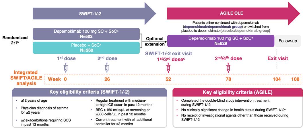
aIncluding treatment with medium-to-high ICS dose plus ≥1 additional controller and excluding biologics. bDepemokimab:placebo.
cAGILE Visit 1 (SWIFT-1/-2, Week 52) was considered as baseline for AGILE. The exit visit in SWIFT-1/-2 was used as baseline for AGILE if it was on the same day or within 7 days of AGILE Visit 1; however, if Visit 1 was >7 days (max 14 days) after the exit visit, baseline assessments were performed pre-dose at Visit 1.
dDefinitions based on GINA 2021 guidelines.
ePatients excluded only if an investigator deemed that their change in health status made them unsuitable for participation.
The sample size was determined by the number of available participants who were randomized into SWIFT-1 and SWIFT-2 and who were eligible for the current study based on inclusion and exclusion criteria.
BEC: blood eosinophil count; GINA: Global Initiative for Asthma; ICS: inhaled corticosteroid; OLE: open-label extension; SC: subcutaneous; SCS: systemic corticosteroid(s); SoC: standard of care.
of inflammation, as assessed by blood eosinophil count (82% reduction in SWIFT-1 and 83% reduction in SWIFT-2 at Week 52).11 Depemokimab also showed numerical improvements versus placebo in patientreported outcome measures. The change in St George’s Respiratory Questionnaire (SGRQ) total score (standard error [SE]) from baseline to Week 52 with depemokimab versus placebo was −13.92 (0.76) versus −11.04 (1.06), respectively, and in Asthma Control Questionnaire-5 (ACQ-5) score (SE) it was −0.81 (0.05) versus −0.73 (0.06), respectively.11 In prespecified subgroup analyses,
depemokimab was found to reduce exacerbations compared with placebo across both medium- and high-dose ICS patient subgroups,12 and in patients with comorbid CRSwNP.13
The overall incidence of adverse events (AE) was similar between depemokimab and placebo treatment groups.11 The most common AEs were upper respiratory tract infection, allergic rhinitis, influenza, arthralgia, and pharyngitis.11 Among patients receiving depemokimab, 72% in SWIFT-1 and 73% in SWIFT-2 had an AE, of which 3% and 4%, respectively, were related to
depemokimab and 1% in each study led to discontinuation or withdrawal. A serious AE (SAE) was reported in 6% and 8% of patients, respectively, none of which were considered related to depemokimab. Among patients receiving placebo, 73% in SWIFT-1 and 78% in SWIFT-2 had an AE: 4% and 1%, respectively, were related to placebo, and 2% and 1% led to discontinuation or withdrawal. An SAE was reported in 17% and 10% of patients, respectively.11
The long-term safety and efficacy of depemokimab in asthma was subsequently assessed in the single-arm, 52-week AGILE OLE study (Figure 1).14 Patients who completed SWIFT-1/-2 were invited to join the OLE study. Of the 762 randomized patients, 641 (84%) entered the OLE; 419 continued with depemokimab (depemokimab/depemokimab) and 210 switched from placebo to depemokimab (placebo/depemokimab), with a total follow-up period of 104 weeks. Twiceyearly depemokimab was generally well tolerated up to 104 weeks; AEs occurred in similar proportions of patients in each group (depemokimab/depemokimab: 72%; placebo/ depemokimab: 70%), and no treatmentrelated SAEs were reported. Efficacy was sustained in patients previously treated with depemokimab, and those switching from placebo experienced numerical reductions in exacerbations; annualized exacerbation rates (AER; 95% CI) were 0.55 (0.47–0.66) and 0.58 (0.45–0.73), respectively.14
At the ATS 2026 Conference, data were presented from an integrated post-hoc analysis of the SWIFT-1/-2 and AGILE studies (N=629), reporting long-term data for depemokimab in terms of overall safety and efficacy, efficacy by baseline ICS dose, and efficacy in patients with comorbid CRSwNP.10,15 These data are summarized in this article.
In the integrated analysis of SWIFT-1/-2 and AGILE, key patient demographics and baseline characteristics were evenly distributed across treatment groups. Reduction in AER was sustained over the full 2-year SWIFT/AGILE period in the depemokimab/depemokimab group (AER: 0.52 [95% CI: 0.45–0.60]), while those in the placebo/depemokimab group experienced greater reductions in exacerbations versus Year 1 (Figure 2). In addition, time to first exacerbation was longer for patients in the depemokimab/ depemokimab group across the 2-year period compared with the placebo/depemokimab group. This difference was less pronounced, however, after Week 52, when all patients received depemokimab.10
Reductions in SGRQ total score and ACQ-5 score were sustained over the 2-year SWIFT/AGILE period in the depemokimab/ depemokimab group, and numerical improvements were observed in the placebo/ depemokimab group following depemokimab initiation in AGILE. Change from baseline in SGRQ total score (SE) in the depemokimab/ depemokimab group was −13.92 (0.76) at Week 52 and −16.29 (0.85) at Week 104. Similarly, change from baseline in ACQ-5 score (SE) in the depemokimab/ depemokimab group was −0.81 (0.05) at Week 52 and −0.89 (0.05) at Week 104.10
Eosinophil levels remained suppressed in the depemokimab/depemokimab group over the 2-year period, while those in the placebo/depemokimab group experienced an 85% reduction from baseline in blood eosinophil count following depemokimab initiation. Accordingly, suppression of Type 2 inflammation, as assessed by blood eosinophil count, was consistent throughout the 2-year period in the depemokimab/ depemokimab group; the ratio to baseline (SE logs) at Week 104 was 0.174 (0.04).10
(95% CI)
Figure 2: Reduction in AER during the SWIFT-1/-2 and AGILE studies.10,a
All patients receive depemokimab
(0.47–0.66) 1.12 (0.91–1.37) 0.51 (0.42–0.61)
(0.45–0.73)
Year 1 (SWIFT-1/-2)
Year 2 (AGILE)

2-year period
(SWIFT-1/-2/AGILE integrated)
Placebo/depemokimab 0.84 (0.69–1.01)
Depemokimab/depemokimab 0.52 (0.45–0.60)
Placebo (N=210)
Depemokimab (N=419)
Placebo/depemokimab (N=210)
Depemokimab/depemokimab (N=419)
aAt pre-SWIFT-1/-2 baseline, the mean (SD) number of exacerbations requiring OCS/SCS in the past 12 months was 2.5 (1.7) in the depemokimab/depemokimab group, and 2.4 (1.1) in the placebo/depemokimab group.
AER: annualized exacerbation rates; OCS: oral corticosteroids; SCS: systemic corticosteroids.
Overall, the integrated post-hoc analysis of SWIFT-1/-2 and AGILE showed sustained suppression of inflammation and efficacy over a 2-year period, supporting the longterm efficacy of twice-yearly depemokimab in patients with Type 2 asthma.10
In SWIFT-1/-2 (N=762), AER (95% CI) over 52 weeks in the depemokimab group was 0.36 (0.27–0.48) in the medium-dose ICS subgroup and 0.65 (0.53–0.79) in the highdose ICS subgroup.12 Improvements in SGRQ and ACQ-5 scores were also observed with depemokimab regardless of baseline ICS dose: least squares mean (SE) change from baseline through Week 52 in the medium- and high-dose ICS subgroups, respectively, was −14.38 (1.19) and −13.60 (1.01) for SGRQ, and −0.89 (0.07) and −0.76 (0.06) for ACQ-5.16
In the integrated analysis of SWIFT-1/-2 and AGILE, a similar proportion of patients received medium-dose and high-dose ICS at baseline. Patients receiving high-dose ICS had a history of more exacerbations,
higher baseline symptom scores, and higher baseline blood eosinophil counts compared with patients receiving medium-dose ICS.16
Patients in both ICS-dose subgroups experienced reductions in exacerbations with depemokimab versus placebo during SWIFT-1/-2, and these reductions were maintained during the AGILE OLE. In addition, those patients who switched from placebo during SWIFT-1/-2 to depemokimab during AGILE showed reductions in exacerbations following the switch, regardless of baseline ICS dose.16
Among patients in the depemokimab/ depemokimab group who received depemokimab throughout the 2-year study period (n=419), 172 patients were on mediumdose ICS at baseline, and 247 were on high-dose ICS. In these patients, AER was maintained over 2 years in both the mediumdose (0.41 [95% CI: 0.32–0.52]) and high-dose (0.62 [95% CI: 0.52–0.75]) ICS subgroups.16
Improvements in SGRQ total score and ACQ5 score from baseline through Week 52 were also maintained throughout Week 104 in
patients receiving medium-dose or high-dose ICS.16
Overall, twice-yearly depemokimab showed sustained efficacy over 2 years, irrespective of baseline ICS dose.16
Several comorbidities are common in patients with severe asthma, and these can contribute to symptom burden and impair quality of life.2 CRSwNP is a common comorbidity among patients with Type 2 asthma, and is characterized by persistent symptoms such as nasal obstruction, rhinorrhea, facial pain, and loss of smell.17 The presence of CRSwNP is considered a potential predictor of an enhanced response to IL-5 therapy.2
A prespecified analysis of SWIFT-1/-2 (N=762) investigated the efficacy of depemokimab in a subgroup of patients with comorbid CRSwNP (n=113; depemokimab: n=80; placebo: n=33). Overall, there was a 69% (95% CI: 42–83) reduction in AER versus placebo in the CRSwNP subgroup (0.51; n=80 versus 1.61; n=33, respectively) compared with a 51% (95% CI: 36–62) reduction in those without CRSwNP (0.51; n=421 versus 1.03; n=227). SGRQ and ACQ5 scores also showed greater improvements in the CRSwNP subgroup than in the overall population receiving depemokimab.13
Among 419 patients treated with depemokimab over the full 2 years of the SWIFT/AGILE study period, 66 (15.8%) had comorbid CRSwNP at baseline. Improvements observed in SWIFT-1/-2 were maintained in AGILE, regardless of CRSwNP status; AER (95% CI) was 0.45 (0.31–0.65) for patients with CRSwNP and 0.54 (0.46–0.63) for patients without CRSwNP over 2 years.15
Numerically greater improvements in SGRQ and ACQ-5 scores were also maintained over the 2-year period, regardless of CRSwNP status at baseline, with more
pronounced improvements observed in those with CRSwNP.15
Overall, depemokimab showed sustained efficacy over the SWIFT/AGILE 2-year period in patients with Type 2 asthma, regardless of CRSwNP comorbidity status at baseline.15 Comorbid CRSwNP in asthma is indicative that disease is primarily driven by Type 2 inflammation,17 which may explain the enhanced clinical benefit observed with depemokimab in patients with this comorbidity.
During the 52-week AGILE OLE, where all patients received depemokimab (N=629), AEs and SAEs were reported in similar proportions of patients across depemokimab/ depemokimab and placebo/depemokimab groups. There were no treatment-related SAEs or fatal SAEs, and low rates of adverse events of special interest (AESI).18
The long-term safety of depemokimab over 2 years was assessed in the integrated posthoc analysis, including AEs, serious AEs, and AESIs reported in patients who received depemokimab for the full 2-year period (n=419; Figure 3).18
AEs were reported in 86% of patients, of which COVID-19 (25%), nasopharyngitis (23%), and upper respiratory tract infection (16%) were the most common. Treatmentrelated AEs (per investigator assessment) occurred in 6% of patients, of which headache (1%), injection-site reaction (<1%), and leukopenia (<1%) were the most common. AEs leading to discontinuation or dose interruption were infrequent (both <1%), and SAEs occurred in 13% of patients. Asthma (3%) was the only SAE to occur in ≥1% of patients. No treatment-related SAEs (per investigator assessment) or fatal SAEs occurred during the 2-year period.18
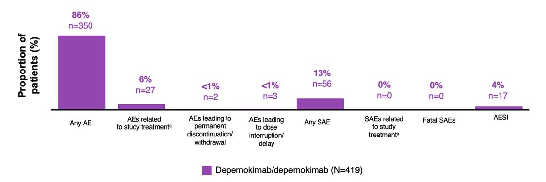
ᵃAE/SAE relation to study treatment was assessed by investigator.
AE: adverse event; AESI: adverse event of special interest; SAE: serious adverse event.
AESIs were reported in 4% of patients: nervous system disorders in 2% of patients, general disorders and administration site conditions in 1%, and skin and subcutaneous tissue disorders in <1%. No single AESI occurred in ≥1% of patients. Anti-drug antibodies (ADA) and neutralizing antibodies occurred infrequently, and there was no association found between ADA status and depemokimab efficacy or safety.18
Overall, twice-yearly depemokimab had low discontinuation rates, no treatment-related or fatal SAEs and AESIs, and few ADAs and neutralizing antibodies over the 2-year SWIFT/AGILE period. The most commonly reported AEs were transient in duration and were similar to findings in Phase III programs for asthma and CRSwNP.19
In addition, no significant safety concerns were identified among patients who switched from placebo to depemokimab during the AGILE OLE; the incidence of AEs was similar between treatment groups and when compared with the placebo group in SWIFT-1/-2.11
The integrated analysis of SWIFT-1/-2 and AGILE supports the long-term efficacy and tolerability of twice-yearly depemokimab in patients with Type 2 asthma. The post-hoc analyses showed sustained reductions in AER and maintenance of patient-reported outcome improvements over 2 years of depemokimab treatment.10 Suppression of Type 2 inflammation, assessed by blood eosinophil levels, was also maintained. Findings were similar regardless of baseline ICS dose and comorbid CRSwNP status, with greater reductions in exacerbation rates in patients with comorbid CRSwNP.12,15,16
The safety profile of depemokimab was similar to placebo in Year 1 and remained consistent through Year 2, with no treatmentrelated or fatal SAEs reported and no new safety concerns identified with long-term depemokimab exposure. Most on-treatment AEs reflected expected events such as COVID-19, nasopharyngitis, and upper respiratory tract infection.18
Overall, these findings provide further insights into the efficacy and safety of twice-yearly depemokimab in patients with severe Type 2 asthma, and durable, long-
References
1. Davis AE et al. Emerging biologic targets in type 2 and non-type 2 asthma. Curr Opin Pulm Med. 2026;32(3):210-9.
2. Global Initative for Asthma (GINA). Global strategy for asthma management and prevention. 2021. Available at: https://ginasthma.org/wpcontent/uploads/2023/04/GINA-MainReport-2021-V2-WMSA.pdf. Last accessed: May 1 2026.
3. Faria N et al. Biologic therapies for severe asthma: current insights and future directions. J Clin Med. 2025;14(9):3153.
4. Food and Drug Administration (FDA). EXDENSUR (depemokimab-ulaa) prescribing information. 2025. Available at: https://www.accessdata. fda.gov/drugsatfda_docs/ label/2026/761458Orig1s000lbl.pdf. Last accessed: May 1 2026.
5. Singh D et al. A phase 1 study of the long-acting anti-IL-5 monoclonal antibody GSK3511294 in patients with asthma.Br J Clin Pharmacol. 2022;88(2):702-12.
6. Orecchia M et al. Generation and preclinical assessment of depemokimab, an enhanced IL-5 antagonist monoclonal antibody. Heliyon. 2026;12:e44247.
7. MHRA Products. EXDENSUR (depemokimab) Summary of Product Characteristics. 2025. Available at: https://mhraproducts4853. blob.core.windows.net/docs/ 9e6a6cf9c16da1c769180069b8c 8229475640294. Last accessed: May 1 2026.
term effects up to 2 years. The full details of the integrated analysis are expected to be published later this year.
8. European Medicines Agency (EMA). EXDENSUR (depemokimab) Summary of Product Characteristics. 2026. Available at: https://www.ema. europa.eu/en/documents/productinformation/exdensur-epar-productinformation_en.pdf. Last accessed: May 1 2026.
9. GSK. Exdensur (depemokimab) approved in China for the treatment of chronic rhinosinusitis with nasal polyps (CRSwNP). 2026. Available at: https:// www.gsk.com/en-gb/media/pressreleases/exdensur-depemokimabapproved-in-china/. Last accessed: May 1 2026.
10. Pavord ID et al. Depemokimab demonstrates sustained long-term efficacy and consistent patientreported outcomes over 2 years in patients with type 2 asthma: an integrated analysis of the SWIFT-1/-2 and AGILE Studies. Poster P1396. ATS International Conference, May 15-20, 2026.
11. Jackson DJ et al. Twice-yearly depemokimab in severe asthma with an eosinophilic phenotype. N Engl J Med. 2024;391(24):2337-49.
12. Pavord I et al. Twice-yearly depemokimab demonstrates efficacy in patients with asthma across baseline medium- and high-dose ICS subgroups: phase III SWIFT-1/2 studies. Eur Respir J. 2025;66(Suppl 69):PA2469.
13. Heffler E et al. Depemokimab demonstrates efficacy in patients with type 2 asthma with comorbid CRSwNP: Phase III SWIFT-1/-2 analysis. Front Allergy. 2026;7:1766647.
14. Wechsler M et al. Long-term safety and efficacy of depemokimab in patients with type 2 asthma: a single-arm, open-label extension study (AGILE). J Allergy Clin Immunol. 2026;157(2):AB278.
15. Pavord ID et al. Depemokimab demonstrates sustained long-term efficacy and consistent patientreported outcomes over 2 years in patients with type 2 asthma and chronic rhinosinusitis with nasal polyps: an integrated analysis of the Phase III SWIFT-1/-2 and the open-label extension AGILE Studies. Poster P520. ATS International Conference, May 15-20, 2026.
16. Panettieri RA et al. Depemokimab demonstrates sustained long-term efficacy and consistent patientreported outcomes over 2 years in patients with type 2 asthma irrespective of baseline inhaled corticosteroid dose: an integrated analysis of the Phase III SWIFT-1/-2 and the open-label extension AGILE studies. Poster P519. ATS International Conference, May 15-20, 2026.
17. Bachert C et al. Burden of disease in chronic rhinosinusitis with nasal polyps. J Asthma Allergy. 2021;14:127-34.
18. Bourdin A et al. Twice-yearly depemokimab is well tolerated over 2 years in patients with type 2 asthma: an integrated safety analysis of the Phase III SWIFT-1/-2 and the open-label extension AGILE studies. Poster P1391. ATS International Conference, May 15-20, 2026.
19. Jackson DJ et al. Safety and tolerability of twice-yearly depemokimab in patients with asthma and chronic rhinosinusitis with nasal polyps: pooled results from SWIFT-1/-2 and ANCHOR-1/-2. Adv Ther. 2026;43(2):880-97.
Job code: PSE-US-5218.
This poster presentation took place at the American Thoracic Society (ATS) 2026 International Conference, May 15–20, 2026, in Orlando, Florida, USA
Support: The studies included herein, and the publication of this poster review article, were developed and funded by Insmed Incorporated. The content of this article reviews one poster presentation from the American Thoracic Society (ATS) 2026 International Conference that took place in Orlando, Florida, USA, between May 15–20, 2026.
Presenter: Eva Polverino1
1. Pneumology Department, Vall d’Hebron Institut de Recerca (VHIR), Barcelona, Spain
Disclosure: Polverino has reported receiving support and honoraria from Insmed Incorporated for the submitted work; grants from Grifols; consulting fees from Chiesi, CSL Behring, Grifols, GSK, Insmed Incorporated, Moderna, and Pfizer; and participation in advisory boards for Boehringer Ingelheim, Chiesi, Insmed Incorporated, Moderna, Pari, and Pfizer.
Acknowledgements: Michal Shteinberg, Sunjay Devaranjan, Takanori Asakura, Christina Hoenig, Prina Donga, Roger Legtenberg, Melanie Lauterio, and Joseph Feliciano were co-authors. The authors would like to thank Justine Hamaïde and Kathy Hart for their contributions as patient experts in the co-creation of the BURDEN initiative. Medical writing assistance was provided by Bethany Cooper, Ownership Health Limited, UK.
Disclaimer: Insmed Incorporated participated in the study design, research, analysis, data collection, and interpretation of the original poster data, as well as review and approval of this poster review article.
Keywords: Bronchiectasis (BE), caregiver burden, daily functioning, disease burden, emotional well-being, exacerbations, patient-reported outcomes, quality of life.
Citation: Respir AMJ. 2026;4[1]:62-67. https://doi.org/10.33590/respiramj/03952E92
The Bronchiectasis Understanding and Research on Daily Experiences and Needs (BURDEN) survey is the first multinational initiative assessing the impact of bronchiectasis from the perspectives of people living with the condition and their caregivers. This poster review article highlights a presentation from the American Thoracic Society (ATS) International Conference, which took place in Orlando, Florida, USA, between May 15–20, 2026, where results from the BURDEN survey were presented. Eva Polverino, Pneumology Department, Vall d'Hebron Institut de Recerca (VHIR), Barcelona, Spain, presented realworld evidence of the social and emotional burden associated with bronchiectasis (BE) for both patients and their caregivers, highlighting the impact of exacerbations on disease burden and daily living. BE has an increasing prevalence worldwide and is associated with a socioeconomic burden. Findings serve to reframe BE as a condition with persistent emotional and societal impact, highlighting the importance of managing and reporting exacerbations effectively.
BE is a chronic, progressive, inflammatory lung disease characterized by abnormal dilatation of the bronchi, impaired mucociliary clearance, neutrophilic inflammation, and structural lung damage.1 BE, as a chronic disorder, includes symptoms such as cough, purulent sputum production, and bronchial infection, and in many patients is marked by recurrent pulmonary exacerbations.2,3 Exacerbations are associated with progressive disease burden, lung function decline, impaired quality of life, hospitalizations, and an increased risk of mortality.4,5 The primary goal of treatment is to prioritize airway clearance and prevent exacerbations.6 Despite the significant burden of bronchiectasis, there is limited information on patient-reported impact of the disease and exacerbations on their physical and emotional wellbeing.6
Eva Polverino Polverino presented the results of the BURDEN study, the first multinational initiative assessing the impact of BE from the perspectives of people living with the
condition and their caregivers, during ATS 2026.7
Findings were presented from a mixedmethods research study, whereby qualitative interviews were conducted to inform the development of a quantitative online survey, co-created with both healthcare providers and patient experts. Study participants included adults from the EU (France, Germany, UK, Italy, and Spain) and the US, with a diagnosis of BE (excluding BE caused by cystic fibrosis), and who experienced ≥1 exacerbation in the past year.7 Recruitment lasted from December 2024–April 2025, and included family members and caregivers who lived with and provided daily support to people with BE.7
To extract data specific on the impact of exacerbation burden, participants with BE were required to have experienced ≥1 exacerbation in the past year (Figure 1). Impacts associated with exacerbations, such as daily activities, emotional wellbeing, work, and finances, were rated on a scale of 1 (“No additional negative impact at all”) to 10 (“Extreme additional negative impact”).7
1: Proportion of patients with 1, 2, or ≥3 exacerbationsa over the previous 12 months by country.
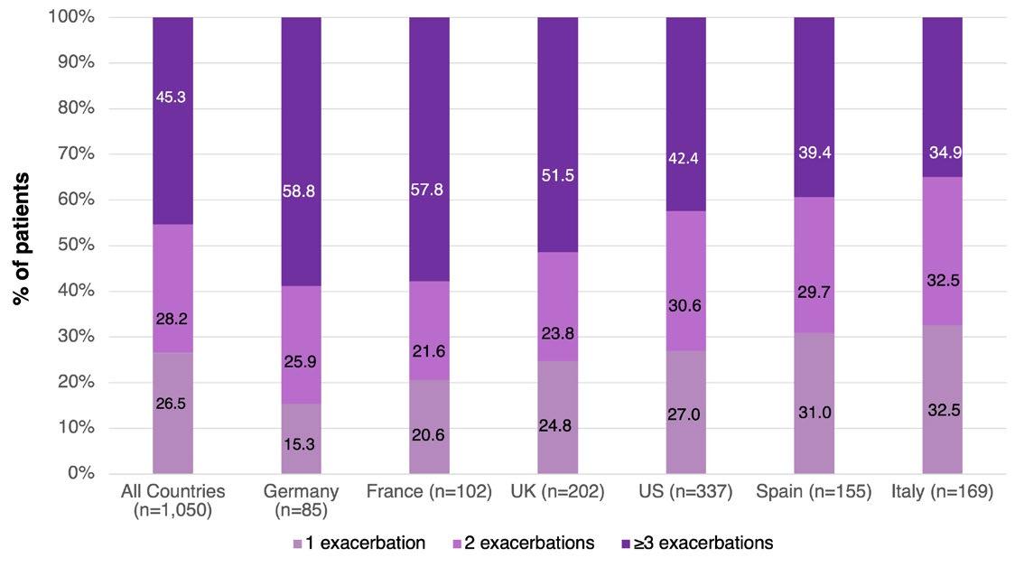
aExacerbations were defined as “a change in therapy suggested by your doctor and could be associated, among others, with the following signs: increased coughing; increased sputum production; bloody sputum production; change of color and viscosity of sputum; bouts of severe breathlessness/dispnea and/or discomfort in the chest; increased fatigue.”
bExacerbation frequency in the past 12 months.
Differences among patients with 1, 2, or ≥3 exacerbations were evaluated.7
In total, 1,050 people with bronchiectasis and 88 caregivers were included, representing a diverse cohort reflective of the heterogeneity of the disease (Table 1).7 Participants included a mix of retired and employed individuals, covering a range of day-to-day experiences when considering the impact of exacerbations on daily life. Approximately half
(54.2%) of patients had been living with BE for 5+ years, and one in three for 10+ years. Common comorbidities included asthma, gastroesophageal reflux disease, joint disease, hypertension, COPD, sinusitis, and non-tuberculous mycobacterial lung disease
Key Findings
Impact of exacerbations on daily living, emotional wellbeing, and caregiver burden
Among patients surveyed, 73.5% reported ≥2 exacerbations in the past year, and 45.0% experienced ≥3 exacerbations annually.7
Table 1: Demographics.




Figure 2: Additional negative impacta of exacerbations on dimensions of daily living in people with bronchiectasis.
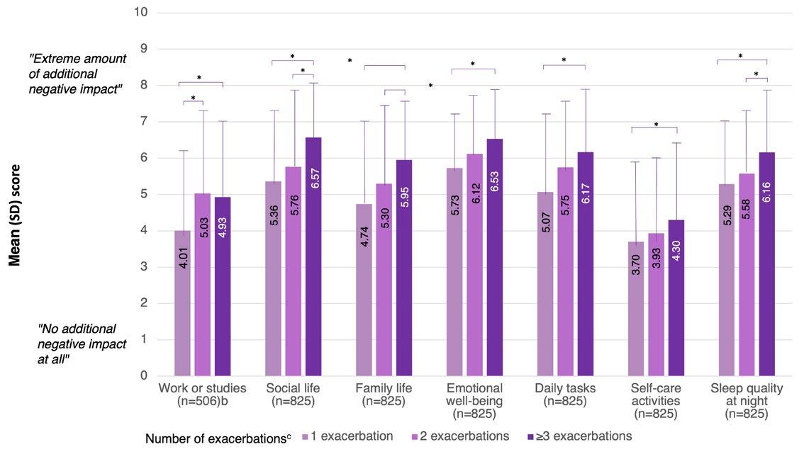
"Extreme amount of additional negative impact"
"No additional negative impact at all"
Work or studies (n=506)b
Number of exacerbationsc
*p<0.05.
aPeople with bronchiectasis were asked: “Compared to the times when you are NOT experiencing a bronchiectasis flare/ exacerbation, is there any additional negative impact of bronchiectasis flares/exacerbations on any of the following aspects of your life?” They were asked to rate each dimension of daily living on a scale from 1 (“No additional negative impact at all”) to 10 (“Extreme amount of additional negative impact”).
bIncludes only people who have worked or studied since their bronchiectasis diagnosis.
cExacerbation frequency in the past 12 months.
Exacerbations imposed an additional negative impact across all dimensions of daily living, with emotional well-being and social life most affected. Negative impact of emotional well-being and social life increased progressively with exacerbation frequency, with statistically significant differences observed between patients with one exacerbation and those with ≥3 exacerbations (Figure 2).7
Emotional wellbeing had a mean (SD) of 6.2 (2.8), and 40.5% of patients rated the
additional negative impact of an exacerbation as extremely high (8 or more).7 Exacerbations also impacted all areas of daily functioning; participants missed an average of 13.2 days of work in the previous 6 months as a result of an exacerbation, and 31.5% of them had to make lifestyle or financial adjustments.7 A clear relationship was observed, with increasing exacerbation frequency associated with progressively greater emotional, social, and quality-of-life impacts. Prevention and effective management of exacerbations, therefore, is an important area of clinical
focus, as each exacerbation is associated with a worsening quality of life. Notably, Polverino and co-authors reported that 76.8% of people with BE felt that every exacerbation worsened their condition.
The study also reported that even in the absence of exacerbations, bronchiectasis symptoms imposed a substantial negative impact on both patients and caregivers, with a particularly pronounced emotional burden driven by anxiety about future exacerbations and insecurity regarding the future.7 Caregivers reported that the impact of supporting someone with BE included an increase in feeling anxious or nervous (53.6%) and depressed or sad (53.7%).7
The BURDEN study found that exacerbations in BE carry a profound and lasting impact on patients and caregivers alike. The cumulative
References
1. Aliberti S et al. Clinical phenotypes in adult patients with bronchiectasis. Eur Respir J. 2016;47(4):1113-22.
2. Polverino E et al. European Respiratory Society guidelines for the management of adult bronchiectasis. Eur Respir J. 2017;50(3):1700629.
3. Choi H et al. Bronchiectasis management in adults: state of the art and future directions. Eur Respir J. 2024;63(6):2400518.
physical, emotional, and functional toll underscores the critical importance of exacerbation prevention and optimized disease management. Evaluation of patientreported outcomes and evidence-based strategies in exacerbation management can help meaningfully improve quality of life in patients with BE as well as their caregivers
Diagnoses and exacerbations were self-reported and medical records were not verified. Participants may have been subject to recall bias. It may have been difficult for participants to distinguish between experiences with and without pulmonary exacerbations.
4. Bertuccio FR et al. Phenotyping bronchiectasis frequent exacerbator: a single centre retrospective cluster analysis. Biomedicines. 2025;13(9):2124.
5. McDonnell MJ et al. Comorbidities and the risk of mortality in patients with bronchiectasis: an international cohort study. Lancet Respir Med. 2016;4(12):969-79.
6. Chalmers JD et al. European Respiratory Society clinical practice guideline for the management of
adult bronchiectasis. Eur Respir J. 2025;66(6):2501126.
7. Polverino E et al. B107-06 Patient and caregiver survey of burden of bronchiectasis in US and Europe. Am J Respir Crit Care Med. 2026;212(S1):aamag162.1278.
This collection of abstract reviews highlights key developments across respiratory and critical care medicine and include acute inpatient outcomes, rehabilitation after respiratory illness, and emerging evidence in lung transplantation. They emphasize the importance of earlier risk stratification, equitable care delivery, and targeted post-discharge and post-transplant management strategies.


Authors: Rakeem Levy,1,2 Carlos A. Gracidas Carrion,2,3 *Joseph Varon1,2,4
1. Caribbean Medical University, Willemstad, Curaçao
2. Dorrington Medical Associates, Houston, Texas, USA
3. Universidad Marista de Mérida, Mexico
4. Independent Medical Alliance, Houston, Texas, USA
*Correspondence to jvaron@imahealth.org
Disclosure: The authors have declared no conflicts of interest.
Acknowledgements: The authors would like to thank the National Inpatient Sample (NIS) database contributors and participating institutions for providing access to nationally representative hospitalization data.
Keywords: COPD exacerbation, critical care, inhospital mortality, invasive mechanical ventilation, National Inpatient Sample, respiratory failure.
Citation: Respir AMJ. 2026;4[1]:69-70. https://doi.org/10.33590/respiramj/0B9Q05K2
Acute exacerbations of COPD requiring invasive mechanical ventilation represent a severe clinical state associated with high morbidity and mortality.1,2 Despite advances in critical care, contemporary nationwide data describing outcomes in this population remain limited.2 Understanding demographic and clinical factors associated with mortality may improve risk stratification and inform management in critically ill patients with COPD.3 The study aimed to evaluate in-hospital outcomes and identify independent predictors of mortality among adults hospitalized with COPD exacerbations requiring invasive mechanical ventilation using a nationally representative US inpatient cohort.3
A retrospective analysis of the 2021 National Inpatient Sample (NIS) was performed.4 Adults aged ≥18 years hospitalized with COPD exacerbation (ICD-10 code J44.1) receiving invasive mechanical ventilation (5A19) were included. The primary outcome was in-hospital mortality. Secondary outcomes included length of stay and total hospital charges. Survey-weighted multivariable logistic regression was conducted to identify predictors of mortality. Model calibration was assessed using the Hosmer-Lemeshow test and discrimination with area under the curve.
A total of 76,680 unweighted hospitalizations met inclusion criteria. The mean age was 67±13 years, and 57% of patients were female.3 Overall in-hospital mortality was 28.4%.3 Adjusted predictors of in-hospital mortality identified on multivariable logistic regression analysis are presented in Figure 1. Female sex, Medicaid insurance, private insurance, and self-pay status were associated with lower adjusted odds of mortality, whereas Hispanic race and “Other” race categories demonstrated increased mortality risk. Model calibration demonstrated good fit with a Hosmer-Lemeshow p-value of 0.76, while discrimination analysis yielded an area under the curve of 0.72.3
Nearly one-third of patients hospitalized with COPD exacerbations requiring invasive mechanical ventilation died during hospitalization, highlighting the severity of illness in this population.2,3 Differences in mortality across race and insurance groups suggest persistent disparities in outcomes that may reflect differences in access to
Figure 1: Multivariable-adjusted predictors of in-hospital mortality in mechanically ventilated COPD exacerbations.
Adjusted odds ratio (95% CI)
Forest plot demonstrating adjusted odds ratios with 95% CIs derived from survey-weighted multivariable logistic regression analysis. Female sex, Medicaid insurance, private insurance, self-pay status, and acute respiratory failure coding were statistically associated with lower odds of in-hospital mortality, whereas Hispanic race and “Other” race categories were associated with increased mortality risk.
care, comorbidity burden, or treatment patterns. The inverse association observed with acute respiratory failure coding likely reflects heterogeneity in coding practices rather than a true protective effect. Despite advances in ventilatory strategies and critical care management, mortality remains substantial in this population.2,3,5
COPD exacerbations requiring invasive mechanical ventilation are associated with high in-hospital mortality.3 Significant variation in outcomes across demographic and socioeconomic groups highlights the need for improved risk stratification and more equitable delivery of care. Standardization of administrative coding
and further investigation into drivers of observed disparities are warranted.
References
1. Global Initiative for Chronic Obstructive Lung Disease. 2025 GOLD report. Available at: https://goldcopd. org/2025-gold-report/. Last accessed: May 18, 2026.
2. Esteban A et al. Characteristics and outcomes in adult patients receiving mechanical ventilation: a 28-day international study. JAMA. 2002;287(3):345-55.
3. Levy R et al. In-hospital outcomes and predictors of mortality among COPD exacerbations requiring invasive mechanical ventilation: insights from a national inpatient cohort. Poster P1573. ATS International Conference, May 15-20, 2026.
4. Agency for Healthcare Research and Quality (AHRQ). AHRQ HCUP NIS overview. 2026. Available at: https:// hcup-us.ahrq.gov/nisoverview.jsp. Last accessed: May 18, 2026
5. Wunsch H et al. ICU occupancy and mechanical ventilator use in the United States. Crit Care Med. 2013;41(12):2712-9.
Authors: *Sai Anusha Akella,1 Samuel Sule-Saa,1
Yorquiris Acevedo,1 Aditi Parulkar,1
Kalpani Panigrahi1
1. One Brooklyn Health – Interfaith Medical Centre, Brooklyn, New York, USA *Correspondence to saianusha.akella@obhny.org
Disclosure: The authors have declared no conflicts of interest.
Keywords: Decubitus ulcer, mortality, sepsis.
Citation: Respir AMJ. 2026;4[1]:71-72. https://doi.org/10.33590/respiramj/YDQZV38E
Sepsis is a life-threatening condition associated with significant morbidity and mortality.1 Decubitus ulcers (pressure injuries) are common in hospitalized patients and may become a source of infection.2,3 Pressure injuries may become complicated by local infection, osteomyelitis, bacteremia, and sepsis.4 This study seeks to investigate the association between the presence of decubitus ulcers and inpatient outcomes, including mortality, resource utilization, and complications, among patients hospitalized with sepsis in the United States, as largescale data on this association are limited.5
This retrospective cohort study utilized the Nationwide Inpatient Sample (NIS) database from 2016–2021. Patients hospitalized with a primary diagnosis of sepsis were identified and stratified based on the presence or absence of a concurrent diagnosis of decubitus ulcer. Multivariable logistic regression was used to assess the odds of in-hospital mortality and specific complications (severe sepsis, septic shock,
disseminated intravascular coagulation, acute respiratory distress syndrome, mechanical ventilation, and acute kidney injury).
Multivariable linear regression was used to compare total hospital charges, costs, and length of stay (LOS). Models were adjusted for patient demographics (age, gender, race, income quartile, and insurance), the Charlson Comorbidity Index, patient residence location, and hospital characteristics (region, bed size, and teaching status).
Of approximately 13.1 million weighted sepsis admissions, 1,046,110 (8.0%) had comorbid decubitus ulcers (2016–2021; Table 1).
Patients with decubitus ulcers were slightly older (mean age: 64.5 versus 69.3 years; p<0.01) and had higher comorbidity burdens. After multivariable adjustment, the presence of a decubitus ulcer was significantly associated with 35% increased odds of inhospital mortality (1.35; p<0.01). Decubitus ulcers were also associated with significantly higher adjusted total hospital charges (40,776.83 USD; p<0.01), total costs (8,175.51 USD; p<0.01), and longer LOS (3.85 days; p<0.01). Furthermore, decubitus ulcers were associated with significantly higher adjusted odds of developing severe sepsis (adjusted odds ratio [AOR]: 1.35), septic shock (AOR: 1.58), disseminated intravascular coagulation (AOR: 1.18), requiring mechanical ventilation (AOR: 3.99), and acute kidney injury (AOR: 1.05; all p<0.01). No significant association was found with acute respiratory distress syndrome (AOR: 1.04; p=0.068).
The study suggests the presence of a decubitus ulcer was independently
associated with significantly increased inpatient mortality, higher resource utilization (cost, LOS), and greater odds of major sepsis-related complications. These findings, with similar results to prior literature6 highlight
decubitus ulcers as an important indicator of adverse outcomes in patients with sepsis, emphasizing the need for targeted prevention and management strategies in this vulnerable population.
Table 1: Adjusted odds ratio of complications comparing teaching versus non-teaching hospitals.
p<0.05 indicates statistical significance. Multivariable regression models were adjusted for age at admission, gender, race, median household income national quartile for patient ZIP Code, Charlson Comorbidity Index, location/teaching status of the hospital, region of the hospital, patient’s residence, and insurance.
AKI: acute kidney injury; ARDS: acute respiratory distress syndrome; DIC: disseminated intravascular coagulation.
References
1. Complex Wounds Working Group; Elena Espejo et al. Bacteremia associated with pressure ulcers: a prospective cohort study. Eur J Clin Microbiol Infect Dis. 2018;37(5): 969-75.
2. Zhaoyu Li et al. Global prevalence and incidence of pressure injuries in hospitalised adult patients: a systematic review and meta-analysis. Int J Nurs Stud. 2020;DOI:10.1016/j.ijnurstu.2020.103546.
3. Padula et al. Delarmente. The national cost of hospital‐acquired pressure injuries in the United States. Int Wound J. 2019;16(3):634-40.
4. Mervyn Singer et al. The Third International Consensus Definitions for Sepsis and Septic Shock (Sepsis-3). JAMA. 2016;315(8):801.
5. Akella S et al. The effect of decubitus ulcer on inpatient outcomes in patients with sepsis: a nationwide analysis (2016-2021). Poster B25-16. ATS International Conference, May 15-20, 2026.
6. Christina L Wassel et al. Risk of readmissions, mortality, and hospital‐acquired conditions across hospital‐acquired pressure injury (HAPI) stages in a us national hospital discharge database. Int Wound J. 2020;17(6):1924-34.
Authors: Amandeep Singh,1 Efstathia
Polychronopoulou,2 *Alexander G. Duarte1
1. Pulmonary, Critical Care and Sleep Medicine, University of Texas Medical Branch, Galveston, Texas, USA
2. Department of Biostatistics and Data Science, University of Texas Medical Branch, Galveston, Texas, USA
*Correspondence to aduarte@utmb.edu
Disclosure: The authors have declared no conflicts of interest.
Keywords: Antifibrotics, idiopathic pulmonary fibrosis (IPF), interstitial lung disease (ILD), lung transplantation, pulmonary fibrosis, pulmonary rehabilitation (PR), respiratory hospitalization.
Citation: Respir AMJ. 2026;4[1]:73-74. https://doi.org/10.33590/respiramj/4GK1H02W
Interstitial lung disease (ILD) is associated with progressive, debilitating symptoms, and hospitalizations occur frequently, with a report from the Pulmonary Fibrosis Foundation registry that described 34.7% of patients with ILD were hospitalized between March 2016–May 2021.1 Although respiratory-related hospitalizations were associated with poor clinical outcomes, postdischarge use of pulmonary rehabilitation (PR) was not reported. In other chronic respiratory disorders, such as COPD, PR after hospital discharge is associated with reduced healthcare use and improved patient outcomes.2 However, there is limited data concerning post-hospital discharge use of PR in patients with ILD, and the aim of this study was to assess the use of PR at the population level for post-hospital discharge patients with ILD in the United States.3
The authors used IBM MarketScan® Research Database (Merative, Ann Arbor, Michigan, USA) to identify adults hospitalized with idiopathic pulmonary fibrosis (IPF) or non-IPF ILD between January 1, 2014–December 31, 2023. PR use was defined as identification of ≥1 claim within 3 months after hospital discharge and excluded patients with neuromuscular conditions, dementia, or discharge to a skilled nursing facility, longterm acute care hospital or hospice.
Among 11,413 eligible patients, 548 (4.8%) participated in PR within 90 days of hospital discharge (Figure 1). PR participants had a mean age of 62.9±11.0 years, which was similar to patients who did not participate in PR. Greater PR participation was noted in males, prior oxygen prescription, antifibrotic use, PR participation in the previous 12 months, and lung transplantation or evaluation in the previous 12 months. Logistic regression model revealed male sex and prior oxygen prescriptions were associated with higher odds of PR utilization, while factors associated with a lower likelihood of PR utilization included greater comorbidities and, in the preceding 12 months, no prior lung transplant surgery or transplant evaluation. Prior prescriptions for antifibrotics or immunosuppressive therapy were not associated with PR participation. Of note, patients with IPF participated in more than eight PR sessions more often compared to patients with non-IPF-ILD.
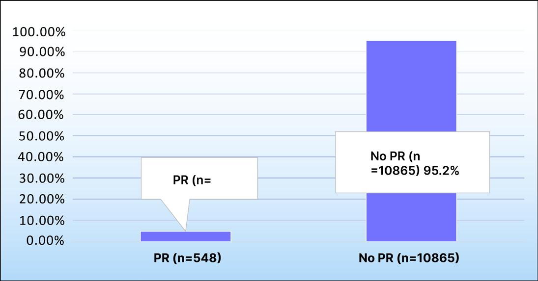
PR: pulmonary rehabilitation.
In conclusion, PR utilization after hospital discharge for patients with IPF and non-IPF ILD was low, and factors associated with increased PR included males and patients with prior oxygen prescription.
References
1. King CS et al. Hospitalization rate in interstitial lung Disease. Am J Respir Crit Care Med. 2024;210:801-13.
2. Jenkins AR et al. Do pulmonary rehabilitation programmes improve outcomes in patients with COPD posthospital discharge for exacerbation: as systematic review and meta-analysis. Thorax. 2024;79:438-47.
3. Singh A et al. Population assessment of postdischarge pulmonary rehabilitation use in patients with interstitial lung disease. Poster 206. ATS International Conference, May 15-20, 2026.
Authors: *Inga Jarosch,1,2 Tessa Schneeberger,1,2
Rainer Gloeckl,1,2 Rixa Hahn,1,2 Daniela Kroll,1,2 Martina Boensch,1 Wolfgang Hitzl,3 Andreas Rembert Koczulla1,2,4
1. Institute for Pulmonary Rehabilitation Research, Schoen Klinik Berchtesgadener Land, Schoenau am Koenigssee, Germany
2. Department of Pulmonary Rehabilitation, Philipps-University of Marburg, German Center for Lung Research (DZL), Germany
3. Research and Innovation Management, Biostatistics, Department of Ophthalmology and Optometry, Research Program Experimental Ophthalmology and Glaucoma Research, Paracelsus Medical University, Salzburg, Austria
4. Teaching hospital, Paracelsus Medical University, Salzburg, Austria *Correspondence to ijarosch@schoen-klinik.de
Disclosure: Jaroch and Schneeberger report being shareholder of Pneumo Factory UG, which was involved in the delivery of the virtual maintenance intervention evaluated in this study. The other authors have no conflict of interest to declare. The trial was funded by the Bavarian State Ministry of Health, Care and Prevention (Germany).
Acknowledgements: The authors would like to thank all participants for their time, commitment, and valuable contribution to this study.
Keywords: Exercise training, fatigue, long COVID, post-exertional malaise (PEM), synchronous maintenance, work ability.
Citation: Respir AMJ. 2026;4[1]:75-76. https://doi.org/10.33590/respiramj/93U1V2YD
Although rehabilitation improves functional outcomes in post-COVID-19 condition (PCC),1,2 many patients do not fully recover and require long-term care. Evidence-based and accessible maintenance strategies following rehabilitation are lacking.
The aim of this study was to investigate whether extending inpatient rehabilitation with a virtual maintenance program improves quality of life in individuals with PCC.
In this single-center RCT, participants with PCC were randomized (1:1) to inpatient rehabilitation (3 weeks) followed by usual care (control) or by a 12-week virtual, synchronous maintenance program (intervention, INT).
Effectiveness outcomes (SF-12 mental [MCS] and physical component score [PCS], quality of life [EQ-5D], Work Ability Index [WAI], Pittsburgh Sleep Quality Index [PSQI], Generalized Anxiety Disorder-7 [GAD-7], Patient Health Questionnaire-9 [PHQ-9], and 1-minute sit-to-stand test) were assessed at baseline (T1) and at 15 weeks (T3). Safety outcomes (Fatigue Assessment Scale [FAS], healthcare contacts, sick days) were assessed after inpatient rehabilitation (T2) and at T3. The primary outcome was the change in SF-12 PCS from T1 to T3. Analyses followed the intention-to-treat principle using multiple imputation. Post-hoc subgroup analyses compared adherent (INTADH+, ≥80% maintenance attendance) and less adherent INT participants (INTADH-, <80%). Satisfaction and usability were rated on a 5-point Likert scale.
Ninety participants were included (67% female; mean age: 51±12 years; 22+14 months between COVID-19 and rehabilitation). SF-12 PCS improved similarly in both groups, with no between-group difference (Δ=0.4 points; 95% CI: −3.8–3.1; p=0.84). No significant between-group
differences were found for secondary outcomes, except for WAI score, which improved significantly more in the INT than in the control group (Δ=2.4 points; 95% CI: 0.2–4.6; p=0.03; Cohen’s d=0.49). Adherence to the virtual program was high (85.5%). INTADH+ showed significantly greater improvements in EQ-5D, PSQI, and GAD-7 compared with INTADH-. Satisfaction with the virtual program was high, and no intervention-related adverse events were observed.
The extended rehabilitation approach was not superior to inpatient rehabilitation alone in improving physical quality of life in PCC. Nevertheless, the observed improvement in work ability indicates that
continued structured support may enhance participation-related recovery beyond the initial rehabilitation phase. High adherence and patient satisfaction support synchronous virtual maintenance programs as a feasible and scalable model for long-term PCC care.3
References
1. Schneeberger S et al. Symptom-based multimodal rehabilitation in people with post COVID-19 (RELOAD): a randomized controlled trial. BMJ Open Res. 2026;DOI:10.1136/bmjresp-2026-004177.
2. Chakraverty S et al. Rehabilitation in long COVID: a systematic review and meta-analysis. ERJ Open Res. 2026;DOI:10.1183/23120541.00133-2026.
3. Jarosch I et al. Feasibility of a digital, synchronous rehabilitation maintenance program for people with post-COVID-19 condition – results from the Reload 2.0 study. Abstract #9475. ATS International Conference, May 15-20, 2026.
Authors: *Abdul Wahab,1 Kavin Parmar,2 Kelly M. Pennington3
1. Division of Pulmonary and Critical Care Medicine, Virginia Commonwealth University, Richmond, Virginia, USA
2. Division of Gastroenterology, Hepatology and Nutrition, Virginia Commonwealth University, Richmond, Virginia, USA
3. Division of Pulmonary and Critical Care Medicine, William J. von Liebig Center for Transplantation, Mayo Clinic, Rochester, Minnesota, USA
*Correspondence to abdul.wahab@vcuhealth.org
Disclosure: The authors have declared no conflicts of interest.
Keywords: Aspergillus, Candida, fungal infection, lung transplant.
Citation: Respir AMJ. 2026;4[1]:77-79. https://doi.org/10.33590/respiramj/8J48GF0I
Fungal infections remain a major cause of morbidity and mortality post-lung transplant due to the unique exposure of the allograft to the external environment and the intensity of immunosuppression. Single-center studies have described variable rates of fungal infections.1 Contemporary populationlevel data with temporal trends, pathogen distribution, and associated outcomes are limited. Leveraging a large multicenter real-world dataset, the authors aimed to characterize incidence, prevalence, and clinical outcomes of fungal infections among adult lung-transplant recipients.2
Data were obtained from the TriNetX™ (TriNetX, LLC, Cambridge, Massachusetts, USA) Global Collaborative Network (160
healthcare organizations). Adult lungtransplant recipients (≥18 years) were identified between 2005–2024, using International Classification of Diseases (ICD) and Current Procedural Terminology (CPT) procedure codes. Patients with solid organ transplants other than lung, combined-organ transplants, or stem-cell transplants, and patients with HIV were excluded. Patients with pre-transplant fungal infections were also excluded. Pre-transplant characteristics included demographics, comorbidities within a year, laboratory values within 7 days, and medication within 90 days before transplantation. Post-transplant fungal infections were identified using ICD-10 codes for Aspergillus, Candida, Cryptococcus, Mucorales, Histoplasma, Blastomyces, Coccidioides, and Pneumocystis TriNetX platform was utilized for statistical analysis. Incidence and prevalence of infections, all-cause mortality, and transplant rejection were analyzed.
A total of 12,857 adult lung-transplant recipients were identified. The mean age at transplantation was 57.6±13.4 years, with a male predominance of 57%, and majority were White (65%). Average BMI was 26.8±5.7 kg/m². The most common pulmonary conditions were COPD (15%), pulmonary fibrosis (17%), pulmonary hypertension (9%), bronchiectasis (3%), and cystic fibrosis (3%). Pre-transplant fungal and viral screening assays were rarely available (<1%). Medication exposure within 90 days prior to transplant demonstrated prednisone prescription (17%), methylprednisolone (7%), tacrolimus (6%), and mycophenolate mofetil (7%). Azathioprine and basiliximab were used in 2% each. Systemic antifungals
(voriconazole, posaconazole, or fluconazole) in approximately 1% each.
The overall incidence of post-transplant fungal infection was 3% across the full study period, rising to 7% in the modern era (2015–2024). Most frequently identified pathogens were Candida spp. (4.8%) and Aspergillus spp. (2.9%), followed by Pneumocystis Jirovecii (0.5%). Infection rates increased from 22 per 1,000 (2005–2014) to 75 per 1,000 (2015–2024; Table 1).
In this large multicenter cohort of adult lung-transplant recipients, the incidence of post-transplant fungal infections increased substantially in the modern transplant era. The rise from 22 per 1,000 recipients in 2005–2014 to 75 per 1,000 recipients in 2015–2024 likely reflects a combination of improved fungal diagnostics, more systematic post-transplant surveillance, better electronic health record capture, and longer survival after transplantation, allowing more time for
Because infections were assessed only during the post-transplant period and typically represented single discrete events, reported incidence and prevalence values are nearly identical within each era. In TriNetX™ (TriNetX, LLC, Cambridge, Massachusetts, USA), prevalence is cumulative within the same observation window, which overlaps with incidence when events occur acutely.
opportunistic infections to emerge.3,4 Candida and Aspergillus were the most identified fungal pathogens, consistent with the high susceptibility of lung-transplant recipients to airway colonization, invasive mold infection, and opportunistic infections related to intense immunosuppression.
The authors’ study highlights a rising burden of fungal infections among lung-transplant recipients with potential implications for early post-transplant surveillance and antifungal prophylaxis strategies.
1. Chang A et al. Epidemiology of invasive fungal infections in lung transplant recipients in Western Australia. Transpl Infect Dis. 2019;21(3):e13085.
2. Wahab A et al. Post lung transplantation fungal infections: a multicenter real-world analysis (20052024). Poster 918. ATS International Conference, May 15-20, 2026.
3. Hosseini‐Moghaddam SM et al. Incidence and outcomes of invasive fungal infection among solid organ transplant recipients: a population‐based cohort study. Transpl Infect Dis. 2020:22(2):e13250.
4. Baker AW et al. Invasive fungal infection after lung transplantation: epidemiology in the setting of antifungal prophylaxis. Clin Infect Dis. 2020;70(1):30-9.
Author: *William W. Mangin1
1. Mayo Clinic, Rochester, Minnesota, USA *Correspondence to drwmangin@gmail.com
Disclosure: The author has declared no conflicts of interest.
Keywords: Aspergillus, fungal colonization, fungal infection, lung transplantation, outcomes.
Citation: Respir AMJ. 2026;4[1]:80-81. https://doi.org/10.33590/respiramj/6MR93OD1
Pre-transplant respiratory tract colonization with Aspergillus species has been associated with an increased incidence of invasive aspergillosis after lung transplant in certain transplant populations.1 Although less common, diagnoses of invasive infections by other non-Aspergillus fungal species are increasing.1,2 Despite this increased recognition of rarer infections from species such as Alternaria, Cladosporium, Fusarium, Lomentospora, Mucorales, Scedosporium, and others, how pre-transplant colonization with these organisms influences posttransplant outcomes is unknown.
This was a multicenter, retrospective cohort study of all adult bilateral lung transplant recipients at Mayo Clinic sites in Rochester, Minnesota, and Jacksonville, Florida, USA, between January 1, 2016–December 31, 2024.3 Patients with pre-transplant respiratory tract fungal colonization were compared to those without colonization. Exclusion criteria included previous organ transplantation and multi-organ transplantation. Fungal colonization was defined as positive respiratory fungal
culture(s) in asymptomatic patients without evidence of infection. Only fungal cultures that were speciated were included. Candida species and Penicillium species were not included in analysis. The primary outcome was severe post-transplant invasive fungal infection (IFI), which was defined as proven or probable IFI (based on European Organization for Research and Treatment of Cancer/Mycoses Study Group [EORTC/ MSG] criteria) that required subsequent hospitalization or additional procedures.4 Baseline characteristics were summarized using descriptive statistics (medians with interquartile ranges or counts with percentages) and groups were compared using standardized mean differences. Unadjusted risk ratios (RR) with 95% CIs were used to analyze associations with posttransplant infection and mortality (Table 1).
Four hundred and forty-nine patients were included in the author’s analysis. In total, 83 patients (18.5%) had pre-transplant fungal respiratory tract colonization, 45 (10%) colonized with Aspergillus species and 38 (8.5%) with non-Aspergillus species. Nineteen of the 45 patients with Aspergillus colonization were also colonized with at least one non-Aspergillus species. Three hundred and sixty-six patients did not have pre-transplant colonization. The most common pre-transplant fungal species were Cladosporium (28), Alternaria (15), and Fusarium (14). Other fungal species included Scapulariopsis (three), Rhizopus (two), and Scedosporium (two). The two cohorts primarily differed in indication for transplant, with a higher percentage of patients with muco-obstructive disease in the colonization group. A total of four patients (4.8%) in the fungal colonization cohort developed severe
fungal infection post-transplant. Fungal species differed pre- and post- transplant in all cases, with none of the species of colonizers being a causative organism in any of the post-transplant infections. There was no significant association between pre-transplant colonization and severe posttransplant fungal infection (RR: 1.36 [0.45–4.06]), severe post-transplant Aspergillus infection (RR: 0.08 [0.18–3.55]), or unadjusted mortality (RR: 0.74 [0.47–1.17]).
Previous studies showing an increased risk of post-transplant aspergillosis in patients with cystic fibrosis could be explained by their sinus involvement, and thus lung transplantation potentially did not remove all sites of fungal airway colonization. The results of this study show that pretransplant fungal colonization is not associated with severe post-transplant IFIs in the author’s cohort of bilateral lung
transplant recipients. Although limited by the retrospective nature of the study and low incidence of the primary outcome, the author’s results do add supporting evidence to the idea that airway fungal colonization should not be a reason to preclude patients from undergoing lung transplantation.
1. Luong ML et al. Pretransplant Aspergillus colonization of cystic fibrosis patients and the incidence of post-lung transplant invasive aspergillosis. Transplantation. 2014;97(3):351-7.
2. Hoenigl M et al. Global guideline for the diagnosis and management of rare mould infections: an initiative of the European Confederation of Medical Mycology in cooperation with the International Society for Human and Animal Mycology and the American Society for Microbiology. Lancet Infect Dis. 2021;21(8):e246-57.
3. Mangin WW et al. Pre-transplant fungal colonization and outcomes in lung transplant recipients. Poster Board 920. ATS International Conference, May 15-20, 2026.
4. Donnelly JP et al. Revision and update of the consensus definitions of invasive fungal disease from the European Organization for Research and Treatment of Cancer and the Mycoses Study Group Education and Research Consortium. Clin Infect Dis. 2019;71(6):1367-76. Outcome
Authors: *Abdul Wahab,1 Kavin Parmar,2 Kelly M. Pennington3
1. Division of Pulmonary and Critical Care Medicine, Virginia Commonwealth University, Richmond, USA
2. Division of Gastroenterology, Hepatology and Nutrition, Virginia Commonwealth University, Richmond, USA
3. Division of Pulmonary and Critical Care Medicine, William J. von Liebig Center for Transplantation, Mayo Clinic, Rochester, Minnesota, USA
*Correspondence to Abdul.wahab@vcuhealth.org
Disclosure: Wahab has received support to attend the ATS Conference 2026 via an ATS 2026 abstract scholarship. The other authors have declared no conflicts of interest.
Keywords: Chronic lung allograft dysfunction (CLAD), fungal infection, lung transplant.
Citation: Respir AMJ. 2026;4[1]:82-84. https://doi.org/10.33590/respiramj/EQ9TF745
Fungal infections are a frequent and clinically important complication after lung transplantation. Lung transplant recipients are uniquely vulnerable because the allograft is directly exposed to the external environment.1,2 In addition, airway instrumentation and structural lung disease may further increase susceptibility to fungal colonization and invasive fungal infection. Aspergillus and Candida species account for most fungal infections in this population, although mucormycosis, endemic fungi, and Pneumocystis jirovecii may also occur depending on host factors, prophylaxis practices, and local epidemiology.2-4
The relationship between post-transplant fungal infection and patient centered outcomes remains incompletely defined
in large, real-world, multicenter cohorts. The authors aimed to evaluate the association between post-transplant fungal infection and mortality, allograft rejection, and chronic lung allograft dysfunction (CLAD) among adult lung transplant recipients.5
The authors conducted a retrospective cohort study using the TriNetX Global Collaborative Network (TriNetX, LLC, Cambridge, Massachusetts, USA), which captures de-identified data from >160 healthcare organizations across North America, Europe, and Asia. Adult lung transplant recipients (≥18 years) between 2005–2024 were identified using International Classification of Diseases, 10th Revision (ICD-10) and Current Procedural Terminology (CPT®) codes. Patients with post-transplant fungal infection (aspergillosis, candidiasis, mucormycosis, histoplasmosis, coccidioidomycosis, blastomycosis, and pneumocystosis) and matched controls without fungal infection, within 1 year before or any time after transplant, were identified. The index date was defined as the date of lung transplantation.
Propensity-score matching was performed across demographics, comorbidities, laboratory values, procedures, and immunosuppressive medications. Outcomes included all-cause mortality, allograft rejection, and CLAD at 6 months and 1 year. Kaplan–Meier analysis and Cox proportional hazards models were used to estimate risk ratios (RR), hazard ratios, and 95% CIs. Analyses were conducted using TriNetX Analytics version 25.0 (TriNetX, LLC, Cambridge, Massachusetts, USA).
A total of 3,083 patients with post-transplant fungal infection and 11,625 patients without fungal infection were identified. The mean age was 58±13 years, and 56% were male. The most common pulmonary comorbidities were COPD, unspecified pulmonary fibrosis, and bronchiectasis. After propensity-score matching across 56 clinical variables, 1,790 patients remained in each cohort. Mortality was significantly higher in the fungal
infection cohort, occurring in 13% versus 9% at 6 months (RR: 1.45; 95% CI: 1.20–1.75; p=0.001) and 17.8% versus 13% at 1 year (RR: 1.36; p=0.0001). Allograft rejection was more frequent among patients with fungal infection, occurring in approximately 46 compared with 22 among matched controls at 6 months. CLAD occurred approximately two- to three-fold more frequently in the fungal infection cohort compared with the non-infected cohort (Table 1).
Values shown represent cumulative incidence at each timepoint.
In this large, multicenter, propensitymatched analysis, post-transplant fungal infection was associated with significantly worse outcomes, including higher all-cause mortality, increased allograft rejection, and CLAD.
Fungal organisms may cause direct airway and parenchymal injury, particularly in the setting of impaired mucociliary clearance. Infections may amplify local inflammation and promote epithelial injury, which increases susceptibility to acute rejection. In turn, recurrent injury and inflammation may contribute to CLAD.1,6 The observed two- to three-fold higher risk of CLAD among patients with fungal infection suggests that fungal surveillance and prevention strategies may
have implications beyond short-term infection control.3 Although propensity-score matching reduces measured confounding, residual confounding by disease severity, centerlevel prophylaxis and diagnostic practices, timing of infection, antifungal exposure, and distinction between colonization and invasive infection remains possible.
Post-transplant fungal infection was independently associated with increased mortality, allograft rejection, and CLAD among lung transplant recipients. These findings highlight the need for optimized surveillance, prophylaxis, and individualized post-transplant management strategies.
1. Pennington KM et al. Risk factors for early fungal disease in solid organ transplant recipients: a systematic review and meta-analysis. Transplantation. 2024;108(4):970-84.
2. Wahab A et al. Diagnosis and prevention of invasive fungal infections in the immunocompromised host. Chest. 2025;167(2):374-86.
3. Villalobos APC, Husain S. Infection prophylaxis and management of fungal infections in lung transplant. Ann Transl Med. 2020;8(6):414.
4. Huggins JP et al. Risk factors for invasive fungal infection in lung transplant recipients on universal antifungal prophylaxis. Open Forum Infect Dis. 2023;11(2):ofad640.
5. Wahab A et al. C27-17 Impact of post-transplant fungal infection on mortality and graft outcomes in lung transplant recipients: a multicenter propensitymatched analysis. Am J Respir Crit Care Med. 2026;212(Suppl 1):S4929-30.
6. Aguilar CA et al. Clinical risk factors for invasive aspergillosis in lung transplant recipients: results of an international cohort study. J Heart Lung Transplant. 2018;37(10):1226-34.
In these interviews, Donald B. Middleton discusses the important questions in respiratory research, how to design vaccination strategies, and building sustainable global respiratory surveillance systems in the face of future outbreaks. Tina Q. Tan shares her hopes for the future of pediatric respiratory care.
Featuring: Donald B. Middleton and Tina Q. Tan

Donald B. Middleton
Professor of Family Medicine, University of Pittsburgh School of Medicine, UPMC St. Margaret, Pennsylvania, USA
Citation: Respir AMJ. 2026;4[1]:85-87.
https://doi.org/10.33590/respiramj/TI48S715
Prevention of disease in patients of all ages is clearly the motivator for all primary care clinicians
Q1
You have spent decades working in both family medicine and influenza vaccine research. How has working directly with patients influenced the kinds of respiratory research questions you believe are most important to investigate?
Prevention of disease in patients of all ages is clearly the motivator for all primary care clinicians. Luckily, over time, we have had more tools that are available to achieve this goal. Illnesses with high morbidity and mortality, like influenza, are frightening. Proven, effective vaccines like Tdap (tetanus, diphtheria, and pertussis) are basic parts of my armamentarium. Every year, I embrace vaccines that are fine-tuned to whatever particular virus variants are likely to circulate. Over the years, I have seen remarkably enhanced influenza vaccine development with much improved vaccine effectiveness (VE), but more is needed. Every winter, I would treat many babies with rotavirus infection who were dehydrated, vomiting, and having severe diarrhea, but now I see
almost none. We also need new vaccines to produce long-lasting protection against agents like pertussis and respiratory syncytial virus (RSV). On the downside, lately, convincing individuals to be vaccinated has become more difficult. Research that helps answer how to persuade individuals that vaccination is safe and effective for them, their families, and society is paramount.
Throughout your career, respiratory viruses have evolved from being viewed as largely seasonal challenges to major drivers of healthcare system pressure and public policy. How have you personally seen the clinical and societal perception of respiratory disease change over time?
The failure of our public leaders to commit to appropriate vaccination for everyone is deplorable and has led many individuals and parents to reject vaccination. Individuals seem to think that they are immune to infection, or that infection will be mild, or that treatment will be
available, so there is no need to vaccinate. These opinions are sadly wrong. The commitment to the protection of society through vaccination of everyone against communicable diseases seems to have lost part of its punch. But not for me.
Recent US studies have explored waning influenza vaccine protection across a single respiratory season, particularly among older adults and high-risk groups. What practical lessons should clinicians and policymakers take from this research when designing vaccination strategies?
The 2020 reference provided is from Jill Ferdinands et al.1 She is a remarkable researcher with whom I and others were able to work to produce a VE study in 2021. In that study,2 we showed that the rate of VE waning was actually worse in adults aged 65 years and over; a decline of approximately 10% every 30 days post-vaccination. At that rate, influenza VE, which typically begins at around 50–60%, could decline by more than half
by season’s end. We did not know what percentage of the study participants received an enhanced influenza vaccine, which might reduce this dangerous VE drop. Most of the time, influenza starts in November. So, to provide better protection throughout the influenza season, given the 1–2-week delay in effectiveness post-vaccination, I like to vaccinate my older patients (including myself) in early-tomid-October. The clinician must be committed to making certain that older patients do not fall through the cracks to end up being unvaccinated. A good motto is “If you are older, wait until it's colder.”
Recent respiratory research3 in the US has also focused on co-circulation of influenza, RSV, and SARS-CoV-2, and the challenge this creates for diagnosis, triage, and healthcare capacity planning. Which findings from these studies do you think will have the greatest long-term impact on respiratory care systems?
Co-circulation of respiratory viruses and bacterial pathogens like pertussis is not a new issue. The
new issue is that with the advent of clinically useful diagnostic tests, multiple other viruses besides influenza have been recognized to cause significant diseases, including death, across all ages. An emphasis on co-circulation of respiratory viruses came with the arrival of SARS-CoV-2; its death rate eclipsed the death rate from influenza. The thought that these two potentially deadly agents would cocirculate was alarming. Every year, multiple viruses, some seasonal (RSV) and some not (SARS-CoV-2), including others like human metapneumovirus, which also infects all ages, co-circulate. Many of these agents tend to cause prolonged coughing, lasting several weeks. The cough from bacterium Bordetella pertussis tends to get lost in the thought that only viruses can create this problem. Although recently, the influenza B/Yamagata has disappeared from circulation, I do not see the spontaneous disappearance of most of the other agents which constantly evolve. All health systems should maintain some method of diagnostic detection to provide focused treatment whenever possible.

Q5
Many countries are now rethinking respiratory surveillance after COVID-19. Based on your experience with influenza monitoring programs, what elements are essential for building sustainable global respiratory surveillance systems that can respond to future outbreaks?
Hopefully, many countries have bolstered their detection and reporting systems. Local education about how to detect and report a disease is helpful, including home tests for influenza and SARS-CoV-2. Patients and clinicians must suspect particular agents, like influenza, and take steps to diagnose properly. Most laboratories report to a central agency like a health department, which then reports to a national infection control agency like the CDC. Both health departments and national bodies must then distribute warnings about circulating agents. The WHO remains critically important to provide warnings and control measures.
Q6
Vaccine confidence has become an increasingly important global health issue in respiratory medicine. How can healthcare professionals better communicate uncertainty, effectiveness, and risk in ways that improve public engagement without oversimplifying the science?
References to supportive websites like Immunize.org or the American Academy of Pediatrics (AAP), discussing disease consequences, and explaining the length of time vaccines are studied for safety and effectiveness can all help. I simply tell a hesitant patient that
I vaccinate myself and my family, that I like them, and that they need vaccination too.
Q7
Emerging technologies such as universal influenza vaccines, mRNA-based respiratory vaccines, and AIassisted outbreak prediction are rapidly changing the field. Which of these developments do you believe has the greatest potential to transform respiratory medicine over the next decade?
I do not endorse any single approach. New technologies and new combinations are coming for many vaccines, like the pneumococcal vaccine and Lyme disease. However, mRNA vaccines have great potential to be widely effective. Influenza and SARSCoV-2 protection can be combined in one injection, and others could be added down the line. The mRNA Lyme vaccine is under investigation. Many hope for better mRNA cancer vaccines. So, I think that is where I would put my money. AI may help to truncate outbreaks, but patients and clinicians first need to explore AI recommendations and second need to have reassurance against errors in advice. AI must also be informed: false anti-vaccine information must not be repeated.
Q8
Finally, if you were advising the next generation of clinicians and researchers entering respiratory and infectious disease medicine today, what unanswered questions or underexplored areas do you believe deserve far more attention in the years ahead?
The public’s trust in the science of vaccination needs to be restored. A growing number of hesitant medical
practitioners need to reaffirm their faith in vaccination. How to accomplish these tasks is the major issue facing us now. For clinicians, vaccine fatigue must be rejected. More improvements of current vaccines should be embraced. Vaccines to stop other diseases like human metapneumovirus should be embraced.
Co-circulation of respiratory viruses and bacterial pathogens like pertussis is not a new issue
References
1. Jill Ferdinands et al. Waning of influenza vaccine protection: exploring the tradeoffs of changes in vaccination timing among older adults. 2020;70(8):1550-9.
2. Jill Ferdinands et al. Waning vaccine effectiveness against influenzaassociated hospitalizations among adults, 2015–2016 to 2018–2019, United States hospitalized adult influenza vaccine effectiveness network. Clinical Infectious Diseases®. 2021;73(4):726-9.
3. Aguilar Figueroa D et al.; SiVIRA surveillance and vaccine effectiveness working group. Burden of severe disease associated with influenza, SARS-CoV-2 and RSV in Spain during the 2024-2025 winter season. Influenza Other Respir Viruses. 2025;19(11):e70190.

Tina Q. Tan
Attending Physician, Division of Infectious Diseases; Medical Director, International Adoptee Clinic; President, Lurie Medical/ Dental Staff, Ann & Robert H. Lurie Children’s Hospital of Chicago; Professor of Pediatrics, Northwestern University Feinberg School of Medicine, Chicago, Illinois, USA; Immediate Past President, Infectious Diseases Society of America (IDSA)
Citation: Respir AMJ. 2026;4[1]:88-89. https://doi.org/10.33590/respiramj/076TM94Q
Over the course of your career, your role has evolved from frontline clinician to national and international thought leader in infectious diseases and immunization policy. How has your understanding of respiratory disease management changed over time, particularly in relation to prevention, multidisciplinary care, and public health preparedness?
My understanding of respiratory disease management has definitely increased substantially over time, and I really emphasize the importance of vaccinating to protect against diseases if a vaccine is available. Healthcare providers in all areas of practice should provide information to their patients on the benefits and importance of vaccines against pertussis, influenza, COVID-19, respiratory syncytial virus (RSV), and pneumococcal disease.
Q2
Infections are just a plane, bus, train, and boat ride away,” given the massive and effective means of transportation that are currently available globally
Several recent US respiratory studies have examined the long-term pulmonary impact of viral respiratory infections in children, including postCOVID-19 respiratory sequelae and RSV-associated morbidity. From your perspective, what are the most important lessons clinicians should take from these emerging findings, and how should they influence pediatric respiratory practice?
These studies really illustrate that these diseases are serious and not benign, and they can cause serious sequelae that may last a very long time. The findings should motivate healthcare practitioners to find available ways to prevent patients from developing these diseases,
with vaccination being the most effective tool. The providers should take the time and make the effort to explain that vaccination is very safe and effective and is the best method for protecting patients against disease.
Recent US-based research has also highlighted the growing challenge of antimicrobial resistance in respiratory infections and the importance of vaccination strategies in reducing severe respiratory disease burden. Which findings from these studies do you believe are most likely to change clinical practice in the coming years, and why?
The findings that are most likely to have an impact on clinical practice are those regarding the development of resistance to all common oral antibiotics. This will be most important in situations where a patient is placed on an antibiotic “just in case” they have a bacterial infection, when they really have a viral infection, and in countries where antibiotics are available over the counter and where patients are frequently placed on antibiotics “just in case.” The findings also emphasize the importance of preventing patients from developing diseases for which vaccines are the most effective tool.
Many respiratory challenges, including vaccine inequity, antimicrobial resistance, and the resurgence of vaccine-preventable respiratory illnesses, extend far beyond national borders. How do you see the relationship between pediatric respiratory care in the US and broader global health trends?
“Infections are just a plane, bus, train, and boat ride away,” given the massive and effective means of transportation that are currently available globally. Organisms that cause respiratory infections can easily spread around the world. For example, this has been seen with measles, where unvaccinated individuals contract the disease while they are abroad and bring it to the US. This has occurred with pertussis, mycoplasma, RSV, influenza, and COVID-19, to name a few organisms. Many countries are not fortunate enough to be able to vaccinate against all the diseases and provide vaccines for people of all ages, as we are able to do here in the US. Vaccine-preventable diseases endemically circulate in these countries and increase the risk of unvaccinated individuals acquiring them.
Last year, I gave several keynote lectures at the Puerto Rico Infectious Diseases Society (SEIPR) Annual Meeting, one of which was on the importance of vaccines for persons of all ages. After the lecture, I was asked by multiple adult infectious diseases practitioners if they should get a tetanus, diphtheria, and pertussis vaccine because they now understand that they are at risk for pertussis. Following this Meeting, I gave several keynote lectures at the Japanese Infectious Diseases
Society Annual Meeting. Three weeks prior to the Meeting, I was contacted by the chair of the session and asked if I could give a lecture on pertussis and pertussis vaccines, given that a massive pertussis epidemic had just started in Japan. I gave the lecture and was swamped by healthcare providers asking if they should get a pertussis vaccine for themselves and for their families. It is evident that more vaccine education is needed for adult providers.
Q5
Given your work in international patient services, travel medicine, and adoption medicine, what global respiratory health issues do you believe deserve greater international collaboration and policy attention over the next decade?
The global respiratory health issues that deserve greater international collaboration and policy attention include pertussis, influenza, RSV, COVID-19, pneumococcal disease, diphtheria, tuberculosis, and mycoplasma. One of the biggest issues worldwide is a massive increase in vaccine hesitancy. The American Academy of Pediatrics (AAP)’s Global Immunization Advocacy Pediatric Advisory Committee, which I chair, has been working for years to put together training on vaccines to address vaccine hesitancy for healthcare practitioners in other countries and increase vaccination rates of all available vaccines in the country. This has been very successful at increasing the ability of healthcare providers to address vaccine hesitancy, discuss the importance of vaccines with parents, and increase vaccination rates.
Q6 Looking ahead, which innovations or developments in respiratory medicine and infectious disease prevention are you most excited about, from next-generation vaccines and monoclonal antibodies to advances in diagnostics, surveillance, or precision medicine?
The innovations and developments that I am most excited about include the development of technology to produce next-generation vaccines and monoclonal antibodies, major advances in our ability to make diagnoses more rapidly, and increased development of local, statewide, and nationwide surveillance and precision medicine programs. All of these will provide increased protection for people of all ages against potentially serious infections.
Q7
As respiratory medicine continues to evolve in the post-pandemic era, what do you hope the future of pediatric respiratory care will look like, and what areas of research or clinical practice do you believe will define the next generation of progress?
My hope for the future of pediatric respiratory care is that effective vaccines continue to be developed, precision diagnostic testing becomes available, and that healthcare practitioners continue to be strong vaccine advocates and utilize vaccines to protect their patients.
My hope for the future of pediatric respiratory care is that effective vaccines continue to be developed

Editor's Pick
This review provides a thoughtful and clinically relevant overview of the rapidly evolving biomarker landscape in interstitial lung disease, spanning physiologic measures, AI-assisted imaging, molecular profiling, and multidimensional risk models. Its emphasis on precision medicine and individualized risk prediction highlights an exciting and highly translational direction for the future of respiratory care.
Kelly Pennington Mayo Clinic, Rochester, Minnesota, USA


Authors: Kathryn Hulme,1 Kelly Chen,1 Taryn Reddy,2 *Lauren K. Troy1,3-5
1. Department of Respiratory and Sleep Medicine, Royal Prince Alfred Hospital, Sydney, Australia
2. Department of Radiology, The Prince Charles Hospital, Brisbane, Australia
3. Faculty of Medicine and Health, University of Sydney, Australia
4. National Health and Medical Research Council (NHMRC) Centre of Research Excellence in Pulmonary Fibrosis, Australia
5. Institute for Academic Medicine, Royal Prince Alfred Hospital, Sydney, Australia
*Correspondence to lauren.troy@sydney.edu.au
Disclosure: Chen has previously received speaker fees from Boehringer Ingelheim. Troy has received speaker fees from Boehringer Ingelheim and Erbe; consultancy fees from Moderna and Boehringer Ingelheim; educational grant support from AstraZeneca; and travel support from Device Technologies and Olympus. The other authors have declared no conflicts of interest.
Received: 04.10.25
Accepted: 11.12.25
Keywords: Biomarker, interstitial lung disease (ILD), pulmonary fibrosis.
Citation: Respir AMJ. 2026;4[1]:90-102. https://doi.org/10.33590/respiramj/HIRK8895
Abstract
Within the heterogeneous group of diseases that constitute interstitial lung disease, biomarkers allowing for early disease identification could provide an opportunity for timely and tailored intervention to preserve lung function and quality of life. Tools for accurate
forecasting of individual disease behavior, disease activity monitoring, prognosis, expected treatment response, and treatment safety risks from the point of diagnosis would be highly advantageous, yet remain largely elusive. Limitations in reliability, poor reproducibility, and prohibitive costs have been factors impacting the widespread implementation of most candidate biomarkers to date. This review evaluates the current and emerging biomarkers that may enable personalized assessment in patients with interstitial lung disease to augment clinical decision-making. Readily available clinical and physiological parameters are considered, along with emerging AI-assisted, high-resolution CT imaging, and molecular biomarkers found in blood, exhaled breath, and bronchoscopic sampling. The emerging data for multidimensional index or risk scores are also discussed.
1. Interstitial lung diseases are often progressive and fatal, with no current therapies able to cure or reverse associated fibrotic processes. Biomarkers to improve diagnostic and prognostic precision, treatment response prediction, and new therapeutic target identification are critical.
2. This is a narrative review synthesizing all relevant interstitial lung disease biomarker literature from 2003 onward, with a focus on existing and emerging biomarkers for refining approaches to clinically meaningful endpoints and management guidance.
3. The future of interstitial lung disease biomarker science lies in combining multiple biomarkers into robust, multidimensional risk indices that are likely to outperform single markers for more precise, individualized guidance for clinical decision-making.
Clinically relevant and reliable biomarkers in interstitial lung disease (ILD) have been challenging to identify and validate, with very few translated into clinical practice to support therapeutic decision-making. A panel of robust biomarkers to enable delivery of personalized medicine in patients with ILD remains an important goal, particularly in the setting of early disease, where interventions could be most impactful. Compared with the field of oncology, precision medicine in ILD is still in its infancy. Yet within this complex and heterogeneous group of disorders, a model of individualized treatment strategies based on careful disease classification and behavior is evolving.
Biomarkers can be utilized for diagnosis, prognostication, monitoring, treatment responsiveness, drug safety assessment, and disease susceptibility in unaffected at-risk
individuals.1 Those that are already available and applicable at minimal (or no) additional cost to healthcare systems are of greatest utility. For clinical translation, potential biomarkers need to be validated as reliable and reproducible, demonstrate cost-effectiveness, and substantially augment existing clinical options. Importantly, they need to be used in appropriate settings to minimize ambiguity in interpretation. Ideally, biomarker acquisition should pose minimal risk to the patient; however, some obtained through more invasive means may also prove valuable.
Disease-specific biomarkers needed for precision management in ILD remain largely in the research domain. Future alignment of clinical and preclinical biomarkers may further aid therapeutic development. Several wellestablished biomarkers are in current clinical practice, derived from high-resolution CT (HRCT) imaging, serology, physiologic testing, and bronchoalveolar lavage (BAL) fluid.2-7
Table 1: Summary of established and emerging biomarkers in interstitial lung disease.
Established Biomarkers
Biomaker Category
Physiologic
Bronchoscopic
Emerging Biomarkers
Biomaker Category
Clinical
Radiological
Bronchoscopic
Cellular
Molecular
Genomic
Multicompartment
Biomarker/Tool
%FVC baseline and decline
%DLCO baseline and decline
Composite risk scores (GAP, ILD-GAP, DO-GAP)
6MWT
Nocturnal hypoxemia
BAL fluid analysis
Biomarker/Tool
Respiratory sound analysis
Nailfold capillaroscopy
Data-driven textural analysis
Measurement of lung shrinkage
CALIPER
AI-based QCT image analysis
Endobronchial optical coherence tomography
Peripheral leukocyte telomere length
Monocyte count
KL-6
CA19-9
SP-D
MMP-7
CA-125
eNose (volatile organic compounds analysis)
MUC5B promoter gene
SAMS
FLAIR model (amyopathic dermatomyositis)
MPF model (microscopic polyangiitis-related ILD)
HTM Score (fibrotic ILD)
Clinical Utility
Prognostic
Prognostic
Prognostic
Prognostic
Prognostic
Diagnostic
Clinical Utility
Diagnostic
Diagnostic, prognostic, treatment-related adverse events
Diagnostic, prognostic
Prognostic
Prognostic
Prognostic
Diagnostic
Prognostic, theragnostic
Prognostic
Diagnostic, prognostic, monitoring
Prognostic
Prognostic
Prognostic
Prognostic
Diagnostic
Diagnostic, prognostic
Prognostic
Prognostic
Prognostic
Prognostic
6MWT: 6-minute walk test; %DLCO: percentage predicted diffusing lung capacity for carbon monoxide; %FVC: percentage predicted forced vital capacity; BAL: bronchoalveolar lavage; CA 19-9: carbohydrate antigen 19-9; CA-125: cancer antigen 125; CALIPER: Computer-Aided Lung Informatics for Pathology Evaluation and Rating; DO-GAP: distanceoxygen-GAP index; GAP: Gender-Age-Physiology score; FLAIR model: serum ferritin, lactate dehydrogenase, antimelanoma differentiation-associated gene 5 antibody (MDA5), HRCT imaging scores, and presence of rapid disease progression; HRCT: high-resolution computed tomography; HTM score: honeycombing, traction bronchiectasis, and peripheral blood monocyte count; ILD-GAP: interstitial lung disease GAP; KL-6: Krebs von den Lungen-6; MMP-7: matrix metalloproteinase 7; MPF model: %FVC, %DLCO, and presence of honeycombing on chest HRCT; QCT: quantitative computed tomography; SAMS: Scoring Algorithm for Molecular Sub-phenotypes; SP-D: surfactant protein D.
Emerging techniques such as AI-supported HRCT analysis, eNose breath analysis, and peripheral blood leukocyte telomere length quantification are increasingly available in specialized centers, requiring further validation ahead of broader implementation (Table 1). This narrative review evaluates current and incipient biomarkers that could be feasibly incorporated into the personalized assessment of adult patients with ILD, based on evolving data.
A pragmatic narrative literature review was performed by the authors using PubMed to search MEDLINE for English language articles related to ILD biomarkers from January 2003–March 2025. Core search terms included “interstitial lung disease” OR “pulmonary fibrosis” AND “biomarkers,” with additional search terms selected as appropriate for the clinical question (e.g., AND "diagnosis"). Relevant publications involving adult patients were selected by the authors, with screening of publication bibliographies for additional evidence sources. Standardised definitions developed by the FDA and National Institutes of Health (NIH) in the 2016 Biomarkers, EndpointS, and other Tools (BEST) framework are used within this review.1 The BEST framework distinguishes biomarkers (measured indicators of normal biological and pathogenic processes or responses to an exposure or intervention) from clinical outcome assessments (how someone feels, functions, or survives), noting the importance of both types of tools, often used simultaneously in clinical research.1
Clinical outcome assessments with demonstrated prognostic value include clinician-assessed prediction of survival, and several disease-specific symptom and functional status evaluation tools (e.g., the King’s Brief ILD [K-BILD] health status questionnaire).3 These clinician and patientrelated outcome measures address important components of holistic care but are outside of the scope of this review.
Several physiologic biomarkers are well established for severity assessment and monitoring of ILD, although they have limited diagnostic utility. Of multiple indices generated through comprehensive lung function testing, percentage predicted forced vital capacity (%FVC) and diffusing lung capacity for carbon monoxide (%DLCO) are the most robust for predicting outcomes.4 Lung function measures combined with other variables in composite risk prediction models have also been validated in various ILD populations.
Whilst baseline FVC and DLCO consistently predict mortality and disease progression in ILD cohorts, longitudinal changes in these parameters appear to be better prognostic biomarkers. Even small relative declines in %FVC over 3–6 months have been linked with poorer survival,5 with one study
reporting a minimally important difference of 3–6% decline over 24 weeks.6 A 12-month decline in FVC of ≥10% appears to be the most consistent predictor of increased mortality in fibrotic ILD subgroups, including idiopathic pulmonary fibrosis (IPF), nonspecific interstitial pneumonia, connective tissue disease-associated ILD (CTD-ILD), and fibrotic hypersensitivity pneumonitis (HP).4,7-10 Due to its relationship with disease progression and mortality, change in %FVC has been used as the primary endpoint in most IPF and progressive fibrosing ILD clinical trials.11-13
Decline in %DLCO is also predictive of increased mortality in ILD cohorts; however, measurement variability due to technical and disease-related factors has limited its reliability as a clinical trial endpoint.14 Lung function measurements also become less robust where there is mixed obstructive and restrictive physiology, as seen with the syndrome of combined pulmonary fibrosis and emphysema. In combined pulmonary fibrosis and emphysema, spirometry and lung volumes appear relatively preserved and are not reliable disease severity indices.15 DLCO is disproportionately reduced with concomitant emphysema and/or pulmonary hypertension.
Additionally, an absolute FVC decline of 5–9%, a ≥10% absolute DLCO decline, radiological progression of fibrosis, or worsening symptoms as stand-alone or in combination criteria have also been shown to predict prognosis in non-IPF ILD cohorts.2
Several clinical risk prediction models have been developed to evaluate mortality risk, incorporating physiologic and demographic variables. The Gender-Age-Physiology (GAP) score (including baseline %FVC and %DLCO) was derived for mortality prediction in IPF but has been validated in other ILD subtypes (HP, CT-ILD, idiopathic non-specific interstitial pneumonia, and unclassifiable ILD) at different stages of disease (ILD-GAP).16,17
Various GAP model iterations have been studied to improve its predictive value.18,19 The distance-oxygen-GAP index (DO-GAP), incorporating 6-minute walk distance <250 m and exertional hypoxemia (peripheral oxygen saturation [SpO2] falling below 88%), was shown to outperform the traditional GAP model for all-cause mortality prediction (C-statistic: 0.756 versus 0.683; p=0.014).18
Lung physiology assessed in exercise and sleep can reveal key prognostic information. SpO2 <88% during baseline 6-minute walk test (6MWT) or cardiopulmonary exercise testing (CPET) is strongly predictive of mortality in IPF and other ILD populations.20 The 6MWT also more closely correlates with healthrelated quality of life than FVC, noting that concomitant comorbidities such as pulmonary hypertension may impact test performance.21,22
Supine posture and reduced ventilatory drive during sleep also exacerbate oxygen desaturation relating to impaired respiratory function in patients with ILD.23 Nocturnal desaturation indices have been shown to predict mortality and the development of pulmonary hypertension in several studies utilizing polysomnography or overnight pulse oximetry. Nocturnal hypoxemia, measured as total sleep time with SpO2 <90% (total sleep time <90), was predictive of overall and progression-free survival in patients with ILD who were fibrotic.24 In a similar population, nocturnal hypoxemia was observed frequently in the absence of resting and exertional hypoxemia, with desaturation index (number of desaturation events >4% per hour overnight) found to be independently predictive of mortality.25
Detailed clinical assessment, including thorough history taking and examination, remains the cornerstone of ILD diagnosis. Through an iterative process, clinicians develop a probability for a specific diagnosis. The degree of confidence in the working
diagnosis is modified by new data from supplementary testing. This Bayesian approach generates a likelihood or clinical "pre-test probability" for specific disease classification, an important determinant of performance characteristics of any subsequent diagnostic tests. Whilst many clinical examination findings are subjective and non-specific, some are quantifiable and appear to align with distinct diagnoses and clinical outcomes.
Fine 'velcro-like' crackles auscultated with a traditional stethoscope are independently associated with the presence of the usual interstitial pneumonia (UIP) pattern characteristic of IPF, on HRCT, which in turn independently confers a worse prognosis.26,27 Digitally recorded velcro crackles similarly predict the extent of various fibrotic features on HRCT and the presence of UIP.28 This biomarker may be particularly valuable in early detection of ILD, and with assistance from digital recording stethoscopes, may feasibly improve remote patient assessment. Machine learning-based quantification of fine crackles has shown promise for ILD identification, severity scoring, and detection of image-based fibrotic features in a recent proof-of-concept study.29
Nailfold capillaroscopy (NFC) may be used as an adjunct to diagnose CTD-ILD where other established criteria for CTD diagnosis are not met.30 The non-invasive technique uses conventional microscopy or a smartphone dermatoscope to assess nailbed vascular patterns, where abnormalities are prevalent in scleroderma, dermatomyositis, and other CTDs.
While ILD-specific validation is limited, NFC has shown promise as a biomarker in scleroderma disease monitoring and
prognostication.31 In 334 patients with scleroderma enrolled in the European Scleroderma Trials and Research (EUSTAR) registry, baseline NFC patterns were associated with new or progressive organ dysfunction (including lung fibrosis).32 Associations were not established for individual organ systems, due to small subgroup numbers. A single-center study of 79 patients with scleroderma found NFC features were independently associated with lower baseline %DLCO and %FVC but did not predict longitudinal disease progression.33
The HRCT is recognized as highly reliable for evaluating suspected IPF in international guidelines.15 The discriminatory power of HRCT for identifying UIP has been convincingly demonstrated in studies using comparative histopathologic specimens. Where key features of UIP are all or mostly present, and pre-test probability of IPF is high, HRCT is sufficiently sensitive and specific to negate the need for lung biopsy.2 For patients with lower pre-test probability (e.g., female, non-smoker, younger age), HRCT reliability diminishes.34 Furthermore, HRCT analysis is affected by inter-observer variability. To address these limitations, more objective and reproducible imaging biomarkers for ILD screening, diagnosis, and prognosis have been developed. Automated HRCT scoring provides a quantitative measurement of fibrosis and can detect subtle disease progression.35 Different models with applied machine learning are summarized below.
The utilization of AI for ILD classification has been explored. Deep learning (DL), an AI subset, employs multi-layer neural networks for extraction of high-level image data.36 By training with different ILD cohort images, DL allows for automated detection and classification of various patterns and quantification of disease extent. Data-driven
textural analysis (DTA) is a DL technique employing convolutional neural network algorithms to quantify the extent of fibrosis. DTA fibrosis extent assessment in patients with IPF predicts mortality and decline in pulmonary function tests.37 Measurement of lung shrinkage, a method that uses elastic registration combined with DL classifiers, has been shown to correlate with morphological and functional disease progression.38
Computer-aided lung informatics for pathology evaluation and rating (CALIPER; Mayo Clinic, Rochester, Minnesota, USA) is a widely used machine learning algorithm for texture analysis, demonstrating strong correlation with pulmonary function tests, survival, and lung function decline in fibrotic ILD.35,39 AI-based quantitative CT image analysis for lung volume quantification has been found to independently predict prognosis in patients with IPF.40
DL algorithms trained with radiologist UIP categorizations can help to classify and predict outcomes in fibrotic lung disease.41,42 AI detection of early disease, or interstitial lung abnormalities (ILA), has also been studied across several platforms. Using an ensemble of convolutional neural networks, researchers identified eight distinct ILA patterns preceding the development of ILD, with a reported sensitivity of 91.4% and specificity of 98.1%.43
For routine integration of AI-based HRCT imaging analysis into clinical practice, several limitations need to be addressed.44 Accuracy of AI-derived outcomes is largely dependent on the quality and quantity of input data. Furthermore, validation of results in large and diverse population cohorts is essential.
Bronchoalveolar lavage (BAL) fluid analysis can be a valuable adjunctive diagnostic biomarker for certain ILD subtypes. It is particularly useful in diagnosing chronic
eosinophilic pneumonia and pulmonary alveolar proteinosis, or for excluding differential diagnoses such as infection.45
International guidelines recommend BAL fluid cellular analysis in the diagnostic work-up of suspected fibrotic and non-fibrotic HP.46 Various BAL fluid lymphocytosis thresholds have been reported in HP populations, with a pooled estimate of 43% (95% CI: 37–48%) in meta-analysis.47 Whilst this threshold could discriminate between IPF and chronic HP, HP and sarcoidosis were not distinguishable, with a mean lymphocyte count of 31% observed for sarcoidosis.
Lower cutoffs resulted in lower specificity for HP, necessitating consideration of other causes of BAL lymphocytosis.47 Notably, elevated BAL lymphocyte counts are more likely to be present in nonfibrotic versus fibrotic HP, making this diagnostic biomarker less useful for the latter.47 Limited data suggest that high BAL neutrophil count may correlate with more severe disease,48 and worse prognosis,49 particularly during acute exacerbations of ILD.49,50 Diagnostic utility, however, is more limited to identifying lower respiratory tract infection, aspiration, or acute lung injury.45 Similarly, BAL macrophage profile may be supportive of various conditions relevant to ILD, but not of sufficient specificity to provide a definitive diagnosis.51,52 Specific BAL biomarkers are summarized in Table 2; however, validated BAL biomarkers for prognosis and treatment responsiveness are lacking.
Optical coherence tomography (OCT) is a novel cross-sectional imaging technique showing promise as an adjunct to HRCT for diagnostic discrimination and utility for therapeutic and monitoring strategies, without the need for invasive tissue biopsy. OCT uses light interference to generate real-time 3D images with resolution <10 µm.53 Endobronchial OCT is performed
Table 2: Diagnostic utility of bronchoalveolar fluid analysis in interstitial lung disease.
Biomarker
Lymphocyte count
>25%
>50%
Eosinophil count >1%
Neutrophil count
>25%
>50%
Macrophages Foamy cytoplasm
HP, sarcoidosis, chronic berylliosis, drug-induced pneumonitis, COP, lymphoproliferative disease
HP, drug-induced pneumonitis
Hemosiderin-laden (coarse pigment)
Hemosiderin-laden (fine pigment, bronchiolocentric)
Eosinophilic pneumonia, druginduced pneumonitis (non-ILD lung disease, including EGPA, ABPA, asthma, lymphoma, infection)
Acute or chronic eosinophilic pneumonia
Acute interstitial pneumonitis, infective exacerbation of ILD
Amiodarone lung, diffuse panbronchiolitis, HP, COP, exogenous lipoid pneumonia, metabolic disorders (e.g., Type B Niemann-Pick disease)
Pulmonary haemorrhage, DAH, pneumoconiosis
DIP, RBILD
Blood Increasing in successive aliquots DAH
CD1a+ cells >4% PLCH
Cloudy, PAS-positive amorphous debris
With or without electron microscopy
PAP
Supportive of diagnosis in the appropriate clinical setting, but non-specific.45-47
Recommended for HP diagnosis; more sensitive in non-fibrotic than fibrotic HP. Cut-off varies (>25–54%); higher specificity with >50% but loss of sensitivity
Not specific.45
Diagnostic in an appropriate clinical context.45
Diagnostic of acute lung injury or acute suppurative infection, depending on clinical context.45
Supportive of diagnosis in the appropriate clinical setting; however, not specific.51,52
Supportive of diagnosis if typical HRCT changes present.51,52
Supportive of diagnosis if typical HRCT changes present.51,52
Diagnostic of DAH but not etiology.45
Highly consistent and diagnostic if typical HRCT changes present.52
Diagnostic if typical HRCT changes present (microbiological testing for concomitant infections should also be undertaken).52
ABPA: allergic bronchopulmonary aspergillosis; CD1a: cluster of differentiation 1a; COP: cryptogenic organizing pneumonia; DAH: diffuse alveolar hemorrhage; DIP: desquamative interstitial pneumonia; EGPA: eosinophilic granulomatosis with polyangiitis; HRCT: high-resolution computed tomography; HP: hypersensitivity pneumonitis; ILD: interstitial lung disease; PAP: pulmonary alveolar proteinosis; PAS: periodic acid–Schiff staining; PLCH: pulmonary Langerhans cell histiocytosis; RBILD: respiratory bronchiolitis–associated interstitial lung disease.
by passing a narrow probe through a bronchoscope to the lung periphery and pulling back to identify near-histologic UIP features such as microscopic honeycombing, airway-centered fibrosis, and traction bronchiectasis. In one study, the technique demonstrated 100% sensitivity and specificity in differentiating UIP and non-UIP ILD.54
High agreement with traditional surgical lung biopsy for specific fibrotic ILD subtypes was also found. Polarisation applied to conventional OCT enables detection of collagen birefringence for in vivo fibrosis quantification. Increased birefringence was shown to correlate with greater fibrosis in comparative histologic samples, highlighting a potential non-invasive biomarker for future utility.55
Molecular biomarkers for ILD have been identified in serum, exhaled breath, tissue, and BAL. These biomarkers are usually noninvasively obtained, ideal for largescale evaluation.
The epithelial glycoprotein Krebs von de Lungen-6 (KL-6) has been extensively studied as a candidate diagnostic, prognostic, and monitoring serum biomarker. Elevated KL-6 concentrations have shown high sensitivity and specificity for identifying patients with ILD compared to controls in several populations.56,57 KL-6 has also been associated with IPF mortality or progression, although findings have been inconsistent.57 Serial KL-6 measurement is used particularly
in the Asia-region ILD clinics, with evidence for predicting disease progression, prognosis, and occurrence of acute IPF exacerbations.58 Considerable heterogeneity across studies and lack of a standardized cut-off or global assay method have limited the broader applicability of this biomarker.57
Other protein biomarkers, including matrix metalloproteinase 7 (MMP7), surfactant protein D (SP-D), and chemokine ligand 18 (CCL18), have been proposed, alone or in combination, to distinguish IPF from other ILD.58 With insufficient validation, they are not yet recommended for IPF diagnosis in international guidelines.
The landmark PROFILE study examined epithelial-derived serum biomarkers (CA199, CA-125, MMP7, SP-D), measured both at baseline and serially in treatment-naïve patients with IPF.59 Baseline SP-D and CA19-9 values were higher in patients with progressive phenotype versus those with stable disease. Increased MMP7 concentrations were associated with worse survival, and increased CA-125 concentrations over 3 months predicted mortality in patients with IPF. These results are confirmed in other cohorts where elevated SP-D, MMP7, and CA-125 were associated with increased mortality in both treatment-naïve and antifibrotic-treated patients with IPF and non-IPF ILD cohorts.60-62
Peripheral blood leukocyte telomere length and telomere dysfunction markers are genetic biomarkers with increasing potential for guiding ILD treatment and prognostication. Post-hoc analysis of the PANTHER-IPF trial found that immunosuppressive therapy was
associated with higher mortality and disease progression in those with telomere lengths <10th centile.63 Similarly, a retrospective study in patients with HP found worse outcomes in those with the shortest telomere lengths when treated with mycophenolate mofetil.64 Variable reliability and availability of platforms for measuring telomere length have limited widespread uptake of this testing to date.
Higher baseline blood monocyte counts (i.e., >0.6 K/μL) have been found to be predictive of disease progression, hospitalization, and mortality in several IPF cohorts.65 Levels, however, were not found to change over time with the introduction of antifibrotic therapy, limiting the utility of this inexpensive and widely available biomarker.
Composite protein biomarker signatures may more accurately reflect the heterogeneity of ILD subtypes and strengthen the individual protein performance characteristics. Accordingly, a composite three-biomarker index has been studied in patients with scleroderma to identify those at high risk of developing ILD.66 Another exploratory study in patients with fibrotic ILD found that a derived 12-biomarker proteomic signature from blood drawn at diagnosis could predict disease progression across different subtypes with a sensitivity of 90% in the validation cohort.67 Most of the proteins were of epithelial and mesenchymal cell origin.
Breath-print assessment with electronic nose technology (eNose) involves non-invasive analysis of volatile organic compounds (VOC) in exhaled breath. VOC composition identified by eNose has been studied for identifying ILD versus healthy controls, and for phenotyping
ILD subclasses.68 In a study of 322 patients with ILD, eNose could accurately identify distinct VOCs for IPF, interstitial pneumonia with autoimmune features (IPAF), and CTDILD with high sensitivity and specificity.69 This emerging composite molecular biomarker may have a role in rapid non-invasive diagnosis, particularly in early undifferentiated ILD.
Genome-wide association studies (GWAS), chiefly performed in IPF and familial PF cohorts, have identified independent sequences associated with diagnosis, disease behavior, and survival.70 Polymorphisms in the MUC5B promoter gene have been found to be strongly associated with IPF, with >50% carrying the high-risk allele, rs35705950.71 More recently, this has been applied to at-risk populations and identified as an independent risk factor for ILD development in rheumatoid arthritis,72 and the presence and progression of ILAs are often described as ‘pre-clinical ILD’ in general populations.73 Using blood samples from IPF and ILA populations, polygenic risk scores (PRS) have been developed, both with and without inclusion of the MUC5B region due to its effect size.74 Whilst the PRS with MUC5B was more strongly predictive of IPF and ILA than the PRS without MUC5B, associations were strongest when clinical indices (age, sex, smoking history) were incorporated with either PRS. The PRS without MUC5B was also associated with ILA progression.
Separately, a 52-gene signature from peripheral blood was shown to be predictive of transplant-free survival in IPF.75 A PRS system ‘Scoring Algorithm for Molecular Sub-phenotypes’ (SAMS) incorporating this signature, was tested in multiple populations.76 The SAMS algorithm could discriminate patients with IPF into high and low-risk mortality groups after adjusting for clinical covariates. When combined with the GAP index, prognostic accuracy significantly improved over GAP alone.75 Temporal change in SAMS scores was associated with FVC
changes in some but not all cohorts, with the use of this tool in disease monitoring yet to be established.
To date, novel biomarkers have fallen short of established physiologic measures for the prediction of ILD outcomes, and few have achieved general clinical application Models incorporating multi-compartment biomarkers may be more useful for risk prediction for this heterogeneous disease group. For example, baseline monocyte count has been integrated into GAP models for IPF, showing modest improvements in predictive power.19
The FLAIR Model, combining serum ferritin, lactate dehydrogenase (LDH), antimelanoma differentiation-associated gene 5 antibody (MDA5), HRCT imaging scores, and presence of rapid disease progression, predicted 1-year mortality in amyopathic dermatomyositisassociated ILD.77 In 51 patients with MDA5 dermatomyositis, automated quantitative CT scoring combined with serum Ro-52 could predict the 6-month risk of developing severe ILD.78 Similarly, the MPF model portended respiratory mortality in microscopic polyangiitis-related ILD, combining physiological biomarker thresholds FVC <79.6%, DLCO <34.9%, and HRCT honeycombing.79 An easily-applied algorithm incorporating thresholds of HRCT fibrosis
References
1. FDA-NIH Biomarker Working Group, BEST (Biomarkers, EndpointS, and other Tools) resource [Internet] (2016) Silver Spring (MD): Food and Drug Administration (US). Available at: https://www.ncbi.nlm.nih. gov/books/NBK338448/. Last accessed: 6 April 2025.
2. Raghu G et al. Idiopathic pulmonary fibrosis (an update) and progressive pulmonary fibrosis in adults: an official ATS/ERS/JRS/ALAT clinical practice
extent of 20% and FVC 70%, was strongly predictive of mortality in scleroderma-ILD.80
While many models are tailored to specific diseases, the HTM score, combining honeycombing and traction bronchiectasis scores with monocyte count (>0.65K/ μL), was developed to assess risk of progression in fibrotic ILD of any subtype.81 In a retrospective, registry-based evaluation and validation study, the model showed sensitivity of 70.6% and specificity of 67.9% for HTM scores ≥2 in identifying patients at higher risk of progression and requiring antifibrotic treatment.81 Multi-compartment biomarker models may overcome the failings of single biomarkers; however, require further validation in larger prospective cohorts ahead of widespread implementation.
The field of ILD biomarker science has made important advances through the integration of well-established and widely available measures with novel and emerging technologies. AI-assisted imaging techniques and molecular risk signatures show promise for addressing limitations of previous candidate biomarkers. Future integration of multiple biomarkers into comprehensive multidimensional indices or risk scores is likely to enhance the effectiveness of individual biomarkers and better guide clinical decisionmaking, ultimately improving outcomes.
guideline. Am J Respir Crit Care Med. 2022;205:e18-47.
3. Moor CC et al. The value of the surprise question to predict one-year mortality in idiopathic pulmonary fibrosis: a prospective cohort study. Respiration. 2021;100:780-5.
4. Collard HR et al. Changes in clinical and physiologic variables predict survival in idiopathic pulmonary fibrosis. Am J Respir Crit Care Med. 2003;168:538-42.
5. Khan FA et al. Three-month FVC change: a trial endpoint for idiopathic pulmonary fibrosis based on individual participant data meta-analysis. Am J Respir Crit Care Med. 2022;205:93648.
6. du Bois RM et al. Forced vital capacity in patients with idiopathic pulmonary fibrosis: test properties and minimal clinically important difference. Am J Respir Crit Care Med. 2011;184:1382-9.
7. Zappala CJ et al. Marginal decline in forced vital capacity is associated with a poor outcome in idiopathic pulmonary fibrosis. Eur Respir J. 2010;35:830-6.
8. Goh NS et al. Short-term pulmonary function trends are predictive of mortality in interstitial lung disease associated with systemic sclerosis. Arthritis Rheumatol. 2017;69:1670-8.
9. Solomon JJ et al. Predictors of mortality in rheumatoid arthritis-associated interstitial lung disease. Eur Respir J. 2016;47:588-96.
10. Gimenez A et al. Change in FVC and survival in chronic fibrotic hypersensitivity pneumonitis. Thorax. 2018;73:391-2.
11. Flaherty KR et al. Nintedanib in progressive fibrosing interstitial lung diseases. N Engl J Med. 2019;381:171827.
12. King TE Jr et al. A phase 3 trial of pirfenidone in patients with idiopathic pulmonary fibrosis. N Engl J Med. 2014;370:2083-92.
13. Richeldi L et al. Efficacy and safety of nintedanib in idiopathic pulmonary fibrosis. N Engl J Med. 2014;370:207182.
14. Ryerson CJ et al. Prevalence and prognosis of unclassifiable interstitial lung disease. Eur Respir J. 2013;42:7507.
15. Cottin V et al. Syndrome of combined pulmonary fibrosis and emphysema: an official ATS/ERS/JRS/ALAT research statement. Am J Respir Crit Care Med. 2022;206:e7-41.
16. Ley B et al. A multidimensional index and staging system for idiopathic pulmonary fibrosis. Ann Intern Med. 2012;156:684-91.
17. Ryerson CJ et al. Predicting survival across chronic interstitial lung disease: the ILD-GAP model. Chest. 2014;145:723-8.
18. Chandel A et al. Derivation and validation of a simple multidimensional index incorporating exercise capacity parameters for survival prediction in idiopathic pulmonary fibrosis. Thorax. 2023;78:368-75.
19. Kreuter M et al. Modified blood cell GAP model as a prognostic biomarker in idiopathic pulmonary fibrosis. ERJ Open Res. 2024;10(4):00666-2023.
20. Lama VN et al. Prognostic value of desaturation during a 6-minute walk test in idiopathic interstitial pneumonia. Am J Respir Crit Care Med. 2003;168:1084-90.
21. du Bois RM et al. Six-minute walk distance is an independent predictor of mortality in patients with idiopathic pulmonary fibrosis. Eur Respir J. 2014;43:1421-9.
22. Harari S et al. The 6-min walk test as a primary end-point in interstitial lung disease. Eur Respir Rev. 2022;31(165):220087.
23. Khor YH et al. Nocturnal hypoxaemia in interstitial lung disease: a systematic review. Thorax. 2021;76:1200-8.
24. Troy LK et al. Nocturnal hypoxaemia is associated with adverse outcomes in interstitial lung disease. Respirology. 2019;24:996-1004.
25. Corte TJ et al. Elevated nocturnal desaturation index predicts mortality in interstitial lung disease. Sarcoidosis Vasc Diffuse Lung Dis. 2012;29:41-50.
26. Lynch DA et al. High-resolution computed tomography in idiopathic pulmonary fibrosis: diagnosis and prognosis. Am J Respir Crit Care Med. 2005;172:488-93.
27. Kim EJ et al. Usual interstitial pneumonia in rheumatoid arthritisassociated interstitial lung disease. Eur Respir J. 2010;35:1322-8.
28. Sgalla G et al. “Velcro-type” crackles predict specific radiologic features of fibrotic interstitial lung disease. BMC Pulm Med. 2018;18:103.
29. Horimasu Y et al. A machine-learningbased approach to quantify fine crackles in interstitial pneumonia: a proof-of-concept study. Medicine (Baltimore). 2021;100:e24738.
30. Umashankar E et al. Assessing the role for nailfold videocapillaroscopy in interstitial lung disease classification: a systematic review and metaanalysis. Rheumatology (Oxford). 2022;61:2221-34.
31. Cutolo M et al. Nailfold videocapillaroscopic features and other clinical risk factors for digital ulcers in systemic sclerosis: a multicenter, prospective cohort study. Arthritis Rheumatol. 2016;68:2527-39.
32. Vanhaecke A et al. Nailfold capillaroscopy in SSc: innocent bystander or promising biomarker for severe organ involvement/ progression? Rheumatology (Oxford). 2022;61:4384-96.
33. Vilela VS et al. Is there a link between nailfold videocapillaroscopy and pulmonary function tests in systemic sclerosis patients? J Clin Rheumatol. 2022;28:26-32.
34. Tibana RCC et al. Clinical diagnosis of patients subjected to surgical lung biopsy with a probable usual interstitial pneumonia pattern on high-resolution computed tomography. BMC Pulm Med. 2020;20:299.
35. Wu X et al. Computed tomographic biomarkers in idiopathic pulmonary fibrosis: the future of quantitative analysis. Am J Respir Crit Care Med. 2019;199:12-21.
36. Humphries SM et al. Deep learning classification of usual interstitial pneumonia predicts outcomes. Am J Respir Crit Care Med. 2024;209:1121-31.
37. Oh AS et al. Deep learning-based fibrosis extent on computed tomography predicts outcome of fibrosing interstitial lung disease independent of visually assessed pattern. Ann Am Thorac Soc. 2024;21:218-27.
38. Chassagnon G et al. Elastic registration-driven deep learning for longitudinal assessment of systemic sclerosis interstitial lung disease at CT. Radiology. 2021;298:189-98.
39. Rea G et al. Beyond visual interpretation: quantitative analysis and artificial intelligence in interstitial lung disease diagnosis. Diagnostics (Basel). 2023;13(14):2333.
40. Handa T et al. Novel artificial intelligence-based technology for chest CT analysis of idiopathic pulmonary fibrosis. Ann Am Thorac Soc. 2022;19:399-406.
41. Walsh SLF et al. Deep learning for classifying fibrotic lung disease on high-resolution computed tomography: a case-cohort study. Lancet Respir Med. 2018;6:837-45.
42. Chung JH et al. A deep learningbased radiomic classifier for usual interstitial pneumonia. Chest. 2024;165:371-80.
43. Bermejo-Peláez D et al. Classification of interstitial lung abnormality patterns with an ensemble of deep convolutional neural networks. Sci Rep. 2020;10:338.
44. Exarchos KP et al. Recent advances of artificial intelligence applications in interstitial lung diseases. Diagnostics (Basel). 2023;13(13):2303.
45. Meyer KC et al. Clinical practice guideline: clinical utility of bronchoalveolar lavage cellular analysis in interstitial lung disease. Am J Respir Crit Care Med. 2012;185:1004-14.
46. Raghu G et al. Diagnosis of hypersensitivity pneumonitis in adults: an official ATS/JRS/ALAT guideline. Am J Respir Crit Care Med. 2020;202(3): e36-69.
47. Adderley N et al. Bronchoalveolar lavage fluid lymphocytosis in chronic hypersensitivity pneumonitis: a systematic review and meta-analysis. Eur Respir J. 2020;56(2):2000206.
48. Shadrach BJ et al. Clinical utility of bronchoalveolar lavage neutrophilia and biomarkers in chronic fibrosing interstitial lung diseases. Cureus. 2023;15:e42162.
49. Kinder BW, King TE Jr. Prognostic significance of bronchoalveolar lavage cellular analysis in scleroderma lung disease. Am J Respir Crit Care Med. 2008;177:1292-3.
50. Salonen J et al. Bronchoalveolar lavage differential cell count and prognosis in interstitial lung disease. Clin Immunol. 2020;220:108594.
51. Rossi G et al. The role of macrophages in interstitial lung diseases. Eur Respir Rev. 2017;26(145):170009.
52. Meyer KC, Raghu G. Bronchoalveolar lavage for evaluation of interstitial lung disease: is it clinically useful? Eur Respir J. 2011;38:761-9.
53. Popescu DP et al. Optical coherence tomography: principles, designs and biomedical applications. Biophys Rev. 2011;3:155.
54. Nandy S et al. Diagnostic accuracy of endobronchial optical coherence tomography for microscopic diagnosis of usual interstitial pneumonia. Am J Respir Crit Care Med. 2021;204(10): 1164-79.
55. Vaselli M et al. In vivo polarizationsensitive optical coherence tomography for fibrosis assessment. BMJ Open Respir Res. 2023;10(1):e001628.
56. Wang T et al. Diagnostic and therapeutic significance of KL-6 in interstitial lung diseases. Heliyon. 2024;10:e27561.
57. Zhang H et al. Diagnostic and prognostic values of circulating KL-6 for interstitial lung disease: a systematic review and
meta-analysis. Medicine (Baltimore). 2020;99:e19493.
58. Majewski S et al. Serial measurements of circulating biomarkers in idiopathic pulmonary fibrosis on antifibrotic therapy. J Clin Med. 2021;10(17):3864.
59. Maher TM et al. An epithelial biomarker signature for idiopathic pulmonary fibrosis: PROFILE study. Lancet Respir Med. 2017;5:946-55.
60. Richards TJ et al. Peripheral blood proteins predict mortality in idiopathic pulmonary fibrosis. Am J Respir Crit Care Med. 2012;185:67-76.
61. Adegunsoye A et al. Circulating plasma biomarkers of survival in antifibrotictreated idiopathic pulmonary fibrosis. Chest. 2020;158:1526-34.
62. Alqalyoobi S et al. Circulating plasma biomarkers of progressive interstitial lung disease. Am J Respir Crit Care Med. 2020;201(2):250-3.
63. Newton CA et al. Telomere length and immunosuppressive medication use in idiopathic pulmonary fibrosis. Am J Respir Crit Care Med. 2019;200:336-47.
64. Adegunsoye A et al. Leukocyte telomere length and mycophenolate therapy in chronic hypersensitivity pneumonitis. Eur Respir J. 2021;57(3):2002872.
65. Kreuter M et al. Monocyte count as a prognostic biomarker in idiopathic pulmonary fibrosis. Am J Respir Crit Care Med. 2021;204:74-81.
66. Jee AS et al. Composite serum biomarker index for diagnosing systemic sclerosisassociated interstitial lung disease. Arthritis Rheumatol. 2023;75:1424-33.
67. Bowman WS et al. Proteomic biomarkers of progressive fibrosing interstitial lung disease. Lancet Respir Med. 2022;10:593-602.
68. Dragonieri S et al. An electronic nose discriminates exhaled breath of pulmonary sarcoidosis patients. Respir Med. 2013;107:1073-8.
69. Moor CC et al. Exhaled breath analysis using eNose technology: a novel diagnostic tool. Eur Respir J. 2021;57(1):2002042.
70. Allen RJ et al. Genome-wide association study identifies loci linked to idiopathic pulmonary fibrosis. Thorax. 2022;77: 829-33.
71. Seibold MA et al. A common MUC5B promoter polymorphism and pulmonary fibrosis. N Engl J Med. 2011;364:1503-12.
72. Juge PA et al. MUC5B promoter variant and rheumatoid arthritis–associated interstitial lung disease. Semin Arthritis Rheum. 2021;51:996-1004.
73. Hunninghake GM et al. MUC5B promoter polymorphism and interstitial lung abnormalities. N Engl J Med. 2013;368:2192-200.
74. Moll M et al. Polygenic risk score for idiopathic pulmonary fibrosis and interstitial lung abnormalities. Am J Respir Crit Care Med. 2023;208:791-801.
75. Herazo-Maya JD et al. Peripheral blood mononuclear cell gene expression predicts poor outcome in idiopathic pulmonary fibrosis. Sci Transl Med. 2013;5:205ra136.
76. Herazo-Maya JD et al. Validation of a 52-gene risk profile for idiopathic pulmonary fibrosis. Lancet Respir Med. 2017;5:857-68.
77. Lian X et al. Mortality risk prediction in amyopathic dermatomyositis-associated interstitial lung disease: the FLAIR model. Chest. 2020;158:1535-45.
78. Xiao F et al. Severe interstitial lung disease risk prediction in MDA5-positive dermatomyositis: STRAD-Ro52 model. Ann Med. 2025;57:2440621.
79. Matsuda S et al. Prediction model for respiratory mortality in microscopic polyangiitis with interstitial lung disease. Rheumatology (Oxford). 2024;63:1607-15.
80. Goh NS et al. Interstitial lung disease in systemic sclerosis: a simple staging system. Am J Respir Crit Care Med. 2008;177:1248-54.
81. Shao G et al. Clinical, imaging and blood biomarkers for progression risk in fibrotic interstitial lung diseases: the HTM-score. Front Med (Lausanne). 2022;9:1043720.
Authors:
Bushra Ahmed,1,2 Helen Spencer,3 *Rossa Brugha2,3
1. Department of Respiratory Paediatrics, Great Ormond Street Hospital for Children, London, UK
2. Great Ormond Street Institute of Child Health, University College London, UK
3. Cardiothoracic Transplantation, Great Ormond Street Hospital for Children, London, UK
*Correspondence to Rossa.Brugha@gosh.nhs.uk
Disclosure: The authors have declared no conflicts of interest.
Received: 04.14.25
Accepted: 08.05.25
Keywords: Lung, pediatric, transplantation.
Citation: Respir AMJ. 2026;4[1]:103-111. https://doi.org/10.33590/respiramj/HTNQ1062
Abstract
Pediatric lung transplantation has been performed in thousands of children with end-stage pulmonary disease internationally over the past 4 decades. Whilst significant improvements have been made in pre- and post-transplant care, waitlist mortality for children awaiting lung transplantation remains high. Specific challenges exist in pediatric lung transplantation, particularly in relation to a shortage of suitable pediatric donors. This narrative review will summarize recent advancements in surgical procedures and immunosuppression strategies in lung transplantation, focusing particularly on the evidence for these in the pediatric population, where this exists.
1. Indications for lung transplantation in children have changed significantly in recent decades, with cystic fibrosis no longer the most frequent indication for transplantation and pulmonary arterial hypertension now the most common indication worldwide.
2. Demand for donor lungs continues to exceed supply, and several promising advancements have been made with the potential to significantly increase the pool of donor lungs available to children, including downsizing donor lungs and ex vivo lung perfusion.
3. Chronic lung allograft dysfunction resulting from antibody-mediated rejection remains one of the greatest challenges in pediatric lung transplantation. Novel therapies to prevent and treat antibody-mediated rejection, such as human leukocyte antigen desensitization, may help to improve survival post-lung transplantation
Since pediatric lung transplantation (LTx) was first successfully performed in October 1987,1 it has become an established treatment for end-stage respiratory and pulmonary vascular disease. By June 2018, a total of 2,777 either lung or heart-lung transplants had been recorded worldwide in the International Society for Heart and Lung Transplantation (ISHLT) registry.2
Specific challenges exist in pediatric LTx owing in part to the reduced pool of sizematched donors, expected somatic growth, and the developing immune system during childhood. However, recent advancements in transplant care have improved both the chances of receiving suitable organs and outcomes for pediatric LTx, which is now associated with a median 1-year conditional survival of 9.1 years.3 These strategies include surgical downsizing of donor lungs to address size mismatch, ex vivo lung perfusion of marginal donor lungs, the use of extracorporeal membrane oxygenation (ECMO) to bridge patients to transplantation, and improvements in the recognition, prevention, and treatment of antibodymediated rejection (AMR). Here, the authors will discuss the available evidence for these advancements and highlight promising areas for future research in pediatric LTx.
A literature search was carried out on PubMed to identify relevant studies using the headings “lung transplant” and “indications,” “lung volume reduction,” “downsize donor lungs,” “ex-vivo lung perfusion,” “living-donor lobar lung transplantation,” “ABO-incompatible,” “storage,” “ECMO-bridge,” or “antibody mediated rejection.” Only English-language abstracts were screened for applicability.
Indications for pediatric LTx have changed significantly in recent decades. Historically, cystic fibrosis (CF) was the most common
indication, accounting for 60% of pediatric LTx worldwide. This far outnumbered the then second most common indication, pulmonary vascular disease, which accounted for 17% of pediatric LTx until early 2019.4 However, recent international data have demonstrated a significant decline in the number of pediatric LTx being performed for CF.2 Whilst this trend began in 2009 and is partly due to improvements in multidisciplinary care,5 there has been a notable decline since the advent of highly effective CF transmembrane conductance regulator modulator therapy (CFTRm)6 which has transformed outcomes for patients with CF. However, access to these expensive CFTRm is not universal. There are notable disparities internationally, particularly in low- and middle-income countries,7 where CF is likely to remain a significant indication for pediatric LTx.8
In contrast, increasing numbers of children are undergoing LTx for pulmonary arterial hypertension (PAH) and interstitial lung disease.3 Due to improvements in management using pulmonary vasodilators, balloon atrial septostomy, and Potts shunt, increasing numbers of children with PAH are surviving to transplantation. An increasing proportion requires bridging on ECMO,3 suggesting not only that these children are more unwell, but that there is growing confidence in managing such patients.
Size matching in LTx is of paramount importance. Size mismatch is a significant barrier to LTx. It was the primary reason for lung offer declines in one study,9 and contributes to the longer waitlist times in children than in adults, with associated increased mortality.10-12 Improving the availability of suitable donor lungs is a high priority as demand continues to exceed supply.
In recent years, an increasing number of LTx have been performed without a concomitant increase in the donor pool. This suggests that there has been better utilization of
available organs.12 To this end, downsizing of donor lungs has been successfully used, with growing data to support its use. These largely involve lobar resections or wedge resections of peripheral lung tissue, usually at the discretion of the transplanting surgeon. The reported complication rates from downsizing donor lungs are low.13,14 These include bronchial stenosis and atelectasis for lobar resections, and air leaks and pleural effusions due to the larger resection borders associated with wedge resections.15
The potential benefit of downsizing donor lungs in pediatric LTx has been demonstrated through several small retrospective studies. Collectively, these have demonstrated comparable outcomes in terms of survival, post-operative complications, and lung function,13-17 compared with children receiving full-sized lungs. Whilst there have been reports of increased ICU and hospital stays in patients with reduced-size grafts, further analysis in one study suggested this may be due to greater pre-transplant illness severity in children receiving reduced-size lungs.15 Overall, this supports the feasibility of using size-reduced donor lungs for LTx in children, which has the potential to increase the available donor pool and reduce waitlist times and mortality.
The “ideal” donor criteria, namely a clear chest radiograph, a partial pressure of oxygen to fraction of inspired oxygen (PaO2/ FiO2) ratio >300 mmHg, a clear chest radiograph and bronchoscopy, and minimal ischemia time,18 are challenging to satisfy. This results in less than 30% of available donor lungs being utilized.19 Consequently, the use of marginal donor lungs that do not meet these criteria is increasingly considered. Ex vivo lung perfusion (EVLP) is an innovative technique for assessing and potentially rescuing marginal donor lungs.20 EVLP is performed by normothermic perfusion of the lungs with a bloodless solution containing oxygen, protein, and nutrients. The proposed mechanisms of
action of EVLP include reducing extracellular lung fluid, reinflating areas of atelectasis, and reducing intrapulmonary shunt and circulating cytokines.21
There have previously been concerns about the risk of primary graft dysfunction (PGD) using marginal donor lungs. However, increasing evidence from adult LTx data suggests that EVLP in marginal lungs is safe and associated with outcomes comparable to lungs that satisfy the standard criteria for transplantation. In the 2011 study from the Toronto Lung Transplant Programme, 20 lungs that underwent EVLP for 4 hours had similar rates of PGD, 30-day mortality, and ICU and hospital stay compared with standard lung transplantation without EVLP.22 Whilst one prospective multicenter trial found higher rates of PGD in those undergoing EVLP at 24 hours, this difference was not evident at 48 and 72 hours.23 Long-term outcomes following EVLP, such as chronic lung allograft rejection (CLAD) and mortality, are comparable at 9 years of follow-up. By enabling greater use of marginal donors, the use of EVLP doubled the number of adult LTx performed annually in Toronto, Canada, from 50 to 100 per year, without an increase in available donors.24 Thus, EVLP has the potential to significantly increase the utilization of available donor lungs that might otherwise have been rejected for transplantation.
There is only one reported case of using EVLP in pediatric LTx to date, which may reflect the lack of available pediatric-sized EVLP systems. In this case, a 3-year-old child with pulmonary vein stenosis was successfully transplanted from a 3-year-old donation after cardiac death (DCD) donor treated with EVLP. At the time of the report, the recipient was well at 1-year of follow-up from a respiratory perspective.25 Greater evidence of the use of EVLP in children is needed to demonstrate its safety. One of the greatest potential advantages of EVLP is that it could extend total preservation time to allow an increased pool of donors across a greater geographical distance, which could be particularly advantageous in the pediatric population, where the donor pool is already scarce.
An alternative strategy to address the shortage of cadaveric lung donors in children is the use of living-donor lobar lung transplantation (LDLLT). Conventionally, this involves transplanting the right and left lower lobes from two living, healthy donors each. LDLLT has primarily been used in patients who might not otherwise survive on the waiting list for a cadaveric donor. It was first performed over 30 years ago in the USA. However, since the introduction of the Lung Allocation Score to prioritize transplantation in those with the most urgent need, the use of LDLLT in the USA has fallen and it is not performed in many countries worldwide.
The greatest recent experience of its use is in Japan, where comparable outcomes have been seen between LDLLT and cadaveric LTx (CLT).26,27 In a multicenter study comparing 70 children undergoing LDLLT with 24 children undergoing CLT over 21 years, comparable 5-year, 10-year, and CLAD-free survival was seen between groups.27 Similar outcome data were seen in a study describing the decade-long experience of LDLLT in California, USA, which included 39 pediatric recipients. Despite the critical condition of these patients, survival rates were 70%, 54%, and 45% at 1, 3, and 5 years, respectively, comparable to ISHLT CLT data.28 Mature adult lobes in pediatric LDLLT recipients have also shown growth on CT scanning, coupled with a median increase in forced vital capacity and forced expiratory volume in 1 second of approximately 60% and 40%, respectively.26 However, LDLLT may not apply to children of all ages, as children under 7 years of age may have a higher risk of early mortality.27
The greatest concern regarding LDLLT is the risk to the two donors. Reported postoperative complications in donors range from approximately 30–40%.29,30 These include pneumothorax, pleural effusion, chylothorax, and delayed pulmonary fistula.30 There are a few reports of long-term donor outcomes. In one study of 174 living lung donors with a median 12-year follow-up, whilst many
donors reported high quality-of-life scores (QOL), low response rates were seen in those donors whose recipients had died. Of those who responded, QOL scores were lower than for donors whose recipients had survived.29 This suggests that there may be over-reporting of positive QOL scores in those donors whose recipients had better outcomes. Furthermore, first-degree relatives are frequently used as donors, which raises ethical dilemmas as care is required to prevent coercion of potential donors. Concerns have been raised about the rates of recipient airway complications with LDLLT. Whilst the overall rates of airway complications are similar between LDLLT and CLT, the pattern of airway complications differs. Bronchial stenosis, particularly of lobar or segmental bronchi, is more common with LDLLT, often necessitating earlier intervention with airway stenting, and is associated with a detrimental impact on survival.31 These concerns have limited the use of LDLLT worldwide.
Major ABO-incompatible LTx is largely not performed due to concerns about interactions between donor antigens and the recipient’s anti-A or anti-B antibodies, resulting in AMR.32 However, evidence from other solid organ transplants in children and rare case reports in LTx suggests that ABO-incompatible LTx is feasible. This is potentially transformative for addressing long waiting times on the transplant list, especially for infants and younger children. This has been clear in pediatric cardiac transplantation, where the utilization of ABOincompatible donors has been found to have excellent early and long-term outcomes which are comparable to using ABO-compatible donor hearts. The use of ABO-incompatible organs has become commonplace in infant cardiac transplantation and has significantly reduced the waitlist mortality from 50% in the early 1990s to 15% in children under 6 months of age.33
To date, most reports of ABO-incompatible lung transplants have been performed
accidentally, but outcomes and mortality have been demonstrated to be similar to ABO-compatible LTx.32 There have been two pediatric case reports to date of deliberate ABO-incompatible LTx, both of whom had positive outcomes. The first was a 32-dayold infant with blood group A and surfactant protein B deficiency who received a successful transplant from a blood group B DCD donor.34 The second was a 14-year-old girl with blood group O who underwent living donor transplantation, where one lobe was donated by her blood group B father.35 Both children remained well post-transplantation with no evidence of graft rejection. Thus, it is plausible that ABO-incompatible LTx may be safe, and that its use could significantly increase the available pediatric LTx donor pool.
Debate remains over the optimum storage temperature and the impact of this on time from harvesting to transplantation of donor organs. Currently, many organs are stored on ice at approximately 4 °C with the aim of transplantation within 6–8 hours. However, in vitro evidence suggests that static storage at 10 °C results in better maintenance of mitochondrial health and cellular membrane function compared with the conventional ice cooler method. Using a pig model, prolonged storage at 10 °C for up to 36 hours led to greater mitochondrial preservation as demonstrated by raised metabolites associated with mitochondrial health and lower airway pressures, higher lung compliance, and better oxygenation capabilities compared to conventional storage at 4 °C.36 A prospective, multicenter, non-randomized trial assessed the impact of overnight storage at 10 oC in 70 patients compared with 140 matched controls. Despite longer total preservation time and more DCD donors in the 10 oC group, there were no significant differences in PGD, time in ICU or hospital, or 30-day and 1-year survival.37 These results are encouraging and suggest that lung preservation time could be
extended to between 12–18 hours without compromising patient outcomes. The ability to store donor organs at 10 °C overnight could reduce the need to perform out-of-hours LTx to improve safety and increase training opportunities, changing LTx from a timepressured emergency to a planned procedure. Further evidence is needed to demonstrate the safety of prolonged storage of organs, both in pediatric and adult populations.
Improvements in ECMO technology and administration have resulted in increased use of venovenous ECMO to bridge patients to lung transplant. There is growing evidence to support the use of ECMO as a bridge to pediatric LTx. Using data from the United Network for Organ Sharing (UNOS) in the USA from the previous 2 decades, several studies have demonstrated that children who are bridged to LTx on ECMO had similar 1- and 5-year mortality to patients who either required mechanical ventilation (MV) only or neither MV nor ECMO.38,39 However, children who had either ECMO or MV prior to transplantation had a two-to-three times higher odds of mortality before discharge following LTx, which likely reflects their increased severity at listing as reflected in their higher Lung Allocation Score. There appears to be an increased risk of immediate post-transplant complications which stabilize over time.38 In one study, however, just over 50% of those children on ECMO did not undergo LTx as they either died or were removed from the waitlist due to worsening clinical status, thus questioning the success rates of ECMO as a bridge to transplantation.39 This may be of greater impact in younger children, as demonstrated in a study of 15 children with a median age of 1.3 years who were bridged on ECMO, in whom only six (40%) survived to hospital discharge.40
Recent advancements in the use of singlesite, double-lumen catheters have enabled
ambulatory, awake venovenous ECMO to be used in LTx. This has the advantage of enabling patients to talk, and continue with physiotherapy and rehabilitation pretransplant, which has an important impact on post-transplant recovery. Although there are only limited case reports to date of the use of ambulatory ECMO in children, survival rates to LTx on ambulatory ECMO are encouraging, with approximately twothirds of patients surviving to transplantation, albeit in only a small handful of patients.41,42
Of 10 patients aged <21 years who received ECMO pre-transplant in one study, six survived to transplantation, of whom three were on ambulatory ECMO and three were non-ambulatory. The authors concluded that improved survival of patients requiring ECMO is likely due to better LTx selection criteria combined with technological advances and growing confidence in the use of ECMO in critically ill patients with severe lung disease.43
Whilst data for the use of ECMO as a bridge to pediatric LTx are interesting, there are many unanswered questions. It is unclear how long ECMO should be continued for and whether there are increased risks from prolonged ECMO use in pediatric LTx patients, such as HLA sensitization from repeated blood product transfusions. There are no clear indications for when to commence or discontinue ECMO, and as such, decisions should be made based on individual criteria, working closely with the multidisciplinary team, the patient, and their family.
Immune activation by donor-specific antibodies in lung transplant recipients to human leukocyte antigen (HLA) in the donor lung plays an important role in the development of antibody-mediated rejection (AMR) and early CLAD, which remains the leading cause of reduced survival following LTx.44,45 Preformed alloantibodies to donor HLA pose a significant challenge in LTx.
Advancements in solid-phase immunoassays for antibody detection have enabled the development of a calculated panel-reactive antibody (cPRA) to identify patients who are HLA-sensitized. This provides an estimate of the likelihood of identifying a crossmatchcompatible donor with a higher cPRA, indicative of a reduced donor pool. HLAincompatible LTx in adults is associated with higher mortality due to longer waitlist times and increased risk of hyperacute rejection in the immediate post-transplant period,46 along with subsequent AMR and CLAD, and consequently is avoided by many centers.
A small number of adult LTx centers have sought to “desensitize” HLA antibody-positive patients prior to transplantation. Although different combinations of immunosuppressive agents have been used in different studies, in broad terms these regimens aim to reduce the number or function of antibody producing cells with agents such as rituximab to target B cells, and bortezomib to target plasma cells; and reduce levels of circulating antibodies through antibody removal, for example using plasmapheresis, or antibody destruction or neutralization, for example using intravenous Ig. However, there are limited data on HLA desensitization in LTx, with great variation in desensitization protocols used and no consensus on the threshold at which DSAs are significant,47 with one study defining a DSA mean fluorescence intensity of 5,000 as “high” whilst another study defined this as “low.”48,49 This may reflect that the level of increase of a single DSA value is more clinically important than a cumulative DSA value.
The optimal timing of desensitization is also unclear. Pre-transplant desensitization risks a lag between desensitization and transplantation, with the potential for antibodies to redevelop. Perioperative desensitization commenced at the time of transplant risks increasing cold ischemia times. The majority of tested regimens to date have involved perioperative desensitization with overall encouraging results. Excellent long- and shortterm outcomes following perioperative
desensitization have been seen in several studies, with no significant differences in PGD Grade 3 after 72 hours and comparable 30-day survival, 1-, 5-, and 8-year graft survival,50 median allograft- or CLAD-free survival, and forced vital capacity and forced expiratory volume in 1 second at follow-up.50-52
In comparison, there has only been one study of pretransplant desensitization in adult LTxs to date. This demonstrated that in highly sensitized adults with cPRA of at least 80%, there was no significant decrease in Class I or II PRA or cPRA using a multimodal pretransplant desensitization regimen consisting of plasmapheresis together with intravenous Ig, methylprednisolone, bortezomib, and rituximab.53 This may suggest that the success of HLA desensitization may vary according to the degree of sensitization. However, each of the above studies has been performed in small numbers of adult transplant patients. Further work is needed to ascertain not only the short and long-term outcomes following desensitization regimens, but also the safety and tolerability of these often aggressive treatment regimens, which to date only a few studies have reported.50,53 Furthermore, consensus over which patients should be desensitized, the timing of this, and the optimal desensitization protocol has yet to be reached.
There is a paucity of data on the effectiveness of desensitization regimens in children, with no reported trials to date. At the authors’ center, they have successfully performed perioperative desensitization using plasma exchange and anti-thymocyte globulin in an 18-month-old child with PAH and interstitial lung disease who had positive DSAs identified at transplantation. Whilst preformed DSAs remained negative post-transplantation, this strategy did not prevent the formation of de novo DSAs, thought to be triggered by post-transplant cytomegalovirus infection.54 Nonetheless, this demonstrates the feasibility of performing HLA desensitization in young children and the need to further evaluate this approach.
Whilst there is limited evidence to inform the treatment of AMR in patients undergoing LTx, lessons can be learned from the treatment of other solid organ transplants. Promise has been shown in the reduction of DSAs following LTx with the use of tocilizumab, an IL-6 inhibitor, which has previously been used in patients who have had renal transplantation. In the first reported use of tocilizumab in patients with LTx, using a dosing strategy derived from the treatment of rheumatoid arthritis, tocilizumab was associated with greater clearance and lower recurrence of existing DSAs and a lower frequency of de novo DSA development. Graft failure rates were remarkably lower in those who received tocilizumab at 11.1% compared to 52.6% in controls.55 Whether these findings can be replicated in larger studies remains to be seen.
In summary, in recent years, there have been significant changes in pediatric LTx, most notably in the changing indications for transplantation since the reduction in referrals for CF in the era of highly effective CFTRm. Whilst demand for donor lungs continues to exceed supply, several advancements have been made which have the potential to increase the donor pool and improve waitlist mortality significantly. Some of these, such as downsizing donor lungs, have already been successfully used in pediatric LTx. In contrast, others, such as EVLP, have shown promise in adult patients but require further evaluation before becoming adopted into pediatric practice. CLAD remains the greatest cause of reduced survival following LTx, and novel therapies to reduce AMR, such as HLA desensitization, may help to improve survival post-LTx. Addressing these problems through future research has the potential to significantly improve survival in children undergoing LTx.
1. Grossman RF et al. Results of singlelung transplantation for bilateral pulmonary fibrosis. The Toronto Lung Transplant Group. N Engl J Med. 1990;322(11):727-33.
2. Hayes D et al.; International Society for Heart and Lung Transplantation. The international thoracic organ transplant registry of the international society for heart and lung transplantation: twentyfifth pediatric lung transplantation report - 2022; focus on pulmonary vascular diseases. J Heart Lung Transplant. 2022;41(10):1348-56.
3. Hayes D et al.; International Society for Heart and Lung Transplantation. The international thoracic organ transplant registry of the international society for heart and lung transplantation: twenty-second pediatric lung and heart-lung transplantation report-2019; focus theme: donor and recipient size match. J Heart Lung Transplant. 2019;38(10):1015-27.
4. Singh TP et al.; International Society for Heart and Lung Transplantation. Graft survival in primary thoracic organ transplant recipients: a special report from the International thoracic organ transplant registry of the international society for heart and lung transplantation. J Heart Lung Transplant. 2023;42(10):1321-33.
5. Avdimiretz N, Benden C. The changing landscape of pediatric lung transplantation. Clin Transplant. 2022;36(4):e14634.
6. Avdimiretz N, Benden C. Pediatric lung transplantation: a new landscape following the height of the COVID-19 pandemic. J Heart Lung Transplant. 2024;43(10):1763-5.
7. Bell SC et al. The future of cystic fibrosis care: a global perspective. Lancet Respir Med. 2020;8(1):65-124.
8. Brugha R et al. Disparities in lung transplantation in children. Pediatr Pulmonol. 2024;59(12):3798-805.
9. Luc JG, Nagendran J. The evolving potential for pediatric ex vivo lung perfusion. Pediatr Transplant. 2016;20(1):13-22.
10. NHS Blood and Transplant. Annual Report on Lung Transplantation. 2023. Available at: https://nhsbtdbe.blob. core.windows.net/umbraco-assetscorp/30885/nhsbt-lung-transplantationreport-2223.pdf. Last accessed: 13 April 2025.
11. Lancaster TS et al. Improved waitlist and
transplant outcomes for pediatric lung transplantation after implementation of the lung allocation score. J Heart Lung Transplant. 2017;36(5):520-8.
12. Keeshan BC et al. Lung transplant waitlist mortality: height as a predictor of poor outcomes. Pediatr Transplant. 2015;19(3):294-300.
13. Benden C et al. Size-reduced lung transplantation in children--an option worth to consider! Pediatr Transplant. 2010;14(4):529-33.
14. Mangiameli G et al. Lung transplantation in childhood and adolescence: unicentric 14-year experience with sex matching as the main prognosticator. Eur J Cardiothorac Surg. 2016;49(3):810-7.
15. Mueller C et al. Size reduction of donor organs in pediatric lung transplantation. Pediatr Transplant. 2010;14(3):364-8.
16. Schmid FA et al. Favorable outcome of children and adolescents undergoing lung transplantation at a European adult center in the new era. Pediatr Pulmonol. 2016;51(11):1222-8.
17. Martens T et al. Pediatric lung transplantation: results of volume reduction in smaller children. Pediatr Transplant. 2020;24(5):e13752.
18. Orens JB et al.; Pulmonary Council, International Society for Heart and Lung Transplantation. A review of lung transplant donor acceptability criteria. J Heart Lung Transplant. 2003;22(11):1183-200.
19. Neizer H et al. Addressing donor-organ shortages using extended criteria in lung transplantation. Ann Cardiothorac Surg. 2020;9(1):49-50.
20. Nakata K et al. Ex vivo lung perfusion: recent advancements and future directions. Front Immunol. 2025;16:1513546.
21. Slama A et al. Standard donor lung procurement with normothermic ex vivo lung perfusion: a prospective randomized clinical trial. J Heart Lung Transplant. 2017;36(7):744-53.
22. Cypel M et al. Normothermic ex vivo lung perfusion in clinical lung transplantation. N Engl J Med. 2011;364(15):1431-40.
23. Sanchez PG et al. Normothermic ex vivo lung perfusion (novel) as an assessment of extended criteria donor lungs: a prospective multi-center clinical trial. J Heart Lung Transplant. 2022;41(4):S4041.
24. Divithotawela C et al. Long-term
outcomes of lung transplant with ex vivo lung perfusion. JAMA Surg. 2019;154(12):1143-50.
25. Galasso M et al. A successful lung transplant from a 3-year-old donor after controlled cardiac death followed by ex vivo lung perfusion: a case report. J Thorac Cardiovasc Surg. 2019;158(4):e149-152.
26. Tanaka S et al. Outcome and growth of lobar graft after pediatric living-donor lobar lung transplantation. J Heart Lung Transplant. 2023;42(5):660-8.
27. Morimura Y et al. Survival and functional outcomes after pediatric livingdonor lobar lung transplantation and deceased-donor lung transplantation: a Japanese multicenter retrospective study. J Heart Lung Transplant. 2025;44(7):1096-105.
28. Starnes VA et al. A decade of living lobar lung transplantation: recipient outcomes. J Thorac Cardiovasc Surg. 2004;127(1):114-22.
29. Fujii K et al. Donor's long-term quality of life following living-donor lobar lung transplantation. Clin Transplant. 2023;37(4):e14927.
30. Tanaka S et al. Postoperative complications in living donors for lung transplantation. Transplant Direct. 2024;10(5):e1617.
31. Sugimoto S et al. Airway complications have a greater impact on the outcomes of living-donor lobar lung transplantation recipients than cadaveric lung transplantation recipients. Surg Today. 2018;48(9):84855.
32. Chen-Yoshikawa TF. ABO blood type incompatible lung transplantation. J Thorac Dis. 2023;15(6):3437-42.
33. Bansal N et al. ABO-incompatible heart transplantation-evolution of a revolution. J Heart Lung Transplant. 2024;43(9):1514-20.
34. Grasemann H et al. ABO-Incompatible lung transplantation in an infant. Am J Transplant. 2012;12(3):779-81.
35. Nakajima D et al. The first successful case of ABO-incompatible livingdonor lobar lung transplantation following desensitization therapy. Am J Transplant. 2023;23(9):1451-4.
36. Ali A et al. Static lung storage at 10°C maintains mitochondrial health and preserves donor organ function. Sci Transl Med. 2021;13(611):eabf7601.
37. Ali A et al. Extension of cold static donor lung preservation at 10°C. NEJM Evid.
2023;2(6):EVIDoa2300008.
38. Thompson K et al. Mortality after lung transplantation for children bridged with extracorporeal membrane oxygenation. Ann Am Thorac Soc. 2022;19(3):415-23.
39. Koh W et al. Extracorporeal membrane oxygenation bridge to pediatric lung transplantation: modern era analysis. Pediatr Transplant. 2023;27(6):e14570.
40. Puri V et al. Extracorporeal membrane oxygenation in pediatric lung transplantation. J Thorac Cardiovasc Surg. 2010;140(2):427-32.
41. Schmidt F et al. Concept of "awake venovenous extracorporeal membrane oxygenation" in pediatric patients awaiting lung transplantation. Pediatr Transplant. 2013;17(3):224-30.
42. Schmidt F et al. Back to the roots? Dual cannulation strategy for ambulatory ECMO in adolescent lung transplant candidates: an alternative? Pediatr Transplant. 2017;21(4):e12907.
43. Toprak D et al. Outcomes of mechanical support in a pediatric lung transplant center. Pediatr Pulmonol. 2017;52(3):360-6.
44. Benden C, Wikenheiser-Brokamp KA. Antibody-mediated rejection (AMR) in
pediatric lung transplantation-current state and future directions. Pediatr Transplant. 2024;28(2):e14739.
45. Levine DJ et al. Antibody-mediated rejection of the lung: a consensus report of the International Society for Heart and Lung Transplantation. J Heart Lung Transplant. 2016;35(4):397-406.
46. Tague LK et al. Association between allosensitization and waiting list outcomes among adult lung transplant candidates in the United States. Ann Am Thorac Soc. 2019;16(7):846-52.
47. Leard LE et al. Consensus document for the selection of lung transplant candidates: an update from the International Society for Heart and Lung Transplantation. J Heart Lung Transplant. 2021;40(11):1349-79.
48. Parquin F et al. A virtual crossmatchbased strategy for perioperative desensitisation in lung transplant recipients with pre-formed donorspecific antibodies: 3-year outcome. Eur Respir J. 2021;58(5):2004090.
49. Courtwright AM et al. Lung transplantation outcomes after crossing low-level donor specific antibodies without planned augmented immunosuppression. Clin Transplant. 2021;35(11):e14447.
50. Heise EL et al. Lung transplantation despite preformed donor-specific antihuman leukocyte antigen antibodies: a 9-year single-center experience. Am J Transplant. 2023;23(11):1740-56.
51. Tinckam KJ et al. Survival in sensitized lung transplant recipients with perioperative desensitization. Am J Transplant. 2015;15(2):417-26.
52. Aversa M et al. Long-term outcomes of sensitized lung transplant recipients after peri-operative desensitization. Am J Transplant. 2021;21(10):3444-8.
53. Snyder LD et al. Antibody desensitization therapy in highly sensitized lung transplant candidates. Am J Transplant. 2014;14(4):849-56.
54. Issitt R et al. Lung transplantation in an 18-month-old with donor specific antibodies - the use of intraoperative, targeted plasma exchange. Perfusion. 2023;38(7):1530-3.
55. January SE et al. Tocilizumab for antibody-mediated rejection treatment in lung transplantation. J Heart Lung Transplant. 2023;42(10):1353-7.
If you like it,why not share it...

Share with a colleague today