Maciej Mrugala discusses neuro-oncology, glioblastomas, and complex CNS tumors Interview:
Updates in Multiple Sclerosis from the AAN Annual Meeting 2026 Congress Feature:
Rajiv R. Ratan
Weill Cornell Medicine, New York, USA
of the American Academy of Neurology (AAN) Annual Meeting, April 18th–22nd 2026
Sclerosis:
Disentangling Age-Related and DiseaseSpecific Upper Cervical Cord Atrophy in Multiple Sclerosis
Rocca
et al.
Aerobic Training and Potential Neurogenesis in Progressive Multiple Sclerosis
Morozumi T et al.
Ong
Yan
Persico
Editorial Board
Prof Nils Erik Gilhus
University of Bergen, Norway
Prof Amos Korczyn
Tel Aviv University, Israel
Dr Natan Bornstein
Shaare-Zedek Medical Center, Israel
Dr Giuseppe Lanza
University of Catania, Italy
Dr Maciej Mrugala
Mayo Clinic, Phoenix, Arizona, USA
Dr Rajiv R. Ratan
Weill Cornell Medicine, New York, USA
Dr Katherine B. Peters
Duke University School of Medicine, Durham, North Carolina, USA
Aims and Scope
AMJ Neurology is an open access, peer-reviewed eJournal committed to committed to publishing the highest quality medical research concerning all aspects of the function and disease of the nervous system to help advance the development of this field.
The journal is published annually, 6 weeks after the American Academy of Neurology (AAN) Annual Meeting, and features highlights from this event, alongside interviews with experts in the field, reviews of abstracts presented at the event, and indepth features on congress sessions. The journal also covers advances within the clinical and pharmaceutical arenas by publishing sponsored content from congress symposia, which is of high educational value for healthcare professionals. This undergoes rigorous quality control checks by independent experts and the in-house Editorial team.
AMJ Neurology also publishes peer-reviewed research papers, review articles, and case reports in the field. In addition, the journal welcomes the submission of features and opinion pieces intended to create a discussion around key topics in the field, and broaden readers’ professional interests. The journal is managed by a dedicated Editorial team that adheres to a rigorous double-blind peer-review process, maintains high standards of copyediting, and ensures timely publication.
AMJ endeavours to increase knowledge, stimulate discussion, and contribute to the delivery of world-class updates in the clinical realm. We do not publish veterinary science papers or laboratory studies that are not linked to patient outcomes. Further details on coverage can be found here: www.emjreviews.com/en-us/amj/.
Editorial Expertise
AMJ is supported by various levels of expertise:
• Guidance from an Editorial Board consisting of leading authorities from a wide variety of disciplines.
• Invited contributors who are recognised authorities in their respective fields.
• Peer review, which is conducted by expert reviewers who are invited by the Editorial Team and appointed based on their knowledge of a specific topic.
• An experienced team of editors and technical editors.
• A team of internal and independent medical writers.
Peer Review
Every review article, case report, feature, and research article published in AMJ Neurology undergoes peer review by at least two independent experts.
On submission, all manuscripts are assessed and undergo a technical check by the AMJ Editorial staff to determine their suitability for the journal and appropriateness for peer review. Editorial staff identify appropriate reviewers who are selected based on their specialist knowledge in the relevant area. All peer review is double-blind.
Following review, manuscripts are either accepted without modification, returned to the author(s) to incorporate required changes, or rejected. Editorial staff are responsible for ensuring that necessary amendments to the manuscript have been made, with input from our Editorial Board or the original reviewers where necessary. The Editor of AMJ has final discretion over any proposed amendments. Manuscripts authored by members of the Editorial Board are subjected to the same double-blind process. Short opinion pieces are published following internal review and publication is at the discretion of the Editor. Congress-related content sponsored or funded by our industry partners undergoes
quality control checks independently. Industry-supported content that falls into any of the categories that are eligible for peer review, undergoes the same peer review process.
Submissions
We welcome contributions from professionals, consultants, academics, and industry leaders on relevant and topical subjects. We seek papers with the most current, interesting, and relevant information in neurology and accept original research, review articles, case reports, and features.
To discuss potential submissions, please email: editorial@americanmedicaljournal.com
To submit a paper, use our online submission site: emj.kriyadocs.com/submissions/submit/emj/emj/login.
Submission details can be found through our website: www.emjreviews.com/en-us/amj/contributors/authors/.
Reprints
All articles included in AMJ are available as reprints (minimum order 1,000). Please contact hello@emjreviews.com if you would like to order reprints.
Distribution and Readership
AMJ is distributed through controlled circulation to healthcare professionals in the relevant fields globally.
Indexing and Availability
EMJ is indexed on DOAJ, the Royal Society of Medicine, and Google Scholar®; selected articles are indexed in PubMed Central®
AMJ is available through the websites of our leading partners and collaborating societies. AMJ publications are all available via our website: www.emjreviews.com/en-us/amj/.
Open Access
This is an open-access journal in accordance with the Creative Commons Attribution-Non Commercial 4.0 (CC BY-NC 4.0) license.
Congress Notice
Staff members attend medical congresses as reporters when required.
This Publication Launch Date: 2024 Frequency: Annually Online ISSN: 3033-375X
High-level perspectives. Global experts. Essential updates.
Join us for conversations with the minds shaping healthcare's future. Gain the distilled insights you need to lead in your field and make maximum impact.
Jonathan Sackier: Non Executive Director & CMO, AiM Medical Robotics, Florida, USA
Saranya Ravindran: Paediatric Emergency Medicine Registrar, Imperial College Healthcare NHS Trust
Catherine Glass: Associate NHS GP and Senior Appraiser, NHS England
Editorial Director
Andrea Charles
Vice President of Content
Anaya Malik
Copy Editors
Noémie Fouarge, Meghan Garcka, Sarah Jahncke
Editorial Helena Bradbury, Katrina Thornber, Aleksandra Zurowska, Jess Nicholson, Bertie Pearcey, Alena Sofieva, Niamh Holmes, Roli Omamuli, Alexander Perkins
Creative Director
Tim Uden
Design Manager
Stacey White
Design
Tamara Kondolomo, Owen Silcox, Shanjok Gurung, Fabio van Paris, Fraser Hoey, Helena Spicer, Caleb Wylie
Head of Marketing
Stephanie Corbett
Vice President of Customer Success
Alexander Skedd
Vice President of Business Development
Robert Hancox
Chief Executive Officer
Justin Levett
Chief Commercial Officer
Dan Healy
Founder and Chairman
Spencer Gore
Welcome
Dear Readers,
I’m very pleased to welcome you to this issue of AMJ Neurology In this publication, we bring you exclusive features and abstract highlights in our review of the American Academy of Neurology (AAN) 2026 Annual Meeting. Whether you attended the meeting or want to catch up on the latest guidelines and developments, we offer a selection of content to keep you abreast of the hot topics impacting neurological practice.
Our AAN coverage includes multiple abstract reviews, authored by the presenters at the meeting. With topics including multiple sclerosis, migraine, sleep medicine, neuro-oncology, rare neuroinflammatory disease, and neurologic education, this issue reflects the breadth of innovation shaping modern neurological care.
Of particular interest is our expert-led congress feature ‘Updates in Multiple Sclerosis’ by Andrew Dugue and Tyler Ellis Smith. Dugue previously authored the 2025 commentary, ‘The 2024 Revised McDonald Criteria: AAN 2025 Highlights’,1 which provided a timely overview of the latest revisions to the McDonald criteria for the diagnosis of multiple sclerosis. In this latest feature, he offers a focused update on the most clinically relevant multiple sclerosis research presented at AAN 2026, including advances in diagnosis, disease monitoring, and emerging treatment considerations.
All that remains is for me to thank the Editorial Board, contributors, Editorial team, interviewees, and you, our readers, for your continued support as we work to provide a trusted resource for healthcare professionals in neurology.
Anaya Malik Vice President of Content
1. Dugue A. The 2024 Revised McDonald Criteria: AAN 2025 highlights. 2025. Available at: https://www.emjreviews.com/en-us/amj/neurology/congress-review/the-2024-revisedmcdonald-criteria-aan-2025-highlights-j310125/. Last accessed: May 13 2026.
Permissions and copyright: accountsreceivable@emjreviews.com
Reprints: info@emjreviews.com
Media enquiries: marketing@emjreviews.com
Foreword
Dear Colleagues,
This issue of AMJ Neurology presents a curated review of the American Academy of Neurology (AAN) Annual Meeting 2026, spotlighting expertled features and abstract reviews spanning highly active research areas and clinical care, as well as those that are underrepresented and potentially underfunded.
The AAN Annual Meeting in Chicago, Illinois, USA, was a spectacle of innovation taking place across neurology. The major themes from the meeting spanned advances in multiple sclerosis, migraine, sleep medicine, neuro-oncology, rare neuroinflammatory disease, and neurologic education. Across these topics, a shared priority was clear: improving how we diagnose, monitor, treat, and support patients with complex neurological conditions. The AAN program balanced scientific advances with meaningful clinical progress, giving rise to inspiring discussions and debates.
This issue also features a wide-ranging collection of AAN abstract reviews. Highlights include research exploring cervical cord atrophy and aerobic training in multiple sclerosis, vestibular rehabilitation and light exposure in migraine, immune findings in narcolepsy, the role of leucine-rich repeat
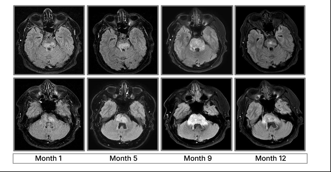
kinase 2 (LRRK2) in IDH-mutant Grade 4 astrocytomas, diagnostic challenges in Erdheim-Chester disease mimicking chronic lymphocytic inflammation with pontine perivascular enhancement responsive to steroids, and the impact of a brain death workshop on clinical knowledge. Together, these summaries reflect the breadth of work carried out in neurological care, from mechanisms of disease to practical educational interventions.
The AAN program balanced scientific advances with meaningful clinical progress, giving rise to inspiring discussions and debates
Rajiv R. Ratan
Weill Cornell Medicine, New York, USA
AAN 2026
The congress featured more than 3,500 scientific abstracts and over 190 educational programs, demonstrating the scale of the meeting and fast pace of neurological innovation
THE 2026 Annual Meeting of the American Academy of Neurology (AAN), held in Chicago, Illinois, from April 18th–22nd, brought together the global neurology community for 5 days of scientific discourse, clinical education, and discussion surrounding the advances in neurologic care. The meeting boasted a hybrid format with in-person and online participation, spanning neurodegenerative disease, stroke, neuroimmunology, epilepsy, headache medicine, movement disorders, neuro-oncology, and emerging technologies, giving those in attendance and at home plenty to explore.
Across plenary presentations, late-breaking science sessions, poster discussions, and educational forums, several recurring themes emerged: precision medicine expanding, prevention and brain health gaining traction, advances in biomarker-driven diagnosis, and the growing integration of AI and digital technologies into research and patient care.
The congress featured more than 3,500 scientific abstracts and over 190 educational programs, demonstrating the scale of the meeting and fast pace of neurological innovation. Particularly notable was the strong presence of late-breaking clinical trial data across several therapeutic areas, including Alzheimer disease, multiple sclerosis, migraine, neuromuscular disorders, and rare genetic neurologic diseases. Discussions increasingly focused not only on symptom management, but also on disease
modification, earlier intervention, and longterm quality of life outcomes.
Neuroimmunology and neurodegenerative disease remained dominant areas of interest throughout the meeting. New findings presented in multiple sclerosis explored the impact of obesity, glucagonlike peptide-1 receptor agonists, and pediatric disease management, and much more, reflecting the growing emphasis on individualized care and long-term disease monitoring in the field. Emerging data in Huntington disease, generalized myasthenia gravis, essential tremor, and stiff person syndrome highlighted the continued momentum behind targeted therapies and gene-based treatment strategies.
Another defining feature of AAN 2026 was the prominence of brain health as a common concept across subspecialties, mirroring shifts in both research priorities and patient expectations. It was no surprise that the
congress also showcased growing interest in AI predictive analytics and digital neurology tools.
The meeting maintained a strong focus on practical clinical relevance. Attendees and audience members were exposed to highprofile trial data. Many sessions explored real-world implementation challenges, healthcare disparities, telehealth integration, and multidisciplinary care delivery.
This congress review highlights a selection of the most clinically impactful studies, emerging trends, and expert perspectives presented at AAN 2026.
Neuroimmunology and neurodegenerative disease remained dominant areas of interest throughout the meeting
Stroke Rehabilitation Frequency Emerges as Key Recovery Factor
RESEARCH presented at AAN 2026 has suggested that increasing the frequency of inpatient physical therapy for people hospitalized with acute stroke may significantly reduce hospital length of stay without negatively affecting functional outcomes at discharge.1
In the retrospective BID-FAST study, investigators from Medical University of South Carolina, USA, evaluated whether implementing twice-daily physical therapy sessions during acute stroke admission could improve patient outcomes compared with the standard approach of 3–5 sessions per week. The study was designed in response to ongoing uncertainty surrounding the optimal intensity and timing of rehabilitation after stroke. While very early, intensive therapy can sometimes be poorly tolerated, the researchers proposed that shorter, more frequent sessions may improve participation and recovery during the acute phase.
The analysis included 296 patients treated across two consecutive 4-month periods at a comprehensive stroke center. The preimplementation cohort received standardfrequency physical therapy, whereas the postimplementation cohort received the BID-FAST intervention consisting of twice-daily sessions. Outcomes assessed included hospital length of stay, discharge modified Rankin Scale (mRS), and discharge destination.
Following implementation of BID-FAST, mean hospital length of stay fell from 6.83 days to 4.61 days, representing a statistically significant reduction of 2.22 days (95% CI: 0.57–3.87; p=0.0086). Importantly, this reduction was not accompanied by worse functional outcomes. Discharge mRS scores remained comparable between the groups, with averages of 2.13 in the standard-frequency cohort and 2.07 in the BID-FAST cohort. Similarly, the proportion of patients discharged home showed no meaningful difference, increasing slightly from 56.1% to 58.2%.
Discharge mRS scores remained comparable between the groups, with averages of 2.13 in the standardfrequency cohort and 2.07 in the BID-FAST cohort
The authors concluded that therapy frequency may represent an important and modifiable aspect of acute stroke rehabilitation. Although the study design does not establish causality, the findings suggest that increasing physical therapy frequency could improve healthcare efficiency by shortening admissions without compromising patient recovery. The investigators noted that prospective studies will now be needed to confirm these observations and determine whether intensified rehabilitation strategies can also improve longer-term disability outcomes after stroke.
Mean hospital length of stay fell from 6.83 days to 4.61 days
Growing Burden of Headache Disorders Among Older Adults
HEADACHE disorders affected more than half a billion older adults globally in 2021, according to new research highlighting substantial sex and socioeconomic disparities in disease burden across regions.2
The analysis examined the global epidemiology of headache disorders in adults aged ≥55 years using data from the Global Burden of Disease (GBD) study between 1990–2021. Researchers evaluated prevalence, incidence, and disability-adjusted life-years (DALY) at global, regional, and national levels, while also investigating associations with socioeconomic development using the Sociodemographic Index (SDI).
In 2021, approximately 538 million older adults worldwide were living with headache disorders, corresponding to a prevalence of 36,219 cases per 100,000 population. The USA ranked among the top three countries globally for prevalence, with 43,133 cases per 100,000 population.
Although global age-standardized rates declined slightly over the study period (estimated annual percentage change: −0.056), the absolute number of DALYs increased by 115% since 1990, indicating a substantial rise in the overall burden of headache disorders among ageing populations.
Migraine incidence increased by 1.90% during the study period, whereas tensiontype headache incidence declined by 2.73%. Female adults consistently experienced a greater burden than males across all older age groups.
Significant regional and socioeconomic disparities were also identified. Middle-SDI regions showed the greatest increase in prevalence over time, while high-SDI regions had the highest prevalence rates overall in 2021 at 39,639 cases per 100,000 population. Eastern Europe recorded the highest DALY
rates globally, suggesting a particularly severe disability burden in this region. East Asia had the greatest number of overall cases, reaching 117.9 million.
The findings underline the growing impact of headache disorders in older populations worldwide, particularly as global demographics continue to shift towards ageing societies. Researchers emphasized that headache disorders remain an underrecognized contributor to disability among older adults, despite their substantial effect on quality of life and daily functioning.
The authors concluded that integrating headache management into geriatric care pathways may help address this rising burden. They also highlighted the need for regionspecific public health strategies to reduce disparities associated with sex and SDI.
The study was based on estimates from the GBD database and may therefore reflect variations in healthcare access, diagnostic practices, and reporting quality between countries. However, the analysis provides one of the most comprehensive assessments to date of headache disorders in older adults globally.
Digital Multiple Sclerosis Quality Measures Face Major Challenges
A
NEW study has raised concerns about the feasibility of converting existing multiple sclerosis (MS) quality measures into reliable digital tools using electronic health record data.3
The retrospective cohort study evaluated the completion rate of the 2020 AAN MS quality measures among 1,101 adults with MS at a single specialist center. Participants had attended at least two clinic visits approximately 24 months apart. Investigators developed digital quality measures (dQMs) using electronic health record data and compared these with manually abstracted quality measures (mQMs) obtained through chart review in a random subgroup of 218 patients.
symptom management (8.7% versus 24%; p<0.001), cognitive screening (77% versus 88%; p<0.001), and fatigue management (2.1% versus 32%; p=0.046).
In contrast, fatigue screening rates were comparable between digital and manual measures (91% versus 89%; p=0.43), while cognitive management completion remained low across both approaches.
MRI screening completion rates were significantly lower using dQMs compared with mQMs
MS is a chronic neurological disease affecting the brain and spinal cord, often leading to progressive disability and a wide range of physical and cognitive symptoms. Quality measures are designed to standardize care and assess whether patients are receiving recommended monitoring, screening, and symptom management. The findings showed substantial differences between dQMs and mQMs for several important care metrics.
MRI screening completion rates were significantly lower using dQMs compared with mQMs (82% versus 93%; p<0.001). Similar discrepancies were observed for
Researchers also reported that several AAN quality measures could not be converted into digital measures at all because the required information was not available as structured electronic health record data. These findings suggest that current electronic systems may not adequately capture the complexity of neurological care delivered in routine clinical practice.
The authors suggested that future MS quality measures should be designed with electronic health record integration in mind, helping clinicians use digital tools more consistently in practice while ensuring care delivery is captured more accurately.
Community Stroke Rehabilitation Priorities Identified in Zambia
IMPROVING post-stroke rehabilitation in Zambia may require integrated community-based services and expanded caregiver support, according to findings presented at AAN 2026 from co-creation workshops involving stroke survivors and caregivers.
Stroke, a leading cause of long-term disability, is associated with impairments affecting mobility, communication, and cognition, yet rehabilitation access remains limited across much of Zambia.4
Researchers used a human-centered design approach to explore perceptions of potential community-based stroke rehabilitation models. Two co-creation workshops were conducted with 36 participants, including 18 stroke survivors and 18 caregivers, using facilitator-led activities such as term association exercises and journey mapping to evaluate seven possible rehabilitation services.
Participants identified several rehabilitation modalities as empowering and beneficial. Speech, physical health, mental health, and occupational therapy were consistently viewed positively. In contrast, employment retraining and social support services were less clearly understood by participants, while pastoral support generated skepticism because of concerns regarding potential corruption.
Journey mapping exercises identified several recurring themes influencing rehabilitation engagement. Participants described initial resistance to seeking support following hospital discharge, often followed by pursuit of external assistance because of social isolation or challenges managing caregiving responsibilities at home. Healthcare provider endorsement emerged as an important factor influencing willingness to engage with rehabilitation services.
Financial barriers were also consistently reported, particularly costs associated with transportation and accessing care. Caregiver training for delivery of rehabilitation at home was repeatedly prioritized. Participants emphasized the value of equipping caregivers with practical skills to support ongoing rehabilitation outside formal healthcare settings.
As a qualitative co-creation study, the findings reflect perspectives from a relatively small participant sample and may not be generalizable across all regions or healthcare settings in Zambia.
Researchers concluded that future stroke rehabilitation initiatives in Zambia should consider integrated local rehabilitation centers capable of delivering multidisciplinary services in a single setting, alongside pilot caregiver training programs designed to support in-home rehabilitation delivery. Further research could help determine how these models influence rehabilitation uptake, continuity of care, and long-term functional recovery following stroke.
Ocrelizumab Delayed Disability Progression in Progressive Multiple Sclerosis
NEW findings from the ORATORIO-HAND trial showed that ocrelizumab significantly delayed disability progression and worsening upper-limb function in patients with primary progressive multiple sclerosis (PPMS), including older patients and those with more advanced disease.5
PPMS is a chronic neurodegenerative condition characterized by steadily worsening neurological disability, often with limited treatment options. While previous studies established the benefit of ocrelizumab in delaying disease progression in PPMS, evidence in older patients and those with advanced disability has remained limited, particularly regarding preservation of hand and upper-limb function.
The Phase III ORATORIO-HAND trial evaluated the efficacy and safety of ocrelizumab compared with placebo in a broad PPMS population. Researchers enrolled adult patients aged up to 65 years with Expanded Disability Status Scale (EDSS) scores ranging from 3.0–8.0, reflecting substantial disability in many participants. Patients were randomized 1:1 to receive ocrelizumab 600 mg or placebo every 6 months for up to 144 weeks or until at least 340 progression events occurred.
The primary endpoint assessed 12-week confirmed composite disability progression, including worsening in the 9-Hole Peg Test, a measure of hand and upper-limb function, or EDSS progression.
Among 505 patients receiving ocrelizumab and 508 receiving placebo, median baseline age was 48 versus 47 years, respectively, while median EDSS score was 6.0 in both groups. Over the treatment period, confirmed disability progression occurred in 32.7% of patients receiving ocrelizumab, compared with 40.4% receiving placebo, corresponding to a 30% risk reduction (HR: 0.70; 95% CI: 0.57–0.86; p=0.0007).
Ocrelizumab also significantly reduced upperlimb disability worsening. Progression based on the 9-Hole Peg Test occurred in 16.7% of ocrelizumab-treated patients versus 24.9% of placebo-treated patients, representing a 41% risk reduction (p=0.0002). EDSS-confirmed progression was similarly reduced by 33% (p=0.0013).
Over the treatment period, confirmed disability progression occurred in 32.7% of patients receiving ocrelizumab
Benefits appeared even greater in patients with MRI-active disease. In this subgroup, ocrelizumab reduced the risk of confirmed disability progression by 55% (HR: 0.45; 95% CI: 0.31–0.64; p<0.0001), alongside substantial reductions in hand function and EDSS worsening.
The safety profile of ocrelizumab was broadly consistent with previous studies. Infusionrelated reactions occurred more frequently with ocrelizumab than placebo (20.8% versus 4.3%), while infections were reported in 48.4% and 44.7% of patients, respectively.
These findings suggest that ocrelizumab may provide clinically meaningful benefits in a wider PPMS population than previously established, including patients with more advanced disability and older age.
Sleep and Anxiety May Shape Persisting PostConcussive Symptom Severity
PERSISTENT post-concussive symptoms (PPCS) may be shaped not only by injury severity, but also by anxiety, resilience, and sleep quality, according to new research presented at AAN 2026.6
Post-concussive symptoms can include headaches, fatigue, dizziness, memory difficulties, and emotional changes that continue for more than 3 months after head injury.
Researchers examined whether psychological and physiological factors independently predicted symptom burden in 30 people enrolled in a clinical trial investigating neuromodulation and recovery. These participants had a high burden of PPCS, defined as a score of ≥20 on the Rivermead Post Concussion Symptom Questionnaire at least 3 months after injury.
Participants completed a range of psychosocial and sleep assessments, while sleep was also tracked over 1 week using the Oura Ring (Oura Health, Oulu, Finland), a wearable device designed to monitor sleep and physiological patterns. Wearable monitoring may provide a practical way to
assess sleep in real-world settings, but it offers less detail than laboratory-based sleep studies.
The findings showed that anxiety and resilience independently predicted symptom burden, supporting previous research in PPCS. When sleep measures were added to the analysis, anxiety, objective sleep efficiency, and subjective sleep impairment together explained 65% of the variation in post-concussion symptom burden.
Poor sleep efficiency was linked with greater symptom burden. Researchers suggest that these findings could be implemented to help clinicians better identify patients who may benefit from more individualized management approaches.
As the small cohort of participants were all already experiencing a high burden of symptoms, this may limit how broadly the findings apply. In addition, wearable sleep tracking provides indirect estimates of sleep quality rather than comprehensive clinical sleep assessment.
Still, the findings suggest that targeting anxiety, sleep impairment, and resilience simultaneously could improve care strategies for people with PPCS. The incorporation of wearable physiological monitoring into post-injury assessment could also support more personalized approaches to recovery in future studies.
Anxiety, objective sleep efficiency, and subjective sleep impairment together explained 65% of the variation in post-concussion symptom burden
Machine Learning Model Highlights Institutional Influence on WLST Decisions
A
MACHINE learning (ML) analysis of more than 155,000 patients with severe traumatic brain injury (TBI) found that institutional withdrawal of lifesustaining therapy (WLST) rates were among the strongest predictors of whether care would be withdrawn, independent of patient clinical condition. The findings suggest that local treatment culture may significantly influence WLST decisions and raise concerns about potential self-fulfilling prognostic bias in severe TBI care.7
Investigators developed ML models to predict WLST in patients with severe TBI using data from the American College of Surgeons Trauma Quality Improvement Program National Trauma Databank between 2017–2021. The researchers aimed to identify the most influential determinants of WLST and hypothesized that facility-specific WLST rates would emerge as a major predictive factor.
The observational study included patients with severe TBI defined by an Abbreviated Injury Scale (AIS)-Head score ≥1 and presenting Glasgow Coma Scale (GCS) score <9. Of more than 5.4 million trauma records screened, 155,639 patients met inclusion criteria. Among these, 32,385 patients (20.8%) underwent WLST. Mean patient age was 43 years, 26.5% were female, and the median time to WLST was 46.4 hours after presentation.
ML models were trained using variables available at different stages of admission and hospitalization. Predictive performance was high across all models, with area under the receiver operating characteristic curve values ranging from 0.875 for the admission model to 0.896 for the total length-of-stay model.
Shapley additive explanation analysis identified age, highest emergency department GCS score, and facility WLST rate as the most influential predictors of WLST decisions. The strong impact of institutional WLST rates suggested that local practice patterns and withdrawal culture may independently shape decision-making
beyond objective measures of neurological injury severity.
The findings suggest that local treatment culture may significantly influence WLST decisions and raise concerns about potential selffulfilling prognostic bias in severe TBI care
The authors concluded that ML models can reliably predict WLST decisions in severe TBI, but the findings also highlight the need for more refined prognostic tools and greater awareness of potential institutional bias. As outcomes in severe TBI continue to improve, the researchers warned that premature WLST decisions influenced by pessimistic prognostication or center-specific culture may contribute to avoidable mortality.
Digital Dashboard Tracks Inpatient Neurology Care Quality
RESEARCHERS have developed a digital inpatient neurology dashboard that integrates electronic health records, billing data, and patient satisfaction metrics, enabling real-time tracking of quality indicators and supporting measurable improvements in hospital care delivery.8
Stroke-related neurology quality measures are routinely monitored because of established stroke center requirements, but equivalent standards for general inpatient neurology remain less developed. Limited program funding has also restricted large-scale quality initiatives. Investigators therefore sought to create a cost-conscious quality dashboard capable of tracking inpatient neurology performance indicators and supporting targeted improvement projects.
This healthcare quality improvement initiative was conducted between 2023–2025 by a core inpatient general neurology committee working alongside a health system Clinical Data Operations team. Researchers reviewed neurohospitalist literature to identify inpatient quality gaps and selected key performance indicators for monitoring. Data were extracted from three primary sources: Epic electronic health records, MSX billing and claims data for readmissions, and HCAHPS patient satisfaction surveys. Information was integrated into the Tableau visual analytics platform to create a dashboard capable of filtering results by date range, location, inpatient service, and provider.
The neurology dashboard continuously integrated new data and enabled visualization of longitudinal trends across inpatient services. One initial quality improvement project using dashboard data demonstrated a 20% improvement in medication reconciliation within 24 hours of hospital admission following an inpatient education intervention. Additional tracked indicators included MRI timing, readmission patterns, patient satisfaction scores, and rapid EEG utilization.
The researchers concluded that modern healthcare systems generate large volumes of underused clinical data, and that these data can support meaningful improvements in patient care when organized into accessible analytics platforms. They suggested that broader implementation of inpatient neurology dashboards may facilitate development of standardized quality indicators while supporting fiscally sustainable quality improvement initiatives across healthcare systems.
Care Partners of Anoxic Brain Injury Survivors Face Lasting Trauma
CAREGIVERS of survivors with severe anoxic brain injury experience high rates of prolonged grief, post-traumatic stress, and conflict with healthcare teams months after injury, according to new research highlighting the hidden psychological burden carried by care partners after cardiac arrest.9
Researchers investigated the emotional and practical challenges faced by surrogates caring for survivors with severe neurological impairment following anoxic brain injury. While previous studies have largely focused on patients with favorable neurological recovery, little is known about families supporting survivors with poor functional outcomes, chronic disability, or dependence on life-sustaining therapies.
The study enrolled care partners of severe anoxic brain injury survivors who had significant disability at discharge, radiological evidence of anoxic injury, or ongoing life-sustaining treatment requirements. Participants completed validated surveys assessing symptom burden, quality of life, grief, depression, anxiety, post-traumatic stress, caregiver burden, and advance care planning. Semi-structured interviews were also conducted to explore unmet care needs and experiences with healthcare systems.
Overall, 26 care partners representing 20 survivors completed 54 surveys and 55 interviews across inpatient and nursing home settings. Participants had a mean age of 57 years, 69% were female, and 38% identified as Black.
At 6 months after injury, psychological distress remained common. Investigators reported prolonged grief in 39% of care partners and post-traumatic stress symptoms in 56%. Many participants also described substantial caregiver burden and persistent disagreement with healthcare staff regarding goals of care, with 59%
reporting conflict around decisions relating to survivorship and treatment.
Participants reported experiencing complex grief linked not only to fear of death, but also to the profound personality and functional changes experienced by survivors
Qualitative analysis identified four major themes emerging from interviews. Care partners frequently perceived conflicting values and staff bias regarding prognosis and treatment decisions. Many expressed a strong desire for more meaningful relationships and communication with healthcare professionals. Lasting distrust of healthcare systems was also commonly described, particularly following difficult decision-making experiences. Finally, participants reported experiencing complex grief linked not only to fear of death, but also to the profound personality and functional changes experienced by survivors.
The authors concluded that care partners of severe anoxic brain injury survivors face enduring and under-recognized psychological sequelae long after hospital discharge. They emphasized the need for targeted clinical interventions, improved communication strategies, and greater psychosocial support to better address caregiver trauma and reduce conflict during long-term survivorship care.
References
1. Vorobyev A et al. High-frequency PT in acute stroke inpatients: implementation and early outcomes (BID-FAST). Abstract S17.001. AAN Annual Meeting, April 18-22, 2026.
2. Sohail A et al. Global burden of headache disorders in older adults (aged ≥55 years) from 1990–2021: an analysis of epidemiology, trends, and socioeconomic disparities. Presentation 009. AAN Annual Meeting, April 18-22, 2026.
3. Tidd S et al. Completion rate and feasibility of converting AAN multiple sclerosis quality measures to digital measures. Presentation S12.009. AAN Annual Meeting, April 18-22, 2026.
4. Nair S et al. A human-centered design approach to implementing communitybased stroke rehabilitation services in Lusaka Zambia. Abstract 61772. AAN Annual Meeting, April 18-22, 2026.
5. Giovannoni G et al. ORATORIOHAND: results of the primary analysis of ocrelizumab versus placebo in primary progressive multiple sclerosis, including older patients and those with more advanced disease. Neurology. 2026;DOI:10.1212/ WNL.0000000000215551.
6. Ismail R et al. Subjective and objective sleep quality independently predict persistent post-concussion symptoms. Abstract S34.001. AAN Annual Meeting, April 18-22, 2026.
7. Cobler-Lichter M et al. Machine learning models to predict withdrawal of lifesustaining therapy in patients with severe traumatic brain injury. Abstract 61618. AAN Annual Meeting, April 18-22, 2026.
8. Raval B et al. Improving quality for inpatient neurology: customizing a dynamic neurology-specific key performance indicators dashboard leveraging health system data streams technology. Abstract 61753. AAN Annual Meeting, April 18-22, 2026.
9. Huang A et al. Hidden from view: assessing psychological sequelae of carepartners of anoxic brain injury survivors. Poster P1. AAN Annual Meeting, April 18-22, 2026.
Updates in Multiple Sclerosis: AAN Annual Meeting 2026
Authors: *Andrew Dugue,1 Tyler Ellis Smith1
1. Department of Neurology, NYU Grossman School of Medicine, New York City, USA
*Correspondence to Andrew.Dugue@nyulangone.org
Disclosure:
Keywords:
Smith has received research funding from PCORI and is set to receive research funding from Neurogenesis; is set to receive royalties from Essential Neurology Board Review Q&A, a book published through Springer; and has participated on the TREAT-MS Trial Study Advisory Committee, funded by PCORI. Dugue has declared no conflicts of interest.
ACROSS the spectrum of multiple sclerosis (MS) care, the American Academy of Neurology (AAN) Annual Meeting 2026 highlighted changes in the diagnosis of MS, impacting both asymptomatic and symptomatic patients. Additionally, a new mechanism of action for the treatment of MS, targeting Bruton’s tyrosine kinase (BTK), showed positive results in both progressive and relapsing MS. Together, these changes, along with updates in biomarkers and upcoming studies on the approach to treatment of MS, underscore the rapid state of change in MS care and opportunities for patients.
INTRODUCTION
Aaron Miller, Icahn School of Medicine at Mount Sinai, New York, USA, reviewed the latest 2024 McDonald Criteria, noting that it was developed through a consensus methodology. The 2024 McDonald Criteria incorporates clinical and radiographic data, as well as biomarkers, to make the diagnosis of MS earlier and in more individuals. Miller emphasized that these criteria maintain accuracy to ultimately reduce the burden of disability among individuals with MS.1
Highlighting a key change to the McDonald Criteria, Laura Balcer, NYU Grossman School
of Medicine, New York, USA, presented the inclusion of the optic nerve as the fifth topographical region when evaluating for dissemination in space. Both asymptomatic and symptomatic optic nerve lesions are now included and can be detected with orbital MRI with gadolinium, and optical coherence tomography, along with visual evoked potentials (Table 1).
The addition of iron-sensitive MRI techniques to detect the presence of a central vein sign (CVS) and paramagnetic rim lesions (PRL) in the 2024 Revised McDonald Criteria were reviewed. The CVS enables the identification of white matter lesions with a central vein,
Significant intereye difference in OCT peripapillary RNFL and/or macular GC-IPL. At least 6 µm peripapillary RNFL, at least 4 µm macular GC-IPL.
Delayed P100 latency on visual evoked potentials.
Presence of optic nerve T2 hyperintensity and/or gadolinium enhancement on MRI orbits.
which has demonstrated high sensitivity and specificity for MS, when using the 'Select 6' method to identify a sufficient number of CVSs to be consistent with MS. PRLs are thought to indicate chronic inflammation in demyelinating lesions, related to iron-laden macrophages and microglia at the lesion edge. While PRLs have not been as sensitive for the diagnosis of MS as a CVS, they remain highly specific. Together, these biomarkers can increase diagnostic specificity for MS.
In addition to substituting oligoclonal bands unique to the cerebrospinal fluid to fulfil the dissemination in time criteria (present in the 2017 McDonald Criteria), the updated MS diagnostic criteria also allow for the use of kappa free light chains to substitute for dissemination in time. Balcer highlighted the advantages of kappa free light chains: high concordance with oligoclonal bands, while remaining rater-independent. Furthermore, if sufficient criteria are met, dissemination in
Table 1: Paraclinical test criteria for optic nerve lesions.
time is no longer required to diagnose MS in the 2024 McDonald Criteria.
The updated criteria also renovate the diagnosis of asymptomatic patients. As discussed by Miller and Jiwon Oh, University of Toronto, Canada, individuals who would have been diagnosed with radiologically isolated syndrome under the prior criteria will now be diagnosed with MS if they meet the appropriate criteria, despite being asymptomatic.
Miller and Oh both emphasized the importance of applying these latest criteria in an individual with a typical syndrome (such as unilateral optic neuritis, partial myelopathy, focal supratentorial, brainstem, or cerebellar syndrome), and to use caution when examining atypical presentations (e.g., encephalopathy, isolated fatigue). While these updated criteria can be applied across diverse situations, Oh emphasized the need to confirm additional features of MS, such as spinal cord lesions, presence of oligoclonal bands and/or kappa free light chains, and the CVS, in individuals with a higher chance of misdiagnosis (e.g., older patients, younger patients, and those with vascular and headache comorbidities).
Validation of the updated criteria was reviewed by Miller, who presented early data from Brownlee et al.3 demonstrating a substantial increase (28%) in the ability of the 2024 McDonald Criteria to diagnose MS with high accuracy, when compared to the 2017 criteria.
Importantly, individuals who met the prior McDonald Criteria4 will continue to maintain the diagnosis of MS under the 2024 criteria. The latest McDonald Criteria also maintain that the presentation must not be better explained by another disorder. For further reading on a consensus approach to the differential diagnosis of MS, see Solomon et al.5
Ludwig Kappos, University Hospital Basel, Switzerland, presented his thoughts on the inclusion of a spectrum of biomarkers in trials, including blood-derived biomarkers
(neurofilament light chain, glial fibrillary acidic protein), imaging biomarkers (quantitative MRI, optical coherence tomography, PET), and digital biomarkers. Additionally, Kappos highlighted the role of progression in MS and advocated for the inclusion of progression independent of relapse activity as a clinical endpoint. Gloria von Geldern, University of Washington School of Medicine, Seattle, USA, elaborated on the concept of progression independent of relapse activity, characterized by “microglial activation and dysfunctional astrocytes, ... [resulting] in [central nervous system]-centric inflammation and neurodegeneration” and leading to a progressive decline in symptoms and function without distinct relapse activity, that has characterized the endpoints of trials focused on relapsing MS.
Annette Wundes, University of Washington School of Medicine, Seattle, USA, presented defective oligodendrocyte progenitor cell recruitment as the primary reason that remyelination fails in chronic MS. Remyelination trials are ongoing to look at exercise and vagus nerve stimulation, as well a number of molecules that may be helpful in affecting remyelination.
TREATMENTS
Veronica Cipriani, University of Chicago Medicine, Illinois, USA, presented the importance of high efficacy disease-modifying therapy (DMT) in preventing irreversible disability and decreasing the number of relapses, MRI activity, and atrophy. Based on work by Langer-Gould et al.,6 Cipriani highlighted some key risk factors for longterm disability, including bowel and bladder symptoms at onset, incomplete recovery from first relapse, short interval between first and second relapses, early accumulation of disability, and Black or Hispanic race/ ethnicity (Table 2). In addition to preventing relapses, Cipriani presented data supporting the association of higher efficacy treatments with a delay to the development of secondary progressive MS.
Table 2: Risk factors associated with long-term disability.
Bowel and bladder symptoms at onset
Incomplete recovery from first relapse
Short interval between first and second relapses
Early accumulation of disability
'Black or Hispanic race/ethnicity'
Adapted from Langer-Gould et al.6
Leslie Benson, Harvard Medical School, Boston, Massachusetts, USA; and Vaishnavi Vaidyanathan, University of California Davis School of Medicine, Sacramento, USA, similarly recommended initial treatment with high-efficacy DMT in pediatric-onset MS (POMS). Vaidyanathan noted that patients with POMS are particularly prone to delays in treatment initiation. She remarked that patients with POMS with the highest risk of disability at 9 years included those with optic nerve, brainstem, and cervical cord lesions, and those with both two new brain T2 lesions and an Expanded Disability Status Scale (EDSS) change in the first 2 years. Benson noted that the PARADIGMS trial led to fingolimod FDA approval in POMS, although potential adverse effects, such as seizures, first dose bradycardia, macular edema, infections, and rebound disease activity with discontinuation, can occur. She cited ongoing trials examining high efficacy DMT in POMS, such as OPERETTA 2 (ocrelizumab) and NEOS (ofatumumab). Elizabeth Wilson, University of Cincinnati College of Medicine, Ohio, USA, highlighted non-pharmacologic treatments in pediatric MS, such as cognitive therapy, cognitive behavioral therapy, the modified Story Memory Technique, aerobic
and resistance exercise training multiple days per week, and early referral to physical medicine and rehabilitation. She stressed the importance of baseline and subsequent annual cognitive screening with tools such as the Symbol Digit Modalities Test in management.
Marwa Kaisey, Cedars-Sinai Medical Center, Los Angeles, California, USA, emphasized using an individualized and shared decision-making approach to DMT selection, incorporating pregnancy planning, side effects, method of drug delivery (e.g., intravenous, injection, oral), insurance coverage, efficacy, lifestyle, and comorbidities, as there is not a standardized protocol for DMT selection at this time.
Ruth Dobson, Queen Mary University of London, UK, presented the final analyses of the MINORE and SOPRANINO trials, which examined the humoral vaccine responses and 1-year follow-up of infants potentially exposed to ocrelizumab during pregnancy (ocrelizumab administered at 0–3 months before last menstrual period or during first trimester, n=16) and breastfeeding. MINORE demonstrated minimal placental transfer of ocrelizumab, normal infant B cell levels at
6 weeks and 13 months of life, and similar positive seroprotective vaccine response to infants not exposed to ocrelizumab. SOPRANINO (n=11) demonstrated minimal transfer of ocrelizumab in breastmilk, normal infant B cell levels at 30 days and 12 months post maternal ocrelizumab infusion, and positive humoral vaccine responses in line with reported rates.
Oh and Amit Bar-Or, Perelman School of Medicine, Philadelphia, Pennsylvania, USA, presented the results of two studies of fenebrutinib, a BTK inhibitor. Bar-Or reviewed the primary results of the Phase III FENtrepid study, comparing the efficacy and safety of fenebrutinib versus ocrelizumab in primary progressive MS. In this study, fenebrutinib demonstrated non-inferiority, with a primary endpoint of composite confirmed disability progression at 12 weeks compared to ocrelizumab. Fenebrutinib had a higher incidence of fatalities compared to ocrelizumab, but these fatal events were thought to be unrelated to fenebrutinib by the investigators. In relapsing MS, Oh presented the results of the FENhance 1 and 2 Phase III trials, evaluating the efficacy and safety of fenebrutinib relative to teriflunomide. The primary endpoint in FENhance 1 and 2 was annualized relapse rate, and
fenebrutinib demonstrated superiority versus teriflunomide in reducing relapses and disease activity on MRI. Similar to the FENtrepid study, FENhance 1 and 2 identified an imbalance of fatalities with fenebrutinib, although only two of seven were thought to be related to fenebrutinib. While both FENtrepid and FENhance demonstrated an increased rate of liver enzyme elevations with fenebrutinib, all were reversible.
FUTURE DIRECTIONS
Highlighting the difficulty in treating progressive MS, Kaisey projected excitement regarding BTK inhibitors that may penetrate the central nervous system to target inflammation localized to the area as an untapped treatment target. As one of these novel BTK inhibitors, if fenebrutinib is approved, it will add a novel mechanism of action to the MS treatment armamentarium, and will be only the second DMT to demonstrate efficacy in the treatment of primary progressive MS. Other potential therapies under study include CAR-T cell therapy, hematopoietic stem cell transplant, anti-CD40L, anti-CD3, anti-CD20 'brain shuttle', and mRNA vaccines targeting Epstein-Barr virus.
Daniel Kurz, University of Chicago Medicine, Illinois, USA, among others, reported on neurofilament light chain as a potential biomarker, but caution is advised in older individuals, and BMI and other forms of neurologic disease or injury must be considered, as these can also cause changes to the neurofilament light chain level.
Cipriani highlighted two ongoing trials, TREAT-MS and DELIVER-MS, the results of which will hopefully provide evidence on the best approach to select a DMT: starting with a lower-efficacy treatment and escalating versus starting with a high-efficacy DMT.
References
1. Montalban X et al. Diagnosis of multiple sclerosis: 2024 revisions of the McDonald criteria. Lancet Neurol. 2025;24(10):850-65. Erratum in: Lancet Neurol. 2025;24(11):e13.
2. Dugue A. The 2024 revised McDonald Criteria: AAN 2025 highlights. Neurol AMJ. 2025;2(1):26-9.
3. Brownlee WJ et al. Performance of the 2024 McDonald Criteria in patients under evaluation for suspected
CONCLUSION
In the world of MS, the past year has been an exciting one. We saw the publication of the latest McDonald Criteria, the use of the CVS, PRLs, kappa free light chains, and the recategorization of radiologically isolated syndrome as MS in some cases. Additionally, there were positive results for fenebrutinib, a BTK inhibitor, in both relapsing and primary progressive MS trials, which will potentially bring a new mechanism of action to the DMTs available to treat MS. There are ongoing trials targeting progressive MS to prevent disability. Finally, there are innovative trials targeting repair and remyelination, the next frontier in managing the disease.
4. Thompson AJ et al. Diagnosis of multiple sclerosis: 2017 revisions of the McDonald criteria. Lancet Neurol. 2018;17(2):162-73.
5. Solomon AJ et al. Differential diagnosis of suspected multiple sclerosis: an updated consensus approach. Lancet Neurol. 2023;22(8):750-68.
6. Langer-Gould A et al. Clinical and demographic predictors of longterm disability in patients with relapsing-remitting multiple sclerosis: a systematic review. Arch Neurol. 2006;63(12):1686-91.
AAN 2026
Abstract Reviews
This collection of abstract reviews brings together research presented at the American Academy of Neurology (AAN) Annual Meeting 2026. Ranging from multiple sclerosis, migraine, and sleep medicine to neuro-oncology, rare neuroinflammatory mimics, and neurologic education, these summaries reflect the breadth of disease-area research and approaches to improving clinical knowledge highlighted at the conference.
Disentangling Age-Related and Disease-Specific Upper Cervical Cord Atrophy in Multiple Sclerosis
Authors: Maria A. Rocca,1-3 Kshiteeja Jain,1 Loredana Storelli,1 Paola Valsasina,1 Paolo Preziosa,1-3 Alessandro Meani,1 Patrizia Pantano,4,5 Claudia Piervincenzi,4 Antonio Gallo,6 Alessandro D’Ambrosio,6 Nicola De Stefano,7 Rosa Cortese,7 *Massimo Filippi1-3,8,9
1. Neuroimaging Research Unit, Division of Neuroscience, IRCCS San Raffaele Scientific Institute, Milan, Italy
2. Neurology Unit, IRCCS San Raffaele Scientific Institute, Milan, Italy
3. Vita-Salute San Raffaele University, Milan, Italy
4. Department of Human Neuroscience, Sapienza University, Rome, Italy
5. IRCCS Neuromed, Pozzilli, Italy
6. Department of Advanced Medical and Surgical Sciences, University of Campania “Luigi Vanvitelli”, Naples, Italy
7. Department of Medicine, Surgery and Neuroscience, University of Siena, Italy
8. Neurorehabilitation Unit, IRCCS San Raffaele Scientific Institute, Milan, Italy
9. Neurophysiology Service, IRCCS San Raffaele Scientific Institute, Milan, Italy
*Correspondence to filippi.massimo@hsr.it
Disclosure: To view the disclosure click here
Acknowledgements: This study was supported by Fondazione Italiana Sclerosi Multipla (FISM) cod. 2025/S/2 and financed or co-financed with the ‘5 per mille’ public funding.
Spinal cord atrophy has been consistently reported in multiple sclerosis (MS), and represents a key predictor of clinical disability and disease progression.1-3 Mean upper cervical cord area (MUCCA), i.e., cross-sectional area measured at the level of the first two cervical segments, has
proven to be a reliable surrogate marker of cord atrophy measurement in both early and late MS.4 However, the distinct contributions of physiological aging versus MS-related neurodegeneration5 remain poorly characterized. The aim of this study1 was to model aging trajectories of upper cervical cord atrophy in healthy controls (HC) and subsequently identify MS-specific patterns of spinal cord atrophy in a large, multicenter cohort.
MATERIALS AND METHODS
The authors analyzed 3D T1-weighted brain MRIs and clinical data from 480 HC and 1,295 patients with MS (ages: 18–70 years). MUCCA was measured between the C1 and C2/3 intervertebral discs using the active surface method and normalized for head size (nMUCCA). Aging trajectories of nMUCCA were modelled in HC using a polynomial regression model accounting for age, agesquared, sex, scanner, and interaction terms. This model was then applied to the MS cohort generating nMUCCA Z-scores, capturing disease-specific atrophy beyond normal aging. Effect of sex and age at onset (pediatric-onset <18 years, adult-onset 18–49 years, late-onset ≥50 years) on Z-scores age dependence was evaluated.
RESULTS
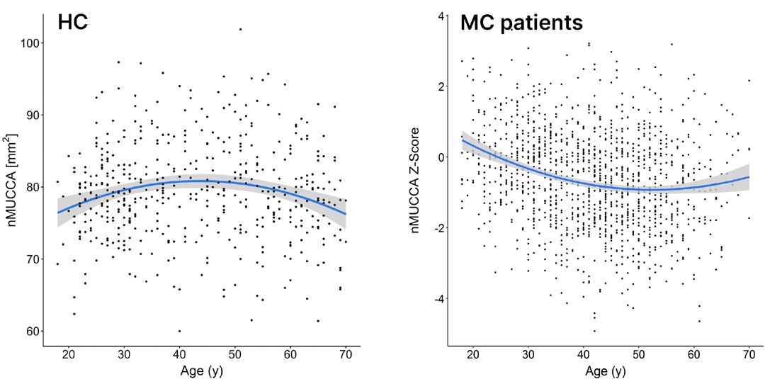
In HC, nMUCCA showed a non-linear age relationship, increasing until late 30s (p≤0.033) and subsequently declining after age 50 years (p≤0.008; Figure 1). In patients with MS, nMUCCA Z-scores exhibited a nonlinear decline with age: a steep reduction was observed in early adulthood, while decline attenuated up to late 40s (p<0.001); deviations from age-expected values
BAPatients with MS
Lifespan average nMUCCA and nMUCCA Z-score trajectories in HC and patients with MS.
A) Sex- and scanner-adjusted estimated lifespan trajectory of nMUCCA in HC, illustrating the significant non-linear relationship with age. B) Non-linear nMUCCA Z-score trajectory with age in patients with MS. The shaded gray area represents the 95% CI.
HC: healthy control; MS: multiple sclerosis; nMUCCA: normalized mean upper cervical cord area; y: years.
decreased in later decades (Figure 1). Sex did not significantly influence atrophy patterns in either HC (p=0.256) or patients with MS (p=0.422). Patients with pediatric-onset MS exhibited significantly lower Z-scores compared to adult-onset MS and late-onset MS (p<0.001), although MS-driven decline with age did not differ among onset groups. Lower Z-scores were associated with higher disability and worse structural MRI measures (rho range=−0.355–0.372, all p<0.001).
CONCLUSION
In MS, upper cervical cord atrophy exceeded effects of normal aging, especially during
early adulthood and midlife, suggesting that the relative contribution of normal aging increased with age, while disease-specific atrophy predominated early, independent of sex. Differences across onset groups largely reflected disease duration. Upper cord atrophy was associated with disability, supporting its role as a sensitive biomarker of MS-related neurodegeneration beyond aging.
References
1. Filippi M et al. Disentangling age-related and diseasespecific upper cervical cord atrophy in multiple sclerosis. Abstract S40.006. AAN Annual Meeting, April 18-22, 2026.
Figure 1: Cord age.
nMUCCA Z-score
2. Rocca MA et al. Clinically relevant cranio-caudal patterns of cervical cord atrophy evolution in MS. Neurology 2019;93(20):e1852-66.
3. Moccia M et al. Advances in spinal cord imaging in multiple sclerosis. Ther Adv Neurol Disord 2019;12:1756286419840593.
4. Weeda MM et al. Validation of mean upper cervical cord area (MUCCA) measurement techniques in multiple sclerosis (MS): high reproducibility and robustness to lesions, but large software and scanner effects. Neuroimage Clin. 2019;24:101962.
5. Filippi M et al. The ageing central nervous system in multiple sclerosis: the imaging perspective. Brain. 2024;147:3665-80.
Aerobic Training and Potential Neurogenesis in Progressive Multiple Sclerosis: Focus on the Hippocampus
and the Subventricular Zone
Authors: Tetsu Morozumi,1 Paola Valsasina,1 Paolo
Preziosa,1-3 Alessandro Meani,1 Robert W. Motl,4
Maria Pia Amato,5,6 Giampaolo Brichetto,7,8 Daniele
Boccia,9 Jeremy Chataway,10,11 Nancy Chiaravalloti,12,13 Gary Cutter,14 Ulrik Dalgas,15 John DeLuca,12,13 Rachel Farrell,10 Peter Feys,16,17
Jenny Freeman,18 Matilde Inglese,9,19 Cecilia Meza,20,21 Amber Salter,22 Brian M. Sandroff,12,13
Anthony Feinstein,20,21 Massimo Filippi,1-3,23,24
*Maria A. Rocca,1-3 CogEx Research Team
1. Neuroimaging Research Unit, Division of Neuroscience, IRCCS San Raffaele Scientific Institute, Milan, Italy
2. Neurology Unit, IRCCS San Raffaele Scientific Institute, Milan, Italy
3. Vita-Salute San Raffaele University, Milan, Italy
4. Department of Kinesiology and Nutrition, University of Illinois Chicago, Illinois, USA
5. Department NEUROFARBA, Section Neurosciences, University of Florence, Italy
6. IRCCS Fondazione Don Carlo Gnocchi, Florence, Italy
7. Scientific Research Area, Italian Multiple Sclerosis Foundation (FISM), Genoa, Italy
8. AISM Rehabilitation Service, Italian Multiple Sclerosis Society, Genoa, Italy
9. Department of Neuroscience, Rehabilitation, Ophthalmology, Genetics, Maternal and Child Health, and Center of Excellence for Biomedical Research, University of Genoa, Italy
10. Department of Neuroinflammation, Queen Square Multiple Sclerosis Centre, UCL Queen Square Institute of Neurology, Faculty of Brain Sciences, University College London, UK
11. National Institute for Health Research, University College London Hospitals, Biomedical Research Centre, UK
12. Kessler Foundation, West Orange, New Jersey, USA
13. Department of Physical Medicine & Rehabilitation, Rutgers New Jersey Medical School, Newark, USA
14. Department of Biostatistics, University of Alabama at Birmingham, USA
15. Exercise Biology, Department of Public Health, Aarhus University, Denmark
16. REVAL, Faculty of Rehabilitation Sciences, Hasselt University, Diepenbeek, Belgium
17. UMSC Hasselt, Pelt, Belgium
18. Faculty of Health, School of Health Professions, University of Plymouth, Devon, UK
19. IRCCS Ospedale Policlinico San Martino, Genoa, Italy
20. Department of Psychiatry, University of Toronto, Canada
21. Sunnybrook Health Sciences Centre, Toronto, Canada
22. Department of Neurology, Section on Statistical Planning and Analysis, UT Southwestern Medical Center, Dallas, Texas, USA
23. Neurorehabilitation Unit, IRCCS San Raffaele Scientific Institute, Milan, Italy
24. Neurophysiology Service, IRCCS San Raffaele Scientific Institute, Milan, Italy
*Correspondence to rocca.mara@hsr.it
Disclosure: To view the disclosure click here
Acknowledgements: The study received support from the MS Society of Canada (EGID3185). Ancillary funding was received from the CMSC, Danish MS Society, and US MS Society.
Hippocampal damage is frequent in patients with multiple sclerosis (MS), and is clinically significant due to its impact on cognitive function and mood disorders.1 Also, damage of the subventricular zone (SVZ) has been associated with impaired processing speed in patients with MS.2 Neurogenesis may occur in the dentate gyrus (DG) of the hippocampus and the SVZ throughout the whole lifespan.3,4 In patients with MS, aerobic training may exert a neuroprotective role on the brain by stimulating neurogenesis in DG and SVZ.5
The aim of this study6 was to assess the effects of aerobic exercise on the volume of the hippocampus and its subfields and on diffusivity measures of the SVZ in patients with progressive MS.
MATERIALS AND METHODS
The authors retrospectively analyzed data from 84 patients with progressive MS enrolled at four sites participating in the CogEx MRI substudy.7 Thirty-nine patients with MS performed aerobic training, while 45 patients undertook a balance- and stretching-based sham exercise intervention. Both groups trained twice weekly for 12 weeks. At baseline, post-intervention, and 6-month follow-up, patients underwent MRI assessment on a 3.0 T scanner. FreeSurfer’s (CorTechs Labs, Inc. [now Cortechs.ai], San Diego, California; and The General Hospital Corporation doing business as Massachusetts General Hospital, Boston, Massachusetts, USA) longitudinal processing stream was used to analyze hippocampal
subfields’ volumes. Fractional anisotropy and mean diffusivity were extracted from the SVZ and the thalamus, serving as control region.
RESULTS
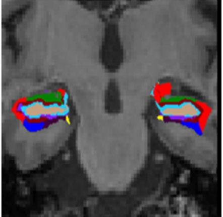
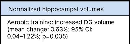
There were no differences between the two groups in the assessed variables at baseline (p≥0.070). The DG showed a significant volume increase post-intervention in the aerobic exercise group (mean change: 0.63%; 95% CI: 0.04–1.22%; p=0.035; Figure 1), but not in the sham group (mean change: −0.26%; 95% CI: −0.81–0.28%; p=0.337). DG volume increase was significantly greater in the aerobic group compared to the sham group (p=0.029).
Figure 1: Hippocampal subfield segmentation and results of longitudinal analysis.
Results of hippocampal subfield segmentation using FreeSurfer (CorTechs Labs, Inc. [now Cortechs.ai], San Diego, California; and The General Hospital Corporation doing business as Massachusetts General Hospital, Boston, Massachusetts, USA) longitudinal processing, along with significant post-intervention volumetric changes observed in patients with multiple sclerosis undergoing aerobic training.
DG: dentate gyrus.
No significant volumetric changes were found in the other hippocampal subfields, nor in the diffusivity metrics of the SVZ (p≥0.057). No significant changes in either group were found between end of training and the 6-month follow-up (p≥0.061).
CONCLUSION
Aerobic exercise increased DG volume in patients with progressive MS, but did not modify microstructural integrity of the SVZ. The possible neuroprotective effect of aerobic training is particularly relevant, since the hippocampus is a common site of atrophy in MS, contributing to cognitive deficits experienced by these patients.
References
1. Preziosa P et al. Structural MRI correlates of cognitive impairment in patients with multiple sclerosis: a multicenter study. Hum Brain Mapp. 2016;37(4):1627-44.
2. Butti E et al. Neural precursor cells tune striatal connectivity through the release of IGFBPL1. Nat Commun. 2022;13(1):7579.
3. Christian KM et al. Adult neurogenesis and the dentate gyrus: predicting function from form. Behav Brain Res. 2020;379:112346.
4. David-Bercholz J et al. Astrocyte and oligodendrocyte responses from the subventricular zone after injury. Front Cell Neurosci. 2021;15:797553.
5. Erickson KI et al. Exercise training increases size of hippocampus and improves memory. Proc Natl Acad Sci U S A. 2011;108(7):3017-22.
6. Morozumi T et al. Aerobic training and potential neurogenesis in progressive multiple sclerosis: focus on the hippocampus and the subventricular zone. Abstract 006. AAN Annual Meeting, April 18-22, 2026.
7. Rocca MA et al. Cognitive rehabilitation effects on grey matter volume and Go-NoGo activity in progressive multiple sclerosis: results from the CogEx trial. J Neurol Neurosurg Psychiatry. 2024;95:1139-49.
The Effect of Vestibular Rehabilitation in the Management of Vestibular Migraine in Adults: A Systematic Review and
Authors: *Bradley Ong,1 Jad El Ahdab,2 Marina Vilardo,3 Nicolas R. Thompson,4 Neil Nero,5 Ahmet Günkan,6 Neil Cherian,7 Julia Bucklan7
1. Sleep Disorders Center, Neurological Institute, Cleveland, Ohio, USA
2. Department of Neurology, Massachusetts General Hospital, Harvard Medical School, Boston, USA
3. Department of Neurology, Neurological Institute, Cleveland Clinic, Ohio, USA
4. Neurological Institute Center for Outcomes, Research, and Evaluation (NI-CORE), Cleveland Clinic, Ohio, USA
5. Education Institute, Floyd D. Loop Alumni Library, Cleveland Clinic, Ohio, USA
6. Division of Vascular and Interventional Radiology, Department of Radiology and Imaging Sciences, University of Arizona, Tucson, USA
7. Center for Neurological Restoration, Cleveland Clinic, Ohio, USA
*Correspondence to ongb9@ccf.org
Disclosure: The institution of Ong has received research support from the American Academy of Neurology (AAN) and the American Headache Society. Ong has a non-compensated relationship as an Editorial Board Member with the Neurology Resident & Fellow Section that is relevant to AAN interests or activities; and a non-compensated relationship as a contributor with Neurology Minute that is relevant to AAN interests or activities. The other authors have declared no conflicts of interest.
Vestibular migraine (VM) is a common migraine subtype characterized by recurrent vestibular symptoms. Despite its prevalence, evidence-based treatment guidelines are lacking.1-4 Vestibular rehabilitation (VR) has been proven effective in many vestibular
Meta-Analysis
disorders, but its role in managing VM has not been well established.5 This systematic review aimed to summarize and pool the evidence on the effectiveness of VR for VM using standardized outcome measures, primarily focusing on patient reported dizziness-related quality-of-life assessments.
MATERIALS AND METHODS
The authors systematically searched MEDLINE, Embase, Cochrane Library, and Scopus from inception to March 2025. Studies were eligible if they enrolled adults with vestibular migraine diagnosed using recognized criteria and reported at least one relevant clinical outcome after a clearly described VR intervention. Meta-analysis of mean change in Dizziness Handicap Inventory (DHI) scores was performed. Risk of bias was assessed using the Cochrane RoB 2 tool for RCTs and the ROBINS-I tool for observational studies.
RESULTS
Seven studies comprising 413 patients (mean age: 45.4; 76% female) with VM treated with VR were included. The effect of vestibular rehabilitation on DHI scores showed a pooled mean difference of −29.3 (95% CI: −40.2–−18.3), more than the clinically important difference of 18 points (Figure 1). However, the authors’ meta-analysis had high heterogeneity.
Across studies, the most commonly used rehabilitation elements were habituation exercises, adaptation training, gaze stabilization, balance training, and gait training. Programs ranged from 4 weeks to about 30 weeks, and some were supervised in clinic whereas others relied largely on home exercise programs with remote follow-up.
Figure 1: Forest plot of change in DHI scores using a 0.5 correlation assumption.
Study
Aydin et al.⁶ (VR)
−27.0 (−35.8–−18.2)
Aydin et al.⁶ (VR+PT) −43.0 (−51.9–−34.1)
Balci et al.⁷
(−38.0–−30.0)
Illarionova et al.⁸ −39.0 (−42.6–−35.4)
Koc et al.⁹ −44.7 (−47.8–−41.6)
Liu et al.¹⁰ −10.6 (−19.7–−1.5)
Stancel-Lewis et al.¹¹ −17.6 (−23.1–−12.2)
Vitkovic et al.¹² −15.6 (−24.8–−6.3)
Random effects −29.3 (−40.2–−18.3)
Prediction interval −29.3 (−61.0–2.5)
Q=132.9; df=7; p<0.001; I²=0.9%; τ²=159.11
DHI mean difference (95% Cl)
DHI: Dizziness Handicap Inventory; MD: mean difference; VR: vestibular rehabilitation.
This variability is clinically relevant because vestibular rehabilitation is often discussed as a single intervention when, in practice, it encompasses multiple distinct treatment strategies.
CONCLUSION
VR demonstrated a reduction in DHI scores, meeting the clinically significant difference of 18, indicating clinical improvement.13 However, the considerable heterogeneity limits the generalizability of these results. Despite these limitations, the authors’ findings support VR as a promising nonpharmacologic option for vestibular migraine, particularly for reducing dizziness-related disability, while also underscoring the need for betterdesigned randomized studies. Future trials should standardize diagnostic criteria, define
VR protocols more clearly, account for concomitant preventive therapy, incorporate objective balance measures, and evaluate durability of response over longer follow-up.
References
1. Ong B et al. The effect of vestibular rehabilitation in the management of vestibular migraine in adults: a systematic review and meta-analysis. Abstract 150101. AAN Annual Meeting, April 18-22, 2026.
2. Lempert T et al. Vestibular migraine: diagnostic criteria. J Vestib Res. 2022;32(1):1-6.
3. Beh SC et al. The spectrum of vestibular migraine: clinical features, triggers, and examination findings. Headache. 2019;59(5):727-40.
4. Headache Classification Committee of the International Headache Society (IHS) The International Classification of Headache Disorders, 3rd edition. Cephalalgia. 2018;38(1):1-211.
6. Aydin İ et al. Effects of vestibular rehabilitation and pharmacological therapy in patients with vestibular migraine. Neurol Sci Neurophys. 2020;37(3):110-7.
7. Balci B, Akdal G. Outcome of vestibular rehabilitation in vestibular migraine. J Neurol. 2022;269(12):6246-53.
8. Illarionova ЕМ, Gribova NP. Vestibular rehabilitation in vestibular migraine. Bull Rehabil Med. 2021;20(5):47-52.
9. Vitkovic J et al. Vestibular rehabilitation outcomes in patients with and without vestibular migraine. J Neurol. 2013;260(12):3039-48.
10. Koc A, Cevizci Akkılıc E. Effects of vestibular rehabilitation in the management of patients with and without vestibular migraine. Braz J Otorhinolaryngol. 2022;88:S25-33.
11. Liu L et al. Effect of vestibular rehabilitation on spontaneous brain activity in patients with vestibular migraine: a resting-state functional magnetic resonance imaging study. Front Hum Neurosci. 2020;14:227.
12. Stancel-Lewis J et al. Vestibular rehabilitation therapy for the treatment of vestibular migraine, and the impact of traumatic brain injury on outcome: a retrospective study. Otol Neurotol. 2022;43(3):359-67.
13. El Ahdab J et al. The effect of vestibular rehabilitation in the management of vestibular migraine in adults: a systematic review and meta-analysis. Headache. 2026;66(1):77-87.
Daily Light Exposure Habits of Youth with Migraine and the Association with Headache Frequency
Authors: *Carlyn Patterson Gentile,1,2 Ryan Shah,3
Blanca Marquez De Prado,1 Nichelle Raj,1
Christina L. Szperka,1,2 Andrew D. Hershey,4 Geoffrey K. Aguirre2
1. Children’s Hospital of Philadelphia, Pennsylvania, USA
2. University of Pennsylvania, Philadelphia, USA
3. Sidney Kimmel Medical School, Thomas Jefferson University, Philadelphia, Pennsylvania, USA
4. Cincinnati Children’s Hospital Medical Center, University of Cincinnati Medical College, Ohio, USA
*Correspondence to pattersonc@chop.edu
Disclosure: Aguirre has received support for the present manuscript through institute grant funding (R01EY036255), with payment to the individual; grants or contracts from NIH NINDS, the United States-Israel Binational Science Foundation, NIH NEI, and Johnson & Johnson; payment or honoraria from the Burroughs Wellcome Foundation; payment for expert testimony from the U.S. Department of Justice and Shulman Rogers; and holds stock or stock options in Nia Therapeutics.
Hershey has received grants or contracts from NIH/ NINDS, NIH/NICHDS, Amgen, Biohaven, Eli Lilly, Theranica, Upsher-Smith, Pfizer, Teva, and Lundbeck; consulting fees from AbbVie, Amgen, Biohaven, Eli Lilly, Lundbeck, Supernus, Teva, Theranica, and Upsher-Smith; support for attending meetings and/or travel from the American Headache Society and International Headache Society; and held leadership or fiduciary roles with the American Headache Society and International Headache Society.
Szperka has received grants or contracts from PCORI, NIH, AbbVie, Amgen, Biohaven/Pfizer, and Teva; consulting fees from Teva, AbbVie, Lundbeck, and Pfizer through their institution; payment or honoraria from the American Academy of Neurology, donated to a related charity; support for attending meetings and/or travel from the American Headache Society; participated on data safety monitoring boards for Eli Lilly and Upsher-Smith; and held a leadership role with the American Headache Society.
Marquez de Prado has received support for attending meetings and/or travel from AHS; and payment from TEVA for testimony as a parent of a patient with migraines.
Gentile has received support for the present manuscript from the Children’s Hospital of Philadelphia Foerderer Grant and NINDS grant funding (K23NS124986), with payment to the individual; payment or honoraria from the Headache Conference of the Northeast; support for attending meetings and/or travel from the American Headache Society, American Academy of Neurology, and American Neurological Association; participated in an unpaid role with Global Light Commons; and held an unpaid leadership role as Chair of the Post-Traumatic Headache Special Interest Group of the American Headache Society. The other authors have declared no conflicts of interest.
Eighty percent of youth with migraine report photophobia.1 It is unknown if photophobia leads to light avoidant behavior, which may further worsen light sensitivity and lead to sleep disruption. The technological development of wearable continuous light loggers makes it possible to address these open questions. Furthermore, these devices can distinguish light that stimulates the melanopic (i.e., circadian, or ‘blue light’) and photopic (i.e., image forming) visual pathways. The authors conducted an exploratory study2 in 20 youth with migraine to determine the feasibility of measuring daily light exposure with wearable light loggers and explore its relationship to migraine burden.
METHODS
Adolescents and young adults aged 15–21 years with a headache specialist-confirmed International Classification of Headache Disorders (ICHD)-3 diagnosis of migraine were recruited from Children’s Hospital of
Philadelphia (CHOP) headache clinics. Each participant recorded 7 consecutive days of light logging data from the ActLumus (Condor Instruments, São Paulo, Brazil) light logger, worn as a pendant around the neck, paired with a text-based daily migraine symptom diary during a typical school week between November 2024–March 2025. Melanopic equivalent daylight illuminance (mEDI) was derived from light logger measurements and used to calculate percent time spent within recommended light exposure levels for the day (>250 lux mEDI), 3 hours prior to bedtime (<10 lux mEDI), and at night (<1 lux mEDI), optimal for circadian entrainment.3
To measure shifts in daily light exposure, 24-hour light exposure patterns for each participant were compared to the mean across participants.
RESULTS
Twenty participants (median age: 17 years; interquartile range [IQR]: 16–19; 70.0% female) completed 7 days of continuous light logger recording and daily headache diary. Data completion rates were high, with 136/140 (97.1%) useable days of light logger data and 100% adherence on the daily headache diary. While a range of disease burden was represented in the authors' sample, participants tended towards more severe symptoms (Table 1).4-7
Light exposure patterns were delayed on the weekends compared to weekdays and were consistent with reported sleep and wake times. Participants spent an average of 15.1% ± SD 8.0 of daylight hours getting the minimum recommended light exposure, while they were more consistently under the
(60) Headache disability (PedMIDAS)4 mod./sev., n (%)
Light sensitivity (VLSQ-8)5 mod./sev., n (%)
(35)
Fear of pain (FOPQ-C)6 mod./sev., n (%) 16 (80) PROMIS (sleep)7 mod./sev., n (%)
Most common preventives All were on 1+ preventive therapies
maximum light levels recommended 3 hours prior to bed (78.1% ± 20.8 of the time), and at night (99.1% ± 2.9 of the time). Youth whose light exposure was delayed to later in the day reported a greater number of any headache days per month (Spearman correlation coefficient: 0.66; p=0.002) and bad headache days per month (Spearman correlation coefficient: 0.60; p=0.005). The authors also evaluated the effects of weather and daylight length on daily light exposure: days with precipitation, but not colder weather or shorter daylight length, was associated with decreased light exposure.
CONCLUSION
The authors’ exploratory data indicate that youth with migraine get low levels of daylight exposure. Participants in this study spent an average of 1.6 hours a day at or above recommended daylight levels, which was less than half that of a cohort of healthy adults in a similar study done in Manchester, UK.8 While case control comparison is needed to account for other confounding factors (e.g., geography, age, season), the authors' findings indicate that this is an important direction for future research. Additionally, the authors' results indicate that delayed daily light exposure should be explored as a potentially modifiable contributor to migraine burden. For more details on this study, please see the publication.2,9
References
1. Gentile CP et al. Cluster analysis of migraine‐associated symptoms (CAMS) in youth: a retrospective cross‐sectional multicenter study. Headache. 2024;64(10):1230-43.
2. Gentile CP et al. Daily light exposure habits of youth with migraine and its association with headache frequency. Abstract 15-015. AAN Annual Meeting, April 18-22, 2026.
3. Brown TM et al. Recommendations for daytime, evening, and nighttime indoor light exposure to best support physiology, sleep, and wakefulness in healthy adults. PLoS Biol. 2022;20(3):e3001571.
4. Hershey AD et al. PedMIDAS: development of a questionnaire to assess disability of migraines in children. Neurology. 2001;57(11):2034-9.
5. Verriotto JD et al. New methods for quantification of visual photosensitivity threshold and symptoms. Transl Vis Sci Technol. 2017;6(4):18.
6. Simons LE et al. The Fear of Pain Questionnaire (FOPQ): assessment of pain-related fear among children and adolescents with chronic pain. J Pain. 2011;12(6):677-86.
7. van Kooten JAMC et al. Validation of the PROMIS sleep disturbance and sleep-related impairment item banks in Dutch adolescents. Qual Life Res. 2018;27(7):1911-20.
8. Didikoglu A et al. Associations between light exposure and sleep timing and sleepiness while awake in a sample of UK adults in everyday life. Proc Natl Acad Sci U S A. 2023;120(42):e2301608120.
9. Gentile CP et al. Daily light exposure habits of youth with migraine: a prospective exploratory study. NPJ Biol Timing Sleep. 2025;2(1):39.
Elevated Influenza Antibodies in Recent Onset Type-1 Narcolepsy
Authors: Han Yan,1 Bruna de Freitas Dias,1 Ling Lin,1 Jing Zhang,1 Claudia I.P. Macaubas,1 Giorgio Ricciardiello Mejia,1 *Emmanuel Mignot1
1. Center for Narcolepsy, Department of Psychiatry and Behavioral Science, School of Medicine, Stanford University, Palo Alto, California, USA
*Correspondence to mignot@stanford.edu
Disclosure: Mignot has received institutional support for the present manuscript from NIH grants R01-AI144798 and P50-NS23724; received institutional grants or contracts from Jazz Pharmaceuticals, Takeda Pharmaceuticals, Avadel Pharmaceuticals, ResMed, Vanda, and Eisai; personal consulting fees from Jazz Pharmaceuticals, Takeda Pharmaceuticals, Avadel Pharmaceuticals, Lundbeck, Centessa Pharmaceuticals, and Alkermes; personal honoraria from Takeda Pharmaceuticals, Alkermes, and Paladin Labs; travel support from Harmony Biosciences, Takeda Pharmaceuticals, and Alkermes; and stock or stock options in Centessa Pharmaceuticals; participated on an advisory board for Takeda Pharmaceuticals. Mejia has received institutional grants or contracts from the ResMed Foundation for funded studies and an RA position. The other authors have declared no conflicts of interest.
Acknowledgements: The authors would like to thank all study participants.
Epidemiological studies have shown associations between pandemic H1N1 2009 influenza A infection and vaccination (only using Pandemrix®, GSK, London, UK)1 and the onset of narcolepsy,2 an autoimmune disease associated with HLA-DQB1*06:02.3 The authors tested whether patients with recent-onset Type-1 narcolepsy have increased flu antibody titers compared with well-matched controls.4
METHOD
Sera of 173 patients with recent-onset Type-1 narcolepsy (12 [1–25] months) and 185 healthy controls matched by sex, age, and year and season of sample collection were used. Sera were tested for influenza A and B antibodies using hemagglutinin inhibition assays5 and neuraminidase inhibition assay6 against the dominant strains known to circulate at the time of collection. Hemagglutinin inhibition results are shown as %, with titers ≥40, while neuraminidase inhibition titers are shown as geometric mean titers (GMT). Further analysis using multiple variable linear and logistic regression was done to analyze the association between disease status and hemagglutinin/ neuraminidase (HA/NA) antibody titers. Pearson correlation analysis was performed between anti-HA and anti-NA antibodies titers against the same strain of the same samples.
RESULTS
Increasing GMT of HA antibody against H1N1pdm09 (odds ratio [OR]: 1.77 [1.001–3.100]; p=0.05), H1N1pre2009 (OR: 2.32 [1.21–4.44]; p=0.01), and B/Victoria (OR: 3.63 [1.17–11.1]; p=0.03] was associated with narcolepsy by logistic regression. For NA antibodies, elevated GMT of NA antibody against H1N1pdm09 (co-eff: 1.27 [0.56–1.99]; p<0.01) and B/Victoria (co-eff: 1.28 [0.74–1.83]; p<0.01) was found in patients by multivariate linear regression, whereas no association was found with HA and/or NA antibodies against H3N2 and B/Yamagata (Table 1). HA and NA antibody titers against different strains on the same samples were weakly correlated (Pearson coefficient: -0.01–0.26).
Table 1: Hemagglutinin and neuraminidase antibody levels in patients with recent onset Type-1 narcolepsy and controls.
Subtypes/ lineages
Influenza A
Influenza B
aUsing ≥40 as a cut-off value for being positive for HIA.
bAdjusted by age, sex, or other covariates when significant.
Both H1N1pdm09 and B/Victoria, but not other strains, may trigger narcolepsy onset. This result is in line with a recent epidemiological study in Europe that reported a strong increase in narcolepsy onset in 2010 (following the 2009 H1N1 pandemic)2 and a secondary peak in 2013 following a season with a dominant B/Victoria infection.7 Further studies of flu-specific immune responses in DQB1*06:02 individuals may help understand how these infections trigger autoimmunity, offering a pathway to prevention.
References
1. Partinen M et al. Increased incidence and clinical picture of childhood narcolepsy following the 2009 H1N1 pandemic vaccination campaign in Finland. PLoS One 2012;7:e33723.
2. Han F et al. Narcolepsy onset is seasonal and increased following the 2009 H1N1 pandemic in China. Ann Neurol 2011;70:410-7.
3. Matsuki K et al. DQ (rather than DR) gene marks susceptibility to narcolepsy. Lancet 1992;339:1052.
4. Yan H et al. Elevated influenza antibodies in recent onset Type-one narcolepsy. Abstract 14-012. AAN Annual Meeting, April 18-22, 2026.
5. Webster R et al. WHO manual on animal influenza diagnosis and surveillance. WHO/CDS/CSR/NCS, 2002, Geneva: World Health Organization, pp.1-105.
6. Bernard MC et al. Validation of a harmonized enzyme-linked lectin assay (ELLA-NI)–based neuraminidase inhibition assay standard operating procedure (SOP) for quantification of N1 influenza antibodies and the use of a calibrator to improve the reproducibility of the ELLA-NI with reverse genetics viral and recombinant neuraminidase antigens: a FLUCOP collaborative study. Front Immunol 2022;13:909297.
7. Zhang Z et al. New 2013 incidence peak in childhood narcolepsy: more than vaccination? Sleep. 2021;44(2):zsaa172.
Role of LRRK2 in IDH-Mutant Grade 4 Astrocytomas: Oncogene or Tumor Suppressor?
Authors: *Michele Persico,1 Jorge Luis Jimenez Macias,2 Ishan Mohamed,2 Saud Alhusaini,3 Renee Read2,4,5
1. Department of Neurology, Emory University School of Medicine, Atlanta, Georgia, USA
2. Department of Pharmacology and Chemical Biology, Emory University School of Medicine, Atlanta, Georgia, USA
3. Department of Neurology, Brown University, Providence, Rhode Island, USA
4. Department of Hematology and Medical Oncology, Emory University School of Medicine, Atlanta, Georgia, USA
5. Winship Cancer Institute, Emory University School of Medicine, Atlanta, Georgia, USA
*Correspondence to michele.persico@emory.edu
Disclosure: Persico has received a scholarship from the American Academy of Neurology (AAN) for the AAN Annual Meeting 2026 (Future in Neurological Research Award); and is the Editorial Board Member of the AAN: Resident and Fellow Section. Mohamed has received a grant from the American Cancer Society (PF-25-1435269-01-PFCBI); and support for attending the ACS Research Conference from the American Cancer Society. Read has received support for the present manuscript via federal grant funding (R01NS126348) from NINDS, with payment to the institution; federal grant funding from NINDS (R21NS133633, R01NS132725, and R21NS116639), the Department of Defense (HT9425-24-1-0973 and HT94252310323), and the NCI (P30CA138292), with payment to the institution; support for attending meetings and/or travel from the American Brain Tumor Association and the Center for Scientific Review/National Institutes of Health; and equipment, materials, drugs, medical writing, gifts, or other services from Bausch Health for WINSHIP5070-20 (NCT04590664). The other authors have declared no conflicts of interest.
IDH-mutant Grade 4 astrocytomas are aggressive brain tumors with limited treatment options.1 According to the 2021 WHO central nervous system tumor classification, they are defined as diffuse high-grade gliomas characterized by IDH1/2, ATRX, and TP53 mutation, and sometimes CDKN2A/B deletion.2 This replaces the old nomenclature of “secondary glioblastomas” (GBM).2 Leucine-rich repeat kinase 2 (LRRK2) was first documented in a Japanese family with autosomal-dominant Parkinson’s disease.3 Park et al.4 demonstrated that LRRK2 is overexpressed in up to 40% of GBM specimens, with higher levels correlating with poor patient survival. LRRK2 is a cytoplasmic kinase that can activate the MAPK cascade, leading to uncontrolled cellular proliferation and resistance to apoptosis.5
MATERIALS AND METHODS
In silico analyses were performed using GlioVis, a web application for data visualization and analysis to explore brain tumor expression datasets.6 LRRK2 mRNA was examined in relation to overall survival and key high-grade glioma diagnostic and prognostic markers (CDKN2A/B, ATRX, PDGFRA, TERT, MGMT, TP53). Subgroup analyses were performed across transcriptional subtypes (classical, mesenchymal, and proneural).7 The proneural subtype is characterized by IDH1 and PDGFRA mutations, whereas the classical and mesenchymal subtypes are IDH- and PDGFRA-wild-type.7 GlioVis does not strictly follow the 2021 WHO classification of central nervous system tumors. As such, a GBM diagnosis in this
Histology: GBM; subtype: proneural; cut-off: median
LRRK2 high (n=10; events=10; median: 7.95)
LRRK2 low (n=9; events=8; median: 38.5)
HR: 0.23 (0.07–0.82)
Log-rank p=0.017
Wilcoxon p=0.0889
Survival time (Months)
GBM: glioblastoma.
study refers to the GlioVis classification framework rather than the WHO 2021 criteria.
RESULTS
In female patients with GBM, proneural subtype, low LRRK2 mRNA expression levels are associated with improved survival (hazard ratio: 0.23; p=0.017; Figure 1). LRRK2 mRNA expression levels showed statistically significantly lower expression in the classical and mesenchymal subtypes compared to the proneural subtype (diff: 0.65, p=0.00; diff: 0.52, p=0.00) and statistically significantly higher LRRK2 expression levels in gliomaCpG island methylator phenotype (G-CIMP; diff: 0.77; p=0.00). LRRK2 mRNA expression levels revealed a correlation in the proneural
subtype with CDKN2A (r=−0.37; p=0.04), ATRX (r=0.59; p=0.00), PDGFRA (r=0.26; p=0.03), TERT (r=−0.26; p=0.00), and MGMT (r=−0.49; p=0.01). No statistically significant correlations were identified with TP53 and CDKN2B.
DISCUSSION
Elevated LRRK2 mRNA expression, particularly in proneural GBM, is associated with worsened overall survival, which raises the question of LRRK2’s potential oncogenic role (Figure 1). LRRK2 mRNA expression levels appear more elevated in the proneural subtype of GBM, which is associated with IDH1 mutations.7 This evidence is solidified by the higher LRRK2 mRNA expression levels
Figure 1: Kaplan–Meier survival curves for LRRK2 expression in female patients with GBM stratified by proneural subtype.
in G-CIMP, which are associated with an IDH-mutant genotype.8 In addition, a positive correlation with PDGFRA, whose higher mRNA expression levels are associated with IDH-mutant Grade 4 astrocytomas,9 and a negative correlation with CDKN2A, whose deletions are a diagnostic criterion for IDHmutant Grade 4 astrocytomas,2 demonstrate that higher LRRK2 expression levels correlate with an IDH-mutant Grade 4 astrocytoma genotype. This is further supported by the negative correlation with TERT, whose higher expression levels are associated with TERT promoter mutation, which is atypical of IDH-mutant Grade 4 astrocytomas.10 LRRK2 has a negative correlation with MGMT in the proneural subtype. In IDH-mutant Grade 4 astrocytomas, MGMT promoter methylation is not prognostic or predictive of response to temozolomide.1 Hence, the clinical significance of the negative correlation between LRRK2 and MGMT mRNA expression levels in IDH-mutant Grade 4 astrocytomas remains unclear.
CONCLUSION
LRRK2 may represent a key oncogene in IDH-mutant Grade 4 astrocytomas. LRRK2 inhibition could represent a potential therapeutic strategy in these types of high-grade gliomas.
References
1. Youssef G et al. Prognostic significance of O6methylguanine-DNA methyltransferase promoter methylation status in isocitrate dehydrogenasemutant glioma. Neuro Oncol. 2026;28(3):752-64.
2. Louis DN et al. The 2021 WHO classification of tumors of the central nervous system: a summary. Neuro Oncol. 2021;23(8):1231-51.
3. Funayama M et al. A new locus for Parkinson's disease (PARK8) maps to chromosome 12p11.2-q13.1. Ann Neurol. 2002;51(3):296-301.
4. Park S et al. Suppression of glioblastoma stem cell potency and tumor growth via LRRK2 inhibition. Int J Stem Cells. 2024;17(3):319-29.
5. Bahar ME et al. Targeting the RAS/RAF/MAPK pathway for cancer therapy: from mechanism to clinical studies. Signal Transduct Target Ther. 2023;8(1):455.
6. Bowman RL et al. GlioVis data portal for visualization and analysis of brain tumor expression datasets. Neuro Oncol. 2017;19(1):139-41.
7. Verhaak RG et al.; Cancer Genome Atlas Research Network. Integrated genomic analysis identifies clinically relevant subtypes of glioblastoma characterized by abnormalities in PDGFRA, IDH1, EGFR, and NF1. Cancer Cell. 2010;17(1):98-110.
8. Turcan S et al. IDH1 mutation is sufficient to establish the glioma hypermethylator phenotype. Nature. 2012;483(7390):479-83.
9. Rautajoki KJ et al. Genomic characterization of IDH-mutant astrocytoma progression to grade 4 in the treatment setting. Acta Neuropathol Commun. 2023;11(1):176.
10. Jiang H et al. Unraveling the heterogeneity of WHO grade 4 gliomas: insights from clinical, imaging, and molecular characterization. Discov Oncol. 2025;16(1):111.
BRAF-Negative Erdheim-Chester Disease Mimicking CLIPPERS (Chronic Lymphocytic Inflammation with Pontine Perivascular Enhancement Responsive to Steroids): Diagnostic Challenges
Authors: Diamler Vadlamuri,1 *Dan Michael Pineda2
1. Tufts Medical Center, Boston, Massachusetts, USA
2. Lahey Hospital and Medical Center, Burlington, Massachusetts, USA
*Correspondence to danmichael.pineda@lahey.org
Disclosure: Pineda has served as a site principal investigator for Novartis AG, with payments to the institution. Vadlamuri has declared no conflicts of interest.
Keywords: Brainstem lesions, chronic lymphocytic inflammation with pontine perivascular enhancement responsive to steroids (CLIPPERS), ECD versus CLIPPERS, Erdheim–Chester disease (ECD), histiocytosis, non-Langerhans.
Chronic lymphocytic inflammation with pontine perivascular enhancement responsive to steroids (CLIPPERS) is a rare central nervous system inflammatory disorder primarily affecting the pons and cerebellum.1,2 It responds to corticosteroids, but alternative diagnoses should be considered if there is no improvement. The authors present a case of Erdheim–Chester disease (ECD), a histiocytosis that can mimic CLIPPERS,3 highlighting diagnostic and treatment challenges.
CASE PRESENTATION
A 61-year-old woman with an 8-month history of gradually worsening symptoms, including slurred speech, diplopia, and ataxia, was found to have multifocal enhancing lesions in the pons (Figure 1A).
She was treated with pulse steroids based on a presumed diagnosis of CLIPPERS, followed by a prolonged steroid taper. One month later, she reported worsening gait difficulty that required support with a walker. Repeat brain imaging showed persistent enhancement and the emergence of new lesions (Figure 1B, 1C, and 1D), prompting further investigation for an alternative diagnosis. FluorodeoxyglucosePET imaging revealed increased radiotracer uptake in multiple areas, including the bones and kidneys. A biopsy of an iliac crest lesion showed histiocyte infiltration of the bone marrow, suggesting ECD. Negative results for the BRAF-V600 mutation prompted additional biopsies of lesions in the mandible and kidney, which did not yield different results. She was empirically treated with cobimetinib, a mitogen-activated extracellular kinase (MEK) inhibitor. The patient’s clinical course has since stabilized, though residual arm and leg weakness persists, and she is currently undergoing intensive physical therapy.
CONCLUSION
ECD can resemble CLIPPERS and presents with a clinical phenotype consistent with infratentorial involvement. This case emphasizes the need to consider alternative diagnoses when pontine lesions are patchy rather than curvilinear, unresponsive to steroids, and show increased perfusion.4 Diagnosing ECD requires tissue analysis and BRAF-V600E testing; if these are negative and there is nervous system involvement, empiric MEK inhibitor treatment can be considered.5
A C B D
MRI demonstrated multifocal, patchy T2/FLAIR hyperintensities within the central pons extending into the medial aspects of the bilateral middle cerebellar peduncles, consistent with CNS involvement from BRAF-negative Erdheim–Chester disease (A-B). Follow-up imaging showed persistent enhancement and interval development of new lesions despite steroid treatment (C), while subsequent MRI 3 months after treatment with comebinitib, a MEK inhibitor, demonstrated marked reduction in enhancement and decreased extent of T2/FLAIR hyperintensities (D).
1. Vadlamuri D et al. BRAF-negative Erdheim-Chester disease (ECD) mimicking CLIPPERS (chronic lymphocytic inflammation with pontine perivascular enhancement responsive to steroids): diagnostic challenges. Abstract 2750. AAN Annual Meeting, April 18-22, 2026.
2. Tobin WO et al. Diagnostic criteria for chronic lymphocytic inflammation with pontine perivascular enhancement responsive to steroids (CLIPPERS). Brain. 2017;140(9):2415-25.
3. Qazi MS et al. Neurological manifestations of Erdheim-Chester disease and their management: a scoping review. Medicine (Baltimore). 2025;104(12):e41932.
4. Alkabie S, Diamond EL. Erdheim-Chester disease masquerading as CLIPPERS. Neurol Neuroimmunol Neuroinflamm. 2024;11(5):e200294.
5. Gulyás A et al. Case report: targeted treatment strategies for Erdheim-Chester disease. Front Oncol. 2024;14:1305518.
Figure 1: Pontocerebellar involvement in BRAF-negative Erdheim–Chester disease with MEK inhibitor response.
Impact of a Brain Death Workshop on the Knowledge of Attendees
Authors: Haamid H. Siddique,1 Yasser B. Abulhasan,2 Maria Gomez,3 Amina Asghar Randhawa,4 Matthew Kirschen,5 Panayiotis Varelas,6 *Lori Shutter7
1. Cleveland Clinic Abu Dhabi, United Arab Emirates
2. Faculty of Medicine, Health Sciences Center, Kuwait University, Kuwait City, Kuwait
3. National Center for Organ Donation and Transplant, Dubai, United Arab Emirates
4. Ministry of Health and Prevention, Dubai, United Arab Emirates
5. Children’s Hospital of Philadelphia, University of Pennsylvania, USA
6. Albany Medical College, New York, USA
7. University of Pittsburgh School of Medicine, Pennsylvania, USA
*Correspondence to shutterla@upmc.edu
Disclosure: The authors have declared no conflicts of interest.
Acknowledgements: The authors would like to acknowledge support from the United Arab Emirates' Ministry of Health and Prevention, and the Emirates Critical Care Conference organizers and staff.
Keywords: Brain death (BD), determination of neurologic death, education.
The goal of this educational initiative was to assess knowledge about the determination of brain death (BD) through surveys completed before, immediately after, and 10 months following a BD workshop.1
BD occurs less frequently than cardiopulmonary death, and determination of BD requires different skills then the determination of cardiopulmonary death. Guidelines for the determination of BD were first published in 1995, and underwent revisions in 2010 and 2023.2-4 In spite of these guidelines, there is variable uniformity of BD testing policies across institutions.5
Educational programs regarding BD determination have proliferated with unknown effect. This study assessed whether a BD workshop led to improved and retained knowledge of participants regarding the process for determination of BD.
MATERIALS AND METHODS
This prospective observational study was conducted in the United Arab Emirates. A survey pertinent to BD determination in children and adults based on the 2023 American Academy of Neurology (AAN) guidelines was administered to all participants at the beginning and end of the workshop. Participants were then asked to complete the survey 10 months later to assess knowledge retention.
RESULTS
The pre-workshop survey had 250 respondents who were predominantly 20–59 years old (95%), with a slight male majority (53%; Table 1). Most participants were physicians (56%) or nurses (30%) and identified as critical care specialists. Prior to the workshop, 47% reported not performing BD evaluations, and 64% had not performed one in the last 6 months. At the 10-month follow-up, 42% had not performed a BD evaluation. Before the workshop, 62% felt comfortable explaining BD. This increased to 75% immediately after the workshop and remained high at 75% at the 10-month followup. Prior to the workshop 48% of respondents faced opposition from families when testing for BD, and 46% faced opposition from family when withdrawing care, with 19% citing legal restrictions. On follow-up, opposition decreased to 28% for BD testing and 38% for withdrawing care.
Table 1:
results.
Average BD knowledge-based test scores improved from 48% pre-workshop to 71% immediately post-workshop, but declined to 50% on 10-month follow-up.
CONCLUSION
The BD workshop provided education that persisted. Test scores on BD knowledge significantly improved immediately after the workshop, but declined to almost pretest levels on 10-month follow-up. However, there was a sustained gain in comfort when explaining BD over the study period.
References
1. Siddique HH et al. Impact of a brain death workshop on the knowledge of attendees. Poster 19-001. AAN Annual Meeting, April 18-22, 2026.
2. Wijdicks EF. Determining brain death in adults [RETIRED]. Neurology. 1995;45(5):1003-11.
3. Wijdicks EFM et al. Evidence-based guideline update: determining brain death in adults: report of the Quality Standards Subcommittee of the American Academy of Neurology. Neurology. 2010;74(23):1911-8.
4. Greer DM et al. Pediatric and adult brain death/death by Neurologic Criteria Consensus Guideline: report of the AAN Guidelines Subcommittee, AAP, CNS, and SCCM. Neurology. 2023;101(24):1112-32.
5. Wang HH et al. Improving uniformity in brain death determination policies over time. Neurology. 2017;88(6):562-8.
Congress Interviews
Paul M. George is the Chair of the Science Committee at the American Academy of Neurology (AAN) and Associate Professor of Neurology and Neurological Sciences at Stanford University, California, USA. In this AMJ interview, he discusses the future of stroke recovery, how to address health equity, and his role at the AAN, in which he works hard to create a scientific program suitable for all in order to connect neurologists with clinically relevant advances within a global professional community.
Featuring: Paul M. George
Paul M. George Chair, Science Committee, American Academy of Neurology (AAN); Associate Professor of Neurology and Neurological Sciences, Stanford University, California, USA
Q1How did the blend of your academic career, from engineering at the Massachusetts Institute of Technology (MIT), Cambridge, through to clinical education at Harvard, Cambridge, Massachusetts, and Stanford, California, USA, influence the way you think about neurological disease, and what drew you specifically to the challenge of stroke recovery and vascular neurology?
I wish I could say I had the foresight and planned it all out from the start. The truth is, during my training I tried to follow the questions that interested me most and be prepared for opportunities that arose. I think having a multidisciplinary background allows someone to approach a problem from a slightly different angle and hopefully learn something new because of this. Throughout
my career, the brain and how it functions always fascinated me, and I really enjoy the dynamic decisions associated with vascular neurology. We have made so many advances in stroke prevention and acute stroke care, but as I was going through my training and meeting patients, it was very evident that there is a real need to find options to help with recovery after a stroke. The research in my lab is focused on the challenge of how to encourage neural recovery after an injury, and I think approaching this from various angles is a unique way to advance our understanding.
Over the last several years, I think the ability to collect and interpret big datasets from biological systems has really moved diagnostics for neurological disease forward
Q2 What advances from your research program, leading a laboratory developing biomaterials and stem-cell transplantation methods, while running a multidisciplinary stroke clinic, have been most promising for improving stroke diagnostics and rehabilitation?
Over the last several years, I think the ability to collect and interpret big datasets from biological systems has really moved diagnostics for neurological disease forward. Advanced
sequencing techniques, -omics, and now the use of AI to help with signal detection and interpretation will lead to better diagnostics and prognostication of how someone will recover after a stroke. We have used some of these methods to identify potential biomarkers for transient ischemic attacks, which are like mini strokes, where no permanent damage is left. Currently, transient ischemic attack remains a clinical diagnosis, so having a confirmatory biomarker would help us to know which patients are at higher risk of going on to have a stroke.
On the stroke recovery front, we have been excited to learn how electrical modulation can improve recovery. Our work and others’ have added to awareness that the recovering brain responds to electrical cues and these hold potential for rehabilitation. We have also used our ability to electrically modulate the recovering nervous system to find key recovery pathways that we can use to develop more traditional medical
therapies for those recovering from a stroke.
Q3
Stroke remains a leading cause of disability. Given that stroke disproportionately affects underserved communities, what changes in health systems or education are needed to ensure that scientific advances translate into equitable outcomes for all patients with stroke?
One of the real strengths of the American Academy of Neurology (AAN) is their dedication to health equity. As you mention, stroke disproportionately affects underserved communities and because this is such a significant problem, it is important to try to address this through as many strategies as possible. One current goal of the AAN is to increase the number of practices preforming clinical trials. If practices in all communities are studying new therapies, the results will be more reflective of all communities and will improve care. Another goal of the AAN is to increase the number
of practicing neurologists, including those who are underrepresented currently in neurology. Having more providers in all communities will allow for quicker access, greater trust, and better care for those in underserved communities.
Q4
How have your leadership roles within the AAN shaped your understanding of the neurology community and its needs?
It has been such a privilege to serve in various leadership roles in the AAN. Because of the size and the international reach of the AAN, you receive feedback and input from members around the world. Because of this insight into various practice settings, we get to see the impact that treatment advances and technologies such as telemedicine can have in neurologic care and rehabilitation, even in areas that don’t have a nearby neurology clinic for many miles. Because the AAN is able to hear from many
members, including neurologists, advanced practice providers, and neuroscientists, I think it can learn the real needs of the community. I believe these factors have led to many of the current aims of the AAN to improve brain health and neurologic care, and have helped guide the program to bring the latest scientific advances to our members to ultimately improve patient care.
Q5
You have highlighted brain health equity, diversity, advocacy, and scientific advancement as core priorities. What steps should professional societies take to ensure that innovations reach diverse patient populations?
This is a complex problem. The AAN is currently working to increase the number of neurologists so that more communities have a neurologist present to provide outstanding care. The AAN is also working to make clinical trials
available across more communities by increasing the number of practices participating in these trials. This is critical so that all communities are represented in the results of these trials. I think organizations such as the AAN play an important role in community education as well, so that diverse patient populations are familiar with how innovations can improve their health.
Q6 As Chair of the AAN Science Committee, you helped shape the scientific program for the AAN Annual Meeting this year. What themes do you expect will have had the greatest impact on practicing neurologists and researchers?
I think one of the main themes we saw was the role that the immune response has on neurologic disease. There were several exciting clinical trials across the field of neurology such as one for stiff person syndrome or another
focused on myelin oligodendrocyte glycoprotein antibody-associated disease, where modifying the immune system improved the disease dramatically. Whether it was using cell-based therapies like CAR-T cell therapy to modify the immune response or medications to dampen the immune system, I think this area will continue to expand going forward. Of course, the other area was AI and its impact on our field. As we begin to understand the areas that AI can be effective or less effective, I think we can more rapidly design for integration of AI into the field of neurology.
Q7
How do you create a balance between innovative discoveries with content containing immediate clinical relevance in the program?
It is really fun to contribute to the Science Committee and work to bring the latest advances in our field together. One of the great things about the AAN, which is also a challenge, is that the AAN covers all of neurology. Because of this, we work to bring the top science that has occurred across
the field from all specialties to the Meeting. One way in which we try to balance innovative discoveries that may take a few years until they impact our patients and those with immediate clinical relevance is the variety of themes of our plenary sessions. Our Frontiers Plenary is an example of a session that highlights some of the latest innovations impacting our field that may not be in the clinics right away. This plenary usually contains some of the sessions where I am just amazed at the new discoveries science is enabling. On the other side of the spectrum, the Clinical Trial Plenary discusses the biggest trials from across the field that can have an immediate clinical impact. We also have the “Neuroscience in the Clinic” sessions where we try to bring it all together. These sessions typically start with a clinical case and present the science behind the current treatments being used in the clinic and the steps to reach current clinical practice.
Q8
How can attendees optimize their learning and networking with the AAN, where “the best education, innovative science, and your global neurology community come together”?
The AAN Annual Meeting is such a wonderful gathering of neuroscience and neurology professionals. Every year I am energized by attending. There are so many things happening that it is great if attendees can utilize the app to determine what the key sessions are that day. There are also helpful tracks for various interests that can provide suggestions for what is happening in these areas. The other great way to make the most of the Meeting is to look at activities in the various hubs, which offer a smaller venue around themes, such as the Research or the Trainee hubs. The Meeting also includes dynamic networking events, such as the Opening Party, as well as dedicated programs throughout at locations such as the Wellness Hub.
Interview
Maciej M. Mrugala, neuro-oncologist at Mayo Clinic and Mayo Clinic Comprehensive Cancer Center, Phoenix, Arizona, USA, boasts expertise in glioblastoma, neurofibromatosis Type 1, and CNS tumor guideline development. Reflecting on the research journey that has shaped his career, Mrugala shares emerging therapeutic strategies in neuro-oncology, and the practical challenges clinicians face when caring for patients with complex, rare, often fatal neurologic cancers.
Maciej M. Mrugala
Department of Neurology and Division of Medical Oncology, Mayo Clinic and Mayo Clinic Comprehensive Cancer Center, Phoenix, Arizona, USA
Taking care of patients with brain tumors is very rewarding, and because the landscape is changing and progress has been made, more treatment options are now becoming available
How did your early training in Warsaw, Poland, influence your approach to neurology once you began practicing in the USA?
While in medical school in Warsaw, I was involved in basic science research from my second year. I was very interested in biochemistry, as well as the central nervous system (CNS). I was studying detoxifying enzymes, specifically glutathione transferases in the brains of monkeys and then ultimately in the cerebrospinal fluid of human subjects. These experiences triggered my interest in clinical neuroscience and neurology in particular. Once I completed medical school, I was accepted through a scholar exchange program to complete a post-doctoral fellowship at the University of Massachusetts, Boston, Massachusetts, USA. I continued to work on the biological clock and circadian biology, as well as neural stem cells, and the data generated through this research led
to my PhD thesis, which I defended at the Copernicus University in Toruń, Poland. Ultimately, I was accepted to the residency program in neurology at the University of Massachusetts.
Q2 What experiences at the University of Washington, Seattle, Washington, USA, and Seattle Cancer Care Alliance, Washington, USA, led you to prioritize research into devastating brain tumors like glioblastomas?
Following the completion of my residency, as well as my fellowship in neuro-oncology at the Massachusetts General Hospital and the Dana-Farber Cancer Institute, Boston, Massachusetts, USA, while earning the Master of Public Health at Harvard University, Cambridge, Massachusetts, USA, I took my first job at the University of Washington and Fred Hutchinson Cancer Research Center in Seattle, Washington, USA. I joined my mentor and dear
colleague Alexander Spence, Department of Neurology, University of Washington Medical School, Seattle, Washington, USA, and together we set the foundation to create the Alvord Brain Tumor Center, named after prominent neuropathologist Ellsworth (Buster) Alvord, who has worked at the University of Washington for many years and trained generations of clinicians. While at the University of Washington, I developed a specific interest in leptomeningeal carcinomatosis, a devastating complication of systemic cancer. We developed treatment protocols for patients suffering from these conditions, mostly those with metastatic breast and lung cancers. We identified a huge clinical need for this patient population and, given the large number of these patients being seen in Seattle, it provided the opportunity to develop and prioritize research opportunities in this space. In addition, we developed a unique protocol for the treatment of patients with glioblastoma using gene therapy (the first ever). The protocol was developed for patients with very poor prognosis: those with unmethylated MGMT. This was pioneering and translational work, done with the leadership of Hans-Peter Kiem and Jennifer Adair, Fred Hutchinson Cancer Research Center, that led us from the bench to bedside and allowed us to treat
patients with this devastating condition with a novel, experimental therapy.
Q3
For clinicians considering tumor treating fields for recurrent glioblastoma, what key factors should guide their decision to recommend this therapy?
This particular therapy has now become a standard of care for this disease at many centers across the US. Given the nature of the therapy (a wearable device treatment), there are some challenges. One of the main factors is compliance with treatment, which is important and related to survival. Many patients, especially those with neurological deficits, might have a difficult time wearing the device and staying compliant. This is typically the main obstacle that limits the use of this technology on a wider scale. Clinical trials have shown that when this therapy is combined with a chemotherapeutic agent, the results are better. Discussing the treatment with patients, being open about possible challenges, and helping them throughout the journey is important and typically results in successful implementation of this modality.
Q4 Which immunotherapy or cell based strategies for glioblastoma are most poised to impact everyday practice, and what foundational knowledge should general neurologists develop now?
So far, immunotherapy has not been successful in the treatment of primary brain cancer, specifically glioblastoma. There are multiple ongoing clinical trials using this modality. There are several challenges associated with the biology of the immune system, specifically in the brain. Some of the more promising therapies that we have observed in clinical trials include CAR-T cells. This treatment modality may be known to neurologists from other applications, such as for patients with lymphomas and multiple myeloma. There have been several clinical trials in glioblastomas using CAR-T cells delivered directly into the tumor bed or into the spinal fluid, resulting in some very encouraging results. I hope that this technology, as well as other immunotherapy approaches in glioma, will be further developed and we will see success in the near future.
Q5 What practical steps can clinicians take today to incorporate cerebrospinal fluid (CSF) based diagnostics and targeted treatments for leptomeningeal metastasis?
Diagnosis of leptomeningeal metastases is frequently challenging. We usually use imaging as well as CSF analysis and clinical symptoms to make the diagnosis. CSF analysis typically incorporates cytology as well as assessment of the level of protein and glucose, which can indirectly indicate the presence of the condition. Cytology, while very specific, is unfortunately not very sensitive. Thankfully, we now have novel technologies such as circulating tumor cells and circulating tumor DNA, which can increase the sensitivity and specificity of testing. There are several commercially available assays that can aid clinicians in the diagnosis of this condition. Working with a local hospital laboratory, as well as external providers of this type of testing, can help clinicians seamlessly introduce them into the clinical workflow and use them on daily basis. We were successful in doing so at my institution, where we use them routinely.
Q6
What are the most critical gaps in care for adults with neurofibromatosis Type 1 (NF1) that practicing neurologists should be aware of?
I think that the most critical gap is access to multidisciplinary clinics that are comfortable with the diagnosis and management of patients with NF1. Thanks to the Children's Tumors Foundation (CTF), there is a network of designated and certified NF1 clinics
that patients and physicians can identify within their geographic areas. It is highly recommended that patients with NF1 are consulted in those centers and, if possible, managed there as well.
I think another challenge is the transition of pediatric patients to adult medical care. Many of the younger patients with NF1 receive excellent care in local children's hospitals, but once they become adults, there is frequent fragmentation of care, with many patients lost to follow-up altogether. Creating a clear and easy pathway of transition from pediatric to adult care would help many patients and clinicians.
Q7
How do you, as a National Comprehensive Cancer Network (NCCN) panelist, approach guideline development when evidence is limited, and how can clinicians keep pace with these evolving recommendations?
NCCN guidelines for CNS tumors have been developed and expanded significantly over the last several years. While this increases the complexity of the guidelines, it also indicates how much progress in terms of tumor classification, as well as treatment approaches, has been made over the last decade. When I was starting as a panelist on NCCN guidelines, our CNS tumors guideline was very thin, with very limited options. This has thankfully changed. We try to find the best possible evidence to support the recommendations in the guidelines, and we heavily rely on published, Phase III, randomized clinical trials when available. If such evidence is not available, the panelists typically share their experience with particular diagnostic modalities or
treatments, and the panel votes on the options. The recommendations have different categories: 1, 2A, 2B, and 3, with Category 1 associated with the highest level of evidence available and Category 3 where there is no consensus of the panel.
I think that clinicians should review NCCN guidelines on a regular basis, as they are updated annually. Even though I am on the panel, I frequently consult the guidelines myself to make sure I have the most up-to-date information regarding the diagnosis and treatment of specific tumors.
Q8What qualities do you look for in neurology trainees who aspire to specialize in neuro-oncology, and what skills are currently under-emphasized?
Neuro-oncology is definitely a unique specialty, bringing together neuroscience and oncology. There is huge discovery potential and many opportunities for someone who would like to dedicate their time to research as well as clinical work. Taking care of patients with brain tumors is very rewarding, and because the landscape is changing and progress has been made, more treatment options are now becoming available. Patients with certain brain tumors can now be managed successfully, and frequently long term.
As for the skills, I think the ones I would consider to be the most important are: intellectual curiosity; the ability to deal with serious, frequently terminal diagnoses on a daily basis; a high level of empathy, compassion, and coping skills (that would allow for regeneration and recovery); and a positive, optimistic attitude and demeanor.