
Dermatology

Interviews:
Paul Bigliardi, David Fisher, Christopher Tomassian, and Warren Heymann discuss the latest in dermatology

Editor’s Pick:
Rediscovering Bleach: The Enduring and Emerging Role of Sodium Hypochlorite in Dermatology

of the American Academy of Dermatology (AAD) 2026 Annual Meeting, 27th-30st March 2026
Divya
Kattapuram

from AAD 2026
Augmented Intelligence in Dermatology: Reframing AI as a Collaborative Tool
Jani Y et al.
The End of the Steroid Era: Redefining Treatment Goals in Pediatric Atopic Dermatitis
Jani
Efficacy of Sunscreen Filters and VisibleLight Blockers on Photoprotection of Melanated
Sharma
Risk with Dupilumab Versus Upadacitinib in Atopic Dermatitis: A Propensity-Matched Cohort Study
Wang

and Early
Jani
Racial Disparities in Melanoma Knowledge and Skin Self-Exams: Outcomes of a Video Intervention in Rural Georgia
Jani Y et al.
52 Over-The-Counter Ophthalmic Drops: An Eye-Opening Cause of Eyelid Dermatitis
Jani Y et al.
55 Red Light Therapy in Cosmetic Dermatology: Are Marketing Claims Backed by Evidence?
Barton N et al.
Upadacitinib in Lichen Planus: A Multivariant Case Series Highlighting Broad Efficacy Across Six Patients
Jani Y et al.
59 Retrospective Evaluation of Immunomodulating Therapies in the Management of StevensJohnson Syndrome and Toxic Epidermal Necrolysis at a Tertiary Burn ICU
Chyung K et al.
61 Characterizing Consumer-Reported Skin Reactions to Microspicule-Containing Skincare Products via TikTok Content Analysis
Paul Bigliardi
Wang A et al. 90 Christopher Tomassain
63 Diagnostic Specialty Predicts Surgical Modality in Extramammary Paget’s Disease: A Single-Center Case Series (2009–2025)
Mohseni Y et al.
65 Deep Neural Network Prediction of Interview Offers in the Dermatology Residency Match: A Multi-Institutional Analysis
Maan S et al. Warren Heymann
Rediscovering Bleach: The Enduring and Emerging Role of Sodium Hypochlorite in Dermatology
Hopkins and Chovatiya
Dermatologic Disparities in Aboriginal and Torres Strait Islander Children
Valiente AMS et al.
106 A Rare Case of Linear Morphea Arising from a Vaccination in a Filipino Female
Marcelo-De Jesus JAC et al.
114 Quadruple Trouble: A Bisexual Male with Kaposi Sarcoma, Cutaneous Tuberculosis, HIV, and Syphilis
Iglesias RDS et al.
Editorial Board










Editor-in-Chief
Dr Michael Gold
Gold Skin Care Center; Tennessee Clinical Research Center, Nashville, USA
Dr Todd Schlesinger
Clinical Research Center of the Carolinas, South Carolina, USA


Dr Neal Bhatia
Therapeutics Clinical Research, California, USA
Dr Leon Kircik
Indiana University Medical Center, Indiana; Icahn School of Medicine at Mount Sinai Medical Center, New York, USA
Dr Raj Chovatiya
Rosalind Franklin University Chicago Medical School; Center for Medical Dermatoloy and Immunology Research, Illinois, USA
Dr Helen He
Mount Sinai Hospital Medical Center, New York, USA


Dr Jack Levy
Weill Cornell Medicine, New York, USA


Dr Aditya Gupta University of Toronto, Canada
Aims and Scope
AMJ Dermatology is an open access, peer-reviewed eJournal committed to helping elevate the quality of healthcare for skin, hair, and nail diseases. AMJ Dermatology endeavours to increase knowledge, stimulate discussion, and contribute to a better understanding of these conditions.
The journal is published annually, 6 weeks after the American Academy of Dermatology (AAD) Annual Meeting, and features highlights from this event, alongside interviews with experts in the field, reviews of abstracts presented at the event, and indepth features on congress sessions. The journal also covers advances within the clinical and pharmaceutical arenas by publishing sponsored content from congress symposia, which is of high educational value for healthcare professionals. This undergoes rigorous quality control checks by independent experts and the inhouse Editorial team.
AMJ Dermatology also publishes peer-reviewed research papers, review articles, and case reports in the field. In addition, the journal welcomes the submission of features and opinion pieces intended to create a discussion around key topics in the field, and broaden readers’ professional interests. The journal is managed by a dedicated Editorial team that adheres to a rigorous double-blind peer-review process, maintains high standards of copyediting, and ensures timely publication.
AMJ endeavours to increase knowledge, stimulate discussion, and contribute to the delivery of world-class updates in the clinical realm. We do not publish veterinary science papers or laboratory studies that are not linked to patient outcomes. Further details on coverage can be found here: www.emjreviews.com/en-us/amj/.
Editorial Expertise
AMJ is supported by various levels of expertise:
• Guidance from an Editorial Board consisting of leading authorities from a wide variety of disciplines.
• Invited contributors who are recognised authorities in their respective fields.
• Peer review, which is conducted by expert reviewers who are invited by the Editorial Team and appointed based on their knowledge of a specific topic.
• An experienced team of editors and technical editors.
• A team of internal and independent medical writers.
Peer Review
Every review article, case report, feature, and research article published in AMJ Dermatology undergoes peer review by at least two independent experts.
On submission, all manuscripts are assessed and undergo a technical check by the AMJ Editorial staff to determine their suitability for the journal and appropriateness for peer review. Editorial staff identify appropriate reviewers who are selected based on their specialist knowledge in the relevant area. All peer review is double-blind.
Following review, manuscripts are either accepted without modification, returned to the author(s) to incorporate required changes, or rejected. Editorial staff are responsible for ensuring that necessary amendments to the manuscript have been made, with input from our Editorial Board or the original reviewers where necessary. The Editor of AMJ has final discretion over any proposed amendments. Manuscripts authored by members of the
Editorial Board are subjected to the same double-blind process. Short opinion pieces are published following internal review and publication is at the discretion of the Editor. Congress-related content sponsored or funded by our industry partners undergoes quality control checks independently. Industry-supported content that falls into any of the categories that are eligible for peer review, undergoes the same peer review process.
Submissions
We welcome contributions from professionals, consultants, academics, and industry leaders on relevant and topical subjects. We seek papers with the most current, interesting, and relevant information in dermatology and accept original research, review articles, case reports, and features.
To discuss potential submissions, please email: editorial@americanmedicaljournal.com
To submit a paper, use our online submission site: emj.kriyadocs.com/submissions/submit/emj/emj/login
Submission details can be found through our website: www.emjreviews.com/en-us/amj/contributors/authors/.
Reprints
All articles included in AMJ are available as reprints (minimum order 1,000). Please contact hello@emjreviews.com if you would like to order reprints.
Distribution and Readership
AMJ is distributed through controlled circulation to healthcare professionals in the relevant fields globally.
Indexing and Availability
EMJ is indexed on DOAJ, the Royal Society of Medicine, and Google Scholar®; selected articles are indexed in PubMed Central®
AMJ is available through the websites of our leading partners and collaborating societies. AMJ publications are all available via our website: www.emjreviews.com/en-us/amj/.
Open Access
This is an open-access journal in accordance with the Creative Commons Attribution-Non Commercial 4.0 (CC BY-NC 4.0) license.
Congress Notice
Staff members attend medical congresses as reporters when required.
This Publication Launch Date: 2024 Frequency: Annually Online ISSN: 3033-3741
All information obtained by AMJ and each of the contributions from various sources is as current and accurate as possible. However, due to human or mechanical errors, AMJ and the contributors cannot guarantee the accuracy, adequacy, or completeness of any information, and cannot be held responsible for any errors or omissions. This content was developed independently and is not endorsed by the AAD. The content does not constitute a media partnership in any form whatsoever. cover photo is of Denver, Colorado, USA, the location of AAD 2026. Front cover and contents photograph: Digitally enhanced image © f11photo / stock.adobe.com

Editorial Director
Andrea Charles
Editor
Sean Boyle
Vice President of Content
Anaya Malik
Copy Editors
Noémie Fouarge, Meghan Garcka, Sarah Jahncke
Editorial Helena Bradbury, Katrina Thornber, Aleksandra Zurowska, Jess Nicholson, Bertie Pearcey, Alena Sofieva, Niamh Holmes, Roli Omamuli, Alexander Perkins
Creative Director
Tim Uden
Design Manager
Stacey White
Design
Tamara Kondolomo, Owen Silcox, Shanjok Gurung, Fabio van Paris, Fraser Hoey, Helena Spicer, Caleb Wylie
Head of Marketing
Stephanie Corbett
Vice President of Customer
Success
Alexander Skedd
Vice President of Business Development
Robert Hancox
Chief Executive Officer
Justin Levett
Chief Commercial Officer
Dan Healy
Founder and Chairman
Spencer Gore
Welcome
Dear Readers,
Welcome to our new issue of AMJ Dermatology. In this publication, the team and I are proud to bring together carefully curated content reflecting the major themes across the American Academy of Dermatology (AAD) Annual Meeting 2026, alongside broader topics that are sparking conversation in dermatology.
Our coverage offers an exciting mix of perspectives from across the Meeting. Invited attendees have expertly selected the most impactful data and key messages and prepared them as trusted overviews for you to revisit. Covering AI, pediatric dermatology, and cutaneous oncology, these features are designed to package the key takeaways from AAD 2026 to inform clinical practice. The abstract summaries authored by presenters at AAD provide a comprehensive look at the current trends in the field, which translate into new insights to meaningfully benefit patients.
The issue includes a thought-provoking feature on the historical role of sodium hypochlorite in dermatologic care, how it has evolved, and how it is viewed through a modern lens. Also included is a spectrum of case reports with fascinating findings, reinforcing the importance of careful assessment and clinical vigilance.
I would like to extend my personal thanks to our Editor-in-Chief, Michael Gold, our Editorial Board, peer reviewers, authors, interviewees, and the AMJ team for their contributions. Thank you for your continued support, and I hope you enjoy this publication.

Contact us
Editorial enquiries: editor@americanmedicaljournal.com
Sales opportunities: salesadmin@emjreviews.com
Permissions and copyright: accountsreceivable@emjreviews.com
Anaya Malik Vice President of Content
Reprints: info@americanmedicaljournal.com
Media enquiries: marketing@emjreviews.com



Stay at the Forefront of Modern Medicine
High-level perspectives. Global experts. Essential updates.
Join us for conversations with the minds shaping healthcare's future. Gain the distilled insights you need to lead in your field and make maximum impact.

Jonathan Sackier: Non Executive Director & CMO, AiM Medical Robotics, Florida, USA



Saranya Ravindran: Paediatric Emergency Medicine Registrar, Imperial College Healthcare NHS Trust

Catherine Glass: Associate NHS GP and Senior Appraiser, NHS England
Foreword
Dear Colleagues,
This issue of AMJ Dermatology celebrates the vibrancy, energy, and breadth of innovation at the American Academy of Dermatology (AAD) Annual Meeting 2026.
It was a joy to meet with colleagues to discuss the growing impact of new technologies, evolving treatment approaches, and continued momentum in aesthetic innovation.
This publication allows readers to revisit the high points of the Meeting, of which there were many. The journal includes thematic coverage of AI, pediatrics, and cutaneous oncology, with high-level overviews covering the most impactful data and clinical messages from the Meeting.
Beyond the congress content, this publication provides clinically varied articles that speak to wider priorities in our field. My Editor’s Pick, 'Rediscovering Bleach: The Enduring and Emerging Role of Sodium Hypochlorite in Dermatology', revisits a familiar therapy through a modern lens, offering a necessary reflection on how these treatments can still hold relevance.


From disparities in pediatric skin health to rare and diagnostically challenging presentations, the other contributions highlight that this specialty is always moving, while staying grounded in positively impacting patient outcomes.
I hope readers find this issue inspiring, timely, and representative of conversations shaping dermatology today.
It was a joy to meet with colleagues to discuss the growing impact of new technologies, evolving treatment approaches, and continued momentum in aesthetic innovation

Michael Gold Gold Skin Care Center,
Nashville, Tennessee, USA
AAD 2026

New research presented at the American Academy of Dermatology (AAD) Annual Meeting
2026 showcased the breadth of advances shaping the field
Congress Review
Review of the American Academy of Dermatology (AAD) Annual Meeting 2026
Location: Denver, Colorado, USA
Date: March 27th–31st 2026
Citation: Dermatol AMJ. 2026;3[1]:10-22. https://doi.org/10.33590/dermatolamj/Z41X86L8
NEW research presented at the American Academy of Dermatology (AAD) Annual Meeting 2026 showcased the breadth of advances shaping the field. The following studies demonstrate highlights from the Meeting and span an array of topics, including inflammatory skin disease, psoriasis and psoriatic arthritis, pregnancy outcomes, skin cancer risk, and long-term treatment and management challenges across clinical practice.

Overlap of Vulvar Lichen Sclerosus and Lichen Planus Linked to Higher
Clinical Burden
A
RETROSPECTIVE study from a tertiary vulvar dermatology clinic, presented at the AAD Annual Meeting 2026, has highlighted the clinical significance of overlap between vulvar lichen sclerosus (LS) and erosive vulvovaginal lichen planus (EVVLP), suggesting that this underrecognized entity may present with greater disease severity and increased neoplastic risk.1
LS and EVVLP are both chronic, immunemediated dermatoses affecting the vulvovaginal region, but they typically involve distinct anatomical sites. LS predominantly affects keratinized vulvar skin and usually spares the vagina, whereas EVVLP is characterized by painful erosions involving both vulvar and vaginal mucosa. However, a subset of patients appear to exhibit features of both conditions, described as LS/lichen planus (LP) overlap.
In this retrospective chart review, investigators identified 13 patients with confirmed diagnoses of both LS and EVVLP affecting the vulva. Notably, all patients also demonstrated vaginal involvement consistent with EVVLP. The mean age at diagnosis was 69.2 years, which is later than typically reported for either condition in isolation.
Comorbid mucocutaneous and systemic features were common. Oral LP was present in 46% of patients, while 31% had lichen planopilaris. Additionally, autoimmune comorbidities were documented in approximately one-third of the cohort. Of particular concern, differentiated vulvar intraepithelial neoplasia was identified in 15% of cases, exceeding rates generally reported for LS or LP alone.
Management strategies reflected the complexity of this overlap phenotype. All patients were treated with high-potency topical corticosteroids, while 69% also used topical estrogen therapy. Vaginal dilators were employed in a minority (15%), and nearly half of patients (46%) required systemic immunomodulatory treatment.
The authors concluded that LS/LP overlap may represent a distinct and underdiagnosed
clinical entity associated with delayed diagnosis, significant mucosal involvement, and a potentially elevated risk of neoplastic transformation. They emphasized the importance of comprehensive mucocutaneous assessment, vigilance for malignancy, and consideration of systemic therapy in management.
Further research is needed to better characterize this overlap syndrome and inform optimal treatment strategies.

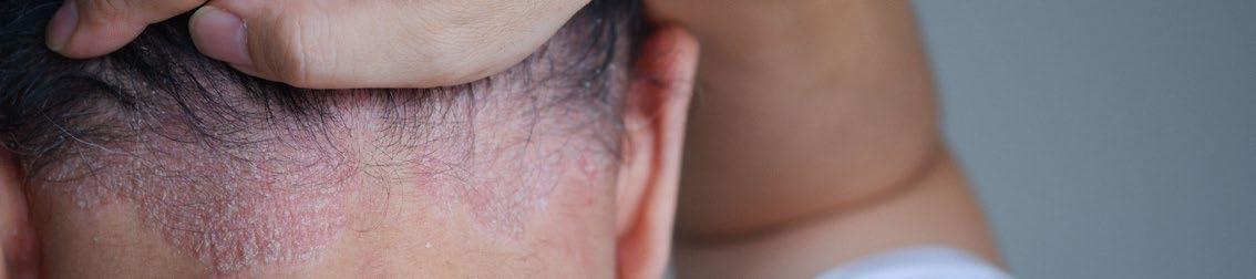
Risankizumab Significantly Improves Quality of Life in Genital and Scalp Psoriasis
NEW FINDINGS from the Phase IV UnlIMMited trial, presented at the AAD Annual Meeting 2026, demonstrate that risankizumab markedly improves quality of life (QoL) outcomes in patients with psoriasis affecting high-impact areas, including the genitals and scalp.2
Psoriasis in these sensitive and visible regions is associated with disproportionate physical discomfort and psychological burden, often leading to increased embarrassment, selfconsciousness, and reduced overall wellbeing. Despite this, such areas remain challenging to treat effectively, underscoring the need for targeted therapeutic options.
The UnlIMMited study (NCT05969223) is a multicenter, randomized, double-blind, placebocontrolled trial evaluating risankizumab, an IL-23 inhibitor, in patients with genital or scalp psoriasis. Participants were randomized 1:1 to receive risankizumab 150 mg or placebo at baseline and Week 4. QoL was assessed at Week 16 using the Dermatology Life Quality Index (DLQI), focusing on symptom burden and psychosocial impact.
Results showed significantly greater improvements in QoL among patients treated with risankizumab compared with placebo. In the genital psoriasis group, 33.3% of patients receiving risankizumab reported no symptoms such as itching, pain, or stinging (DLQI question one, score of 0), compared with just 4.2% in the placebo group. Even more strikingly, 74.5% of risankizumab-treated patients reported no embarrassment or self-consciousness related to
their skin (DLQI question two, score of 0), versus 10.9% of those receiving placebo.
Similar trends were observed in the scalp psoriasis group. Among patients with scalp psoriasis, 25.0% of those treated with risankizumab achieved a score of 0 for symptom burden, compared with 6.3% in the placebo arm. For psychosocial impact, 48.9% of patients receiving risankizumab reported no embarrassment or self-consciousness, compared with 10.4% of placebo-treated patients.
These findings highlight the substantial burden of psoriasis in high-impact areas and demonstrate that risankizumab can deliver meaningful improvements in both physical symptoms and emotional wellbeing within 16 weeks.
The authors concluded that risankizumab offers a valuable treatment option for patients with genital or scalp psoriasis, significantly reducing disease impact and improving patient-reported outcomes.
Psoriasis in these sensitive and visible regions is associated with disproportionate physical discomfort and psychological burden

Guselkumab
Shows Consistent Benefits for Erosive Psoriatic Arthritis
NEW DATA presented at the AAD Annual Meeting 2026 demonstrated consistent Week 24 efficacy of guselkumab in biologic-naïve patients with active, erosive psoriatic arthritis (PsA) across baseline subgroups.3
The Phase IIIb APEX study previously met its primary endpoint, demonstrating significantly higher American College of Rheumatology 20% response criteria (ACR20) response rates at Week 24 with guselkumab versus placebo. This subgroup analysis assessed treatment consistency across baseline demographic, clinical, and radiographic characteristics.
APEX was a randomized Phase IIIb study in biologic-naïve adults with active PsA despite prior non-biologic therapy. Eligibility required swollen joint count ≥3, tender joint count ≥3, C-reactive protein ≥0.3 mg/dL, and at least two erosive joints on hand or foot radiographs. Participants were randomized to guselkumab every 4 weeks (Q4W; n=273); guselkumab at Weeks 0 and 4, then every 8 weeks (n=371); or placebo Q4W (n=376). Outcomes at Week 24 were evaluated in predefined subgroups by sex, BMI, PsA disease characteristics, concomitant methotrexate use, and radiographic features.
At Week 24, ACR20 response rates were 66.6%, 68.3%, and 47.0% in the guselkumab Q4W, guselkumab at Weeks 0 and 4 then every 8
weeks, and placebo groups, respectively. ACR50 responses were 41.4%, 42.2%, and 20.5%; Psoriasis Area and Severity Index (PASI)-90 responses were 69.4%, 60.0%, and 22.0%; and minimal disease activity rates were 29.6%, 27.7%, and 13.7%, respectively. Improvements across endpoints were consistent in guselkumabtreated subgroups. Inhibition of radiographic progression was also generally consistent across baseline subgroups.
In summary, in biologic-naïve patients with active, erosive PsA, guselkumab provided greater clinical improvements than placebo, as well as consistent benefit across key subgroups. These findings support guselkumab as an effective option when both symptom control and structural preservation are treatment priorities.
Minimal disease activity rates were 29.6%, 27.7%, and 13.7%

Patient-Reported Triggers Reveal Complexity of Chronic Hand Eczema
A NEW US study, presented at the AAD Annual Meeting 2026, has revealed that patients with chronic hand eczema (CHE) most commonly attribute their condition to a complex mix of environmental, biological, and psychological triggers, offering important insights for more personalized management strategies.4
When asked to prioritize a single main trigger, environmental factors ranked highest (14.2%), followed by genetic predisposition (11.6%), and stress or emotional triggers (10.4%)
The causes of CHE are often difficult to determine, as the disease is widely recognized as multifactorial, involving both external exposures and internal predispositions. Despite this, patient perceptions of what drives their condition have historically been underexplored in clinical care.
The CHECK-US study, an online survey of adults from general population panels, analyzed responses from 982 participants with physician-diagnosed CHE. Participants were asked to identify perceived triggers and select what they believed to be the primary cause of their condition. Findings showed that most individuals linked their CHE to multiple overlapping factors, with environmental triggers reported by 69.4%, biological causes by 47.4%, psychological factors by 34.1%, and lifestyle or habits by 26.0%.
When asked to prioritize a single main trigger, environmental factors ranked highest (14.2%), followed by genetic predisposition (11.6%), and stress or emotional triggers (10.4%). Notably, differences emerged across subgroups. Females were significantly more
likely to report environmental (73.4% versus 66.1%; p<0.05) and psychological triggers (47.5% versus 22.8%; p<0.01), while males more frequently identified lifestyle factors (29.7% versus 21.7%; p<0.01) and infections (19.1% versus 10.9%; p<0.01). Employed individuals more often cited lifestyle and physical triggers compared with unemployed respondents, and those living in urban areas were more likely to report lifestyle-related causes than those in rural settings.
These findings underscore the heterogeneity of CHE and highlight the importance of incorporating patient perspectives into clinical assessment. While the study relied on selfreported data and may be subject to recall bias, it provides valuable real-world insight into how patients interpret their disease.
The results suggest that understanding patient beliefs about CHE triggers could support more tailored treatment approaches, improve adherence, and ultimately enhance outcomes. Future research may further explore how aligning clinical management with patient perceptions can optimize care in this challenging condition.
Hidradenitis Suppurativa Tied to Poorer Pregnancy Outcomes
A NEW meta-analysis presented at the AAD Annual Meeting 2026 suggested that hidradenitis suppurativa (HS) was associated with a higher risk of several adverse pregnancy outcomes, adding to growing concern about the impact of this chronic inflammatory skin disease during pregnancy.5
HS is a long-term inflammatory condition marked by recurrent nodules, abscesses, sinus tract formation, and scarring. It also frequently occurs alongside comorbidities such as smoking, obesity, diabetes, and hypertension, all of which can complicate pregnancy.
Against this background, researchers examined whether pregnancies in females with HS were more likely to be affected by maternal or fetal complications than pregnancies in females without the condition.
Researchers examined whether pregnancies in females with HS were more likely to be affected by maternal or fetal complications than pregnancies in females without the condition

The systematic review and meta-analysis searched PubMed, Embase, and Cochrane databases in May 2025 for studies comparing pregnancy outcomes in females with HS against those without it. Five studies were included, representing a combined total of 15,192,650 pregnant females. Random-effects models were used to calculate pooled odds ratios with 95% CI.
The analysis showed that pregnancies affected by HS were associated with a significantly increased risk of hypertensive disorders (odds ratio [OR]: 1.81; 95% CI: 1.49–2.20; p<0.001), Caesarean delivery (OR: 1.34; 95% CI: 1.09–1.65; p=0.006), and preterm birth (OR: 1.20; 95% CI: 1.01–1.43; p=0.035) compared with pregnancies not affected by HS.
However, no statistically significant differences were observed for preeclampsia (OR: 1.18; 95% CI: 1.00–1.40; p=0.057) or stillbirth (OR: 1.08; 95% CI: 0.57–2.06; p=0.810). These findings suggested that, while HS was linked to an elevated risk of some adverse outcomes, this pattern was not seen across every complication assessed.
As with all meta-analyses, the findings depended on the quality and consistency of the available studies. Even so, the scale of the dataset adds weight to the observation that HS may be an important consideration in pregnancy risk assessment.
The study added to evidence that females with HS may benefit from closer monitoring before and during pregnancy, particularly with regard to hypertensive disorders, preterm birth, and delivery planning.
Comparative Risk of Psoriatic Arthritis in Patients with Psoriasis on Immunomodulators
PSORIATIC arthritis (PsA) is a progressive disease that can be difficult to prevent, is often diagnosed late, and remains challenging to treat. Current treatments are often symptomdriven rather than preventive, and for patients with psoriasis (PsO), an important clinical question is whether some immunomodulators are more effective than others at reducing the risk of developing PsA. This study, presented at the AAD Annual Meeting 2026, assessed the 3-year risk of PsA development in patients with PsO treated with immunomodulators targeting IL-12, IL-17, IL-23, TNF-α, JAK1, and JAK3.6
The investigators used the TriNetX Research Network (TriNetX, LLC, Cambridge, Massachusetts, USA) to identify patients with PsO who had been treated with one of the listed immunomodulatory agents. To improve comparisons across treatment groups, the investigators matched patients 1:1 by propensity score using demographic, socioeconomic, and clinical factors, including obesity, inflammatory bowel disease, gout, diabetes, hypertension, hyperlipidemia, tobacco use, and alcohol-related disorders.
The study evaluated multiple therapies and revealed notable differences across treatment groups. Among the agents assessed, IL-23 inhibitors were consistently associated with the greatest reduction in the 3-year risk of PsA development. Overall, risankizumab, guselkumab, and ustekinumab demonstrated more favorable outcomes than other agents, including TNF-α, IL-17, and JAK inhibitors. These findings suggest that not all biologics provide the same level of protection against the development of PsA in patients with PsO.
The mechanisms behind these findings remain incompletely understood, but PsA development is thought to be driven by a persistent pro-inflammatory state that contributes to abnormal bone formation and turnover. By targeting key inflammatory pathways, immunomodulators may help interrupt this process and delay disease onset, potentially offering a more effective approach to limiting progression to PsA.
These findings suggest that treatment selection in PsO may play a larger role in long-term disease prevention than previously acknowledged. For patients who are at risk of PsA development, clinicians may consider whether certain immunomodulators offer added benefits beyond symptom control. This approach could help reduce disease progression and improve quality of life.
Further research may help clarify how these therapies can be used most effectively to reduce PsA development risk in patients with PsO.
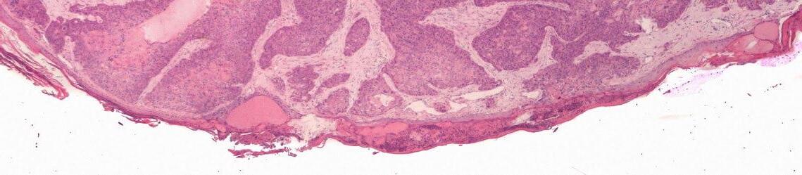
The Safety of Topical Therapies in Transplant-Related Skin Cancer Risk
ALLOGENIC hematopoietic stem cell transplant (allo-HSCT) recipients face an increased risk of developing precancerous lesions and malignant mucocutaneous lesions. These include actinic keratosis, squamous cell carcinoma in situ, and superficial basal cell carcinoma.7
Common topical therapies such as 5-fluorouracil, used alone or combined with 0.005% calcipotriene, work by inducing skin inflammation. This raises questions about their safety in allo-HSCT recipients, particularly regarding graft-versus-host disease, a condition in which donor immune cells attack the patient’s tissues and can be fatal.
A retrospective review, presented at the AAD Annual Meeting 2026, was conducted across three tertiary care centers and evaluated patients treated with these topical therapies between January 2015–September 2024. The study included 136 patients with at least 3 months of follow-up, analyzing 269 treatment exposures. Indications for treatment included actinic keratosis (51.3%), squamous cell carcinoma in situ (31.2%), and superficial basal cell carcinoma (13.4%).
Adverse events were infrequent and mainly consisted of erythema (13.0%), blistering (10.9%), and irritation or pain (7.8%). Treatment was discontinued in 14.0% of cases, although the reasons were not specified in the abstract presented. Within 3 months, graft-versus-host disease flares were reported in 11 patients (4.1%); however none of these flares were attributed to the topical therapy.
Complete response rates with 5-fluorouracil reached 94.7% in squamous cell carcinoma in situ, with rates of 62.3% in actinic keratosis and 56.3% in superficial basal cell carcinoma. Response rates were lower with the combination therapy across all indications (66.7%, 41.7%, and 33.3%, respectively).
As a retrospective study, the findings are limited by the lack of a comparison group, and differences between treatment groups were not detailed. It would also be important to understand which treatments were associated with adverse events, as this was also not specified. However, these results support cautious use of these therapies in patients with allo-HSCT and highlight the need for prospective studies.
Complete response rates with 5-fluorouracil reached 94.7% in squamous cell carcinoma in situ, with rates
62.3 56.3
actinic keratosisrates superficial basal cell carcinoma
Oral Peptide Shows Strong Clearance in Difficult Psoriasis Sites
NEW pooled analyses from four Phase III trials in the ICONIC program, presented at the AAD Annual Meeting 2026, evaluated icotrokinra, a first-in-class targeted oral peptide designed to block the IL-23 receptor and interrupt inflammatory signaling in plaque psoriasis.8
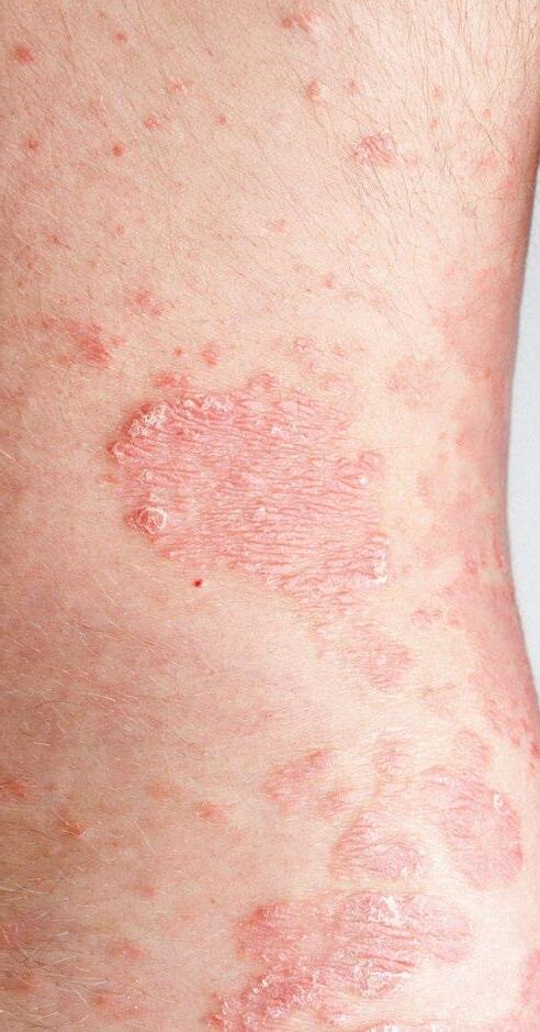
Plaque psoriasis is a chronic immune-mediated inflammatory skin condition. It is characterized by discolored scaly lesions that can vary in appearance depending on skin tone. Lesions are often red with silvery scale on lighter skin, but may appear purple, gray, or dark brown on darker skin tones. These plaques can cause discomfort and significantly impact quality of life, particularly when affecting high-impact areas like the scalp, genitals, hands, or feet.
Across 1,866 patients, many entered the study with moderate or severe psoriasis (defined by a score ≥3 on the Physician’s Global Assessment [PGA]), 72% with scalp involvement, 25% with genital psoriasis, and 24% with hand or foot disease.
Participants were randomized to receive oncedaily oral icotrokinra 200 mg or placebo for 16 weeks. Outcomes were measured using PGA scales tailored to each body region. At Week 16, icotrokinra cleared or almost cleared psoriasis versus placebo in scalp (72% versus 16%), genital (76% versus 29%), and hand/foot (64% versus 23%).
The safety profile of icotrokinra was reported to be similar to placebo over the 16-week period, suggesting short-term tolerability. However, the findings are limited by the relatively short follow-up duration, and longer-term data will be important to confirm durability of response and safety outcomes. Participant demographics showed an overwhelmingly White study population, with smaller proportions of Asian and Black participants, which may restrict broader generalizability.
For clinicians, these results point to the potential of an oral, targeted therapy to address psoriasis
in areas that often remain challenging despite existing treatments. If confirmed in longer-term studies, this approach could broaden treatment options and improve disease management in routine practice.

Survey Highlights Need for Acne Relapse Guidance After Isotretinoin
A NEW practitioner survey, presented at the AAD Annual Meeting 2026, shows highly variable follow-up and maintenance strategies for patients with acne vulgaris after oral isotretinoin therapy, highlighting the need for clearer long-term management pathways.9
Following oral isotretinoin therapy, approximately 10–60% of patients with acne vulgaris experience relapse. Despite this burden, there are no formal posttreatment guidelines. Researchers therefore evaluated how dermatology practitioners currently manage acne relapse risk, maintenance therapy, follow-up schedules, and subsequent treatment decisions after isotretinoin cessation.
Using an anonymous institutional review boardexempt survey, researchers collected data on demographics, isotretinoin maintenance therapy, follow-up protocol, and relapse management. A total of 410 practitioners responded, including 47.4% boardcertified dermatologists. The results revealed that most practitioners aimed for a 150–200 mg/kg (44.2%) or 120–150 mg/kg (37.1%) cumulative dose of isotretinoin, with 53.9% recommending treatment cessation around 3 months after clearance.
Regarding acne maintenance therapy, topical retinoids alone or in combination with benzoyl peroxide were initiated by 69% of respondents immediately after the final dose (44.8%) or 1 month later (37.2%). The survey showed that 47% of those who did not initiate maintenance therapy deemed it unnecessary due to low relapse rates.
Post-treatment follow-up was recommended by 75.2% of respondents, with 61.1% advising ongoing follow-up, primarily biannually (57.3%). For mild and moderate acne relapse, topical retinoids and benzoyl peroxide were commonly used. In moderate relapse, spironolactone was added for females by 53.1%, while doxycycline or minocycline was used for males by 61.0%. More than three-quarters would treat severe acne relapse with a second isotretinoin course, and 60.2% would repeat the same dosing endpoints used initially.
The results of the survey demonstrate that there is large variability in the recommendations given to patients with acne vulgaris following oral isotretinoin therapy. The researchers therefore advocated for evidence-based post-isotretinoin guidelines and increased awareness of relapse risk to optimize care and reduce repeat isotretinoin therapy.
There is large variability in the recommendations given to patients with acne vulgaris following oral isotretinoin therapy
Nicotinamide Exposure for Melanoma Prevention
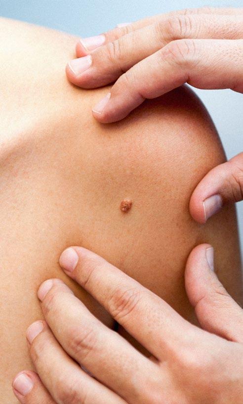
Despite the advances made in dermatology, melanoma remains a persistent threat, with no established medications or drugs for prevention available. Nicotinamide has been considered for the prevention of nonmelanoma skin cancer, but its potential role has remained unclear. As a result, there has been growing interest in understanding the role of nicotinamide in melanoma prevention. To address this gap, researchers conducted a retrospective cohort study in which data were gathered from the Veterans Affairs (VA) Corporate Data Warehouse, and the findings were presented at the AAD Annual Meeting 2026.10
Over 30,000 patients diagnosed with skin cancer were evaluated, with those exposed to nicotinamide compared to similar patients who had not received the supplement. Patients were matched according to demographic and clinical factors, such as age, gender, race, prior skin cancer history, and other relevant conditions.
The findings revealed a correlation between the use of nicotinamide and a lower incidence of melanoma. Overall, nicotinamide exposure was associated with a 25% reduction in melanoma incidence. This tendency was more pronounced among patients who began treatment after developing one to three prior non-melanoma skin cancers. Among patients with no history of melanoma, nicotinamide was associated with a 27% reduction in incidence. Finally, it should be noted that nicotinamide use was associated with fewer cases of invasive melanoma, but not melanoma in situ
While the underlying mechanisms have not been explicitly studied, nicotinamide is thought to help increase DNA repair and reduce ultravioletinduced immunosuppression. The differences noted between invasive and in situ melanomas may point towards a possible role in halting progression, as opposed to influencing tumor initiation, but additional studies are required to verify this possibility.
These findings suggest that there might be an alternative use for nicotinamide in preventing nonmelanoma skin cancers, particularly in patients at risk of melanoma development. Nicotinamide
appears to be most beneficial when given at early stages of cancer development, before melanoma diagnosis. Further studies may help clarify this association and support more individualized preventive treatment in melanoma care.
References
1. Chapman MC et al. Characterizing vulvar lichen sclerosus-lichen planus overlap: a clinical subtype with implications for diagnosis, surveillance, and management. Poster 71348. AAD Annual Meeting, March 27-31, 2026.
2. Song EJ et al. Quality of life improvements in patients with genital or scalp psoriasis receiving risankizumab: 16-week results from the UnIMMited randomized placebo-controlled trial. Poster. AAD Annual Meeting, March 27-31, 2026.
3. Mease PJ et al. APEX: guselkumab response and inhibition of structural damage progression in active psoriatic arthritis by participant baseline characteristics. Poster 72888. AAD Annual Meeting, March 27-31, 2026.
4. Simpson E et al. Patient-perceived factors associated with chronic hand eczema – results from the CHECK study in the United States. Poster. AAD Annual Meeting, March 27-31, 2026.
5. Matos de Albuquerque K et al. Adverse pregnancy outcomes in women with hidradenitis suppurativa: systematic review and meta-analysis. Poster 73335. AAD Annual Meeting, March 27-31, 2026.
6. Madelyn Schmidt et al. Comparative risk of psoriatic arthritis in psoriasis patients on immunomodulators. Poster 71957. AAD Annual Meeting, March 27-31, 2026.
7. Sakunchotpanit G et al. Topical 5-fluorouracil and topical 5-fluorouracil/ calcipotriene in allogeneic stem cell transplant recipients: safety and efficacy. Poster 76919. AAD Annual Meeting, March 27-31, 2026.
8. Soung J et al. Treatment of plaque psoriasis involving high-impact sites with icotrokinra, a targeted oral peptide: pooled analyses of 4 phase 3 placebocontrolled trials. Poster P73233. AAD Annual Meeting, March 27-31, 2026.
9. Zarabian N et al. Post-isotretinoin acne management: a survey of dermatology practitioners. Poster. AAD Annual Meeting, March 27-31, 2026.
10. Knox K et al. Nicotinamide exposure for melanoma prevention. Poster 73232. AAD Annual Meeting, March 27-31, 2026.
Highlights and Insights from the 2026 American Academy of Dermatology (AAD) Annual Meeting
Author: *Divya Sharma1
1. Department of Dermatology, University of Nebraska Medical Center, Omaha, USA
*Correspondence to disharma@unmc.edu
Disclosure: The author has declared no conflicts of interest.
Keywords: Atopic dermatitis, cutaneous oncology, dermatology, hair loss, immunotherapy.
Citation: Dermatol AMJ. 2026;3[1]:23-25.
https://doi.org/10.33590/dermatolamj/U3O2RE60
EVERY year, thousands of dermatologists and dermatology practitioners attend the American Academy of Dermatology (AAD) Annual Meeting. This year’s meeting, located in Denver, Colorado, USA, was no different, with more than 20,000 registrants attending.1 Experts provided valuable insight in numerous areas within dermatology, along with goals for the future of the specialty. Herein, we highlight some of the latest advancements highlighted in the meeting and the common themes shared by leaders in the field.
PLENARY SESSION
The John Kenney Jr. M.D. Lifetime Achievement Award and Lectureship this year was awarded to Iltefat Hamzavi, Henry Ford Health, Detroit, Michigan, USA. During his lecture, he noted that a will to go where patients are suffering is what guided him through his career and led him to create numerous advancements in vitiligo and hidradenitis suppurativa. Similarly, Robert Brodell, University of Mississippi Medical Center, Jackson, USA, provided the Clarence S. Livingood M.D. Memorial Award and Lectureship, in which he highlighted his career-long work on improving access to care in rural areas.
Incoming president of the AAD, Murad Alam, Northwestern University Feinberg School of Medicine, Chicago, Illinois, USA, provided an insightful look into his goals for the upcoming year. Much of his message was focused on the specialty collectively working together to address Medicare reimbursement reform.
ATOPIC DERMATITIS: NEW THERAPEUTICS ON THE HORIZON
Numerous presenters shared practicechanging updates in the realm of atopic dermatitis. Linda Stein Gold from Henry Ford Health shared data from a recent Phase II, multicenter, open-label
study showing efficacy and safety of nemolizumab for atopic dermatitis in children. This was a 52-week, openlabel, single-arm study and involved 109 patients.2 Improvements in itch were seen as quickly as within 1 week and 80% of patients achieved an Eczema Area and Severity Index (EASI)-75 score by 52 weeks. Moreover, no serious adverse events were reported.2
Amlitelimab, an anti-OX40 ligand monoclonal antibody treatment, was discussed by Eric Lawrence Simpson from Oregon Health and Science University, Portland, USA. Specifically, promising data from a Phase III, randomized, double-blind, placebo controlled trial were shared. There were clinically significant improvements in atopic dermatitis noticed while the medication was found to be well tolerated.2
HAIR LOSS: POTENTIAL NEW TREATMENTS TO END A DROUGHT
For hair loss, finasteride and minoxidil have remained the two primary FDAapproved treatments for decades. New data highlighted at this year’s meeting suggest this may not be the case for long.
PP-405, a mitochondrial pyruvate carrier inhibitor developed by Pelage Pharmaceuticals (Los Angeles, California, USA), was discussed again at this year’s meeting by Arash Mostaghimi from Brigham and Women’s Hospital, Boston, Massachusetts, USA. New data from a randomized, controlled, Phase IIa study were shown, which primarily investigated safety and pharmacokinetics while including 78 participants.3 Topical use of PP-405 was found to be well tolerated without any systemic absorption noted. Moreover, photos and data shown
For hair loss, finasteride and minoxidil have remained the two primary FDA-approved treatments for decades
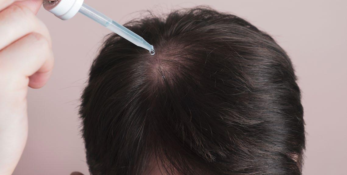
were very encouraging to suggest PP405 can truly increase the number of follicular units and terminal hairs.3 Pelage Pharmaceuticals will be preparing to begin late stage clinical trials for PP-405.
VDPHL01, a form of extended-release oral minoxidil, was similarly highlighted at this year’s meeting. Of note, the data shared by Veradermics (New Haven, Connecticut, USA), the developer of VDPHL01, showed a statistically significant increase in hair growth when compared to topical and standardrelease oral minoxidil.4
INTRATUMORAL IMMUNOTHERAPY: A PROMISING NEW AVENUE FOR CUTANEOUS ONCOLOGY
Mary C. Spellman highlighted a new therapeutic investigational siRNA that
References
1. DermWorld Meeting News. Invest now, pay it forward. 2026. Available at: https://www.aadmeetingnews. org/2026-aad-annual-meeting/ article/22963971/invest-now-pay-itforward. Last accessed: April 21 2026.
2. Tsao H et al. Late-breaking research: session 1. S023. AAD Annual Meeting, March 27-31, 2026.
targets the PDCD1 gene. Called PH762, this intratumoral immunotherapy was created to help reduce the adverse effects of systemic immunotherapy while still targeting cutaneous tumors. Phase I data were shared, which included a large range of doses of PH762, and showed no serious adverse events or doselimiting toxicities.5
The vast majority of patients in this trial had cutaneous squamous carcinoma and pathologic response, meaning the tumor did respond to the therapy, was seen in 65% of patients.5 While these data are very early and further trials and studies are needed, the impact of limiting systemic side effects and potentially surgery for patients while still targeting tumors could change the field of cutaneous oncology.
3. Mostaghimi A. Emerging treatments for hair loss targeting dormant follicles. S020. AAD Annual Meeting, March 2731, 2026.
4. Aguh C et al. Faster onset and superior efficacy of an investigational extended-release oral minoxidil tablet (VDPHL01) versus topical and immediate-release oral minoxidil for androgenetic alopecia: results from
a blinded retrospective investigator global assessment. Poster 76221. AAD Annual Meeting, March 27-31, 2026.
5. Spellman MC. PD-1 directed intratumoral immunotherapy: results of an escalating dose study of INTASYL PH-762 for cutaneous carcinomas. S034. AAD Annual Meeting, March 27-31, 2026.
Updates in Cutaneous Oncology from the 2026 AAD Annual Meeting: A Focus on Cutaneous Squamous Cell Carcinoma and Merkel Cell Carcinoma
Authors: Nathan Kattapuram1, *Divya Sharma2
1. Georgetown University School of Medicine, Washington, D.C., USA
2. Department of Dermatology, University of Nebraska Medical Center, Omaha, Nebraska, USA
*Correspondence to disharma@unmc.edu
Disclosure: The authors have declared no conflicts of interest.
Keywords: Biomarkers, cutaneous oncology, cutaneous squamous cell carcinoma (cSCC), immunotherapy, Merkel cell carcinoma (MCC).
Citation: Dermatol AMJ. 2026;3[1]:26-28.
https://doi.org/10.33590/dermatolamj/RYVY233I
THE AMERICAN Academy of Dermatology (AAD) Annual Meeting represents the largest gathering of leaders in dermatology each year. The most recent conference in Denver, Colorado, provided several key updates in the management of high-risk cutaneous malignancies, specifically cutaneous squamous cell carcinoma (cSCC) and Merkel cell carcinoma (MCC). In this article, the authors highlight sessions at the 2026 Annual Meeting relevant to these areas.
UPDATES IN CUTANEOUS ONCOLOGY
There is a compelling need to stratify cSCC based on risk.1 The Brigham and Women’s Hospital (BWH) and American Joint Committee on Cancer (AJCC) staging systems have traditionally provided a framework to stratify this cancer. AI may serve as an additional tool to stratify cSCC.
A prior study demonstrated that a large language model, trained on a comprehensive dataset, can derive a prognostic model that has higher sensitivity than both the BWH and AJCC systems.2 AI-derived prognostic models may assist providers in determining which patients may benefit from sentinel lymph node biopsy or nodal imaging at
baseline. Gene expression profiling is another promising tool for identifying high-risk tumors. A novel 14-gene expression profile was shown to have a sensitivity of 65.1% and a specificity of 80.5% for cSCC metastasis.3
For patients with virally mediated MCC, anti-MCPyV antibody titers (AMERK) may be used in surveillance. Roughly 50% of patients will have AMERK seropositivity at baseline.4 Patients who are seropositive at baseline have lower recurrence risk than seronegative
There is a compelling need to stratify cSCC based on risk
patients.4 In seropositive patients, AMERK titers can be followed during surveillance. A meaningful decline in AMERK titer, at least a 30% reduction, was associated with a 99.3% likelihood of being recurrence-free in the subsequent 3 months, whereas a rising titer was associated with a 58% risk of recurrence within the year.5
AI-derived prognostic models may assist providers in determining which patients may benefit from sentinel lymph node biopsy or nodal imaging at baseline
Circulating tumor DNA (ctDNA) is another promising biomarker for the detection of MCC recurrence. In a study of 319 patients with Stage I–IV MCC, a positive ctDNA test during surveillance was associated with increased recurrence risk, with a hazard ratio of 6.8.6 In a subset analysis of 84 patients who underwent curative-intent treatment, a positive ctDNA test within 4 months after treatment was associated with a 1-year recurrence rate of 74%, compared with 21% in those for whom ctDNA was negative during the same interval.6 A positive ctDNA value
precedes clinical evidence of recurrence by a median of 2.7 months.7
ADVANCES IN THE MANAGEMENT OF CUTANEOUS ONCOLOGY
Pre-operative management of cSCC, specifically the use of neoadjuvant immunotherapy, is an area of significant research. The MATISSE trial, which consisted of 40 patients who had an indication for extensive surgery, found that neoadjuvant nivolumab yielded major pathologic response in 40% of patients, whereas neoadjuvant combination immunotherapy (nivolumab with ipilimumab) yielded major pathologic response in 53%.8 Notably, nine patients declined surgery with or without adjuvant radiotherapy due to self-reported remission after neoadjuvant immunotherapy, and all nine remained disease-free in followup.8 Any potential benefit of neoadjuvant immunotherapy must be weighed carefully against risks, specifically immune-related adverse events. Further study is needed to elucidate the dose response to neoadjuvant immunotherapy, helping providers identify the minimum dose required to yield durable benefit.
There are parallels in the emerging understanding of neoadjuvant immunotherapy between cSCC and MCC. In a prospective study of 39 patients with Stage IIA-IV resectable MCC who received at least one pre-operative dose of nivolumab before resection, approximately half of the patients achieved a pathologic complete response.9 Both pre-operative radiographic reduction in tumor burden by ≥30% and a pathologic complete response were associated with longer recurrence-free survival.9
LILA AND MURRAY GRUBER MEMORIAL CANCER RESEARCH AWARD AND LECTURESHIP
The management of advanced MCC has undergone significant transformation in the past decade.10 Cytotoxic chemotherapy used to be the standard of care for advanced
References
1. Joo J. Updates in cutaneous oncology - cutaneous squamous cell carcinoma updates. AAD Annual Meeting, March 27-31, 2026.
2. Jairath NK et al. Retrieval augmented generation–enabled large language model for risk stratification of cutaneous squamous cell carcinoma. JAMA Dermatol. 2025;161(8):796-804.
3. Leibovit-Reiben Z et al. Development of a gene-expression panel predictive of local recurrence, metastasis, and overall survival in intermediate- to high-risk cutaneous squamous cell carcinoma: a cohort study. J Am Acad Dermatol. 2026;94(3):843-51.
4. Miller DM et al. The prognostic value of the Merkel cell polyomavirus serum antibody test: a dual institutional observational study. Cancer. 2024;130(15):2670-82.
5. Gunnell L et al. Polyomavirus antibodies for Merkel cell carcinoma
MCC. However, responses to chemotherapy were typically not durable, with a median progression-free survival of 3 months.11 Immunotherapy has revolutionized care for patients with advanced MCC. In 2016, a prospective trial demonstrated that pembrolizumab yielded a 62% response rate among virus-positive MCC and a 44% response rate among virus-negative MCC.12 Since 2017, three immune checkpoint inhibitors have been approved for advanced MCC, including avelumab, pembrolizumab, and retifanlimab.13 On a population-level, the introduction of immune checkpoint inhibitors has aligned with an at least two-fold increase in survival for advanced MCC.14 Further study is needed to determine therapeutic options for the roughly 50% of patients with advanced MCC who do not have a sustained response to immune checkpoint inhibitor therapies.
recurrence detection. JAMA Dermatol. 2025;161(11):1132-9.
6. Akaike T et al. Circulating tumor DNA assay detects Merkel cell carcinoma recurrence, disease progression, and minimal residual disease: surveillance and prognostic implications. J Clin Oncol. 2024;42(26):3151-61.
7. Akaike T et al. Circulating tumor DNA level is associated with time to clinical recurrence in Merkel cell carcinoma: implications for patient management. J Am Acad Dermatol. 2026;94(2):548-56.
8. Miller D. What’s new in the management of advanced skin cancer- peri-operative management of high-risk resectable cutaneous squamous cell carcinoma. AAD Annual Meeting, March 27-31, 2026.
9. Topalian SL et al. Neoadjuvant nivolumab for patients with resectable Merkel cell carcinoma in the CheckMate 358 Trial. J Clin Oncol. 2020;38(22):2476-87.
10. Nghiem P. Plenary - 2026 Lila and Murray Gruber memorial cancer research award and lectureship. AAD Annual Meeting, March 27-31, 2026.
11. Iyer JG et al. Response rates and durability of chemotherapy among 62 patients with metastatic Merkel cell carcinoma. Cancer Med. 2016;5(9):2294-301.
12. Nghiem PT et al. PD-1 blockade with pembrolizumab in advanced Merkel cell carcinoma. N Engl J Med. 2016;374(26):2542-52.
13. Schmults CD et al. NCCN Guidelines® Insights: Merkel Cell Carcinoma, Version 1.2024. J Natl Compr Canc Netw. 2024;22(1D):1-11.
14. Paulson KG et al. Improved survival at the population level for patients with advanced Merkel cell carcinoma following availability of immunotherapy. J Am Acad Dermatol. 2025;93(1):89-94.
Augmented Intelligence in Dermatology: Reframing AI as a Collaborative Tool
Authors: *Yash Jani,¹ Sahana Sharma,¹ Geneveive Patrick,2 Preet Jani,3 Alexis Coican,4 Milaan Shah5
1. Medical College of Georgia, Augusta, USA
2. Department of Dermatology, University of Central Florida, Tallahassee, USA
3. Mercer University School of Medicine, Macon, Georgia, USA
4. Department of Graduate Medical Education, Hospital Corporation of America (HCA) Orange Park Medical Center, Florida, USA
5. Department of Dermatology, Medical University of South Carolina, Charleston, USA
*Correspondence to yjani@augusta.edu
Disclosure: The authors have declared no conflicts of interest.
Keywords:
Citation:
Algorithmic bias, AI, augmented intelligence (AuI), clinical decision support, dermatology, diagnostic accuracy, digital health, health equity, real-world validation, skin of color (SOC).
Dermatol AMJ. 2026;3[1]:29-33. https://doi.org/10.33590/dermatolamj/07M81QW3
RECENT discussions at the American Academy of Dermatology (AAD) 2026 Annual Meeting have underscored a pivotal shift in how AI is framed within dermatology, moving away from replacement-based narratives toward a model of augmented intelligence (Aul) that supports clinician expertise. This congress feature highlights key evidence surrounding human–machine collaboration, AI in diagnosis, triage, and clinical decision support, and the broader implications of these tools for dermatologic practice. It explores ongoing challenges related to algorithmic bias, realworld validation, skin of color representation, and health equity, underscoring the need for dermatologists to actively guide the responsible integration of AI into patient care.
INTRODUCTION
The rapid advancement of AI in medicine has prompted both enthusiasm and apprehensions amongst clinicians. In dermatology, a specialty deep-rooted in visual pattern recognition, these developments carry particular significance. A critical distinction must be made between AI, autonomous machine-based decision making, and AuI, in which AI-driven tools are
integrated into clinical practice to enhance, rather than replace, physician judgment. This reframing is not merely semantics. The AAD’s position statement on AuI explicitly endorses AuI as a model where AI technologies work in ‘harmony’ with dermatologists to improve patient care,1 and recent continuing medical education initiatives suggest this concept is already entering mainstream dermatologic education.
The practical applications of AuI in dermatology are already substantial, particularly in skin cancer detection, triage, and deep neural networks capable of supporting diagnosis across more than 100 skin disorders. Yet its evolution remains incomplete. Algorithmic bias remains a pressing concern, as models trained on lighter skin tones demonstrate reduced performance across diverse skin types. Thus, the goal of AuI extends beyond technological adoption to cultivating AI literacy among dermatologists, equipping them to critically evaluate, co-create, and implement these tools equitably.2 Rather than signaling obsolescence, AuI positions dermatologists as active participants in shaping the responsible development of the technologies that will define the future of the specialty.
The goal of AuI extends beyond technological adoption to cultivating AI literacy among dermatologists
EVIDENCE FOR HUMAN-MACHINE SYNERGY
The Performance Data
The case for AuI in dermatology rests not on promise alone, but rather on a growing body of evidence demonstrating the performance of clinician and algorithm compared to either alone. This ‘human with machine’ paradigm has been validated across multiple study designs, within clinical settings, and among levels of provider expertise.
One foundational study training a deep neural network algorithm on 220,680 clinical images demonstrated that AuI can support malignancy detection, treatment prediction, and multi-class disease classification for 134 skin conditions. With AI assistance, clinician sensitivity and specificity significantly improved for malignancy prediction, with high accuracy in narrowing to Top 5 differential
diagnoses and modest gain in Top 1.3 This underscores AuI’s role in differential generation support rather than as an autonomous diagnostician. A RCT matching patients with concerning lesions to either AIassisted or unaided diagnostic groups found significantly higher diagnostic accuracy with AI assistance. Greater benefit was observed among non-dermatology trainees, whereas improvements in dermatology residents were not statistically significant, and a decline in Top 1 accuracy for AI-assisted differentials was observed.4
A more compelling showcase of humanmachine synergy comes from a prospective clinical study of dermatologists collaborating with a conventional neural network (CNN) in real-world melanoma screening. Strikingly, cooperating with the CNN, dermatologists achieved 100% sensitivity with melanoma detection with increased specificity, approaching the clinical ideal of minimizing missed melanomas while reducing unnecessary excisions. Dermatologists with less dermoscopy experience experienced the greatest benefit. However, limitations from small sample size, unmasked design, lack of acral and subungual lesions, and training on predominantly White European populations underscore the need for further studies among skin of color (SOC) and diverse lesions.5
Across the literature, AuI benefits vary by expertise level. One review found CNNs achieved the highest diagnostic accuracy among AI models, with support vector machines also performing strongly, particularly in melanoma detection. AI-assistance improved diagnostic accuracy among all clinicians, but generalists and trainees benefited more than experienced dermatologists. Narrow classification tasks outperformed broad melanoma detection across all skin types, informing how these tools should be best deployed.6 Another meta-analysis similarly found AI sensitivity and specificity for melanoma comparable to dermatologists, with one review reporting non-inferior or superior performance relative to dermatologists
and general practitioners. However, many studies were subject to selection bias and overrepresentation of malignant lesions, underscoring the gap between curated datasets and real-world clinical practice as a key barrier to clinical translation.7
THE QUINTUPLE AIM FRAMEWORK
The AAD’s 2019 Position Statement on Augmented Artificial Intelligence established foundational principles for AI integration into dermatology.1 In the years since, emerging evidence has begun to operationalize these principles across each of the Quintuple Aims: enhancing patient experience, improving population health, reducing costs, improving professional fulfillment, and increasing diversity, equity, and inclusivity. This framework provides a useful lens through which to evaluate the promise and limitations of AuI in dermatological practice.
Enhancing Patient Experience
AuI has the potential to transform the patient encounter through real-time diagnostic support, reduced wait times via optimized triage, and improved communication through visual explanations of diagnostic reasoning.8 Consumer-directed AI applications may also improve access to dermatologic information and patient confidence in identifying concerns. However, risks include inaccurate predictions, anxiety, and unnecessary healthcare utilization, highlighting the need for these tools to complement rather than replace clinical evaluation.9
Improving Population Health
A compelling application of AuI lies in expanding access to dermatology expertise in underserved areas. Pediatric dermatology AI models remain limited, highlighting opportunities for decision support in complex cases with restricted subspecialty access and for improving triage in rural settings. Early applications are already being tested for diagnosis of facial infantile hemangiomas and
X-linked hypohidrotic ectodermal dysplasia.10 Applications to teledermatology, in-person visits, and dermatopathology could also facilitate earlier detection. However, realworld clinical validation is lacking, with persistent challenges including dataset bias, reduced generalizability across skin tones, and issues with interpretability.11
Reducing Costs
AuI may reduce unnecessary biopsy and referrals while maintaining or improving diagnostic accuracy. As demonstrated by Winkler et al.,5 dermatologist-CNN cooperation reduced unnecessary excisions without sacrificing safety, achieving 100% melanoma sensitivity. AI-assisted triage systems may additionally optimize resource allocation by directing patients to appropriate levels of care, reducing both over and underreferrals for lesions.8
Improving Professional Fulfillment
The AAD position statement endorses a model in which clinicians focus on tasks aligned with their expertise while delegating algorithmic processes to machines.1 Rather than threatening professional identity, AuI may reduce administrative burden and mitigate burnout through workflow optimization, allowing dermatologists to focus on clinical reasoning and patient relationships.2 Evidence that AI assistance benefits generalists and trainees more than experienced dermatologists further supports this paradigm.6
Increasing Diversity, Equity, and Inclusivity
The fifth aim represents both the greatest opportunity and most significant barrier to responsible AuI implementation. Current AI systems demonstrate substantial performance disparities across skin tones, raising concern about whether AuI will mitigate or exacerbate existing inequities.

One study using a pathologically confirmed, diverse image dataset found that AI models performed significantly worse on darker skin tones and uncommon diseases. Notably, dermatologists labeling the dataset also showed reduced accuracy in these categories, suggesting bias exists at multiple levels of model development. Fine-tuning models helped close this performance gap between skin tones, highlighting the importance of diverse training data.12 Another review documented that only 30% of AI programs had reported dermatological data specifically in SOC populations, underscoring persistent underrepresentation and challenges with image quality and standardization. These are factors that make current AI programs inevitable to perform worse at identifying lesions in SOC.13 Therefore, AuI cannot overcome this barrier until explicit attention to training dataset diversity, validation across populations, and equity-focused framework developments are addressed.
Fine-tuning models helped close this performance gap between skin tones, highlighting the importance of diverse training data
CRITICAL IMPLEMENTATION CHALLENGES
The Bias and Equity Crisis
The growth of AI in dermatology has been rapid, but the evidence supporting many of these tools has not kept pace. A large proportion of currently available applications still lack meaningful clinical validation or transparency. An analysis found that about 88% of AI dermatology apps had no supporting evidence, and nearly 90% did not report their regulatory status.14 That raises real concerns about how ready these tools are for clinical use.
At the same time, performance differences across skin types remain a major issue. Models trained mostly on lighter skin continue to perform worse on darker skin tones.12 This is not just a technical limitation; it has direct implications for equity in care. Many studies also fail to clearly describe the demographics of their datasets, making it difficult to know whether a model will actually work in diverse patient populations. The AAD has addressed this directly, stating that datasets need to reflect the
populations where these tools are used. Without that, even well-designed models risk being unreliable in practice and may worsen existing disparities instead of improving them.
The Real-World Validation Gap
A lot of the excitement around AI in dermatology comes from studies using controlled or retrospective datasets. While those results can look impressive, they do not always translate to real clinical settings. This becomes clear in prospective studies. In one 2023 primary care study, AI had a Top 1 diagnostic accuracy of 39%, compared to 64% for general practitioners and 72% for dermatologists under routine conditions.15 That gap highlights how different real-world performance can be compared to what is reported in curated datasets.
The takeaway is straightforward: strong results in controlled environments do not necessarily mean a tool is ready for clinical use. These systems need to be tested in actual workflows, where variability and uncertainty are much higher. The AAD emphasizes that validation should happen in
References
1. American Academy of Dermatology (AAD). Position statement on augmented artificial intelligence. Revised August 12, 2023. Available at: https://staging.aad.org/dw/weekly/may22. Last accessed: April 20 2026.
2. Schlessinger D et al. Augmented intelligence and dermatology-part I: core concepts and applications. J Am Acad Dermatol. 2026;94(1):1-8.
3. Han SS et al. Augmented intelligence dermatology: deep neural networks empower medical professionals in diagnosing skin cancer and predicting treatment options for 134 skin disorders. J Invest Dermatol. 2020;140(9):1753-61.
4. Han SS et al. Evaluation of artificial intelligence-assisted diagnosis of skin neoplasms: a single-center, paralleled, unmasked, randomized controlled trial. J Invest Dermatol. 2022;142(9): 2353-62.e2.
5. Winkler JK et al. Assessment of diagnostic performance of
real-world settings, with ongoing monitoring after deployment.
CONCLUSION: A CALL FOR ENGAGED LEADERSHIP
Dermatology is in a position to help shape how AI is integrated into clinical care. The specialty’s reliance on visual diagnosis and pattern recognition makes it especially relevant in this space, but it also means the risks and limitations of these tools are highly visible.
The goal is not to replace dermatologists, but to support them. AI has the potential to improve diagnostic accuracy, expand access, and make care more efficient. But those benefits depend on how these tools are developed and implemented. Without careful validation, diverse datasets, and continued clinician involvement, the same systems could just as easily reinforce existing gaps in care. This is ultimately a question of ownership. Will dermatologists take an active role in shaping how these tools are used, or will they be introduced without enough clinical oversight?
dermatologists cooperating with a convolutional neural network in a prospective clinical study: human with machine. JAMA Dermatol. 2023;159(6):621-7.
6. Karimzadhagh S et al. Performance of artificial intelligence in skin cancer detection: an umbrella review of systematic reviews and meta-analyses. Int J Dermatol. 2026;65(1):69-85.
7. Nadour N et al. Diagnostic accuracy of artificial intelligence compared to family physi-cians and dermatologists for skin conditions: a systematic review and meta-analysis. BMC Prim Care. 2025;26(1):384.
8. Nahm WJ et al. artificial intelligence in dermatology: a comprehensive review of ap-proved applications, clinical implementation, and future directions. Int J Dermatol. 2025;64(9):1568-83.
9. Sayres R et al. Consumer understanding of skin concerns with an ai-powered informa-tional tool. JAMA Dermatol. 2026;DOI:10.1001/ jamadermatol.2026.0597.
10. Issa CJ et al. A call for implementing augmented intelligence in pediatric dermatology. Pediatr Dermatol. 2023;40(3):584-6.
11. Young AT et al. Artificial intelligence in dermatology: a primer. J Invest Dermatol. 2020;140(8):1504-12.
12. Daneshjou R et al. Disparities in dermatology AI performance on a diverse, curated clini-cal image set. Sci Adv. 2022;8(32):eabq6147.
13. Fliorent R et al. Artificial intelligence in dermatology: advancements and challenges in skin of color. Int J Dermatol. 2024;63(4):455-61.
14. Wongvibulsin S et al. Current state of dermatology mobile applications with artificial in-telligence features. JAMA Dermatol. 2024;160(6):646-50.
15. Escalé-Besa A et al. (2023). Exploring the potential of artificial intelligence in improving skin lesion diagnosis in primary care. Sci Rep. 2023;13(1):4293.
The End of the Steroid Era: Redefining Treatment Goals in Pediatric Atopic Dermatitis
Authors: *Yash Jani,¹ Mitchell Hanson,¹ Preet Jani,2 Riggs Newell,3 McKenzie Maloney4
1. Medical College of Georgia, Augusta, USA
2. Mercer University School of Medicine, Macon, Georgia, USA
3. Florida State University, Tallahassee, USA
4. Department of Medicine, Massachusetts General Hospital, Boston, USA
*Correspondence to yjani@augusta.edu
Disclosure: The authors have declared no conflicts of interest.
Acknowledgements:
Keywords:
The authors would like to thank the American Academy of Dermatology for their dedication to improving dermatologic care for children.
Atopic dermatitis, biologics, clinical practice guidelines, dupilumab, health equity, immunomodulatory therapy, JAK inhibitors, pediatric dermatology, systemic corticosteroids, treatment outcomes.
Citation: Dermatol AMJ. 2026;3[1]:34-37.
https://doi.org/10.33590/dermatolamj/A01I6L47
RECENT updates to the American Academy of Dermatology (AAD) guidelines have marked a pivotal shift in the management of pediatric atopic dermatitis, reflecting the rapid evolution of targeted immunomodulatory therapies and a growing emphasis on long-term disease control. This congress feature highlights key discussions surrounding the transition away from systemic corticosteroids, the expanding role of biologics and JAK inhibitors, and the broader implications of these changes for clinical practice. In addition, it explores ongoing challenges related to access, safety, and health equity, underscoring the need to translate therapeutic innovation into meaningful improvements in outcomes for pediatric patients.
INTRODUCTION
After more than a decade of relative therapeutic stagnation, the AAD has released updated guidelines for the management of pediatric atopic dermatitis, marking a pivotal inflection point in the field.¹ These recommendations arrive at a time when the therapeutic landscape has been fundamentally reshaped by the rapid expansion of targeted immunomodulatory therapies, including biologics and JAK
inhibitors. Together, these advances have transformed what was once a disease managed largely with topical agents and systemic corticosteroids into one that can now be treated with precision-based, mechanism-driven interventions.
Perhaps the most striking aspect of the updated guidelines is the strong recommendation against systemic corticosteroids.1,2 This shift reflects not only a growing body of evidence highlighting the
risks associated with corticosteroid use in children, but also the availability of safer and more effective alternatives. In this context, the 2026 guidelines do not merely update prior recommendations, they redefine the standard of care.
The benefits of therapeutic innovation can only be realized if they are available to all patients
THE THERAPEUTIC REVOLUTION
The scope of therapeutic advancement in atopic dermatitis over the past decade is unprecedented. Dupilumab, the first biologic approved for atopic dermatitis, is now indicated for children as young as 6 months.1,3 Clinical trials in infants and young children have demonstrated robust efficacy, with significantly higher rates of skin clearance and rapid improvement in disease severity compared to placebo.³ Notably, treatment responses are observed within weeks, and safety profiles appear consistent across pediatric and adult populations.³
Beyond dupilumab, the biologic landscape has expanded to include agents targeting IL-13 (tralokinumab, lebrikizumab) and IL-31 (nemolizumab) pathways, further refining the ability to modulate Type 2 inflammation.4 These therapies offer the potential for sustained disease control with fewer systemic adverse effects compared to traditional immunosuppressive agents. In parallel, JAK inhibitors, including upadacitinib, abrocitinib, and baricitinib, have emerged as powerful oral alternatives that act downstream of multiple cytokine signaling pathways.3,4 Network metaanalyses suggest that certain JAK inhibitors may achieve higher rates of near-complete skin clearance and more rapid itch reduction compared to biologics, although these benefits must be weighed against evolving safety considerations.3,5
Importantly, real-world data increasingly support the durability of targeted therapies. Dupilumab demonstrates superior long-term drug survival compared to conventional systemic agents such as methotrexate and cyclosporine, with lower discontinuation rates due to treatment failure.3 These findings underscore a paradigm shift from episodic symptom suppression to sustained disease modification.

THE CASE AGAINST SYSTEMIC CORTICOSTEROIDS
The strong recommendation against systemic corticosteroids represents a long-overdue reassessment of their role in pediatric atopic dermatitis.1,2 Although corticosteroids provide rapid symptomatic relief, their effects are often transient, with disease recurrence frequently occurring upon discontinuation.6-8
More concerning, however, are the welldocumented risks associated with systemic corticosteroid use, even when administered for short durations. These include increased risks of infection, venous thromboembolism, and fracture.2,9,10 In pediatric populations, additional concerns include growth suppression, adrenal insufficiency, metabolic disturbances, and neuropsychiatric effects.11
In clinical practice, corticosteroids are often used in repeated short courses, creating a cycle of temporary improvement followed by rebound flares.2 This pattern not only exposes children to cumulative toxicity, but also delays the initiation of more appropriate long-term therapies. As newer, targeted agents become increasingly accessible, the continued reliance on systemic corticosteroids appears increasingly difficult to justify.
REDEFINING TREATMENT GOALS
The emergence of targeted therapies has fundamentally altered treatment goals in pediatric atopic dermatitis. Historically, management focused on reducing acute inflammation and alleviating symptoms. Today, the emphasis has shifted toward achieving sustained disease control, improving quality of life, and minimizing long-term morbidity.
Biologic therapies have demonstrated meaningful improvements not only in clinical severity, but also in patient-reported outcomes, including sleep quality, psychosocial functioning, and caregiver burden.12 These benefits are particularly important in pediatric populations, where chronic skin disease can
have profound effects on development, selfesteem, and social integration.
At the same time, advances in nonsteroidal topical therapies, including phosphodiesterase-4 inhibitors (crisaborole, roflumilast), topical JAK inhibitors (ruxolitinib), and aryl hydrocarbon receptor modulators (tapinarof), have expanded treatment options for patients with milder disease.13,14 This growing therapeutic armamentarium allows for more individualized, stepwise approaches to care.
However, treatment selection remains complex. While JAK inhibitors may offer rapid onset of action and high levels of efficacy, biologics such as dupilumab provide a longer safety track record and broader applicability across age groups.15 The decision between these options must be guided by patient-specific factors, including age, comorbid conditions, route of administration preferences, and risk tolerance.
IMPLEMENTATION CHALLENGES AND HEALTH EQUITY
Despite these advances, significant barriers to implementation persist. Cost and insurance coverage remain major obstacles to accessing biologic therapies, particularly for younger children.² Prior authorization requirements, variable payer policies, and high out-ofpocket costs can delay or prevent initiation of appropriate treatment.
Disparities in access are further compounded by gaps in clinical trial data for the youngest patients. Although dupilumab is approved for infants as young as 6 months, many newer agents remain restricted to older pediatric populations, leading to reliance on off-label use in younger age groups.13,15
These challenges highlight a critical need for coordinated efforts to improve access to care. Dermatologists, pediatricians, and healthcare systems must work collaboratively to ensure that advances in therapy translate into realworld improvements in patient outcomes.
LOOKING FORWARD
The 2026 AAD guidelines represent more than an update: they signal a fundamental shift in how pediatric atopic dermatitis is conceptualized and managed. The move away from systemic corticosteroids toward targeted, mechanism-based therapies reflects a maturation of the evidence base and a redefinition of therapeutic priorities.
However, important questions remain. Long-term safety data for newer agents, particularly in very young children, are still evolving. Additionally, the optimal sequencing of therapies and strategies for treatment de-escalation have yet to be fully defined.
Perhaps most importantly, the field must address persistent disparities in access to care. The benefits of therapeutic innovation can only be realized if they are available to all patients, regardless of socioeconomic status or geographic location.
References
1. Davis DMR et al. Guidelines of care for the management of atopic dermatitis in pediatric patients. J Am Acad Dermatol. 2026;DOI:10.1016/j.jaad.2026.02.113.
2. Chu DK et al. Atopic dermatitis (eczema) guidelines: 2023 American Academy of Allergy, Asthma and Immunology/American College of Allergy, Asthma and Immunology Joint Task Force on Practice Parameters GRADE– and Institute of Medicine–based recommendations. Ann Allergy Asthma Immunol. 2024;132(3):274-312.
3. Paller AS et al. Dupilumab in children aged 6 months to younger than 6 years with uncontrolled atopic dermatitis: a randomised, double-blind, placebocontrolled, phase 3 trial. Lancet. 2022;400(10356):908-19.
4. Guttman-Yassky E et al. Atopic dermatitis. Lancet. 2025;405(10478):583-96.
5. Drucker AM et al. Systemic immunomodulatory treatments for atopic dermatitis: living systematic review and network meta-analysis update. JAMA Dermatol. 2024;160(9):936-44.
CONCLUSION
Pediatric atopic dermatitis is no longer a condition defined by therapeutic limitation. The emergence of biologics and JAK inhibitors has ushered in a new era of precision medicine, enabling clinicians to move beyond symptom control toward sustained disease modification.
In this context, the strong recommendation against systemic corticosteroids is both timely and necessary. It reflects not only a growing recognition of their risks, but also the availability of superior alternatives. As the field continues to evolve, the focus must shift from expanding therapeutic options to ensuring their appropriate and equitable use. Only then can the full potential of these advances be realized for the millions of children affected by atopic dermatitis worldwide.
6. Sawangjit R et al. Systemic treatments for eczema: a network meta-analysis. Cochrane Database Syst Rev. 2020;9(9):CD013206.
7. Drucker AM et al. Use of systemic corticosteroids for atopic dermatitis: International Eczema Council Consensus Statement. Br J Dermatol. 2018;178(3):768-75.
8. Tribolet-de-Abreu I et al. Topical and systemic corticosteroids in the modern management of atopic eczema: a scoping review. Int Immunopharmacol. 2026;176:116477.
9. Waljee AK et al. Short term use of oral corticosteroids and related harms among adults in the United States: population based cohort study. BMJ. 2017;357:j1415.
10. Johannesdottir SA et al. Use of glucocorticoids and risk of venous thromboembolism: a nationwide population-based case-control study. JAMA Intern Med. 2013;173(9):743-752.
11. Lima JP et al. Adverse events following short-course systemic corticosteroids among children and adolescents: a systematic review and meta-analysis. JAMA Netw Open. 2025;8(9):e2534953.
12. Paller AS et al. The effect of dupilumab on caregiver- and patient-reported outcomes in young children with moderate-to-severe atopic dermatitis: results from a placebo-controlled, phase 3 study. J Am Acad Dermatol. 2025;92(1):116-26.
13. Drucker AM. Treatment of atopic dermatitis. JAMA. 2025;334(12): 1103-4.
14. Armstrong AW et al. Advanced topical nonsteroidal therapies for atopic dermatitis: consensus statements from an expert panel. J Drugs Dermatol. 2026;25(3):221-7.
15. Kamata M et al. Deciding which patients with atopic dermatitis to prioritize for biologics and Janus kinase inhibitors. J Allergy Clin Immunol Pract. 2025;13(8):1901-10.
Addressing Barriers to Sunscreen Use in Athletes: New Data from AAD 2026
This poster was presented at the American Academy of Dermatology (AAD) Annual Meeting in Denver, Colorado, USA, from March 27th–31st, 2026
Support: The publication of this article was funded by La Roche-Posay, L’Oréal USA.
Presenters:
Hawasatu Dumbuya,1 Tania Katherine Hernandez,1 Mallory Hamilton Dennis,2 Emma DiMartini,1 Stephanie Laga,2 Iqra Iqbal,2 Carly Ortiz,2 Katharine Podimatis1
1. La Roche-Posay Laboratoire Dermatologique, L’Oréal USA, New York, USA
2. L’Oréal USA Research and Innovation, Clark, New Jersey, USA
Disclosure: All presenters are employees of L'Oréal USA.
Acknowledgements: Medical writing assistance was provided by Brigitte Scott, MarYas Editorial Services, Cowlinge, UK.
Keywords: Active lifestyle sunscreen, athlete sun protection, barriers, diverse racial/ ethnic populations, skin cancer prevention, sport sunscreen performance, sun exposure, sun protective behaviors, sunscreen, UV, UV risk.
Citation: Dermatol AMJ. 2026;3[1]:38-42. https://doi.org/10.33590/dermatolamj/IZQR666E
Meeting Summary
Outdoor athletes and active individuals have frequent and prolonged UV exposure and are at increased risk of skin cancer. Although athletes are generally aware of the risks of UV exposure, they often practice suboptimal sunprotection behaviors. Common barriers to sunscreen use include perceived impact on performance, uncomfortable sensory feel, sticky hands, and eye irritation. Understanding and addressing these barriers is essential to improving sun-protection behaviors and promoting sunscreen use in this population. This article summarizes a poster, ‘Evaluation of a Sunscreen Formulation by Athletes and Active Individuals’, presented as part of the American Academy of Dermatology (AAD) Annual Meeting in Denver, Colorado, USA, from March 27th–31st, 2026. An exploratory, 2-week, home-use study evaluated perceptions of sunscreen attributes and usage over time in 180 athletes and active individuals. Participants were aged 18–65 years and were from diverse racial/ethnic populations (skin phototypes Fitzpatrick I–VI). They were instructed to apply an organic, waterproof, broadspectrum sunscreen (sun protection factor [SPF] 50) at least five times per week, and to reapply after sweating and after 2 hours of sun exposure. Participants completed selfassessment questionnaires immediately after each application, and after 1 and 2 weeks of use. After application, most participants reported that the sunscreen dried quickly, their
PHARMA
hands did not feel sticky, and the product was invisible on their skin. After 2 weeks of use, nearly all participants noted that the sunscreen did not run into their eyes while sweating, was not visible after sweating, and was suitable for their active lifestyle. Participants across skin phototypes were satisfied with the look and feel of the sunscreen. The proportion of highly satisfied participants increased after 2 weeks of use.
ATHLETES, UV RISK, AND SUNSCREEN USE
Outdoor athletes and active individuals have frequent and prolonged UV exposure and are at increased risk of skin cancer.1-3 Athletes are generally aware of the risks of UV exposure, but this does not necessarily translate to adoption of sun-safety behaviors.1-3 A study of 347 golfers showed that 5.8% never, 12.4% rarely, and 22.5% only sometimes (i.e., not often or always) wore sunscreen.2 In a study of 290 collegiate athletes, 96% agreed that sunscreen helps prevent skin cancer, yet over 50% never used sunscreen, and 75% used sunscreen ≤3 days per week.3 Common barriers to sunscreen use in athletes and active individuals include perceived impact on performance, for example, due to slippery hands, uncomfortable sensory feel, sticky hands, and eye irritation.4
METHODS
An exploratory, 2-week, home-use study was conducted in athletes and active individuals from diverse racial/ethnic populations to evaluate their perceptions of sunscreen attributes and usage over time, and to enhance understanding of the barriers to sunscreen use. A total of 180 male and female athletes and active individuals aged 18–65 years who engaged in physical activity leading to sweating at least 5 days per week were included in the study.5 Participants had a diverse range of skin phototypes (Fitzpatrick I–VI).6 There were 60 participants in each of the following three skin phototype subgroups: Fitzpatrick I or II, Fitzpatrick III or IV, and Fitzpatrick V or VI.5 More than half the participants played at least one sport, the most common being tennis/pickleball (34%), soccer (20%), and basketball (13%; Figure 1).5
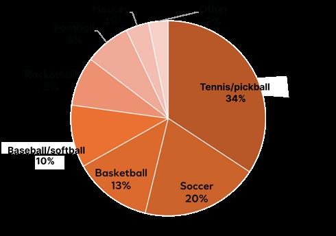
A total of 130/180 (72%) athletes and active individuals in the study participated in sports activities. The figure shows the sports activities of this subgroup. Some participants took part in more than one sport.
Figure 1: Participants’ sports activities.
The test product was an organic, waterproof, broad-spectrum sunscreen with an SPF of 50 (Anthelios UV Pro-Sport Sunscreen SPF 50; La Roche-Posay Laboratoire Dermatologique, L’Oréal USA). Participants were instructed to apply the test product, instead of their current sunscreen, on their face and body. They were asked to maintain their normal skincare routine, without introducing anything new. Application of the test product was required at least five times per week, 15 minutes before sun exposure. Reapplication was required every 80 minutes after sweating and after every 2 hours of sun exposure.5
Participants completed self-assessment questionnaires immediately after applying the test product, and after 1 and 2 weeks of use. Responses were given in the form of a semistructured 10-point scale, from 0 (the most negative rating)–9 (the most positive rating). The results are presented as the percentage of participants who rated the evaluated
attribute with a score from 6–9 (i.e., high positive ratings).5
RESULTS
All participants completed the study. Immediately after application, 93.9% responded that the sunscreen dried quickly, 90.6% reported that their hands did not feel sticky or tacky, and 93.3% noted that the product was invisible on their skin (Figure 2A). After 1 week of test product use, 91.7% noted that sand brushed off their skin easily, 96.1% observed that the sunscreen was not visible after sweating, and 95.0% considered the product to be suitable for their active lifestyle (data not shown). After 2 weeks of use, 92.2% specified that the sunscreen formulation did not run into their eyes while sweating, 97.2% responded that the product was not visible after sweating, and 95.0% found it suitable for their active lifestyle (Figure 2B).5
Figure 2: Athletes’ and active individuals’ perceptions of sunscreen formulation over time.
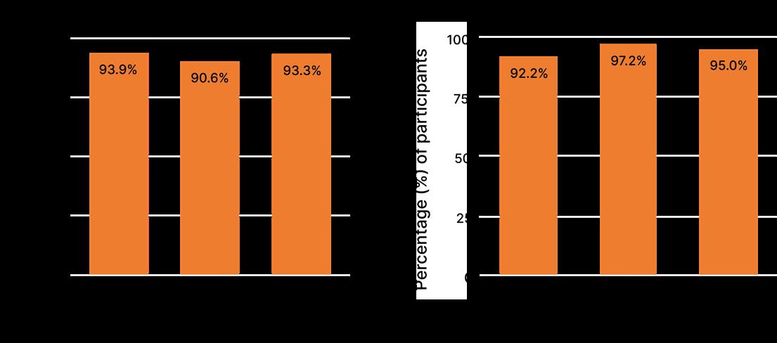
A total of 180 athletes and active individuals completed the questionnaire and rated the attributes on a scale of 0 (the most negative rating) to 9 (the most positive rating). The figure shows the percentage of participants who rated the evaluated attribute with a score from 6–9 (i.e., high positive ratings).
A total of 93.3% of participants were highly satisfied overall with the sunscreen formulation immediately after application. This increased to 94.4% and 97.2% after 1 and 2 weeks of sunscreen use, respectively (data not shown).
Participants were also asked whether they agreed with the following statements about the sunscreen formulation: ‘No chalky/ashy appearance’, ‘No white cast’, and ‘Invisible on skin’. Nearly all participants strongly agreed with these statements, with similar results across the three Fitzpatrick6 skin phototype subgroups (Figure 3).5
Figure 3: Perceptions of sunscreen formulation appearance immediately after application by Fitzpatrick skin phototype subgroup.

A total of 180 athletes and active individuals (60 per skin phenotype subgroup) completed the questionnaire and rated the attributes on a scale of 0 (the most negative rating) to 9 (the most positive rating). The figure shows the percentage of participants who rated the evaluated attribute with a score from 6–9 (i.e., high positive ratings).
CONCLUSIONS
Understanding the barriers to sunscreen use in outdoor athletes is essential to improve sun-protection behaviors and promote sunscreen use in this population. In this exploratory study, participants from diverse racial/ethnic populations who played a range of sports and/or had active lifestyles were satisfied with the look and feel of the sunscreen test product. Most participants noted that the sunscreen dried
quickly, did not leave their hands feeling sticky or tacky, and was invisible on the skin, even after sweating, thus mitigating common barriers to use. The lack of runoff into the eyes while sweating was noted by most participants. This is a key feature to avoid eye irritation and minimize impact on performance during sports and other activities. The sunscreen formulation was considered suitable for an active lifestyle.5
The study results indicate this formulation may successfully address key barriers to sunscreen use, such as perceived impact on performance, uncomfortable sensory feel, sticky hands, and eye irritation. The increase in the proportion of participants in the study
References
1. Fernandez-Ruiz J et al. Knowledge, behaviour and attitudes related to sun exposure in sportspeople: a systematic review. Int J Environ Res Public Health. 2022;19(16):10175.
2. Weikert AE et al. Golfers' interest in multilevel sun-protection strategies. Int J Environ Res Public Health. 2021;18(14):7253.
who were highly satisfied overall with the sunscreen formulation after 2 weeks of use was a positive outcome, particularly for a population known to practice suboptimal sun-protection behaviors, and may encourage more compliant sunscreen use.
3. Wysong A et al. Sunscreen use in NCAA collegiate athletes: identifying targets for intervention and barriers to use. Prev Med. 2012;55(5):493-6.
4. Gilaberte Y et al. Photoprotection in outdoor sports: a review of the literature and recommendations to reduce risk among athletes. Dermatol Ther (Heidelb). 2022;12(2):329-43.
5. Dumbuya H et al. Evaluation of a sunscreen formulation by athletes and
active individuals. Poster 73650. AAD Annual Meeting, March 27-31, 2026.
6. Callender VD et al. Beyond Fitzpatrick skin types: a Delphi consensus on key considerations for a universal skin typing classification. J Am Acad Dermatol. 2026;94(2):577-81.
AAD 2026

Abstract Reviews
This issue features research by abstract presenters at the 2026 American Academy of Dermatology (AAD) Annual Meeting. Covering topics such as conjunctivitis in atopic dermatitis, photoprotection for melanated skin, online dermatological trends, these abstracts reflect the current landscape of the evolving dermatological field.
Efficacy of Sunscreen Filters and Visible-Light Blockers on Photoprotection of Melanated Skin
Authors: Sahana Sharma,¹ *Yash Jani,¹ Renee Copeland,² Kathryn Potter²
1. Medical College of Georgia, Augusta, Georgia, USA
2. Department of Dermatology, Medical College of Georgia, Augusta, Georgia, USA *Correspondence to yjani@augusta.edu
Disclosure: The authors have declared no conflicts of interest.
Acknowledgements: The authors would like to thank Kathryn Potter for her guidance.
Keywords: Iron oxide, melasma, photoprotection, post-inflammatory hyperpigmentation, skin of color, sunscreen formulations, UVA1, visible light.
Citation: Dermatol AMJ. 2026;3[1]:44-45. https://doi.org/10.33590/dermatolamj/389K27BJ
BACKGROUND AND AIMS
Photoprotection in individuals with skin of color requires consideration beyond traditional UVB-focused sunscreen strategies. While darker Fitzpatrick skin types (IV–VI) have lower rates of skin cancer, they are disproportionately affected by pigmentary disorders such as melasma and post-inflammatory hyperpigmentation, which are exacerbated by long-wave UVA and visible light exposure.¹,² Despite this, current sunscreen guidance remains largely UVB-centric and lacks ingredient-specific recommendations tailored to melanated skin.
This systematic review evaluates clinical evidence on sunscreen filters and formulations relevant to skin of color, with a focus on iron oxides, extended UVA1/visible light organic filters, and antioxidant-enriched products. Following Preferred Reporting Items for Systematic reviews and MetaAnalyses (PRISMA)-guided methodology, studies involving Fitzpatrick III–VI participants
were analyzed for outcomes, including pigmentation, melasma severity, erythema, and tolerability.³
RESULTS AND DISCUSSION
Iron oxide-containing formulations consistently demonstrated superior protection against visible light-induced pigmentation compared to non-tinted mineral sunscreens.⁴ In randomized studies, sunscreens with iron oxide achieved earlier and greater improvement in melasma, with significant changes observed as early as 8 weeks and sustained through 12 weeks.⁵ However, variability in optimal concentration and limited long-term data highlight the need for further investigation.⁶
Organic filters extending into the UVA1 and visible light spectrum provided additional protection beyond standard SPF formulations. Filters targeting wavelengths around 350–400 nm were associated with measurable reductions in melanin index within 4 weeks, independent of UVB strength.⁷ Broadspectrum filters such as phenylene bisdiphenyltriazine (PBDT) also demonstrated short-term efficacy against blue lightinduced pigmentation in Fitzpatrick III–V individuals.⁸ Notably, many of these studies were industry-sponsored, underscoring the importance of independent validation.
Adjunctive formulations incorporating antioxidants and anti-inflammatory agents showed promising additive benefits. Combination therapies, including glycolic acid, hydroquinone, and vitamins C and E, improved melasma outcomes in up to 75% of participants compared to sunscreen alone.⁹ In post-procedural settings, antioxidant-enriched sunscreens achieved complete prevention of post-inflammatory
hyperpigmentation in select cohorts, suggesting a role in mitigating inflammationdriven pigmentation pathways.¹⁰
Despite these advances, significant barriers remain. Current literature is limited by small sample sizes, short follow-up durations, and underrepresentation of diverse skin tones.¹,² Additionally, clinical translation is hindered by the limited availability of iron oxide-containing products, inadequate shade inclusivity, cosmetic acceptability concerns such as white cast, and cost-related adherence barriers.¹⁰
Opportunities for advancement include the development of cosmetically elegant, shade-inclusive formulations, expansion of antioxidant-based photoprotective strategies, and incorporation of visible light and UVA1 coverage into clinical guidelines. Improved patient education and targeted counseling are also essential to address misconceptions and enhance sunscreen adherence in skin of color populations.¹
CONCLUSION
Overall, iron oxides, extended-spectrum organic filters, and antioxidant-enriched formulations demonstrate measurable benefits in reducing photodamage and pigmentary disorders in melanated skin. Larger, independent clinical trials and broader access to tailored formulations
are necessary to establish evidencebased recommendations and optimize photoprotection in this population.³
References
1. Sharma S et al. Efficacy of sunscreen filters and visible-light blockers in photoprotection of melanated skin. Abstract 75383. AAD Annual Meeting, March 27-31, 2026.
2. Ward WH et al, “Clinical Presentation and Staging of Melanoma,” Ward WH, Farma JM, (eds), Cutaneous Melanoma: Etiology and Therapy (2017), Brisbane: Codon Publications.
3. Rigel DS et al. Photoprotection for skin of all color: consensus and clinical guidance from an expert panel. J Am Acad Dermatol. 2022;86(3S):S1-8.
4. Taylor SC et al. Misconceptions of photoprotection in skin of color. J Am Acad Dermatol. 2022;86(3S):S9-17.
5. Mahmoud BH et al. Impact of long-wavelength UVA and visible light on melanocompetent skin. J Invest Dermatol. 2010;130(8):2092-7.
6. Dumbuya H et al. Impact of iron-oxide containing formulations against visible light-induced skin pigmentation in skin of color individuals. J Drugs Dermatol. 2020;19(7):712-7.
7. Grimes PE et al. Photoprotection efficacy of sun protection factor and iron oxide formulations in diverse skin with melasma and photodamage. J Drugs Dermatol. 2025;24(7):662-7.
8. Ezekwe N et al. Evaluate of the protection of sunscreen products against long wavelength ultraviolet A1 and visible light-induced biological effects. Photodermatol Photoimmunol Photomed. 2024;40(1):e12937.
9. Moyal D. Prevention of ultraviolet-induced skin pigmentation. Photodermatol. Photoimmunol. Photomed. 2004;20(5):243-7.
10. Zhang X et al. Evaluation of the efficacy of a sunscreen containing ultra-long UVA1 and other UVR broad-spectrum filters on skin barrier protection and melanin content reduction in Chinese adults: a singlecenter study. Health Sci Rep. 2024;7(2):e1923.
Conjunctivitis Risk with Dupilumab Versus Upadacitinib in Atopic Dermatitis: A
PropensityMatched Cohort Study
Authors: David Wang,1 Omar Alani,2
Iyla Draw,3 Samer Wahood,4 Lara Shqair,2
*Christopher Bunick5
1. Boston University Chobanian and Avedisian School of Medicine, Massachusetts, USA
2. Department of Dermatology, Icahn School of Medicine at Mount Sinai, New York, USA
3. University of Louisville School of Medicine, Kentucky, USA
4. The Warren Alpert Medical School of Brown University, Providence, Rhode Island, USA
5. Department of Dermatology and Program in Translational Biomedicine, Yale School of Medicine, New Haven, Connecticut, USA
*Correspondence to christopher.bunick@yale.edu
Disclosure: Bunick has served as an investigator and/or consultant for AbbVie, AbSci, Almirall, Alumis, Amgen, Apogee, Arcutis, Botanix, Castle Biosciences, Connect BioPharma, Daiichi Sankyo, Dermavant, Disc Medicine, Eli Lilly, EPI Health/Novan, Galderma, Highlight Therapeutics, Incyte, LEO Pharma, Novartis, Ortho Dermatologics, Palvella, Pfizer, Priovant, Regeneron, Sanofi, South Beach Symposium, Sun Pharma, Takeda, Timber, Teladoc, Triveni, UCB, and Veradermics (with honoraria and fees paid to himself); received travel support from Dermsquared (South Beach Symposium); and is Editor in Chief of Dermatology Times. The other authors have declared no conflicts of interest.
Keywords: Atopic dermatitis (AD), conjunctivitis, dupilumab (DUPI), upadacitinib (UPA), real-world evidence, TriNetX.
Citation: Dermatol AMJ. 2026;3[1]46-47. https://doi.org/10.33590/dermatolamj/7I0Q06LH
INTRODUCTION
Biologic-associated conjunctivitis represents a clinically meaningful adverse effect in patients with atopic dermatitis (AD), with implications for treatment selection and patient quality of life.1 Dupilumab (DUPI), an IL-4 receptor alpha antagonist widely used for moderate-to-severe AD, has demonstrated an increased incidence of
conjunctivitis in clinical trials and real-world settings.2 In contrast, upadacitinib (UPA), a selective JAK-1 inhibitor, has not shown a similar signal in trials, prompting interest in comparative safety.3
MATERIALS AND METHODS
In this retrospective cohort study using the TriNetX Global Collaborative Network (TriNetX, Cambridge, Massachusetts, USA), adult patients with AD initiating DUPI or UPA were identified. Following 1:1 propensity score matching on demographic variables (age, sex, race, and ethnicity), 1,369 patients were included in each treatment group. Conjunctivitis risk was assessed using both odds ratios and time-to-event analyses. Median follow-up duration was longer in the DUPI cohort (664 days) compared to the UPA cohort (310 days).
RESULTS
DUPI use was associated with a significantly increased likelihood of conjunctivitis compared to UPA (odds ratio: 2.71; 95% CI: 1.65–4.45; p<0.001). Time-to-event analysis supported this finding, demonstrating a 70% higher hazard of conjunctivitis among DUPItreated patients (hazard ratio: 1.70; 95% CI: 1.26–2.29; p=0.001). These findings suggest a consistent elevation in both cumulative and longitudinal risk.
Mechanistically, the increased risk observed with DUPI may be related to IL-4/ IL-13 pathway inhibition, which has been associated with reduced conjunctival goblet cell density, impaired mucin production, and tear film instability.4 In contrast, JAK-1 inhibition with UPA modulates inflammatory signaling through multiple cytokine pathways
without direct IL-4 receptor alpha blockade, which may preserve epithelial homeostasis and avoid these downstream ocular effects.
CONCLUSION
Overall, these real-world data support a differential ocular safety profile between DUPI and UPA. UPA may represent a reasonable therapeutic alternative in patients at higher risk for conjunctivitis or those who develop ocular adverse events while receiving DUPI. Further prospective studies are warranted to validate these findings and clarify underlying mechanisms.
References
1. Wang D et al. Conjunctivitis risk with dupilumab versus upadacitinib in atopic dermatitis: a propensitymatched cohort study. AAD Annual Meeting, March 27-31, 2026.
2. Silverberg JI et al. Efficacy and safety of upadacitinib versus dupilumab in adults and adolescents with moderate-to-severe atopic dermatitis: week 16 results of an open-label randomized efficacy assessor-blinded head-to-head phase IIIb/IV study (Level Up). Br J of Dermatol. 2025;192(1):36-45.
3. Paganini C et al. Impact of upadacitinib on atopic keratoconjunctivitis exacerbated by dupilumab treatment in atopic dermatitis patients: a prospective dermatological and ophthalmological clinical evaluation in common clinical practice. J Clin Med. 2024;13(13):3818.
4. Hansen PM et al. IL‐4 and IL‐13 both contribute to the homeostasis of human conjunctival goblet cells in vitro. Allergy. 2022;77(8):2555-8.
Closing the Gap: Video-Based Education Enhances Melanoma Awareness and Early Detection in
Underserved Communities
Authors: *Yash Jani,¹ John Dean Christiansen,²
Kaitlyn J. Blanchard,³ Brittany Ange,³
Matthew Belcher³
1. Medical College of Georgia, Augusta, USA
2. Emory University School of Medicine, Department of Internal Medicine, Atlanta, Georgia, USA
3. Department of Dermatology, Medical College of Georgia, Augusta, USA
*Correspondence to yjani@augusta.edu
Disclosure: The authors have declared no conflicts of interest.
Acknowledgements: The authors would like to thank Kathryn Potter for her guidance.
Keywords: Early detection, health disparities, health literacy, melanoma, patient education, rural health, self-skin examination (SSE), skin of color.
Citation: Dermatol AMJ. 2026;3[1]:48-49. https://doi.org/10.33590/dermatolamj/UH5F14FY
BACKGROUND AND AIMS
Melanoma remains the deadliest form of skin cancer, accounting for a disproportionate share of skin cancer-related mortality despite relatively low incidence.1,2 Early detection is critical, with 5-year survival rates exceeding 99% in localized disease compared to approximately 27% in advanced-stage melanoma.2 However, persistent disparities exist, particularly among rural populations and individuals with skin of color, who experience higher mortality due to reduced access to dermatologic care, lower health literacy, and underrepresentation in skin cancer education initiatives.2,3
This prospective interventional study evaluates the impact of a brief, video-based educational intervention on melanoma knowledge, lesion recognition, and selfskin examination (SSE) behaviors in an underserved population.
MATERIALS AND METHODS
The study was conducted at a regional cancer center in Albany, Georgia, USA, and included 201 adult participants recruited from clinic waiting areas and survivorship programs. Participants completed surveys at baseline, immediately following the intervention, and at 3-month follow-up (Table 1). The intervention consisted of a 5-minute, visually driven educational video written at a fifth-grade reading level, covering melanoma risk factors, ABCDE detection criteria (asymmetry, border irregularities, color variation, diameter >6 mm, and evolution), sun protection strategies, and SSE techniques.¹
RESULTS
The intervention produced significant improvements across all primary outcomes, including melanoma knowledge scores, confidence in lesion identification, ABCDE recall, and frequency of SSE. Knowledge scores increased from a baseline mean of 3.2 to 7.5 post-intervention and remained elevated at 6.7 at 3 months. Confidence in lesion identification improved from 21.4% at baseline to 96% immediately post-intervention, with sustained improvement at 84.8% at follow-up. Similarly, ABCDE recall increased from 4% to 89.1%, and monthly SSE frequency improved from 12.4% to 59.2%, with retention at 53.7% at 3 months.¹
Importantly, subgroup analysis demonstrated that participants with lower baseline awareness, disproportionately represented by underserved and minority populations, experienced the greatest improvements. Racial disparities in melanoma knowledge and SSE practices narrowed following the intervention, highlighting its potential to address inequities in dermatologic education and early detection.1
Table 1: Participant outcomes from pre-survey to post-survey and 3-month follow-up.
Knowledge scores (1–10) improved from 3.2 at baseline to 7.5 post-survey, with partial retention at 6.7 at follow-up. Confidence in lesion identification increased from 21.4% to 96.0% post-survey, remaining high at 84.8% after 3 months. ABCDE recall rose from 4.0% to 89.1% and was sustained at 81.0% at follow-up. Monthly self-skin exam practice improved from 12.4% to 59.2% post-survey, with continued adherence of 53.7% at 3 months.
ABCDE: Asymmetry, border irregularities, color variation, diameter >6 mm, and evolution; SSE: self-skin examination.
These findings emphasize the effectiveness of brief, accessible educational tools in overcoming structural barriers to care. In settings where access to dermatology is limited, empowering patients with practical knowledge and self-examination skills may facilitate earlier detection and reduce reliance on specialty services.3 The low cost, scalability, and minimal resource requirements of this intervention make it particularly well suited for implementation in primary care settings, oncology clinics, and underserved communities.
CONCLUSION
In conclusion, a short, literacy-conscious video intervention delivered at the point of care can produce significant and sustained improvements in melanoma awareness and preventive behaviors while reducing racial disparities in knowledge and screening practices. This scalable, patient-centered approach represents a practical strategy
to improve early melanoma detection and advance equity in cancer outcomes.¹
References
1. Jani Y et al. Closing the gap: video-based education enhances melanoma awareness and early detection in underserved communities. Poster 76232. AAD Annual Meeting, March 27-31, 2026.
2. Waseh S, Lee JB. Advances in melanoma: epidemiology, diagnosis, and prognosis. Front Med (Lausanne). 2023;10:1268479.
3. Stitzenberg KB et al. Distance to diagnosing provider as a measure of access for patients with melanoma. Arch Dermatol. 2007;143(8):991-8.
Racial Disparities in Melanoma Knowledge and Skin Self-Exams: Outcomes of a Video Intervention in Rural Georgia
Authors: *Yash Jani,1 Joo “Sally” Jung,2 Genevieve Patrick,3 Kaitlyn Blanchard,2 Brittany Ange,1 Matthew Belcher2
1. Medical College of Georgia, Augusta, Georgia, USA
2. Department of Dermatology, Medical College of Georgia, Augusta, USA
3. Department of Dermatology, University of Central Florida, Tallahassee, USA
*Correspondence to yjani@augusta.edu
Disclosure: The authors have declared no conflicts of interest.
Acknowledgements: The authors would like to thank Kathryn Potter for her guidance.
Keywords: Dermatology access, health disparities, health literacy, melanoma, patient education, rural health, self-skin examination (SSE), skin of color.
Citation: Dermatol AMJ. 2026;3[1]:50-51. https://doi.org/10.33590/dermatolamj/P6708369
BACKGROUND AND AIMS
Melanoma remains the deadliest form of skin cancer, accounting for a disproportionate share of skin cancer-related mortality despite relatively low incidence.1,2 Early detection is critical, with 5-year survival rates approaching 99% in Stage I disease compared to 27% in advanced-stage melanoma.2 However, significant disparities persist, particularly among rural populations and individuals with skin of color, who experience higher mortality rates due to reduced access to dermatologic care, lower health literacy, and underrepresentation in public health education efforts.2,3
MATERIALS AND METHODS
This prospective study evaluates the impact of a brief, low-cost educational video
intervention on melanoma knowledge, lesion recognition, and self-skin examination (SSE) behaviors in a rural, underserved population. Conducted at a regional cancer center in Albany, Georgia, USA, participants completed pre-intervention, immediate post-intervention, and 3-month follow-up surveys. The intervention consisted of a 5-minute, visually driven video designed at a fifth grade reading level, covering melanoma risk factors, ABCDE (Asymmetry, Border Irregularities, Color variation, Diameter >6 mm, and Evolution) criteria, sun protection, and SSE techniques.1
RESULTS
The intervention resulted in significant improvements across all primary outcomes, including melanoma knowledge, confidence in lesion identification, recognition of ABCDE criteria, and frequency of SSE. Notably, these improvements were sustained at 3-month follow-up, demonstrating durable retention of knowledge and behavioral change.1
From an equity perspective, participants with the lowest baseline awareness demonstrated the greatest gains, suggesting that the intervention effectively reached those at highest risk for delayed diagnosis. Importantly, racial disparities in melanoma knowledge and SSE practices narrowed following the intervention, highlighting its potential to address long-standing gaps in dermatologic health education.1
These findings underscore the value of accessible, point-of-care educational tools in mitigating structural barriers to early melanoma detection. In resource-limited settings where access to dermatology is constrained, empowering patients with the knowledge and confidence to perform SSE may reduce dependence on specialty care
and facilitate earlier presentation of suspicious lesions.3
The scalability and cost-effectiveness of this intervention further strengthen its public health relevance. A brief, standardized video can be easily implemented across diverse clinical environments, including primary care offices, oncology clinics, and community health centers, without requiring additional personnel or infrastructure. Given the substantial cost differences between earlyand late-stage melanoma treatment, even modest improvements in early detection may translate into significant reductions in healthcare expenditures.4
CONCLUSION
In conclusion, a short, literacy-conscious educational video delivered at the point of care can produce meaningful and sustained
improvements in melanoma knowledge and self-examination behaviors while narrowing racial disparities in awareness. This scalable, patient-centered approach represents a practical strategy to improve early detection and reduce inequities in melanoma outcomes, particularly in underserved and rural populations.1
References
1. Jani YC et al. Racial disparities in melanoma knowledge and skin self-exams: outcomes of a video intervention in rural Georgia. Abstract 76257. AAD Annual Meeting, March 27-31, 2026.
2. Waseh S, Lee JB. Advances in melanoma: epidemiology, diagnosis, and prognosis. Front Med (Lausanne). 2023;10:1268479.
3. Stitzenberg KB et al. Distance to diagnosing provider as a measure of access for patients with melanoma. Arch Dermatol. 2007;143(8):991-8.
4. Alexandrescu DT. Melanoma costs: a dynamic model comparing estimated overall costs of various clinical stages. Dermatol Online J. 2009;15(11):1.
Over-The-Counter Ophthalmic Drops: An EyeOpening Cause of Eyelid Dermatitis
Authors: *Yash Jani,1 Kaitlyn J. Blanchard,2 Cody C. Blanchard,3 Kathryn Anne Potter2
1. Medical College of Georgia, Augusta, USA
2. Department of Dermatology, Medical College of Georgia, Augusta, USA
3. Department of Ophthalmology, University of Kentucky College of Medicine, Lexington, USA *Correspondence to yjani@augusta.edu
Disclosure: The authors have declared no conflicts of interest.
Keywords: Allergic contact dermatitis, benzalkonium chloride, blepharoconjunctivitis, eyelid dermatitis, ophthalmic drops, patch testing, periocular dermatitis, preservatives.
Citation: Dermatol AMJ. 2026;3[1]:52-54. https://doi.org/10.33590/dermatolamj/OM062L8W
BACKGROUND AND AIMS
Eyelid dermatitis represents a unique diagnostic challenge due to the thin periocular skin, increased permeability, and frequent exposure to topical agents, which amplify inflammatory responses even at low levels of exposure.1,2 While cosmetics are wellrecognized triggers, over-the-counter (OTC) ophthalmic drops remain an underrecognized cause of both allergic and irritant contact dermatitis, often resulting in delayed diagnosis and inappropriate management.1-3
This review characterizes the epidemiology, causative agents, pathophysiology, diagnostic challenges, and management implications of eyelid contact dermatitis associated with OTC ophthalmic drops.
MATERIALS AND METHODS
A focused literature review was conducted using PubMed and Embase, emphasizing preservative systems, active pharmaceutical ingredients, and diagnostic strategies
including standard patch testing and patient product (‘as is’) testing.1
Allergic contact dermatitis from OTC ophthalmic drops is primarily a Type IV delayed hypersensitivity reaction mediated by T lymphocytes, typically presenting 24–72 hours after exposure.3 Preservatives, such as benzalkonium chloride, play a central role by disrupting epidermal barrier integrity while simultaneously promoting sensitization, leading to overlapping irritant and allergic mechanisms.4,5 Common offending agents include preservatives (benzalkonium chloride, thimerosal, parabens), antibiotics (neomycin, bacitracin), and excipients such as propylene glycol, many of which have well-documented sensitization potential (Table 1).1
Clinically, patients present with pruritic, erythematous, and edematous plaques involving the upper and/or lower eyelids, often with a characteristic ‘tear-drip’ distribution.6 Conjunctival involvement may result in chronic blepharoconjunctivitis. Importantly, patients frequently fail to recognize OTC ophthalmic drops as potential triggers unless specifically questioned, contributing to diagnostic delay.3 Diagnostic pitfalls commonly arise when clinicians focus exclusively on cosmetic exposures or rely solely on standard patch testing, which may fail to identify ophthalmicspecific allergens.1,3
RESULTS
Identification and avoidance of the offending agent remain the cornerstone of management.5,7 Preservative-free or singleuse ophthalmic formulations are preferred for patients requiring chronic therapy. Acute flares may be treated with low-potency topical corticosteroids, used cautiously in the periocular region. Early dermatologic involvement facilitates targeted allergen
Table 1: Common offending agents in over-the-counter ophthalmic drops associated with eyelid contact dermatitis.3,5-7
Category Agent
Preservatives
Benzalkonium chloride
Preservatives Thimerosal
Preservatives Parabens
Preservatives Chlorobutanol
Preservatives EDTA
Antibiotics Neomycin
Antibiotics Tobramycin
Antibiotics Gentamicin
Antibiotics Bacitracin
Antihistamines/decongestants Ketotifen
Antihistamines/decongestants Pheniramine
Antihistamines/decongestants Phenylephrine
Antihistamines/decongestants Naphazoline
Excipients/other Propylene glycol
Excipients/other Lanolin derivatives
Excipients/other Sodium metabisulfite
Excipients/other Packaging materials (e.g., latex droppers)
Clinical relevance
Present in ~70% of multi-dose OTC drops; irritant and allergenic; patch-test positivity 1.3–3.1%5,6
Patch-test positivity ~9.7%; declining use but continued relevance6
Less common sensitizers reported in eyelid ACD⁷
Preservative associated with delayed hypersensitivity reactions6
Chelating agent implicated in ophthalmic ACD⁷
High clinical relevance; sensitization rates up to 2.6%6
Increasingly reported cause of periorbital ACD5
Aminoglycoside with potential cross-reactivity5
Recognized cause of eyelid ACD
Delayed hypersensitivity reactions reported3
Antihistamine associated with eyelid ACD³
Vasoconstrictor linked to eyelid edema and ACD3
Primarily irritant reactions; occasional allergic responses3
Common excipient and known contact allergen7
Sensitization reported, especially in atopic patients3
Rare but documented cause of eyelid ACD3
Rare but reported source of sensitization
ACD: allergic contact dermatitis; EDTA: ethylenediaminetetraacetic acid; OTC: over-the-counter.
identification, improves counseling, and enables interdisciplinary coordination with ophthalmology to prevent recurrent exposure.7
Despite increasing recognition, OTC ophthalmic drop-induced eyelid dermatitis remains a diagnostic blind spot. Barriers include low clinical suspicion, overlapping irritant and allergic mechanisms, and
incomplete allergen identification with standard testing approaches. Expanded diagnostic strategies, including ophthalmicspecific patch testing and direct product testing, may improve diagnostic accuracy and patient outcomes.6,7
CONCLUSION
In conclusion, OTC ophthalmic drops are a frequent yet underrecognized cause of eyelid contact dermatitis. Preservatives and topical antibiotics represent the most common culprits, with benzalkonium chloride playing a key role in barrier disruption and sensitization. Early recognition, targeted history-taking, and appropriate testing are essential to reduce morbidity and avoid unnecessary therapeutic escalation.1
References
1. Jani Y et al. Over-the-counter ophthalmic drops: an eye-opening cause of eyelid dermatitis. Poster 75986. AAD Annual Meeting, March 27-31, 2026.
2. Pegalajar-García et al. [Allergic contact dermatitis to topical ophthalmic drugs: review of frequently used allergens in Spain]. Actas Dermosifiliogr. 2024;115(6):T572-T82. (In Spanish).
3. Amin KA, Belsito DV. The aetiology of eyelid dermatitis: a 10-year retrospective analysis. Contact Dermatitis. 2006;55(5):280-5.
4. Glass LRD et al. Patch testing for eyelid dermatitis: a report by the American Academy of Ophthalmology. Ophthalmology. 2025;132(7):842-7.
5. Goldstein MH et al. Ocular benzalkonium chloride exposure: problems and solutions. Eye (Lond). 2022;36(2):361-8.
6. Özkaya E et al. Tobramycin and antiglaucoma agents as increasing culprits of periorbital allergic contact dermatitis from topical ophthalmic medications: a 24-year study from Turkey. Contact Dermatitis. 2023;89(1):37-45.
7. Nečas M, Dastychová E. Periorbital allergic contact dermatitis - the most common allergens in the Czech Republic. Int J Ophthalmol Clin Res. 2017:DOI:10.23937/2378-346X/1410074.
Red Light Therapy in Cosmetic Dermatology: Are Marketing Claims Backed by Evidence?
Authors: Natasha Barton,1 Thu Anh Ly,2 *Aaron Wang,1 Nicole Case,1 Gustavo Rodrigues de Moraes,3 Robert Dellavalle,4 Cory Dunnick1
1. University of Colorado School of Medicine, Aurora, USA
2. Rocky Vista University School of Osteopathic Medicine, Centennial, Colorado, USA
3. Kansas City University, Missouri, USA
4. University of Minnesota Medical School, Minneapolis, USA
*Correspondence to aaron.wang@cuanschutz.edu
Disclosure: Dellavalle has received honoraria from UCB; patent royalties or other compensation for intellectual property rights from Wolters Kluwer Health; salary from JMIR Dermatology; and has held an unpaid role with the American Board of Dermatology. Dunnick has received fees for consulting and speaker/faculty education activities from Pfizer Inc. The other authors have declared no conflicts of interest.
Keywords: Cosmeceuticals, marketing, photobiomodulation (PBM), red light therapy (RLT).
Citation: Dermatol AMJ. 2026;3[1]:55-56. https://doi.org/10.33590/dermatolamj/AGDVVM75
BACKGROUND AND AIMS
Red light therapy (RLT) has rapidly expanded from a niche wound-healing modality into a multibillion-dollar skincare trend marketed as a way to ‘heal the skin from within'.1-5 Across social media, medical spas, and direct-toconsumer advertising, RLT is promoted as a transformative tool for various dermatologic conditions.6-8
METHODS
The authors performed a narrative review to identify the conditions most commonly marketed for RLT among top-selling products, characterize how these treatments are advertised, and critically compare these claims with current scientific evidence.
RESULTS
For acne, RLT is marketed as a quick and natural way to clear breakouts. Evidence demonstrates that RLT reduces inflammatory cytokines, modulates sebaceous activity, and disrupts Cutibacterium acnes colonization, resulting in modest improvements in inflammatory lesions.9 However, results take weeks to months of consistent use, contrasting with the ‘overnight clear skin’ marketing.6,7
In photoaging, RLT is advertised as a collagen-boosting alternative to injectables and surgery. Clinical studies confirm fibroblast stimulation and incremental improvements in fine lines, texture, and elasticity,1,5 but benefits are subtle, temporary without maintenance, and far less dramatic than ‘facelift in a mask’ narratives.3,6,7
For hair growth, RLT devices are promoted as effortless solutions to androgenetic alopecia. RCTs demonstrate increased hair density and shaft thickness with helmet and comb devices.5 The outcomes depend on adherence and do not match promises of ‘permanent regrowth'.3,6
CONCLUSION
Overall, while RLT demonstrates biologic plausibility and growing clinical support, commercial messaging often outpaces the evidence. Marketing frequently overstates the speed, magnitude, and consistency of results, at times portraying RLT as a broadly effective solution across multiple dermatologic conditions despite variable and condition-specific data. These claims rarely clarify which indications are wellsupported versus emerging in evidence, or which patient populations may derive limited benefit. Although safety messaging, such as its UV-free nature and lack of association
with skin cancer, is generally accurate, it is often presented as reassurance rather than as context for understanding both the mechanism and limitations of therapy. As with many aesthetic technologies, industry adoption and direct-to-consumer promotion have progressed more rapidly than clinical standardization. Dermatologists, therefore, play a critical role in helping patients interpret these claims, emphasizing appropriate patient selection, realistic expectations, and evidence-based use.
Reference
1. Chaves ME et al. Effects of low-power light therapy on wound healing: LASER x LED. An Bras Dermatol. 2014;89(4):616-23.
2. Cotler HB. A NASA discovery has current applications in orthopaedics. Curr Orthop Pract. 2015;26(1):72-4.
3. Stanford Medicine. What's the deal with red light therapy? 2025. Available at: https://med.stanford. edu/news/insights/2025/02/red-light-therapyskin-hair-medical-clinics.html. Last accessed: May 28, 2025.
4. Chen P et al. Low-level photodynamic therapy in chronic wounds. Photodiagnosis Photodyn Ther. 2024;46:104085.
5. Glass GE. Photobiomodulation: The Clinical Applications of Low-Level Light Therapy Aesthet Surg J. 2021;41(6):723-38. Correction in: Aesthet Surg J. 2022;42(5):566.
6. American Academy of Dermatology Association. Social media skin care trends: Dermatologists reveal the facts. 2024. Available at: https://www.aad.org/ news/social-media-skin-care-trends. Last accessed: June 6, 2025.
7. The New York Times Wirecutter. The best red light therapy skin care devices. 2023. Available at: https:// www.nytimes.com/wirecutter/reviews/best-red-lighttherapy-skin-care-device/. Last accessed: June 6, 2025.
8. Natasha Barton et al. Red light therapy in cosmetic dermatology: are marketing claims backed by evidence? Poster 72315. AAD Annual Meeting, March 27-31, 2026.
9. Akuffo-Addo E et al. Visible light in the treatment of acne vulgaris. J Cutan Med Surg. 2024;28(6):567-71.
10. Li M et al. A wearable and stretchable dualwavelength LED device for home care of chronic infected wounds. Nat Commun. 2024;15(1):9380.
Upadacitinib in Lichen Planus: A Multi-variant Case Series Highlighting Broad Efficacy Across Six Patients
Authors: *Yash Jani,¹ Milaan Shah,² Genevieve Patrick,² McKenzie E. Maloney,² David Kent²
1. Medical College of Georgia, Augusta, USA
2. Department of Dermatology, Medical College of Georgia, Augusta, USA
*Correspondence to yjani@augusta.edu
Disclosure: The authors have declared no conflicts of interest.
Keywords: Inflammatory dermatoses, JAK inhibitors, JAK-STAT pathway, lichen planopilaris, lichen planus, oral lichen planus, targeted therapy, upadacitinib.
Citation: Dermatol AMJ. 2026;3[1]:57-58. https://doi.org/10.33590/dermatolamj/70ME6860
BACKGROUND AND AIMS
Lichen planus is a chronic inflammatory disorder characterized by cytotoxic T cell-mediated interface dermatitis targeting the dermoepidermal junction.¹ The disease demonstrates significant clinical heterogeneity, including cutaneous, mucosal, hypertrophic, and scarring variants such as lichen planopilaris, and remains challenging to manage due to frequent relapse and limited durability of conventional therapies. Systemic corticosteroids, retinoids, and immunosuppressive agents provide incomplete disease control and are associated with substantial adverse effects.¹
Emerging evidence implicates cytokine-driven inflammation mediated through the JAK-STAT pathway as a central driver of disease activity. Interferon γ and other proinflammatory cytokines promote keratinocyte injury and sustained immune activation through this signaling cascade.² Targeting upstream immune signaling, therefore, represents a rational therapeutic approach.
MATERIALS AND METHODS
This case series evaluates the clinical response to upadacitinib, a selective JAK1 inhibitor, in six patients with refractory lichen planus across multiple subtypes. All patients had failed prior therapies, including topical and systemic corticosteroids, cyclosporine, and other immunosuppressive agents. The cohort included patients with oral lichen planus, hypertrophic disease, annular variants, and scarring alopecia consistent with lichen planopilaris (Table 1).¹
RESULTS
Treatment with upadacitinib resulted in consistent clinical improvement across all patients and disease subtypes. Patients with oral involvement demonstrated a reduction in pain and pruritus with improvement in mucosal lesions. Cutaneous variants showed decreased erythema, scale, and plaque thickness, with several cases achieving nearcomplete clearance or marked stabilization. Improvement was observed across a range of dosing strategies, including standard daily dosing and modified regimens, suggesting flexibility in real-world use.¹
The observed responses support the role of JAK-STAT signaling as a key pathogenic mechanism in lichen planus. By inhibiting JAK1, upadacitinib reduces cytokinemediated T cell activation and keratinocyte injury, thereby interrupting the selfperpetuating inflammatory loop characteristic of this disease.²
These findings highlight the potential of targeted immunomodulation in patients with refractory lichen planus who have failed conventional therapies. The consistent
response across diverse clinical phenotypes, including mucosal and scarring disease, suggests the broad applicability of this approach.
CONCLUSION
In conclusion, upadacitinib demonstrates promising efficacy in the treatment of refractory lichen planus across multiple variants. Further studies with larger cohorts and longer follow-up are needed to establish long-term safety and durability of response and to define its role in treatment algorithms.¹
Table 1: Case presentation summary of patients with lichen planus.
Case
Case 1 OLP
Case 2 OLP with atopic dermatitis
Urea 40% cream, triamcinolone acetonide 0.1% dental paste
Topical steroids, antifungal medications, mouthwash, cyclosporine 100 mg twice daily
15 mg daily
30 mg daily (increased from 15 mg)
Case 3 Lichen planus with post-inflammatory hyperpigmentation
Case 4 Hypertrophic lichen planus
Case 5 Annular lichen planus (pediatric patient)
Triamcinolone cream, cyclosporine 100 mg daily
Betamethasone dipropionate 0.05% topical ointment, cyclosporine 100 mg daily, acitretin
Triamcinolone cream, mupirocin 2%, permethrin cream 5%
Case 6 OLP (adult patient) Multiple rounds of steroids, cyclosporine 100–200 mg daily
M/W/F: Monday/Wednesday/Friday; OLP: oral lichen planus
References
1. Jani YC et al. Upadacitinib in lichen planus: a multivariant case series highlighting broad efficacy across seven patients. Abstract 76225. AAD Annual Meeting, March 27-31, 2026.
15 mg daily (samples provided)
Initially 15 mg daily, transitioned to a modified regimen (M/W/F)
Half a 15 mg pill daily (off-label)
15 mg three times a week
Significant improvement with reduced pain and pruritus
Reduction in body surface area affected by dermatitis from over 30% to 20%, significant improvement in oral lesions
Improvement in symptoms, noted post-inflammatory hyperpigmentation
Significant improvement, clear status of both lichen planus and atopic dermatitis
Ongoing improvement
Gradual improvement with new treatment regimen, significant stabilization
2. Yeung K et al. Efficacy of Janus kinase inhibitors (JAKi) in the treatment of biologic-naive and biologicexperienced patients with ulcerative colitis. Int J Pharm Pract. 2024;32(Suppl 2):ii20.
Retrospective Evaluation of Immunomodulating Therapies in the Management of StevensJohnson Syndrome and Toxic Epidermal Necrolysis at a Tertiary Burn ICU
Authors: Kevin Chyung,1 *Aaron Wang,1 Nathaniel Bunnell,1 Evan Mckee,1 Nicole Case,1 Caterina Prizio,1 Sabrina Newman1
1. University of Colorado School of Medicine, Aurora, Colorado, USA
*Correspondence to aaron.wang@cuanschutz.edu
Disclosure: The authors have declared no conflicts of interest.
Acknowledgements: Chyung and Wang are co-first authors.
Keywords: Stevens-Johnson syndrome (SJS), toxic epidermal necrolysis, drug hypersensitivity, severe cutaneous adverse reaction.
Citation: Dermatol AMJ. 2026;3[1]:59-60. https://doi.org/10.33590/dermatolamj/15561AQ0
BACKGROUND AND AIMS
Stevens-Johnson Syndrome (SJS) and Toxic Epidermal Necrolysis (TEN) are rare, life-threatening cutaneous adverse drug reactions characterized by epidermal detachment and mucosal involvement, with estimated mortality rates ranging from 5% for SJS to 30–50% for TEN.1,2 SJS/TEN is most often triggered by medications such as sulfonamides, antiepileptics, and nonsteroidal anti-inflammatory drugs. These conditions are managed primarily through immediate withdrawal of the causative agent and intensive supportive care, yet controversy remains around the role of systemic immunomodulating therapies.3,4
METHODS
To generate risk-adjusted estimates, the authors used Day-1 Score of Toxic
Epidermal Necrolysis (SCORTEN) to derive expected mortality rate and calculated the standardized mortality ratio while tracking acute complications in a single-center retrospective cohort admitted to the Burn ICU, University of Colorado Hospital, Aurora, USA, from January 2000–August 2025.5 The authors compared outcomes between supportive care and supportive care with systemic immunomodulation.
RESULTS
Among the 75 patients evaluated (38 immunomodulated; 37 supportive care), there were 6/38 (15.79%) versus 6/37 (16.22%) in-hospital deaths. Based on SCORTEN, expected mortality rates were 7.28 (immunomodulated) and 6.73 (supportive care), yielding standardized mortality ratios (SMR) of 0.82 (95% CI: 0.30–1.79) and 0.89 (95% CI: 0.33–1.94), respectively. The SMR ratio (immunomodulated/supportive) was 0.92 (95% CI: 0.30–2.86; p=0.89). Among immunomodulated patients versus supportive care, sepsis occurred in 9/38 (23.7%) versus 10/37 (27.0%; p=0.79), acute kidney injury in 12/38 (31.6%) versus 12/37 (32.4%; p=1.00), and secondary infection in 14/38 (36.8%) versus 7/37 (18.9%; p=0.12). Among cyclosporine-treated patients (n=14), there was one death, with SMR 0.35 (95% CI: 0.01–1.94). Sepsis occurred in 2/14 (14.3%; cyclosporine-treated) versus 10/37 (27.0%; supportive; p=0.471), acute kidney injury in 5/14 (35.7%) versus 12/37 (32.4%; p=1.000), and secondary infection in 7/14 (50.0%) versus 7/37 (18.9%; p=0.038).
CONCLUSION
These data suggest there is no significant difference in mortality between care plans, but an increase in secondary infection rates with immunomodulation. Cyclosporine remains a viable option, with a moderate reduction in SMR, but more investigation is required to confirm this trend.6,7
References
1. Shah H et al. Update on Stevens-Johnson syndrome and toxic epidermal necrolysis: diagnosis and management. Am J Clin Dermatol. 2024;25(6): 891-908.
2. Micheletti RG et al. Stevens-Johnson syndrome/toxic epidermal necrolysis: a multicenter retrospective study of 377 adult patients from the United States. J Invest Dermatol. 2018;138(11):2315-21.
3. Zimmermann S et al. Systemic immunomodulating therapies for Stevens-Johnson syndrome and toxic epidermal necrolysis: a systematic review and metaanalysis. JAMA Dermatol. 2017;153(6):514-22.
4. Ye L et al. The effect of intravenous immunoglobulin combined with corticosteroid on the progression of Stevens-Johnson syndrome and toxic epidermal necrolysis: a meta-analysis. PLoS One. 2016;11(11):e0167120.
5. Chyung K et al. Retrospective evaluation of immunomodulating therapies in the management of Stevens-Johnson syndrome and toxic epidermal necrolysis at a tertiary burn ICU. Poster 73834. AAD Annual Meeting, March 27-31, 2026.
6. Paradisi A et al. Etanercept therapy for toxic epidermal necrolysis. J Am Acad Dermatol. 2014;71(2):278-83
7. St John J et al. Successful use of cyclosporin A for Stevens–Johnson syndrome and toxic epidermal necrolysis in three children. Pediatr Dermatol. 2017;34(5):540-46.
Characterizing Consumer-Reported Skin Reactions to Microspicule-Containing Skincare Products via TikTok Content Analysis
Authors: *Aaron Wang,1 Thu Anh Ly,2 Nicole Case,1 Sandy Zhang,3 Laavanya Joshi Malik,1 Parker Juels,1 Victoria Asuquo,4 Robert Dellavalle,5 Cory A. Dunnick6
1. University of Colorado School of Medicine, Aurora, USA
2. Rocky Vista University School of Osteopathic Medicine, Centennial, Colorado, USA
3. University of Minnesota Medical School, Minneapolis, USA
4. Morehouse School of Medicine, Atlanta, Georgia, USA
5. Department of Dermatology, University of Minnesota Medical School, Minneapolis, USA
6. Dermatology Service, Rocky Mountain Regional VAMC, Aurora, Colorado, USA
*Correspondence to aaron.wang@cuanschutz.edu
Disclosure: Dunnick has received fees from Pfizer Inc. for consulting and speaker/faculty education activities. Dellavalle has received honoraria from UCB; patent royalties or other compensation for intellectual property rights from WoltersKluwer Health; salary from JMIR Dermatology; and has held an unpaid role with the American Board of Dermatology. The other authors have declared no conflicts of interest.
Acknowledgements: Wang and Ly are co-first authors. These authors contributed equally.
Keywords: Microspicules, sponge spicules, TikTok (Los Angeles, California, USA).
Citation: Dermatol AMJ. 2026;3[1]:61-62. https://doi.org/10.33590/dermatolamj/94RZ190Q
BACKGROUND AND AIMS
Microspicule technology refers to the use of sharp, rod- or pyramid-shaped microneedlelike structures, typically composed of siliceous sponge spicules (e.g., from Haliclona or Spongilla species) or biodegradable materials such as hyaluronic acid, that mechanically disrupt the stratum corneum and facilitate transdermal delivery of hydrophilic or macromolecular compounds.1-7
These structures, approximately 100–250 μm in length and 7–12 μm in diameter, can create hundreds to thousands of microchannels per mm² in the skin through simple mechanical application. Increasingly incorporated into creams, ampules, or patches, these systems are marketed for cosmetic and dermatologic indications such as anti-aging/wrinkle reduction, hyperpigmentation, hydration, and scar reduction.1-7 Although microspicules have been promoted for cosmetic benefits, safety data remain limited.1-7 This study aims to characterize how microspicule-based products are portrayed on TikTok.
MATERIALS AND METHODS
TikTok videos related to microspicule products posted within the last 6 months were evaluated. Of 455 videos, 274 were analyzed after excluding 181 as irrelevant or duplicate. Video data were processed using Python libraries and statistical comparisons were conducted with χ2 tests. Outcomes were coded for product type, user-reported reactions, sponsorship, and sentiment.
RESULTS
Tingling/stinging was most frequently reported (23.7%), often framed as desirable. Other adverse reactions including redness, burning, prickliness, and breakouts were rare (<2%). About 21.5% of videos explicitly reported no side effects and 46.7% made no mention of side effects. Among users who noted visible improvement, 93% encouraged continued use. Irritant reactions were reported at similar rates in both sponsored and unsponsored videos. However, sponsored content was less likely to lack mention of skin irritation (54%; χ2=18.7; df=2; p<0.001).
CONCLUSION
Microspicules are a novel delivery system. Overall, they appear to be well tolerated in skincare products, with the minority of videos noting irritant reactions. Future studies should evaluate the efficacy of the cosmetic claims of the products. This study highlights social media’s influence on perceptions of emerging skincare technologies and emphasizes the need for dermatologists to counsel patients on both benefits and potential risks of microspiculebased products.
References
1. Zhang S et al. Skin delivery of hydrophilic biomacromolecules using marine sponge spicules. Mol Pharm. 2017;14(9):3188-200.
2. Kim TG et al. A novel dermal delivery system using natural spicules for cosmetics and therapeutics. J Cosmet Dermatol. 2022;21(10):4754-64.
3. Ha J-M et al. The effect of micro-spicule containing epidermal growth factor on periocular wrinkles. Ann Dermatol. 2017;29(2):187-93.
4. Tansathien K et al. Development of sponge microspicule cream as a transdermal delivery system for protein and growth factors from deer antler velvet extract. Biol Pharm Bull. 2019;42(7):1207-15.
5. Tansathien K et al. Investigation of lipid nanocarriers and microspicule gel for dermal delivery of porcine placenta extract. J Curr Sci Technol. 2022;12(3):505-16.
6. Tansathien K et al. Hair growth promoting effect of bioactive extract from deer antler velvet-loaded niosomes and microspicules serum. Int J Pharm. 2021;597:120352.
7. Zhang K et al. Topical application of exosomes derived from human umbilical cord mesenchymal stem cells in combination with sponge spicules for treatment of photoaging. Int J Nanomedicine. 2020;15:2859-72.
8. Wang A et al. Characterizing consumer-reported skin reactions to microspicule-containing skincare products via TikTok content analysis. Poster 76093. AAD Annual Meeting, March 27-31, 2026.
Diagnostic Specialty Predicts Surgical Modality in Extramammary Paget’s Disease: A Single-Center Case Series (2009–2025)
Authors: *Yasmine Mohseni,1 Olivia M. Burke,1 John Tsatalis,1 Clara Milikowski,2 Alyx C. Rosen Aigen1
1. Dr. Phillip Frost Department of Dermatology and Cutaneous Surgery, University of Miami Miller School of Medicine, Florida, USA
2. Department of Pathology and Laboratory Medicine, University of Miami Miller School of Medicine, Florida, USA
*Correspondence to yxm823@med.miami.edu
Disclosure: The authors have declared no conflicts of interest.
Keywords: Dermatology, extramammary Paget’s disease (EMPD), Mohs micrographic surgery (MMS), multidisciplinary care, recurrence, skin cancer, surgical oncology, vulvar cancer.
Citation: Dermatol AMJ. 2026;3[1]63-64. https://doi.org/10.33590/dermatolamj/944344S6
BACKGROUND AND AIMS
Extramammary Paget’s disease (EMPD) is a rare intraepithelial adenocarcinoma that typically arises in apocrine gland-rich areas such as the vulva, scrotum, and perineum. Clinically, EMPD often presents as a chronic, pruritic, erythematous plaque that can mimic benign dermatologic conditions, frequently leading to delays in diagnosis and treatment.1 Despite its often indolent appearance, EMPD is associated with high recurrence rates and, in some cases, underlying malignancy, making optimal management critical.2
MATERIALS AND METHODS
Surgical excision remains the standard of care. Historically, wide local excision (WLE) has been the primary treatment modality.3 However, its effectiveness is limited, contributing to high rates of positive margins and recurrence.4,5 Mohs micrographic surgery (MMS), in contrast, allows for complete margin
assessment through staged excision with realtime histologic evaluation, offering improved tissue conservation and lower recurrence rates. Prior studies have demonstrated recurrence rates of approximately 7–22% with MMS compared to 26–33% with WLE, supporting MMS as a superior approach in appropriately selected patients.6,7
Despite this evidence, treatment patterns for EMPD remain highly variable across specialties. The authors' recent single-center case series of 23 patients with biopsyconfirmed EMPD demonstrated that both referring and treating specialty significantly influenced surgical management decisions.8 Dermatologists were the only providers performing MMS, while gynecologic oncology and obstetrics/gynecology specialists exclusively utilized excisional approaches such as WLE or vulvectomy. Notably, no patients referred by gynecologic oncology or obstetrics/gynecology underwent MMS, highlighting a lack of cross-specialty referral even when evidence supports its use.
RESULTS
These findings suggest that EMPD management may be driven more by specialty-specific practice patterns than by evidence-based decision-making. This discrepancy has important clinical implications. In the authors’ cohort, recurrence rates were higher among patients treated with traditional excisional approaches compared to those undergoing MMS, consistent with existing literature (Figure 1). Persistent reliance on WLE in certain specialties may therefore contribute to increased recurrence, repeat procedures, and patient morbidity.
Several factors likely contribute to these patterns, including limited access to MMS outside of dermatology, established referral
Figure 1: Recurrence rates by treating specialty in patients with extramammary Paget’s disease.
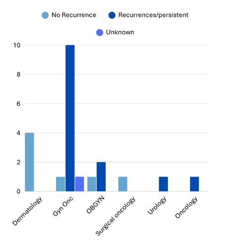
Note that one patient did not have treatment after diagnosis.
Gyn Onc: gynecologic oncologist; OBGYN: obstetrician-gynecologist.
networks that reinforce specialty-specific care pathways, and the absence of consensus treatment guidelines for EMPD. Without standardized recommendations, treatment decisions may default to provider familiarity rather than comparative outcomes data.
A multidisciplinary approach is essential to optimize EMPD management. Improved collaboration between dermatology, gynecologic oncology, and surgical oncology may facilitate appropriate patient selection for MMS and reduce recurrence rates. Additionally, the development of standardized referral pathways and increased awareness of MMS outcomes among non-dermatologic providers could help address existing gaps in care.
CONCLUSION
In conclusion, although MMS demonstrates superior outcomes for EMPD, its utilization remains limited by specialty-driven practice patterns. Addressing these disparities through multidisciplinary collaboration and guideline
development is critical to improving outcomes in this rare malignancy.
References
1. Ishizuki S, Nakamura Y. Extramammary Paget’s disease: diagnosis, pathogenesis, and treatment with focus on recent developments. Curr Oncol. 2021;28(4):2969-86.
2. Shah RR et al. Extramammary Paget disease. Part I: Epidemiology, pathogenesis, clinical features, and diagnosis. J Am Acad Dermatol. 2024;91(3):409-18.
3. Leong JY, Chung PH. A primer on extramammary Paget’s disease for the urologist. Transl Androl Urol. 2020;9(1):93-105.
4. McCarter MD et al. Long-term outcome of perianal Paget’s disease. Dis Colon Rectum. 2003;46(5):612-16.
5. Berardi RS et al. Perianal extramammary Paget’s disease. Surg Gynecol Obstet. 1988;167(4):359-66.
6. Kim GY et al. Local recurrence rates of extramammary Paget disease are lower after Mohs micrographic surgery compared with wide local excision: a systematic review and meta-analysis. Dermatol Surg. 2023;49(1): 8-12.
7. Shah RR. Extramammary Paget disease. Part II: evidence-based approach to management. J Am Acad Dermatol. 2024;91(3):421-30.
8. Mohseni Y et al. Diagnostic specialty predicts surgical modality in extramammary Paget’s disease: a singlecenter case series (2009–2025). Abstract 72663. AAD Annual Meeting, March 27-31, 2026.
Deep Neural Network Prediction of Interview Offers in the Dermatology Residency Match: A Multi-Institutional Analysis
Authors: *Saad Maan,1 Sahil Bhatt,1 Adam Kashlan,1 Angela
P. Mihalic,2 Loren Krueger1
1. Emory School of Medicine, Atlanta, Georgia, USA
2. University of Texas Southwestern Medical Center, Dallas, USA
*Correspondence to smaan@emory.edu
Disclosure: The authors have declared no conflicts of interest.
Keywords: AI, applicant advising, deep neural network, dermatology, interview prediction, medical education, residency match, Texas STAR.
Citation: Dermatol AMJ. 2026;3[1]:65-66. https://doi.org/10.33590/dermatolamj/W6KG9XO6
BACKGROUND AND AIMS
AI has shown promise in medical education,1-3 yet its ability to predict residency application outcomes in dermatology has not been studied. The authors sought to evaluate whether a deep neural network could accurately predict interview offers in dermatology using multivariable applicant and program data.
MATERIALS AND METHODS
The authors developed a dual tower neural network in Python (Python Software Foundation, Beaverton, Oregon, USA) using dermatology application data from the Texas STAR database (n=74,534 applications; 943 applicants; 144 programs). One tower encoded applicant-level features, including Step 2 Clinical Knowledge (CK) scores, research productivity, geographic factors, and signaling behavior. The second tower encoded program-level features, including institutional ranking and program size. Model performance was assessed by comparing
predicted interview offer rates across several key applicant and program variables.4
RESULTS
Away rotations were the strongest predictor of interview offers (86.1% offer rate versus 10.6% without). Program signaling was also highly predictive (49.2% versus 12.5%; Figure 1). Step 2 CK scores between 270–274 were associated with the highest interview rates (31.9%), while scores lower than 250 corresponded to rates under 11.6%. Geographic differences were also notable, as applicants from the New England region had the highest interview rate at 17.3% compared to the lowest 9.9% rate among applicants from the Pacific Northwest. Collectively, these findings demonstrate that both applicant behaviors and regional/program context contribute meaningfully to interview outcomes.
CONCLUSION
A dual tower neural network trained on a large, multi-institutional dataset demonstrated strong predictive performance for dermatology interview offers. The model identified variables such as away rotations, signaling, Step 2 CK score range, and geographic origin as significant determinants of interview rates. These findings suggest that AI-based tools have the potential to serve as a valuable resource for data-driven advising in the dermatology residency match process. Importantly, this model is intended to augment, not replace, holistic applicant review by mentors and programs. Because Texas STAR data is self-reported and historically derived, unmeasured confounding and reporting bias may affect model estimates. External validation across future
match cycles and independent datasets is needed to confirm generalizability and assess equity across applicant subgroups. If prospectively validated, this framework could
support personalized advising, strategic signaling decisions, and more transparent applicant-program alignment.5,6
Val: validation.
References
1. Gottlieb-Smith R et al. Artificial intelligence in residency recruitment: impact on equity. Neurol Educ. 2024;3(3):e200150.
2. Patel R et al. Neural network–based model predictive control framework incorporating first-principles knowledge for process systems. Ind Eng Chem Res. 2025;64(18):9287-302.
3. Sumner MD et al. The use of artificial intelligence in residency application evaluation-a scoping review. J Grad Med Educ. 2025;17(3):308-19.
4. UT Southwestern Medical Center. Texas STAR: Seeking Transparency in Application to Residency.
Available at: https://medschool.utsouthwestern.edu/ about-the-school/student-affairs/texas-star.html. Last accessed: April 8 2026.
5. Kashlan A et al. Artificial intelligence-based prediction of dermatology residency interview success. Poster 77235. AAD Annual Meeting, March 27-31, 2026.
6. Burk-Rafel J et al. Development and validation of a machine learning-based decision support tool for residency applicant screening and review. Acad Med. 2021;96(11S):S54-61.
Figure 1: Dermatology model training and validation metrics across epochs.
Congress Interview
In this issue, we present a perceptive interview with Tammie Ferringer, Geisinger Medical Center, Danville, Pennsylvania, USA, a leader in dermopathology and medical education. She shines a light on how dermatology education has evolved, the importance of clinicopathologic correlation, and the importance of leadership as a physician. With her vast leadership experience in the American Academy of Dermatology (AAD), she provides invaluable insights on the Academy's role in shaping the future of the field, and offers a deeper perspective on education, diagnosis, and leadership in modern dermatology.
Featuring: Tammie Ferringer


Tammie Ferringer
Division Chief of Dermatologic, Oral and Soft Tissue Pathology, and Section Head and Fellowship Director of Dermatopathology, Geisinger Medical Center, Danville, Pennsylvania, USA
Citation:
Q1Dermatol AMJ. 2026;3[1]:67-72. https://doi.org/10.33590/dermatolamj/3H431ZEB
Your career has brought together clinical dermatology, dermatopathology, education, and leadership. What first drew you to dermatopathology, and how has that shaped the kind of physician and educator you’ve become?
At the Medical College of Pennsylvania, Philadelphia, USA, my first 2 years of medical school were all didactic, and I loved the second year when we did pathology. I felt like I was finally getting the answer to things. The histopathology is the final diagnosis; you can actually see it under the microscope. Something that isn’t necessarily clear just by seeing the patient in the clinic.
I thought very seriously about doing a pathology residency and chose to do pathology as my first elective during my third year at Allegheny General Hospital, Pittsburgh,
Pennsylvania, USA, to get a better sense if it was for me. During that month, the chief resident at the time was going to graduate and go into a dermatopathology fellowship. I spent a lot of time with her going over dermatopathology specimens, and loved combining and correlating them with the clinical side.
I went into my pathology rotation, really enjoyed it, but it didn’t include the direct patient interaction I desired. I resigned myself to thinking you probably can’t have both, but when I saw dermatopathology and the correlation with the clinic, I wanted to see more clinical dermatology. A friend kindly switched rotations to give me a chance, and I was so lucky to do the coveted dermatology rotation in Ira Cohen’s private practice in Pittsburgh. I saw patients and performed minor procedures on a
daily basis, spoke and connected with them, and even reviewed the dermatopathology from some of the patients. At that point, there was no question in my mind that I wanted to practice dermatopathology.
As far as what kind of physician and educator I am, thanks to Cohen’s recommendation, I was fortunate enough to be accepted into dermatology residency at Geisinger Medical Center, Danville, Pennsylvania, USA. Fred Miller, former department head of Dermatology at Geisinger Medical Center, was the chair at the time. He really connected with every patient, and that became my goal when seeing patients in clinic.
After residency, I stayed on as a staff member at Geisinger, the same time that Dirk Elston, Professor and Chairman, Department of Dermatology and Dermatologic Surgery, Medical University of South Carolina, Charleston, USA, a master in
dermatopathology, joined the department. He set up a much more structured dermatopathology curriculum for the dermatology residents with innumerable glass slide teaching cases, and I watched him teach every Friday session with the dermatology residents. I absolutely loved his teaching style. He kept them engaged and interested. Something I have strived to emulate ever since.
When I joined the staff at Geisinger, I wanted to do half-time clinic and half dermatopathology because I really loved being with patients, but I also loved being behind a microscope in the lab. At the time, I thought this 50/50 split was perfect, but over time, I realized that I gravitated more toward dermatopathology.
Now, I mostly do dermatopathology, only seeing patients once a month in the melanoma clinic with the dermatology residents. Although
working behind a microscope to read dermatopathology can seem isolating, I actually have lots of interaction with my fellow colleagues, and through my other passion, teaching residents and fellows, while I am signing out cases. I still get to teach and interact with people, even if it’s not a patient.
Q2 You’ve spent years training fellows at Geisinger. What has it meant to you to help shape the next generation of dermatopathologists, and what do you think distinguishes a truly strong training program?
Teaching fellows is probably the most rewarding thing I get to do. I love to see their passion and interest in dermatopathology, and help them build on and take it into the future, even teaching new generations. I’m proud of what they’ve all become, and I’ve been incredibly lucky to always have excellent fellows.
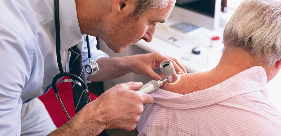
We make sure all aspects of dermatopathology are covered in a structured and comprehensive curriculum, because you never know what you’re going to see in day-to-day sign-out
For a strong training program, I think it is important to have a high volume and variety of specimens that cover the spectrum of the field, so that fellows really get the breadth of training.
They need to be able to start previewing cases from Day 1. I’ve heard of some programs where fellows are just observing faculty sign out cases, and I don’t think that prepares them for their own practice. They also need to have a structured curriculum. We make sure all aspects of dermatopathology are covered in a structured and comprehensive curriculum, because you never know what you’re going to see in day-to-day sign-out.
Q3
As a co-author of ‘Dermatopathology, Fourth Edition’, what did revisiting the field in that format reveal to you about how dermatopathology is evolving, and what do you think today’s learners need most from a resource like that?
It’s fun to work on a new edition of a book because you really must revisit every aspect of dermatopathology again. It’s interesting to see all the new discoveries. There is always something more to add, new entities, and tons of molecular developments are going on right now. We are able to understand the
pathophysiology of so many things, define entities better, and even think about treatments based on the molecular findings. All of this is really exciting.
As far as our book, what I think distinguishes it as a strong resource is the fact that it is very image-heavy. One thing I remember from learning dermatopathology myself is that I would look at all these pictures with captions saying what it was, but I’d think, ‘What specifically are they referring to?’ Therefore, we annotated the images with arrows and circles to indicate exactly what we are talking about, so that trainees can really make sure they’re looking at the right thing.
We also tried to keep the text very short with bullet key points. Attention spans are short. They don’t need an encyclopedic explanation of the history of a lesion. They just need to know the defining features.
Q4Clinicopathologic correlation is central to dermatopathology. Why is it such a challenging skill to develop, and what do clinicians misunderstand most about what dermatopathologists need for an accurate diagnosis?
Dermatology training requires dermatopathology. It makes up
one-quarter or more of the training in dermatology, and I think this is incredibly important.
As people become clinical dermatologists and go into practice, especially surgeons, but others as well, they forget a lot of their dermatopathology training and the importance of clinicopathologic correlation. It is hard for this to remain at the forefront of their minds when seeing patients in a busy schedule. I often get invited to speak at Grand Rounds in different programs, where I usually talk about clinicopathologic correlation to re-emphasize its importance.
When doing biopsies, the provider needs to think about where the pathology lies. Sometimes we get superficial shave biopsies when the pathology they’re looking for is in the dermis, much deeper than the surface. So, if they really want to know the diagnosis, a deeper biopsy, like a punch or excision, is needed. I try to remind folks: where is the pathology of the things in your differential diagnosis? Make sure the biopsy includes this area so that you get an answer.
Some clinicians, mostly primary care physicians, submit specimens without a clinical history or differential diagnosis, believing this avoids biasing the interpretation. This is surprising to me, because the more clinical information I have,
the more likely I am to provide an accurate diagnosis. When a case is submitted simply as a “rash,” and I observe a spongiotic process under the microscope, the possibilities are broad. Numerous different things could be spongiotic.
When doing biopsies, the provider needs to think about where the pathology lies
However, if I have a clinical picture of the patient, they tell me how long the rash has been there, how it started, whether it’s itchy or not, I may be able to tell them it is pityriasis rosea. I would have never been able to say that just by looking under the microscope and seeing spongiotic dermatitis.
I sometimes give talks about how to get the most out of your biopsy, and I always emphasize the information clinicians need to make sure they tell their dermatopathologist. They need to let us know if there was a prior biopsy at that site, because nevi can mimic melanoma when they are recurrent, and if the dermatopathologist doesn’t know about the previous biopsy, they may overcall it. We also need to know if it’s been treated. A rash currently treated with a steroid may not have the diagnostic microscopic features to arrive at the diagnosis.
We need the duration, distribution, and most importantly, the clinician’s differential diagnosis, even if it’s incorrect. While clinicians may worry about biasing the interpretation, it can help picture what it is they’re looking at clinically.
I have access to the clinical photos of essentially every biopsy that I read. If there is any way to provide a photo in a Health Insurance
Portability and Accountability Act (HIPAA)-compliant manner, clinicians should give their dermatopathologist those images, because they are going to get a lot more out of their reports.
Q5
You’ve served the Academy in many leadership roles, including on the American Academy of Dermatology (AAD) Board of Directors. Looking back, what did that experience teach you about the profession and the Academy that you might not have seen otherwise?
Serving on the AAD Board of Directors, I learned about the governance structure behind the society. I never had any idea how complicated it is and who reports to whom, and how all the different committees and councils fit together. When you see the diagram with all of the committees and their relationship, it gives you a much greater appreciation of the incredible number of things going on in the AAD at any given time, across so many key areas.
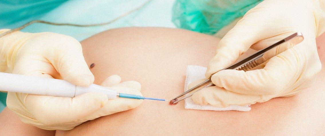
I also learned that there are ways to bring your questions, concerns, and issues to the academy that I did not really understand before. For example, I knew the Advisory Board existed, but I did not know what it was, how it worked, or how states could bring things to the AAD for consideration.
I also learned more about the membership itself and how difficult it is to represent such a large number of people with different interests. For example, I love dermatopathology, but dermatopathologists make up a small portion of AAD members. Pediatric dermatologists are also another relatively small group. Trying to address everyone’s issues is difficult.
As for the profession, it showed me how vast it is and how many different aspects there are within it. It gave me a greater appreciation for some of the challenges across the field. For example, I knew there was a critical shortage of pediatric dermatologists, but now I have a greater appreciation of the problem and why it is such a challenge to address.
Q6
What stood out to you most at this year’s AAD Annual Meeting in Denver, and are there any themes from the meeting that you think will meaningfully shape dermatology practice going forward?
I would say the main theme was: ‘Be curious’. There is always more to know. It is just unbelievable how much new information is coming out on a daily basis. When you are a resident, you think you are going to learn it all during training, and then that is it, but that is really just the beginning.
Be curious, keep learning, and recognize that there is always more to know
Every day, there is something new to learn: new drugs, new molecular findings, new procedures that somebody designed, and new diagnoses coined. There are constant developments, so it is important for us to keep up, or at least do the best we possibly can to keep up.
It really requires lifelong learning. At the same time, it is exciting that all of these new things are coming out. Be curious, keep learning, and recognize that there is always more to know.
Q7From your experience within leadership, where do you see AAD playing the most critical role in shaping the future of dermatology and dermatopathology, particularly in areas like education, workforce development, and emerging technologies?
I was not involved in much advocacy before I was on the AAD Board of Directors, and the importance of that has become very clear. I think it is one of the most critical things the AAD is doing.
I also think education is incredibly important. Education is one of my passions. I can say that the AAD stays at the forefront and gets that information out to its members. Approximately 95% of all dermatologists are members of the AAD, which is almost unheard of for
most specialty societies, so they really do reach almost everyone.
They work to keep members at the cutting edge of what is going on in dermatology through the annual meetings, publications, messaging, and public-facing communication. They have an important public-facing campaign to help people understand the importance of certain issues and to avoid getting caught up in some of the misinformation that spreads on social media. There are definitely a lot of myths that need to be corrected.
I alluded to workforce development earlier with pediatric dermatology. I do not think we have figured out the answer yet. As I mentioned, it is a small proportion of dermatologists, so it can be difficult to make that a primary advocacy issue or focus, but they are definitely aware of the pediatric dermatologist shortage and are looking into it.
As far as emerging technologies are concerned, this is certainly something we need to be at the forefront of. The academy stays current with what is coming and what is changing, and they create work groups, task forces, or whatever is needed to address those issues.
For example, the Augmented Intelligence committee is working to be at the initial table, instead of waiting for things to happen and then asking, “Why didn’t you involve a dermatologist?” It should not work that way. We should be at the drawing board when things are first developed so that they are built correctly from the beginning.
It is analogous to the electronic medical record, which was created by people who had no real understanding of medicine, and then handed to physicians who didn’t find it appropriate for what they needed and had to try to work with developers to make it more appropriate. Instead of doing that, we need dermatologists involved from the start. Something the AAD is taking very seriously.
One other thing about the AAD that I really respect, and that is probably part of the reason I am where I am today, is how much they value leadership training. Something that is really not taught in medical school or residency programs, even though it should be.
Residents finish training and then, suddenly, they need to run their own practice. AAD has been extremely good at building leadership skills. I was a member of the Academic Dermatology Leadership Program (ADLP), which was a year-long program, and attended the Leadership Forum multiple years in various capacities, including as Chair of the Leadership Forum and then Chair of the Leadership Development Steering Committee. I also gave talks at the academy meeting on leadership skills.
All of these things are really valuable, and they are things that a lot of other societies do not put the time and effort into.
Q8At this year’s AAD meeting, you were involved in sessions on clinicopathologic selfassessment and diagnostic mimics and masqueraders. Was there a common message you hoped attendees would take away from those discussions?
The main message is about getting the diagnosis, which sometimes you cannot do just by looking at the patient. Having that histopathologic component can provide the final answer. While that’s not always the case, most situations involve either clinical mimics or histologic mimics, but rarely both at once, so you can usually arrive at a diagnosis.
I also gave another talk at the AAD Annual Meeting, ‘Pearls for Running a Successful Dermatopathology Practice’. I talked about leadership skills in general, more than specifically about dermatopathology. We talked a little bit about Strengths, Weaknesses, Opportunities, Threats (SWOT) analysis.
One of the take-home points is the importance of setting aside time
to work on your practice, not just in your practice. Everyone goes to work, sees as many patients as possible from the minute they walk through the door to the minute they leave, but they need to set aside a little time to think about the practice itself. Look at the practice’s strengths, weaknesses, opportunities, threats, and then make a plan and put it into action. If you do not set aside time to do this, it is going to get lost in the process, and things can unravel very easily.
The other thing I emphasized was setting culture; taking the time to think about your culture, your mission, and what your goals are for your practice. It was funny when I was preparing the talk, I spoke with several colleagues I really respect, who set up dermatopathology practices and accomplished a great deal in the field. One of them said, “Well, what do you mean by successful?” I replied, “That is a good question.”
To me, success means getting the right answer for the patient and working with a group of people who love learning together. While others may define success in terms of financial gain, it’s important to determine what success means to you personally.
Society Update
SCALE 2026
Music City SCALE 2026 returns to Nashville, TN, from May 13–17, at the Grand Hyatt Nashville, bringing together the brightest minds in aesthetics and medical dermatology for five days of innovation, education, and connection. Now in its 21st year, SCALE has established itself as one of the premier multi-specialty dermatology and aesthetic medicine conferences in the country, attracting leading physicians, advanced practice providers, and industry experts from across the globe. Set in the heart of downtown Nashville, SCALE combines high-impact education with the energy of Music City. In addition to the education, attendees can expect vibrant networking events, live entertainment, and memorable experiences that make this meeting unlike any other in the field.
Why Attend SCALE 2026
• World-Class Education
Gain insights from over 90+ expert faculty across dermatology, plastic surgery, and aesthetic medicine, covering the latest clinical advancements, technologies, and treatment strategies.
• Hands-On & Interactive Learning
Participate in highly sought-after workshops, live demonstrations, and ultrasound-guided anatomy sessions designed to enhance precision, safety, and outcomes in clinical practice.
• Cutting-Edge Topics
Explore emerging trends including injectables, energy-based devices, regenerative aesthetics, longevity science, and practice management strategies, all curated to elevate your practice.
• CME/CE Opportunities
Earn up to approximately 30.5 hours of accredited continuing education credits while engaging in clinically relevant, evidence-based programming.
• Unmatched Networking
Connect with more than 1,400+ attendees and 125+ industry partners, fostering collaboration, partnerships, and business growth in a dynamic, highenergy environment.
From foundational techniques to advanced innovations, SCALE 2026 will deliver practical, practice-changing education that attendees can immediately implement, making it a must-attend event for anyone serious about staying at the forefront of aesthetics and medical dermatology. Register now at www.scalemusiccity.com
Interviews
In this issue, we bring forward four thoughtful interviews with key leaders, educators, and researchers in dermatology. First, David Fisher, Harvard Medical School, Boston, Massachusetts, USA, speaks on understanding how UV exposure, pigmentation, and the immune system drive cancer treatment. Next, Warren Heymann, Director, American Board of Dermatology (ABD), highlights how great dermatologists are not defined by knowledge alone, but by curiosity, empathy, and genuine care for their patients. Then, Paul Bigliardi, University of Minnesota, Minneapolis, USA, explains how dermatology outcomes also depend on recognizing drug allergies and signals that the skin reveals. Finally, Christopher Tomassian, The Dermatology Collective, Glendora, California, USA, sheds a light on how building a successful dermatology practice today means pairing strong business fundamentals with ethical, evidence-based care.
Featuring: David Fisher, Warren Heymann, Paul Bigliardiand, and Christopher Tomassian

David Fisher
Edward Wigglesworth Professor of Dermatology; Department of Dermatology, Massachusetts General Hospital, Harvard Medical School, Boston, Massachusetts, USA
Citation: Dermatol AMJ. 2026;3[1]:74-80. https://doi.org/10.33590/dermatolamj/OGRK4881
Q1The discovery that UV exposure can trigger an endorphin response in the skin suggests a biological basis for sun-seeking behavior. What does this reveal about the evolutionary role of UV exposure, and how might this knowledge be leveraged for a new approach to skin cancer prevention?
The discovery that UV, when it hits our skin, not only induces things we've known about for a long time, like DNA damage, mutations, and the ability to induce melanin synthesis, the so-called tanning
response, but also simultaneously triggers the production of endorphin, is relatively new. It was discovered and reported just over 10 years ago, and it turns out that this response, the molecular activation of endorphin synthesis, is linked molecularly to the very pathway that triggers melanin production. You cannot induce melanin, the tanning response, without making endorphin; it is part of a common precursor that involves both of these. So it is organically linked, embedded in our genomes, and it is part of evolution over presumably countless millennia.
UV is triggering the body's opiate-like response, producing measurable behavioral effects
There is something extraordinarily counterintuitive about this; UV is triggering the body's opiate-like response, producing measurable behavioral effects. This has been observed in mice and identified in humans as well. There isn’t a more powerful type of behavioral response than opiate responses, which can even have addictive-like consequences. But here, we see a behavioral inclination to seek UV, the most common carcinogen in our environment. How could we possibly have evolved to expose ourselves to one of the most dangerous environmental factors associated with the most common cancers in humans, skin cancers? It feels absolutely backwards.
When something is so counterintuitive, an evolutionary perspective suggests it has to be
true. There must be some beneficial effect that would have been selected for evolution, despite the fact that UV is so damaging to our skin and heavily implicated in skin cancer risk. Research explored this and found that UV participates in the synthesis of vitamin D in our skin. There is one carbon–carbon bond in the precursors to vitamin D that the human body, and indeed the entire animal kingdom, cannot cleave with an enzyme, but it must be cleaved to produce vitamin D; UV does this.
In evolutionary terms, if you lived in low-light regions, like Scandinavia, you risked vitamin D deficiency, which could be fatal in childhood, causing rickets. Anything that enhanced the ability of light to produce vitamin D could have conferred an evolutionary advantage. This likely contributed to the evolution of lighter skin in highlatitude populations, and endorphinmediated sun-seeking behavior may have reinforced this adaptation.
Interestingly, studies, mostly in animals, showed that vitamin D
levels feedback on endorphin responses. Vitamin D-deficient animals experience stronger euphoria from endorphins, whereas animals with normal vitamin D levels do not. This forms a selfcorrecting loop that would have helped sustain behaviors ensuring sufficient vitamin D intake over evolutionary time.
The modern challenge is that this pathway still exists, but we no longer need UV to maintain vitamin D levels, we can get it safely from inexpensive supplements. Yet the behavioral response to sunlight remains, which continues to drive skin cancer risk. This is why, despite public health campaigns and sunscreen use, skin cancer incidence remains high.
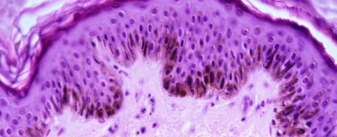
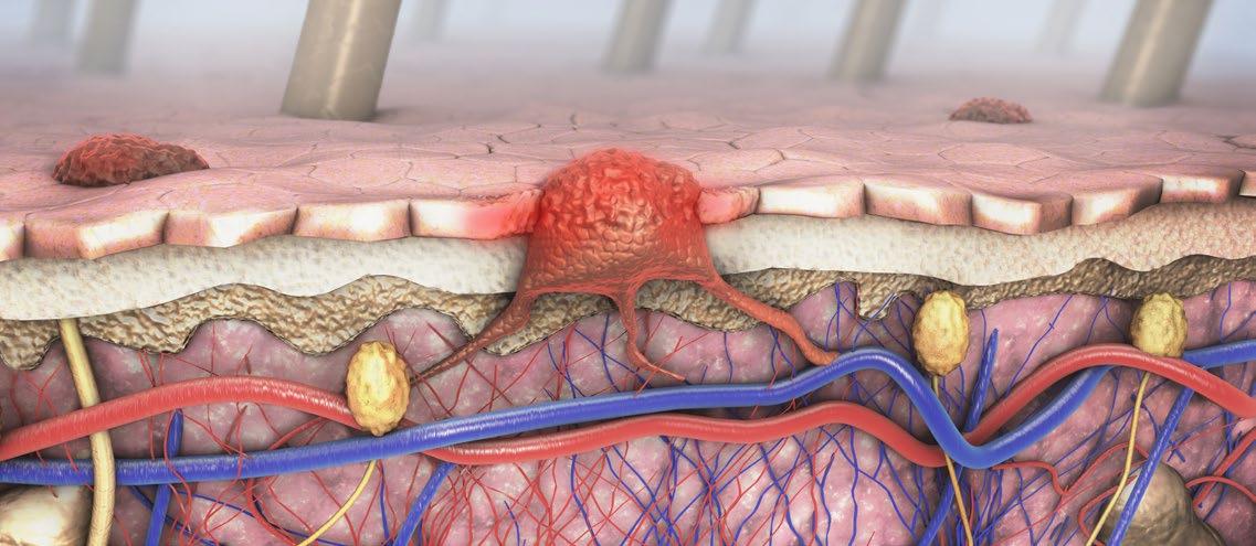
Q2
Red hair and fair skin pigmentation are known risk factors for skin cancer. What specific molecular mechanisms, identified through your research, are responsible for this increased risk, and how can that knowledge lead to more targeted preventative measures?
There have been some very important developments in our understanding of why it is that lightskinned individuals and red-haired people are at higher risk. Red hair is one extreme example, but for lightskinned people like myself, even though my hair is not red, my skin is probably closer to a red-haired person than a darkly pigmented person. That turns out to be very important in terms of the risk of melanoma formation.
There was a study from my lab about 15 years ago that generated a lot of press. It was a disruptive observation made in the laboratory using mouse models of redheads. This was a mouse model where the exact same gene associated with
the red hair phenotype in humans was engineered into mice and used as a background for studying melanoma risk. In this experiment, one of the oncogenes associated with the formation of nevi, benign moles, was crossed into the redhaired mouse background. The plan was to use UV to add additional mutations and then, if these mice developed melanomas, try to discover what those other mutations were and how melanoma gets caused on a light-skinned genetic background in a mouse model.
However, something unexpected happened. When this common oncogene, BRAF V600E, present in about two-thirds of moles, was combined with a red-haired mouse background, about half of the mice spontaneously developed melanomas over the course of 6–12 months, which is half to a full lifespan in mice, even without UV. Genetically black mice did not show this effect. This observation suggested that red-blonde pigment, called pheomelanin, has a unique chemical activity that is
weakly carcinogenic. Even in the absence of UV, it produces reactive oxygen species that constantly mutate DNA in the skin.
People with red hair have an elevated risk of melanoma, which is well known, and this risk is evident even in skin that is not sunexposed, consistent with the idea that pheomelanin itself contributes to mutations. Interestingly, albino individuals and mice that produce no pigment have a dramatically lower risk of melanoma, although they have a much higher risk of nonmelanoma skin cancers, highlighting the unique role of pheomelanin.
From a prevention perspective, it is important to be careful with sun exposure. UV interacts not only with DNA but also with redblonde pigment, amplifying reactive oxygen damage. Sun protection, clothing, and sunscreen are key.
However, traditional sunscreens, calibrated to prevent sunburn, may not provide optimal melanoma protection. SPF measures UVB
protection, which prevents burning. UVB protection is unequivocally of great importance. However UVA, which contributes to oxidative damage and photoaging, is less well filtered by most chemical sunscreen ingredients. Improving UVA protection and understanding the chemistry of pheomelanin–UV interactions is essential for more effective melanoma prevention.
Q3
Looking toward the future of melanoma therapy, what are the key milestones that you believe must be achieved to fundamentally transform how we treat the disease?
It ought to be preventable. We're not there yet in terms of prevention, and hopefully that will be coming, but for patients who have melanoma, the traditional therapy for early melanoma is surgery. You remove it, and hopefully it will never come back. Unfortunately, sometimes it does because it's a very invasive tumor, and even melanomas just thicker than 1 millimeter have, unfortunately, a significant risk of invading and metastasizing.
The treatment for metastatic melanoma has dramatically improved in recent years, and the most important component in our armamentarium has been the ability to harness the immune system through a class of drugs known as immune checkpoint inhibitors. These are antibodies that, without going into too much mechanistic detail, activate the immune system and allow it to attack the melanoma cells and kill them.
The idea of harnessing the immune system to kill melanoma cells is over a century old, and unfortunately, did not work for
most of that century. There was a breakthrough back in the 1990s and 2000s when a very specific set of molecular pathways were discovered that function as checkpoints to prevent the immune system from killing our own cells. When those checkpoints could be blocked to allow the immune system to attack the cancer cells, because cancer is our own cells, it liberated the ability of the immune system to attack melanoma cells. Between a third and a half of patients with metastatic melanoma produce major, durable remissions or complete remissions. This is a huge breakthrough. It was science-driven, based on understanding the pathways that regulate the immune system.
Why was it melanoma that benefited first? Probably because melanoma is one of the cancers with the most mutations in its genome. It has an enormous density of mutations. These mutations come largely from UV, from sunlight, because melanoma is a cancer that develops in the skin from cells exposed throughout life to UV. It turns out that when a melanoma harboring many of these mutations sets up shop wherever it metastasizes, it expresses slightly mutated genes due to these frequent UV mutations. The immune system, if lucky, may recognize some of these mutations as though they're foreign. It never saw them before.
Our immune systems are educated to avoid attacking our own cells, but these mutations produce what we call neoantigens, or UV-derived neoantigens, which are not part of our normal genome, but are clonally amplified as the tumor grows from one original cell. The
immune system can look at these and say: "I never saw this before." On the other hand, if it were that simple, melanoma wouldn't exist. As soon as melanomas grew, the immune system would kill them, and we wouldn't even see them. But until recently, when these immune checkpoint inhibitors were developed, melanoma still grew and killed people.
The reason the immune system was stopped is that the tumor would fight back by telling the immune system: "I'm one of you, I'm self, you can't kill me." This is part of the tolerance mechanism. The checkpoints activated by the tumor to the immune system prevented these tumor cells from being attacked. When drugs were developed to block that tolerance mechanism, the immune system was liberated to attack and destroy the tumor. This works in about a third to half of melanoma patients. There can be simultaneous autoimmune side effects involving immune attack of separate healthy cells, in some people, as you might expect, but frequently this can be managed safely.
There is good news and frustration. The good news is that a significant fraction of patients with metastatic melanoma are cured, who otherwise would have had almost no chance of surviving for long. Even better, in recent years, these same treatments have been moved to earlier stages of the disease, such as thicker cutaneous melanomas or those spread to local lymph nodes, like under the arm or neck. These patients have a statistical risk of around 25–50% of relapse. Using immune checkpoint inhibitors in these highrisk patients shows about a 50%
reduction in relapse. This is even better because there are more patients with earlier melanoma than late melanoma. These discoveries improved outcomes by applying a rational, mechanism-based therapy first in advanced disease, and now in earlier stages.
Where does this leave us? What do we still need to overcome? The other 50% of patients who do not respond are very important. Why are they not responding? In some cases, the density of UV mutations in the tumors is not as high, so they do not make as many neoantigens, and the immune system does not recognize them as foreign. This is a problem for most human cancers, such as breast, lung, colon, etc., which generally do not respond as well to immune checkpoint inhibitors. Researchers are exploring ways to artificially inflame tumors to trick the immune system into attacking them.
For earlier melanoma, another challenge arises: some immune treatments carry rare but serious autoimmune side effects. If you have a population where 25% are at risk of relapse, offering immunotherapy reduces that risk but exposes the other 75% to potential harm. This creates a dilemma because the principle ‘do no harm’ applies. We need biomarkers to predict which patients are most likely to benefit, so the therapy is targeted.
Another area is targeted therapies, or drugs that go after specific mutated oncogenes driving melanoma growth. Some have impressive activity, but tumors often develop resistance. Understanding and overcoming these resistance mechanisms is crucial to make responses more durable.
Q4Your work on giant congenital nevi involves modeling this condition in mice and developing potential topical drug treatments. Could you discuss the process of translating a laboratory finding into a viable clinical approach for a childhood condition?
Giant congenital nevi are among the most common causes of pediatric melanoma, which is a relatively uncommon but devastating disease. You can imagine a child with melanoma, and unfortunately, these are melanomas that do not arise as much from UV, and therefore do not tend to have a good response to immune checkpoint inhibitors.
Removing congenital giant nevi or diagnosing melanomas and removing them very early would, of course, be the preferred approach. The problem with congenital giant nevi is that when a baby is born with these large moles over much of their body, they are very difficult to remove surgically. If you remove them surgically, you are essentially converting a giant mole into a giant scar, which will be a debility for the rest of their lives. Parents are put in an absolutely anguishing position of whether to leave it or watch very carefully, but even if they watch, the child grows up with a somewhat disfiguring, psychologically challenging condition to live with.
We wanted to study this because the oncogenes causing the growth of those nevus cells in giant congenital nevi are known in most cases, and we were able to genetically engineer models in animals. Several other research groups have been able to do this as well, creating mice born with giant congenital nevi driven by the exact same oncogene. We were able to
test whether there were any drugs that might trigger regression of these lesions, avoiding the need for surgery and potentially reducing the risk of melanoma developing later.
Several years ago, we reported a study in which we used a form of immune therapy to inflame the skin and topically administer treatment to trigger infiltration by macrophages, which would gobble up and kill the nevus cells. The skin would lose hyperpigmentation after recovery. These treated mice had no melanoma formation in the treated areas, whereas untreated areas still developed melanoma, making this almost like a controlled study. It was a promising approach to developing a drug treatment.
This has not yet been studied in humans for one reason: it is highly irritating to the skin. It is a pro-inflammatory, immunebased therapy, and very itchy and annoying, although temporary. We have been deliberating whether to start treating now or try to ameliorate the itch and toxicity without losing efficacy. The risk of melanoma is significantly elevated compared to a normal child, but still only about 10%, meaning there is a 90% chance of not having melanoma. This allows time to explore anti-inflammatory approaches, adjusting dose or schedule, to make the treatment better tolerated for children, parents, and doctors.
Other groups have been testing oral drugs that do not regress lesions much but may reduce some spontaneous symptoms. The long-term effect on melanoma risk remains unclear. Fortunately, multiple research groups worldwide are working on this problem.
Although relatively uncommon, giant congenital nevi affect many children globally, and a better therapy would be a significant improvement.
Q5Your research has identified a direct link between the cell death pathways involved in hair graying and those that control melanocyte survival. Could you explain this intriguing connection and its therapeutic implications for both hair pigmentation and melanoma?
We have been very interested in why your hair turns gray, not just because we know it is inevitable, but actually because the melanocyte, the pigment cell, which is the origin of melanoma, is a very difficult cell to kill.
If you think about it, we spend our lives under the sun, bombarded by UV. These cells accumulate mutations, but they don’t die. If
they died, UV would cause our skin and hair to turn white just from radiation. But melanocytes survive, somehow fending off the cell death response to all that DNA damage. There are unique survival behaviors, and we have known for many years that traditional chemotherapy drugs don’t work in melanoma, as it grows right through cytotoxic chemotherapy drugs.
About 20 years ago, I became interested in understanding why melanocytes die in our hair follicles as part of the normal cycle. Hair follicles go through cycles of growth, regression, and rest. During the regression phase, melanocytes and keratinocytes undergo apoptosis or cell suicide. Keratinocytes make the hair fiber, and melanocytes make the melanin that colors the hair. At the end of each follicle cycle, which can last a few months to a few years, these cells die, and stem cells for keratinocytes and melanocytes are
reawakened to form a new follicle. After growth, during which the hair is pigmented, the cells involute and die, and the cycle continues.
I wondered if understanding the efficient death of melanocytes in hair follicles could provide clues for treating melanoma. We started using hair graying as a model, where pigment cells die prematurely or stop producing pigment. In the early 2000s, the cause of hair graying was unclear. It turned out that melanocyte stem cells in the hair follicle were dying prematurely. Each hair follicle has a finite number of maybe 10–20 melanocyte stem cells. By age 20, there are fewer than at birth, and by 40 or 50, almost none remain. When there are no melanocyte stem cells in a follicle, the hair produced has no pigment and appears white. The follicle cycles are asynchronous, which is why hair looks gray first and then white over time.
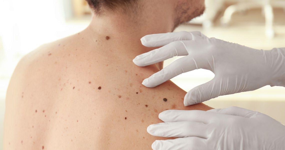
More recently, in collaboration with Ya-Chieh Hsu, a stem cell biologist at Harvard University, we discovered that stress accelerates the death of melanocyte stem cells in hair follicles. This confirmed the old wives’ tale that stress can turn your hair gray. In mice, various forms of stress, from social isolation to painful injections, caused subsequent hair cycles to grow hairs lacking pigment. The mechanism involves the sympathetic nervous system, which has nerve endings in the stem
cell compartment of the hair follicle. It releases a neurotransmitter that causes melanocyte stem cells to differentiate prematurely, produce inappropriate melanin, and eventually die. Blocking this neurotransmitter protected against stress-associated hair graying in mice.
What does this tell us about melanoma treatment? We are not quite there yet. It shows that previously unrecognized pathways regulate life and death decisions in
melanocytes. Perhaps analogous approaches could target hyperdifferentiated melanoma cells to induce their demise, but this remains an open question for future research.

Warren Heymann Director, American Board of Dermatology
(ABD)
We have individual studies on what works and what doesn't work, but we need more comparative studies
Disclaimer:
These answers are Heymann’s personal opinions only and do not represent the views of the American Board of Dermatology (ABD).
Citation: Dermatol AMJ. 2026;3[1]:81-85. https://doi.org/10.33590/dermatolamj/707QV0W0
Q1What initially inspired you to pursue dermatology, and how did your early experience help shape the way you approach patient care today?
When I was in medical school, my brother had a rash. In retrospect, it was probably pityriasis rosea, but I don't know that for a fact. At the time he showed it to me, I was a second-year student, and I really had no idea what it was. He went running to my father and told him that he was throwing money out on me in medical school, so I thought maybe it would be a good idea to take a dermatology elective.
I was leaning toward internal medicine and that is what I originally applied to. One of my internal medicine residents, who I admired a lot, went into dermatology, so I decided the month before the Match that it was time to take on the elective. I was a fourth-year student, and within a few days I realized that dermatology was fun and interesting, and it clicked.
It was the first time I really felt that I could excel at something in medical school. Not just do okay, but do really well; it was just a gut feeling. But it was a very different world back then, in 1979, because it was not that competitive; there was no Match in dermatology. I was able to apply and, a couple of weeks later, had a spot where I went to medical school.
I was very impressed with our Chief of Dermatology at the time, Michael Fisher, especially about a case on the wards, which may have convinced me. This young man who had Crohn's disease, just like my brother, had a blistering rash that no one could figure out. Fisher just looked at the patient’s hand and said: "He's deficient in zinc from his total parenteral nutrition. Supplement zinc." That person had acrodermatitis enteropathica, and this mysterious rash went away very quickly with the zinc supplementation. To me, that was magical.
Q2Your publication record spans over 300 articles. Looking across the entirety of your work, where do you believe that the most significant gaps in dermatology currently lie, and what would you like to see more research on?
It's always an evolving issue. We're in the biologic era of targeted medicine, and these medicines are very expensive. We have individual studies on what works and what doesn't work, but we need more comparative studies, and we need to understand molecularly what the right medication is for the right person. So, there's a lot more research needed. Even though treatment is targeted, we need comparative studies that make it truly personalized with what's best for the individual. And we’re not there yet.
We have an increasing number of biologics for a variety of indications and a variety of diseases, like psoriasis and atopic dermatitis, and increasingly alopecia and dermatomyositis. But with all these new offerings, right now, decisions are made based on insurance, and what an insurance company will pay for, rather than really understanding what the right biologic for the condition is.
Q3Over the past decade, dermatology has seen rapid developments in areas such as teledermatology, targeted immunomodulatory therapies, and new models of care delivery. From your clinical perspective, which of these advances have most significantly improved patients’ quality of life, and are there any unintended consequences?
Teledermatology was being used a little bit in the past, and then COVID-19 came along, and it became a dramatic lifesaver, in a way. I think the use has backed off now in dermatology because of reimbursement and time. When we couldn't do anything else, it was incredibly valuable. But it doesn't compare to a real visit. You can't
hold a hand, you can't do a biopsy, you can't do a culture.
It still has its value if people are really underserved and can't be seen, but not to the degree that you could have in person. For example, if you are a college kid on isotretinoin, and you can't make it to the office, it’s valuable, but while it will always be there, it is not optimal.
While I can't think of an unintended consequence, it has its limitations. Maybe there is a false sense of security. You can't do a full-body skin exam, and you can look at something that a patient is concerned about, but you didn't get their shirt off and see the melanoma on their back, because they weren't worried about it.


From the targeted immunomodulatory therapies, there are some unintended consequences as you use these. The one that comes to mind first when we were first using biologics such as tumor necrosis factor-alpha inhibitors like adalimumab was the development of paradoxical psoriasis. You gave it for psoriasis, and then it would get worse. I don't view it as paradoxical. I think it's an adverse reaction.
It can also happen with IL-17 inhibitors, like secukinumab. But we weren't aware of that. Dupilumab, for example, is still controversial. Are we unmasking cutaneous T cell lymphoma, or was it there from the start? That's a little bit of a debate. As you use new medications, sometimes you discover things you just didn't anticipate, and we can learn from those unintended consequences.
Q4From your vantage point as a clinician and prolific medical writer, how would you use AI to accelerate dermatology R&D responsibly, and what safeguards do you think are non-negotiable for any healthcare field that wants speed without sacrificing truth?
AI is not limited to dermatology. It's in every aspect of our lives, right? And, like any tool, it could be used wonderfully or in a dastardly way.
Within the last year, AI has changed my approach to how I look at literature. When I was a dermatology resident, if I had to look something up, I'd have to spend a Sunday afternoon at the library, look at Index Medicus, pull books off the shelves, find articles, and photocopy them. It would take all afternoon to get three articles. Then we computerized, and PubMed came along, and now it would take you a few minutes. But now, with AI, you can ask any question, which is marvelous.
Now, the trouble is: do you stop there? I think you need real fundamental knowledge of your field to ask appropriate
questions and to have a deep enough knowledge to know when you're being duped, and when it's hallucinating or making things up.
So, you can't just stop there. If it includes the reference, you need to go to the original sources of the references to form your own opinion. But I think in clinic, when you're working quickly, and somebody asks a question that is something you hadn't thought about before, it can be marvelous. If used properly, to our advantage, and if you have healthy skepticism and don't take things at face value, it's remarkable.
Q5 As a mentor, having supported generations of trainees, in your experience, what distinguishes a competent dermatologist from an exceptional one?
That’s an excellent question. The first reason has nothing to do with knowledge. It has to do with heart and compassion: being a person who really cares about their patients and their problems, and who is curious about finding the right answers and advocating for
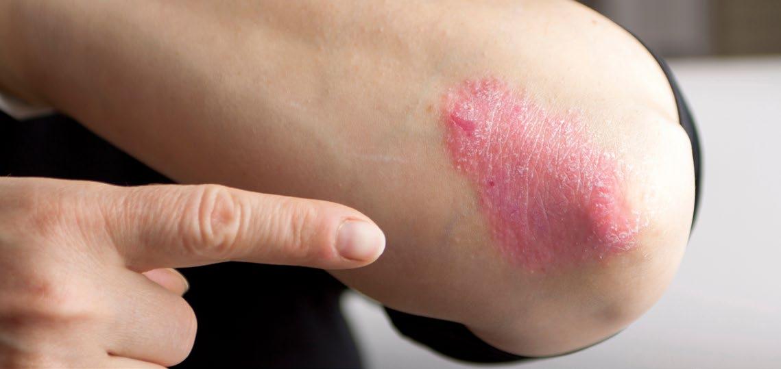
their patient. In this day, anyone can get information instantly, so I think the most important thing is what is in a person’s heart.
Anyone who goes through training and passes their boards is, by definition, good, but what makes them great is curiosity and recognizing potential problems when other people don't realize it. This can be paying attention to detail that uncovers illness that could easily get overlooked; recognizing a small lesion; not looking too quickly at the patient and, for example, recognizing arteriovenous malformations; and recognizing some large veins that could uncover thoracic outlet syndromes. It's about paying attention to detail and being curious.
Q6 Have you seen a shift in curiosity from previous generations of dermatologists versus current ones?
I wouldn't say it's a shift in curiosity. When I started, the
field was mostly general medical dermatology. For example, when I was a resident, there were only two Mohs surgeons in New York City, a city of 8 million people. Now, about 20% or so of all graduating dermatologists are surgeons. So, the sub-specialization has grown tremendously as the knowledge has grown. I think people are curious, and as knowledge expands exponentially, the field has become sub-specialized.
Q7
Throughout your career, you have been involved in teaching, research, clinical care, and leadership within dermatology. How have you seen the field evolve over the past several decades, and which innovations or shifts in thinking do you believe have had the greatest impact on the way dermatologists practice today?
Dermatologic surgery went from Mohs surgery to lasers, cosmetics, and plastic repairs, and it's magnificent. You have pediatric dermatology, advancements
in diagnosing and managing angiomas, and genetics, and the whole classification of ichthyosis is now changing based on the molecular defects of recognizing syndromes. So, whole exome sequencing and next-generation sequencing are redefining how we look at illness, based on a molecular level.
There have been huge advances in imaging in dermatology, targeted therapies, and biologics. I mean, 15 years ago, metastatic melanoma was essentially a death sentence, and now there are many cures. How fantastic is that?
When I started at my first job, I ran the psoriasis daycare center. It was tough work, and people would be grateful if you could get them to a Psoriasis Area and Severity Index (PASI) 40 with methotrexate and phototherapy and the like. Now, with the biologics, within a few shots, so many people have a PASI 100. They're completely clear, and they're upset if a spot comes back.
It's just mind-boggling to me how it has revolutionized. What used to be chronic, really hard conditions to control are now amazingly well controlled with biologics such as dupilumab and JAK inhibitors.
When I was in medical school, and I graduated medical school in 1979, we were learning about monoclonal antibodies and what they could theoretically do one day. And boy, is that day here. The advances have been incredible. Now we're moving into the oral IL-23 inhibitors, which are not even an injection anymore.
Q8You were recently recognized with the 2025 Master Dermatologist Award. Reflecting on your career, what achievement are you most proud of, and what lessons from that experience would you share with the next generation of physicians entering dermatology and medicine more broadly?
I've been very fortunate in my career, and there are a lot of things I've done that I'm very proud of. But I guess the most important one was when I was getting started with my private practice and I was able to be involved with a new medical school at the time. That is now the Cooper Medical School of Rowan University, Camden, New Jersey, USA.
For 6 years I was alone, but my goal was to set up an academic division in private practice, with the goal of being in practice, but academically oriented, so that we could ultimately teach the next generation and learn to always be as current as could be.
After being head of that division for 35 years, I stepped down a few years ago. But we now have a respected academic division of dermatology, and that setup, with that goal, enabled me to be involved in other things that I'm certainly proud of, like writing Dermatology World Insights and Inquiries, and getting involved with the first six editions of this book called Treatment of Skin Disease.
We started the residency program a little over 30 years ago, and being able to watch it grow is the wellspring of everything, so I'm most proud of that. It's a wonderful experience to have trainees and watch them go on to have successful careers. Not everybody is going to go out and set up a residency program, but I would encourage anybody to set their sights high, to always keep learning, and to never be complacent.

Paul Bigliardi
Dermato-Allergy Division, Department of Dermatology, University of Minnesota, Minneapolis, USA
Drug allergies and hypersensitivity reactions are becoming more common as the population ages and multimorbidity increases
Citation: Dermatol AMJ. 2026;3[1]:86-89. https://doi.org/10.33590/dermatolamj/5846F740
Q1How has practicing in a wide range of healthcare systems, including the US, Europe, and Singapore, shaped the way you approach skin conditions, and are there any perspectives or practices from those experiences you think could add value here in the US?
All three are highly advanced healthcare systems, but they are organized very differently. For example, in Switzerland, the state decides pricing for medication, so we know how much a medication will cost. In Singapore, when I prescribed drugs, I saw the pricing on my computer, which was interesting and certainly helpful for discussions with the patients about alternatives and their affordability. In the US, that is a little bit more difficult, because the individual drug pricing is variable and not very transparent. However, with increasingly limited resources and strain on the healthcare system worldwide, access to treatments and medications is restricted because of constantly increasing healthcare costs. Switzerland has a mandatory health insurance, and everybody is insured, including a federal insurance for professional diseases and accidents. This is a very useful and important backup for patients with occupational allergies, where all the medical evaluations and changes in the workplace and profession are financially supported. Working across these very different systems and in different dermatology departments has reinforced for me that dermatology in the US
needs to integrate allergic skin disease more fully, especially in the emerging area of drug allergies and hypersensitivities. Many of these are delayed-type reactions, and both allergists and dermatologists should be trained to diagnose and manage these complex, highly variable disorders.
In practice, dermatologists are often the first to recognize delayedtype reactions, which makes close collaboration between dermatology and allergy essential. Specialized training in skin diagnosis and testing is crucial in both fields. As treatments become more targeted, making the right diagnosis before starting therapy becomes even more important, so we can avoid expensive and ineffective treatment trials with biologics.
I’m a member of the American Contact Dermatitis Society (ACDS), and patch testing remains an important tool, but it only addresses contact dermatitis. Drug allergies, especially delayed-type reactions, are still underdiagnosed in dermatology and only partially addressed in allergy, leaving a real gap. To diagnose complex, often severe drug reactions properly, we need stronger diagnostic capabilities, including the ability to perform both immediate- and delayed-type testing in the same clinic. We also need continued progress in in vitro testing, particularly for drug allergies.
Drug allergies and hypersensitivity reactions are becoming more common as the population ages
and multimorbidity increases. Most patients who come to a clinic or hospital already have some kind of drug reaction documented in the electronic health record. For that reason, dermato-allergology should have a much stronger place in the training of future allergists and dermatologists.
Q2
Your research spans contact dermatitis, skin-barrier immunology, nerve receptors, and chronic itch. Can you give some examples where doctors are still missing signals that a skin problem is pointing to something bigger going on in the body?
One of the unique strengths of dermatology is that we can learn an enormous amount simply by looking at the skin and examining it carefully. From the beginning of my training, I learned that observation and touch can be extraordinarily valuable in a multidisciplinary setting, whether we are helping identify autoimmune disease, cancer, allergy, or another systemic process.
That clinical skill has become even more important in the era of targeted therapy. In the past, prednisone improved many inflammatory conditions. Today, we have far more targeted treatments, which is a major advance because they can reduce side effects. But precision therapy requires precision diagnosis. We need to determine which biologic is truly appropriate and what the best path forward is for each patient.
As the population ages, treatment regimens become more complex, and patients take more medications at the same time, I believe we will see more drug reactions and drug allergies, not fewer. Dermatology will therefore continue to play a crucial multidisciplinary role, especially when the skin is the first visible sign that something larger is happening in the body.
Q3 Where do doctors most often go wrong in diagnosing contact dermatitis, and what should clinicians across specialties watch out for when a reaction isn’t behaving as expected?
A classic problem is the patient with dermatitis whose presentation could represent atopic dermatitis or allergic contact dermatitis. Histology cannot reliably distinguish the two, and clinically, they can look very similar. A patient may appear to have atopic dermatitis, receive a biologic, and improve only partially. Then the question becomes: Is the biologic failing, or is something else contributing? Very often, the answer is an undiagnosed contact allergy superimposed on underlying atopic dermatitis. That has to be identified through patch testing and addressed with careful counseling on allergen avoidance.
In many cases, management is a combination of identifying the allergen, helping the patient avoid it, and treating the atopic dermatitis itself. A large proportion of patients likely fall into this mixed category.
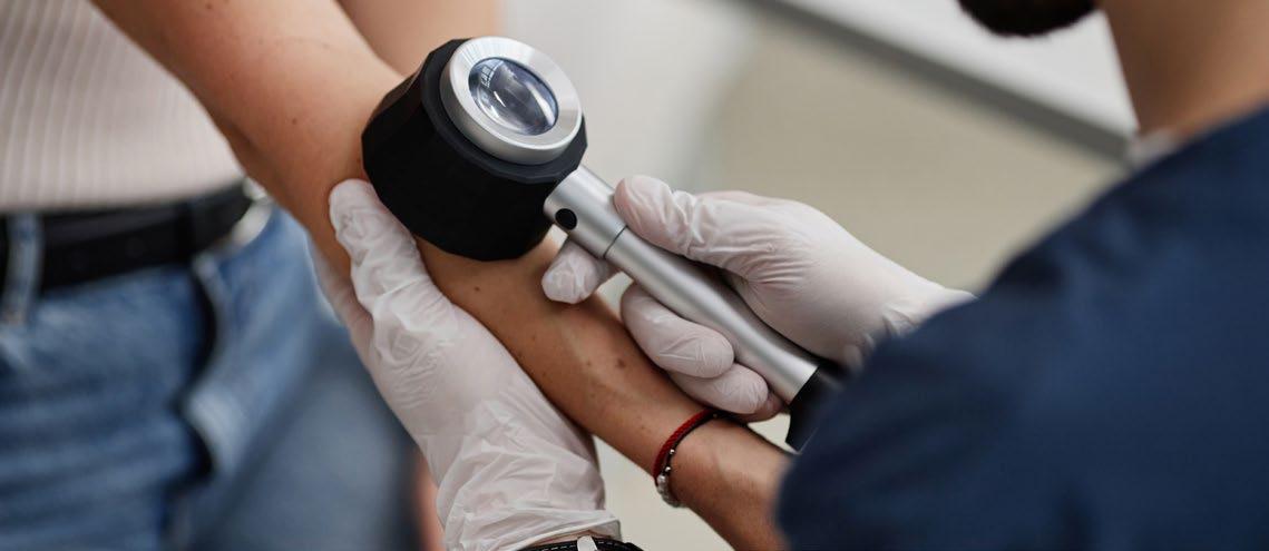
Another common error, especially outside dermatology, is to mistake eczema or an allergic reaction for an infection. There are often clinical clues that distinguish bacterial infection or cellulitis from eczema and allergic contact dermatitis, but recognizing them requires training in skin morphology and careful history-taking. The skin often reacts in similar ways, yet small differences in lesion appearance and patient history can lead us to the correct diagnosis. Itching indicates more of an allergic reaction, and pain points more towards infection. Combining all those skills is the art of an experienced clinician, and they will remain essential even as AI becomes more integrated into practice.
Q4How has our understanding of itch and skin neurology evolved, and what treatment possibilities do you think are still being overlooked?
We have come a long way in our understanding of itch, and the interactions between skin, immune, and nervous systems are particularly important in itch disorders. There are now more studies, more treatment options, and more opportunities to reduce the burden of skin disease at a lower cost and with fewer side effects. One area that may deserve renewed emphasis is topical therapy for localized skin disorders, rather than defaulting too quickly to systemic treatment.
We are only beginning to understand how central the skin is to the two-way interaction between the immune system and the nervous system. As the body’s largest organ, the skin can both reflect and influence what is happening elsewhere. Recent research also suggests that the
skin has its own circadian rhythm. Cytokine expression in the skin differs between morning and evening, which may help explain why itching worsens at night in certain conditions. It also means that some cytokine studies could be misleading if we do not account for the time of day when biopsies are taken.
The skin is not only our largest barrier organ; it is also a highly sophisticated peripheral sensory and immune interface. We have identified an intricate sensory system in the skin, particularly in keratinocytes, including light, olfactory, taste, and pain/itch receptors, along with cytokines and neurotransmitters expressed in the skin. Skin cells can release cytokines and neuropeptides/ transmitters; they not only shape local immune response, but also affect sensory signaling.
We still have much to learn about these peripheral interactions. Clinically, I would like to see more emphasis on localized, non-systemic treatments in dermatology. They have lost some ground, partly for financial reasons, but when only a limited area of skin is involved, we should ask whether it makes sense to treat the whole body rather than the affected area.
Q5 Do you think simple treatment approaches are an area that needs more attention or research? What aspects do you think clinicians and researchers should be looking at more closely?
Yes. If the issue is allergic contact dermatitis, the first step is to identify the allergen and avoid it. Today, we have helpful tools such as the ACDS (Milwaukee, Wisconsin, USA) and SkinSAFE
(SkinSAFE Products Inc., Phoenix, Arizona, USA), which can guide patients toward safer products.
I tend to prefer simple formulations with as few additives as possible. Less is more. We do not always need expensive ingredients or complicated regimens.
That does not mean systemic therapy has no place. If 50% to 60% of the body surface is involved, we now have effective medications and should use them appropriately. But when the disease is limited to a smaller area, I believe we should place more emphasis on topical approaches that carry fewer systemic side effects and drug interactions.
Q6
As you look at the field today, which emerging allergens or exposure patterns do you believe clinicians should be paying much closer attention to, and why?
Patch-test panels should evolve continuously, and they do. I review my standard panel every year to decide which allergens remain important and which have become less relevant.
One substance I am seeing more often is propolis, a bee-derived product used as a “natural” disinfectant and increasingly found in natural cosmetics. The question is whether it is primarily an irritant or a true allergen. In my practice, I am seeing more genuine allergic reactions to it, so it is something clinicians should watch closely.
More broadly, it is important to follow the American Contact Dermatitis Society’s Allergen of the Year and the exposure patterns it highlights.
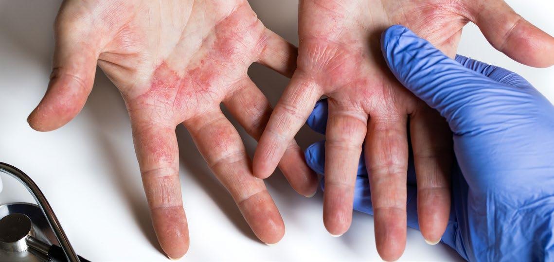
Q7Across your research, where do you think dermatology is closest to a meaningful breakthrough in diagnosis or treatment, and what would that change look like for patients and clinicians in practice?
I think dermatology is closest to a meaningful breakthrough in drug allergy evaluation. Our current research places strong emphasis on combining results from in vivo skin tests with future in vitro testing, especially for delayedtype drug reactions, so we can distinguish true drug eruptions from microbial exanthema or other immunologic disorders, including graft-versus-host disease. Moreover, all our efforts have to concentrate on identifying the specific drug responsible.
At the same time, new imaging methods and AI-assisted analysis are likely to change how dermatology is practiced. In
underserved areas, telemedicine and supportive AI could significantly expand diagnostic support in allergology and dermatology, improve prevention, and strengthen early detection of skin cancer.
We also need more objective ways to evaluate what we see clinically. We want to know precisely how lesions change over time, how extensive the inflammation is, and what biological processes are driving it, and then correlate those findings with objective clinical data and molecular evaluation.
In one hair-growth study in Singapore, for example, we used the subjective Hamilton scale as one measure, but it did not reveal much. When we added objective measurements, such as hair papilla length, hair follicle length, and papilla size, clear biological pathways began to emerge.
Modern molecular medicine, including single-cell analysis, proteomics, lipidomics, transcriptomics, and metabolomics, will open new horizons in research, diagnosis, and treatment. But for those highly precise tools to be clinically meaningful, they must be paired with equally precise, objective clinical measurements and outcomes, including variations in circadian rhythm in skin expression patterns and organspecific differences. Moreover, we should work in the future to develop meaningful organotypic skin culture models, to study toxicity, efficacy, particularly for cosmetics, but also for any product that can affect skin integrity. These models are human-specific and can reduce or avoid animal experiments for skin applications.

Christopher Tomassian Founder/CEO, The Dermatology Collective, Glendora, California, USA
Citation:
Every physician today should have some presence on social media; the need to combat misinformation alone makes it worth it
Q1
Dermatol AMJ. 2026;3[1]:90-92. https://doi.org/10.33590/dermatolamj/0H3512J8
What drew you into dermatology? How did your training at the University of Kansas Medical Center, Kansas City, USA, where you served as Cosmetic Chief and were later recognized as Best Dermatologist in Kansas City, shape your clinical philosophy going into private practice?
Dermatology has always been a specialty I've been drawn to because of its variety and the hands-on skill set it demands. Unlike many conditions in medicine, dermatologic disease is visible and having the knowledge to look at something, quickly diagnose it, and treat it was deeply appealing to me. The blend of cosmetics and surgery also made for a career that was never dull and was always evolving.
My residency training prepared me well for private practice. We were seeing 45–50 patients a day in a setting that functioned much like a private office, so the transition felt natural. I was fortunate to train under one of the best dermatologists I've encountered, Anand Rajpara, Department Chair of Dermatology, University of Missouri–Kansas City, USA, who shaped not only how I approach clinical care but how I connect with patients. His bedside manner was something I studied closely and carried with me.
I always knew I wanted to be in private practice, even before training began. That clarity pushed me to be intentional throughout residency, paying attention to what worked, what didn't, and what I
would do differently. My goal was always to have full ownership of the patient experience and to redefine what "going to the doctor" could and should feel like.
Q2
Having opened a private practice shortly after residency, what are the most consequential operational mistakes you made early on, and what advice would you give dermatologists who want to grow their practice while maintaining evidence-based patient care?
Hire slow, keep your team lean, and always keep margins in mind. When I first started my practice, I wanted all the newest lasers and a full team, but you can put yourself in a financial hole very quickly.
For any new dermatologist looking to open a practice, my advice is this: first, learn the business. Understand your baseline costs and know what every supply and material is running you: this is non-negotiable. Second, hire key team members intentionally and build a standard operational procedure that clearly defines your expectations for each role. This keeps both you and your staff accountable. Third, get out into your community and network. It may feel old-fashioned, but it's still one of the most powerful things you can do. I recommend personally visiting every local personal care provider in your area, asking to speak directly with the physician, handing them your phone number, and telling them to text you anytime they need a
patient seen urgently. That single habit was instrumental in building my practice.
Q3 You built an impressive TikTok (Los Angeles, California, USA)/Instagram (Meta Platforms, Menlo Park, California, USA) audience that centers around patient education. How do you ensure that short-form content is translated into measurable clinical benefit (e.g., better adherence, triage, fewer unnecessary visits)?
I always made my content not to replace a doctor’s visit, but to give patients a chance to try to improve their skin with evidence-backed products and ingredients. The more value you give to a patient online, the more they want to come and see you in person.
Q4 What safeguards do you use to maintain scientific accuracy and mitigate ethical concerns and criticism on shortform content?
At the end of the day, social media is an extension of what I do, but my
primary role is as a dermatologist, and that keeps me grounded. I try to keep my short-form content fun and engaging while making sure it's always evidence based. When I make a mistake or recommend a product that turns out to have issues, I own it and I'm transparent with my audience. It takes years to build authority and seconds to destroy it.
Q5
Which recent advances in energy-based therapies have meaningfully impacted how you approach treatment decisions, and where do you see the next substantive shifts coming from?
I don't think there's been a dramatic shift in energy-based technologies themselves, but what's producing more meaningful results is how we're combining them. Using ablative, non-ablative, vascular, and picosecond technologies together allows us to give patients a truly global improvement in their skin rather than addressing one concern at a time.
There continues to be a clear shift toward non-invasive, minimalto-no-downtime procedures like radio frequency and ultrasound technology. What I'm most excited to see is how these platforms begin incorporating AI software to make treatments more seamless, more personalized, and, ultimately, more effective.
Q6 When working with brands and media, what is your decision framework for endorsing products or participating in sponsored content so that recommendations remain evidencebased and ethically sound for both colleagues and patients?
I only work with brands and, more specifically, products I believe in, use myself, and would normally recommend to my patients in the clinic. It is easy to get carried away with opportunities from companies, especially when you start, but if you want to keep your authority in the space you have to make sure that what you are promoting is legitimate.
Q7
For dermatology clinicians who want to build a patient education or digital outreach program from their practice, what safeguards are essential to ensure online content remains medically accurate, ethically responsible, and beneficial for patient care?
Every physician today should have some presence on social media; the need to combat misinformation alone makes it worth it. The guardrails are simple: be professional, be ethical, and remember that you are always in control of what you put out into the world. The internet is forever, so be deliberate. And if you're sharing any patient photos, always secure both verbal and written consent, no exceptions.

Rediscovering Bleach: The Enduring and Emerging Role of Sodium Hypochlorite in Dermatology
Editor's Pick
This Editor’s Pick offers a timely revisit to the rich clinical history of sodium hypochlorite. As we continue to explore strategies for managing infection, inflammation, and skin barrier-related disease in order to treat our patients safely and effectively, this feature provides renewed attention to current practice.
Michael Gold
Founder and Medical Director, Gold Skin Care Center, Nashville; Advanced Aesthetics Medical Spa, Nashville; The Laser & Rejuvenation Center, Nashville; Tennessee Clinical Research Center, Nashville, Tennessee, USA
Authors: Theresa Hopkins,1 *Raj Chovatiya1,2


1. Rosalind Franklin University of Medicine and Science Chicago Medical School, North Chicago, Illinois, USA
2. Center for Medical Dermatology + Immunology Research, Chicago, Illinois, USA
*Correspondence to raj.chovatiya@gmail.com
Disclosure: Chovatiya has served as an advisor, consultant, speaker, and/or investigator for AbbVie, Acelyrin, Alumis, Amgen, AnaptysBio, Apogee Therapeutics, Arcutis Biotherapeutics, Argenx, Astria Therapeutics, Avalere Health, Beiersdorf, Boehringer Ingelheim, Bristol Myers Squibb, Cara Therapeutics, Castle Biosciences, Celldex Therapeutics, CLn Skin Care, Dermavant, DKSH, Eli Lilly and Company, EMD Serono, EPI Health, Formation Bio, Forte Biosciences, Galderma, Genentech, GSK, Incyte, Imagene Bio Inc., Indero, Johnson & Johnson, Kenvue, LEO Pharma, L’Oréal, Mestag Therapeutics, Nektar Therapeutics, Nia Health, Novan, Novartis, Opsidio, Organon, Pfizer, RAPT, Regeneron, Sanofi, Sitryx, SUN Pharma, Takeda, TRex Bio, UCB, Zai Lab, ZenZema, and Zuellig Pharma. Hopkins has declared no conflicts of interest.
Received: 02.23.26
Accepted: 04.12.26
Keywords: Anti-inflammatory, atopic dermatitis (AD), bleach baths, dermatology, eczema, hidradenitis suppurativa (HS), sodium hypochlorite (NAOCl), wound care.
Citation: Dermatol AMJ. 2026;3[1]:93-98. https://doi.org/10.33590/dermatolamj/28RABNC8
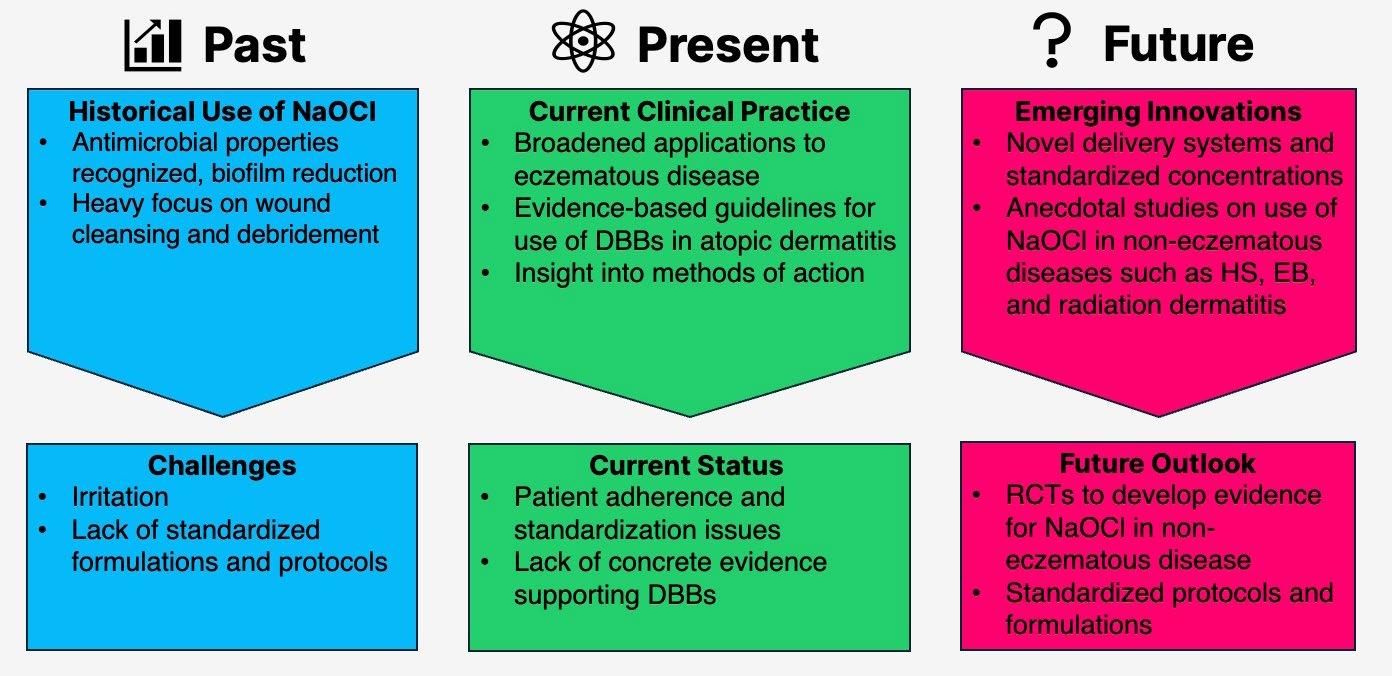
INTRODUCTION
Sodium hypochlorite (NaOCl), the active ingredient in household bleach, has long been a staple in medical history, yet its full potential in dermatology remains underexplored. A 2025 scoping review by Chang et al.1 synthesized available literature, analyzing 222 studies from 1915–2024 to map NaOCl’s past applications, current evidence, and future directions (Figure 1). This feature appraises that review, critically examining established knowledge while highlighting NaOCl’s relevance in modern contexts and suggesting areas for future advancements. Originally referred to as Dakin’s solution, NaOCl was used during World War I to irrigate infected wounds. In an era before antibiotics, NaOCl played an important role in reducing sepsis and saving limbs on the front lines due to its low cost, broad antimicrobial spectrum, and unique ability to dissolve necrotic tissue. Its use faded when
penicillin was introduced in the 20th century, and it became referred to as outdated and irritative. However, among rising antibiotic resistance and new research exploring its antiinflammatory mechanisms, the use of NaCOl has been rediscovered in recent decades.
HISTORICAL PERSPECTIVES AND MODERN MECHANISTIC INSIGHTS
Initially, the success of Dakin’s solution was attributed to mechanical debridement, but research later showed that the chemical composition of NaOCl played a role in sloughing of necrotic skin to facilitate healing. When in an aqueous solution, NaOCl and hypochlorous acid (HOCl) are at an equilibrium. A lower, more acidic pH favors a shift toward HOCl with more potent antimicrobial and cytotoxic effects. Therefore, it is important that the pH of NaOCl solutions used for medical purposes is regulated to
Figure 1: The evolving landscape of NaOCL in dermatology.
DBB: dilute bleach bath; EB: epidermolysis bullosa; HS: hidradenitis suppurativa; NaOCl: sodium hypochlorite.
confer a balance between antimicrobial and toxic effects that can be corrosive to healthy cells. These findings were initially highlighted in studies from the 1960s and 70s, which focused on optimizing the balance between NaOCl and HOCl to maximize efficacy and minimize cytotoxicity. A key finding was dose-dependent effects and trade-offs to treatment. The highest dose may not be the most effective dose due to increased risk for adverse reactions including skin irritation, fibroblast damage, contact dermatitis, and patient discomfort. Antimicrobial properties against Staphylococcus aureus and Pseudomonas aeruginosa could be harnessed with concentrations of 0.005–0.5% NaOCl, incurring a lower risk for side effects.1 This review highlights how historical insights paved the way for the modern, broader application of NaOCl, though inconsistencies in preparation (e.g., household bleach strength, dilution methods, and storage) still hinder comparability and the development of specific guidelines for use in evidencebased medicine. Early studies were often observational or in animal models, limiting translation but establishing NaOCl’s application beyond antiseptics.1
KEY FINDINGS AND EVIDENCE
The scoping review organized evidence into four clinical domains, revealing robust support for NaOCl across dermatoses. By synthesizing information based on date and domain, this review effectively summarized historical perspectives, current patterns, and future directions. Additionally, domains with limited research could be highlighted. Among the 222 articles included for review, 57 described antimicrobial properties, 64 wound care, 78 eczematous skin disease, and 23 noneczematous inflammatory skin conditions.1 Direct grading of the quality of evidence was not included in the original review.
Antimicrobial Properties
Antimicrobial properties take precedence in early literature, describing broad-spectrum
activity against bacteria, fungi, viruses, and parasites. The largest breadth of studies focused on common skin pathogens like S. aureus (including methicillin-resistant strains), P. aeruginosa, and Candida species. However, a wide variety of pathogens were targeted, including herpes simplex virus, Streptococcus spp., Enterococcus spp., Escherichia coli, dermatophytes, Morganella morganii, Enterobacter cloacae, Klebsiella pneumoniae, Malassezia spp., Sporothrix schenckii, Aspergillus spp., and even maggots like Lucilia sericata. Antimicrobial effects were evaluated using bacterial cultures and a wide range of concentrations from as low as 0.000025% to 10%.1-3 Not only did concentration affect outcomes, but longer exposure times were found to increase the efficacy of more diluted solutions. Cell toxicity has been a consistent concern, and, most recently, concentrations under 0.05% have been favored to avoid skin irritation, although doses as low as 0.005% have shown antimicrobial effects. Factors like solution pH, age, and the skin’s natural barriers affect real-world performance.1
Wound Care
NaOCl’s capacity to disrupt biofilms and multidrug-resistant organisms has been established in chronic wounds and burns. Outside of its role in infection control, NaOCl can positively affect wound healing across multiple stages. Low concentrations have been found to facilitate debridement by softening necrotic tissue, reduce excessive neutrophil activation that may prolong inflammation, increase or sustain fibroblast activity, and stimulate granulation tissue. While higher concentrations are typically associated with negative effects, one in vivo model found that 4% NaOCl promoted angiogenesis.1 Clinical reports show faster ulcer closure and fewer amputations in diabetic foot cases, though evidence is mixed for burns, with some trials noting discomfort or graft sloughing at higher doses.1 It is important to note that the benefit of NaOCl in wound care and as an antimicrobial agent is dependent on dose, exposure time, and
pathogen strain. A study comparing 0.1% and 0.5% NaOCl-soaked gauze for maintenance of burn wounds found that while 0.5% NaOCl was effective in eradicating S. aureus and P. aeruginosa, it could not be used for more than 2 weeks continuously due to epidermal toxicity.2 Comparatively, 0.1% NaOCl was able to reduce microorganisms with no epidermal damage but needed to be used for a longer period.2 Further studies are needed to elucidate the exact role and precise concentrations of NaOCl required to optimize wound healing.1
Eczematous Skin Disease
Most literature regarding NaOCl use in eczematous conditions is centered on atopic dermatitis (AD). Dilute bleach baths (DBB; often approximately 0.005% NaOCl) have been used as an adjunctive treatment option for moderate-to-severe AD colonized by S. aureus. The objective of DBBs is to decolonize S. aureus and reduce the incidence of skin and soft tissue infections. However, recent studies highlight inconsistent results and question the validity of DBBs.1,4,5 Meta-analyses found no superiority over plain water for severity scores like Eczema Area and Severity Index (EASI) or body surface area.1,4,5 Additionally, there is no standardized dilution formulation for DDBs, and the household use of DBBs poses significant heterogeneity, as bath tub sizes vary.1
Emerging data suggest direct antiinflammatory effects via inhibition of the nuclear factor κB pathway, which, when activated, promotes pro-inflammatory cytokine production, T cell activation, and histamine release.6 This mechanism is likely why DBBs can ease symptoms like redness, itching, and skin barrier issues in AD, even when bacterial reduction is inconsistent. This study highlights that NaOCl’s benefits go beyond killing bacteria and emphasizes its broader therapeutic value.
Non-eczematous Disease
Non-eczematous applications are the newest and smallest area of research. Emerging areas of study include reduction in the severity of radiation dermatitis, improved desquamation in rare ichthyoses, infection prevention in epidermolysis bullosa and hidradenitis suppurativa (HS), and papule reduction in acne vulgaris. These findings underscore NaOCl’s versatility, but the review justly critiques the predominance of case reports and observational data, with heterogeneity confounding meta-analyses.1
DISCUSSION
The review’s strengths lie in its comprehensive nature, including historical perspectives, novel applications, and critical lens on the strength (or lack thereof) among cited literature. Limitations include the exclusion of studies with a focus on HOCl, comparative studies using another component in combination with NaOCl, and non-English language publications. Due to these exclusion criteria, there may be other applications and mechanisms on NaOCl not included in the scoping review. Additionally, specific clinical protocols and evaluation of evidence strength remain lacking. All things considered, the importance of NaOCl was well-described across multiple applications. Among the ever-changing medical landscape, NaOCI addresses enduring concerns regarding antibiotic resistance and resource limitations. It remains a viable treatment option in guidelines from the USA, Canada, Europe, and Asia, which endorse its use in DBBs for moderate-severe AD, though consensus on mild cases or exact dosing is lacking.1,7
FUTURE DIRECTIONS
Despite a lengthy history, clinical application of NaOCl has ample room for innovation to determine the optimal concentrations and formulations to use in each setting: antimicrobial, anti-inflammatory, and wound healing (Table 1). Prioritizing dosing to
maximize effectiveness in each unique setting may be the future, rather than the current one-bathtub-fits-all application. Standardized formulations, such as pH-buffered cleansers or sprays, can mitigate instability, enhance compliance, and bypass variability. This benefit is highlighted by commercially available NaOCl cleansers, which exemplify this approach and have shown practical utility in the management of eczematous skin disease.10 Comparative trials pitting NaOCl against HOCl, biologic therapies, and examining a combination approach would clarify mechanisms and offer better guidance for personalized regimens.1
1: Current recommended
In non-eczematous realms, RCTs for HS, radiation dermatitis, epidermolysis bullosa, and rare ichthyoses could validate anecdotal benefits and stimulate development of standard guidelines. This process has already begun, as evidenced by a recent study using a NaOCl body wash of standardized concentration (0.006% NaOCl; CLn® BodyWash, TopMD Skin Care, Dallas, Texas, USA) for HS management.8 This uncontrolled study demonstrated significant reductions in HS symptoms, illustrating the potential of standardized NaOCl formulations while underscoring the need for future RCTs to confirm these findings and strengthen the
concentrations by dermatologic application.1-3,5,8-10 Concentrations adapted from multiple supporting studies.1-3,5,8-10 DBB: dilute bleach bath.
Adjunctive; significant reduction in selfreported redness, swelling, drainage, pain, itching, and odor found in recent study10
Ichthyoses Not specified1 Anecdotal desquamation aid
Table
NaOCl
evidence base. Advancing standardized protocols and innovative delivery systems through RCTs will be essential to unlock NaOCl’s full therapeutic potential and establish clear, evidence-based roles across dermatologic indications.1
CONCLUSION
NaOCl embodies dermatology’s blend of tradition and innovation, representing an accessible, promisingly effective, and yet under-optimized therapeutic approach. Chang et al.1 charts a path forward to make what is old new, urging refinement of protocols
References
1. Chang CH et al. Past, present, and future of sodium hypochlorite in dermatology: a scoping review. Am J Clin Dermatol. 2025;DOI:10.1007/ s40257-025-00999-9.
2. Cotter JL et al. Chemical parameters, antimicrobial activities, and tissue toxicity of 0.1 and 0.5% sodium hypochlorite solutions. Antimicrob Agents Chemother. 1985;28(1):118-22.
3. Yang C et al. Effect of negatie pressure wound therapy with instillation on bioburden in chronically infected wounds. Wounds. 2017;29(8):240-6.
4. Wong SM et al. Efficacy and safety of sodium hypochlorite (bleach) baths in patients with moderate to severe atopic
and expansion of trials. Currently, NaOCl is clinically used for wound debridement; viral, bacterial, and fungal infections; and as an adjunct treatment for AD. Further studies are needed to determine standardized dilution and the role of NaOCl in non-eczematous cutaneous disease. By bridging gaps in evidence, NaOCl could reclaim its place as a reliable and highly utilized therapy, offering safe, equitable solutions in an era of complex skin conditions.
dermatitis in Malaysia. J Dermatol. 20213;40(11):874-80.
5. Bakaa L et al. Bleach baths for atopic dermatitis: a systematic review and meta-analysis including unpublished data, Bayesian interpretation, and GRADE. Ann Allergy Asthma Immunol. 2022;128(6):660-8.
6. Leung TH et al. Topical hypochlorite ameliorates NF-κB-mediated skin diseases in mice. J Clin Invest. 2013;123(12):5361-70.
7. Chu DK et al.; AAAAI/ACAAI JTF Atopic Dermatitis Guideline Panel. Atopic dermatitis (eczema) guidelines: 2023 American Academy of Allergy, Asthma and Immunology/American College of Allergy, Asthma and Immunology Joint Task Force on Practice Parameters
GRADE- and Institute of Medicinebased recommendations. Ann Allergy Asthma Immunol. 2024;132(3):274-312.
8. Omole I et al. Real-world efficacy of sodium hypochlorite body wash in managing hidradenitis suppurativa. J Clin Aesthet Dermatol. 2025;18(8):13-15.
9. Hidalgo E et al. Cytotoxicity mechanisms of sodium hypochlorite in cultured human dermal fibroblasts and its bactericidal effectiveness. Chem Biol Interact. 2002;139(3):265-82.
10. Majewski S et al. Sodium hypochlorite body wash in the management of Staphylococcus aureus-colonized moderate-to-severe atopic dermatitis in infants, children, and adolescents. Pediatr Dermatol. 2019;36(4):442-8.
Dermatologic Disparities in Aboriginal and Torres Strait Islander Children
Authors: *Ana
María Sierra Valiente,1,2 Markus Augusto Martínez Holst,1 Thomas Duncan Nichols3
1. Universidad Anáhuac México, Mexico City, Mexico
2. University of New South Wales, Sydney, Australia
3. Private Practice, Dermatology & Skin Surgery, Houston, Texas, USA
*Correspondence to z5342685@zmail.unsw.edu.au
Disclosure: The authors have declared no conflicts of interest.
Acknowledgements: The authors are non-Aboriginal medical students with clinical experience in underserved settings. They acknowledge that they do not speak from lived experience and write from outside Aboriginal and Torres Strait Islander populations. Even so, the authors recognize a responsibility to engage with this topic to generate awareness and support equity in pediatric skin health while affirming the essential role and leadership of Aboriginal and Torres Strait Islander peoples in all matters of their healthcare. The authors' goal is to amplify evidence and support ongoing, community-led efforts toward self-determined health systems. Their perspective is grounded in a commitment to health equity, critical appraisal of the literature, and training in skin diseases, health policy, and culturally safe health practices.
This work was conducted under the supervision of Thomas Nichols, a board-certified dermatologist with over 48 years of clinical experience, with a private practice in Houston Texas Medical Center, Texas, USA. Drawing on his extensive expertise in dermatologic disease and patient education, Nichols provided supervisory oversight through critical review of dermatologic content, ensuring clinical accuracy, relevance, and clarity. His contributions strengthened the scientific rigor of the manuscript while supporting the responsible translation of dermatologic knowledge into patient-centered health promotion.
Received: 01.26.26
Accepted: 04.09.26
Keywords: Aboriginal and Torres Strait Islander children, racism in dermatology, skin diseases, social determinants of health.
Citation: Dermatol AMJ. 2026;3[1]:99-105. https://doi.org/10.33590/dermatolamj/B57WK395
Abstract
The wellbeing of Aboriginal and Torres Strait Islander children is threatened by a disproportionate burden of dermatologic diseases, which reflects the environmental disadvantages and systemic racism experienced by this population. Despite the improvement of healthcare accessibility and health initiatives targeted at Aboriginal and Torres Strait Islander populations in Australia, skin infections such as impetigo and scabies continue to affect nearly half of children in remote
communities. Underrepresentation of darker skin tones in dermatology education and housing conditions plays a significant part in Aboriginal children’s high skin disease incidence and diagnostic inequity. Aboriginal and Torres Strait Islander children often experience a delay in receiving dermatological treatment. Beyond the burden of skin conditions, severe health complications, such as post-streptococcal glomerulonephritis and rheumatic heart disease, have an alarming prevalence in the study group. This narrative review synthesizes dermatological conditions and determinants shaping the pediatric skin health among Aboriginal and Torres Strait Islander peoples. Initiatives such as the SToP trial and The Gija Healthy Skin Story are discussed, as they demonstrated promising outcomes in skin disease control in the community. Addressing pediatric dermatologic disparities is essential for improving child development, health equity, and, overall, quality of life among the population.
Key Points
1. Aboriginal and Torres Strait Islander children face a disproportionate burden of dermatologic conditions, including preventable skin infections with life-threatening sequelae, driven by structural inequity, household crowding, and systemic racism.
2. This narrative review synthesizes the epidemiological burdens and barriers to care shaping pediatric skin health in Aboriginal and Torres Strait Islander children, integrating evidence across remote and urban settings and examining culturally grounded interventions.
3. Interventions such as the SToP trial demonstrate that skin health initiatives are most effective when co-designed with Aboriginal communities, underscoring the importance of partnership alongside sustainable improvements in infrastructure and clinician training to achieve dermatologic equity.
INTRODUCTION
Skin conditions that could be avoided keep affecting Aboriginal and Torres Strait Islander children more than others.1,2 Recurrent, often visible, and treatable skin conditions extend far beyond the skin, manifesting as systemic pathologies and having psychosocial consequences, including lower self-esteem, reduced school participation, and decreased community engagement.2,3 Recurrent infections and inflammatory skin disorders in these children disrupt proper development, perpetuating cycles of social disadvantage and health inequity.4,5 Despite substantial national advances in healthcare accessibility, skin health inequities persist as one of the most visible indicators of systemic disadvantage and the ongoing failure to deliver culturally safe, equitable dermatologic care.6,7 Health disparities awareness has sparked research studies focused on the better care and prompt diagnosis of skin
pathologies in Australia’s Aboriginal groups.8 This narrative review synthesizes current evidence on pediatric skin health inequities among Aboriginal and Torres Strait Islander peoples, emphasizing the contexts that shape these disparities.9,10 Barriers to healthcare and community-driven solutions are discussed. Evidence highlights skin health as a core component of child wellbeing and an essential domain of health equity, one that deserves global attention and concerted dermatologic action.
SCOPE AND NOVELTY OF THIS REVIEW
Dermatologic disparities in Aboriginal and Torres Strait Islander children remain critically underrepresented in international literature, despite their magnitude. The authors position their work in relation to two key existing publications. Ricciardo et al.8
provides an important global perspective on skin conditions in children with skin of color, centering clinical recognition, morphological presentation, and diagnostic features across diverse darker skin tones worldwide. While Aboriginal and Torres Strait Islander children are mentioned as illustrative examples, the review’s primary focus is on equipping clinicians globally to recognize and manage skin conditions across global populations with skin of color. The authors' manuscript addresses diagnostic challenges related to darker skin tones, but complements this landmark work by providing a dedicated, in-depth analysis of the epidemiological burden, systemic sequelae, and social architecture of skin diseases specifically in Aboriginal and Torres Strait Islander children. Davidson et al.2 offers a valuable Australian-focused synthesis of bacterial skin infections (impetigo, scabies, cellulitis, and abscesses) in Aboriginal children, with emphasis on remote communities in northern Australia. The authors acknowledge that few data are available for other Australian states and note that research on communitywide skin health programs was ‘underway’. The authors' review extends this foundation by incorporating 5 years of new evidence (2020–2025), integrating data from both remote and urban settings, and broadening the dermatologic scope beyond bacterial skin infections to encompass inflammatory, fungal, and pigmentary conditions. Furthermore, this review synthesizes recent evidence from community-led, culturally grounded intervention programs, including outcome data from the SToP trial, which Davidson cited as starting in the remote Kimberley region in 2019, as well as process evaluations of the Gija Healthy Skin Story educational resource, and the HipHop2SToP youth engagement initiative.
Together, this brief review provides a comprehensive resource for clinicians and policymakers seeking to address skin health inequities in Aboriginal and Torres Strait Islander children. More broadly, it offers a paradigm for understanding how structural inequity manifests as dermatologic disease
in Indigenous communities worldwide, with transferable lessons for First Nations populations facing similar burdens of preventable skin disease.
DERMATOLOGIC HEALTH SOCIAL DETERMINANTS
Skin problems open a window into deep social inequities. Not just illness; they show how history still shapes the lives of First Nations’ people today. Colonization displaced Aboriginal and Torres Strait Islander populations into environments that today are characterized by household crowding and a lack of adequate access to basic services and health.6,7 Environmental and historic determinants have made Aboriginal and Torres Strait Islander children particularly vulnerable to preventable skin diseases and their severe complications, such as poststreptococcal glomerulonephritis (PSGN) and rheumatic heart disease (RHD).4,5 Care shaped by racist biases further compounds this disparity. Dermatology as a field has long centered on lighter skin tones, with minimal representation of Indigenous patients or patients with darker skin in medical textbooks, teaching materials, and diagnostic imagery.8 This visual and curricular bias results in diagnostic inequities, delays in care, and a limited capacity of clinicians to recognize and manage skin disease presented in darker skin.10 Medical teaching often unfoundedly links darker skin to diagnoses with stigmas, such as sexually transmitted infections.10 People of color with skin diseases may experience stigmatization, with consequent discrimination and social rejection even by dermatologic care providers.10
COMMON DERMATOLOGICAL CONDITIONS AMONG ABORIGINAL AND TORRES STRAIT ISLANDER CHILDREN
Among Aboriginal and Torres Strait Islander children in remote communities, the prevalence of skin infections is alarmingly high. Hospitalization rates for skin infections among Aboriginal children are approximately 15-times higher than those of non-Aboriginal children in Western Australia.7,11 Complications from skin infections, such as bacteremia and sepsis, are higher in Aboriginal children compared with non‐Aboriginal children. The annual incidence of invasive Staphylococcus aureus infections among Aboriginal children in northern Australia was 46.6 cases per 100,000 population, compared with 4.4 cases per 100,000 in non-Aboriginal children.12 A study found that Aboriginal and Torres Strait Islander children had significantly higher ICU admission rates for invasive infections, particularly S. aureus, compared with nonAboriginal children.13 Although ICU case fatality rates were similar between groups, the population-based mortality from these infections was more than twice as high among Aboriginal and Torres Strait Islander children, highlighting major health inequities.13
Impetigo
Research has discovered that the prevalence of impetigo among Australian Aboriginal and Torres Strait Islander children is among the highest reported worldwide, particularly in those residing in remote communities.2,4,14,15 The point prevalence of impetigo commonly ranges from 10–70%, with a pooled community prevalence of approximately 28%.2,4,14,15 Community-based studies have documented that up to 49% of children in Aboriginal communities in northern Australia were affected at a given time,16 with 84% of remote Aboriginal and Torres Strait Islander children presenting to a clinic with impetigo at least once before their first birthday.13 The burden of impetigo in remote Aboriginal communities has significant implications onmorbidity, healthcare utilization, and long-term health equity.14,16
Scabies
Scabies remains highly prevalent, affecting up to 33% of Aboriginal children in remote communities and contributing to secondary complications such as impetigo, PSGN, and RHD.5 Scabies leads to intense pruritus and skin lesions, which are frequently superinfected by S. aureus and Streptococcus pyogenes, resulting in impetigo, cellulitis, and abscesses.5 Crusted scabies imposes a significant economic burden, with an estimated healthcare cost of AUD 35,418 per patient in the Northern Territory, primarily borne by hospital systems.16 Cellulitis is a frequent complication, with extremities being the most commonly affected site in children presenting to healthcare facilities.17 Considering the disproportionately high rates of severe skin and bacterial infections reported among Aboriginal children, these findings emphasize the vital need to prioritize skin health in Aboriginal and Torres Strait Islander populations.12,13
From Skin Infection to Systemic Disease
Acute PSGN and RHD after streptococcal impetigo or scabies are severe complications that occur at the highest rates globally in remote Aboriginal communities.2,18 The incidence of acute PSGN in Aboriginal children under 15 years in remote Australia ranges as high as 94–124 per 100,000 person-years, representing nearly 19–54-times the rate seen in non-Aboriginal children.19 Though less common elsewhere, RHD among Aboriginal and Torres Strait Islander youth living in remote areas is hyper-endemic. About 3.3% of children show signs of it during heart scans, and most have the antecedent of repeated skin infections like scabies or impetigo.20 Survival rates for those diagnosed remain significantly lower than for non-Aboriginal children, with premature mortality attributable to delayed diagnosis.3,20
Other Causes of Clinic Visits for Skin Disease Among Aboriginal Children
Outside of remote areas, dermatologic conditions also remain a major driver of
healthcare encounters among Aboriginal children.21,22 Atopic dermatitis, the most prevalent chronic inflammatory skin disease in this population, affects approximately 18% of urban Aboriginal children, and is frequently associated with undertreatment, secondary bacterial infection, and sleep disturbances.21,23 Head lice and tinea are also widespread, each affecting 12–18% of children.24
Autoimmune connective tissue disorders, pigmentary disorders, and benign neoplasms are noteworthy, but less frequently reported. These conditions are often underdiagnosed due to diagnostic complexity and limited access to specialists.22,25 In the Kulunga Moorditj Healthy Skin (KMHS) project, the first Australian co-designed researchservice study to describe skin health in urban-living Aboriginal children and young people, skin of color was defined as Fitzpatrick Skin Phototypes IV–VI, and a substantial proportion of participants were classified within these darker phototypes.24 A study found that pigment issues made up about 15% of skin diagnoses, especially dyspigmentation after inflammation, showing that they often appear in Aboriginal children.24 Skin color matters during diagnosis; pigmentary disorders pose a particular diagnostic challenge in children with darker skin.2
SYSTEMIC BARRIERS TO CARE AND CONSEQUENCES IN ABORIGINAL AND TORRES STRAIT ISLANDER CHILDREN
Systemic neglect, the lack of public investment, and the scarcity of basic services, such as functioning bathrooms, laundry facilities, and clean water, complicate skin disease management in this population.2 Although subsidized healthcare exists through Medicare and the Pharmaceutical Benefits Scheme, access to dermatologic care remains limited by expenses related to medications and transportation, particularly for families residing in remote areas.2,3
Implicit bias among healthcare providers undermines the trust necessary for care utilization and continuity of treatment by Aboriginal Australians.7,22 The normalization of skin infections within communities, coupled with under-recognition of dermatologic diseases, sustains delayed or inadequate methods for treating and preventing skin infections in Aboriginal and Torres Strait Islander children.25 The perceived normalization of skin infections is probably a consequence of limited access to timely care and a history of healthcare services not meeting community needs, rather than community indifference. Shortages in staff, along with broken referral systems, make it harder to get skin care quickly.26 These limitations are further exacerbated by the underrepresentation of Aboriginal and Torres Strait Islander populations in health screening programs, and of populations with skin of color in dermatologic research and education.10,25,27
Even though schools frequently act as key spots for teaching health habits and running screenings, they struggle due to inadequate funding and unclear directives fading over time.7,9 Together, these barriers lead to more disease burden and poorer health outcomes for Aboriginal and Torres Strait Islander children, whether they live in remote or urban areas.
The high burden of pediatric skin disease poses downstream effects on early childhood development, school readiness, and long-term social and economic participation.3 Chronic pain, itching, and discomfort associated with dermatologic diseases can cause significant sleep disturbance, impaired concentration, and frequent absenteeism from school, all of which negatively affect Aboriginal and Torres Strait Islander children. Recurrent or visibly apparent skin conditions are also strongly associated with stigma, diminished selfesteem, and disrupted peer relationships.11,23,24
Untreated or recurrent skin infections significantly elevate the risk of serious systemic complications. As previously
mentioned, among Aboriginal and Torres Strait Islander children, the most critical sequelae include invasive bacterial infections, PSGN, and rheumatic fever. Chronic complications contribute to increasing morbidity and mortality, affecting Aboriginal and Torres Strait Islander children’s overall wellbeing.
INTERVENTIONS
In Australia, some public initiatives have begun to integrate medical, educational, and environmental strategies to enhance pediatric skin health. The SToP trial enrolled 915 children aged 5–9 years across nine Kimberley, Australia, communities, with 777 completing at least one skin check. The trimodal approach, combining See (skin checks), Treat (treatment training), and Prevent (co-designed health promotion), achieved a 17–19% decline in impetigo prevalence between 2019–2022. Multi-method evaluation confirmed sustained community engagement throughout the trial.9,28
Community-led educational initiatives have demonstrated effectiveness in promoting skin health. For instance, the Berrembi JarragbooBoorroo Wajawoorroo Men’Gawoom Gijam (Gija Healthy Skin Story) resource had over 500 copies distributed, including a Kriol version. Co-designed with community members, this resource aims to support families in recognizing skin infections and understanding their connection to complications such as RHD, with process evaluation demonstrating high community acceptability and uptake.29
On the other hand, the HipHop2SToP project utilized music and youth engagement to teach healthy skin and hygiene practices.7 Such culturally appropriate and Aboriginalled interventions, alongside the multimethod SToP trial evaluations, have demonstrated high acceptance, uptake, and reductions in skin infections.9,28 Virtual support interventions have shown potential to increase engagement with skin health;
however, studies emphasize the necessity of in-person community engagement and environmental health interventions to achieve meaningful improvements in Aboriginal and Torres Strait Islander populations.28,30
Active case detection campaigns have proven essential in identifying cases of crusted scabies, particularly when followup and engagement with close contacts are implemented.31 Aboriginal community members emphasized the need for faceto-face engagement, highlighting that interventions are most effective when guided by local knowledge and priorities.32 Skilled personnel and community engagement maximize effectiveness of health interventions.30 Improving clinician knowledge of dermatologic presentations in skin of color is an important strategy to enhance care accessibility and outcomes among Aboriginal children.8,25 Despite the SToP trial achieving a decline in impetigo prevalence, it reported that the sustainability of such interventions is threatened by extreme workforce instability in remote clinics, where annual turnover rates exceed 150%, meaning almost twice as many individual employees are required each year to maintain staffing.9,28 Lessons from these programs offer transferable insights for Indigenous and minority populations globally.
CONCLUSION
Aboriginal and Torres Strait Islander children carry a disproportionate burden of skin conditions, particularly skin infections. Higher incidence of skin diseases ties back to deep-rooted social challenges faced by Aboriginal and Torres Strait Islander populations. Reducing disparities in dermatological care requires collaboration. In both city neighborhoods and remote regions, current initiatives prove the importance of culturally appropriate communityled interventions, including educational initiatives, hygiene programs, and active case detection campaigns to control this issue. Despite drawing on community initiatives, this paper lacks input from Aboriginal and
Torres Strait Islander peoples. For future research, the authors strongly recommend incorporating this input to ensure genuine partnership, promote health literacy, and advance dermatologic equity. Future research
References
1. McMeniman E et al. Skin disease in the first two years of life in Aboriginal children in East Arnhem Land. Australas J Dermatol. 2011;52(4):270-3.
2. Davidson L et al. Skin infections in Australian Aboriginal children: a narrative review. Med J Aust. 2020;212(5):231-7.
3. Hendrickx D et al. Ascertaining infectious disease burden through primary care clinic at-tendance among young Aboriginal children living in four remote communities in Western Australia. PLoS One. 2018;13(9):e0203684.
4. Wiegele S et al. The epidemiology of superficial Streptococcal A (impetigo and pharyngitis) infections in Australia: a systematic review. PLoS One. 2023;18(11):e0288016.
5. Gramp P et al. Scabies in remote Aboriginal and Torres Strait Islander populations in Australia: a narrative review. PLoS Negl Trop Dis. 2021;15:e0009751.
6. Bailie RS et al. The impact of housing improvement and socio-environmental factors on common childhood illnesses: a cohort study in Indigenous Australian communities. J Epidemiol Community Health. 2012;66(9):821-31.
7. McRae T et al. HipHop2SToP a community-led health promotion initiative empowering Aboriginal youth in the Kimberley region of Western Australia: a process evaluation. Front Public Health. 2023;11:1258517.
8. Ricciardo BM et al. Healthy skin for children and young people with skin of colour starts with clinician knowledge and recognition: a narrative review. Lancet Child Adolesc Health. 2025;9(4):262-73.
9. Thomas HMM et al. Multi-methods process evaluation of the SToP (see, treat, prevent) trial: a cluster randomised, stepped wedge trial to support healthy skin. EClinicalMedicine. 2024;77:102793.
10. Narla S et al. Racial disparities in dermatology. Arch Dermatol Res. 2023;315(5):1215-23.
should focus on evaluating the long-term sustainability and scalability of interventions to ensure improved dermatologic outcomes across diverse Indigenous populations.
11. Abdalla T et al. Hospital admissions for skin infections among Western Australian children and adolescents from 1996 to 2012. PLoS One. 2017;12(11):e0188803.
12. Engelman D et al. Invasive Staphylococcus aureus infections in children in tropical Northern Australia. J Pediatr Infect Dis Soc. 2014;3(4):304-11.
13. Ostrowski JA et al. The burden of invasive infections in critically ill Indigenous children in Australia. Med J Aust. 2017;206(2):78-84.
14. Bowen AC. The global epidemiology of impetigo: a systematic review of the population prevalence of impetigo and pyoderma. PLoS One. 2015;10(8):e0136789.
15. Ricciardo B et al. Skin health of Aboriginal children living in urban communities. Australas J Dermatol. 2024;65(8):e224-37.
16. Romani L et al. Prevalence of scabies and impetigo worldwide: a systematic review. Lancet Infect Dis. 2015;15(8):960-7.
17. Campbell M et al. Health care cost of crusted scabies in Aboriginal communities in the Northern Territory, Australia. PLoS Negl Trop Dis. 2022;16(3):e0010288.
18. Salleo E et al. Cellulitis in children: a retrospective single centre study from Australia. BMJ Paediatr Open. 2021;5(1):e001130.
19. Aung PTZ et al. Scabies and risk of skin sores in remote Australian Aboriginal communities: a self-controlled case series study. PLoS Negl Trop Dis. 2018;12(7):e0006668.
20. Chaturvedi S et al. Acute poststreptococcal glomerulonephritis in the Northern Territory of Australia: a review of data from 2009 to 2016 and comparison with the literature. Am J Trop Med Hyg. 2018;99(6):1643-8.
21. Baker MG. Risk factors for acute rheumatic fever: literature review. Int J Environ Res Public Health. 2019;16(22):4515.
22. Courtney A et al. Burden of disease and unmet needs in the diagnosis and management of atopic dermatitis in
diverse skin types in Australia. J Clin Med. 2023;12(11):3812.
23. Ricciardo BM et al. Describing skin health and disease in urban-living Aboriginal children: co-design, development and feasibility testing of the Koolungar Moorditj Healthy Skin pilot project. Pilot Feasibility Studies. 2014;10(1):6.
24. Ricciardo BM et al. Koolungar (Children) Moorditj (Strong) Healthy Skin Project part II: skin health in urban-living Australian Aboriginal children. Pediatr Dermatol. 2025;DOI:10.1111/pde.70016.
25. Silverberg NB et al. Training in pediatric skin of color: suggested curricular guidelines of the Pediatric Dermatology Research Alliance special interest group in pediatric skin of color. Pediatr Dermatol. 2021;38(Suppl 2):90-5.
26. Thorn H et al. Barriers and facilitators to accessing medical services in rural and remote Australia: a systematic review. Asia Pac J Health Management. 2023;18(1):e1755.
27. Nepal S et al. Systematic literature review to identify methods for treating and preventing bacterial skin infections in Indigenous children. Australas J Dermatol. 2018;59(3):194-200.
28. Enkel SL et al. Berrembi JarragbooBoorroo Wajawoorroo Men'Gawoom Gijam (Gija Healthy Skin Story): twoway learning for healthy skin. Health Promot J Aus. 2025;36(4):e70111.
29. Porchak E et al. Multi-method evaluation of community engagement in the SToP trial. BMC Health Serv Res. 2025;24(1):1164.
30. Moecke DP et al. Scoping review of telehealth use by Indigenous populations from Australia, Canada, New Zealand, and the United States. J Telemed Telecare. 2024;30(9):1398-416.
31. Glennie M et al. Active case detection methods for crusted scabies and leprosy: a systematic review. PLoS Negl Trop Dis. 2021;15(7):e0009577.
32. Dalton R et al. Yarning with a remote Aboriginal community about the next steps for achieving healthy skin. Aust N Z J Public Health. 2025;49(3):100242.
A Rare Case of Linear Morphea Arising from a Vaccination in a Filipino Female
Authors: *Jean Anne Camille Marcelo-De Jesus,1 Roxanne Louise Salazar,1 Rosalie Deeneye Santiago-Iglesias1
1. East Avenue Medical Center, Quezon City, Philippines *Correspondence to Jeacamarcelo@gmail.com
Disclosure: The authors have declared no conflicts of interest.
Disclaimer: The authors confirm that informed consent was obtained from the patient, including consent to publish images and other relevant clinical information in the journal. Patient anonymity was strictly maintained, and no identifying information was disclosed in this manuscript.
Received: 02.06.26
Accepted: 04.15.26
Keywords: Case report, linear morphea, treatment, UVA1 phototherapy, vaccination.
Citation: Dermatol AMJ. 2026;3[1]:106-113. https://doi.org/10.33590/dermatolamj/52T3XS27
Abstract
This case report highlights the successful use of UVA1 phototherapy in the treatment of linear morphea temporally associated with vaccination. A 13-year-old female presented at the dermatology department with localized skin hardening and tightening of the right gluteal region, which developed a few days following Bacille Calmette–Guérin vaccination. Histopathologic examination revealed sparse perivascular and peri-adnexal inflammatory infiltrates with thickened collagen bundles, findings consistent with morphea. Treatment with UVA1 phototherapy resulted in marked clinical improvement, with a reduction in skin firmness, tightness, and induration, supporting the effectiveness of UVA1 phototherapy in managing inflammatory and sclerosing cutaneous disorders.
Key Points
1. Morphea is a rare inflammatory disorder characterized by skin fibrosis, with linear morphea representing a distinct subtype that can lead to significant functional and cosmetic complications. Reports of morphea following vaccination are extremely uncommon, with only a few cases documented globally.
2. This case report describes the effective use of UVA1 phototherapy in treating linear morphea potentially triggered by vaccination. A 13-year-old girl presented to the dermatology clinic with localized skin induration and tightening over the right gluteal region, which developed a few days after receiving the Bacille Calmette–Guérin vaccine.
3. Because there is no gold standard in the treatment of Morphea, management must be highly patient centered and individualized. Treatment selection is guided by several factors such as severity of the disease, its activity and extent, anatomic distribution, depth of distribution, and rate of progression, although non-life-threatening, functional, and cosmetic issues must be addressed. Vaccine-related morphea is rare yet treatable and may be self-limited.
INTRODUCTION
Morphea, also called localized scleroderma, is a rare fibrosing disorder affecting the skin and underlying tissues. Its incidence is estimated at 0.4–2.7 per 100,000 people. Morphea is more frequently seen in White individuals and women, with a female-to-male ratio of about 2:1 to 4:1. It is distinguished from systemic sclerosis by the absence of sclerodactyly, Raynaud phenomenon, and nailfold capillary changes.
PATIENT INFORMATION
The authors report the case of a 13-yearold female who presented with localized skin hardening and tightening over the right gluteal region, which developed a few days following Bacille Calmette–Guérin (BCG) vaccination. Subsequently, similar lesions appeared on the left anterior and lateral thigh, characterized clinically as firm and indurated plaques. Past medical history was unremarkable. Family history revealed no similar lesions within the family.
Approximately 4 years after the initial onset, the patient’s mother noted the development of an abnormal gait, prompting dermatologic consultation. Baseline laboratory investigations, including complete blood count, liver function tests, and renal function tests, were within normal limits. A skin biopsy performed at that time demonstrated histopathologic findings, which included compact hyperkeratosis and flattened epidermis, and, within the dermis, there was a superficial and deep perivascular and periadnexal infiltrate of lymphocytes
and histiocytes. The deep reticular dermis was characterized by thick compact collagen bundles; all were consistent with early morphea. Treatment with topical calcipotriol was initiated but resulted in no clinical improvement, after which the patient was lost to follow-up.
During the intervening period, the lesions persisted and progressively increased in size. Upon re-evaluation, the patient underwent narrowband UVB phototherapy three times weekly for nine sessions, without a significant clinical response. Consequently, UVA1 phototherapy was recommended as the subsequent therapeutic intervention, hence referral to the dermatology department.
CLINICAL FINDINGS
Cutaneous examination revealed multiple illdefined, irregularly shaped plaques ranging from skin-colored to hyperpigmented, with areas of induration and focal atrophy. The lesions were located on the left anterior thigh (measuring 6×10 cm), left lateral thigh (27×11 cm), and right gluteal region (5×11 cm). There was noted limb length discrepancy. Notably, there was effacement of the intergluteal cleft, attributed to cutaneous fibrosis resulting in skin tightening (Figure 1).
DIAGNOSTIC ASSESSMENT
Dermoscopy demonstrated fibrotic beams along with loss of hair follicles within the affected areas (Figure 2). A repeat skin biopsy demonstrated superficial and deep
perivascular and periadnexal inflammatory infiltrates, with thick, compact collagen bundles in the deep reticular dermis. These findings were consisten with morphea (Figure 3).
THERAPEUTIC INTERVENTION
UVA1 phototherapy was started at 2,000 mJ/cm2, with increments of 1,000 mJ/cm2 twice a week for a total of 82 sessions. Future plans would include continuing UVA1 phototherapy at 40,000 mJ/cm2, one to two times a week. As the patient’s treatment duration has progressively increased on a weekly basis, she has developed difficulty in tolerating standing for periods exceeding 1.5 hours. Notably, the patient is 13 years old. In light of this, the treatment plan is to transition from UVA1 phototherapy to narrowband UVB phototherapy, which is also an effective modality for the management of morphea. The patient was also referred to rehabilitation medicine to evaluate limb length discrepancy and
abnormal gait. The complete timeline is depicted in Supplementary Table 1
LIMITATIONS OF THIS CASE
This report has several limitations. First, as a single case report, the findings are inherently limited in generalizability, and a causal relationship between vaccination and the development of morphea cannot be established. The observed association is temporal and may be coincidental.
Second, although the onset of lesions occurred shortly after BCG vaccination, there was a prolonged interval before definitive diagnosis and treatment, which may introduce recall bias and limits the precision in establishing the exact timeline of disease progression.
Third, comprehensive immunologic workup was not performed, which limits further characterization of underlying autoimmune predisposition or systemic involvement.
Figure 1: Multiple atrophic, indurated plaques on thighs and buttocks.
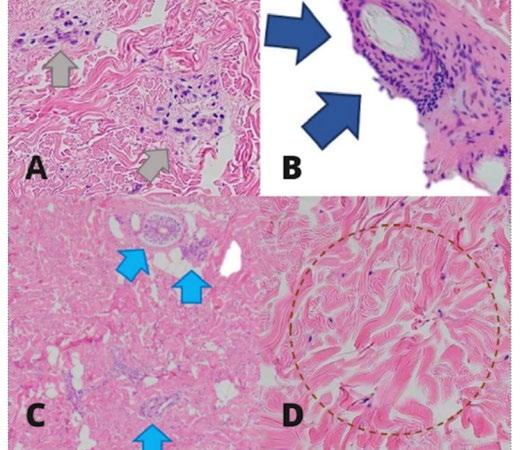
Fourth, the lack of standardized objective outcome measures (e.g., validated skin scoring systems or imaging-based assessment) restricts the ability to quantitatively evaluate treatment response to UVA1 phototherapy.
Finally, while the patient demonstrated significant clinical improvement, longterm follow-up data are limited, and the durability of response, as well as risk of relapse, remain uncertain.
DISCUSSION
Morphea is a chronic inflammatory disease that affects the dermis and subcutaneous tissue. It is also known as localized scleroderma. Progressive skin sclerosis, thickness, induration, and variable degrees of atrophy are its defining characteristics. Morphea is rarely fatal, though it can lead to functional and aesthetic problems. In rare instances, it is self-limited, but more frequently it follows a chronic, relapsing–remitting pattern, which over time leads to a considerable cumulative disease burden.
Given the clinical presentation, several conditions were considered in the differential diagnosis, including systemic sclerosis, atrophoderma of Pasini and Pierini, stiff skin syndrome, and eosinophilic fasciitis. Systemic sclerosis was considered due to the presence of cutaneous sclerosis; however, Raynaud phenomenon and other signs and symptoms were not present, hence this diagnosis is less likely. Atrophoderma of Pasini and Pierini was also entertained given the presence of dermal atrophy, but the lack of the typical cliff-drop borders argued against this entity. Stiff skin syndrome was considered in view of skin induration; however, the clinical course and distribution were not consistent with its usual presentation. Eosinophilic fasciitis was likewise included in the differential due to skin thickening, but the absence of peripheral eosinophilia, fascial involvement, and groove sign made this diagnosis unlikely. Taken together, these findings support the final diagnosis of morphea.
Figure 2: Dermoscopy: fibrotic beams (red arrows) and loss of hair.
Figure 3: A and B) Sparse perivascular periadnexal infiltrates; C) entrapped eccrine glands; D) thick collagen bundles.
The pathogenesis of morphea remains incompletely understood; however, accumulating evidence points to multiple immunoinflammatory and profibrotic pathways. In linear morphea there is immune activation, vascular injury, and progressive fibrosis. In the early phase, Th1/Th17-mediated inflammation leads to the release of cytokines such as IL-6 and TNF-α, causing endothelial damage and immune cell infiltration into the skin and deeper tissues.1 The disease then shifts toward a Th2-dominant response, where IL-4 and IL-13 promote fibroblast activation and collagen deposition, with TGF-β acting as a key profibrotic mediator.1 In linear morphea, these processes extend beyond the dermis to involve subcutaneous tissue, muscle, and bone, leading to structural deformity and functional impairment.1
The development of morphea is thought to result from an interplay between predisposing host factors and external triggers.2 Predisposing factors include a personal or family history of autoimmune disease, concurrent autoimmune conditions, cutaneous mosaicism, and specific human leukocyte antigen (HLA) subtypes, particularly HLA-DRB104:04 and HLA-B37, which have been associated with increased susceptibility to generalized and linear forms of morphea.2,3 These genetic associations were also present in the authors’ patient.
Epigenetic mechanisms are thought to serve as a link between genetic susceptibility and environmental factors.4 External triggers such as injuries, injections, or vaccinations can initiate disease in individuals who are genetically predisposed, as seen in this case. After such triggers, a complex process involving epidermal keratinocytes and dermal fibroblasts occurs, engaging innate and adaptive immune responses along with profibrotic signaling pathways. This process ultimately results in excessive collagen deposition and fibrosis.4
In the present case, the onset of cutaneous induration occurred within a few days
following BCG vaccination, suggesting a close temporal association. Similar temporal relationships have been described in previously reported cases of vaccineassociated morphea, where lesions developed days to weeks after immunization, often at or near the injection site. This pattern supports the hypothesis that vaccination may act as a triggering event in predisposed individuals rather than a direct causative factor.
From a pathophysiological standpoint, vaccination may induce localized immune activation through multiple mechanisms. Mechanical trauma from injection may initiate an exaggerated wound-healing response, while vaccine components or adjuvants may stimulate innate immune pathways, including Toll-like receptor signaling, leading to the release of proinflammatory and profibrotic cytokines such as TGF-β. This cascade promotes fibroblast activation, excessive collagen deposition, and, ultimately, cutaneous sclerosis.
Despite these proposed mechanisms, current evidence remains largely limited to case reports and small case series, and a definitive causal relationship between vaccination and morphea has not been established. Therefore, the observed association should be interpreted with caution.
Eight cases of morphea have been associated with different vaccines, including measles–mumps–rubella, diphtheria–tetanus–pertussis, BCG, hepatitis B, and tetanus.5 Given the variety of vaccines involved, the authors propose that injectionrelated trauma or vaccine preservatives may serve as the actual triggers, rather than specific vaccine antigens.6 It is also suggested that mechanical trauma from vaccination could provoke an exaggerated wound-healing response, resulting in abnormal fibrosis.
Recently, several cases of morphea were reported following COVID-19 vaccination. One case involved an 80-year-old man who developed morphea profunda after
vaccination and was successfully treated with oral methotrexate.6 Although a temporal association does not establish causality, these reports highlight the potential for vaccine-related immune activation to serve as a trigger in individuals who are genetically or immunologically predisposed. Current evidence supporting vaccine-associated autoimmunity is largely derived from case reports and observational data, and a definitive causal relationship has not been established. The management of morphea remains challenging, since there is no single therapy that is regarded as the gold standard.7 Treatment selection is directed by disease activity, extent, anatomical distribution, depth of involvement, and rate of progression.8 Consequently, a patientcentered therapeutic approach is essential.9
In pediatric patients with linear morphea, management is guided by disease severity and depth of involvement, with the primary goal of controlling inflammation early to prevent permanent fibrosis and deformity. For mild, superficial lesions, especially in cosmetically sensitive areas like the face, topical tacrolimus may be used as a firstline or adjunctive therapy.9,10 It works by inhibiting T cell activation and reducing inflammatory cytokines, helping to decrease erythema and soften lesions; however, its limited tissue penetration makes it insufficient for deeper or progressive disease.1,11 In cases of moderate-to-severe, rapidly progressive, or function-threatening linear morphea, systemic therapy is required, with methotrexate serving as the cornerstone of treatment.9 Methotrexate suppresses immune activity and fibrosis and has the strongest evidence for reducing disease activity and preventing progression in children; it is typically administered weekly and continued for at least 12 months or longer.9 Because methotrexate has a delayed onset of action, it is commonly combined initially with systemic corticosteroids such as prednisone or intravenous methylprednisolone to achieve rapid control of active inflammation; these steroids are used as short-term
induction therapy and then tapered, as they are not suitable for long-term disease control.9 For patients who do not respond adequately to or cannot tolerate methotrexate, mycophenolate mofetil is an effective second-line option, offering immunosuppressive effects through inhibition of lymphocyte proliferation and serving as a useful steroid-sparing agent.9,11 Overall, treatment often involves a combination approach (early systemic therapy for active disease, with adjunctive topical agents for superficial lesions) to optimize outcomes and minimize long-term complications.
UVA1 phototherapy uses longwavelength UV radiation (340–400 nm), which penetrates more deeply into the dermis than UVB or broadband UVA.8 Its therapeutic effect in morphea is attributed to immunomodulatory actions, including attenuation of inflammatory responses, suppression of fibroblast activity and collagen synthesis, and upregulation of matrix metalloproteinases (e.g., matrix metalloproteinase-1) involved in collagen degradation.8 These effects are thought to be mediated by activation of the aryl hydrocarbon receptor pathway, resulting in antifibrotic activity at the cellular level.8
Several studies have demonstrated that UVA1 phototherapy significantly improves cutaneous sclerosis in morphea, leading to plaque softening and enhanced skin elasticity.1,3,10 Both medium and highdose regimens have shown efficacy, with documented improvements in clinical severity scores and objective assessments of skin elasticity.12 In a prospective study, approximately 82% of patients reported symptomatic improvement, accompanied by ultrasonographic evidence of reduced skin thickness following UVA1 treatment.6
In the short term, adverse effects of UVA are generally mild and include erythema (a sunburn-like reaction), skin dryness and pruritus due to epidermal barrier disruption, and hyperpigmentation from increased
melanogenesis; patients may also experience heat-related discomfort during treatment because UVA1 devices generate significant warmth, and, rarely, phototoxic reactions can occur if combined with photosensitizing agents.1 Long-term risks are more concerning, especially in pediatric patients, and include photoaging from cumulative UV exposure leading to collagen breakdown, resulting in skin laxity, wrinkling, and dyspigmentation.1 There is also a theoretical risk of carcinogenesis due to deep dermal penetration of UVA1 and oxidative DNA damage, with potential associations with melanoma, basal cell carcinoma, and squamous cell carcinoma, although pediatric data remain limited and much of the risk is extrapolated from adult and psoralen plus UVA studies.1,13
Cutaneous involvement in morphea can result in restricted range of motion, limblength discrepancy, joint deformities, and contractures, particularly in cases of linear morphea, where musculoskeletal complications have been reported in approximately 45–56% of patients.8 Lesions that cross joint lines confer the highest risk for functional impairment.8 In pediatric patients, morphea may be associated with substantial morbidity, including adverse effects on growth, physical function, and quality of life, whereas fewer studies have evaluated these outcomes in adults.8
The relationship between morphea subtype and quality of life using the Dermatology Life Quality Index (DLQI) and Children’s Dermatology Life Quality Index (CDLQI) were evaluated. Among 202 adult patients, 75 individuals experienced a moderate or greater impact on quality of life (DLQI >5), with the greatest impairment observed in those with generalized morphea; however, no statistically significant correlation was identified between morphea subtype and DLQI scores (F=1.97; p=0.12). In contrast, among 75 pediatric patients, 21 (28%) reported a moderate or greater impact on quality of life, nearly all of whom had linear morphea.11 Similarly, no significant association was found between morphea
subtype and CDLQI scores (F=0.45; p=0.72).11 Overall, children demonstrated a lower frequency of moderate-to-severe quality-oflife impairment compared with adults.13
Although morphea is rarely life-threatening, it can lead to significant functional and cosmetic morbidity.7 While the disease may be selflimited in some cases, it more commonly follows a chronic or relapsing–remitting course, resulting in a substantial cumulative disease burden over time.8
Morphea temporally associated with vaccination is a rare occurrence and, despite its chronic and potentially relapsing course, remains a treatable condition.14 Notably, spontaneous remission has been reported in a subset of patients.6 These observations underscore the importance of avoiding undue alarm and emphasize the need for individualized clinical assessment and management, as patient responses to disease processes, pharmacologic therapies, and vaccinations may vary.14 Importantly, the well-established benefits of vaccination substantially outweigh the potential risks.14 Consequently, rare adverse cutaneous events such as morphea should not deter vaccination, particularly in the context of ongoing public health challenges.
CONCLUSION
Although morphea can result in functional and cosmetic impairment, it is rarely lifethreatening. The disease may be selflimited in some individuals; however, it more commonly follows a chronic or relapsing–remitting course, leading to a substantial cumulative disease burden over time. Given the wide range of vaccines reported in association with morphea, it has been suggested that injection-related trauma or vaccine preservatives, rather than specific vaccine antigens, may act as potential triggers. Mechanical trauma from vaccination may initiate an exaggerated woundhealing response, resulting in dysregulated fibrosis. UVA1 phototherapy is an effective
therapeutic modality for the management of inflammatory and sclerosing cutaneous disorders, including morphea.
PATIENT PERSPECTIVE
Before starting the treatment, my skin felt very tight and hard, which made it difficult for me to move comfortably and do activities of daily living. I cannot play sports with my friends due to the limitation of movement. When I started phototherapy, I did not know what
References
1. Knobler R et al. European Dermatology Forum S1-guideline on the diagnosis and treatment of sclerosing diseases of the skin, part 1: localized scleroderma. J Eur Acad Dermatol Venereol. 2017;31(9):1401-24.
2. Fett N et al. Update on morphea: part I. Epidemiology, clinical presentation, and pathogenesis. J Am Acad Dermatol. 2011;64(2):217-28.
3. Leitenberger JJ et al. Distinct autoimmune syndromes in morphea: a review of 245 adult and pediatric cases. Arch Dermatol. 2009;145(5): 545-50.
4. Wolska-Gawron K et al. MicroRNA in localized scleroderma: a review of literature. Arch Dermatol Res. 2019;312(5):317-24.
to expect, but the treatments were generally tolerated. One downside was I had to stand up for a long period of time since the duration of treatment became progressive over time. During my phototherapy sessions, I watched short films to keep myself entertained, which helped pass the time without me noticing it. After completing the treatment, I noticed that my skin became softer and less tight. I was able to move more easily, and my daily activities were less difficult. I felt relieved and more confident with the improvement.
5. Kreuter A. Localised scleroderma. Dermatol Ther. 2013;26(2):135-47.
6. Andres C et al. Successful ultraviolet A1 phototherapy in the treatment of localized scleroderma: a retrospective and prospective study. Br J Dermatol. 2010;162(2):445-7.
7. Oba Ç et al. Morphea: Clinical considerations and management. J Turk Acad Dermatol. 2020;14(1):1-5.
8. Kang S et al. Fitzpatrick's Dermatology (2019) 9th edition, New York: McGraw Hill Education.
9. Martini G et al. Disease course and long-term outcome of juvenile localized scleroderma: experience from a single center. Arthritis Rheum. 2008;59(2):292-9.
10. Kliegman R, St. Geme JW III. Nelson Textbook of Pediatrics, 2-Volume Set (2019) 21st edition, Philadelphia: Elsevier.
11. Li SC et al. Treatment of pediatric localized scleroderma: current concepts and future directions. Curr Treat Options Rheumatol. 2015;1(2):143-60.
12. Kreuter A et al. Diagnosis and therapy of localized scleroderma. J Dtsch Dermatol Ges. 2009;7(6):489-503.
13. Das S et al. Correlates of self-reported quality of life in adults and children with morphea. J Am Acad Dermatol. 2014;70(5):904-10.
14. McPherson GC et al. Generalized morphea profunda following COVID-19 messenger ribonucleic acid vaccination. Dermatol Online J. 2025;31(4):207.

Quadruple Trouble: A Bisexual Male with Kaposi Sarcoma, Cutaneous Tuberculosis, HIV, and Syphilis
Authors: *Rosalie Deeneye Santiago Iglesias,¹ Carelle Lois See,¹ Jean Anne Camille Marcelo-De Jesus,¹ Roxanne Louise Salazar,¹ Karlo Edwin R. Balisi,¹ Maria Franchesca Quinio-Calayag¹
1. East Avenue Medical Center, Metro Manila, Philippines *Correspondence to deeneyesantiago@gmail.com
Received: 01.28.26
Accepted: 04.10.26
Keywords: AIDS-associated Kaposi sarcoma, case report, cutaneous tuberculosis, extrapulmonary tuberculosis, Kaposi sarcoma (KS), syphilis and HIV coinfection, tuberculosis and HIV.
Citation: Dermatol AMJ. 2026;3[1]:114-123. https://doi.org/10.33590/dermatolamj/19FR0078
Abstract
AIDS is defined by several opportunistic infections, including Kaposi sarcoma (KS) and tuberculosis. This report describes a complicated case of AIDS-associated Kaposi sarcoma with syphilis and cutaneous tuberculosis coinfection. A 32-year-old bisexual male presented with a 3-month history of violaceous patches initially on the right sole spreading to the whole body. Cutaneous examination showed violaceous to brownish patches and plaques on the face, hard palate, trunk, both upper and lower extremities, with a nodule on the right sole. He was also diagnosed with gastrointestinal tuberculosis with a finding of a mesentery abscess positive for acid-fast bacilli stain. Sexually transmitted diseases laboratory work-up revealed late latent syphilis treated with benzathine penicillin G, and AIDS with a CD4 count of 29 cells/µL, and was started on anti-retroviral medications. Skin punch biopsy of the patch on the right thigh with CD31 and CD34 stain confirmed KS. While on antiretroviral and antituberculosis medications, the patient developed scrofuloderma. Skin punch biopsy of the right neck showed cutaneous tuberculosis with KS features. After 5 months of antiretroviral therapy and imiquimoid 5% cream as local therapy, there was 50% improvement in cutaneous lesions, and the patient was then lost to follow-up. A high index of suspicion and a thorough examination should be done in this population for early diagnosis and management of these complications.
Key Points
1. Advanced AIDS is a diagnostic challenge. Profound immunosuppression (CD4 count <30) allows for the simultaneous occurrence of multiple opportunistic pathologies. A high index of suspicion must be upheld by the clinician in order to personalize treatment strategies for each patient.
2. This case is of a 32-year-old male presenting with disseminated Kaposi sarcoma, late latent syphilis, and gastrointestinal tuberculosis. It also highlights the clinical challenge of immune reconstitution inflammatory syndrome, where the patient's improving immune system, following antiretroviral therapy, paradoxically triggers a new inflammatory response in the form of cutaneous tuberculosis (scrofuloderma).
3. Clinicians must perform thorough examinations, as Kaposi sarcoma can coexist with other infections. Patients should be monitored for immune reconstitution inflammatory syndrome and visceral involvement. A multidisciplinary approach involving dermatology, oncology, and infectious diseases is recommended. Treatment plans should also focus on wellness and prevention for a more holistic approach.
INTRODUCTION
HIV is part of the Retroviridae family of viruses, which induces suppression of the immune system and dysregulation primarily via depletion of CD4+ lymphocytes.1 AIDS is the late stage of HIV, which can be diagnosed when CD4 cell counts plummet to levels of <200 cells/µL or infections caused by opportunistic pathogens are identified, including both Kaposi sarcoma (KS) and Mycobacterium tuberculosis (M. tuberculosis) infection.1 One of the most frequently seen cancers among individuals affected by HIV is KS,2 occurring mostly in homosexual or bisexual men with abnormally low levels of CD4 count, usually below 350 cells/mm.1,3 It is an angioproliferative disorder arising from endothelial cell infection by KS Herpesvirus or Human Herpesvirus-8 (HHV8).4 M. tuberculosis, on the other hand, causes an endemic pulmonary infection and is a major public health concern in the Philippines.5 Extrapulmonary tuberculosis, a less common form of M. tuberculosis infection, occurs in patients infected with HIV. Seventy percent of individuals afflicted by tuberculosis and concomitant AIDS infection are usually affected.6 Lastly, syphilis is a venereal disease primarily caused by Treponema pallidum, a part of the Spirochaetaceae family of bacteria.7 A resurgence of this bacterial infection has
caused it to be identified as a global health risk, especially among individuals who are HIV-positive.8 Due to risk-taking behaviors such as sexual intercourse without the use of condoms among the younger men who have sex with men (MSM) population, concomitant infection of syphilis among the HIV afflicted patients is increasing.8
This study describes a case of a Filipino bisexual male, who was HIV-positive with co-existing KS, syphilis, gastrointestinal, and cutaneous tuberculosis.
CASE SUMMARY
This is a case of a 32-year-old, bisexual male from Manila, Philippines, who presented with a 3-month history of multiple violaceous patches and plaques starting on the right sole, gradually spreading to the face, upper and lower extremities, with no other associated symptoms such as pain or pruritus.
Two months prior to the consult, the patient also had fever, body malaise, right lower quadrant pain, anorexia, and vomiting. He was admitted under an impression of appendicitis, hence underwent exploratory laparotomy and appendectomy. Intraoperatively, a mesentery abscess
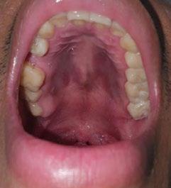
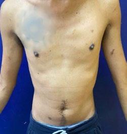
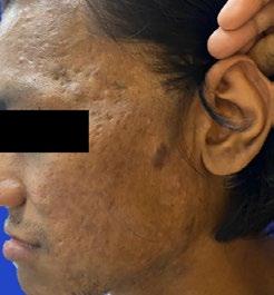
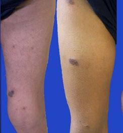
was seen, which revealed 3+ for acid-fast bacilli stain. Hence, an assessment with gastrointestinal tuberculosis was made, and he was started on an anti-tuberculosis singletablet regimen: rifampicin 150 mg, isoniazid 75 mg, pyrazinamide 400 mg, ethambutol 275 mg once daily. There was relief from the abdominal symptoms.
One month prior to the consult, the patient had intermittent fever and was admitted for sexually transmitted diseases workup at a tertiary government hospital. He tested positive at both screening and confirmatory testing for HIV with a CD4 cell count of 29 cells/µL. Further laboratory work-ups were done, revealing non-reactive Rapid Plasma Reagin (RPR) and hepatitis B surface antigen (HBsAg), while the treponemal test (TPPA) was positive. He was started on a fixed-dose combination of lamivudine 300 mg, efavirenz 600 mg, and tenofovir disoproxil fumarate 300 mg for HIV-1 treatment. Benzathine
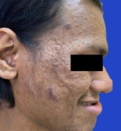
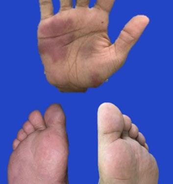
penicillin G 2,400 IU was administered weekly intramuscularly in the buttocks for 3 doses as treatment for late latent syphilis. Prophylaxis for opportunistic infections was also started, including fluconazole (cryptococcosis) and cotrimoxazole (Pneumocystis pneumonia). The patient was referred to the dermatology service for further evaluation of the skin lesions.
The patient works as a civil engineer. He is single, with several sexual partners, including 3 female and 12 male partners, and engages in unprotected oral, anal, and vaginal sexual intercourse wherein he assumes both penetrative and receptive roles. The review of systems was unremarkable. The patient’s medical and family history were non-contributory.
Cutaneous examination showed violaceous to brownish patches and plaques on the tip of the nose, preauricular and temporal area,
Figure 1: Clinical presentation of Kaposi sarcoma in a 32-year-old male.
left clavicular area, both arms and thighs, palms, soles, and hard palate (Figure 1).
Dermoscopy shows a homogeneous pattern consisting of white lines on a pink to brown background on the patches and plaques, and a rainbow pattern on the nodule (Figure 2).
Skin punch biopsy, CD34, and CD31 staining showed KS (Figure 2).
The patient was lost to follow-up for 3 months but continued his antiretroviral and anti-tuberculosis medications. During the interim, the patient developed erythematous, non-painful nodules on both sides of the
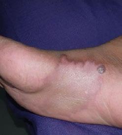
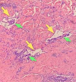
staining for acid-fast bacilli, CD34, and CD31, assessed as cutaneous tuberculosis with KS features (Figure 3).
To assess the visceral involvement of KS, chest X-ray and fecal occult blood test were
neck, which spontaneously ruptured. On physical examination, non-tender cervical lymphadenopathies were noted, and a cutaneous examination showed round to oval, erythematous, non-tender plaques with ulceration and purulent discharge with hemorrhagic crusting on the neck. The clinical impression was scrofuloderma; hence, Tissue Gene Xpert® (Cepheid, Sunnyvale, California, USA) of the neck was requested, which was negative. Wound Gram stain and culture and sensitivity were also negative. Another skin punch biopsy of the right neck was done, which had positive
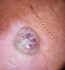
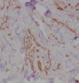
requested, which were both negative. A CT scan of the neck, chest, and whole abdomen was also requested; however, it was not done due to financial constraints. Imiquimod 5% cream applied three times weekly was started as local therapy for the cutaneous lesions.
A B
C D
Figure 2: Dermoscopy, histopathology and, immunohistochemistry of Kaposi sarcoma.
A B
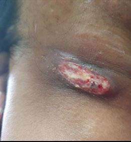
After 5 months of antiretroviral therapy (ART), there was a 50% improvement in the lesions with noted flattening of the plaques and lightening of the violaceous color of some patches and plaques on the face, upper, and lower extremities. However, efforts have been made to reestablish communication, but the patient eventually discontinued follow-up. A detailed chronological summary of the patient’s clinical course, including presentation, diagnostic workup, and management, is presented in Table 1
DISCUSSION
Advanced HIV disease (CD4 count <200 cells/µL) remains a diagnostic challenge due to the coexistence of multiple opportunistic infections and malignancies.9 This case is particularly significant for its simultaneous presentation of KS, late latent syphilis, and extrapulmonary tuberculosis, making this case rare and of clinical value.
KS is a variety of an angioproliferative disorder stemming from infection of an endothelial cell by Kaposi sarcomaassociated herpesvirus (KSHV) or HHV8.4 Hyper-proliferation of KSHV-infected cells is usually seen together with immunomodulatory cell infiltration, which results in chronic inflammatory states,
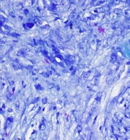
angiogenesis, and tumor progression.4 This occurs statistically in 481.54 per 100,000 person-years among individuals who are HIVpositive, with the highest incidence observed in HIV-positive MSM.10 Local incidence based on the Philippine Dermatological Society Health Information System shows that there are 67 patients from 2011–2019 with KS, and the cases are predominantly in males.
There are 4 clinical and epidemiologic types:11,12 1) the classic type affecting older men of Mediterranean or Eastern European origin, which presents with a slow, indolent course with lesions commonly appearing on the lower extremities; 2) the endemic type occurs in sub-Saharan Africa and demonstrates a broader clinical spectrum, ranging from indolent cutaneous disease to aggressive forms involving lymph nodes and visceral organs, particularly among young adults and children; 3) the iatrogenic type arises in individuals receiving immunosuppressive therapy following organ transplantation and may show partial or complete regression upon reduction of immunosuppressive agents; and the last type 4) the epidemic type, which is associated with HIV/AIDS, present in the authors’ patient, represents the most aggressive form characterized by widespread mucocutaneous and visceral involvement due to profound immunosuppression.
Figure 3: Scrofuloderma.
Table 1: Timeline of clinical course and management.
3 months PTC Onset of a violaceous patch on the right sole -
2 months PTC Fever, right lower quadrant pain, anorexia, and vomiting
Diagnosis: appendicitis
Intraoperative finding of mesentery abscess, AFB stain positive 3+
Diagnosis: gastrointestinal tuberculosis
1 month PTC Intermittent fever
Admitted for sexually transmitted disease workup:
• HIV testing: positive
• HBsAg: nonreactive
• RPR: nonreactive
• TPPA: positive
• CD4 cell count of 29 cells/µL
Diagnosis: acquired immunodeficiency syndrome late latent syphilis
Increase in violaceous patches spreading to the hard palate, left clavicular area, both upper and lower extremities, including palms and soles
Day of consult Referral to dermatology
Dermoscopy: homogeneous pattern consisting of white lines on a pink to brown background (patches and plaques) and a rainbow pattern (nodule)
Skin punch biopsy (right thigh and right palm):
Kaposi sarcoma with positive staining for CD34 than CD31
Diagnosis: Kaposi sarcoma
Appendectomy
Antituberculosis medications started: isoniazid, rifampicin, pyrazinamide, ethambutol
Antiretroviral initiated: lamivudine, efavirenz, and tenofovir
Prophylaxis: cotrimoxazole (Pneumocystis pneumonia) fluconazole (cryptococcosis)
Benzathine penicillin G 2,400 IU weekly for three doses
Advised patient to continue antiretroviral medications
Month 3 Developed erythematous non-painful nodules on both sides of the neck, which eroded into ulcers -
Month 4
Workup for visceral involvement of Kaposi sarcoma
• Chest X-ray: Normal
• Fecal occult blood test: Negative
• Chest and whole abdominal CT scan with IV Contrast for metastatic workup: requested but not done
Workup for the ulcer on the neck
• Wound Gram stain and culture and sensitivity: No growth
• Tissue Gene Xpert® (Cepheid, Sunnyvale, California, USA): Mycobacterium tuberculosis bacilli not detected
• CT scan of the neck: requested but not done
Diagnosis: scrofuloderma
Month 5 Skin punch biopsy (right neck):
Cutaneous tuberculosis with Kaposi sarcoma features, positive staining for AFB, CD34, and CD31
Diagnosis: AIDS-associated Kaposi sarcoma scrofuloderma
Advised patient to continue antituberculosis medications taken for the gastrointestinal tuberculosis
Imiquimod 5% cream three times weekly as local treatment initiated on Kaposi lesions
Lost to follow-up
Plans:
Repeat CD4 count and viral load
Oncology service
• Requested chest and whole abdominal CT scan with IV contrast for metastatic workup
Infectious disease
• Clearance given to start chemotherapy -
AFB: acid-fast bacilli; HBsAg: hepatitis B surface antigen; PTC: prior to consult; RPR: Rapid Plasma Reagin; TPPA: Treponemal test.
The clinical manifestations of KS usually present with multiple, pigmented, nonblanching, painless macules and papules on the lower extremities.1 Larger oval plaques located on the torso often line up along the skin creases.13 Protruding, ulcerated, and hemorrhagic nodules can develop with edema associated with tenderness.13 AIDSassociated KS commonly presents with oral lesions in the palate, which is present in the authors’ patient, and may be one of the first symptoms of underlying immunosuppression.14 Visceral lesions are also more common among individuals with AIDS-related KS, frequently occurring in the lungs, lymph nodes, and gastrointestinal tract.15
A very important tool in dermatology in this era is dermoscopy. The rainbow pattern was seen in a nodular lesion using a contact polarized-light dermatoscope, a common finding in KS lesions.16 KS is commonly diagnosed based on the distinctive appearance of the lesions alone, but confirmation of the diagnosis must be based on histologic examinations.17 Histopathology usually shows spindle cells, growth of irregular vessels with slit-like configurations, extravasation of erythrocytes, and leukocytic infiltration encompassing full dermal thickness with associated plasma cells, and intra- and extracellular hyaline globules as well as the promontory sign (Figure 2).18
The histopathology result of the right neck of this patient showed cutaneous tuberculosis with KS features. In patients with HIV, more than one pathology in a single biopsy site can be seen, and case reports documented KS in association with tuberculosis, cryptococcosis, and Mycobacterium avium intracellulare in the same sample.19 A case report in Zimbabwe showed a 25-year-old patient who was HIVpositive whose biopsy of the mass of the right neck showed features of both cutaneous tuberculosis and KS, similar to this patient.20
Several markers can be used to differentiate KS from other vascular lesions.21 Latencyassociated nuclear antigen (LNA1) is a highly sensitive and specific marker, given the
strong correlation between HHV-8 and KS.21 However, since this is not available locally, the authors utilized other endothelial markers for vascular conditions, particularly CD31 and CD34, which both have positive staining in their sample. CD31 (platelet endothelial cell adhesion molecule-1) is considered one of the most sensitive and specific markers for endothelial differentiation; it highlights the spindle cells and abnormal vascular channels characteristic of KS, making it particularly useful in distinguishing it from other spindle cell neoplasms. CD34, another endothelial marker, is also commonly expressed in KS and stains both the tumor cells and surrounding vascular structures, although it is less specific because it can be positive in a wider range of soft tissue tumors.11 In practice, both CD31 and CD34 are used together as part of an immunohistochemical panel to support the diagnosis, typically alongside detection of HHV-8, which is more specific for KS.22 While CD31 offers higher specificity for vascular differentiation, CD34 provides complementary sensitivity, and their combined expression strengthens diagnostic confidence, especially in early or atypical lesions.11,22 In advanced stages of KS, CD34 exhibits stronger immunoreactivity than CD31, which was seen in the case.17
The AIDS Clinical Trial Group (ACTG) of the National Institute of Health developed the most frequently used staging system for AIDS-related KS.23 There are three categories in this staging, including tumor, immune system, and systemic disease. The patient in this case had lesions confined to the skin with minimal oral disease, CD4 count of <29 cells/µL, and systemic disease, showing a history of opportunistic infection, particularly extrapulmonary tuberculosis, but with the Kanofsky score of 90 defined as capable of normal activity but with few symptoms or signs of the disease T0I1S1. Pending the metastatic workup, the author’s patient falls into poor-risk.
Based on the NCCN Clinical Guidelines in Oncology, AIDS associated KS can be classified as having limited or advanced
cutaneous disease.23 Asymptomatic individuals who are cosmetically unremarkable are considered to have limited cutaneous disease, and these patients may be treated with ART alone.23 As observed in the patient, there was 50% improvement on the patches and plaques with 5 months of ART.
Topical imiquimod has been investigated as a local therapy for KS, particularly for cutaneous and localized lesions.23-27 Imiquimod acts as a toll-like receptor 7 agonist, inducing local production of cytokines such as interferon-α and promoting antitumor immune responses as well as antiangiogenic effects.24 A systematic review of topical therapies for KS identified imiquimod as one of the most commonly studied agents, where 5% imiquimod cream was typically applied for several weeks (median duration approximately 17 weeks), sometimes under occlusion.24 Clinical outcomes showed complete response in approximately 34.6% of patients and partial response in 30.8%, with overall good tolerability and mainly mild local adverse effects such as erythema and pruritus.24 Individual case reports further support its efficacy. One study reported complete clearance of classic KS lesions after 20 weeks of imiquimod 5% cream applied three times weekly under occlusion, with no recurrence at 6-month follow-up.25 Another case demonstrated near-complete regression of lesions within 12 weeks, again with minimal toxicity.26 Additional reports have shown successful treatment of localized genital KS, highlighting its usefulness as a noninvasive option in anatomically sensitive areas.27 Available studies suggest that topical imiquimod is a promising, well-tolerated local therapy for selected patients with limited cutaneous KS; hence, this was also used for this case.
The prognosis of KS varies widely and depends on several factors, particularly immune status, extent of disease, and response to therapy.28 The introduction of ART has significantly improved survival in AIDS-associated KS by promoting immune reconstitution and reducing viral replication,
which can lead to stabilization or regression of lesions. Patients with limited disease, higher CD4 counts, and absence of systemic symptoms generally have better outcomes.23 The determinants significantly associated with increased mortality or the necessity for chemotherapeutic regimens include tumor Stage of T1, a decreased CD4 count of less than 200 cells/µL, and a positive test for HHV8.28 As with the case, a CD4 count of 29 cells/ µL puts the authors’ patient at poor risk.
Scrofuloderma is an unusual variety of extrapulmonary tuberculosis, encompassing 1–2% of cases, but it is the most common form of extrapulmonary tuberculosis in patients who are HIV-positive and in the pediatric age group.29 While on treatment, this patient also developed scrofuloderma signifying immune reconstitution inflammatory syndrome (IRIS), which occurs in 18% of patients who are HIV-TB co-infected.30 Occult tuberculosis was subsequently unmasked in this patient as the immune system recovered, presenting with multiple neck nodules. IRIS is a paradoxical exacerbation of a pre-existing infection or disease process or the emergence of a new infection/disease process soon after initial therapeutic modalities.1 Upon initiation of ART, individuals with concurrent infections of HIV-1 and tuberculosis (TB) are vulnerable to developing TB-associated IRIS (TB-IRIS).31 Patients with low CD4+T cell counts at the time of ART initiation, followed by a rapid increase in CD4 counts post-ART, are more likely to develop TB-IRIS.31
On the sexually transmitted disease workup for this patient, a diagnosis of late latent syphilis was made through serology. The presence of syphilis alongside TB and KS further complicates this case. Recent data shows a rising trend in syphilis and HIV coinfection, with approximately 45.5% of highrisk populations showing positive serology for both.32 Syphilis is a venereal disease with protean manifestations stemming from Treponema pallidum infection. Systemic illness is of initial consideration due to vasculitis as its primary pathology.33 With negative non-treponemal test RPR and
positive treponemal test TPPA, this patient was diagnosed with the late latent syphilis34,35 and underwent treatment with benzathine penicillin G 2,400 IU weekly for three doses based on the CDC Sexually Transmitted Guidelines 2015.36 Monitoring should be done using RPR, and there should be a four-fold decrease in titers to determine if the patient had been treated completely.36 Hicks et al.37 described a repeated non-reactive Veneral Disease Research Laboratory Test (VDRL) and Fluorescent Treponemal Antibody Absorption (FTA-ABS) in an HIV-positive individual with concomitant Kaposi’s sarcoma and secondary syphilis. Impairments in antibody responses among HIV afflicted individuals are shown by various studies as well. As seen in these cases, the antigenic response may range from complete seronegativity to exaggeration inconsistent with the clinical findings.38
HIV is associated with many opportunistic infections and malignancies, and the presence of syphilis and TB coinfections with KS makes this a rare case. A similar case described a 30-yearold Indian male with HIV, with generalized purplish or brown rash, where histopathology of cutaneous lesions confirmed the diagnosis of KS.39 During workup, he was also positive for
References
1. Jameson JL et al., “Human Immunodeficiency Virus Disease: AIDS and Related Disorders,” Fauci AS et al. (eds.), Harrison’s Principles of Internal Medicine (2019), United States: McGraw-Hill Education, pp. 1448-49.
2. Lamers SL et al. HIV-1 Evolutionary patterns associated with metastatic Kaposi’s sarcoma during AIDS. Sarcoma. 2016;2026:1-10.
3. Crum-Cianflone et al. Is Kaposi's sarcoma occurring at higher CD4 cell counts over the course of the HIV epidemic? AIDS. 2010;24(18):2881-83.
4. Douglas JL et al. Kaposi sarcoma pathogenesis: a triad of viral infection, oncogenesis and chronic inflammation. Transl Biomed. 2010;1(2):172.
5. Department of Health Philippines. National Tuberculosis Control Program
syphilis, pulmonary tuberculosis, and primary effusion lymphoma. A high index of suspicion for early diagnosis and long-term monitoring must be advised to these patients.
Ethics and Consent
Written informed consent was obtained from the patient for publication of this case report and accompanying clinical images. Patient anonymity was strictly maintained, and no identifying information was disclosed in this manuscript.
CONCLUSION
This is a complicated case of AIDS associated KS with extrapulmonary tuberculosis and syphilis. This case differs from previous literature by highlighting the prognosis and clinical improvement despite its quadruple presentation. Timely intervention and a multidisciplinary approach are necessary. Clinicians must always maintain a high index of suspicion in patients who are severely immunocompromised.
Manual of Procedures (2020) 5th edition, Manila: Department of Health.
6. Slutsker L et al. Epidemiology of extrapulmonary tuberculosis among persons with AIDS in the United States. Clin Infect Dis. 1993;16(4):513-18.
7. Kang S et al., “Syphilis,” Tuddenham S et al (eds.), Fitzpatrick’s Dermatology (2019), New York: McGraw-Hill Education, pp.3145.
8. Shilaih M et al. Factors associated with syphilis incidence in the HIV-infected in the era of highly active antiretrovirals. Medicine. 2017;96(2):e5849.
9. Lehman A et al. Advanced HIV disease: A review of diagnostic and prophylactic strategies. HIV Med. 2023;24(8):859-76.
10. Liu Z et al. The world-wide incidence of Kaposi's sarcoma in the HIV/AIDS era. HIV Med. 2018;19(5):355-364.
11. Kumar V et al., Robbins and Cotran Pathologic Basis of Disease (2020) 10th edition, Philadelphia: Elsevier.
12. Bolognia JL et al., Dermatology (2018) 4th edition, Philadelphia: Elsevier.
13. Cesarman E et al. Kaposi sarcoma. Nat Rev Dis Primers. 2019;5(1):9.
14. Martins-de-Barros AV et al. AIDSrelated Kaposi sarcoma of the oral cavity. Rev Soc Bras Med Trop. 2023;56:e0133.
15. El Mawla Z et al. Visceral kaposi's sarcoma as a presentation in a newly diagnosed hiv-infected man: a case report. Cureus. 2022;14(3):e23339.
16. Satta R et al. Dermoscopic rainbow pattern in kaposi's sarcoma lesions: our experience. Arch Dermatol. 2012;148(10):1207-08.
17. Amerson et al. Diagnosing Kaposi’s sarcoma (KS) in East Africa: how
accurate are clinicians and pathologists? Infect Agent Cancer. 2012;7(Suppl 1):P6.
18. Requena C et al. Kaposi sarcoma and cutaneous angiosarcoma: guidelines for diagnosis and treatment. Actas Dermosifiliogr (Engl Ed). 2018;109(10):878-87.
19. Calonje E et al., “Vascular tumors of low grade or borderline malignancy,” Calonje E et al. (eds.), McKee’s Pathology of the Skin: With Clinical Correlations (2020) 5th edition, Edinburgh: Elsevier, pp.1288.
20. Paksoy N. Simultaneous occurrence of Kaposi sarcoma and tuberculosis; Kaposi sarcoma and lymphoma in the same lymph node: a report on two HIV-positive patients from Zimbabwe. Rev Soc Bras Med Trop. 2019;52:e20180188.
21. Pantanowitz L et al. Immunohistochemistry in Kaposi’s sarcoma. Clinical and Experimental Dermatology, 2010;35(1):68-72.
22. Fletcher CDM et al., Diagnostic Histopathology of Tumors (2020). 5th edition. Philadelphia: Elsevier.
23. Reid E et al. AIDS-related Kaposi sarcoma, Version 2.2019. J Natl Compr Canc Netw. 2019;17(2):171-89.
24. Htet KZ et al. Topical treatments for Kaposi sarcoma: A systematic review. Skin Health Dis. 2022;2(2):e107.
25. Gündüz K et al. Efficacy of 5% imiquimod cream in a patient with classic Kaposi
sarcoma. J Dermatol Case Rep. 2012;6(2):52-3.
26. Benomar S et al. Kaposi's sarcoma responding to topical imiquimod 5% cream: a case report. Cases J. 2009;2:7092.
27. Fairley JL et al. Topical imiquimod 5% as a treatment for localized genital Kaposi's sarcoma in an HIV-negative man: a case report. Int J STD AIDS. 2012;23(12):907-8.
28. Boffi El Amari E et al. Predicting the evolution of Kaposi sarcoma in the highly active antiretroviral therapy era. AIDS. 2008;22(9):1019-28.
29. Mello R et al. Scrofuloderma: a diagnostic challenge. Anais brasileiros de dermatologia. 2019;94(1):102-04.
30. Walker NF et al. The tuberculosisassociated immune reconstitution inflammatory syndrome: recent advances in clinical and pathogenesis research. Curr Opin HIV AIDS. 2018;13(6):512-21.
31. Lai R et al. HIV-1 tuberculosis-associated immune reconstitution inflammatory syndrome. Semin Immunopathol. 2016;38(2):185-98.
32. Melin UN et al. Secondary syphilis with multiple co-infections of human immunodeficiency virus (HIV), tuberculosis (TB), and oral candidiasis in a 19-year-old men who have sex with men (MSM): a case report. BIKKK. 2024;36(3):225-31.
33. Hook EW III et al. Acquired syphilis in adults. N Engl J Med. 1992;326(16):1060-69.
34. Henao-Martínez AF, Johnson SC. Diagnostic tests for syphilis: new tests and new algorithms. Neurol Clin Pract. 2014;4(2):114-22.
35. Forrestel A et al. Sexually acquired syphilis. J Am Acad Dermatol. 2019;82(1):17-28.
36. Centers for Disease Control and Prevention (CDC). Sexually transmitted disease treatment guidelines. 2015. Available at: https://stacks.cdc.gov/view/ cdc/53167. Last accessed: April 22 2026.
37. Hicks CB et al. Seronegative secondary syphilis in a patient infected with the human immunodeficiency virus (HIV) with Kaposi sarcoma: a diagnostic dilemma. Ann Intern Med. 1987;107(4):492-5.
38. Gregory N et al. The spectrum of syphilis in patients with human immunodeficiency virus infection. J Am Acad Dermatol. 1990;22(6):1061-67.
39. Bangar S et al. Kaposi sarcoma (KS) with primary effusion lymphoma in HIVinfected MSM (men having sex with men) co-infected with pulmonary tuberculosis and syphilis: a case report from India. AIDS Res Ther. 2022;19:36.
If you like it,why not share it...

Share with a colleague today