Probing the protection mechanisms of colorectal cancer


The microenvironment around a tumour acts as a protective layer and helps prevent immune cells from entering. The team behind the PLASTICAN project aim to characterise the cellular composition of the microenvironment around a tumour, and are looking at whether interfering with it could lead to improved treatment of colorectal cancer, as Professor Florian Greten explains.
Colorectal cancer is one of the most common forms of cancer amongst both men and women, particularly in developed countries. Late stage tumours continue to have a very poor prognosis due to the lack of convincing therapeutic options, while the growing number of people below the age of 50 that are being diagnosed with colorectal cancer is causing widespread concern. “It’s a worrying development,” says Professor Florian Greten, director of the GeorgSpeyer Haus Institute for Tumour Biology and Experimental Therapy in Frankfurt, where he leads a research group. As Principal Investigator of the ERC-backed PLASTICAN project, Professor Greten is investigating how colorectal cancer depends on the surrounding microenvironment. “The microenvironment is the host tissue, the non-mutated cells,” he explains. “A tumour cell is a malignant cell with mutations in the DNA profile, while the

Credit: Andreas Reeg, Georg-Speyer-Haus.
stroma around a tumour is just host tissue that is not mutated.”
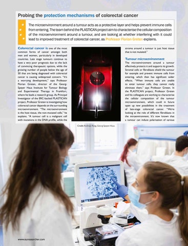
Tumour microenvironment
The microenvironment around a tumour effectively protects it and supports its growth. Stromal cells or fibroblasts shield the tumour for example and prevent immune cells from entering, which then has significant wider effects. “When immune cells are unable to enter tumour cells, they cannot really eliminate them,” says Professor Greten. In the PLASTICAN project, Professor Greten and his colleagues are working to characterise the cellular composition of the tumour microenvironment, which could in future open up new possibilities in the treatment of late-stage colorectal cancer. “We’re looking at the role of different fibroblasts in the miroenvironment. It’s now known that a tumour can induce polarisation of various
forms of fibroblasts that have distinct features. We aim to characterise these fibroblasts in greater detail and to investigate whether interfering with them could then allow for better therapies,” he continues.
This research involves using organoids, tumour cells grown in 3-D cultures in a matrix composition, which then allows for 3-D growth of those cells. Those organoids are then re-transplanted into mice, where they form single tumours in the colon, an approach which has some significant advantages over other genetic models. “With other models you get mutation or activation of oncogenes in the entire intestine, and then there are many tumours, especially in the small intestine. We are really trying to look at individual, single tumours, just like in a patient, and this is a very effective, neat model to mimic this,” outlines Professor Greten. Researchers are also able to manipulate the organoids, insert distinct mutations using gene-editing tools like

CRISPR-Cas9, then monitor the progression of a tumour. “We can put the organoids back into an immune-competent mouse, with an intact immune system, then monitor the spontaneous growth,” continues Professor Greten.
The organoids typically spontaneously metastasise in the space of just a couple of weeks, with cancer cells spreading out beyond the site of the primary tumour. The project team are investigating the cells around this metastatic growth, focusing particularly on the liver, as Professor Greten explains. “We are trying to compare the composition of the microenvironment in the liver to the primary tumour, as we believe there are some slight differences,” he says. Comparing cells from the areas of metastatic growth to those from the primary tumour may lead to fresh insights in this respect, which could in future lead to improved treatment of colorectal cancer. “We want to see whether the same signalling pathways are in place or not, or if the tumour cells behave differently. If we can then identify structures and signalling pathways that we can interfere with, that could allow improved immune cell infiltration,” says Professor Greten.
A biobank has been established to bring together this information, while the project team have also established ex vivo coculture systems of these organoids, together with fibroblasts from the same patients. Researchers have been able to study the interaction between them, and Professor

Greten says this has led to some fresh insights.
“There were certain differences in therapy response – depending on the molecular subtype of tumour – and that was markedly altered by the presence of the fibroblasts,” he outlines. Tumour cells respond differently to treatment with a particular compound if cancer associated fibroblasts (CAFs) are present, now with this co-culture system researchers can dig deeper.
“Sometimes it’s not enough just to look at the tumour cells, you really have to study them in context. With this ex vivo co-culture system we can now specifically do this,” says Professor Greten. “We have also been able to do highthroughput drug screening.”
Therapeutic potential
This work holds wider importance in terms of the treatment of colorectal cancer and the goal of making tumours more sensitive to immunotherapy. The advent of immunotherapies like checkpoint inhibitors and CAR T-cell therapies over the last 10-15 years has greatly improved the treatment of many forms of cancer, yet these approaches are largely ineffective on colorectal cancer.
“There’s only a sub-set of tumours that do respond to checkpoint inhibition, depending on the genetic profile. While a group of patients respond very positively, around 90 percent do not have this genetic profile, and therefore don’t really respond to checkpoint inhibitors,” explains Professor Greten. “We are trying to
find ways to increase the sensitivity of tumours to checkpoint inhibitors. We believe that interfering with the stromal compartments, or with other cells in the microenvironment, can lead to combinatorial treatments that in the end would allow checkpoint inhibitors to be more effective.”
The team have conducted two clinical trials so far, while further trials are planned in future to assess the effectiveness of different drugs, with the long-term aim of improving the treatment of colorectal cancer. While a lot of energy is devoted to developing entirely new drugs, Professor Greten and his colleagues are also looking into whether already available pharmacological compounds can be used to treat colorectal cancer. “We were able to use a compound that’s already been approved to treat patients with rheumatoid arthritis. This compound blocks IL-1 signalling and it’s been approved, so large numbers of patients have already received this and tolerated it very well,” he outlines. A re-purposing study has now been

findings have already been validated in an initial clinical trial, now Professor Greten is looking to take the next steps. “We are going for a phase-2 trial to validate these findings in a larger cohort,” he continues. If the results of this trial are again positive, and the findings are validated, then the drug could be applied in colorectal cancer treatment
“We want to see whether the same signalling pathways are in place or not, or if the tumour cells behave differently. If we can then identify structures and signalling pathways that we can interfere with, that could allow improved immune cell infiltration.”
conducted on this compound, looking at its potential in treating colorectal cancer, and the initial results are positive. “We’ve found that blocking IL-1 signalling would have a positive impact on the polarisation of the surrounding fibroblasts,” says Professor Greten.
This would weaken the surrounding microenvironment and leave tumours more sensitive to radiation therapy, enhancing the prospects of a successful outcome. These
relatively quickly, as it is already available. The PLASTICAN team are currently working to initiate this phase-2 trial, while plans are in train to pursue further research following on from the conclusion of the project, building on the progress that has been achieved so far.
“We’re thinking about how we can build on these results in a future project on the same topic, but more refined, with a narrower focus,” says Professor Greten.

PLASTICAN
Cell Plasticity in Metastatic Colorectal Cancer
Project Objectives
Colorectal cancer (CRC) is common and lethal at the metastatic stage, with under 10% five-year survival. Tumour and stromal cell plasticity, particularly of heterogeneous mesenchymal subsets, crucially influences progression. PLASTICAN investigates cancer–stroma interactions using hypothesis-driven studies and in vivo models to identify mechanisms driving metastasis and novel therapeutic targets.
Project Funding
Work in the laboratory of FRG is funded by institutional funds from the GeorgSpeyer-Haus, the LOEWE Center Frankfurt Cancer Institute (FCI) funded by the Hessen State Ministry for Higher Education, Research and the Arts, grants from the Deutsche Forschungsgemeinschaft, and Advanced Grant PLASTICAN-101021078.
Contact Details
Project Coordinator, Prof. Dr. Florian R. Greten Georg-Speyer-Haus Institute for Tumor Biology and Experimental Therapy Paul-Ehrlich-Str. 42-44 60596 Frankfurt am Main Germany
T: +49-(0)69-63395-232
E: greten@gsh.uni-frankfurt.de W: https://georg-speyer-haus.de/en/ forschung/forschungsgruppen/prof-drflorian-greten
Florian Greten

Florian Greten is Scientific Director of the Georg-Speyer Haus Institute for Tumour Biology and Experimental Therapy in Frankfurt. He is also Speaker of the LOEWE centre “Frankfurt Cancer Institute” and Speaker of the DFG-funded Transregio 417 “Cellular Communication in the Stroma of Colorectal Cancer From Pathophysiology to Clinical Translation”.


