Unless otherwise noted, data for reference interval tables were compiled from multiple sources and may vary slightly from intervals listed within chapters. Each laboratory must establish its particular intervals based on its instrumentation, methodology and demographics of the population it serves.
Assay
REFERENCE INTERVALS FOR OTHER COMMONLY ORDERED TESTS (ADULTS)
*The RDW is markedly elevated in newborns, with a range of 14.2% to 19.9% in the first few days of life, gradually decreasing until it reaches adult levels by 6 months of age. Pediatric reference intervals are from Riley Hospital for Children, Indiana University Health, Indianapolis, IN.
Some reference intervals are listed in common units and in international system of units (SI units) in parenthesis. ANC, absolute neutrophil count (includes segmented neutrophils and bands); BAND, neutrophil bands; BASO, basophils; d, days; EO, eosinophils; ESR, erythrocyte sedimentation rate; Hb, hemoglobin fraction; HCT, hematocrit; HGB, hemoglobin; lpf, low power field; LYMPH, lymphocytes; MCH, mean cell hemoglobin; MCHC, mean cell hemoglobin concentration; MCV, mean cell volume; M:E, myeloid:erythroid; mo, month; MONO, monocytes; MPV, mean platelet volume; N, neutrophilic; NB, normoblast; NEUT, neutrophils; NRBC, nucleated red blood cells; PLT, platelets; RBC, red blood cells; RDW, red blood cell distribution width; RETIC, reticulocytes; WBC, white blood cells; y, year.
Please see inside back cover for additional reference interval tables.
RODAK’S
vip.persianss.ir Hematology
CLINICAL PRINCIPLES AND APPLICATIONS
Evolve Student Resources for Keohane: Rodak’s Hematology: Clinical Principles and Practice, 5th Edition, include the following:
• Glossary
Activate the complete learning experience that comes with each textbook purchase by registering at http://evolve.elsevier.com/Keohane/
Go to evolve.elsevier.com/html/shop-promo.html
CLINICAL PRINCIPLES AND APPLICATIONS Fifth Edition
Elaine M. Keohane, PhD, MLS
Chair and Professor, Department of Clinical Laboratory Sciences
School of Health Related Professions
Rutgers, The State University of New Jersey Newark, New Jersey
vip.persianss.ir Hematology RODAK’S
Larry J. Smith, PhD, SH(ASCP), HCLD/CC(ABB)
Assistant Attending Scientist and Director, Coagulation Laboratory
Department of Laboratory Medicine
Memorial Sloan-Kettering Cancer Center
New York, New York
Adjunct Professor, Department of Health Professions
York College, The City University of New York Jamaica, New York
Adjunct Associate Professor, Department of Clinical Laboratory Sciences
School of Health Related Professions
Rutgers, The State University of New Jersey
Newark, New Jersey
Jeanine M. Walenga, PhD, MT(ASCP)
Professor, Thoracic-Cardiovascular Surgery, Pathology, and Physiology
Co-Director, Hemostasis and Thrombosis Research Unit
Stritch School of Medicine
Loyola University Chicago Maywood, Illinois
Director, Clinical Coagulation Core Laboratory and Special Coagulation Laboratory
Director, Urinalysis and Medical Microscopy
Associate Director, Point of Care Testing
Loyola University Hospital Maywood, Illinois
3251 Riverport Lane
St. Louis, Missouri 63043
RODAK’S HEMATOLOGY: CLINICAL PRINCIPLES AND APPLICATIONS, FIFTH EDITION
All rights reserved. No part of this publication may be reproduced or transmitted in any form or by any means, electronic or mechanical, including photocopying, recording, or any information storage and retrieval system, without permission in writing from the publisher. Details on how to seek permission, further information about the Publisher’s permissions policies and our arrangements with organizations such as the Copyright Clearance Center and the Copyright Licensing Agency, can be found at our website: www.elsevier.com/permissions
This book and the individual contributions contained in it are protected under copyright by the Publisher (other than as may be noted herein).
Notices
Knowledge and best practice in this field are constantly changing. As new research and experience broaden our understanding, changes in research methods, professional practices, or medical treatment may become necessary.
Practitioners and researchers must always rely on their own experience and knowledge in evaluating and using any information, methods, compounds, or experiments described herein. In using such information or methods they should be mindful of their own safety and the safety of others, including parties for whom they have a professional responsibility.
With respect to any drug or pharmaceutical products identified, readers are advised to check the most current information provided (i) on procedures featured or (ii) by the manufacturer of each product to be administered, to verify the recommended dose or formula, the method and duration of administration, and contraindications. It is the responsibility of practitioners, relying on their own experience and knowledge of their patients, to make diagnoses, to determine dosages and the best treatment for each individual patient, and to take all appropriate safety precautions.
To the fullest extent of the law, neither the Publisher nor the authors, contributors, or editors, assume any liability for any injury and/or damage to persons or property as a matter of products liability, negligence or otherwise, or from any use or operation of any methods, products, instructions, or ideas contained in the material herein.
The Publisher
ISBN: 978-0-323-23906-6
vip.persianss.ir
Executive Content Strategist: Kellie White
Content Development Manager: Laurie Gower
Content Development Specialist: Rebecca Corradetti
Publishing Services Manager: Julie Eddy
Project Manager: Sara Alsup
Design Direction: Teresa McBryan
Text Designer: Ashley Miner
To my students for being great teachers, and to Camryn, Riley, Harper, Stella, Jackie, Alana, Ken, and Jake for reminding me about the important things in life.
EMK
To my wonderful mentors and students who have taught me so much about laboratory medicine.
LJS
vip.persianss.ir
To my teachers, both formal and informal, for all this fascinating knowledge in clinical laboratory sciences which made possible my interesting career.
JMW
Special Dedication
To Bernadette “Bunny” F. Rodak, with great admiration and gratitude for your vision, perseverance, and courage to first publish Hematology: Clinical Principles and Applications in 1995; for your over 20-year commitment to publish the highest quality text through five editions; for your mentorship and guidance of five co-editors and over 50 authors; and for sharing your great enthusiasm for hematology and hemostasis and lifelong learning that has inspired a generation of students and faculty in this country and around the world.
Special Acknowledgment
To George A. Fritsma, with our sincere gratitude for your dedication and reasoned approach that has kept Hematology: Clinical Principles and Applications at the leading edge as a comprehensive, state-of-the-art, yet practical textbook, guided by you as co-editor for two editions and through the multiple number of chapters that you have authored. We are indebted to you for sharing your vast knowledge in hematology and hemostasis and for your unwavering commitment to the profession of clinical laboratory science.
vip.persianss.ir
Reviewers
Keith Bellinger, PBT(ASCP)
Medical Technologist
The United States Department of Veterans Affairs New Jersey Health Care System
East Orange, New Jersey; Adjunct Assistant Professor, Clinical Laboratory Sciences
Rutgers, The State University of New Jersey Newark, New Jersey
Susan Conforti, EdD, MLS(ASCP)SBB
Associate Professor, Medical Laboratory Technology Farmingdale State College Farmingdale, New York
Shamina Davis, MS, MT(ASCP)
Faculty, College of Biomedical Sciences and Health Professions
University of Texas at Brownsville Brownsville, Texas
Kathleen Doyle, PhD, M(ASCP), MLS(ASCP)CM
Medical Laboratory Scientist, Consultant Professor Emeritus, Clinical Laboratory and Nutritional Sciences
University of Massachusetts Lowell Lowell, Massachusetts
Michele Harms, MS, MLS(ASCP)
Program Director, School of Medical Technology WCA Hospital Jamestown, New York
Jeanne Isabel, MS, MT(ASCP), CLSpH(NCA)
Steve Johnson, MS, MT(ASCP)
Program Director, School of Medical Technology
Saint Vincent Health Center Erie, Pennsylvania
Haywood B. Joiner, Jr., EdD, MT(ASCP) Chair, Department of Allied Health
Louisiana State University at Alexandria Alexandria, Louisiana
vip.persianss.ir
Associate Professor and Program Director, Allied Health and Communicative Disorders Northern Illinois University DeKalb, Illinois
Chair and Program Director, Clinical Laboratory Science University of Illinois Springfield Springfield, Illinois
Christine Nebocat, MS, MT(ASCP)CM
Assistant Professor Farmingdale State College Farmingdale, New York
Tania Puro, CLS, MS, MT(ASCP) Instructor, Clinical Lab Science Program
San Francisco State University San Francisco, California
Contributors
Sameer Al Diffalha, MD
Pathology Resident PGY3 Loyola University Medical Center Maywood, Illinois
Larry D. Brace, PhD, MT(ASCP)SH
Clinical Pathology/Laboratory Consultant
Emeritus Professor of Pathology University of Illinois at Chicago Chicago, Illinois; Scientific Director of Laboratories Laboratory and Pathology Diagnostics at Edward Hospital Naperville, Illinois
Karen S. Clark, BS, MT(ASCP)SH Point of Care Manager
Baptist Memorial Hospital Memphis, Tennessee
Magdalena Czader, MD, PhD
Director, Division of Hematopathology Director, Clinical Flow Cytometry Laboratory Department of Pathology and Laboratory Medicine Indiana University School of Medicine Indianapolis, Indiana
Kathryn Doig, PhD, MLS(ASCP)CMSH(ASCP)CM
Professor, Biomedical Laboratory Diagnostics College of Natural Science Michigan State University East Lansing, Michigan
Sheila A. Finch, CHSP, CHMM, MS, BS, MT(ASCP) Executive Director, Environment of Care/Emergency Management Detroit Medical Center Detroit, Michigan
Pranav Gandhi, MD, MS
Hematopathology Fellow
Scripps Clinic La Jolla, California
Bertil Glader, MD, PhD Professor, Pediatric Hematology/Oncology
Stanford University Palo Alto, California
vip.persianss.ir
Linda H. Goossen, PhD, MT(ASCP) Professor, Medical Laboratory Science
Associate Dean, College of Health Professions Grand Valley State University Grand Valley, Michigan
Teresa G. Hippel, BS, MT(ASCP)SH
Laboratory Manager
Baptist Memorial Hospital Memphis, Tennessee
Debra A. Hoppensteadt, BS, MT(ASCP), MS, PhD, DIC
Professor of Pathology and Pharmacology
Loyola University Chicago Maywood, Illinois
Cynthia L. Jackson, PhD
Director of Clinical Molecular Biology
Lifespan Academic Medical Center
Associate Professor of Pathology
Warren Alpert Medical School at Brown University Providence, Rhode Island
Ameet R. Kini, MD, PhD
Director, Division of Hematopathology
Medical Director, Hematology & Flow Cytometry
Associate Director, Molecular Diagnostics
George A. Fritsma, MS, MLS Manager
The Fritsma Factor, Your Interactive Hemostasis Resource Birmingham, Alabama
Margaret G. Fritsma, MA, MT(ASCP)SBB
Associate Professor, Retired School of Health Professions Division of Laboratory Medicine, Department of Pathology University of Alabama at Birmingham Birmingham, Alabama
Associate Professor of Pathology, Stritch School of Medicine
Loyola University Medical Center Maywood, Illinois
Clara Lo, MD
Instructor, Pediatric Hematology/Oncology
Stanford University Palo Alto, California
Sharral Longanbach, MT, SH(ASCP)
Senior Technical Application Specialist
Siemens Healthcare Diagnostics
Deerfield, Illinois
Lynn B. Maedel, MS, MLS(ASCP)CMSHCM
Executive Director
Colorado Association for Continuing Medical Laboratory Education, Inc. (CACMLE) Denver, Colorado
Naveen Manchanda, MBBS
Associate Professor of Clinical Medicine, Division of Hematology-Oncology
Indiana University School of Medicine Indianapolis, Indiana
Steven Marionneaux, MS, MT(ASCP)
Manager, Clinical Hematology Laboratories
Memorial Sloan Kettering Cancer Center New York, New York
Adjunct Assistant Professor, Clinical Laboratory Sciences
Rutgers, The State University of New Jersey
Newark, New Jersey
Richard C. Meagher, PhD
Section Chief, Cell Therapy Laboratory Department of Laboratory Medicine
Memorial Sloan Kettering Cancer Center New York, New York
Shashi Mehta, PhD
Associate Professor, Clinical Laboratory Sciences
School of Health Related Professions
Rutgers University, The State University of New Jersey Newark, New Jersey
Martha K. Miers, MS, MBA
Assistant Professor, Division of Medical Education and Administration
Tim R. Randolph, PhD, MT(ASCP)
Chair and Associate Professor, Department of Biomedical Laboratory Science
Doisy College of Health Sciences
Saint Louis University
St. Louis, Missouri
Bernadette F. Rodak, MS, CLSpH(NCA), MT(ASCP)SH
Professor, Clinical Laboratory Science Program Department of Pathology and Laboratory Medicine
Indiana University School of Medicine Indianapolis, Indiana
Woodlyne Roquiz, DO
Hematopathology Fellow
Loyola University Medical Center
vip.persianss.ir
Vice Chair, Finance and Administration
Department of Pathology, Microbiology, and Immunology
Vanderbilt University School of Medicine
Nashville, Tennessee
JoAnn Molnar, MT(ASCP)
Core Laboratory Technical Specialist
Loyola University Medical Center Maywood, Illinois
Kim A. Przekop, MBA, MLS(ASCP)CM
Assistant Professor, Clinical Laboratory Sciences
School of Health Related Professions
Rutgers, The State University of New Jersey
Newark, New Jersey
Maywood, Illinois
Kathleen M. Sakamoto, MD, PhD
Professor and Chief, Division of Hematology/Oncology
Department of Pediatrics
Stanford University School of Medicine
Lucile Packard Children’s Hospital at Stanford Stanford, California
Gail H. Vance, MD
Sutphin Professor of Cancer Genetics
Department of Medical and Molecular Genetics
Indiana University School of Medicine
Indianapolis, Indiana
Staff Physician
Indiana University Health Hospitals
Carmel, Indiana
Instructor and Student Ancillaries
Case Studies, Instructor’s Guide, Test Bank
Susan Conforti, EdD, MLS(ASCP)SBB
Associate Professor, Medical Laboratory Technology
Farmingdale State College Farmingdale, New York
PowerPoint Slides
Kathleen Doyle, PhD, M(ASCP), MLS(ASCP)CM
Medical Laboratory Scientist, Consultant
Professor Emeritus, Clinical Laboratory and Nutritional Sciences
University of Massachusetts Lowell Lowell, Massachusetts
PowerPoint Slides
Carolina Vilchez, MS, MLS(ASCP)H
Assistant Professor, Clinical Laboratory Sciences
School of Health Related Professions
Rutgers, The State University of New Jersey
Newark, New Jersey
Preface
The science of clinical laboratory hematology provides for the analysis of normal and pathologic peripheral blood cells, hematopoietic (blood-producing) tissue, and the cells in non-vascular body cavities such as cerebrospinal and serous fluids. Laboratory hematology also includes the analysis of the cells and coagulation proteins essential to clinical hemostasis. Hematology laboratory assay results are critical for the diagnosis, prognosis, and monitoring treatment for primary and secondary hematologic disorders. Similarly, hematology results are used to establish safety in the perioperative period, monitor treatments during surgical procedures, and monitor transfusion needs in trauma patients.
Clinical laboratory hematology has been enhanced by profound changes as reflected in the numerous updates in the fifth edition of Rodak’s Hematology: Clinical Principles and Applications Automation and digital data management have revolutionized the way blood specimens are transported and stored, how assays are ordered, and how results are validated, reported, and interpreted.
Molecular diagnosis has augmented and in many instances replaced long-indispensable laboratory assays. Hematologic disorders have been reclassified on the basis of phenotypic, cytogenetic, and molecular genetic analyses. Diagnoses that once depended on the analysis of cell morphology and cytochemical stains now rely on flow cytometry, cytogenetic testing, fluorescence in situ hybridization (FISH), end-point and real-time polymerase chain reaction assays, gene sequencing, and microarrays. Traditional chemotherapeutic monitoring of leukemias and lymphomas at the cellular level has shifted to the management of biologic response modifiers and detection of minimal residual disease at the molecular level. Hemostasis has grown to encompass expanded thrombophilia testing, methods that more reliably monitor newly available antiplatelet and anticoagulant drugs, molecular analysis, and a shift from clot-based to functional and chromogenic assays.
Rodak’s Hematology: Clinical Principles and Applications systematically presents basic to advanced concepts to provide a solid foundation of normal and pathologic states upon which readers can build their skills in interpreting and correlating laboratory findings in anemias, leukocyte disorders, and hemorrhagic and thrombotic conditions. It provides key features for accurate identification of normal and pathologic cells in blood, bone marrow, and body fluids. The focus, level, and detail of hematology and hemostasis testing, along with the related clinical applications, interpretation, and testing algorithms, make this text a valuable resource for all healthcare professionals managing these disorders.
ORGANIZATION
Part I: Introduction to Hematology
Chapters 1 to 5 preview the science of clinical laboratory hematology and include laboratory safety, blood specimen collection, microscopy, and quality assurance. The quality assurance chapter was significantly updated to include enhanced sections on statistical significance; assay validation with applications of the Student’s t test, ANOVA, linear regression, and Bland-Altman difference plots; and assessment of diagnostic efficacy.
Part II: Blood Cell Production, Structure, and Function
vip.persianss.ir
Chapters 6 and 7 use photomicrographs and figures to describe general cellular structure and function and the morphologic and molecular details of hematopoiesis. Chapters 8, 12, and 13 discuss erythropoiesis, leukopoiesis, and megakaryopoiesis using numerous photomicrographs demonstrating ultrastructure and microscopic morphology. Chapters 9 and 10 examine mature red blood cell metabolism, hemoglobin structure and function, and red blood cell senescence and destruction. Iron kinetics and laboratory assessment in Chapter 11 was substantially updated with new figures and updated coverage of systemic and cellular regulation of iron. Chapter 13 includes detailed descriptions of platelet adhesion, aggregation, and activation with updated figures.
Part III: Laboratory Evaluation of Blood Cells
Chapter 14 describes manual procedures such as microscopybased cell counts, hemoglobin and hematocrit determinations, and point-of-care technology. Chapter 15 has been substantially updated to include descriptions and figures of the latest automated hematology analyzers. Chapter 16 describes peripheral blood film examination and the differential count correlation to the complete blood count. New figures correlate red blood cell and platelet histograms to their morphology. Chapter 17 follows up with bone marrow aspirate and biopsy collection, preparation, examination, and reporting. Chapter 18 describes methods for analyzing normal and pathologic cells of cerebrospinal fluid, joint fluid, transudates, and exudates, illustrated with many excellent photomicrographs.
Part IV: Erythrocyte Disorders
Rodak’s Hematology: Clinical Principles and Applications fifth edition is reorganized into 7 parts and 45 chapters for enhanced pedagogy. Chapter highlights and new content are described as follows:
Chapter 19 provides an overview of anemia and describes costeffective approaches that integrate patient history, physical examination, and symptoms with the hemoglobin, red blood cell indices, reticulocyte count, and abnormal red blood cell morphology. Chapters 20 to 22 describe disorders of iron and DNA metabolism and bone marrow failure. New algorithms help the reader to distinguish types of microcytic and macrocytic anemias. Chapters 23 to 26 discuss hemolytic anemias due to intrinsic or extrinsic defects. Chapter 23 is fully updated with new figures that detail extravascular and intravascular hemolysis and hemoglobin catabolism. Chapters 27 and 28 provide updates in
pathophysiology, diagnosis, and treatment of hemoglobinopathies (such as sickle cell disease) and the thalassemias.
Part V: Leukocyte Disorders
Chapter 29 is significantly updated with many excellent photomicrographs and summary boxes of nonmalignant systemic disorders manifested by the abnormal distribution or morphology of leukocytes. These include bacterial and viral infections, various systemic disorders, and benign lymphoproliferative disorders. Chapter 30 provides details on traditional cytogenetic procedures for detection of quantitative and qualitative chromosome abnormalities and more sensitive methods such as FISH and genomic hybridization arrays. Chapter 31 covers molecular diagnostics and was fully updated with new and enhanced figures on basic molecular biology, end-point and real-time polymerase chain reaction, microarrays, and DNA sequencing, including next generation sequencing. Chapter 32 describes flow cytometry and its diagnostic applications. It includes numerous scatterplots of normal and leukemic conditions. Chapters 33 to 36, with significant updating, provide the latest classifications and pathophysiologic models for myeloproliferative neoplasms, myelodysplastic syndromes, acute lymphoblastic and myeloid leukemias, chronic lymphocytic leukemia, and solid tumor lymphoid neoplasms, such as lymphoma and myeloma, with numerous full-color photomicrographs and illustrations.
Part VI: Hemostasis and Thrombosis
Chapter 37 provides the plasma-based and cell-based coagulation models and the interactions between primary and secondary hemostasis and fibrinolysis with updated illustrations. Chapter 38 details hemorrhagic disorders, including the management of the acute coagulopathy of trauma and shock. Chapter 39 updates the currently recognized risk factors of thrombosis and describes laboratory tests that identify venous and arterial thrombotic diseases, particularly for lupus anticoagulant and heparin-induced thrombocytopenia (HIT) testing. Chapters 40 and 41 detail the quantitative and qualitative platelet disorders using additional tables and figures, and Chapter 42 details laboratory assays of platelets and the coagulation mechanisms with helpful new figures and diagrams. Chapter 43 covers the mechanisms and monitoring methods of the traditional warfarin and heparin-derived antithrombotic drugs, as well as all thrombin and factor Xa inhibitor drugs. It also includes methods for monitoring the different classes of antiplatelet drugs, including aspirin. Chapter 44 reviews the latest coagulation analyzers and point of care instrumentation.
Preface xi
technicians, and the faculty of undergraduate and graduate educational programs in the clinical laboratory sciences. This text is also a helpful study guide for pathology and hematologyoncology residents and fellows and a valuable shelf reference for hematologists, pathologists, and hematology and hemostasis laboratory managers.
TEXTBOOK FEATURES
Elaine M. Keohane, PhD, MLS, Professor, Rutgers University, School of Health Related Professions, Department of Clinical Laboratory Sciences, co-editor in the fourth edition, and lead editor in the fifth edition, is joined by Larry J. Smith, PhD, Coagulation and Satellite Laboratory Director, Memorial Sloan Kettering Cancer Center, Adjunct Professor at Rutgers University, School of Health Related Professions and York College, CUNY, Department of Health Professions, and Jeanine M. Walenga, PhD, MT(ASCP), Professor, Loyola University Chicago, Stritch School of Medicine, Clinical Coagulation Laboratories Director, Loyola University Health System.
vip.persianss.ir
The outstanding value and quality of Rodak’s Hematology: Clinical Principles and Applications reflect the educational and clinical expertise of its current and previous authors and editors. The text is enhanced by nearly 700 full-color digital photomicrographs, figures, and line art. Detailed text boxes and tables clearly summarize important information. Reference intervals are provided on the inside front and back covers for quick lookup.
Each chapter contains the following pedagogical features:
• Learning objectives at all taxonomy levels in the cognitive domain.
• One or two case studies with open-ended discussion questions at the beginning of the chapter that stimulate interest and provide opportunities for application of chapter content in real-life scenarios.
• A bulleted summary at the end of each chapter that provides a comprehensive review of essential material.
• Review questions at the end of each chapter that correlate to chapter objectives and are in the multiple-choice format used by certification examinations.
• Answers to case studies and review questions that are provided in the Appendix.
The Evolve website has multiple features for the instructor:
• An ExamView test bank contains multiple-choice questions with rationales and cognitive levels.
• Instructor’s manuals for every chapter contain key terms, objectives, outlines, and study questions.
Part VII: Hematology and Hemostasis in Selected Populations
Chapter 45 provides valuable information on the hematology and hemostasis laboratory findings in the pediatric and geriatric populations correlated with information from previous chapters.
READERS
Rodak’s Hematology: Clinical Principles and Applications is designed for medical laboratory scientists, medical laboratory
• Learning Objectives with taxonomy levels are provided to supplement lesson plans.
• Case studies have been updated and feature discussion questions and photomicrographs when applicable.
• PowerPoint presentations for every chapter can be used “as is” or as a template to prepare lectures.
• The Image Collection provides electronic files of all the chapter figures that can be downloaded into PowerPoint presentations.
For the student, a Glossary is available as a quick reference to look up unfamiliar terms electronically.
Acknowledgments
The editors express their immense gratitude to Bernadette F. (Bunny) Rodak, who laid the foundation for this textbook with her expert writing, editing, detailed figures, and especially her contribution of over 200 outstanding digital photomicrographs over the past 2 decades. Now in its fifth edition, she has authored three chapters, provided invaluable contributions and assistance with additional photomicrographs and figures, and provided the opportunity for us to continue her work on this outstanding textbook. We sincerely thank George A. Fritsma for his significant contribution to this text as a previous coeditor and author, for sharing his immense expertise in hemostasis, for updating and authoring ten chapters in the fifth edition, and for his constant support and encouragement. We thank Kathryn Doig for her contributions as coeditor for the third edition; author in previous editions; and for her tenaciousness, creativity, and care in updating the five chapters authored in the fifth edition. The editors also thank the many authors who have made and continue to make significant contributions to this work. All of these outstanding professionals have generously
shared their time and expertise to make Rodak’s Hematology: Clinical Principles and Applications into a worldwide educational resource and premier reference textbook for medical laboratory scientists and technicians, as well as pathology and hematology practitioners, residents, and fellows.
We also express our appreciation to Elsevier, especially Ellen Wurm-Cutter, Laurie Gower, Kellie White, Sara Alsup, Megan Knight, and Rebecca Corradetti, whose professional support and reminders kept the project on track, and to Debbie Prato for her editorial assistance.
Finally, and with the utmost gratitude, we acknowledge our families, friends, and professional colleagues who have supported and encouraged us through this project.
vip.persianss.ir
Elaine M. Keohane, PhD, MLS
Larry J. Smith, PhD, SH(ASCP), HCLD/CC(ABB)
Jeanine M. Walenga, PhD, MT(ASCP)
PART I Introduction to Hematology
CHAPTER 1 An Overview of Clinical Laboratory Hematology, 1
George A. Fritsma
CHAPTER 2 Safety in the Hematology Laboratory, 8
Sheila A. Finch
CHAPTER 3 Blood Specimen Collection, 19
Elaine M. Keohane
CHAPTER 4 Care and Use of the Microscope, 34
Bernadette F. Rodak
CHAPTER 5 Quality Assurance in Hematology and Hemostasis Testing, 42
George A. Fritsma
PART II Blood Cell Production, Structure, and Function
CHAPTER 6 Cellular Structure and Function, 65
Elaine M. Keohane
CHAPTER 7 Hematopoiesis, 76
Richard C. Meagher
CHAPTER 8 Erythrocyte Production and Destruction, 95
Kathryn Doig
CHAPTER 9 Erythrocyte Metabolism and Membrane Structure and Function, 112
George A. Fritsma
PART III Laboratory Evaluation of Blood Cells
CHAPTER 14 Manual, Semiautomated, and Point-of-Care Testing in Hematology, 187
Karen S. Clark and Teresa G. Hippel
CHAPTER 15 Automated Blood Cell Analysis, 208
Sharral Longanbach and Martha K. Miers
CHAPTER 16 Examination of the Peripheral Blood Film and Correlation with the Complete Blood Count, 235
vip.persianss.ir
CHAPTER 10 Hemoglobin Metabolism, 124
Elaine M. Keohane
CHAPTER 11 Iron Kinetics and Laboratory Assessment, 137
Kathryn Doig
CHAPTER 12 Leukocyte Development, Kinetics, and Functions, 149
Woodlyne Roquiz, Sameer Al Diffalha, and Ameet R. Kini
CHAPTER 13 Platelet Production, Structure, and Function, 167
George A. Fritsma
Lynn B. Maedel and Kathryn Doig
CHAPTER 17 Bone Marrow Examination, 253
George A. Fritsma
CHAPTER 18 Body Fluid Analysis in the Hematology Laboratory, 269
Bernadette F. Rodak
PART IV Erythrocyte Disorders
CHAPTER 19 Anemias: Red Blood Cell Morphology and Approach to Diagnosis, 284 Naveen Manchanda
CHAPTER 20 Disorders of Iron Kinetics and Heme Metabolism, 297
Kathryn Doig
CHAPTER 21 Anemias Caused by Defects of DNA Metabolism, 314
Linda H. Goossen
CHAPTER 22 Bone Marrow Failure, 331
Clara Lo, Bertil Glader, and Kathleen M. Sakamoto
CHAPTER 23 Introduction to Increased Destruction of Erythrocytes, 348
Kathryn Doig
CHAPTER 24 Intrinsic Defects Leading to Increased Erythrocyte Destruction, 367
Elaine M. Keohane
CHAPTER 25 Extrinsic Defects Leading to Increased Erythrocyte Destruction—Nonimmune Causes, 394
Elaine M. Keohane
CHAPTER 26 Extrinsic Defects Leading to Increased Erythrocyte Destruction—Immune Causes, 411
Kim A. Przekop
CHAPTER 27 Hemoglobinopathies (Structural Defects in Hemoglobin), 426
Tim R. Randolph
CHAPTER 28 Thalassemias, 454
Elaine M. Keohane
PART V Leukocyte Disorders
CHAPTER 29 Nonmalignant Leukocyte Disorders, 475
Steven Marionneaux
CHAPTER 30 Cytogenetics, 498
Gail H. Vance
CHAPTER 31 Molecular Diagnostics in Hematopathology, 513
Cynthia L. Jackson and Shashi Mehta
CHAPTER 32 Flow Cytometric Analysis in Hematologic Disorders, 543
Magdalena Czader
CHAPTER 33 Myeloproliferative Neoplasms, 561
Tim R. Randolph
CHAPTER 34 Myelodysplastic Syndromes, 591
Bernadette F. Rodak
CHAPTER 35 Acute Leukemias, 604
Woodlyne Roquiz, Pranav Gandhi, and Ameet R. Kini
PART VI Hemostasis and Thrombosis
CHAPTER 37 Normal Hemostasis and Coagulation, 642
Margaret G. Fritsma and George A. Fritsma
CHAPTER 38 Hemorrhagic Disorders and Laboratory Assessment, 667
George A. Fritsma
CHAPTER 39 Thrombotic Disorders and Laboratory Assessment, 689
George A. Fritsma
CHAPTER 40 Thrombocytopenia and Thrombocytosis, 713
Larry D. Brace
vip.persianss.ir
CHAPTER 36 Mature Lymphoid Neoplasms, 619
Magdalena Czader
CHAPTER 41 Qualitative Disorders of Platelets and Vasculature, 739
Larry D. Brace
CHAPTER 42 Laboratory Evaluation of Hemostasis, 760
George A. Fritsma
CHAPTER 43 Antithrombotic Therapies and Their Laboratory Assessment, 790
George A. Fritsma
CHAPTER 44 Hemostasis and Coagulation Instrumentation, 810
Debra A. Hoppensteadt and JoAnn Molnar
PART VII Hematology and Hemostasis in Selected Populations
CHAPTER 45 Pediatric and Geriatric Hematology and Hemostasis, 829
Linda H. Goossen
APPENDIX
Answers, 847
Glossary, 857
Index, 877
An Overview of Clinical Laboratory Hematology
George A. Fritsma
OUTLINE
History
Red Blood Cells
Hemoglobin, Hematocrit, and Red Blood Cell Indices
Reticulocytes
White Blood Cells
Platelets
Complete Blood Count
Blood Film Examination
Endothelial Cells
Coagulation
Advanced Hematology Procedures
Additional Hematology Procedures
Hematology Quality
Assurance and Quality Control
The average human possesses 5 liters of blood. Blood transports oxygen from lungs to tissues; clears tissues of carbon dioxide; transports glucose, proteins, and fats; and moves wastes to the liver and kidneys. The liquid portion is plasma, which, among many components, provides coagulation enzymes that protect vessels from trauma and maintain the circulation.
Plasma transports and nourishes blood cells. There are three categories of blood cells: red blood cells (RBCs), or erythrocytes; white blood cells (WBCs), or leukocytes; and platelets (PLTs), or thrombocytes 1 Hematology is the study of these blood cells. By expertly staining, counting, analyzing, and recording the appearance, phenotype, and genotype of all three types of cells, the medical laboratory professional (technician or scientist) is able to predict, detect, and diagnose blood diseases and many systemic diseases that affect blood cells. Physicians rely on hematology laboratory test results to select and monitor therapy for these disorders; consequently, a complete blood count (CBC) is ordered on nearly everyone who visits a physician or is admitted to a hospital.
HISTORY
The first scientists such as Athanasius Kircher in 1657 described “worms” in the blood, and Anton van Leeuwenhoek in 1674 gave an account of RBCs,2 but it was not until the late 1800s that Giulio Bizzozero described platelets as “petites plaques.”3 The development of Wright stain by James Homer Wright in 1902 opened a new world of visual blood film examination through the microscope. Although automated instruments now differentiate and enumerate blood cells, Wright’s Romanowsky-type stain (polychromatic, a mixture of acidic and basic dyes), and refinements thereof, remains the foundation of blood cell identification.4
In the present-day hematology laboratory, RBC, WBC, and platelet appearance is analyzed through automation or visually using 5003 to 10003 light microscopy examination of cells fixed to a glass microscope slide and stained with Wright or Wright-Giemsa stain (Chapters 15 and 16). The scientific term for cell appearance is morphology, which encompasses cell color, size, shape, cytoplasmic inclusions, and nuclear condensation.
RED BLOOD CELLS
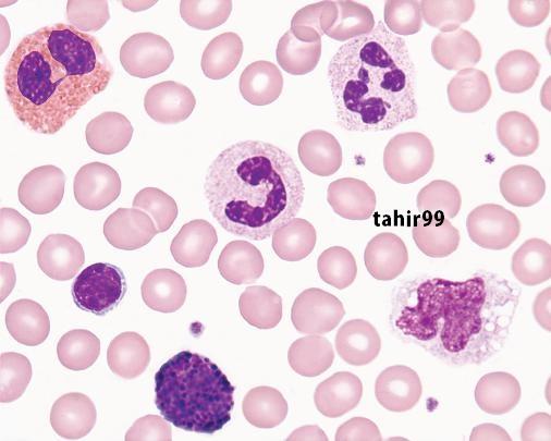
RBCs are anucleate, biconcave, discoid cells filled with a reddish protein, hemoglobin (HGB), which transports oxygen and carbon dioxide (Chapter 10). RBCs appear pink to red and measure 6 to 8 mm in diameter with a zone of pallor that occupies one third of their center (Figure 1-1, A), reflecting their biconcavity (Chapters 8 and 9).
Since before 1900, physicians and medical laboratory professionals counted RBCs in measured volumes to detect anemia or polycythemia. Anemia means loss of oxygen-carrying capacity and is often reflected in a reduced RBC count or decreased RBC hemoglobin concentration (Chapter 19). Polycythemia means an increased RBC count reflecting increased circulating RBC mass, a condition that leads to hyperviscosity (Chapter 33). Historically, microscopists counted RBCs by carefully pipetting a tiny aliquot of whole blood and mixing it with 0.85% (normal) saline. Normal saline matches the osmolality of blood; consequently, the suspended RBCs retained their intrinsic morphology, neither swelling nor shrinking. A 1:200 dilution was typical for RBC counts, and a glass
Figure 1-1 Normal cells in peripheral blood: A, Erythrocyte (red blood cell, RBC); B, Neutrophil (segmented neutrophil, NEUT, SEG, polymorphonuclear neutrophil, PMN); C, Band (band neutrophil, BAND); D, Eosinophil (EO); E, Basophil (BASO); F, Lymphocyte (LYMPH); G, Monocyte (MONO); H, Platelet (PLT).
pipette designed to provide this dilution, the Thoma pipette, was used routinely until the advent of automation.
The diluted blood was transferred to a glass counting chamber called a hemacytometer (Figure 14-1). The microscopist observed and counted RBCs in selected areas of the hemacytometer, applied a mathematical formula based on the dilution and on the area of the hemacytometer counted (Chapter 14), and reported the RBC count in cells per microliter (mL, mcL, also called cubic millimeter, mm3), milliliter (mL, also called cubic centimeter, or cc), or liter (L).
Visual RBC counting was developed before 1900 and, although inaccurate, was the only way to count RBCs until 1958, when automated particle counters became available in the clinical laboratory. The first electronic counter, patented in 1953 by Joseph and Wallace Coulter of Chicago, Illinois, was used so widely that today automated cell counters are often called Coulter counters, although many high-quality competitors exist (Chapter 15).5 The Coulter principle of direct current electrical impedance is still used to count RBCs in many automated hematology profiling instruments. Fortunately, the widespread availability of automated cell counters has replaced visual RBC counting, although visual counting skills remain useful where automated counters are unavailable.
with that of a known standard and is mathematically converted to hemoglobin concentration. Modifications of the cyanmethemoglobin method are used in most automated applications, although some automated hematology profiling instruments replace it with a formulation of the ionic surfactant (detergent) sodium lauryl sulfate to reduce environmental cyanide.
Hematocrit is the ratio of the volume of packed RBCs to the volume of whole blood and is manually determined by transferring blood to a graduated plastic tube with a uniform bore, centrifuging, measuring the column of RBCs, and dividing by the total length of the column of RBCs plus plasma.7 The normal ratio approaches 50% (refer to inside front cover for reference intervals). Hematocrit is also called packed cell volume (PCV), the packed cells referring to RBCs. Often one can see a light-colored layer between the RBCs and plasma. This is the buffy coat and contains WBCs and platelets, and it is excluded from the hematocrit determination. The medical laboratory professional may use the three numerical results— RBC count, HGB, and HCT—to compute the RBC indices mean cell volume (MCV), mean cell hemoglobin (MCH), and mean cell hemoglobin concentration (MCHC) (Chapter 14). The MCV, although a volume measurement recorded in femtoliters (fL), reflects RBC diameter on a Wright-stained blood film. The MCHC, expressed in g/dL, reflects RBC staining intensity and amount of central pallor. The MCH in picograms (pg) expresses the mass of hemoglobin and parallels the MCHC. A fourth RBC index, RBC distribution width (RDW), expresses the degree of variation in RBC volume. Extreme RBC volume variability is visible on the Wright-stained blood film as variation in diameter and is called anisocytosis. The RDW is based on the standard deviation of RBC volume and is routinely reported by automated cell counters. In addition to aiding in diagnosis of anemia, the RBC indices provide stable measurements for internal quality control of counting instruments (Chapter 5).
vip.persianss.ir
Hemoglobin, Hematocrit, and Red Blood Cell Indices
RBCs also are assayed for hemoglobin concentration (HGB) and hematocrit (HCT) (Chapter 14). Hemoglobin measurement relies on a weak solution of potassium cyanide and potassium ferricyanide, called Drabkin reagent. An aliquot of whole blood is mixed with a measured volume of Drabkin reagent, hemoglobin is converted to stable cyanmethemoglobin (hemiglobincyanide), and the absorbance or color intensity of the solution is measured in a spectrophotometer at 540 nm wavelength. 6 The color intensity is compared
Medical laboratory professionals routinely use light microscopy at 5003 or 10003 magnification (Chapters 4 and 16) to visually review RBC morphology, commenting on RBC diameter, color or hemoglobinization, shape, and the presence of cytoplasmic inclusions (Chapters 16 and 19). All these parameters—RBC count, HGB, HCT, indices, and RBC morphology—are employed to detect, diagnose, assess the severity of, and monitor the treatment of anemia, polycythemia, and the numerous systemic conditions that affect RBCs. Automated hematology profiling instruments are used in nearly all laboratories to generate these data, although visual examination of the Wright-stained blood film is still essential to verify abnormal results.8
Reticulocytes
In the Wright-stained blood film, 0.5% to 2% of RBCs exceed the 6- to 8-mm average diameter and stain slightly blue-gray. These are polychromatic (polychromatophilic) erythrocytes, newly released from the RBC production site: the bone marrow (Chapters 8 and 17). Polychromatic erythrocytes are closely observed because they indicate the ability of the bone marrow to increase RBC production in anemia due to blood loss or excessive RBC destruction (Chapters 23 to 26).
Methylene blue dyes, called nucleic acid stains or vital stains, are used to differentiate and count these young RBCs. Vital (or “supravital”) stains are dyes absorbed by live cells.9 Young RBCs contain ribonucleic acid (RNA) and are called reticulocytes when the RNA is visualized using vital stains. Counting reticulocytes visually by microscopy was (and remains) a tedious and imprecise procedure until the development of automated reticulocyte counting by the TOA Corporation (presently Sysmex Corporation, Kobe, Japan) in 1990. Now all fully automated profiling instruments provide an absolute reticulocyte count and, in addition, an especially sensitive measure of RBC production, the immature reticulocyte count or immature reticulocyte fraction (Chapter 15). However, it is still necessary to confirm instrument counts visually from time to time, so medical laboratory professionals must retain this skill.
WHITE BLOOD CELLS
WBCs, or leukocytes, are a loosely related category of cell types dedicated to protecting their host from infection and injury (Chapter 12). WBCs are transported in the blood from their source, usually bone marrow or lymphoid tissue, to their tissue or body cavity destination. WBCs are so named because they are nearly colorless in an unstained cell suspension.
WBCs may be counted visually using a microscope and hemacytometer. The technique is the same as RBC counting, but the typical dilution is 1:20, and the diluent is a dilute acid solution. The acid causes RBCs to lyse or rupture; without it, RBCs, which are 500 to 1000 times more numerous than WBCs, would obscure the WBCs. The WBC count ranges from 4500 to 11,500/mL. Visual WBC counting has been largely replaced by automated hematology profiling instruments, but it is accurate and useful in situations in which no automation is available. Medical laboratory professionals who analyze body fluids such as cerebrospinal fluid or pleural fluid may employ visual WBC counting.
A decreased WBC count (fewer than 4500/mL) is called leukopenia, and an increased WBC count (more than 11,500/mL) is called leukocytosis, but the WBC count alone has modest clinical value. The microscopist must differentiate the categories of WBCs in the blood by using a Wright-stained blood film and light microscopy (Chapter 16). The types of WBCs are as follows:
• Neutrophils (NEUTs, segmented neutrophils, SEGs, polymorphonuclear neutrophils, PMNs; Figure 1-1 , B ). Neutrophils are phagocytic cells whose major purpose is to engulf and destroy microorganisms and foreign material, either directly or after they have been labeled for destruction by the immune system. The term segmented refers to their multilobed nuclei. An increase in neutrophils is called neutrophilia and often signals bacterial infection. A decrease is called neutropenia and has many causes, but it is often caused by certain medications or viral infections.
• Bands (band neutrophils, BANDs; Figure 1-1, C). Bands are less differentiated or less mature neutrophils. An increase in bands also signals bacterial infection and is customarily called a left shift. The cytoplasm of neutrophils and bands
contains submicroscopic, pink- or lavender-staining granules filled with bactericidal secretions.
• Eosinophils (EOs; Figure 1-1, D). Eosinophils are cells with bright orange-red, regular cytoplasmic granules filled with proteins involved in immune system regulation. An elevated eosinophil count is called eosinophilia and often signals a response to allergy or parasitic infection.
• Basophils (BASOs; Figure 1-1, E). Basophils are cells with dark purple, irregular cytoplasmic granules that obscure the nucleus. The basophil granules contain histamines and various other proteins. An elevated basophil count is called basophilia. Basophilia is rare and often signals a hematologic disease.
• The distribution of basophils and eosinophils in blood is so small compared with that of neutrophils that the terms eosinopenia and basopenia are theoretical and not used. Neutrophils, bands, eosinophils, and basophils are collectively called granulocytes because of their prominent cytoplasmic granules, although their functions differ.
• Leukemia is an uncontrolled proliferation of WBCs. Leukemia may be chronic—for example, chronic myelogenous (granulocytic) leukemia—or acute—for example, acute myeloid leukemia. There are several forms of granulocytic leukemias that involve any one of or all three cell lines, categorized by their respective genetic aberrations (Chapters 30, 33 to 35). Medical laboratory scientists are responsible for their identification using Wright-stained bone marrow smears, cytogenetics, flow cytometric immunophenotyping, molecular diagnostic technology, and occasionally, cytochemical staining (Chapter 17 and Chapters 30 to 32).
• Lymphocytes (LYMPHs; Figure 1-1, F). Lymphocytes comprise a complex system of cells that provide for host immunity. Lymphocytes recognize foreign antigens and mount humoral (antibodies) and cell-mediated antagonistic responses. On a Wright-stained blood film, most lymphocytes are nearly round, are slightly larger than RBCs, and have round featureless nuclei and a thin rim of nongranular cytoplasm. An increase in the lymphocyte count is called lymphocytosis and often is associated with viral infections. Accompanying lymphocytosis are often variant or reactive lymphocytes with characteristic morphology (Chapter 29). An abnormally low lymphocyte count is called lymphopenia or lymphocytopenia and is often associated with drug therapy or immunodeficiency. Lymphocytes are also implicated in leukemia; chronic lymphocytic leukemia is more prevalent in people older than 65 years, whereas acute lymphoblastic leukemia is the most common form of childhood leukemia (Chapters 35 and 36). Medical laboratory scientists and hematopathologists classify lymphocytic leukemias largely based on Wright-stained blood films, flow cytometric immunophenotyping, and molecular diagnostic techniques (Chapters 31 to 32).
• Monocytes (MONOs; Figure 1-1, G). The monocyte is an immature macrophage passing through the blood from its point of origin, usually the bone marrow, to a targeted tissue location. Macrophages are the most abundant cell type in the body, more abundant than RBCs or skin cells, although monocytes comprise a minor component of peripheral
blood WBCs. Macrophages occupy every body cavity; some are motile and some are immobilized. Their tasks are to identify and phagocytose (engulf and consume) foreign particles and assist the lymphocytes in mounting an immune response through the assembly and presentation of immunogenic epitopes. On a Wright-stained blood film, monocytes have a slightly larger diameter than other WBCs, blue-gray cytoplasm with fine azure granules, and a nucleus that is usually indented or folded. An increase in the number of monocytes is called monocytosis. Monocytosis may be found in certain infections, collagen-vascular diseases, or in acute and chronic leukemias (Chapters 29, 33, and 35). Medical laboratory professionals seldom document a decreased monocyte count, so the theoretical term monocytopenia is seldom used.
PLATELETS
Platelets, or thrombocytes, are true blood cells that maintain blood vessel integrity by initiating vessel wall repairs (Chapter 13). Platelets rapidly adhere to the surfaces of damaged blood vessels, form aggregates with neighboring platelets to plug the vessels, and secrete proteins and small molecules that trigger thrombosis, or clot formation. Platelets are the major cells that control hemostasis, a series of cellular and plasmabased mechanisms that seal wounds, repair vessel walls, and maintain vascular patency (unimpeded blood flow). Platelets are only 2 to 4 mm in diameter, round or oval, anucleate (for this reason some hematologists prefer to call platelets “cell fragments”), and slightly granular (Figure 1-1, H). Their small size makes them appear insignificant, but they are essential to life and are extensively studied for their complex physiology. Uncontrolled platelet and hemostatic activation is responsible for deep vein thrombosis, pulmonary emboli, acute myocardial infarctions (heart attacks), cerebrovascular accidents (strokes), peripheral artery disease, and repeated spontaneous abortions (miscarriages).
The microscopist counts platelets using the same technique used in counting WBCs on a hemacytometer, although a different counting area and dilution is usually used (Chapter 14). Owing to their small volume, platelets are hard to distinguish visually in a hemacytometer, and phase microscopy provides for easier identification (Chapter 4). Automated profiling instruments have largely replaced visual platelet counting and provide greater accuracy (see Chapter 15).
One advantage of automated profiling instruments is their ability to generate a mean platelet volume (MPV), which is unavailable through visual methods. The presence of predominantly larger platelets generates an elevated MPV value, which sometimes signals a regenerative bone marrow response to platelet consumption (Chapters 13 and 40).
Elevated platelet counts, called thrombocytosis, signal inflammation or trauma but convey modest intrinsic significance. Essential thrombocythemia is a rare malignant condition characterized by extremely high platelet counts and uncontrolled platelet production. Essential thrombocythemia is a life-threatening hematologic disorder (Chapter 33).
A low platelet count, called thrombocytopenia, is a common consequence of drug treatment and may be life-threatening. Because the platelet is responsible for normal blood vessel maintenance and repair, thrombocytopenia is usually accompanied by easy bruising and uncontrolled hemorrhage (Chapter 40). Thrombocytopenia accounts for many hemorrhage-related emergency department visits. Accurate platelet counting contributes to patient safety because it provides for the diagnosis of thrombocytopenia in many disorders or therapeutic regimens.
COMPLETE BLOOD COUNT
A complete blood count (CBC) is performed on automated hematology profiling instruments and includes the RBC, WBC, and platelet measurements indicated in Box 1-1. The medical laboratory professional may collect a blood specimen for the CBC, but often a phlebotomist, nurse, physician assistant, physician, or patient care technician may also collect the specimen (Chapters 3 and 42). No matter who collects, the medical laboratory professional is responsible for the integrity of the specimen and ensures that it is submitted in the appropriate anticoagulant and tube and is free of clots and hemolysis (redtinted plasma indicating RBC damage). The specimen must be of sufficient volume, as “short draws” result in incorrect anticoagulant-to-specimen ratios. The specimen must be tested or prepared for storage within the appropriate time frame to ensure accurate analysis (Chapter 5) and must be accurately registered in the work list, a process known as specimen accession. Accession may be automated, relying on bar code or radiofrequency identification technology, thus reducing instances of identification error.
Although all laboratory scientists and technicians are equipped to perform visual RBC, WBC, and platelet counts
BOX 1-1 Complete Blood Count Measurements
Generated by Automated Hematology Profiling Instruments
RBC Parameters
RBC count
HGB
HCT
MCV
MCH
MCHC
RDW
RETIC
Platelet Parameters
PLT count
MPV
WBC Parameters
WBC count
NEUT count: % and absolute
LYMPH count: % and absolute
MONO count: % and absolute
EO and BASO counts: % and absolute
BASO, Basophil; EO, eosinophil; HGB, hemoglobin; HCT, hematocrit; LYMPH, lymphocyte; MCH, mean cell hemoglobin; MCHC, mean cell hemoglobin concentration; MCV, mean cell volume; MONO, monocyte; MPV, mean platelet volume; NEUT, segmented neutrophil; PLT, platelet; RBC, red blood cell; RDW, RBC distribution width; RETIC, reticulocyte; WBC, white blood cell.
using dilution pipettes, hemacytometers, and microscopes, most laboratories employ automated profiling instruments to generate the CBC. Many profiling instruments also provide comments on RBC, WBC, and platelet morphology (Chapter 15). When one of the results from the profiling instrument is abnormal, the instrument provides an indication of this, sometimes called a flag. In this case, a “reflex” blood film examination is performed (Chapter 16).
The blood film examination (described next) is a specialized, demanding, and fundamental CBC activity. Nevertheless, if all profiling instrument results are normal, the blood film examination is usually omitted from the CBC. However, physicians may request a blood film examination on the basis of clinical suspicion even when the profiling instrument results fall within their respective reference intervals.
BLOOD FILM EXAMINATION
To accomplish a blood film examination, the microscopist prepares a “wedge-prep” blood film on a glass microscope slide, allows it to dry, and fixes and stains it using Wright or Wright-Giemsa stain (Chapter 16). The microscopist examines the RBCs and platelets by light microscopy for abnormalities of shape, diameter, color, or inclusions using the 503 or 1003 oil immersion lens to generate 5003 or 10003 magnification (Chapter 4). The microscopist then visually estimates the WBC count and platelet count for comparison with their respective instrument counts and investigates discrepancies. Next, the microscopist systematically reviews, identifies, and tabulates 100 (or more) WBCs to determine their percent distribution. This process is referred to as determining the WBC differential (“diff”). The WBC differential relies on the microscopist’s skill, visual acuity, and integrity, and it provides extensive diagnostic information. Medical laboratory professionals pride themselves on their technical and analytical skills in performing the blood film examination and differential count. Visual recognition systems such as the Cellavision® DM96 or the Bloodhound automate the RBC and platelet morphology and WBC differential processes, but the medical laboratory professional or the hematopathologist is the final arbiter for all cell identification. The results of the CBC, including all profiling and blood film examination parameters and interpretive comments, are provided in paper or digital formats for physician review with abnormal results highlighted.
ENDOTHELIAL CELLS
Because they are structural and do not flow in the bloodstream, endothelial cells—the endodermal cells that form the inner surface of the blood vessel—are seldom studied in the hematology laboratory. Nevertheless, endothelial cells are important in maintaining normal blood flow, in tethering (decelerating) platelets during times of injury, and in enabling WBCs to escape from the vessel to the surrounding tissue when needed. Increasingly refined laboratory methods are becoming available to assay and characterize the secretions (cytokines) of these important cells.
COAGULATION
Most hematology laboratories include a blood coagulation–testing department (Chapters 42 and 44). Platelets are a key component of hemostasis, as previously described; plasma coagulation is the second component. The coagulation system employs a complex sequence of plasma proteins, some enzymes, and some enzyme cofactors to produce clot formation after blood vessel injury. Another 6 to 8 enzymes exert control over the coagulation mechanism, and a third system of enzymes and cofactors digests clots to restore vessel patency, a process called fibrinolysis. Bleeding and clotting disorders are numerous and complex, and the coagulation section of the hematology laboratory provides a series of plasma-based laboratory assays that assess the interactions of hematologic cells with plasma proteins (Chapters 42 and 44).
The medical laboratory professional focuses especially on blood specimen integrity for the coagulation laboratory, because minor blood specimen defects, including clots, hemolysis, lipemia, plasma bilirubin, and short draws, render the specimen useless (Chapters 3 and 42). High-volume coagulation tests suited to the acute care facility include the platelet count and MPV as described earlier, prothrombin time and partial thromboplastin time (or activated partial thromboplastin time), thrombin time (or thrombin clotting time), fibrinogen assay, and D-dimer assay (Chapter 42). The prothrombin time and partial thromboplastin time are particularly high-volume assays used in screening profiles. These tests assess each portion of the coagulation pathway for deficiencies and are used to monitor anticoagulant therapy. Another 30 to 40 moderate-volume assays, mostly clot-based, are available in specialized or tertiary care facilities. The specialized or tertiary care coagulation laboratory with its interpretive complexities attracts advanced medical laboratory scientists with specialized knowledge and communication skills.
ADVANCED HEMATOLOGY PROCEDURES
Besides performing the CBC, the hematology laboratory provides bone marrow examinations, flow cytometry immunophenotyping, cytogenetic analysis, and molecular diagnosis assays. Performing these tests may require advanced preparation or particular dedication by medical laboratory scientists with a desire to specialize.
Medical laboratory scientists assist physicians with bedside bone marrow collection, then prepare, stain, and microscopically review bone marrow smears (Chapter 17). Bone marrow aspirates and biopsy specimens are collected and stained to analyze nucleated cells that are the immature precursors to blood cells. Cells of the erythroid series are precursors to RBCs (Chapter 8); myeloid series cells mature to form bands and neutrophils, eosinophils, and basophils (Chapter 12); and megakaryocytes produce platelets (Chapter 13). Medical laboratory scientists, clinical pathologists, and hematologists review Wright-stained aspirate smears for morphologic abnormalities, high or low bone marrow cell concentration, and inappropriate cell line distributions. For instance, an increase
in the erythroid cell line may indicate bone marrow compensation for excessive RBC destruction or blood loss (Chapter 19 and Chapters 23 to 26). The biopsy specimen, enhanced by hematoxylin and eosin (H&E) staining, may reveal abnormalities in bone marrow architecture indicating leukemia, bone marrow failure, or one of a host of additional hematologic disorders. Results of examination of bone marrow aspirates and biopsy specimens are compared with CBC results generated from the peripheral blood to correlate findings and develop pattern-based diagnoses.
In the bone marrow laboratory, cytochemical stains may occasionally be employed to differentiate abnormal myeloid, erythroid, and lymphoid cells. These stains include myeloperoxidase, Sudan black B, nonspecific and specific esterase, periodic acid–Schiff, tartrate-resistant acid phosphatase, and alkaline phosphatase. The cytochemical stains are time-honored stains that in most laboratories have been replaced by flow cytometry immunophenotyping, cytogenetics, and molecular diagnostic techniques (Chapters 30 to 32). Since 1980, however, immunostaining methods have enabled identification of cell lines by detecting lineage-specific antigens on the surface or in the cytoplasm of leukemia and lymphoma cells. An example of immunostaining is a visible dye that is bound to antibodies to CD42b, a membrane protein that is present in the megakaryocytic lineage and may be diagnostic for megakaryoblastic leukemia (Chapter 35).
Flow cytometers may be quantitative, such as clinical flow cytometers that have grown from the original Coulter principle, or qualitative, including laser-based instruments that have migrated from research applications to the clinical laboratory (Chapters 15 and 32). The former devices are automated clinical profiling instruments that generate the quantitative parameters of the CBC through application of electrical impedance and laser or light beam interruption. Qualitative laser-based flow cytometers are mechanically simpler but technically more demanding. Both qualitative and quantitative flow cytometers are employed to analyze cell populations by measuring the effects of individual cells on laser light, such as forward-angle fluorescent light scatter and right-angle fluorescent light scatter, and by immunophenotyping for cell membrane epitopes using monoclonal antibodies labeled with fluorescent dyes. The qualitative flow cytometry laboratory is indispensable to leukemia and lymphoma diagnosis.
Cytogenetics, a time-honored form of molecular technology, is employed in bone marrow aspirate examination to find gross genetic errors such as the Philadelphia chromosome, a reciprocal translocation between chromosomes 9 and 22 that is associated with chronic myelogenous leukemia, and t(15;17), a translocation between chromosomes 15 and 17 associated with acute promyelocytic leukemia (Chapter 30). Cytogenetic analysis remains essential to the diagnosis and treatment of leukemia. Molecular diagnostic techniques, the fastest-growing area of laboratory medicine, enhance and even replace some of the advanced hematologic methods. Real-time polymerase chain reaction, microarray analysis, fluorescence in situ hybridization, and DNA sequencing systems are sensitive and specific methods that enable medical laboratory scientists to detect
various chromosome translocations and gene mutations that confirm specific types of leukemia, establish their therapeutic profile and prognosis, and monitor the effectiveness of treatment (Chapter 31).
ADDITIONAL HEMATOLOGY PROCEDURES
Medical laboratory professionals provide several time-honored manual whole-blood methods to support hematologic diagnosis. The osmotic fragility test uses graduated concentrations of saline solutions to detect spherocytes (RBCs with proportionally reduced surface membrane area) in hereditary spherocytosis or warm autoimmune hemolytic anemia (Chapters 24 and 26). Likewise, the glucose-6-phosphate dehydrogenase assay phenotypically detects an inherited RBC enzyme deficiency causing severe episodic hemolytic anemia (Chapter 24). The sickle cell solubility screening assay and its follow-up tests, hemoglobin electrophoresis and high performance liquid chromatography, are used to detect and diagnose sickle cell anemia and other inherited qualitative hemoglobin abnormalities and thalassemias (Chapters 27 and 28). One of the oldest hematology tests, the erythrocyte sedimentation rate, detects inflammation and roughly estimates its intensity (Chapter 14).
Finally, the medical laboratory professional reviews the cellular counts, distribution, and morphology in body fluids other than blood (Chapter 18). These include cerebrospinal fluid, synovial (joint) fluid, pericardial fluid, pleural fluid, and peritoneal fluid, in which RBCs and WBCs may be present in disease and in which additional malignant cells may be present that require specialized detection skills. Analysis of nonblood body fluids is always performed with a rapid turnaround, because cells in these hostile environments rapidly lose their integrity. The conditions leading to a need for body fluid analysis are invariably acute.
HEMATOLOGY QUALITY ASSURANCE AND QUALITY CONTROL
Medical laboratory professionals employ particularly complex quality control systems in the hematology laboratory (Chapter 5). Owing to the unavailability of weighed standards, the measurement of cells and biological systems defies chemical standardization and requires elaborate calibration, validation, matrix effect examination, linearity, and reference interval determinations. An internal standard methodology known as the moving average also supports hematology laboratory applications.10 Medical laboratory professionals in all disciplines compare methods through clinical efficacy calculations that produce clinical sensitivity, specificity, and positive and negative predictive values for each assay. They must monitor specimen integrity and test ordering patterns and ensure the integrity and delivery of reports, including numerical and narrative statements and reference interval comparisons. As in most branches of laboratory science, the hematology laboratory places an enormous responsibility for accuracy, integrity, judgment, and timeliness on the medical laboratory professional.
REFERENCES
1. Perkins, S. L. (2009). Examination of the blood and bone marrow. In Greer, J. P., Foerster, J, Rodgers, G. M., et al, (Eds.), Wintrobe’s Clinical Hematology. (12th ed.). Philadelphia: Lippincott Williams and Wilkins.
2. Wintrobe, M. M. (1985). Hematology, the Blossoming of a Science: A Story of Inspiration and Effort. Philadelphia: Lea & Febiger.
3. Bizzozero, J. (1882). Über einem neuen formbestandtheil des blutes und dessen rolle bei der Thrombose und der Blutgerinnung. Virchows Arch Pathol Anat Physiol Klin Med, 90, 261–332.
4. Woronzoff-Dashkoff, K. K. (2002). The Wright-Giemsa stain. Secrets revealed. Clin Lab Med, 22, 15–23.
5. Blades, A. N., Flavell, H. C. (1963). Observations on the use of the Coulter model D electronic cell counter in clinical haematology. J Clin Pathol, 16, 158–163.
6. Klungsöyr, L., Stöa, K. F. (1954). Spectrophotometric determination of hemoglobin oxygen saturation: the method of Drabkin & Schmidt as modified for its use in clinical routine analysis. Scand J Clin Lab Invest, 6, 270–276.
7. Mann, L. S. (1948). A rapid method of filling and cleaning Wintrobe hematocrit tubes. Am J Clin Pathol, 18, 916.
8. Barth, D. (2012). Approach to peripheral blood film assessment for pathologists. Semin Diagn Pathol, 29, 31–48.
9. Biggs, R. (1948). Error in counting reticulocytes. Nature, 162, 457.
10. Gulati, G. L., Hyun, B. H. (1986). Quality control in hematology. Clin Lab Med, 6, 675–688.
2
Safety in the Hematology Laboratory
Sheila A. Finch
OUTLINE
Standard Precautions
Applicable Safety Practices
Required by the OSHA
Standard
Housekeeping
Laundry
Hepatitis B Virus Vaccination
Training and Documentation
Regulated Medical Waste Management
Occupational Hazards
Fire Hazard
Chemical Hazards
Electrical Hazard
Needle Puncture
Developing a Safety Management Program
Planning Stage: Hazard
Assessment and Regulatory Review
Safety Program Elements
CASE STUDY
OBJECTIVES
After completion of this chapter, the reader will be able to:
1. Define standard precautions and list infectious materials included in standard precautions.
2. Describe the safe practices required in the Occupational Exposure to Bloodborne Pathogens Standard.
3. Identify occupational hazards that exist in the hematology laboratory.
4. Describe appropriate methods to decontaminate work surfaces after contamination with blood or other potentially infectious material.
5. Identify the regulatory requirements of the Occupational Exposure to Hazardous Chemicals in Laboratories standard.
6. Describe the principles of a fire prevention program, including details such as the frequency of testing equipment.
7. Name the most important practice to prevent the spread of infection.
8. Given a written laboratory scenario, assess for safety hazards and recommend corrective action for any deficiencies or unsafe practices identified.
9. Select the proper class of fire extinguisher for a given type of fire.
10. Explain the purpose of Safety Data Sheets (SDSs), list information contained on SDSs, and determine when SDSs would be used in a laboratory activity.
11. Name the specific practice during which most needle stick injuries occur.
12. Describe elements of a safety management program.
After studying the material in this chapter, the reader should be able to respond to the following case study:
Hematology Services, Inc., had a proactive safety program. Quarterly safety audits were conducted by members of the safety committee. The following statements were recorded in the safety audit report. Which statements describe good work practices, and which statements represent deficiencies? List the corrective actions required for identified unsafe practices.
1. A hematology laboratory scientist was observed removing gloves and immediately left the laboratory for a meeting. She did not remove her laboratory coat.
2. Food was found in the specimen refrigerator.
3. Hematology laboratory employees were seen in the lunchroom, wearing laboratory coats.
4. Fire extinguishers were found every 75 feet of the laboratory.
5. Fire extinguishers were inspected quarterly and maintained annually.
6. Unlabeled bottles were found at a workstation.
7. A 1:10 solution of bleach was found near an automated hematology analyzer. Further investigation revealed that the bleach solution was made 6 months ago.
8. Gloves were worn by the staff receiving specimens.
9. Safety data sheets were obtained by fax.
10. Chemicals were stored alphabetically.
Many conditions in the laboratory have the potential for causing injury to staff and damage to the building or to the community. Patients’ specimens, needles, chemicals, electrical equipment, reagents, and glassware all are potential causes of accidents or injury. Managers and employees must be knowledgeable about safe work practices and incorporate these practices into the operation of the hematology laboratory. The key to prevention of accidents and laboratory-acquired infections is a well-defined safety program.
Safety is a broad subject and cannot be covered in one chapter. This chapter simply highlights some of the key safe practices that should be followed in the hematology laboratory. Omission of a safe practice from this chapter does not imply that it is not important or that it should not be considered in the development of a safety curriculum or a safety program.
STANDARD PRECAUTIONS
One of the greatest risks associated with the hematology laboratory is the exposure to blood and body fluids. In December 1991, the Occupational Safety and Health Administration (OSHA) issued the final rule for the Occupational Exposure to Bloodborne Pathogens Standard. The rule that specifies standard precautions to protect laboratory workers and other health care professionals became effective on March 6, 1992. Universal precautions was the original term; OSHA’s current terminology is standard precautions. Throughout this text, the term standard precautions is used to remind the reader that all blood, body fluids, and unfixed tissues are to be handled as though they were potentially infectious.
Standard precautions must be adopted by the laboratory. Standard precautions apply to blood, semen, vaginal secretions, cerebrospinal fluid, synovial fluid, pleural fluid, any body fluid with visible blood, any unidentified body fluid, unfixed slides, microhematocrit clay, and saliva from dental procedures. Adopting standard precautions lessens the risk of health care worker exposures to blood and body fluids, decreasing the risk of injury and illness.
Bloodborne pathogens are pathogenic microorganisms that, when present in human blood, can cause disease. They include, but are not limited to, hepatitis B virus (HBV), hepatitis C virus (HCV), and human immunodeficiency virus (HIV). This chapter does not cover the complete details of the standard; it discusses only the sections that apply directly to the hematology laboratory. Additional information can be found in the references at the end of this chapter.
Applicable Safety Practices Required by the OSHA Standard
The following standards are applicable in a hematology laboratory and must be enforced:
1. Hand washing is one of the most important safety practices. Hands must be washed with soap and water. If water is not readily available, alcohol hand gels (minimum 62%
alcohol) may be used. Hands must be thoroughly dried. The proper technique for hand washing is as follows:
a. Wet hands and wrists thoroughly under running water.
b. Apply germicidal soap and rub hands vigorously for at least 15 seconds, including between the fingers and around and over the fingernails (Figure 2-1, A).
c. Rinse hands thoroughly under running water in a downward flow from wrist to fingertips (Figure 2-1, B).
d. Dry hands with a paper towel (Figure 2-1, C). Use the paper towel to turn off the faucet handles (Figure 2-1, D).
Hands must be washed:
a. Whenever there is visible contamination with blood or body fluids
b. After completion of work
c. After gloves are removed and between glove changes
d. Before leaving the laboratory
e. Before and after eating and drinking, smoking, applying cosmetics or lip balm, changing a contact lens, and using the lavatory
f. Before and after all other activities that entail hand contact with mucous membranes, eyes, or breaks in skin
2. Eating, drinking, smoking, and applying cosmetics or lip balm must be prohibited in the laboratory work area.
3. Hands, pens, and other fomites must be kept away from the mouth and all mucous membranes.
4. Food and drink, including oral medications and tolerancetesting beverages, must not be kept in the same refrigerator as laboratory specimens or reagents or where potentially infectious materials are stored or tested.
5. Mouth pipetting must be prohibited.
6. Needles and other sharp objects contaminated with blood and other potentially infectious materials should not be manipulated in any way. Such manipulation includes resheathing, bending, clipping, or removing the sharp object. Resheathing or recapping is permitted only when there are no other alternatives or when the recapping is required by specific medical procedures. Recapping is permitted by use of a method other than the traditional two-handed procedure. The one-handed method or a resheathing device is often used. Documentation in the exposure control plan should identify the specific procedure in which resheathing is permitted.
7. Contaminated sharps (including, but not limited to, needles, blades, pipettes, syringes with needles, and glass slides) must be placed in a puncture-resistant container that is appropriately labeled with the universal biohazard symbol (Figure 2-2) or a red container that adheres to the standard. The container must be leakproof (Figure 2-3).
8. Procedures such as removing caps when checking for clots, filling hemacytometer chambers, making slides, discarding specimens, making dilutions, and pouring specimens or fluids must be performed so that splashing, spraying, or production of droplets of the specimen being manipulated is prevented. These procedures may be performed behind a barrier, such as a plastic shield, or protective eyewear should be worn (Figure 2-4).