Fundamentals of Goat Practice
Overview 3
Distribution of Goats 3
Use of Goats 3
Current Interest in Goats 4
Distinguishing Goats from Sheep 4
Goat Behavior 5
General Characteristics 5
Ingestive and Eliminative Behavior 5
Sexual Behavior 6
Maternal Behavior 6
Handling Goats 7
Group Considerations 7
Individual Restraint 7
Administering Medications 7
Clinical Examination of Goats 9
History Taking 9
Special Considerations for Range and Pastured Goats 10
Special Considerations for Intensively Managed Goats 10
OVERVIEW
Distribution of Goats
According to the Food and Agriculture Organization (FAO) of the United Nations, in 2006 there were an estimated 837.2 million goats in the world, approximately 64.2% of which were in Asia, 28.8% in Africa, 4.3% in South and Central America, 2.2% in Europe, 0.3% in North America, and 0.1% in Oceania. Approximately 4.2% of the world’s goats are found in developed countries and 95.8% in developing countries (FAO 2007). Goats are highly adaptable to a broad range of climatic and geographic conditions and are more widely distributed than any other mammalian livestock. Goats are managed under every imaginable production system, including feral, transhumant, nomadic, extensive, intensive, and total confinement systems.
Use of Goats
Goats are exploited for diverse purposes, including meat production, cashmere and mohair fiber production, milk and cheese production, and skins for leather making. Specialty uses include brush and weed control, pack and draft use, animal experimentation (particu-
Special Considerations for Hobby Farms 11
Special Considerations for Organic Goat Production 11
Special Considerations for Transgenic Goats 11
Physical Examination 13
Inspection from a Distance 13
Direct Physical Examination of Individual Goats 14
General Inspection 14
Examination of the Integument 15
Examination of the Head 16
Examination of the Neck 17
Examination of the Chest 17
Examination of the Abdomen 18
Examination of the Limbs 18
Examination of the Reproductive System 18
Examination of the Environment 19
Field Necropsies and Slaughterhouse Checks 20
References 20
larly as models of ruminant digestion and human heart disease and as transgenic animals), commercial antibody production, and companionship. Goat horn and bone are sometimes used for ornamental purposes and musical instruments, while goat skins are used for drum making.
Meat production is the major use of goats on a worldwide basis, particularly in Asia, Africa, the Middle East, and Latin America, and world goat meat production more than doubled between 1980 and 2000 (Morand-Fehr et al. 2004). In 2006, the seven leading goat meat producing nations in descending order were China, India, Pakistan, Sudan, Nigeria, Bangladesh, and Iran (FAO 2007a). A myriad of local and regional breeds exists around the world that are used mainly for meat. In recent years, more attention has been paid to selective breeding in goats for meat production, leading to the development of two highly efficient, purpose-bred meat goat breeds. These are the South African Boer goat (Mahan 2000) and the Kiko goat of New Zealand (Batten 1988), both of which have gained popularity in the United States, particularly in the southeast where commercial goat meat production has been expanding.
The major milking breeds of goats originated primarily in Europe. These breeds include the Saanen, Toggenburg, Anglo-Nubian, and Alpine breeds. The more recently developed La Mancha breed originated in the United States. The Jamnapari and Beetal breeds of India are also important dairy breeds that are well adapted to and becoming more widely distributed in the humid tropics. The use of goat milk to manufacture cheese is an important industry in France, Spain, and other European countries.
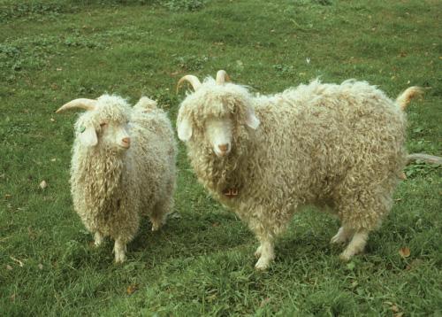
Angora goats, the source of mohair fiber, have traditionally been concentrated in a number of distinct areas, notably Turkey, where they originated, South Africa, Texas, Argentina, and some central Asian republics formerly in the USSR. Cashmere or Pashmina goats, which produce cashmere fiber, are found primarily in the mountainous regions of Central Asia, including parts of Tibet, China, Mongolia, Iran, Afghanistan, Kazakstan, Kyrgyzstan, and Tajikistan. Skins are usually a byproduct of goat slaughter for meat, but skins of certain goat breeds such as the Red Sokoto of Niger are prized for high-quality leather goods such as kidskin gloves and purses. Details of the various goat industries are beyond the scope of this veterinary text. The interested reader is referred to other sources (Gall 1981; Coop 1982; DeVendra and Burns 1983; Dubeuf et al. 2004; Morand-Fehr et al. 2004).
Current Interest in Goats
Worldwide interest in goats has continued to increase dramatically during the last decade. There is greater understanding of the importance of goats in agricultural systems in low-income countries. A number of humanitarian organizations, such as Heifer International and FARM-Africa, have recognized the value of using goats as a tool in rural development programs to improve the social and economic conditions of subsistence farmers and the rural poor. Methodologies for improved goat production in the tropics in support of rural development have been published (Peacock 1996).
There is also increased demand for goat products in developed countries, especially goat cheese, cashmere goods, and even goat meat. Demand for goat meat in the United States has exceeded domestic supply in recent years. In 2004, the United States imported 2,400 metric tons of goat meat, mostly from Australia (Ward 2006).
This expanding interest in goats has increased the demand for goat-related veterinary services in the areas of clinical medicine, research, and extension. In response to this need, interested veterinarians must familiarize themselves with goats as a species distinct from sheep and cattle, recognizing their often characteristic behavior, physiology, and response to disease.
Fortunately, a number of resources have become available to provide information in these areas. The International Goat Association (www.iga-goatworld. org/) sponsors a quadrennial international conference on goats and regularly publishes the peer-reviewed, international research journal, Small Ruminant Research, which reports research findings on all aspects of goat production including health, nutrition, genetics, physiology, and husbandry from all over the world. The American Sheep Industry Association regularly produces a similar, multidisciplinary research publication, Sheep and Goat Research Journal, which focuses specifically on small ruminant production in North America and is available on the Internet (http://www.sheepusa.org/).
The American Association of Small Ruminant Practitioners (AASRP) is an excellent resource for veterinary practitioners in North America. This member organization produces a regular newsletter, Wool and Wattles, full of current, relevant information on regulatory and clinical issues as well as an e-mail discussion forum for AASRP members. The AASRP Website (www.aasrp.org/) provides links to other useful resources for goat health and production. Another useful Web-based resource for veterinarians is Consultant, which generates differential diagnoses based on clinical signs entered by the user on a species basis, with goats recognized as a distinct species. It is available on the Internet at http://www.vet.cornell.edu/ consultant/consult.asp. Finally, many state extension agencies now have much more information available on goat husbandry and production than they had in the past, and much of it accessible on the Internet.
Distinguishing Goats from Sheep
Source of Confusion
For those whose experience with sheep and goats is limited to the common European wool breeds of sheep and the European dairy breeds of goats, the notion that individuals of the two species could be confused may seem ridiculous. However, in tropical and subtropical regions, various breeds of hair sheep are common. These breeds are often maintained in mixed flocks with local breeds of goats, and may not be readily differentiated. The following information can help in distinguishing the two species.
Genetic Distinctions
Goats have 60 chromosomes and sheep have 54. Though very uncommon, fertile goat-sheep hybrids have been reported. These hybrids have 57 chromosomes. The phenomenon is discussed in Chapter 13.
Behavioral Distinctions
A major difference between sheep and goats is feeding behavior. Sheep are grazing animals, consis-
tently feeding at ground level, while the goat is more of a browsing animal, readily feeding on shrubs, bushes, and trees. While both species are social, individual goats are less anxious than sheep when separated from the group. Goats are less tolerant of rain and more readily seek shelter in wet weather.
The males of both species will fight, buck goats by rearing up on their hind feet and coming down forcefully to butt heads, while rams back up and then charge forward to butt heads. The anatomic structure of the horns, frontal sinuses, and neck muscles of each species is appropriate to its method of fighting, minimizing the risk of injury to combatants (Reed and Schaffer 1972). When young bucks and rams are maintained together, the rams become dominant because they preemptively strike bucks in the abdomen while the male goats are still in the act of rearing up.
Whereas lambs are almost constantly at the side of ewes in early life, goats practice “lying out” or “planting” behavior with kids left in “camps” for a good part of the day while does feed.
Anatomic Distinctions
When wool is not obvious in sheep, other anatomic differences may be observed. Most goat breeds have an erect tail, while the tail of sheep always hangs down. The sheep has an upper lip divided by a distinct philtrum and the goat does not. Male goats, and to a lesser extent female goats, have beards, which are lacking in sheep. Goats do not have infraorbital, interdigital, or inguinal glands, while sheep do. Goats have sebaceous glands beneath the tailhead that sheep lack.
GOAT BEHAVIOR
General Characteristics
Goats exhibit some very distinct behavior patterns (Hafez 1975; Kilgour and Dalton 1984). Many aspects of goat behavior are conditioned by the circumstances in which the animals are kept. Many natural behavior patterns observed in free-ranging feral goats may be altered or not expressed at all under different degrees of confinement. Nevertheless, some behavior patterns are widely characteristic.
Goats tend to flock together in extended family groups. They have a strong hierarchical structure in the flock or herd. Both males and females will establish social dominance in their respective groups through head to head fighting. Goats use their horns to advantage when fighting to establish their social dominance. Therefore, all goats in a group should be either horned or hornless to avoid excessive bullying by horned goats.
When goats are accustomed to human contact, they will approach strangers rather than flee. When threatened or upset, they will turn and face an intruder and
make a characteristic sneezing noise. In keeping with their browsing behavior, goats orally investigate everything in their environment. This includes veterinary equipment, paperwork, clothing, and jewelry brought within their reach. When drawing blood samples or writing health papers, it is essential to keep the paperwork in a safe place or it will be eaten or destroyed. Goats will chew on pen partitions and other structures made of wood, and a large group of goats can actually devour pen walls over a period of months. They will also eat the paint off walls, so lead paint should be avoided.
Goats are very agile and are excellent climbers. They are occasionally found in barn rafters, in trees, or on the hood of the veterinarian’s vehicle, if allowed access. Providing a rock pile in paddocks or pastures can foster recreation and will help to control hoof overgrowth. Goats will stand on their hind legs and lean against fences, causing considerable damage over time. Broken limbs may occur if legs are caught in the openings of chain link fences. The goats’ agility combined with their curiosity can be fatal if their heads get caught and they are strangled in fences, gates, doors, windows, or other structures. Backward curving horns contribute to this problem.
Goats are notorious for successfully undoing simple gate closures and latches. This is a common occurrence in accidental grain overload cases, so goat keepers must ensure that gates are securely fastened. Goats can easily jump fences designed for sheep and also will dig under fences that do not closely skirt the ground. Goat fencing should never have interior sloping support posts because goats will use them to climb out of the enclosure. Goats ignore barbed wire and therefore it should not be used because it can inflict serious damage. Thus, electric fencing has become popular for goat operations because the animals quickly learn to respect it.
Ingestive and Eliminative Behavior
A key to the adaptability of goats worldwide is their efficient browsing ability. This same efficiency, however, has given the goat notoriety as an important cause of desertification in some regions of the world. The reputation is not always deserved because overgrazing by numerous livestock species may be at fault, but only the goats are left surviving when vegetation is almost gone (Dunbar 1984). Goats may climb into trees to reach food when it is scarce. If permitted, they can girdle the bark from trees, thus killing them. Goats are used to clear brush to reclaim pasture land for sheep and cattle. When run simultaneously with sheep and cattle, they may improve pasture quality for these other species by contributing manure for fertilization, removing toxic plants such as oak to which they are more resistant than the other ruminant species, and
eliminating brush to allow more sunlight for improved grass growth (Ward 2006).
Owners feeding goats in confinement often complain that the animals waste a good deal of hay, particularly leaving behind the nutritious leafy parts of good legume hay. This tendency can be countered to some extent by feeder designs that inhibit the goat from pulling hay out of the feeder and dropping it on the floor. Goats are also finicky about contaminated feed and water supplies and may refuse water containing fecal pellets or hay and grain in wet troughs that smell moldy.
In free-ranging feral and Angora goats, approximately 30% of the day is spent in feeding, usually divided into sunrise, midday, and sunset periods. Onethird of this is grazing time, two-thirds browsing. About half the day is spent resting, 10% ruminating, and 12% traveling (Askins and Turner 1972; Kilgour and Ross 1980). In contrast, intensively managed Saanen milking goats eat for 20% of the day, ruminate 25%, travel 20%, sleep 11%, rest recumbent 14%, and rest standing approximately 8% of each day. They defecate on average 11.2 times and urinate 8.3 times daily (Pu, personal communication 1990).
Goats raise their tails to defecate and normally produce pelleted feces. Female goats squat to urinate. During the non-breeding season, males urinate on the ground with little or no extension of the penis beyond the prepuce. However, during the breeding season, the pattern of urination is markedly different and associated with sexual behavior as discussed below. Goats cannot be prompted easily to urinate by holding off their nares, as is done with sheep. This makes simple collection of a urine specimen problematic.
Sexual Behavior
In tropical and subtropical regions, estrus generally occurs year-round while in temperate regions goats are seasonally polyestrus, with breeding season triggered by decreasing day length. Breed factors may also play a role in this pattern because relocation of some indigenous breeds to new climatic zones does not result in a change of estrus pattern. Specific information on the frequency, signs, and patterns of estrus are provided in Chapter 13. Male sexual behavior reflects the pattern seen in does. Libido and sperm quality may be depressed during anestrous seasons. However, if females are brought into estrus by hormonal manipulation, bucks quickly respond out of season.
The obnoxious behavior and strong odor of bucks during breeding season are notorious. At least two factors contribute to buck odor. First, the aroused buck repeatedly urinates on himself, soaking his head, neck, and forequarters. He will sometimes take his erect penis into his mouth. Afterwards, the buck may yawn
and demonstrate the flehmen reaction, curling his upper lip. Second, the buck possesses sebaceous scent glands on his head, caudomedial to the base of the horn, which during active rutting produce an odiferous compound identified as 6-trans nonenal (Smith et al. 1984). This compound may also be released from the sebaceous gland under the tail. It acts as a potent pheromone and the odor alone can induce estrus in the doe.
Bucks show active fighting behavior at the beginning of and during the breeding season to establish dominance. Veterinarians and owners should exercise caution when working around sexually active bucks. A full grown buck striking from the standing position can produce serious or fatal injury. For this reason, bucks of different sizes in confinement operations should be segregated so that smaller, younger bucks are not injured or killed. Do not turn your back to an unrestrained buck!
During courtship, the buck will sniff the urine of does and follow with the flehmen response. To display to does, a buck holds his head erect and high or he lowers his extended head and neck to the ground. He may also kick out at the doe with an extended forelimb, but rarely actually strike her. Courtship is accompanied by much frenzied vocalizing and flicking the tongue in and out. Sexually active bucks commonly lose weight during the breeding season.
Maternal Behavior
Free-ranging goats separate from the herd and hide to kid. Confined goats may attempt to conceal themselves. As parturition approaches, does become restless and paw at the ground, making rudimentary efforts to “nest build.” Details of parturition and the recognition of dystocia are discussed in Chapter 13. Following parturition, the doe actively licks the kids, and this is considered to be critical to successful bonding. If does are frightened or disturbed at this point, or if licking is delayed longer than one hour, bonding may be impaired and kids may be abandoned or mothered less effectively.
Kids are precocial, standing and seeking the hairless udder shortly after birth. In free-ranging herds, there is a “lying-out” period of several days to several weeks when does may leave kids in sheltered areas for periods of two to eight hours while they feed. Does must be familiar with the geography to return successfully to their kids. Therefore, it is not advisable to move does to new grazing areas immediately before kidding. Does will respond to alarm calls from their distant kids and return to defend them if bonding is strong. Kids gradually begin to follow their dams, learning to browse and graze. The infrequent nursing pattern of young kids makes the goat adaptable to the twice-aday feeding regimens that are often practiced under
intensive management. If given boxes to hide in, kids raised in confinement will use them for the first week, coming out only to suckle the dam.
HANDLING GOATS
Group Considerations
Goats are highly adaptable and trainable animals. Feral goats captured in Australia and New Zealand may become used to handling in confinement within weeks, although if frightened suddenly they can clear sheep fences with ease. Dairy goats are readily trained into milking routines involving parlors and machine milking. Although Angora kids may scream the first time they are sheared, they get used to the procedure.
Goats used to human contact can be mustered by calling. Moving less tame goats on open range is similar to moving sheep. Dogs can be used, but they must be well trained. The flight distance of feral goats is eight to ten meters. Goats are more likely to turn and fight than sheep if provoked by a dog. Animals that break away from the group should be left to follow along rather than chased. The presence of sheep with the goats can actually facilitate flocking and driving, although on hills, goats tend to move upward and sheep downward. When collected in yards, anxious goats may pile up in a corner and some may suffocate, so they should be divided into small groups. When possible goats should be allowed to spend 24 hours in a handling facility before they are worked so they are more comfortable with their surroundings. Horned goats may be very wary of entering narrow races and gates. When working horned goats in close quarters, the danger of face and eye injuries to handlers is high, and protective eye wear should be used.
Individual Restraint
Tame goats will stop when caught by the gastrocnemius tendon. However, if a frightened or wary goat is actively fleeing or struggling, capture by the limb can lead to serious dislocations of joints or fractures of long bones, particularly in young animals. It is preferable to catch animals by hooking an arm around the neck or torso or by grabbing the collar, horns, beard, or, less desirably, the ears.
Goats used to human contact can be trained to lead. Goats that pull strongly against neck chains will commonly cough and, rarely, cause trauma to the trachea. Goats used to handling are usually easily restrained for examination, administration of medication, or routine sample collection. Such goats can be haltered or lead shanks can be tied to neck straps and then secured. Uncooperative goats can be straddled over the withers by a handler with the goat’s hind end backed into a corner and the head held firmly by the
handler (Figure 1.1). If a goat is horned, the horns should be held when restraining goats in close quarters to avoid injury to the handler. Bearded goats can be led by the beard and non-bearded goats by the ears, though some owners may object to the latter practice. For smaller, uncooperative goats, flipping the animal into lateral recumbency and then placing the handler’s knee on the goat’s neck may provide effective restraint.
Goats do not become passive when tipped up on the rump in the manner used for sheep, so this method of restraint is less useful; this is a problem regarding shearing. A modification of the technique to avoid struggling is to first tip up the goat, and then allow the head to fall backward between the handler’s thighs so that the goat’s back is resting on the handler’s shins. This redistributes the goat’s weight from the bony rump to the back, making it more comfortable for the goat. Tipping up the goat is useful for examining the prepuce and penis of male goats suspected of urolithiasis. In this case, the weight of the goat’s upper body needs to be shifted forward to facilitate extension of the penis. Foot trimming is most easily carried out by raising the distal limbs of the standing goat.
Administering Medications
Oral Medications
Mass medication of feed and water is no more reliable in goats than in other species, because sick animals are likely to have reduced feed and possibly water intake. In addition, goats, being fastidious about water supplies, may detect a change in the odor or flavor of the water and refuse to drink it.
When drenching individual goats, the head should be held horizontally and not tilted up, reducing the chances of aspiration pneumonia. The drenching gun should be inserted at the commissure of the lips and the nostrils held off while the medication is quickly dispensed. To successfully administer boluses with a balling gun, the gun must be carefully worked over the base of the tongue before dispensing the bolus or the pill will be chewed and spit out. Put the gun into the mouth at the commissure of the lips to facilitate this process. Do not force the gun into the pharynx or traumatic injury can occur. Balling and drenching guns should be examined before use to ensure that they do not have sharp defects that could injure the goats. Passing a stomach tube in goats via the mouth is not particularly difficult if proper restraint and a suitable speculum are available. Commercially available sheep speculums work well with goats, as does a block of wood with a circular hole cut through it. Small diameter, well lubricated tubes can be passed through the nose to the stomach.
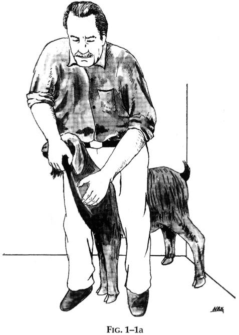
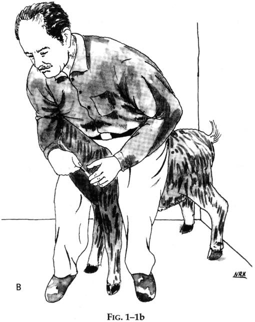
Figure 1.1. Useful restraint methods for intravenous blood sampling or medicine administration via the jugular vein of a goat. Backing the goat into a corner, as shown, improves control. In Figure. 1.1a, the goat is restrained so that an assistant, kneeling in front of the goat, can easily take the sample or give the medicine. In Figure. 1.1b, the goat is positioned with the head tucked under the handler’s arm so that sampling or medicine administration can be accomplished by the handler alone. (Illustrations by Mr. Nadir Kohzad.)
Injections
Mass medication or vaccination using a common needle has long been practiced by some farmers and veterinarians. In the case of goats, as with other species, it is time to rethink this practice, particularly in light of the growing importance of caprine arthritis encephalitis virus (CAEV) in goats. This retrovirus may be transmissible by blood-contaminated needles. Certainly, in herds where the virus is known to exist, or attempts are under way to control it, it would be counterproductive and negligent to use common needles. Additional discussion of this important caprine disease is found in Chapters 4 and 5.
Another reason to use individual needles in conjunction with other good hygienic practices during vaccination is the tendency for goats to develop large swellings and even abscesses at injection sites after vaccination with clostridial, chlamydial, and paratuberculosis vaccines. If the veterinarian’s technique is exemplary, he or she is unlikely to be held accountable for any problems that develop later.
If skins are marketed from a goat herd, the veterinarian should avoid injections of any kind in regions of the body that become part of the marketed skin, because defects can occur secondary to injection site reactions and devalue the skin. Therefore, the back and upper flanks should be avoided even though they are often convenient.
Intramuscular injections can cause difficulties in goats. The preferred site is in the neck, in a triangular region bounded by the vertebral column ventrally, the nuchal ligament dorsally, and the shoulder caudally. The triceps can also be used. If skin quality is not a consideration, the longissimus muscles over the back in the lumbar region may also be used. In all cases, the volume of drug administered in one site should not be greater than 5 ml. Needles should be 2 to 3 cm long and no larger than 18-gauge unless the medication is highly viscous. Shorter needles should be used for young kids.
The thigh muscles should be avoided as a site of intramuscular injection in adults and especially young
goats. The muscle mass is small compared to other ruminants, and sciatic nerve damage is not uncommon. Owners should be counseled against using this site. Even when the nerve is not damaged, marked lameness can occur when irritating drugs such as oxytetracycline are given in the leg. Permanent muscle damage can also occur that devalues the carcasses of meat goats.
Subcutaneous injections are commonly given in the neck in the same region described for intramuscular injections, or on the chest wall about 5 cm behind the point of the elbow. Injections ahead of the shoulder should be avoided in show goats, because local reactions near the superficial cervical (prescapular) lymph node may be confused with caseous lymphadenitis. Needles should be 18- to 20-gauge. The risk of accidental intramuscular injection may be increased if long needles are used.
Intravenous drugs are given via the jugular vein generally using 2 to 3 cm long needles of 18- or 20gauge. Blood samples can be taken from the jugular vein using an 18-gauge needle. Intradermal injections are given using 26-gauge, 1-cm long tuberculin needles. Intraperitoneal injections are rarely used except to treat neonates for hypoglycemia with glucose solutions or navel infections with aqueous based antibiotics. With the kid held hanging by the front legs, an 18- or 20-gauge needle is inserted perpendicular to the skin about 1 cm to the left of the navel no deeper than 1 cm.
When intramammary infusions are given, the teat should first be cleaned and swabbed with alcohol. As in cattle, single use teat cannulae should be used for each infusion with the cannula inserted into the teat only enough to gain entry into the teat cistern. For very small teat openings, sterile tomcat catheters can be used to infuse the teat.
CLINICAL EXAMINATION OF GOATS
A complete clinical examination consists of three major elements: history taking, physical examination, and inspection of the environment. Many diseases seen in individual goats are likely to represent potential herd problems; therefore, prompt diagnosis of clinical cases is essential so that, in addition to therapy, appropriate preventive measures can be introduced into the overall management program. In many caprine diseases, subclinical cases often exist in addition to the obvious clinical ones, and additional diagnostic testing may be required to identify them. The existence of subclinical infections and carrier states is a troublesome one for veterinarians performing prepurchase health examinations or writing health certificates for exportation or interstate travel. A list of such caprine diseases that the veterinarian must be aware of is given in Table 1.1.
History Taking
Very few diseases or health-related problems are randomly distributed in a flock or herd of goats; rather, they are concentrated in specific groups, usually by sex, function, production status, or age. Always establish early on what age, sex, breed, or group of goats is dying, showing signs of illness, aborting, or showing decreased productivity. If it is a mixed farm operation, the number of other types of livestock and their degree of contact with goats should be ascertained.
Detailed history should include a determination of the total flock or herd population and estimation of its breakdown by sex, age, breed, and pregnancy status. Having determined the total animal population, the population at risk, and the number of animals affected and dying, it is possible to determine rates of disease occurrence and case fatality rates. By counting cases,
Viral/prionRickettsialBacterialProtozoalUnknown
Caprine arthritis encephalitis
Foot and mouth disease
Scrapie
Chlamydiosis Coxiellosis (Q fever)
Caseous lymphadenitis
Paratuberculosis
Salmonellosis
Listeriosis
Brucellosis
Melioidosis
Tuberculosis
Mycoplasmosis
Staphylococcal mastitis
ToxoplasmosisUdder warts in white goats
Table 1.1. Goat diseases characterized by chronic infection or a carrier state.
the investigator is also in a better position to determine the actual significance of a problem as compared to the farmer’s perception of it. In some cases, the loss of a few animals may be insignificant compared to a more serious unrecognized problem such as endoparasitism or ectoparasitism.
Such an epidemiologically based history should aim to identify not only specific problems but also specific risk factors that appear to be associated with mortality, morbidity, or suboptimal performance. For example, when a primary complaint of kids developing diarrhea after weaning suggests coccidiosis, additional questions concerning the segregation of kids from adults, the manner that kids are fed, the design of feeders, the frequency and manner of barn cleaning, and details on the use of coccidiostats are necessary. In such cases, modification of management practices may halt the spread of disease.
Temporal relationships are important to note. Some diseases may occur seasonally, in association with abrupt weather changes, or in relation to specific events such as breeding, pregnancy, shearing, parturition, and lactation. For example, an unexpected cold snap or heavy rain right after shearing of Angora goats can increase pneumonia, abortion, and death rates, particularly if adequate shelter and supplemental feed have not been provided.
Localization of death or disease to specific areas on the premises is helpful. For example, if losses are seen only in certain areas of the farm, specific pastures, or particular barns, then suspicion of poisoning is increased.
Other important aspects of the history include questions pertaining to the actual ration being fed and its consumption, methods of feeding, changes in feeding, access to grazing, and water supply type and water availability.
If management interventions or preventive health procedures have been undertaken recently, they should be identified. Shearing, drenching, dehorning, spraying or dipping, castration, or vaccination can be associated with increases in morbidity and mortality. When range animals are mobbed for such procedures, sudden close confinement, temporary feed deprivation, and abrupt weather changes can predispose to outbreaks of conditions such as abortion, coccidiosis, salmonellosis, hypocalcemia, or starvation as a result of mismothering. When drugs or vaccines are used, the products and dosages, number of treatments, and method of administration should be determined, particularly because many goat farmers traditionally obtain their drugs and biologicals from nonveterinary sources.
If animals have been transported recently, dates, origins, means of transportation, and quarantine times should be determined. Information should also be col-
lected on visits to shows or fairs and on the origin of purchased animals, be it other farms, stockyards, or specialized goat sales. If animals have come from out of state, the relevant health certificates should be examined and the disease situation in the state of origin reviewed.
Finally, the reliability of information obtained should be checked with the actual goat keepers if the owner is not involved with day-to-day management decisions. If the veterinarian has prior knowledge of the local disease patterns in goats, such knowledge should not be used to make hasty and possibly incorrect judgments.
Special Considerations for Range and Pastured Goats
Extensively managed animals may not be closely observed, and histories can be sketchy. With large flocks, it should be determined if the animals are managed as a single flock or in smaller, self-contained units. The seasonal pattern of grazing and the length of grazing periods should be noted. Pasture composition, seasonal stocking rates per acre, the degree of pasture subdivision, and length of resting periods between grazing should be established. Note if supplemental feeding is practiced and the types of feed used. This may be important in terms of meeting specific nutritional needs, and, in the case of silage, may be associated with diseases such as listeriosis or rumen acidosis. Inquiries should also be made about whether crops are fed or grazed, the type and stage of growth, and whether there is a recent history of fertilizer or herbicide application. Knowledge of local trace element deficiencies or excesses may be helpful.
The type of grazing, whether set stocked or rotational, may be relevant to some disease outbreaks, particularly to gastrointestinal helminthiasis. The presence of other livestock species and feral, predatory, or scavenging animals or birds should be established if relevant to the problem under investigation.
Special Considerations for Intensively Managed Goats
A complete history can usually be obtained from the owner or herdsman of intensively managed goats. The patterns of disease are also likely to be different, with pneumonia and enteric diseases of young goats assuming much greater significance than the foot rot, helminthiasis, predation, or toxic plant problems more often seen under grazing systems. Feed composition and intake are more regulated but the veterinarian must inquire about episodes of sudden changes, excesses, or deprivations in the feed and water supplies.
Under close quarters, the movement, mixing, or introduction of new animals is more likely to cause an outbreak of disease. Kidding is often assisted in inten-
sively managed operations, and artificial kid rearing methods are commonly used. These procedures should be carefully reviewed when morbidity and mortality are concentrated in young kids. Weather, per se, should not adversely affect intensively managed animals. However, extremes in temperature may tax the ventilatory capacity of confinement buildings and extremely cold weather may freeze water supplies or incapacitate mechanized feeding equipment. Answers to questions about changes in dairy herd milking procedures or personnel may help to explain mastitis problems.
Special Considerations for Hobby Farms
Because hobbyists often have little previous agricultural or livestock experience, it might be helpful to practitioners to gauge the owners’ knowledge and attitudes regarding basic animal husbandry before history taking. Some fundamental misunderstandings about the care and management of goats may be revealed, such as non-recognition of basic ruminant physiology and the need for roughage in the diet. In other situations, owners may know about basic husbandry and disease problems, but may have seemingly unorthodox ideas about management and treatment. A good deal of tact may be required to obtain a useful history and prescribe appropriate therapy while not offending the hobbyist’s sensibilities.
In addition, hobby farmers often perceive goats more as companion animals than as livestock production units. While they may seek the expertise of a livestock clinician, they often expect the “bedside manner” of the companion animal practitioner. Therefore, the veterinarian who appears insensitive to the client’s emotions or indifferent to pain of the goat or who emphasizes only the economic value of the animal may not be called to the farm again.
Special Considerations for Organic Goat Production
Consumer interest in organically produced food has grown considerably over the past twenty years or so and producers have responded by producing and marketing an expanding variety of foodstuffs certified as organic. Increasingly, this includes foods of animal origin. Goat owners may choose to raise their goats under organic conditions. Veterinary practitioners with such clients need to be aware of and familiar with the constraints on conventional therapy that are associated with organic livestock production, which is now strictly regulated by law (Karreman 2006).
In the United States, the Organic Food Production Act (OFPA) was signed into law in 1990, creating the framework for regulation and certification of organically produced foods of plant and animal origin. The OFPA created the National Organic Standards Board (NOSB) which reviews materials for consideration as
acceptable for use in organic food production, including veterinary inputs used to maintain animal health. As a general rule, all natural materials are allowed for use in organic agriculture, unless specifically prohibited, while all synthetic materials are prohibited unless specifically permitted, following a successful petition process to the NOSB. The specific regulations of the National Organic Program are found in the United States Code of Federal Regulations at 7 CFR 205. These regulations became effective in 2002.
Vaccination is promoted as an organic livestock health care practice under 7 CFR 205, but the use of antibiotics and most anthelmintics is prohibited. Veterinarians must approach therapeutic interventions in organically raised animals differently than in conventionally raised animals, relying heavily on so-called natural treatments, including botanicals, acupuncture, homeopathy, etc. The standards of livestock health care practice which must be observed under the OFPA are given in 7 CFR 205.238. Veterinarians should be aware, however, that 7 CFR 205.238 considers the welfare of organically raised livestock by stipulating that an organic livestock producer may “not withhold medical treatment from a sick animal in an effort to preserve its organic status. All appropriate medications must be used to restore an animal to health when methods acceptable to organic production fail. Livestock treated with a prohibited substance must be clearly identified and shall not be sold, labeled, or represented as organically produced.” The synthetic substances allowed for use in organic livestock production are found in 7 CFR 205.603. The full text of the regulations can be found at www.ams.usda.gov/nop/ NOP/standards/FullRegTextOnly.html.
In Europe, organic production is regulated throughout the European Union (EU). EU regulation number 1804/99, which became effective in 2000, sets forth the rules for organic livestock production including animal health and veterinary interventions. All EU member states at a minimum comply with these rules, but some individual countries have included additional rules of their own. Specifications of the European and U.S. regulations have been compared (Nardone et al. 2004).
Special Considerations for Transgenic Goats
Production of transgenic animals using microinjection or nuclear transfer and the propagation of desirable animals using cloning are no longer just scientific research endeavors. They have become established production systems for the propagation and management of transgenic goats. It behooves veterinarians with active goat practice to be familiar with the basic techniques and health issues associated with transgenic goat production.
Transgenic technology began in 1980 when the first transgenic mouse was developed (Gordon et al. 1980).
The first transgenic goat was developed in 1989, producing rhtPA (recombinant human tissue plasminogen activator) in the milk (Ebert et al. 1991) as a potential human therapeutic agent. Since then, the field has markedly expanded with transgenic animals becoming commonplace within many programs and facilities.
The applications for transgenic animals are considerable and include not only the investigation of gene function but also the development of animal models, increased disease resistance through either transgene insertion or knock-out techniques, and production of recombinant, biopharmaceutical proteins in a number of biological fluids such as milk, blood, urine, and semen (Nieman and Kues 2003). In fact, distribution of the first transgenically derived human therapeutic recombinant protein from goat milk (ATryn®) was approved by the European Agency for the Evaluation of Medicinal Products in 2006 and by the Food and Drug Administration in the United States in 2009.
The two main techniques employed for making transgenic animals are microinjection and nuclear transfer (cloning). While microinjection was the first technology to be used in making large transgenic animals (Hammer et al. 1985), and specifically the goat (Gavin 1996), the process is inefficient with only a small percentage of the resulting animals being transgenic. Large animal nuclear transfer (Campbell et al. 1996; Wilmut et al. 1997) was developed later and provides for a near 100% transgenic rate when compared to microinjection. The cloning of the first transgenic goat soon followed (Baguisi et al. 1999; Keefer et al. 2001). There are other techniques for producing transgenic animals such as retroviral gene transfer and artificial chromosome insertion. However, these techniques have not been used yet in goats and are not mentioned further.
Most transgenic goats are maintained in USDAAPHIS-AC Licensed Research Facilities. Under the auspices of the Animal Welfare Act (AWA), these licensed facilities must maintain strict adherence to rules and regulations specifically governing animal care, health, and welfare (housing, lighting, feeding, veterinary care, and environmental enrichment at a minimum). Depending upon the type of research and the funding source, the National Institutes of Health (NIH) may also be involved through their Office of Laboratory Animal Welfare (OLAW) as government funding brings along its own slightly different set of rules and regulations for animals used in a research setting. A growing number of institutions are also striving for accreditation by the Association for the Assessment and Accreditation of Laboratory Animal Care, International (AAALAC-Int.), considered by many to set the gold standard for animal care in licensed research programs and facilities. Lastly, depending upon the intended use of any tissues/fluids
from the transgenic animal, the FDA may also have regulatory oversight and impose its own set of rules and regulations.
The use of microinjection to produce a transgenic animal involves microinjection of the transgene into the pronucleus of a fertilized, one-cell embryo and then the transfer of surviving embryos to a surrogate mother. One of the first areas for possible concern, and for which observation and monitoring are appropriate, is the physical/mechanical effects on the nucleus/gene due to the actual microinjection process at the one-cell stage. If any negative impacts occur or gene functions are altered or impaired, one may see outcomes ranging from decreased pregnancy rates from transferred embryos to increased pregnancy loss, late term abortions, or possible physiological abnormalities at birth with clinical sequelae. However, years of experience now indicate that these phenomena, while possible, occurs at a very low incidence.
Regardless of the technique used to produce a transgenic goat, another possible concern involves endogenous gene function and potential transgene insertional site effects. The gene of interest inserts randomly into the genome following transgene introduction. Hence, there is a chance that an endogenous gene could be negatively impacted, leading to potential adverse physiological effects and a transgenic goat presenting with clinical signs of abnormal physiology or health. Therefore, appropriate post-parturitional monitoring of animal health is warranted for any transgenic founder animal.
Introduction of a transgene produces a goat that is hemizygous for that given transgene. Subsequent breeding within a lineage may be aimed at achieving a homozygous state for the transgene. Possible concerns may arise through this approach. First, inbreeding of related goats is the primary route to achieving a homozygous animal. Therefore, inbreeding coefficients need to be considered and animals need to be monitored for ill effects from this relatedness and for possible impacts on overall health and ability to thrive. Second, achieving a homozygous state may bring to light an insertional gene effect since both copies of an endogenous gene may now be affected thereby causing physiological or clinical issues that were not seen in the hemizygous state. Again, appropriate monitoring of animal health is warranted for the first homozygous animals produced. Lastly, the potential exists that breeding for the production of a transgenic homozygous animal will reveal a lethal outcome. A lethality issue may be suspected when: breedings of two hemizygous animals produce no detectable pregnancies; pregnancies do not hold to term with either resorptions or abortions; or, offspring succumb soon after birth. Thus, production of a homozygous transgenic animal may not always be possible and close animal
health monitoring is warranted when homozygosity is pursued.
One additional set of concerns related to transgene effects is the possibility of systemic circulation of the recombinant protein being expressed and the potential health impacts arising from expression of pharmacologically active molecules. Depending on the tissue or fluid where the recombinant protein may be directed for expression (e.g., milk, blood, urine, semen, etc.), one must be vigilant for systemic effects as the protein usually will be found systemically due to leaky vasculature and normal lymphatic drainage. Therefore, the biological nature and function of the recombinant protein being introduced must be known so that any effects which may be exerted can be anticipated and recognized. Consideration must also be given to potential adverse health impacts if this is a new gene and novel protein not normally physiologically found in the genome or animal. Lastly, the quantity of the recombinant protein that is expressed and then found systemically in the transgenic animal must be considered. Even if the target protein is endogenous to the animal, it may be found at significantly higher levels than normal and may cause physiological effects that alter normal homeostasis.
As with any traditional goat agricultural production operation for meat, milk, or fiber, optimizing health and product output starts with a sound nutritional program. Relative to transgenic production, nutritional programs should consider the nature of the recombinant protein to be produced. Specifically, if the recombinant protein is novel to the physiological output of the goat’s normal cellular machinery, or if quantities are above what is normally produced in vivo, then one may need to augment the diet. This modified or fortified diet may need to contain increased levels of vitamins, minerals, or specific amino acids. One should understand the normal cellular machinery and biochemical pathways involved in protein production to know if or what supplementation may be appropriate or necessary.
With the development of cloning technology, nuclear transfer has become the preferred method for producing transgenic goats and has greatly improved the overall efficiency of the process. However, the use of nuclear transfer has added some additional health concerns in a small percentage of animals.
Nuclear transfer starts by removing the maternal DNA from an unfertilized oocyte through enucleation. A full complement of genetic material is subsequently replaced by addition of a somatic cell (e.g. fetal or adult skin fibroblast cell) through a process termed reconstruction. Thereafter, in vitro techniques are used to fuse the oocyte and somatic cell and activate the couplet to begin dividing. Following a brief in vitro culture period, these newly developed cloned embryos
are then transferred to recipient goats using traditional embryo transfer techniques.
With nuclear transfer, a decreased in utero fetal survival rate can be seen very early in pregnancy and has been well documented in many species (Campbell et al. 1996; Wilmut et al. 1997; Baguisi et al. 1999). This inability to thrive may be associated with inappropriate or inadequate reprogramming (Dean et al. 2001) of the nuclear/genetic material of the donor cell line or karyoplast and has been postulated to be at the level of the DNA (e.g., methylation pattern). An altered inheritance of cellular mitochondria (Wells 2005) has also been shown to occur in cloned embryos, adding to the possible causes for some of the abnormalities in homeostasis. Both of these phenomena may be directly linked to the small percentage of physiological problems seen in utero for some cloned animals such as abnormal placentation and/or organogenesis (Farin et al. 2006; Loi et al. 2006; Fletcher et al. 2007). Abnormal placentation can also lead to abnormal uterine fluid homeostasis and fluid retention in does carrying cloned embryos, which may warrant close clinical monitoring or intervention where appropriate. Other possible outcomes of abnormal placentation include: a tendency toward decreased pregnancy rates for animals receiving cloned embryos, an increased in utero loss rate through resorption, or an increased level of abortions if there is late term fetal loss.
The potential abnormal physiology with or without clinical presentations may continue after birth and into the neonatal and early prepuberal stages (Hill et al. 1999). Documented abnormalities in a few large animal species have been shown at the level of the renal, cardiac, respiratory, hepatic, hematopoietic, and immune systems. However, if the small percentage of animals, including goats, that present with these abnormal physiological entities can be clinically supported over time, as the animals grow, many of these abnormalities resolve and they can lead normal and healthy lives (Chavatte-Palmer et al. 2002).
The vast majority of transgenic and cloned animals are normal and healthy (Walsh et al. 2003; Enright et al. 2002; Tayfur Tecirlioglu et al. 2006) and subsequent generations of animals produced from first generation clones have not shown any of the health related issues seen in a small percentage of original clones (Wells 2005). In fact, passage through the germ line has been reported to reverse any abnormal patterns detected at the DNA level in first generation clones (Wells 2005).
PHYSICAL EXAMINATION
Inspection from a Distance
It is often useful diagnostically to observe a group of goats from a distance prior to disturbing them for “hands-on” examination. This is especially true at the
time of the initial visit to identify the existence of common problems in the herd or flock. The animals should be observed at rest, while eating or drinking, and during spontaneous and forced movement. General impressions of body condition, mental attitude, and social hierarchy may be acquired and abnormal behaviors characteristic of certain diseases may be noted. Estimated prevalence of common disease problems such as kid pneumonia, diarrhea, and pinkeye can be roughly assessed, respectively, by counting coughers, stained hindquarters, and runny eyes. Other specific observations that might suggest commonly seen disease problems in goats are briefly discussed below. This is for illustration and is not meant to be comprehensive.
Individual goats that appear listless, separate themselves from the herd, or are not actively feeding when others are should be noted and later caught for careful examination as should animals in very poor body condition. Reluctance to feed may be due to a wide range of systemic diseases or localized conditions such as dental or pharyngeal problems, or the result of inadequate bunk space or bullying by dominant does.
Latent signs of respiratory disease or anemia associated with parasitism can be brought out by forced movement of a flock. Anemia is manifested by rapid fatigue, increased heart and respiratory rates, and sometimes collapse. Increased respiratory rate, dyspnea, and coughing indicate respiratory problems.
Signs of skin irritation or pruritus manifested as hair loss, fleece biting, or rubbing against fences or other solid objects usually suggest ectoparasitism, though scrapie, pseudorabies, and migrating Parelaphostrongylus tenuis are other possibilities. Goats scratching at their ears with their hind limbs or shaking their heads vigorously probably have ear mites.
Goats observed resting or walking on their knees often have chronic CAEV infection or sore feet. Any animals with abnormalities of gait or lameness after forced exercise should be carefully examined for evidence of arthritis, fractures, laminitis, and lesions of foot rot or foot scald.
A variety of clinical signs may be observed in goats with neurologic disease. Among the more common are ataxia, posterior paresis, circling, depression, head pressing, unilateral facial paralysis, and blindness. Details on carrying out a neurologic examination and differential diagnosis for signs of neurologic disease are provided in Chapter 5.
Straining while attempting to urinate, particularly in bucks and wethers, should suggest the possibility of urolithiasis or posthitis.
Cutaneous swellings or discharges may be observed. Draining abscesses associated with lymph nodes are
highly suggestive of caseous lymphadenitis in the flock or herd. A high rate of subcutaneous swellings is often associated with injection site reactions in goats in response to certain adjuvanted vaccines or bacterins or when aseptic injection techniques are not followed.
When kids are left with does, careful observation of suckling behavior can indicate kids that are not successfully nursing. Their does should be examined particularly for signs of mastitis or other udder problems.
Direct Physical Examination of Individual Goats
A topographical approach to physical examination is presented here. Because of the small size of goats, rectal palpation is limited to insertion of a finger into the rectum to assess the pelvic structures and to determine the presence and character of feces. When economics permit, physical examination findings should be supplemented through use of appropriate imaging techniques, diagnostic procedures, and laboratory tests. When numerous individuals in a herd or flock show signs of disease, field necropsy examination may be the most effective way to establish or confirm a diagnosis.
General Inspection
Body condition, mental attitude, and the status of the superficial lymph nodes should be noted. The temperature, pulse, and respiration should be recorded. Fleece in Angora goats makes visual assessment of body condition difficult. Digital palpation of the ribs, spinous and transverse processes of the vertebrae, and the loin muscle may be necessary to evaluate condition. A word of caution about condition scoring in goats: scoring systems derived for sheep are not directly applicable to goats because goats as a species tend to deposit stored fat intra-abdominally rather than subcutaneously. Scoring systems for dairy goats combine palpation of the sternal and lumbar regions and are discussed in Chapter 19. In general, any palpable back fat in a dairy goat would classify her as obese. A scoring system applied to the Small East African goat in Zimbabwe showed a good correlation between a backbone condition score and changes in bodyweight (bw), with a 1-point change in condition score representing an average change of 12% in bw (Honhold et al. 1989).
Normally goats have an alert, attentive, and inquisitive mental attitude. A depressed attitude is characterized by dullness, separation from the flock, and indifference to handling. Depression is present in a wide variety of septic and toxemic conditions but is a particularly prominent sign in pregnancy toxemia and listeriosis. An anxious or apprehensive state is often associated with urethral obstruction in males, sudden
blindness as may occur in polioencephalomalacia, or with persistent irritations such as flies and nasal myiasis. An attitude of extreme excitation is most often associated with neurological diseases such as tetanus and meningitis that may be accompanied by muscular rigidity, or encephalitic conditions such as pseudorabies and rabies.
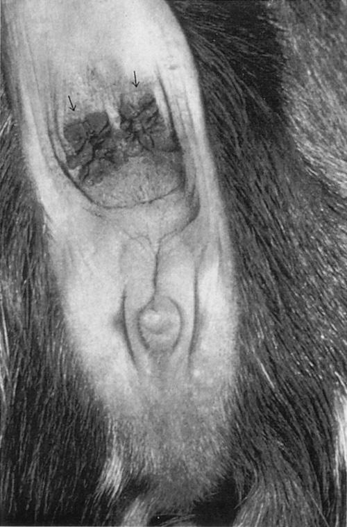
Digital palpation of all superficial lymph nodes should always be a part of the physical exam because of the clinical importance of caseous lymphadenitis in goats. The mandibular, parotid, retropharyngeal, superficial cervical (prescapular), subiliac (prefemoral), and superficial inguinal (supramammary) lymph nodes should be inspected. Normal sized nodes may not be palpable in some of these locations but affected nodes should be readily evident. Any other swellings on the body surface should be noted. Temperature, pulse, and respiration should be measured when the animal is calm because the activity of catching the animal for examination may elevate all three parameters. When taking the goat’s rectal temperature, an accumulation of brown, waxy material may be noted near the anus. This is the normal secretion of the sebaceous gland located below the base of the tail (Figure 1.2).
The normal body temperature of goats is usually reported in the range of 38.6° to 40°C (101.5° to 104°F). However, the body temperature of a normal Angora goat with a full fleece on a hot, humid day can reach 40.3°C (104.5°F) or higher, and goats of lighter bodyweight are more likely to have higher temperatures when exposed to sun than bigger goats (McGregor 1985). To accurately assess the febrile state of the patient, it is useful to record body temperatures in apparently normal herd mates.
The pulse can be measured by stethoscope over the heart or by digital palpation of the femoral artery. Normal pulse rate ranges from 70 to 90 beats per minute (bpm) in resting adults, but can be double that in young, active kids. Fetal heart rates up to 180 bpm have been recorded by ultrasound. It may be useful to assess respiratory rate both at rest and after exercise. Any abnormalities of respiration should also be noted, including flaring of nostrils, extension of head and neck, grunting, abdominal press, and so forth. Normal resting respiratory rate is 10 to 30 per minute in adults and 20 to 40 in kids.
Neonates should be inspected particularly for congenital defects. More commonly observed problems include brachygnathia, cleft palate, hydrocephalus, atresia ani, or rectovaginal fistula, and abnormalities of the genitalia associated with the intersex condition as discussed in Chapter 13. A list of congenital and inherited diseases is provided in Table 1.2. Not all of these, of course, will be evident at birth. Up-to-date information on inherited conditions of goats as well as
Figure 1.2. Typical waxy secretion found at the base of the tail of goats which is produced by the sebaceous glands in that area. This secretion should not be confused with diarrhea, vaginal discharge, or lochia. (Reproduced by permission of Dr. C.S.F. Williams.)
other species is available through the Online Mendelian Inheritance in Animals (OMIA) database at http:// omia.angis.org.au/.
Goat stature is quite diverse. Many small breeds of goats such as the Pygmy or West African Dwarf goat are in fact achondroplastic dwarfs. They appear disproportionate with short legs and normal size torsos. This may draw visual attention to the degree of abdominal distension present, which though quite pronounced, is usually normal. Dwarfism because of pituitary hypoplasia is also seen in goats. These small goats are proportionate in appearance; the Sudan goat is an example (Ricordeau 1981).
Examination of the Integument
In goats the character of the skin and haircoat is a good indicator of general health. A rough, dry, unglossy coat; excessive dander or flakiness; and failure to shed
Table 1.2. Congenital and inherited abnormalities in goats.
Known inherited conditionsKnown acquired conditionsConditions of unclear status
Afibrinogenemia in Saanen goats
Beta mannosidosis in Nubian goats
Bipartite scrotum in Angora goats
Brachygnathia superior or inferior
Cryptorchidism in Angora goats
Excessive facial hair in Angora goats
Gynecomastia
Hereditary goiter in Dutch goats
Inherited abortion in South African Angora goats
Intersexes associated with polled condition
Myotonia congenita
N-acetylglucosamine 6-sulphatase
deficiency in Nubian goats
(mucopolysaccharidosis IIID)
Recessive atrichosis
Robertsonian translocation
Short tendons in Australian Angora goats
Sperm granulomas
Supernumerary teats
Testicular hypoplasia
Anthrogryposis and hydranencephaly caused by Akabane virus
Border disease
Congenital copper deficiency
Cyclopia due to Veratrum californicum
Freemartins
out in the spring are all suggestive of poor nutritional status, parasitism, or other chronic diseases. The hair or fleece should be parted and the skin examined for lice, ticks, fleas (in the tropics), nodules, swellings, crusts, eczema, necrosis, neoplasia, photosensitization, and sunburn and focal or regional alopecia. The differential diagnoses for these findings are discussed in Chapter 2. Because many goats are used primarily for cashmere or mohair production, the veterinarian should know something about the nature of goat hair used in textiles. Detailed information on the subject is given in Chapter 2.
Examination of the Head
Many conditions can cause general asymmetry or focal swellings around the head and these abnormalities should be noted. The differential diagnoses for such swellings are discussed in Chapter 3.
Membranes
Inspection of the conjunctivae and mucous membranes of the mouth may reveal paleness due to anemia, icterus resulting from hemolysis or hepatic dysfunction, or hyperemia and congestion associated with acute febrile or toxemic states.
Absence of hair
Atresia ani
Atresia coli
Cleft palate
Congenital goiter of Boer goats
Double or fused teats
Entropion
Hydrops
Patellar luxation
Precocious milking
Progressive paresis of Angora goats
Rectovaginal fistula
Skeletal malformations
Spastic paresis
Sticky kid syndrome of Golden
Guernsey goats
Umbilical hernia
Oral Cavity
Evidence of brachygnathism, cleft palate, mucosal lesions, dental abnormalities, or dysphagia such as drooling, salivation, dropping food from the mouth, or accumulating food in the buccal space should be noted. The differential diagnoses for these signs are given in Chapter 10. Necrotic odors of the breath may occur. They may reflect necrotic stomatitis, alveolar periostitis, pharyngitis, or even pneumonia.
Thorough examination of the oral cavity requires good restraint, a speculum, a towel, and a penlight. All oral structures should be examined and the molar arcades digitally palpated for missing teeth from outside the mouth. If more direct examination of the teeth is required, extreme caution should be taken because the molars may have sharp, jagged edges and exert powerful grinding motion. Tranquilization is indicated, because fingers can be badly injured. Wearing gloves during oral examination is a wise precautionary measure, particularly if the goat shows neurologic signs.
Eyes
Facial hair covering the eyes is a heritable trait in Angora goats. Affected goats tend to do poorly on
range because their ability to selectively browse is impaired. The body condition of such individuals should be noted as well as their prevalence in the flock. Blindness is initially assessed by testing the menace response, but facial nerve paralysis can render a sighted animal unable to blink. Intact pupillary light responses in a blind goat suggest a cerebrocortical lesion. Lacrimation and hyperemia of the conjunctiva, cloudiness of the cornea, and hypopyon in the anterior chamber should be noted if present. The differential diagnoses for these various findings are given in Chapter 6.
Nares
Both nostrils should be evaluated for symmetry of air flow. If nasal discharge is observed, determine if it is unilateral or bilateral and note its character. Collapse of a nostril may result from facial nerve paralysis. Crusting of the nares occurs when the sick animal does not clean the nostrils or it may be a sign of specific disease problems. The differential diagnoses for nasal discharge and crusting of the nares are given in Chapter 9.
Ears
Ear mites, if suspected, can be identified by collecting debris from the ear canal on a cotton swab and smearing it on a slide for examination.
Goats are often identified by tattoos inside the ear; tattoo numbers may have to be checked against health papers at shows and sales. It may be necessary to clean the inside of the ear with soap and water and use a powerful light source to backlight the tattoo to make it readable. Metal and plastic ear tags commonly tear out of goats’ ears and their use should be avoided, especially in pet and show animals.
Ear tips may be lost on young goats due to prolonged exposure to freezing temperatures. Goats of the La Mancha breed lack a well-developed external ear. Only a vestigial pinna is present and is referred to as an elf (up to 5 cm, with some cartilage) or gopher (up to 2.5 cm, with little or no cartilage) ear. These animals are usually tattooed on the underside of the tail.
Horns
Goats may be horned or polled. Horn buds may be present at birth or become palpable within several days of birth. Generally, horned kids have two irregular whorls of hair over the location of the horn buds whereas hornless kids have a smooth poll with a single central symmetrical whorl of hair. It is important to establish and record which offspring are naturally polled because homozygous polled goats have a high incidence of infertility. The relationship of the polled trait and the intersex condition is discussed in detail in Chapter 13. Deformed horns, or scurs, are often seen
on older goats as a result of incomplete removal of germinal horn tissue at the time of disbudding. Techniques for, and problems with, disbudding and dehorning are discussed in Chapter 18. The glands partially responsible for the characteristic odor of buck goats in the breeding season are located in skin folds just caudomedial to the horn buds.
Examination of the Neck
Traumatic injuries to the pharynx from balling guns and drenching equipment occur in goats. The throat should be palpated for swelling, heat, and pain associated with cellulitis from traumatic injury. A number of normal and abnormal structures and swellings in the neck must be differentiated on physical examination, including goiters, thymus, branchial cleft cysts, wattles, and abscesses. Their differentiation is discussed in Chapter 3. Thorough examination of the neck should include palpation of the jugular furrows for evidence of phlebitis, palpation of the esophagus for evidence of obstruction, and auscultation of the trachea. Prominent distension of the jugular vein, though possibly suggestive of congestive heart failure, is most commonly due to overly tight collars or neck chains in goats. This should be brought to the owner’s attention if found.
Examination of the Chest
The extent and severity of respiratory disease is often difficult to assess in goats. To improve the chances of accurate diagnosis and prognosis, careful attention should be given to auscultation. When possible, the animal should be moved to quiet surroundings. The fleece in Angora goats should be parted to place the stethoscope in contact with the skin. The trachea and the lungs should be ausculted to identify the presence of referred sounds. Eliciting a cough by compression of the pharynx may clear the trachea or reveal the presence of bronchial exudate. Care should be taken to listen with a stethoscope placed well forward under the elbow and in front of the shoulder, otherwise cranial ventral pneumonias, which are common, may be overlooked. The intensity of identifiable sounds can be augmented by forced activity of the animal before auscultation or by placing a plastic bag or exam glove over the nares to increase the depth of respiration by rebreathing of carbon dioxide. Radiographic examination is indicated when questions about the severity of lung disease persist.
Normally the heart can be heard about evenly on both the right and left sides of the chest at the fourth or fifth rib space. Mediastinal abscesses may displace the heart, resulting in a shift in intensity of cardiac sounds. Muffling of the heart sounds because of pericarditis is uncommon in goats. Murmurs are rarely heard and are discussed in Chapter 8.
Examination of the Abdomen
The abdominal contour should be inspected to assess conditions such as bloat, advanced pregnancy in females, and ruptured bladder in wethers and intact males. Characteristic contours and their clinical significance are discussed in Chapter 10. Ballotment may help to detect abdominal fluid accumulations, pregnancy, or rumen impaction. Auscultation of the rumen in the left paralumbar fossa is essential. Normal rumen contractions occur at a rate of one to two per minute. Observation of cud chewing suggests normal rumen activity.
Examination of the Limbs
Locomotor problems are common in goats. Lameness and abnormalities of gait may result from neurological disease, conformational defects, muscular dysfunction, skeletal trauma, infectious and noninfectious arthritides, and diseases of the foot. Localization of the problem by careful physical examination is the first step in making an accurate diagnosis. Differential diagnoses of locomotor problems are discussed in detail in Chapter 4.
Overgrown hooves must be pared with shears or a hoof knife to adequately assess the health of the foot. Hyperemia and swellings at or above the coronary band should be noted. They may represent either local infections or systemic disease.
All joints should be carefully palpated. Distensions of the joint capsule, heat, pain, swelling or fibrosis of periarticular structures, limitations on the range of joint motion, and enlargement of bursae should all be noted. The degree of joint enlargement may not necessarily correlate with the severity of lameness.
The vertebral column and the long bones of the legs should be palpated for evidence of fractures in acutely lame or recumbent animals. A major goal of physical examination in recumbent goats is to differentiate musculoskeletal, metabolic, toxemic, and neurologic causes of recumbency. The differential diagnoses for recumbency are discussed in Chapter 4.
Examination of the Reproductive System
Mammary Gland
Careful examination of the udder is always warranted. Visual inspection may reveal weakness of the suspensory apparatus, slack halves, abnormal swellings, and discolorations of the skin. Digital palpation of the gland will identify udder edema, active inflammation, fibrosis, scarring, or abscesses in the parenchyma or teats. In gangrenous mastitis the udder skin may be blue-black and cold and in time the gland may slough if the animal survives. Patency of the teats should be assessed in lactating animals. Supernumer-
ary teats may be present. Their identification and removal are discussed in Chapter 14.
The milk of lactating does should be observed on a black plate or strip cup to assess color, consistency, and the presence of clots or flakes. Bovine screening tests for subclinical mastitis such as the California Mastitis Test must be used cautiously in goats because normal goat milk tends to have higher cell counts. The somatic cell count issue and interpretation of test results are discussed in Chapter 14.
Vulva
Swelling and hyperemia of the vulva may be signs of heat or impending parturition, but may also be seen in herpes vulvovaginitis in conjunction with vesicles or scabs. Any vulvar discharges should be noted. As females come out of heat, the vulvar discharge, initially serous and mucoid, may become white and tenacious. This is often misinterpreted as a purulent discharge by the inexperienced observer. Often, there is a sanguinous discharge after termination of a pseudopregnancy. Speculum examination of the vaginal canal and cervix should be carried out when there is doubt about the source and nature of the discharge. Occasionally, otherwise normal does may have ectopic mammary tissue present at the vulva which may swell during lactation.
Does may show vaginal eversion or frank prolapse in advanced pregnancy or immediately post partum. After uncomplicated births, normal lochia may be discharged for a period of one to three weeks. It is reddish brown in color and odorless. Placentas are usually passed within four hours of parturition and are frequently eaten.
It is important to carefully examine the external genitalia of young does, particularly when there is a complaint of infertility, because of the high incidence of intersexes among polled goats. Malformations of the genitalia range from the clinically subtle, such as a slightly enlarged clitoris, to the overt, such as male phenotypes in genetically female individuals. Accurate record keeping may help to identify homozygouspolled individuals.
Scrotum
The scrotum and its contents should be palpated. Normally there is bilateral symmetry of all structures. A bipartite or split scrotum is a common congenital condition that some breeders consider a fault. The differential diagnoses for abnormalities of the scrotum, testes, and spermatic cord structures are given in Chapter 13. Semen samples can be collected for evaluation by electroejaculation or by use of an artificial vagina. These procedures are described in Chapter 13. Gynecomastia occurs in male goats and it is not
extraordinary to detect distended teats anterior to the scrotum during physical examination, as discussed in Chapter 13.
Penis and Prepuce
Examining the penis of male goats, especially wethers, can be difficult and is not ordinarily attempted unless there is a history of urinary or breeding problems. Details on special examination and catheterization of the penis are given in Chapter 12.
The preputial opening should be routinely examined, particularly in wethers, for the presence of ulcerative posthitis. The preputial orifice may become occluded in this condition. Crystals or drops of blood may be noted at the orifice in cases of obstructive urolithiasis.
Examination of the Environment
An examination of the environment where goats are raised should include a detailed review of all feeds used, the feeding facilities, water sources and water delivery systems; the yards, pastures, range, or buildings where the animals are kept; and any mechanical or manual equipment used as part of the routine farm procedures. Too often, farmers attempt to make do with equipment and buildings that are inadequate for an expanding operation. Because of prolificacy, goat herds tend to expand faster than owners anticipate. Inadequate feeder space for does, a smell of ammonia in the air due to poor ventilation and/or soaked bedding, and overcrowded kid pens are three examples of common faults found on environmental inspection. Besides visual inspection of equipment, farmers could be asked to demonstrate how common procedures are carried out to detect if they are using inappropriate techniques.
Equipment used for treatment should be examined for its potential to cause injury. Poorly maintained syringes with either contaminated or overly long, large-gauge needles can result in injection abscesses or systemic infections. Drug and vaccine stocks should be examined to determine if they are appropriate, clean, unexpired, and maintained at proper temperatures. Storage facilities for these items should be properly secured. If the farmer blends his own feed or components, the techniques and constituents used should be examined, particularly when toxicities are suspected. Accidental inclusion of agricultural or other farm chemicals is not uncommon. Milking machines used for goats should also be examined to determine their efficiency and the milking procedures studied if it appears that mastitis is a significant problem. Finally, the facilities available and routinely used to dispose of any dead animals should be particularly noted to see that they are consistent with any legal requirements and do not serve as environmental contaminants or
as a source of infection for the remainder of the flock or herd.
In recent years, in association with the expansion of international trade in livestock and livestock products and increasing concerns about bioterrorism, there has been a growing emphasis on food safety and biosecurity, with recognition that food safety encompasses all aspects of food production, from the farm to the table. As such, quality management assurance programs have grown in popularity as have procedures to ensure biosecurity to limit the introduction and spread of disease on and between farms. Environmental inspections should include an assessment of biosecurity and aspects of management and sanitation that can affect food quality.
Special Considerations for Range and Pasture Operations
The quality and quantity of pasture and the availability of supplementary or conserved feed should be assessed. This may require a specialized knowledge of pasture species, weeds, or other potentially toxic plants. A list of plants known to be poisonous to goats is provided in Chapter 19. If possible, animals should be observed while feeding. The source of water available both year-round and seasonally, its quality, and its quantity, should also be inspected. Feed and water samples should, if necessary, be collected for laboratory examinations.
On range or pasture, goats may congregate in certain shaded or sheltered areas, particularly during periods of high or low temperatures or precipitation, and after shearing. Provision of adequate shelter should be documented. Heat stress can be reduced by ensuring the animals have adequate shade and water when the livestock weather safety index is in the danger or emergency zone. Cold stress is particularly likely in recently shorn animals, and temporary shelter and extra feed are necessary for these animals. If shelter areas are limited or overused, buildup of infectious agents can occur, leading to increases in enteric and other diseases, especially in kids. This is aggravated by poor drainage under feeders and water troughs.
Fencing for these types of operations should also be inspected, particularly regarding its effectiveness in keeping goats in and keeping predators out.
Special Considerations for Confinement Operations
In semiconfinement operations adult animals and sometimes young stock are grazed on pasture during the warmer months but confined during the winter and early spring. As herds expand, there is a tendency for small pastures to be overused, with poor nutrition and increased parasitism as the result. Stocking rates should be noted and deworming history verified.
In confinement systems, the type of buildings used for housing should be evaluated for the following: available area (square feet or square meters) per adult breeding animal, type of flooring and bedding, degree of insulation, presence of supplemental heating if any, adequacy of natural or mechanical ventilation systems, source and availability of water, and the amount of trough or bunk space per goat. Requirements for goats are given in Chapter 9. The distribution of animals within buildings should be noted. Ideally, a separate kidding facility should exist and be hygienically maintained. Artificially reared kids should be in a separate facility from adult stock, and bucks should be housed separately from milking does. Methods and times of feeding for all groups within the herd should be noted. If other species of livestock are present, consideration should be given to them as possible sources of disease for goats.
Unfortunately, many confinement goat operations have both rodent and associated feline populations. Cats are a recognized source of Toxoplasma gondii, a major cause of abortion in goats. Farmers should be encouraged to seek alternate means of rodent control.
Special Consideration for Hobby Farms
The principles and procedures of environmental examination already described also apply to the hobby farm. Special considerations that occur are usually the result of inexperience on the part of the hobbyist. Medications, insecticides, and grain supplies may be inadequately secured, allowing access of inquisitive goats with resultant overdoses, poisonings, and/or grain overload. In the barn, overcrowding with inadequate feeder space is often observed along with fecal contamination of feed and water supplies.
In winter, lack of adequate ventilation in closed, overcrowded barns leads to outbreaks of pneumonia. Some owners consistently shut all windows and doors, believing that animals must stay warm in winter. These individuals need to be counseled on the beneficial effects of continuous air exchange on respiratory health. Pasture problems often involve disappearance of animals or predation due to inadequate fencing, bloat due to inadequate adaption to pasture, or parasitism due to placement of young stock on contaminated, overcrowded plots. The veterinarian should examine all elements of the ration being fed on the hobby farm to ensure that the hobbyist understands basic ruminant nutrition.
Field Necropsies and Slaughterhouse Checks
The economic value of most individual goats in commercial herds and flocks is such that most owners can be persuaded to have a necropsy undertaken of an ill or obviously moribund animal. This is an extremely useful adjunct to clinical examination, because nec-
ropsy can confirm the suspected diagnosis or suggest new avenues of investigation. All necropsies should be carried out following a systematic procedure with special regard to the likelihood of any zoonotic infections being transmitted and the safe and legal disposal of carcasses.
Necropsy of all or a random sample of young animals born dead or dying in the first two to three weeks of life is an extremely valuable service that veterinarians can provide to their clients. Categorization of deaths into preparturient, parturient, and postparturient causes can be done with a minimum of laboratory diagnostic techniques and immediately direct the owner to possible ways of reducing these losses.
Whenever possible, goats slaughtered for meat or culled from the herd or flock should be examined post mortem by a veterinarian as an inexpensive and worthwhile means of disease surveillance. Such inspections can provide information on the efficacy of parasite control measures; the presence of subclinical pneumonia; and the occurrence of injection site abscesses, visceral caseous lymphadenitis, and hydatid disease. Opportunities to perform routine slaughter checks on goats at an abattoir are uncommon in the United States because the goat meat industry is decentralized, though this may change as demand for goat meat continues to increase.
REFERENCES
Askins, G.D. and Turner, E.E.: A behavioural study of Angora goats on west Texas range. J. Range Mgmnt., 25:82–87, 1972.
Baguisi, A., et al.: Production of goats by somatic cell nuclear transfer. Nat. Biotech., 17:456–461, 1999.
Batten, G.J.: A new meat goat breed. International Goat Association. Prcdngs, IV International Conference on Goats, Brasilia, March 8–13, 1987, Volume 2, pp. 1330–1331.
Campbell, K.H.S., McWhir, J., Ritchie, W.A., and Wilmut, I. Sheep cloned by nuclear transfer from a cultured cell line. Nature, 380:64–66, 1996.
Chavatte-Palmer, P., et al.: Clinical, hormonal, and hematologic characteristics of bovine calves cloned from nuclei from somatic cells. Biol. Reprod., 66:1596–1603, 2002.
Coop, I.E., (ed): Sheep and Goat Production. Amsterdam, Elsevier Scientific Publ. Co., 1982.
Dean, W., et al.: Conservation of methylation reprogramming in mammalian development: aberrant reprogramming in cloned embryos. Proc. Natl. Acad. Sci. USA, 98:13734–13738, 2001.
Devendra, C. and Burns, M.: Goat Production in the Tropics. Slough, U.K., Commonwealth Agricultural Bureaux, 1983.
Dubeuf, J.-P, Morand-Fehr, P., Rubino, R.: Situation, changes and future of goat industry around the world. Small Rum. Res., 51:165–173, 2004.
Dunbar, R.: Scapegoat for a thousand deserts. New Scientist, 104:30–33, 1984.
Ebert, K.M., et al.: Transgenic production of a variant of human tissue-type plasminogen activator in goat milk: Generation of transgenic goats and analysis of expression. Bio/Tech, 9:835–838, 1991.
Enright, B.P., et al.: Reproductive characteristics of cloned heifers derived from adult somatic cells. Biol. Reprod., 66:291–296, 2002.
FAO: Livestock Units. In: ResourceSTAT Module of FAOSTAT, Food and Agricultural Organization of the United Nations, Rome, 2007. http://faostat.fao.org/site/405/default.aspx
FAO: Livestock Primary and Processed. In: ProdSTAT Module of FAOSTAT, Food and Agricultural Organization of the United Nations, Rome, 2007a. http://faostat.fao. org/site/526/default.aspx
Farin, P.W., Piedrahita, J.A., and Farin, C.E.: Errors in development of fetuses and placentas from in vitro-produced bovine embryos. Theriogenology, 65:178–191, 2006.
Fletcher, C.J., et al.: Somatic cell nuclear transfer in the sheep induces placental defects that likely preceded fetal demise. Reprod., 133:243–255, 2007.
Gall, C., (ed): Goat Production, London, Academic Press, 1981.
Gavin, W.G.: Gene transfer into goat embryos. Transgenic Animals–Generation and Use. L.M. Houdebine, ed. Amsterdam, Harwood Academic Publishers, 1996.
Gordon, J.W., et al.: Genetic transformation of mouse embryos by microinjection of purified DNA. Proc. Natl. Acad. Sci. USA, 77:7380–7384, 1980.
Hafez, E.S.E. (ed): The Behaviour of Domestic Animals, 3rd Ed. Baltimore, The Williams and Wilkins Co., 1975.
Hammer, R.E., et al.: Production of transgenic rabbits, sheep and pigs by microinjection. Nature, 315:680–683, 1985.
Hill, J.R., et al.: Clinical and pathological features of cloned transgenic calves and fetuses (13 case studies). Theriogenology, 51:1451–1465, 1999.
Honhold, N., Petit, H. and Halliwell, R.W.: Condition scoring scheme for Small East African goats in Zimbabwe. Trop. Anim. Health Prod., 21:121–127, 1989.
Karreman, H.J.: Organic Livestock Production: Veterinary Challenges and Opportunities, 2006 Convention Notes, 143rd AVMA Annual Convention, Honolulu, July 15–19, 2006.
Keefer, C.L., et al.: Generation of dwarf goat (Capra hircus) clones following nuclear transfer with transfected and
non-transfected fetal fibroblasts and in vitro matured oocytes. Biol. Reprod., 64:849–856, 2001.
Kilgour, R. and Dalton, C.: Livestock Behaviour: A Practical Guide. Auckland, Methuen Publications Ltd. 1984.
Kilgour, R. and Ross, D.J.: Feral goat behaviour—a management guide. N.Z.J. Agric., 141:15–20, 1980.
Loi, P.L., et al.: Placental abnormalities associated with postnatal mortality in sheep somatic cell clones. Theriogenology, 65:1110–1121, 2006.
Mahan, S.W.: The improved Boer goat. Small Rum. Res., 36:165–170, 2000.
McGregor, B.A.: Heat stress in Angora wether goats. Aust. Vet. J., 62:349–350, 1985.
Morand-Fehr, P., et al.: Strategy for goat farming in the 21st century. Small Rum. Res., 51:175–183, 2004.
Nardone, A., Zervas, G., and Ronchi, B.: Sustainability of small ruminant organic systems of production. Livestock Prod. Sci., 90:27–39, 2004.
Nieman, H. and Kues, W.: Application of transgenesis in livestock for agriculture and biomedicine. An. Reprd. Sci., 79:291–317, 2003.
Peacock, C.: Improving Goat Production in the Tropics. Oxford, UK, Oxfam, 386 pp. 1996.
Pu, Jiabi, Chengdu, Sichuan, China, personal communication, 1990.
Reed, C.A. and Schaffer, W.M.: How to tell the sheep from the goats. Field Museum Nat. Hist. Bull., 43(3):2–7, 1972.
Ricordeau, G.: Genetics: breeding plans. In: Goat Production, C. Gall, ed. London, Academic Press, 1981.
Smith, P.W., Parks, O.W. and Schwartz, D.P.: Characterization of male goat odors: 6-trans nonenal. J. Dairy Sci., 67:794–801, 1984.
Tayfur Tecirlioglu, R., et al.: Semen and reproductive profiles of genetically identical cloned bulls. Theriogenology, 65:1783–1799, 2006.
Walsh, M.K., et al.: Comparison of milk produced by cows cloned by nuclear transfer with milk from non-cloned cows. Cloning and Stem Cells, 5:213–219, 2003.
Ward, M.L.: Cleaning up with goats. Beef Magazine. 42(12):46–47, August, 2006.
Wells, D.N.: Animal cloning: problems and prospects. Rev. sci. tech., Off. Int. Epiz., 24 (1):251–264, 2005.
Wilmut, I., et al.: Viable offspring derived from fetal and adult mammalian cells. Nature, 385:810–813, 1997.