ST4 Anaesthetic Registrar, The Grange University Hospital
Great Clarendon Street, Oxford, OX2 6DP, United Kingdom
Oxford University Press is a department of the University of Oxford. It furthers the University’s objective of excellence in research, scholarship, and education by publishing worldwide. Oxford is a registered trade mark of Oxford University Press in the UK and in certain other countries
All rights reserved. No part of this publication may be reproduced, stored in a retrieval system, or transmitted, in any form or by any means, without the prior permission in writing of Oxford University Press, or as expressly permitted by law, by licence or under terms agreed with the appropriate reprographics rights organization. Enquiries concerning reproduction outside the scope of the above should be sent to the Rights Department, Oxford University Press, at the address above
You must not circulate this work in any other form and you must impose this same condition on any acquirer
Published in the United States of America by Oxford University Press 198 Madison Avenue, New York, NY 10016, United States of America
British Library Cataloguing in Publication Data Data available
Library of Congress Control Number: 2022952182
ISBN 978–0–19–288332–2
eISBN 978–0–19–288334–6
DOI: 10.1093/med/9780192883322.001.0001
Oxford University Press makes no representation, express or implied, that the drug dosages in this book are correct. Readers must therefore always check the product information and clinical procedures with the most up-to-date published product information and data sheets provided by the manufacturers and the most recent codes of conduct and safety regulations. The authors and the publishers do not accept responsibility or legal liability for any errors in the text or for the misuse or misapplication of material in this work. Except where otherwise stated, drug dosages and recommendations are for the non-pregnant adult who is not breast-feeding
Links to third party websites are provided by Oxford in good faith and for information only. Oxford disclaims any responsibility for the materials contained in any third party website referenced in this work.
I applaud Dr Phill Molloy’s work in producing this book. When I was working towards my primary fellowship exam in 1978, I learnt to draw and annotate anatomical diagrams. It was many years later that I helped Phill learn anatomy in the dissecting room! Knowledge of anatomy is important for all doctors and others dealing with patients. It is, of course, vital for surgeons or other practitioners dealing directly with patients’ bodies on a day-to-day basis to understand the intricacies of the parts they are working with.
The greatest advances in medical knowledge in the sixteenth century coincided with the learning of human anatomy after a thousand years of comparative ignorance, based on knowledge of animals rather than humans. Understanding the structure of the human body enabled the scientific minds of the late Middle Ages to work out much of its function and the workings. The most beautiful anatomical drawings were produced for a wider public by Vesalius and for a more limited audience by Leonardo da Vinci after their experiences of dissection. Anatomical textbooks since then have disseminated knowledge to generations of professionals. This book teaches the learner to learn and remember basic anatomy by the application of drawing. Learning how to draw anatomical structures will assist with day-to-day understanding of
anatomy and help students—both undergraduate and postgraduate —revise for anatomy exams.
I hope that this book proves popular and that it helps many people understand and remember anatomy and also pass their exams.
Prior to the creation of this book, I found learning anatomy incredibly difficult. Looking at complicated and intricate textbook diagrams and then applying that knowledge in real life can be cumbersome, whether it be in the dissection room, operating theatre, or during exams.
This book will enable readers to both accurately reproduce anatomical drawings while also offering a template to appreciate variance in clinical practice. It also provides a comprehensive overview of anatomy for medical students and other healthcare professionals alike. Furthermore, these simple but concise illustrations may be kept as a toolkit to enhance explanations for patients visually during consultations without the need for expensive three-dimensional models—something that your patients will love (whether in clinical practice or during your OSCEs!).
The book has been designed with plenty of white space to allow the addition of notes during either anatomy classes or private study. I hope you enjoy this resource and welcome feedback or suggestions!
ACKNOWLEDGEMENTS
I would like to express my gratitude to the following individuals for their kind support, teaching, and help which have ultimately led to the production of this book.
To Professor Ruth Chambers OBE for mentoring me throughout the years as student and doctor, instilling the belief and discipline that almost anything can be achieved.
To Mr Joe Borucki, Dr Taha Haq, and Captain Jake Melhuish for their critical appraisals and moral support throughout.
To the student reviewers for your input and feedback.
To Keele University Medical School for its modern and everadapting and creative curriculum.
CONTENTS
Senior Reviewers
Junior Reviewers
How to … Use the Book
CHAPTER 1 Cardiovascular
The Major cardiac vessels
Branches of the aorta
Great veins of the heart
Cardiac valves
Coronary arteries
Coronary veins
CHAPTER 2 Respiratory
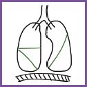
Lung anatomy
Trachea cross section
Bronchus cross section
Bronchiole cross section
Layers of the thorax
Rib anatomy
Venous drainage of the thorax
CHAPTER 3 Upper Gastrointestinal
Layers of the abdomen
Stomach anatomy
Blood supply to the stomach
CHAPTER 4 Lower Gastrointestinal
Large intestine anatomy
Blood supply to the large intestine
CHAPTER 5 Hepatobiliary
Pancreas anatomy
Biliary tree anatomy
Biliary tree and surrounding structures
Anterior view: liver anatomy
Posterior view: liver anatomy
Inferior liver anatomy
Venous drainage of the liver
CHAPTER 6 Neurology
Lobes of brain
The meninges (dura)
Spinal cord cross section
Spinal cord nuclei
Spinal tract overview (template)
Spinothalamic tract
Dorsal column tract
Corticospinal tract
Brain stem (with cranial nerves)
Basal ganglia
Lateral view: ventricular system
Coronal view: ventricular system
Arterial supply of the brain (circle of Willis)
Venous drainage of the head
Blood supply to brain: lateral view
Blood supply to brain: sagittal view
Brachial plexus
CHAPTER 7 Genitourinary
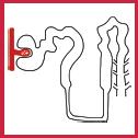
Kidney
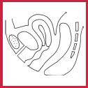
Male reproductive system
Cross section of the penis
Female reproductive system
Surface anatomy of the vulva
Female pelvic peritoneum: cross section
Bladder anatomy
CHAPTER 8 Musculoskeletal
Bones of the foot
Bones of the hand
Carpal tunnel
Knee anatomy
Hip joint
Hip anatomy
Shoulder anatomy
Elbow anatomy
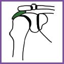
Scapula anatomy
Muscles and ligaments of the scapula
Spine and vertebra anatomy
CHAPTER 9 Ophthalmology
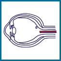
Cross section of the eye
Eye muscle movements
CHAPTER 10 Miscellaneous
Thyroid gland
Transverse section of the anterior neck
Femoral triangle
Anterior cubital fossa
Blood supply to the leg
Venous drainage of the leg
Breast anatomy
Surface areas of the abdomen
Surgical scars
CHAPTER 11 Respiratory Histology
Trachea histology
CHAPTER 12 Gastrointestinal Tract Histology
Gastrointestinal tract histology overview
Stomach histology
Duodenum histology
Jejunum histology
Ileum histology
Colon histology
CHAPTER 13 Hepatic Histology
Liver histology
Index
CHAPTER 14 Renal Histology
Loop of Henle
Adrenal gland histology
CHAPTER 15 Musculoskeletal Histology
Muscle filament layers
Muscle histology
CHAPTER 16 Skin Histology
Hair follicle histology
Thick skin histology
Pacinian and Meissner’s corpuscles histology
CHAPTER 17 Ophthalmic Histology
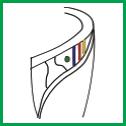
Layers of the retina
CHAPTER 18 Miscellaneous Histology
Thyroid gland histology
Lymph node histology
Artery histology
Vein histology
SENIOR REVIEWERS
Mr Joseph P Borucki MBBS BSc (Hons) PGCert (MedEd)
MRCS
General Surgical Registrar
James Paget University Hospital
Dr Taha Haq MBChB Dist BSc (Hons)
Radiology Registrar
Imperial College Healthcare NHS Trust
Captain Jake Melhuish MBBS DipMCC
Core Anaesthetics Trainee
Portsmouth Queen Alexander
Hospital/British Army
JUNIOR REVIEWERS
Rahul Anil Bhagwat iBSc (Hons)
Year 4 Medical Student
University College London
Georgia Brown
Year 3 Medical Student
University of Newcastle (Australia)
Max Butler
Year 3 Medical Student
University of Cambridge
Kaylise Faull
Year 4 Medical Student
University of Newcastle (Australia)
Simon Hickey
Year 3 Medical Student
University of Newcastle (Australia)
Akos Marton
Year 3 Medical Student
University of Cambridge
Michael McLucas
Year 4 Medical Student
University of Newcastle (Australia)
Maria Elaine Prayle BSc (Hons)
Year 3 Medical Student
University of Keele
Emily Wales BSc (Hons)
Year 3 Medical Student
Cardiff University
Ben Walters
Year 4 Medical Student
University of Keele
Jack FG Wellington
Year 3 Medical Student
Cardiff University
Jessica Xie BSc (Hons)
Year 4 Medical Student
University College London
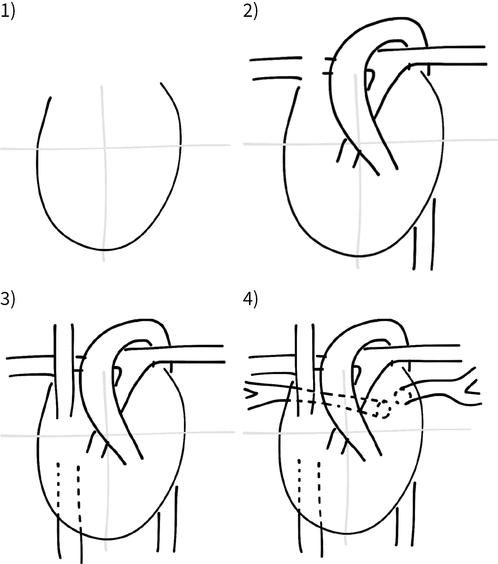
Learn How To Draw, Then How To Label!
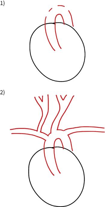
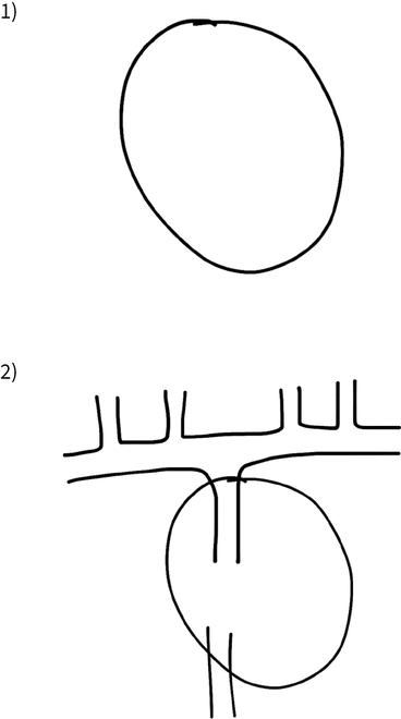
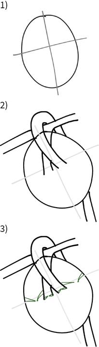
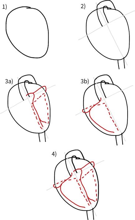
Follow the diagrams in number order. Each consecutive diagram has additional features to add on. Once complete, use the larger drawing (diagram usually on the next page) to learn structures and diagram labelling. Finally, test yourself and your friends!
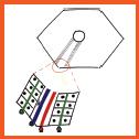
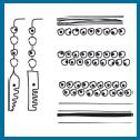
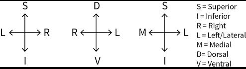
How To … Orientate
Crosshatches (or compasses) are useful for determining your orientation. Whenever drawing, always remember to include one! It will help you determine your orientation for labelling—particularly in exams. It allows you to orientate the diagram in two dimensions. Note below how the second crosshatch shows ‘L’ (left) to be on the right side and ‘R’ (right) to be on the left side (as you look at it). Just as you would interpret a chest radiograph depending whether you are looking at the image from the front (anterior to posterior) or back (posterior to anterior), left or right may be on one side or the other.
Lines Key
CHAPTER ONE CARDIOVASCULAR
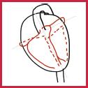
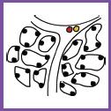
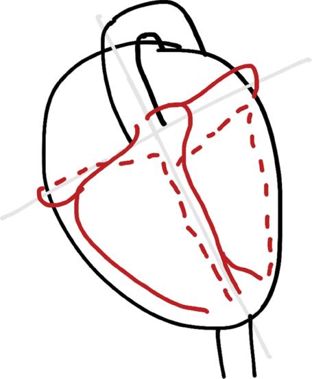
HOW TO … DRAW THE MAJOR CARDIAC VESSELS
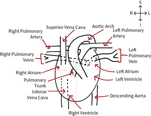
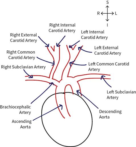
THE MAJOR CARDIAC VESSELS
HOT TIPS
NB: The Aorta runs posterior to the heart (not fully shown in this image to give greater clarity).
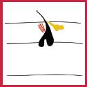
HOW TO … DRAW THE 1ST BRANCHES OF THE AOR
THE 1ST BRANCHES OF THE AORTA
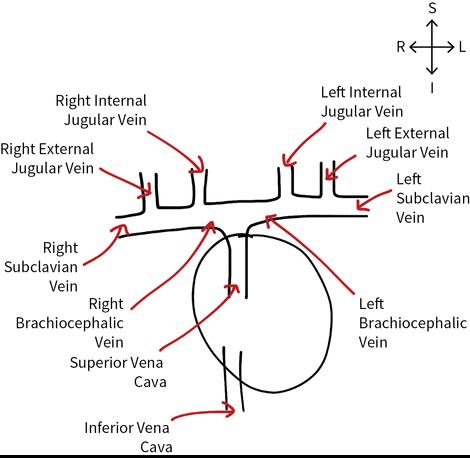
HOW TO … DRAW THE GREAT VEINS
THE GREAT VEINS
HOW TO … DRAW THE CARDIAC VALVES
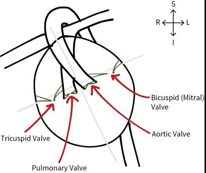
THE CARDIAC VALVES
HOT TIPS Valve structure
Tricuspid has three leaflets.
Bicuspid has two leaflets (‘Mitral’ is derived from mitre—a bishop’s two-pointed hat).
HOW TO … DRAW THE CORONARY ARTERIES
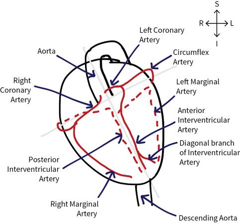
THE CORONARY ARTERIES (ANTERIOR VIEW)
HOT TIPS
NB: the Anterior Interventricular Artery is also known as the Left Anterior Descending (LAD).