C&T
Centre for Veterinary Education
Control & Therapy Series
Issue 324 | September 2026
Major Winner Mucor sp. Isolation in a Cat with Inflammatory Airway Disease Tanya Rajkumar
5
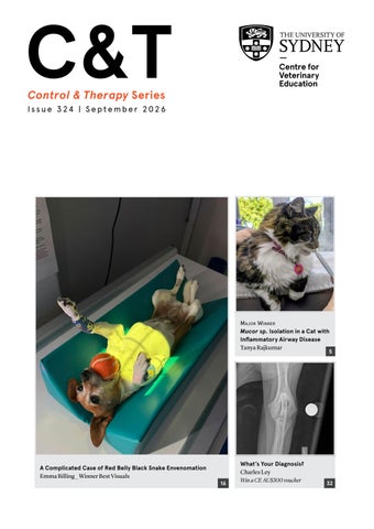
A Complicated Case of Red Belly Black Snake Envenomation Emma Billing _ Winner Best Visuals
16
What’s Your Diagnosis? Charles Ley Win a CE AU$300 voucher
32
ADVERTISEMENT
COMING SOON
PROTECTION
ADVERTISEMENT
TO THE POWER OF
Canine leptospirosis protection is about to evolve.
Are you ready for broader protection? For more information contact your Boehringer Ingelheim Territory Manager
Four serogroups. One powerful vaccine.
Protech® Lepto 4 See product label for full claim details and directions for use. Boehringer Ingelheim Animal Health Australia Pty. Ltd. Level 1, 78 Waterloo Road, North Ryde NSW 2113. ABN 53 071 187 285. ® PROTECH is a registered trademark of the Boehringer Ingelheim Group. All rights reserved. AU-PET-0276-2026
C&T Issue 324 | September 2026
Control & Therapy Series ISSN: 2207-4368 PUBLISHER Centre for Veterinary Education Veterinary Science Conference Centre Regimental Drive The University of Sydney NSW 2006 + 61 2 9351 7979 cve.marketing@sydney.edu.au cve.edu.au Print Post Approval No. 10005007
Engage With Your Profession .................................... 2 Director's Editorial ................................................... 2 The T G Hungerford Award........................................... 3 REWARDING LOYAL CVE MEMBERS Applications OPEN for the CVE CE Scholarship......... 48
Small Animal
CVE Director Associate Professor Kate Patterson kate.patterson@sydney.edu.au
Mucor sp. Isolation in a Cat with Inflammatory Airway Disease Tanya Rajkumar............................... 5
EDITOR Lis Churchward elisabeth.churchward@sydney.edu.au
Focal Pyloroduodenal Junction Carcinoma in a Dog with Minimal Ultrasonographic Abnormalities but Subtle Endoscopic Obstruction Elizabeth Thrift ........................................................ 8
VETERINARY EDITOR Dr Richard Malik VETERINARY SUB-EDITOR Dr Jo Krockenberger joanne.krockenberger@sydney.edu.au DESIGNER Samin Mirgheshmi ADVERTISING Lis Churchward elisabeth.churchward@sydney.edu.au To integrate your brand with C&T in print and digital and to discuss new business opportunities, please contact: MARKETING & SALES MANAGER Ines Borovic ines.borovic@sydney.edu.au DISCLAIMER All content made available in the Control & Therapy (including articles and videos) may be used by readers (You or Your) for educational purposes only. Knowledge and best practice in this field are constantly changing. As new research and experience broadens our knowledge, changes in practice, treatment and drug therapy may become necessary or appropriate. You are advised to check the most current information provided (1) on procedures featured or (2) by the manufacturer of each product to be administered, to verify the recommended dose or formula, the method and duration of administration, and contraindications. To the extent permitted by law You acknowledge and agree that: I. Except for any non-excludable obligations, We give no warranty (express or implied) or guarantee that the content is current, or fit for any use whatsoever. All such information, services and materials are provided ‘as is’ and ‘as available’ without warranty of any kind. II. All conditions, warranties, guarantees, rights, remedies, liabilities or other terms that may be implied or conferred by statute, custom or the general law that impose any liability or obligation on the University (We) in relation to the educational services We provide to You are expressly excluded; and III. We have no liability to You or anyone else (including in negligence) for any type of loss, however incurred, in connection with Your use or reliance on the content, including (without limitation) loss of profits, loss of revenue, loss of goodwill, loss of customers, loss of or damage to reputation, loss of capital, downtime costs, loss under or in relation to any other contract, loss of data, loss of use of data or any direct, indirect, economic, special or consequential loss, harm, damage, cost or expense (including legal fees).
Modified Labial Button Technique Post Temporomandibular Joint Luxation in the Kitten Tanni Sima................................................... 12 A Complicated Case of Red Belly Black Snake Envenomation Emma Billing...................................16 Comment 1 courtesy of Ellie Leister Comment 2 courtesy of Rob Webster Diet & The Canine Faecal Microbiome Louise Campbell, Mary Thompson & Andrew Holmes....................................................... 22 Feline Hypertension: What’s New? Samantha Taylor...................................................... 26 Cat Friendly Blood Pressure Monitoring Sarah Collins........................................................... 30 What’s Your Diagnosis? Charles Ley....................... 32 Periodontal Disease & The Cybernetic Hypothesis Tom Lonsdale...................................... 33
Large Understanding Milking Machines, Mastitis, & Milking Machine-Associated Risk Factors Ian Hodge .............................................................. 38 Snails, Seasons & Sensible Control: A Practical Liver Fluke Update for Vets Nichola Eliza Davies Calvani & Priscilla Huynh........... 42
Replies & Comments Inducing Emesis in Cats Anne Quain...................... 46
Major Winner
Winner
Best Visuals
Engage With Your Profession The Control & Therapy Series was established in 1969 by Director Dr Tom Hungerford. His aim was to publish uncensored and unedited material contributed by vets writing about:
“
...not what he/she should have done, BUT WHAT HE/ SHE DID, right or wrong, the full details, revealing the actual “blood and dung and guts” of real practice as it happened, when tired, at night, in the rain in the paddock, poor lighting, no other vet to help.
The C&T forum gives a ‘voice’ to the profession and everyone interested in animal welfare. You don’t have to be a CVE Member to contribute an article or reply to a 'What's YOUR Diagnosis?'. We welcome contributions from Vets, Techs, Nurses, allied professionals—Non CVE Members included.
Submit your C&T article A template and information about uploading high resolution images for print can be found here cve.edu.au/submit-article.
Questions? Please contact cve.marketing@sydney.edu.au.
Join In! The C&T is not a peer-reviewed journal. Rather, it is a unique forum allowing veterinary professionals to share their cases and experiences with their colleagues. We are keen on publishing short, pithy, practical articles (a simple paragraph is fine) that our members/readers can immediately relate to and utilise. Our editors will assist with English and grammar if required.
“
I enjoy reading the C&T more than any other veterinary publication. -Terry King, Veterinary Specialist Services, QLD
Thank You to All Contributors & Advertisers The C&T Series thrives due to your generosity.
Major Winner Prize: A CVE$500 voucher —Tanya Rajkumar
page 5
Winners Prize: A CVE$300 voucher —Tanni Sima
page 12
Best Visuals
Prize: A CVE$300 voucher
—Emma Billing
There are few things more determined than a dog with a tennis ball and, apparently, not even a radiograph will get in the way. The cover photo of Dolly is a lovely reminder that, alongside the seriousness of disease and complexity of clinical care, our patients bring moments of joy into practice. The article on snake bites in this edition, together with the supporting clinical resources, is packed with practical, timely knowledge and reminds us to remain vigilant, regardless of an early response to treatment. It is my honour to acknowledge an outstanding educator in this issue; Dr Jane Day, who we recently announced as a recipient of the prestigious CVE T G Hungerford Award. Writing her citation was remarkably difficult—not because there was too little to say, but because of the number of contributions describing her mentorship, generosity and impact. Jane’s contribution to veterinary education across the entire spectrum of lifelong learning is particularly impressive: a rare achievement and one she is thoroughly deserving of recognition for. I’m also delighted to announce that applications for the AU$8,000 CVE Continuing Education scholarship opened on 1 September—see page 48. Finally, I encourage the whole veterinary team to see C&T as a place to share what you know. Early findings from a University of Sydney DVM3 student-led readership review show that each issue is being read and shared across entire practice teams. I want to hear from more of you, not just veterinarians, but nurses, receptionists, technicians, hospital managers and everyone involved in patient care. Share your knowledge, experiences and practical discoveries. Others in our profession can learn from them.
page 16
Associate Professor Kate Patterson Director Page 2
The T G HUNGERFORD AWARD 2026 CITATION
Dr Jane Day
Follow the Goanna Track to Success...
BVSc MVS FANZCVS (Radiology) The T G Hungerford Award recognises individuals who have made an outstanding contribution to continuing veterinary education. It celebrates those whose commitment to professional learning strengthens the veterinary profession, advances standards of practice, and leaves a lasting legacy through the veterinarians they teach, mentor and inspire.
“
So splendid is their performance; they adorn the awards for excellence now conferred upon them. _Tom Hungerford
cve.edu.au/Jane_Day_Award
Dr Jane Day Exemplifies These Ideals Over a distinguished career as a specialist veterinary radiologist, educator, mentor and innovator, Jane has devoted herself to helping others learn. Her contribution spans the full spectrum of veterinary education, from continuing professional development for practising veterinarians and the training of specialist radiologists, to undergraduate teaching, curriculum design and educational leadership. Few individuals have influenced veterinary diagnostic imaging education so broadly, so deeply, or so enduringly. At the heart of Jane's contribution is her commitment to lifelong learning. For many years she has delivered dynamic and innovative diagnostic imaging and ultrasound education to practising veterinarians, helping clinicians build confidence, sharpen clinical reasoning and strengthen diagnostic decision-making. Her contribution to the continuing professional development of Australian and international veterinarians has been enormous. She has also developed, empowered and supported Membership-level radiologists, not only as specialists but as educators, creating an impact that extends far beyond her own teaching. Colleagues repeatedly describe Jane's determination to ensure that every learner develops genuine diagnostic imaging skills and reasoning. Her commitment to education is not measured simply by teaching hours or programs developed, but by her willingness to invest personally in the success of others. She makes herself available to learners, offers encouragement generously, and approaches education with an uncommon level of care and attention. Centre for Veterinary Education
|
Est. 1965
Jane is widely recognised as a gifted educator. Those who have learned alongside her or from her speak not only of her expertise, but of her warmth, patience and generosity. She possesses a rare ability to take the genuine complexity of diagnostic imaging and make it clear, clinically meaningful and engaging. She creates learning environments where people feel comfortable asking questions, where curiosity is encouraged, and where confidence grows. She never loses sight of the learner's perspective and understands that how students experience learning is often just as important as what they learn.
This educational philosophy found its fullest expression through Animal Imaging As founder and director of Animal Imaging, Jane has transformed access to high-quality diagnostic imaging education across Australia, New Zealand and, more recently, the United Kingdom. Her leadership has reshaped how diagnostic imaging is taught, creating benefits that will be felt for many years to come. Built on educational scholarship, clinical authenticity and a deep understanding of how people learn, the program now supports veterinary schools across Australasia and has become a benchmark for innovative veterinary education. Jane has gone above and beyond to develop a robust curriculum that combines foundational knowledge with clinically authentic cases. She has sought expert educational advice, embraced modern teaching methods, and continually refined learning resources, assessment processes and educational experiences. Videos,
Celebrating supporting the profession for over 60 years
Page 3
annotations, case-based learning activities and carefully structured frameworks have transformed concepts that once seemed confusing into learning experiences that are accessible, engaging and effective. What distinguishes Jane is her refusal to stand still. Even when programs are already excellent, she continues to refine and improve them, driven by a genuine desire to create the best possible learning experience. Her responsiveness to student and partner feedback, collaborative approach and willingness to listen have made her a trusted educational partner across multiple institutions. The impact of this work is best reflected in the voices of students. They describe the Animal Imaging platform as phenomenal, incredible, organised, detailed and invaluable. They speak of learning a systematic approach to image interpretation that has boosted their confidence and competence. They value the extensive case material, the clarity of the video resources, the practical relevance of the content and the opportunity to practise repeatedly until skills become second nature. Many describe finishing their training feeling more confident, more capable and better prepared for clinical practice. Beyond the classroom, Jane's influence can be seen in strengthened university programs, in the practice readiness of new graduates, in the specialists and educators she has mentored, and in the many veterinarians she has inspired to teach others. Diagnostic imaging's growing accessibility and popularity within veterinary education is, in no small part, a testament to her vision, hard work and dedication. Across all reflections on her career, certain themes emerge consistently: excellence, humility, innovation, mentorship, generosity and service. Despite extraordinary achievements, Jane remains deeply committed to helping others succeed. She continues to approach each new challenge with energy, creativity and an unwavering focus on learner outcomes. Her influence and the value of her contributions are immeasurable and will be long-lasting. Through continuing professional development, specialist mentoring, educational innovation and undergraduate teaching, Dr Jane Day has transformed veterinary diagnostic imaging education and strengthened the veterinary profession. The thousands of veterinarians, students and educators whose careers she has shaped stand as a testament to her extraordinary contribution. Jane, congratulations on receiving the T G Hungerford Award. Thank you for your tireless service to veterinary education, for your generosity in sharing your knowledge, and for the lasting difference you have made to our profession, its practitioners, and the animals entrusted to their care. Page 4
Dr Jane Day in action at the September 2025 Case-based Diagnostic Imaging Conference held in Melbourne: Engaging with a masterclass participant, and sharing the stage with Drs Mary Thompson & Claire Sharp
Control & Therapy Series – Issue 324 September 2026 Authors’ views are not necessarily those of the CVE
MAJOR Winner The prize is a CVE$500 voucher
Small Animal
Mucor sp. Isolation in a Cat with Inflammatory Airway Disease Tanya Rajkumar BVSc
alongside numerous branching fungal hyphae. No bacteria were identified. Fungal culture yielded a pure growth of Mucor sp. with no evidence of oropharyngeal contamination and absence of bacterial co-isolates, supporting a true lower airway sample. The PCR respiratory panel was negative. Despite confirmed fungal presence the patient lacked clinical and radiographic evidence of invasive pulmonary disease. This is significant as true mucormycosis in cats is typically acute, rapidly progressive, and often fatal. In contrast, Astrid remained stable with minimal clinical signs.
Interpretation:
Dural Veterinary Hospital
The findings are most consistent with mucus-associated fungal colonisation ('quasi-fungal ball') secondary to underlying inflammatory airway disease, rather than invasive fungal pneumonia.
687 Old Northern Rd Dural NSW 2158 e. duralvet@duralvet.com.au C&T No. 6140 Tanya graduated from the University of Sydney in 2011 and has since worked in a variety of small animal practices across Sydney. She enjoys all aspects of companion animal medicine and surgery with particular interests in dentistry, soft tissue surgery, fear free medicine and palliative pet care. Outside of work her three cats keep her on her toes, not to mention Astrid—the veterinary nurse’s cat featured in this unusual fungal case report—who has certainly kept everyone guessing! Astrid, a 6-year-old female neutered domestic medium haired cat, presented with a 12-month history of mild, intermittent coughing (approximately 1–2 episodes per month). She lives mostly indoors, only occasionally venturing into the yard. She was otherwise clinically well with a normal physical exam. All testing including routine bloodwork, FIV/FeLV testing and thoracic radiographs were unremarkable.
The presence of increased eosinophils and mucus accumulation is suggestive of feline asthma or inflammatory airway disease. In this context, Mucor spp.—a ubiquitous saprophytic organism—has likely proliferated after germination of inhaled spores from some environmental source and is living within the abnormal mucus environment without causing tissue invasion. This fungal organism may also be contributing to the observed cytological changes. Mucor species (mucormycetes) are found in soil and decaying matter. Although they do not usually cause pathology in immunocompetent cats, they can in rare instances be associated with severe lifethreatening disease in immunocompromised individuals (mucormycosis).¹
During general anaesthesia for a routine dental procedure the patient repeatedly developed almost complete occlusion of the endotracheal tube with thick yellow mucoid material. Bronchoalveolar lavage was not performed due to inability to maintain a patent ET tube. Instead, a sample of mucus collected from the distal end of the tube was submitted for cytology, culture and respiratory PCR. Cytology revealed moderate mixed inflammatory cell infiltrate including neutrophils, eosinophils, macrophages (some reactive) and lymphocytes,
Centre for Veterinary Education
|
Est. 1965
Figure 1. Astrid enjoying the yard
Infections can be acquired through inhalation or ingestion of spores, or enter via skin wounds.2 Several sites of infection have been reported in cats including gastrointestinal, tracheobronchial, subcutaneous nasal lesion, cerebral and disseminated disease.3,4,5,6,7
Celebrating supporting the profession for over 60 years
Page 5
Management Treatment was directed primarily at the underlying airway disease. The aim of this was to reduce inflammation and improve mucus clearance making conditions less suitable for Mucor proliferation. 1. Inhaled therapy: salbutamol (bronchodilator) followed by fluticasone (inhaled corticosteroid) administered via a meter dose inhaler and spacer twice daily. 2. Nebulisation: saline nebulisation for 5–10 minutes, 2–3 times daily to aid mucus clearance.
Figure 2. Low power photomicrograph of the cytology from the respiratory mucus plug, showing mixed inflammatory cells and branching fungal septate hyphal elements
Figure 3. High power photomicrograph of the mucus plug cytology revealing eosinophils, neutrophils, macrophages and branching fungal hyphae later confirmed on culture to be Mucor sp.
3. Gentle thoracic coupage (chest thumping!): following nebulisation to loosen secretions and facilitate expectoration by induced coughing. A cautious adjunctive antifungal approach was elected using low-dose intermittent posaconazole whilst awaiting susceptibility testing. In this instance, 25mg posaconazole (1/4 of a sustained release tablet) was administered orally 4 times per week. Systemic corticosteroids were intentionally avoided due to concerns regarding fungal proliferation. Fungal susceptibility testing demonstrated low minimum inhibitory concentrations (MIC) to amphotericin B (0.12 µg/mL), posaconazole (0.25 µg/mL) and itraconazole (0.25 µg/mL), suggesting favourable in vitro activity against the isolate. Although interpretive veterinary breakpoints are unavailable for this species, reference laboratory commentary and published literature indicate amphotericin B and posaconazole as the most likely effective agents.⁸ These findings supported the current treatment choice of posaconazole in this case. One could equally make a case for nebulisation with saline and amphotericin B deoxycholate.
Figure 4. Astrid being trained when first starting saline nebulisation treatment. Note the use of a creamy treat for encouragement and distraction, and reward.
In Astrid’s case, mucormycetes presented as a non-invasive overgrowth in the airways of an immunocompetent patient.
Page 6
Figure 5. Medications used in Astrid’s treatment plan including posaconazole tablets,a salbutamol inhaler, fluticasone inhaler, spacer with paediatric mask attachment and a portable nebuliser with paediatric mask
Control & Therapy Series – Issue 324 September 2026 Authors’ views are not necessarily those of the CVE
Discussion
Conclusion
This case highlights an unusual but clinically important scenario in which fungal organisms are genuinely present within the lower airways yet are not acting as primary pathogens. Instead, they represent opportunistic colonisers of mucus in a patient with underlying inflammatory airway disease.
Astrid’s case highlights the variety of presentations of inflammatory airway disease in practice.
Key clinical lessons include – Isolation of fungal organisms from respiratory samples should be interpreted in the context of clinical presentation, cytological findings and imaging. Cytological evidence of inflammation with fungi does not necessarily indicate tissue invasion. Chest radiographs are vital to aid interpretation! – Reminder that normal thoracic radiographs do not rule out underlying inflammatory airway disease. – Invasive Mucor spp. infection in cats is typically acute, severe and rapidly progressive. However, this case highlights the possibility of non-invasive disease in some cats causing only mild signs. – Whilst Mucor spp. more commonly cause opportunistic infections in cats with immune compromise, proliferation can also occur in otherwise healthy individuals.⁴ Isolation in these cases requires careful consideration to determine its clinical significance. – In cases with concurrent signs of feline inflammatory airway disease, management should prioritise control of airway inflammation and mucus clearance. Inhaled therapies and airway clearance techniques are effective, systemic steroid-sparing strategies. – The decision to institute antifungal therapy should be made on a case-by-case basis and, if administered, guided by sensitivity testing.
In cats with inflammatory airway disease, abnormal mucus production may create an environment conducive to opportunistic fungal colonisation, including with rare organisms such as Mucor spp. Differentiating between contamination, colonisation and invasive infection is critical in determining suitable management. Culture results must always be interpreted in light of patient clinical signs and cytological findings. Targeting the primary airway disorder is critical, with antifungal therapy in this case considered adjunctive rather than central given the absence of invasive disease. References 1. Hernandez JL, Buckley CJ. 2023. Mucormycosis. StatPearls Publishing, Treasure Island FL. 2. Grooters AM. 2010. Opportunistic mycoses in dogs and cats. ACVIM Forum Proceedings. 3. Cunha SC, Aguero C, Damico CB, et al. Duodenal perforation caused by Rhizomucor species in a cat. J Feline Med Surg 2011; 13 (3): 205-07. 4. Snyder KD, Spaulding K, Edwards J. Imaging diagnosistracheobronchial zygomycosis in a cat. Vet Radiol Ultrasound 2010; 51 (6): 617-20. 5. Wray JD, Sparkes AH, Johnson EM. Infection of the subcutis of the nose in a cat caused by Mucor species: successful treatment using posaconazole. J Feline Med Surg 2008; 10 (5): 523-27. 6. Ravisse P, Fromentin H, Destombes P, et al. Cerebral mucormycosis in the cat caused by Mucor pusillus. Sabouraudia 1978; 16 (4): 29198. 7. Ossent P. Systemic aspergillosis and mucormycosis in 23 cats. Vet Rec 1987; 120 (14): 330-33. 8. Barrs VR, Hobi S, Wong A, et al. Invasive fungal infections and oomycoses in cats 2. Antifungal therapy. J Feline Med Surg 2024; 26 (1):1098612X231220047.
Let’s keep the C&T Series broad and interesting!
Birds, fish, guinea pigs, rabbits, reptiles, snakes, exotics, wildlife, cats, dogs, horses, goats, sheep and pigs. Please write up that interesting case and send it to us for publication. We promise, it’s not an arduous process. Rest assured our veterinary editors will contact you before publication if they have any concerns or queries. Join in! Send your article complete with high resolution images, videos if you have them, figures and tables etc to: cve.marketing@sydney.edu.au and read your contribution in PRINT and digital.
Centre for Veterinary Education
|
Est. 1965
Celebrating supporting the profession for over 60 years
Page 7
Focal Pyloroduodenal Junction Carcinoma in a Dog with Minimal Ultrasonographic Abnormalities but Subtle Endoscopic Obstruction Elizabeth Thrift
shortly after eating. Antiemetic therapy (ondansetron and maropitant) resulted in temporary improvement, but clinical signs recurred following cessation. CBC/ biochemistry/CPL and CRP blood testing did not reveal any clinically significant abnormalities. The dog had become increasingly lethargic, with markedly reduced appetite and reduced faecal output. A history of intermittent gastrointestinal sensitivity was reported but no previous diagnosis of chronic enteropathy had been established.
Physical Examination Pertinent findings on presentation were that the body condition was reduced and cardiothoracic auscultation revealed a grade 3–4/6 left-sided systolic murmur.
BVSc (Hons) FANZCVS (Small Animal Internal Medicine)
Diagnostic Imaging
The Pet Specialists
Abdominal Ultrasonography
16 Myoora Road, Terrey Hills NSW 2084
Abdominal ultrasonography had been performed at the referring veterinary clinic and again at the referral centre. The stomach contained fluid and echogenic material but was not persistently or severely distended, with rugal folds preserved. Gastric wall thickness and layering were within normal limits. No discrete gastric or pyloric mass was identified. At the referral centre, the pyloroduodenal region was subjectively hyperechoic and mildly thickened but without loss of intestinal layering or clear evidence of obstructive disease—importantly these findings were subtle and only noted once the images were reviewed after the endoscopy (see later). The duodenal mucosal margin appeared hyperechoic, with preserved wall layering and normal overall thickness. A mildly enlarged gastric lymph node with normal echogenicity was noted. No free abdominal fluid was identified. Normal emptying from the antrum/pylorus into the duodenum was noted.
e. info@tps.vet e. elizabeth.thrift@tps.vet t. 02 9452 2933 C&T No. 6141
Abstract An 11-year-old neutered male Cocker Spaniel was referred for investigation of chronic vomiting, progressive inappetence and weight loss. Abdominal ultrasonography performed at both the referring practice and a specialist referral centre revealed only subtle abnormalities, with no definitive evidence of gastrointestinal obstruction or mass lesion. Upper gastrointestinal endoscopy identified diffuse gastroduodenal inflammatory changes and subtle pyloroduodenal changes when assessed from the gastric lumen. However, marked resistance and distortion were encountered when traversing the pyloroduodenal junction, with abnormal proliferative tissue only really appreciated during attempted intubation. Histopathology of targeted biopsies confirmed a focal infiltrative carcinoma of the pyloroduodenal junction. This case highlights the diagnostic limitations of ultrasonography in focal pyloroduodenal disease and emphasises the importance of endoscopic tactile feedback and operator experience when evaluating subtle outflow obstruction.
Case Description Signalment & History An 11-year-old neutered male Cocker Spaniel was referred for further investigation of a 3–4-week history of persistent vomiting, progressive hyporexia and weight loss of approximately 3 kg. Vomiting was described as true vomiting rather than regurgitation and occurred
Page 8
Overall, ultrasonographic findings were interpreted as consistent with inflammatory gastrointestinal disease, with neoplasia considered less likely due to the absence of a focal mass, significant wall thickening or layering disruption.
Endoscopic Findings Upper gastrointestinal endoscopy was performed under general anaesthesia for further evaluation at the referral centre. Diffuse likely inflammatory changes were observed throughout the upper gastrointestinal tract, including mild distal oesophagitis, moderate diffuse gastritis, mild superficial gastric erosions and duodenitis. From the gastric aspect, the pylorus was mildly abnormal with some thickening and irregularity. There was initially a small piece of grass sitting within the pyloroduodenal junction (PDJ).
Control & Therapy Series – Issue 324 September 2026 Authors’ views are not necessarily those of the CVE
Figure 1. Gastroscopy: Mildly thickened and irregular PDJ with surrounding superficial multifocal gastric erosions visualised Figure 2. Gastroscopy: PDJ appeared quite proliferative during insufflation of this area to try and facilitate intubation with the endoscope, resulting in an increasingly abnormal PDJ in appearance Figure 3. Gastroscopy: Increased friability causing haemorrhage unusual for this procedure. The mucosal margin of the PDJ also appeared irregular when the opening was insufflated. Figure 4. Duodenoscopy with diffusely abnormal mucosal surface
1
3
2
4 However, significant resistance was encountered when attempting to pass a standard gastroscope through the pyloroduodenal junction. Despite appearing patent on initial inspection, the pylorus proved extremely difficult to traverse. Passage was only possible using a smaller diameter (feline) gastroscope and required marked, atypical manipulation, including scope inversion to achieve intubation. (i.e. I had to turn the scope 180° to get it through. The handle was ‘upside down’ in orientation to the dog’s mouth in relation to how it would normally sit to get it through.) During this process, the pyloroduodenal junction was appreciated to be abnormally thickened, firm and proliferative, with reduced luminal diameter and increased friability (causing increased bleeding associated with the procedure). The abnormality was not overtly obvious as a discrete lesion but was primarily recognised through impaired scope passage rather than visual appearance alone. However, the pyloroduodenal mucosa did appear irregular and thickened but without a focal intraluminal mass. During the endoscopic examination, we requested that the ultrasound imaging of the PDJ be reviewed as the endoscopic findings were significantly different from the
Centre for Veterinary Education
|
Est. 1965
Celebrating supporting the profession for over 60 years
Page 9
initial ultrasound findings. It was reported that there was ‘maybe’ some subtle hyperechoic mucosal change on the PDJ, but still not to the degree that was appreciated endoscopically. Multiple mucosal punch biopsies were obtained from the stomach, pyloroduodenal junction and duodenum.
Histopathology Histopathologic examination of biopsies from the pyloroduodenal junction revealed a focally infiltrative carcinoma arising from glandular epithelium. Neoplastic epithelial cells infiltrated the deeper lamina propria, separating and distorting residual glands, with moderate pleomorphism and a high mitotic count. Biopsies from the stomach and duodenum demonstrated only mild lymphoplasmacytic gastritis and enteritis, respectively, with no evidence of neoplasia at these sites.
Figure. 5. Gastric lymph node on ultrasound
Outcome & Management A diagnosis of focal carcinoma of the pyloroduodenal junction was made. Given the infiltrative nature of the lesion and its anatomically challenging location, options including referral for oncologic consultation, surgical exploration and palliative management were discussed with the owners. Supportive medical management was continued in the interim.
Discussion This case illustrates a diagnostically challenging presentation of pyloroduodenal junction carcinoma, characterised by minimal ultrasonographic abnormalities and only subtle changes on initial endoscopic visual inspection. Despite repeated ultrasonography by experienced operators, findings were limited to mild pyloroduodenal hyperechogenicity and minimal lymphadenopathy, with no convincing evidence of a focal obstructive lesion.
Figure 6. Stomach containing some fluid and ingesta on ultrasound
In contrast, the most clinically significant finding was not what was seen, but what was felt during endoscopy. While the pylorus appeared only mildly irregular when assessed from the gastric lumen initially, attempted traversal of the pyloroduodenal junction revealed marked and unexpected resistance. The degree of difficulty encountered was clearly disproportionate to the visual appearance (although the visual appearance did become more severe through the endoscopy), with passage requiring downsizing of equipment and atypical manoeuvres, including scope inversion. The tissue itself was perceived as firm, thickened and unyielding, with reduced compliance and increased friability, features that became apparent primarily through tactile interaction rather than visual identification of a discrete lesion. Figure 7.: Duodenum on ultrasound Page 10
Control & Therapy Series – Issue 324 September 2026 Authors’ views are not necessarily those of the CVE
This discordance between visual appearance and tactile feedback highlights an important limitation of both ultrasonography and purely visual endoscopic assessment in focal infiltrative disease. In this case, the lesion did not present as a mass or cause obvious architectural disruption but instead altered the mechanical properties of the pyloroduodenal junction. As such, the abnormality was functionally significant before it became overtly visible. Recognition of this pattern relies heavily on operator experience and an understanding of what normal feels like during scope passage. Subtle increases in resistance, loss of expected compliance or the need for non-standard techniques to achieve intubation should not be dismissed, even in the absence of striking visual abnormalities. These findings represent important diagnostic cues and—as demonstrated here—may be the primary indicator of clinically significant disease. Focal infiltrative lesions of the pyloroduodenal junction may therefore be under-recognised on imaging and underestimated on visual inspection alone. This case emphasises that endoscopic evaluation is not solely a visual modality; it is also a tactile one. The ‘feel’ of the pylorus (its compliance, distensibility and response to instrumentation) can provide critical diagnostic information and should be consciously integrated into clinical decision-making.
Key Clinical Message Subtle or focal pyloroduodenal neoplasia may not be readily identified on ultrasonography or by gross endoscopic appearance alone. Difficulty traversing the pyloroduodenal junction, particularly when disproportionate to visual findings, should prompt a high index of suspicion for infiltrative disease and targeted biopsy, even when imaging findings are minimal.
Surface to Stewardship Conference Dermatology, Diagnostics & Smarter Antimicrobial Decisions in Practice 22 - 24 February 2027 | 18 CPD Points Art Gallery of NSW Early Bird rates end 10 January 2027
From Surface to Stewardship The skin may be where the problem starts — but the clinical decisions go much deeper. Skin, ears, wounds and dental disease are everyday presentations in general practice, but working out what is really going on - and what to do about it - is rarely straightforward. From Surface to Stewardship is a three-day deep dive into practical dermatology, diagnostics and antimicrobial stewardship, designed around the cases you see in practice. Across the conference, expert clinicians will take you beyond recognising dermatological patterns to the decisions that matter: Is this local or systemic? What should I sample? Do I need cytology or culture? How do I interpret the results? And what treatment makes sense for this patient, this owner and this situation? Presented in collaboration with the AMR Vet Collective, the program brings these decisions together rather than treating them as separate topics. You’ll explore common and challenging dermatology presentations including atopy, pyoderma, otitis, wounds and immune-mediated skin disease, while following the diagnostic and treatment questions into the clinical situations where they intersect with antimicrobial use - from dental disease and common medical cases to ophthalmology and theriogenology. Come for the dermatology. Leave with a more thoughtful approach to the whole clinical decisionmaking pathway - from surface to stewardship.
cve.edu.au/ february-conference-2027
Winner
Entitled to a CVE$300 voucher
dislocated to the right side of the midline making him unable to close his jaw (Figure 1). The rest of the physical exam was normal. As the mandible shifted to the side opposite the luxation, a rostral dorsal unilateral luxation of the left temporomandibular joint (TMJ) was suspected.
Modified Labial Button Technique Post Imaging Temporomandibular Joint Luxation in the Kitten Tanni Sima BVB/DVM Beecroft Vet Shop 2 & 3/5 Wongala Cres Beecroft NSW 2119
The kitten was sedated with butorphanol and medetomidine. Chest, abdominal, and head X-rays were taken. A full oral exam was performed, and the mouth was unable to be closed under sedation. No obvious oral trauma, fracture, or dental disease were observed (Figure 2). Blood tests, other imaging, and specialist referral for advanced imaging and surgical repair were declined due to cost constraints. The rest of the imaging were within normal limits.
t. 02 8914 0828 e. info@beecroftvet.com.au C&T No. 6142
Signalment, History & Clinical Presentation A 6-month-old male neutered domestic short-haired kitten presented for suspicion of being hit by a car.
Physical Exam On physical exam, the kitten’s nose and face were covered in blood with a laceration on the lip. He had an open-mouth appearance, and the lower mandible was
Figure 2. Head X-rays
Close Reduction of Luxated TMJ The following procedure was replicated from the techniques outlined by the referenced papers (Goodman and Carmichael 2016; Jung et al. 2023; Rocha et al. 2013). The kitten was placed in left lateral recumbency. A wooden hexagonal pencil was placed between the maxillary 4th premolar (PM4) and the mandibular 1st molar (M1), holding both the maxilla and mandible in one hand, then gently closing the mouth whilst rolling the pencil anticlockwise slowly. This allowed the mandible to move rostrally so that the condylar process can be relocated back to the mandibular fossa. Figure 1. Dislocated jaw to the right
Page 12
Control & Therapy Series – Issue 324 September 2026 Authors’ views are not necessarily those of the CVE
In this case, after closed reduction, the TMJ was dislocated again within 30 minutes after the sedation wore off. The kitten did not tolerate a tape muzzle. Stabilisation of the TMJ post closed reduction was therefore required. Therefore, modified labial button technique was selected. General anaesthesia was recommended for the procedure and to insert and secure a feeding tube.
Anaesthesia The kitten was admitted the next day, premedicated with methadone and medetomidine and maintained on isoflurane and oxygen via a size 4 ET tube.
Feeding Tube Placement The affected skin was aseptically prepared. The kitten was placed in right lateral recumbency. A 5 mm diameter stab incision was made guided by a curved haemostat over the left neck area away from the jugular vein. A 14fr MILA feeding tube was placed and stopped at 7-8th intercostal space. X-rays were taken to confirmed tube end in the caudal oesophagus. Purse string suture and Chinese finger trap suture were applied to secure the feeding tube with 2/0 PDS. Paddings were applied (Figure 3).
Figure 3. Feeding tube placement
Surgical Technique
|
Est. 1965
The Technique was as Follows 1. The needle and suture first passed from the left hole of the button on the chin, exiting the left side of the lower lip. 2. It then passed through the opening of the mouth, then entering the left upper lips (lateral to both canines). 3. It exited via the lateral hole of the button on the left upper lip, and returned via the medial hole of the button on the left upper lip. 4. It entered the left lower lip 2 mm away from the initial suture entry point, then back out through the same left hole of the buttons placed on the chin. 5. This is then repeated on the right side. This creates a loop that connects the upper and lower jaws. 6. After both sides were completed, the suture was tightened and tied over the chin button, leaving a 5 mm gap between the jaws to allow limited mouth movement while stabilising proper jaw alignment (Figures 4-5).
Figure 4. Ventrodorsal view post button placement
Once the feeding tube was placed, the TMJ luxation was repositioned back to normal (with the same pencil and rolling technique; as described above). The kitten was then placed in dorsal recumbency. Three buttons were cleaned and disinfected with chlorhexidine. The lips and
Centre for Veterinary Education
chin were prepared aseptically. 2/0 PDS suture was used for the procedure. The buttons need to have minimum two holes and should be orientated horizontal to the opening of the mouth.
Post Surgery
The kitten was put on meloxicam and amoxiclav, and kept indoors with an Elizabethan collar on, and the feeding instructions were given to the owner. The kitten was normal and comfortable at 3 days post operation
Celebrating supporting the profession for over 60 years
Page 13
Figure 5. Left lateral view post button placement
recheck (Figure 6), and the suture and feeding tube were removed at 16 days post-surgery. No post-surgery complication or re-luxation was reported.
Discussion Modified labial button technique is a cost-effective way to stabilise TMJ luxation post trauma. The procedure is fast in both the technique and the healing time, and easy to perform in a general practice setting with readily available material. For X-rays of the TMJ joint, sagittal oblique view with 10 to 30 degrees angle can be done on top of the ventrodorsal and dorsoventral views (Dennis et al, 2010). The main concerns for this procedure include infection post-surgery, the owner’s ability to use a feeding tube, and patient tolerance. In this case, the kitten was able to eat wet food by itself and tolerated the buttons and sutures well.
Figure 6. Post surgery
References Dennis, R., Kirberger, R. M., Barr, F., & Wrigley, R. H. (2010). Chapter 4 - Head and neck. In R. Dennis, R. M. Kirberger, F. Barr, & R. H. Wrigley (Eds.), Handbook of Small Animal Radiology and Ultrasound (Second Edition) (pp. 85-113). Edinburgh: W.B. Saunders. Goodman, A. E., & Carmichael, D. T. (2016). Modified Labial Button Technique for Maintaining Occlusion After Caudal Mandibular Fracture/ Temporomandibular Joint Luxation in the Cat. J Vet Dent, 33(1), 47-52. doi:10.1177/0898756416637172 Jung, J. W., Castejón-González, A. C., Stefanovski, D., & Reiter, A. M. (2023). Etiology, clinical presentation, and outcome of temporomandibular joint luxation in cats: 21 cases (2000–2018). Frontiers in Veterinary Science, Volume 10 - 2023. doi:10.3389/fvets.2023.1274193 Rocha, A., Rosa-Ballaben, N., Moraes, P., Filho, J., & Minto, B. (2013). LABIAL LOCKING WITH BUTTONS FOR MANAGING MANDIBULAR FRACTURES IN CATS/Utilização do bloqueio labial com botões na correção das fraturas mandibulares em gatos. Ars Veterinaria, 29. doi:10.15361/2175-0106.2013v29n2p83-87
Alternative methods for TMJ stabilisation include dental fixation with acrylic composite or wiring, which generally require referral and are not recommended for young and growing kittens. Tape muzzles can be done in a general practice setting but are poorly tolerated by cats.
Page 14
Control & Therapy Series – Issue 324 September 2026 Authors’ views are not necessarily those of the CVE
Patient care is important from the moment they come into the clinic to the time they leave. When drawing up a medication, we check the drug, double check the dose and label the syringe with the appropriate sticker to indicate who the drug is for and what is in the syringe. So why would we not want the same safety check with our infusion lines? Our patients rely on us to provide the highest standard of care. In busy veterinary hospitals, where multiple medications may be administered simultaneously, clear identification of infusion lines can help reduce the risk of medication errors and improve workflow efficiency.
Colour-Coded Perfusor™ Tubing Available in BLUE, MAGENTA, and GREEN, colour-coded Perfusor™ tubing allows rapid visual identification of infusion lines, helping veterinary teams quickly associate the correct drug with the correct patient.
y
Low priming volume (≤1.27 mL) to minimise drug waste and enable rapid therapy initiation.
y
Pressure resistance up to 2 bar for reliable use with syringe drivers.
y
Secure Luer-Lock connections compatible with standard syringe driver systems.
y
Low sorbing, high density material with kink-resistant design to support consistent drug delivery.
Safe Materials for Sensitive Patients Perfusor™ Lines are manufactured from polyethylene (PE) and are free from PVC, DEHP and latex, making them suitable for sensitive patients and supporting safer infusion therapy. Available in 150 cm and 200 cm lengths, they are ideal for anaesthesia, critical care and post-operative care where accuracy and reliability are essential.
Why DEHP-Free Matters DEHP (Di(2-ethylhexyl) phthalate) is a plasticiser commonly used in PVC medical devices. Studies have shown that DEHP can leach from tubing during medical procedures, leading to increasing regulatory scrutiny worldwide, particularly for vulnerable patients. As part of its commitment to patient safety, B. Braun has converted its entire infusion therapy portfolio to DEHP-free materials, with no change to product numbers or pricing.
Benefits at a Glance ;
Colour-coded line identification
;
Reduced risk of medication errors
;
Low priming volume and minimal drug waste
;
DEHP, PVC and latex free
;
Secure Luer-Lock connections
;
Reliable performance for critical care applications
To find out more: Visit DEHP free IV lines & consumables www.bbraun-vetcare.com.au/en/stories/dehp-free-iv-lines References available on request.
ADVERTORIAL
Key features include:
Winner - Best Visuals
Entitled to a a CVE$300 voucher
A Complicated Case of Red Belly Black Snake Envenomation Emma Billing New England Veterinary Services 221 Rusden Street, Armidale NSW 2350 e. info@newenglandvetservices.com.au C&T No. 6143 A 7-year-old female neutered Jack Russell Terrier (Dolly) presented following a witnessed bite from a Red-bellied black snake (Pseudechis porphyriacus). Dolly was presented approximately 30 minutes after the event. Initial clinical signs included mild depression and hypersalivation. Physical examination identified two puncture wounds on the muzzle, one on each side, with no evidence of external trauma elsewhere. Based on the history and physical findings, snake envenomation was considered highly likely.
Figure 1. Urine sample day 5
An intravenous catheter was placed, and blood samples were collected for coagulation testing and packed cell volume (PCV). Empirical treatment was initiated with tiger–brown snake antivenom, administered as a diluted intravenous infusion. Shortly after commencement of the infusion, Dolly developed acute cardiovascular collapse characterised by severe bradycardia, hypotension, pallor of mucous membranes, and weak peripheral pulses. The dog remained conscious but rapidly became recumbent. The differential diagnosis for this acute deterioration included venom-induced cardiovascular compromise and an anaphylactoid reaction. Given the severity of the clinical signs, treatment was instituted immediately with adrenaline (epinephrine), atropine, intravenous crystalloid fluids, and supplemental oxygen delivered via nasal insufflation. The antivenom infusion was continued. The dog responded rapidly over several minutes, with restoration of heart rate, perfusion parameters, and mentation. Coagulation testing demonstrated a prolonged activated partial thromboplastin time (aPTT), with normal prothrombin time (PT). The PCV was within reference intervals. A blood smear identified mild neutrophilia with a left shift and no evidence of spherocytosis. Local swelling of the muzzle developed over the ensuing hours. The dog was maintained on intravenous fluids and monitored. Mild sedation was required to facilitate oxygen supplementation and reduce stress.
Page 16
Figure 2. Urine samples day 6 7 and 8 (from bottom to top).
By the following day, Dolly was bright, ambulatory, and clinically stable, and was discharged. However, she represented the next day with tachypnoea and a restrictive respiratory pattern. Respiratory rate was approximately 60 breaths per minute, with minimal abdominal effort. Heart rate was elevated but difficult to auscultate due to increased respiratory noise. Peripheral perfusion was adequate. A focal swelling was noted in the laryngeal region, associated with a mild stridor. This swelling was not present previously and raised suspicion of either an additional bite site or local inflammatory oedema. Thoracic radiographs demonstrated bilateral pulmonary infiltrates, including a perivascular and mild alveolar pattern, most prominent in the dorsocaudal lung fields.
Control & Therapy Series – Issue 324 September 2026 Authors’ views are not necessarily those of the CVE
Cardiac size and pulmonary vasculature were within normal limits. A trial of frusemide (furosemide) did not result in clinical improvement, making cardiogenic pulmonary oedema unlikely. Based on these findings, a non-cardiogenic pulmonary process was considered most likely. The dog was hospitalised and managed with oxygen therapy, cautious intravenous fluid administration, and empirical antimicrobial therapy (amoxicillin). Given the acute onset, radiographic findings, and lack of cardiac involvement, a diagnosis of acute respiratory distress syndrome (ARDS) was considered. Low-dose corticosteroid therapy was instituted, recognising the limited and conflicting evidence supporting its use in this setting. The presence of laryngeal swelling was considered clinically significant. The larynx forms the narrowest segment of the upper airway and plays a critical role in regulating airflow. Even minor reductions in luminal diameter can markedly increase airway resistance. In this case, focal laryngeal oedema likely contributed to increased work of breathing and may have exacerbated negative intrathoracic pressure during inspiration. This mechanism is recognised to promote non-cardiogenic pulmonary oedema and may have compounded the underlying pulmonary pathology.
Figure 3. Lateral chest radiograph on second presentation (Day 2)
Over the subsequent 48 hours, Dolly remained persistently tachypnoeic but did not develop overt respiratory distress. The dog remained ambulatory and interactive, although anorexia persisted. Oxygen dependence was evident, with deterioration in respiratory rate when removed from supplementation.
4A
On the third day following re-presentation, Dolly developed pigmenturia and became quieter. Laboratory assessment revealed a decline in PCV consistent with intravascular haemolysis. Whole blood transfusion was performed, resulting in improvement in PCV and clinical status. Given ongoing haemolysis, a second dose of antivenom was administered without adverse reaction. Respiratory signs persisted over several days with minimal change. Repeat thoracic radiographs demonstrated progression to a diffuse alveolar pattern with air bronchograms, again most prominent in the dorsocaudal lung fields. Despite these findings, the dog remained stable without progression to respiratory failure. Additional supportive measures were instituted, including nebulisation, coupage, and photobiomodulation therapy. Nutritional support was provided via careful syringe feeding. Gradual improvement was noted from day 5 onwards, with reduction in pigmenturia and stabilisation of PCV. A second blood transfusion was administered in response Centre for Veterinary Education
|
Est. 1965
4B Figure 4A and 2B. Ventrodorsal and lateral radiograph Day 4
Celebrating supporting the profession for over 60 years
Page 17
to persistent anaemia. Over the following 24–48 hours, respiratory rate decreased and effort normalised. Oxygen supplementation was discontinued without deterioration. By Day 8, Dolly was bright, active, and eating voluntarily. Respiratory parameters were within normal limits, and thoracic auscultation was unremarkable. Followup radiographs demonstrated marked improvement in pulmonary changes. She was discharged with instructions for rest and monitoring.
Discussion
5A
5B Figure 5A and 5B. Ventrodorsal and lateral radiograph Day 6
Figure 6. Dolly on Day 4 cve.edu.au/dolly
Page 18
The clinical course in this case was most consistent with snake envenomation complicated by an anaphylactoid reaction, non-cardiogenic pulmonary oedema consistent with acute lung injury, and delayed haemolysis. The presence of concurrent laryngeal oedema likely contributed to respiratory compromise and may have amplified pulmonary pathology through increased airway resistance and negative pressure effects. The prolonged but non-progressive respiratory signs, followed by rapid resolution, support a reversible inflammatory process rather than irreversible structural lung damage. Jack Russell Terriers are disproportionately represented among canine snakebite presentations, and their behaviour likely contributes to this trend. As a terrier breed developed for hunting, they possess a naturally high prey drive and are strongly attracted to moving targets. Selective breeding has also favoured traits such as boldness, persistence, and a willingness to engage without hesitation. Consequently, these dogs often continue interacting with snakes despite the obvious risk, which may explain both the frequency of envenomation and the tendency for multiple bite wounds and potentially greater venom exposure. This case threw me as I’ve treated snakebites regionally for 20 years and yet I had not seen anything like this. Dolly was managing, but she didn’t decline, and she didn’t get better for days and days—she just…lingered. She was tachypnoeic for so long, but never dyspnoeic which was both surprising and puzzling. It made me read a lot, and the budget restrictions meant I had to rely on experience, logical thinking, Dolly’s behaviour, regular hands-on physical exams and a lot of observation to make the best choices. This case highlights the complexity of envenomation syndromes and the potential for atypical presentations. It also demonstrates that careful clinical observation, repeated physical examination, and supportive care can result in a successful outcome despite limited access to advanced diagnostics.
Control & Therapy Series – Issue 324 September 2026 Authors’ views are not necessarily those of the CVE
Comment 1 courtesy of Dr Ellie Leister
BVSc (Hons) FANZCVS (Emergency and Critical Care) Credit to the treating vet: This was a severe envenomation, and a great outcome came from close, repeated observation and intervention over multiple days, including blood transfusions and further antivenom. What makes Dolly such a useful case is that she ran into two complications: – the delayed haemolysis that defines red-bellied black envenomation, and – a respiratory problem that is rare with this species. Figure 7. Dolly having radiographs taken— unwilling to give up the tennis ball. Co-operative care wins!
I’ll take those in turn, then discuss how I decide whether to use 1 vial or 2, and why some of these dogs do so well and others so badly.
The Haemolysis
Scan this QR code to access
Snake Envenomation of Dogs and Cats in Australia—Guidelines on Diagnosis and Treatment. Proudly sponsored by Boehringer Ingelheim, the NexGard SPECTRA® makers of Nex The Suspected Snakebite Treatment Decision Tree is part of the comprehensive flipbook mentioned above made available courtesy of Boehringer Ingelheim
cve.edu.au/bi-guidelines
Centre for Veterinary Education
|
Est. 1965
One of the main clinical syndromes of red-bellied black envenomation is red cell haemolysis, caused by the haemolysin component of the venom. It’s well documented in the case series, including Wun’s review of 91 cases, Finney’s 17 dogs and the Lenske case report. The venom damages the red cell membrane directly, through a phospholipase A₂ (PLA₂), so it isn’t immunemediated, even though you’ll see spherocytes, which automatically make you think of IMHA. Steroids are therefore not indicated! It’s often delayed: a dog can look fine, sometimes go home, then drop its PCV and pass dark urine around Days 3 to 5, which is exactly what Dolly did, and it can be fatal if missed. We’ve had plenty of dogs whose PCV keeps falling after antivenom, and the mechanism isn’t settled. A prospective cohort study one of our residents has under review (Hansen et al.) found the PCV commonly dropped over the first 24 hours despite antivenom, and left the cause open: ongoing haemolysis, extravascular clearance of spherocytes that had already formed, haemodilution, or a combination. That uncertainty is a big part of why the second-vial question is so hard to answer. These dogs need serial PCVs in hospital and close monitoring in the days after, because the ones that come unstuck are almost always those whose haemolysis is picked up too late. That monitoring can be low-tech: ask the owner to catch each urination in a clear container, a Chinese takeaway tub is perfect, and to come straight back if the urine darkens again, or bring the dog in a day or two after discharge for a PCV, total protein and urine check. When a dog still has marked haemolysis at Day 2 to 5, I give a second vial, and in my hands the haemoglobinaemia and pigmenturia usually start to improve within about 12 hours and have largely resolved by 24 hours post the second vial.
Celebrating supporting the profession for over 60 years
Page 19
Catch it early and give more antivenom, and you may spare the dog haemolysing so heavily that it needs a transfusion, though some still do.
The Respiratory Signs
Dolly’s other complication was respiratory, and this is where I’m most cautious, because respiratory disease is rarely reported with red-bellied blacks and, when it is, it usually isn’t primary lung injury. Where these dogs need ventilation, it’s typically for neuromuscular paralysis rather than lung pathology, as in the severe case Andrew Padula and I published, where 18 hours of ventilation was needed for paralysis with no primary lung disease (Padula & Leister 2017a), and in 2 of Finney’s 17, ventilated for neurotoxicity. Primary lung changes like Dolly’s are unusual, and we haven’t examined these cases closely enough to say what is happening pathophysiologically. The report called her disease non-cardiogenic and placed it under acute lung injury, and without further information that’s a reasonable call. One terminology note: the term ‘acute lung injury’ has since been retired, first in human medicine with the 2012 Berlin definition and now in the 2025 veterinary consensus, which replaces the earlier VetALI and VetARDS split with a single, severity-graded ARDSVet (Balakrishnan et al. 2025); the same picture would now be described as ARDSVet. Beyond the label, it’s worth thinking about the mechanisms that could produce her picture, increased vascular permeability and systemic inflammation, and what might have triggered them.
So What Could Have Caused It? She had a documented reaction during her antivenom administration, and anaphylaxis can cause pulmonary changes, but in dogs, the lung is not the primary shock organ the way it is in cats, and her respiratory signs didn’t start until she re-presented a couple of days later, not at the time of the reaction, which argues against it. Her laryngeal oedema and stridor point to upper-airway obstruction and the negative-pressure, post-obstructive pulmonary oedema it can cause, which fits a non-cardiogenic, reversible course. Then there is a pulmonary haemorrhage theory. The coagulopathy these snakes cause is usually a mild anticoagulant one, reversed by antivenom, but at very high venom loads it can shift to a procoagulant, consumptive coagulopathy (Lane 2011), and that is the setting in which they bleed into the lung. The two published red-bellied black pulmonary bleeds were ours (Wun 2022). The Day 3 timing makes it questionable here though, since the reported bleeds all occurred early, while the coagulopathy was active. I wouldn’t commit to any one of these. But a bleed is lifethreatening, so in any envenomation with a coagulopathy
Page 20
and an alveolar or interstitial pattern, or B-lines on POCUS, with or without haemoptysis, treating with fresh frozen plasma would be prudent.
One Vial or Two? On numbers, I lean towards two vials in a dog and one in a cat. The single vial in cats is mostly my own caution, carried over from my tick paralysis work where reactions to tick antiserum can be fatal. So far, cats have also had lower venom levels than dogs (Padula & Leister 2017b). Plenty of clinicians give cats two, and that isn’t wrong. I haven’t had a cat die from a snake antivenom reaction, but I’ve seen a fair few milder ones, from bradycardia and hypotension through to angioedema. In dogs I moved to two vials for red-bellied blacks about five years ago, after researching the species more closely and seeing a run of severe cases, some catastrophic deaths, and the post-mortem changes that went with them (Kelly-Bosma 2021). Anecdotally, the ones I’ve seen with continued haemolysis had only had one vial. Whether two vials up front prevents that continued haemolysis, and the mechanism behind it, is currently unanswered in the literature. A human red-bellied black case (Lim 2016) raises the same question: despite an early vial, the patient’s myopathy worsened, with CK peaking at 116,000 U/L around 50 hours post-bite, and free venom, which had fallen after the first vial, reappeared in the blood as the antivenom cleared, leading the authors to conclude that one vial may not have been enough. Another consideration is the time from envenomation to antivenom. In our series of 88 red-bellied black dogs, those that died had received their antivenom later than the survivors, and dogs treated within 6 hours survived (Wun 2022). That wasn’t an issue for Dolly, who got hers promptly, a credit to the owner and the treating vet. Realistically, the majority of red-bellied black cases will probably do fine on a single vial. It’s the ones that develop severe complications where I wonder whether a second vial would have changed the outcome. When they do run into trouble, the published support for a second vial is thin: just two dogs (Lenske 2018, and the Finney 2020 series) that had an additional vial on Day 2 to 4 post envenomation and improved. Dolly’s second vial for continued haemolysis was a reasonable call.
Why Some Cases Go Badly A lot may come down to the initial dose of venom. Massively envenomated dogs can deteriorate acutely within the first 24 hours despite antivenom, with marked intravascular haemolysis that can be life-threatening and continue over many days. Delayed antivenom is the other clear driver: those dogs have more complications and higher mortality, with acute kidney injury a particular concern (Wun 2022).
Control & Therapy Series – Issue 324 September 2026 Authors’ views are not necessarily those of the CVE
Where I practise, in south-east Queensland, I’d caution against treating red-bellied blacks as the milder snake. They can carry as much morbidity and mortality as eastern browns, if not more. Eastern browns can kill faster, but red-bellied blacks can develop severe, life-threatening complications over the coming days, as Dolly’s case highlighted. In the published series, these dogs need blood products several times more often than brown snake cases. References
7. Padula AM, Leister E. Eastern brown snake (Pseudonaja textilis) envenomation in dogs and cats: clinical signs, coagulation changes, brown snake venom antigen levels and treatment with a novel caprylic acid fractionated bivalent whole IgG equine antivenom. Toxicon. 2017b;138:89–97. 8. Balakrishnan A, Silverstein DC, Bedenice D, et al. Acute respiratory distress syndrome in veterinary medicine: the ARDSVet definitions. J Vet Emerg Crit Care. 2025;35:327–338. 9. Hansen WL, Green J, Padula AM, Xu G, Hall E, Leister E, Webster R. Spherocytosis in red-bellied black snake (Pseudechis porphyriacus) envenomated dogs: a prospective observational cohort study. Under review.
1. Wun MK, Padula AM, Greer RM, Leister EM. A review of 91 canine and feline red-bellied black snake (Pseudechis porphyriacus) envenomation cases and lessons for improved management. Aust Vet J. 2022;100(7):318–328.
10. Lim AYL, Singh PN, Isbister GK. Severe rhabdomyolysis from redbellied black snake (Pseudechis porphyriacus) envenoming despite antivenom. Toxicon. 2016;117:46–48.
2. Finney ER, Padula AM, Leister EM. Red-bellied black snake (Pseudechis porphyriacus) envenomation in 17 dogs: clinical signs, coagulation changes, haematological abnormalities, venom antigen levels and outcomes following treatment with a tiger-brown snake antivenom. Aust Vet J. 2020;98(7):319–325.
Comment 2 courtesy of
3. Lenske E, Padula AM, Leister E, Boyd S. Severe haemolysis and spherocytosis in a dog envenomed by a red-bellied black snake (Pseudechis porphyriacus) and successful treatment with a bivalent whole equine IgG antivenom and blood transfusion. Toxicon. 2018;151:79–83. 4. Lane J, O’Leary MA, Isbister GK. Coagulant effects of black snake (Pseudechis spp.) venoms and in vitro efficacy of commercial antivenom. Toxicon. 2011;58(3):239–246. 5. Kelly-Bosma M, Leister E, Padula A, Schaffer-White A, BielefeldtOhmann H, Haworth M, et al. Pathology of fatal Australian black snake (Pseudechis sp) envenomation in two adult dogs. J Comp Pathol. 2021;186:1–6. 6. Padula AM, Leister EM. Severe neurotoxicity requiring mechanical ventilation in a dog envenomed by a red-bellied black snake (Pseudechis porphyriacus) and successful treatment with an experimental bivalent whole equine IgG antivenom. Toxicon. 2017a;138:159–164.
Centre for Veterinary Education
|
Est. 1965
Dr Rob Webster
BVSc(hons) FANZCVS GAICD This case highlights that snake envenomation is a syndrome-based problem rather than a diagnosisbased problem. Multiple venom-mediated processes may occur simultaneously and clinicians should remain open to more than one mechanism when patients behave unexpectedly. It also reinforces a key message from the Australian Snakebite Advisory Panel (ASAP) Guidelines— some of the most important complications of snake envenomation occur after apparent initial improvement. Early antivenom, continued monitoring and reassessment over the following days remain critical components of successful management. Readers interested in a practical framework for diagnosis and treatment are referred to the ASAP Guidelines—see page 19.
Celebrating supporting the profession for over 60 years
Page 21
Diet & The Canine Faecal Microbiome Findings from a Real-World Feeding Study Louise Campbell, Mary Thompson & Andrew Holmes Charles Perkins Centre School of Life and Environmental Sciences & Sydney School of Veterinary Science e. louise.campbell@sydney.edu.au e. mary.thompson@sydney.edu.au e. andrew.holmes@sydney.edu.au — corresponding author C&T No. 6144
Background Veterinarians are increasingly being asked by dog caregivers about alternative products to traditional commercial kibble or moist diets, such as ‘fresh’ or raw diets. These discussions can be difficult, as claims regarding gut health and long-term wellbeing are often made before there is sufficient evidence to support them clinically. Collecting this kind of evidence is challenging. Individual dogs can respond differently to the same diet, and those responses can be influenced by many factors, including the diet itself, the dog’s previous diet, body condition, age, environment and gut microbiome. Traditional feeding studies often reduce this variation through tightly controlled animals, diets and housing conditions, but this does not always reflect clinical practice. Our recent study aimed to add evidence to this area by comparing short-term responses to two commercially available, nutritionally complete canine diets: an extruded kibble diet and a mildly cooked, minimally processed diet. The study was conducted in healthy pet dogs using a real-world feeding design. Before asking whether diet-associated changes are clinically meaningful over the long term, we first need to know whether they can be measured in pet dogs under real-world conditions. This study therefore asked whether short-term dietary change was associated with detectable differences in the faecal microbiome, faecal consistency and selected host-response markers. These early responses can help identify which markers are worth following in longer-term and disease-focused studies.
Page 22
Study Design Healthy adult pet dogs were recruited into a randomised cross-over feeding study. Each dog was fed both study diets, with the order of feeding assigned by group. Dogs were first monitored while eating their usual at-home diet, before transitioning to one experimental diet and then the other. The cross-over design was chosen to account for the large variability between pet dogs. By allowing each dog to act as its own comparison, the study could account for some individual differences while still being conducted in a community pet dog population. Dogs remained in their normal home environments throughout the study. This was deliberate, as the aim was to measure diet responses without otherwise changing the broader household environment. Faecal samples were collected repeatedly for microbiome analysis using 16S rRNA gene sequencing. Caregiver-reported faecal consistency scores were collected alongside these samples, as stool quality is a practical and commonly used indicator of gastrointestinal tolerance. Postprandial blood glucose and selected gut-and appetite-associated hormones were also measured as short-term hostresponse markers. The test diets were not nutrient-matched. The extruded kibble diet provided a greater proportion of energy from carbohydrate, while the minimally processed diet provided a greater proportion from protein. This was part of the real-world comparison: commercial diet formats, including kibble, wet and minimally processed diets, often differ not only in processing, but also in macronutrient profile, ingredient composition and food structure. The findings should therefore be interpreted as responses to these two specific diets, rather than as the effect of processing alone.
What We Found The pattern of short-term faecal, metabolic and microbiome responses differed between diet periods. Reported faecal consistency scores were significantly lower on the minimally processed diet, with scores closer to the ideal range. This supports improved stool quality under the conditions of this short-term study, while noting that scores were caregiver-reported and the dogs were healthy at enrolment. Postprandial glycaemic response was lower on the minimally processed diet compared with the extruded kibble diet, and some gut-and appetite-associated hormones also differed between diet periods. These findings show that the diets were associated with different short-term physiological responses. However, they are best interpreted as response markers, not health outcomes in themselves. A lower postprandial glycaemic response or difference in gut-associated
Control & Therapy Series – Issue 324 September 2026 Authors’ views are not necessarily those of the CVE
hormones is not the same as demonstrating improved metabolic health, appetite regulation, disease prevention or long-term benefit. The faecal microbiome was one of the main outcomes of interest, and clear diet-associated differences were detected. This was expected, as microbial communities can respond to dietary change over relatively short periods. Although individual dogs retained distinct microbiome profiles, diet was a strong predictor of microbial community composition. Repeated faecal sampling allowed these microbial responses to be followed across the feeding periods, rather than inferred from a single endpoint sample. The study was therefore well suited to detecting short-term microbiome change, while longer studies are needed to determine whether these changes persist or relate to health outcomes over time.
Interpreting the Findings The study provides evidence that short-term biological responses to diet can be measured in a structured realworld feeding study. The responses assessed included faecal consistency, postprandial metabolism, selected hormone responses and faecal microbiome composition. Body weight did not differ between diet periods, as expected for two nutritionally complete diets fed according to recommendations. Importantly, the study does not suggest all dogs responded in the same way. Rather, the distribution of responses differed between diet periods. This is relevant to future precision nutrition approaches, where the aim is to understand not only whether a diet changes a marker at the group level, but why individual animals may respond differently. The findings are best viewed in this context: as shortterm responses to two specific commercial diets. They do not establish long-term clinical outcomes, disease prevention or therapeutic benefit. Similarly, microbiome and biomarker changes could help guide future research but should not be treated as evidence of improved health on their own.
Where Next? This study was intended as a starting point. It showed that short-term, real-world dietary intervention studies in pet dogs are feasible, and that diet-associated differences can be measured across microbiome and host-response markers. The next step is to determine how these markers behave over longer periods, and whether they are associated with clinically meaningful outcomes. Longitudinal and disease-focused studies will be needed, particularly in dogs with gastrointestinal, metabolic and/or inflammatory conditions where diet Centre for Veterinary Education
|
Est. 1965
is commonly used as a key part of management. Deeper microbiome and metabolic analyses are also ongoing. If you and/or your clinic would be interested in partnering with our group (collaboration between the Charles Perkins Centre and the Sydney School of Veterinary Science) in future real-world canine or feline studies, please email Dr Mary Thompson (mary.thompson@sydney.edu.au) and we can add you to our register of associates.
Take Home Points – Short-term dietary change was associated with measurable differences in faecal consistency, postprandial responses and faecal microbiome composition in healthy pet dogs. – The study shows that diet-associated responses can be measured in pet dogs living in their usual home environments. – The comparison reflects two specific commercial diets, which differed in processing format, nutrient profile and formulation. – The findings are best interpreted as short-term biological responses, not as evidence of long-term clinical benefit or diet superiority. – Future longitudinal and disease-focused studies are needed to determine whether these markers are clinically meaningful.
Further Reading For full study methods, statistical analyses and results, readers are referred to the published article: cve.edu.au/campbell-etal Campbell et al. Diet-induced metabolic and faecal microbiome responses in pet dogs fed a minimally processed versus extruded kibble diet. Frontiers in Veterinary Science. 2026. doi: 10.3389/ fvets.2026.1734572
Acknowledgements We are very grateful to the participating dogs and their caregivers, who made this real-world study possible. We also thank the nurses and staff of the University Veterinary Teaching Hospital Sydney, especially the referral medicine team, for their assistance with clinical visits and blood sampling.
Funding
The original study was supported by an Innovation Connections Grant, co-funded by the Australian Government Department of Industry, Science and Resources and Lyka Pet Food. Lyka Pet Food provided logistical support for the study but was not involved in data collection, analysis, interpretation or manuscript writing. This C&T summary was prepared by the listed authors to provide additional context for a veterinary audience.
Celebrating supporting the profession for over 60 years
Page 23
Figure 1. Summary of the real-world diet comparison. The nutrient-space plot shows protein and carbohydrate as proportions of metabolisable energy, with fat represented by the remaining energy fraction. Data from 31 commercial kibbles are simplified as a background cloud for context. ME
Metabolisable energy
GIP
Gastric inhibitory polypeptide
EKD
Extruded kibble diet
PYY
Peptide YY
MPD
Minimally processed diet
FCS
Faecal consistency score
Editor’s Note:
Control & Therapy publishes educational articles that are selected on the basis of their relevance and value to veterinary practitioners. Publication of this article was initiated by the editorial team and was not commissioned, solicited or sponsored by Lyka Pet Food. The editorial content has been prepared independently by the authors and reviewed in accordance with the publication's usual editorial processes. Readers are encouraged to consider the study findings alongside the declared funding statement and the broader body of published evidence.
Page 24
Control & Therapy Series – Issue 324 September 2026 Authors’ views are not necessarily those of the CVE
ADVERTISEMENT
Online course available nationwide
Be rea�y � help when native wil�life needs you � most Taronga’s Veterinary Professional Training in Wildlife Treatment and Care offers flexible online learning and a practical workshop to equip you with the skills in wildlife first aid, treatment and emergency care. • 20-hour online self-paced course (20 CPD points) • Optional one-day practical workshop (12 CPD points) • Designed for vets and vet nurses across Australia • AVA & VNCA certified • Supported by the NSW Department of Climate Change, Energy, the Environment and Water
ENROL TODAY
and start study when it suits you taronga.org.au/vet-professional-training
Course fees start from just $200.
Centre for Veterinary Education
|
Est. 1965
Celebrating supporting the profession for over 60 years G O L D
C O A S T
•
A U S T R A L I A
Page 25
The International Cat Care Veterinary Society (formerly ISFM) is the veterinary membership division of pioneering cat welfare charity International Cat Care (iCatCare), bringing together a global community of veterinary professionals dedicated to improving the lives of cats worldwide. Trusted by vets and nurses, it provides professional development and CPD through access to expert knowledge resources, including the Journal of Feline Medicine and Surgery (JFMS). The iCatCare website is also a trusted resource of information and guidance for veterinary professionals, cat owners and caregivers.
Feline Hypertension: What’s New? Samantha Taylor BVetMed(Hons) PhD CertSAM DipECVIM-CA FHEA PGCert(TLHE) MANZCVS FRCVS C&T No. 6145 Sam is a Royal College of Veterinary Surgeons Specialist in Feline Medicine and European Board of Veterinary Surgeons Specialist in Internal Medicine. She works for International Cat Care as a specialist consultant and at Lumbry Park Veterinary Specialists. She is an associate editor of the Journal of Small Animal Practice, an honorary lecturer at Surrey Vet School, the European/ UK Liaison for the Australia and New Zealand College of Veterinary Science Feline Chapter, a tutor for the Centre for Veterinary Education University of Sydney Feline Medicine Distance Education course and a Veterinary Information Network Consultant. She recently passed her PhD on feline physical health and mental wellbeing. cve.edu.au/de-feline-medicine Hypertension is commonly seen in cats but can be challenging to diagnose and treat. Hypertension often occurs secondarily to underlying diseases, such as hyperthyroidism and chronic kidney disease, and may result in various clinical signs affecting target organs, such as the brain, heart, kidneys and eyes.
A study in 2024 comparing Doppler ultrasonic sphygmomanometer with high-definition oscillometry found that using Doppler was fast, reliable and gave reliable results (Figure 1).¹ However, the high-definition oscillometric devices were equally easy to use (using the tail in this study) and both techniques took a similar length of time (around 4 minutes). The petMAP device measurements were significantly higher than the measurements on the Cardell oscillometric device and the Doppler in this study. Another comparative study in 2024 compared Thames Medical CAT+ Doppler and SunTech Vet 20 oscillometry devices in 50 conscious cats.² An acceptable agreement was found between the two devices; however, the Doppler device had superior repeatable precision, and of the cases with suboptimal agreement, the oscillometric device measured a higher systolic blood pressure (SBP). It is therefore important to interpret results in individual cats according to repeatability of measurements,
This article looks at what is new in feline hypertension and the veterinary nurse’s role in monitoring blood pressure, which will assist with more frequent diagnosis and treatment of the condition.
The Latest Research Hypertension is an area of growing research, with some recent publications providing useful information on the condition.
Page 26
Figure 1. The Doppler technique for measurement of systolic blood pressure. Note the use of headphones, minimal restraint and a comfortable blanket
Control & Therapy Series – Issue 324 September 2026 Authors’ views are not necessarily those of the CVE
comorbidities and the cat’s demeanour. Assessing for target organ damage (particularly retinal examination) can assist in interpreting results.
Telmisartan is an angiotensin receptor blocker and may have some theoretical advantages over amlodipine, such as reduction of intraglomerular pressure and proteinuria.
A 2025 study looked at gross and histopathological findings in the hearts of older cats with hypertension, mainly with chronic kidney disease (CKD).³ Hypertension increases after-load which can lead to left ventricular hypertrophy, and this study showed hypertensive cats had thicker left ventricular free walls and interventricular septa. However, myocardial fibrosis was common in both hypertensive and non-hypertensive aged cats, suggesting that age-related cardiac pathology, exacerbated by azotaemic CKD, is very common. Undoubtably, control of hypertension will benefit the heart, but the study findings suggest there may be other factors also affecting the heart in this age group.
There is no one-size-fits-all approach as dosages may need adjustment and comorbidities considered and managed.
A 2024 study sought to determine the prevalence of systemic systolic hypertension in a large group of cats with untreated hyperthyroidism, reassessing blood pressure after radioiodine treatment.⁴ Of untreated hyperthyroid (non-azotaemic) cats, 27% were hypertensive based on SBP measurement, but these cats were more likely to be nervous/excited than normotensive cats. Just over half of these hypertensive, hyperthyroid cats remained hypertensive after radioiodine treatment and 9.5% developed hypertension. The study suggests that around half of hyperthyroid cats considered hypertensive pretreatment may be normotensive when euthyroid, illustrating the challenges of measuring SBP in hyperthyroid cats and suggesting that higher readings could be situational/stress-related and hence resolve.
Further studies are required to untangle the relationship between prognosis, proteinuria and hypertension in cats, but blood pressure monitoring is vital. Body condition and appetite should also be considered, as well as reassessment of blood urea and creatinine and urine protein–creatinine ratio. Medication formulation preferences (e.g. tablets vs liquid) may also influence choice, and caregivers must be supported in administering medication. This is an important role of the veterinary nurse, who should emphasise that it is likely their cat will need antihypertensive medication for life. It is also important to follow up on diagnosed cases, as well as rechecking the SBP after 7–10 days and adjusting the medication dosage if required. Many cats will have underlying disease (most commonly CKD), so monitoring and management of both conditions is required. These are often fragile cats, and addressing aspects such as nutrition, stress and/or analgesia for osteoarthritis is important for quality of life and longevity (Figure 2). Figure 2. Hypertensive cats are often fragile cats, and it is important to address other aspects of their health to support quality of life and longevity
Finally, a 2024 study examined SBP in obese and nonobese cats.⁵ This was a small study, but the obese and overweight cats had significantly higher SBP than ideal weight cats which, although seemingly obvious considering the association in humans, has not been well-reported in cats. Of note, the obese cats also had higher blood glucose, which is another reason to promote healthy weight loss, if needed.
Key Point Obese and overweight cats have been shown to have significantly higher systolic blood pressure than ideal weight cats.⁵
Treatment Options In the UK and other countries, amlodipine (Amodip; Ceva) and telmisartan (Semintra; Boehringer Ingelheim) are licensed for the treatment of hypertension in cats. Amlodipine is often the treatment of choice when SBP ≥180 mmHg or target organ damage is present.⁶
Centre for Veterinary Education
|
Est. 1965
Key Point A 2024 comparative study2 highlighted the importance of interpreting results in individual cats according to repeatability of measurements, comorbidities and the cat’s demeanour.
Prognosis A study looking at survival after diagnosis of hypertension showed that cats diagnosed as part of screening (on account of pre-existing disease) had improved
Celebrating supporting the profession for over 60 years
Page 27
survival over those diagnosed owing to clinical signs of hypertension (supporting the promotion of early diagnosis).⁷ Estimated median survival time of cats with ≥7 days of follow-up was 400 days. The consultation data showed that only 1 in 23 cats ≥9 years had their blood pressure assessed, showing room for improvement in identifying cases. The findings of this study support the experience that cats treated for hypertension can do well, with prolonged survival and good quality of life.
Measuring Blood Pressure After Anxiolytic Administration Ideally, blood pressure is assessed in cats without sedation or anxiolytics, such as gabapentin or pregabalin being administered. However, for some cats, even with a cat friendly environment and cat friendly interactions and handling, measurement would only be possible with these medications on board. Another situation may be a cat in the clinic after receiving these drugs for other procedures, yet blood pressure measurement is indicated. Research in this area shows that gabapentin can lower SBP in both normal cats and cats with CKD. This could be owing to reductions in situational hypertension or a direct cardiovascular effect. Oral administration of pregabalin at 5 mg/kg also lowered SBP.⁸ However, studies are lacking in cats with hypertension as to the effect of such medications on SBP measurement. Optimising the environment and cat friendly techniques, booking the cat in when the clinic is quiet, using a handsoff approach with an oscillometric device (Figure 3) and combining this information with retinal examination for target organ damage, can achieve a reasonably reliable assessment of SBP in most cats. If not possible, trialling a lower dosage of anxiolytic is indicated, as this could lower the readings.
A Team Approach Hypertension cannot be treated if it is not detected, but there are barriers to bringing cats to the clinic, perhaps even more so with older cats. Clinical signs can be subtle, and cats are not usually presented specifically for SBP assessment, rather for clinical signs of other disease.
Page 28
Figure 3. ‘Hands-off’ oscillometric measurement of blood pressure can facilitate assessment in anxious cats. Note the cat remains in the base of the carrier, allowing minimal handling to avoid stress.
Opportunities can also be missed when cats are in the clinic for other reasons, such as the treated hyperthyroid cat attending for thyroxine measurement. Therefore, raising caregiver awareness of hypertension helps to increase understanding of why it is important to present cats for screening, particularly if they have associated conditions. While senior clinics can include body weight/condition measurement, urinalysis and SBP measurement, this does take time and effort. Instead, starting by measuring SBP in cats with azotaemia and building from this can make increasing SBP measurement feel more achievable in a busy clinic. Veterinary nurses are vital in improving the frequency of SBP measurement in clinics via nurse clinics, measurement of SBP during consulting periods and in hospitalised patients, as well as improving caregiver communication and awareness. Pricing of SBP measurement can be a contentious issue and create its own barrier to diagnosis of hypertension, although this may not be as much of a barrier to caregivers as we perceive or assume.
Control & Therapy Series – Issue 324 September 2026 Authors’ views are not necessarily those of the CVE
Figure 4. A team approach to increasing uptake and value of blood pressure measurement in practice. CKD=chronic kidney disease, SBP=systolic blood pressure.
In some clinics, SBP may be priced in different ways by different staff members and even have more than one code and cost. A clinic meeting to see how this can be affordably incorporated into consultation fees or blood/urine pricing bundles may be useful. Importantly, there is value in recording normal readings in healthy older cats to allow observation of increasing SBP trends in individual cats. Figure 4 shows different areas of focus in improving the detection of hypertension.
Conclusions A growing body of research is helping us understand more about hypertension in our feline patients, but in parallel, implementation of initiatives to normalise routine SBP assessment of senior cats must be the aim. Barriers may vary between clinics, so a bespoke and manageable plan should be implemented, starting with increasing SBP measurement in the most at-risk patients. Veterinary nurses play a key role in the education and support of caregivers as well as in creating a cat friendly clinic environment and implementing cat friendly interactions and handling techniques for blood pressure measurement.
Centre for Veterinary Education
|
Est. 1965
References 1. Knies M, Teske E and Kooistra H. Comparison of Doppler ultrasonic sphygmomanometry, oscillometry and high-definition oscillometry for non-invasive blood pressure measurement in conscious cats. J Feline Med Surg 2024; 26. DOI: 10.1177/1098612X241231471. 2. Cases C and Dye C. Comparison of Thames Medical CAT+ Doppler and SunTech Vet 20 oscillometric devices for non-invasive blood pressure measurement in conscious cats. J Feline Med Surg 2024; 26. DOI: 10.1177/1098612X231216350. 3. Flora Z, Tang PK, Smith K, et al. Cardiac pathology associated with hypertension and chronic kidney disease in aged cats. J Comp Pathol 2025; 216. DOI: 10.1016/j.jcpa.2024.11.006. 4. Stammeleer L, Xifar P, Serrano SI, et al. Blood pressure in hyperthyroid cats before and after radioiodine treatment. J Vet Intern Med 2024; 38. DOI: 10.1111/jvim.17032. 5. Costa Vitor R, Santos Oliveira JT, Navarro ADM, et al. Body condition scores in cats and associations with systolic blood pressure, glucose homeostasis, and systemic inflammation. Vet Sci 2024; 11. DOI: 10.3390/vetsci11040151. 6. International Renal Interest Society. Treatment recommendations for CKD in cats. https://www.iris-kidney.com/iris-guidelines-1 (2025, accessed 25 February 2026). 7. Conroy M, Chang YM, Brodbelt D, et al. Survival after diagnosis of hypertension in cats attending primary care practice in the United Kingdom. J Vet Intern Med 2018; 32: 1846–1855. 8. Meng L, Wu Y, Chen H, et al. Effect of oral administration of pregabalin on physiological and echocardiographic variables in healthy cats. J Feline Med Surg 2024; 26. DOI: 10.1177/1098612X241250245.
Celebrating supporting the profession for over 60 years
Page 29
Cat Friendly Blood Pressure Monitoring Sarah Collins DipAVN(Medical) RVN VTS(ECC) CertCFVHNut ISFM DipFN This practical article explores how veterinary teams can reduce ‘stressor stacking’ from the moment a cat leaves home through to the blood pressure procedure itself. These techniques can help improve both feline welfare and the reliability of your blood pressure readings. Read the article here. cve.edu.au/feline-bp
iCatCare Research Roundup Welcome to Research Roundup where we bring you summaries of the latest feline research. This month’s focus is cardiology, and we have selected three papers on interesting topics including the use of biomarkers, parameters useful for predicting prognosis in thromboembolism, and the relationship between cardiomyopathy and prognosis in cats undergoing subcutaneous ureteral bypass device placement. cve.edu.au/rr-september-26
Feline Medicine Distance Education 1 February 2027 - 30 November 2028 348 CPD Points | RACE approved - 100 CE credits
Managing unwell cats requires precise, thoughtful, and targeted care. You'll learn practical skills and insights that will enable you to achieve optimal outcomes. Tutored by a panel of leading specialists from Australia, the UK, Europe and the US the course is designed to give you individual feedback as you progress. It is designed to be completed over 2 years to allow busy small animal practitioners to fully engage with the comprehensive content. Our partnerships with the iCatCare Veterinary Society and the Feline Veterinary Medical Association facilitate a broad-reaching educational experience across the UK, Europe, the US, and Canada, with equal membership rates for CVE and partner members.
Enrol in any Distance Education course and pay in full by 31 October 2026 for your chance to win. cve.edu.au/de-eb-promo
Centre for Veterinary Education
cve.edu.au/de-feline-medicine
|
Est. 1965
Celebrating supporting the profession for over 60 years
ADVERTISEMENT
Centre for Veterinary Education
Page 31
What’s Your Diagnosis? Charles Ley Specialist in Veterinary Diagnostic Imaging Swedish University of Agricultural Sciences Uppsala Sweden e. charles.ley@slu.se C&T No. 6146 A 5½-months-old female Cavalier King Charles Spaniel presented for intermittent left front limb lameness of 3-4 weeks' duration. On clinical examination, there was mild pain on left elbow palpation.
What are your radiographic findings? What is your diagnosis?
With our compliments
Join in! The author of the best and most complete answer is entitled to this voucher to be usesd for membership or to enrol in a CVE course. cve.edu.au
cve.edu.au
+61 2 9351 7979
CVE$300
Email your answer to us at cve.marketing@sydney.edu.au
1A
2A
1B
2B
Figure 1A & B. Left elbow from a 5-½-month old Cavalier King Charles Spaniel with left front lameness Page 32
Figure 2A & B. Right elbow from a 5-½-month old Cavalier King Charles Spaniel
Control & Therapy Series – Issue 324 September 2026 Authors’ views are not necessarily those of the CVE
Periodontal Disease & The Cybernetic Hypothesis From local infection to biological regulation, 1992-2026 Why reductionism, germ theory and treatment should be subsumed—not discarded
Tom Lonsdale BVetMed MRCVS e. tom@rawmeatybones.com C&T No. 6147
A veterinary hypothesis first formulated in 1992 now intersects with modern periodontal medicine, systems biology and cybernetics. The opportunity is not to reject existing science, but to place its parts inside a more explanatory framework of interfaces, feedback, regulation, co-evolution and prevention.
“
Well, I am surprised. Make sure you cite it in your article in C&T. _Richard Malik, CVE Valentine Charlton Consultant, on reading the 2026 Journal of Periodontal Research letter
I was surprised too—not because the argument was new to me, but because after 34 years it had crossed an important disciplinary boundary. An idea that began
in veterinary practice in 1992 had entered the human periodontal literature.1,3 The event does not prove the Cybernetic Hypothesis of Periodontal Disease. It does, however, create an opportunity to examine it properly. That opportunity has widened during 2026. The Cybernetic Hypothesis webpage now assembles the historical record, the 1994 Journal of Veterinary Dentistry paper, the book-length argument in Raw Meaty Bones: Promote Health, the new periodontal medicine letter, diagrams, working papers and proposed tests.1-3,16 In July, I also participated in the Cybernetics Society’s Conversation at Loughborough University.15 The value of that gathering lay less in agreement than in the habit of asking a different class of question: not merely ‘Which part is defective?’ but ‘What system is being regulated, by what feedback, across what boundary and over what time?’
Figure 1. The conventional sequence from plaque to tissue breakdown is useful but incomplete. The cybernetic model places the oral cavity at an interface and asks how diet, microbial ecology, host response, local inflammation and systemic conditions become linked by feedback. Centre for Veterinary Education
|
Est. 1965
Celebrating supporting the profession for over 60 years
Page 33
Three Successful Paradigms—And Their Limits Modern medicine and veterinary medicine have been shaped by three immensely productive habits of thought: reductionism, germ theory and treatment. Reductionism divides a difficult problem into manageable parts. Germ theory identifies microorganisms and the damage associated with them. The treatment paradigm asks how the damaged part, infectious agent or clinical sign can be controlled. These approaches have delivered anatomy, microbiology, antibiotics, surgery and countless life-saving interventions. The argument for cybernetics is not that these achievements were mistaken.2,13,14
The Problem Arises When Useful Tools Become the Governing Account of Chronic Biological Disease Reductionism can reveal components while obscuring the relationships that give those components meaning. Germ theory can identify organisms while encouraging us to treat a co-evolved microbial community as an invading army. Treatment can relieve suffering while leaving untouched the conditions that repeatedly regenerate the disease. The result is a familiar clinical sequence: plaque accumulates; bacteria proliferate; inflammation follows; tissues break down; the mouth is cleaned; the process returns.
That Sequence Describes Events. It Does Not Explain Persistence Research in periodontology has already moved well beyond a simple one-germ/one-disease model. The polymicrobial synergy and dysbiosis model treats disease as the product of a community, host response and ecological shift.⁵ Chronic periodontitis is also increasingly described as a failure of inflammatory resolution rather than merely an excess of inflammation.⁶ Romandini and colleagues have now organised the oralsystemic literature into seven interwoven mechanisms, including microbial translocation, systemic metainflammation, trained immunity, immune-cell trafficking, masticatory dysfunction, microbiome dysregulation and shared underlying vulnerabilities.⁷ These are important advances. But they become more coherent when regarded not as a longer list of causes, but as interacting elements of a regulatory system.
The Mouth as a Biological Interface The mouth is not an isolated compartment. It is an interface at which food, teeth, gums, saliva, microbes, blood flow, immunity, metabolism, behaviour and environment meet. Interfaces are where systems exchange matter, energy and information—and where disturbance can be either corrected or amplified.
Page 34
In a stable system, inflammatory responses help contain disturbance and then resolve. In an unstable system, outputs return as inputs. Local inflammation contributes to systemic inflammatory signalling; systemic metabolic and immune disturbance alters the susceptibility of periodontal tissues; microbial communities respond to the changed environment; tissue breakdown supplies further nutrients and inflammatory stimuli. The loop sustains itself.
This is Circular Causation, Not a Straight Line The 2026 Journal of Periodontal Research letter argued that the persistent difficulty in isolating the ‘independent causal effect’ of periodontitis may be partly conceptual.3,4 Directed acyclic graphs, Mendelian randomisation and increasingly sophisticated statistical methods can clarify assumptions, but biological systems remain recursive. Diabetes and periodontitis are the obvious example: hyperglycaemia alters immune and tissue responses, while periodontal inflammation adds to systemic inflammatory and metabolic burden. Asking which condition is the single cause may be less useful than asking how the coupled system becomes stable, unstable or capable of recovery. Cybernetics does not abolish causality. It relocates it. Causation becomes distributed, reciprocal, timedependent and conditional on the organisation of the whole.
Why Mammalian Carnivores Provide a Revealing Model Dogs and cats offer an unusually clear view of this organisation because the physical function of the mouth is difficult to separate from nutrition, survival and behaviour. For a wild carnivore, teeth and gums are working organs. Tearing, shearing and crunching carcass material obtains food while mechanically affecting the oral environment. Food quality, food form, chewing effort, salivary flow, microbial habitat and immune activity are therefore coupled. A carnivore unable to obtain and process food enters a downward spiral of poor intake, weakness, declining immunity and oral disease.1,2 The Cybernetic Hypothesis proposes that this coupling has ecological significance. Periodontal decline in a failing wild carnivore may contribute to the attrition of an individual that can no longer perform its regulatory role effectively. In that evolutionary setting, what we call ‘disease’ may also participate in population and ecosystem regulation. This is a hypothesis, not an established fact. It invites attempted refutation. Are there balanced populations
Control & Therapy Series – Issue 324 September 2026 Authors’ views are not necessarily those of the CVE
of mammalian carnivores in which periodontal disease is absent? How do oral health, hunting success, age, nutritional stress and population dynamics change together? Do the predicted feedback relationships occur? Domestication creates a revealing disturbance. Modern pets can receive abundant calories in food that requires little tearing or crushing. Protection from starvation and predators allows them to survive for years with a diseased oral interface. The natural coupling between obtaining food and maintaining oral function is weakened. A regulatory process that may have been rapid and self-limiting in the wild becomes chronic amplification in the home. Two early-life periods deserve particular attention. Deciduous teeth erupt during rapid growth, and permanent teeth later erupt into an already established oral ecosystem. These are likely high-gain periods in which nutrition, chewing, microbial succession and immune development interact intensely. In adulthood, periodontal attrition may play a different role. Lumping these phases together under the singular noun ‘periodontal disease’ may hide more than it reveals. The clinical observations that led me to the hypothesis were simple. Many dogs and cats suffered foul mouths and recurrent systemic illness despite conventional treatment. When established dental disease was treated and the feeding system was changed—especially towards appropriately selected raw meaty bones and carcass-derived foods that demanded oral work—owners frequently reported rapid changes in breath, gums, coat, vitality and recurrent illness.² Clinical experience is not a substitute for controlled research. But consistent, reversible observations are not an excuse for avoiding the research either. A later observational comparison of dogs fed raw meat-based or extruded diets also found differences in clinical scores and blood markers, while emphasising the need for better controlled work.12
What The Hypothesis Does—And Does Not—Claim – Bacteria remain important. The hypothesis places microbes within a larger host-microbe-environment system. – Periodontal disease is not declared the sole cause of chronic illness. Oral and systemic conditions may be coupled expressions of disturbed regulation. – Treatment remains necessary. Established pathology requires competent scaling, extraction, antimicrobial or other care. The further question is why the pathology arose, persists and recurs. – Belief is not requested. The hypothesis asks for tests capable of corroborating, modifying or refuting it.
Human Relevance: Scale Without Overstatement
Humans are not dogs or cats, and crude extrapolation would repeat the reductionist error. Yet humans possess the same broad organisational features: an oral interface, co-evolved microbial communities, immune and metabolic feedback, connective-tissue turnover and continuous environmental input. The scale warrants attention. The World Health Organization estimates that severe periodontal disease affects more than one billion people and that oral diseases overall affect nearly 3.7 billion.⁸ Periodontitis is reported in association with more than 70 communicable and non-communicable conditions.⁷ Association is not proof that periodontal disease independently causes each condition, and many relationships reflect shared determinants. But ‘shared determinants’ may themselves be evidence of shared regulatory architecture. The economic setting is equally sobering. Long-term conditions account for about half of the UK’s National Health Service (NHS) general-practice appointments,
Figure 2. A stylised interface-driven feedback system. Dietary inputs, local inflammation, systemic circulation and tissue effects are connected by amplifying loops within a wider ecological context.
Centre for Veterinary Education
|
Est. 1965
Celebrating supporting the profession for over 60 years
Page 35
64% of outpatient appointments and more than 70% of inpatient bed days.⁹ An NHS page, citing 2012 Department of Health data, estimated that treatment and care for people with long-term conditions consumed roughly £7 in every £10 of health and social care expenditure.⁹ Total UK healthcare expenditure was provisionally estimated at approximately £345 billion in 2025, although that figure is broader than NHS expenditure.10 It would be reckless to allocate these costs to periodontal disease alone. It would be equally reckless to assume that an inflammatory interface affecting more than a billion people contributes nothing to the development, amplification or management of chronic disease.
The Appropriate Inference Modest But Important: Even a Small Preventable Contribution From Oral-Systemic Dysregulation Would Matter Greatly at Population Scale Diet also belongs in this discussion. In people, high exposure to ultra-processed food is associated with adverse cardiometabolic, mental-health and mortality outcomes, although the certainty varies across outcomes.11 In companion animals, ‘complete and balanced’ nutrient calculations have encouraged attention to chemical composition while marginalising food structure, mastication, behaviour and oral ecology. A cybernetic account asks not only what nutrients enter the bowl or packet, but what the whole feeding process does to the organism over time.
From Treatment to Prevention The treatment paradigm starts after failure has become visible. Cybernetic prevention begins earlier by identifying the inputs and feedbacks that maintain stability. For veterinary patients, this means examining the mouth at every consultation, treating pain and established disease, and then changing the conditions that produced it. It means feeding for function: considering physical form, chewing behaviour, frequency, microbial consequences and the animal’s evolutionary equipment, not merely a nutrient panel. Raw meaty bones are relevant because they restore a class of mechanical and behavioural functions removed by pulverised foods; they should be selected for the individual animal and used with informed supervision, not treated as a commercial recipe or magic ingredient. For human patients, the intervention cannot simply be copied from carnivores. The principle, however, transfers. Oral hygiene, periodontal treatment, smoking
Page 36
cessation, metabolic control, diet quality, food texture, mastication, access to care and systemic disease management should be understood as interacting interventions rather than professional silos. Prevention also changes the research question. Instead of asking only whether periodontal treatment lowers the incidence of a distant disease, we can ask which combinations of oral, dietary, behavioural and metabolic intervention shift the coupled system from amplification towards resolution.
A Research Programme Capable of Failing A useful hypothesis must take risks. Several tests are now possible. 1. Compare feeding systems, not isolated nutrients Longitudinal veterinary studies should contrast appropriately designed, functionally natural feeding regimes with conventional ultra-processed regimes, measuring oral status, microbiome dynamics, inflammatory resolution, immune and metabolic markers, behaviour and clinical disease. Existing observational studies and short-term chewing trials are informative, but they do not test the whole cybernetic proposition. 2. Begin before disease is entrenched Puppies and kittens should be followed through deciduous eruption and permanent dentition, with careful ethical oversight. These predicted high-gain periods may reveal feedback changes that adult crosssectional studies cannot detect. 3. Measure trajectories and loops Repeated observations of oral ecology, systemic inflammation, metabolic state, connective-tissue turnover and behaviour can be analysed as a dynamic network. The aim is not to abandon reductionist measurements, but to reconnect them. 4. Seek counter-examples Wild and captive carnivores, herbivores and omnivores offer natural contrasts. The hypothesis should be revised or rejected if its ecological predictions fail. 5. Test system-level interventions Studies should combine competent dental treatment with alteration of the feeding environment, then monitor local and systemic recovery. A system-level intervention should produce patterns of timing, sequence, threshold and relapse that a local treatment model does not predict.
Subsumption, Not Rejection The central proposal is therefore less destructive than it may first appear. Reductionism, germ theory and treatment remain indispensable. Cybernetics places them inside a larger framework of regulation, feedback, interfaces, co-evolution and time.
Control & Therapy Series – Issue 324 September 2026 Authors’ views are not necessarily those of the CVE
That is what Chapters 10 and 14 of Raw Meaty Bones: Promote Health attempted to do in 2001.² Chapter 10 examined how reductionism, germ theory and the treatment paradigm can misdirect science when mistaken for complete explanations. Chapter 14 proposed a replacement framework with greater explanatory and predictive reach. The 2026 periodontal literature has not proved that proposal, but it has moved markedly towards its language: dysbiosis, bidirectionality, shared vulnerabilities, non-resolving inflammation and interconnected mechanisms.3-7 The Important Question Is No Longer Whether Germs, Inflammation or Treatment Matter. Of Course They Do. The Question Is Whether They Are Parts Of A System Whose Organisation We Have Failed to See. Veterinary medicine supplied the original observation and still offers the clearest experimental opportunity. Dentistry and medicine carry the enormous human burden. Evolutionary biology and cybernetics provide the wider frame. It is time for the disciplines to meet at the interface.
12. Hiney K, Sypniewski L, Rudra P, Pezeshki A. Clinical health markers in dogs fed raw meat-based or commercial extruded kibble diets. J Anim Sci. 2021;99(6):skab133. doi:10.1093/jas/skab133. 13. Wiener N. Cybernetics: Or Control and Communication in the Animal and the Machine. New York: John Wiley & Sons; 1948. 14. Ashby WR. An Introduction to Cybernetics. London: Chapman & Hall; 1956. 15. The Cybernetics Society. The Cybernetics Conversation 2026, Loughborough University Business School, 1-3 July 2026. Cybernetics Society event page. Accessed 21 July 2026. 16. Lonsdale T. Cybernetic Hypothesis of Periodontal Disease in Mammalian Carnivores: historical record and working papers. ThePetFoodCon cybernetics page. Accessed 21 July 2026.
Further reading Pete Coleshaw, June 2025, Book Review: Multi-BillionDollar Pet Food Fraud by Tom Lonsdale, C&T No. 6082 Issue 319, Control & Therapy Series cve.edu.au/book-review Tom Lonsdale, June 2018, Raw meaty bones essentials, C&T No. 5682, Control & Therapy Series. cve.edu.au/cnt-5682
References 1. Lonsdale T. Cybernetic hypothesis of periodontal disease in mammalian carnivores. J Vet Dent. 1994;11(1):5-8. PMID: 7993585. 2. Lonsdale T. Raw Meaty Bones: Promote Health. Sydney: Rivetco; 2001. Chapters 10 and 14. 3. Lonsdale T. Periodontal medicine and the limitations of linear causality: time for a cybernetic framework. J Periodontal Res. 2026. doi:10.1111/jre.70118. 4. Bouchard P. Decoding causality in periodontal medicine: background factors and other sources of uncertainty. J Periodontal Res. 2026. doi:10.1111/jre.70106. 5. Hajishengallis G, Lamont RJ. Beyond the red complex and into more complexity: the polymicrobial synergy and dysbiosis model of periodontal disease etiology. Mol Oral Microbiol. 2012;27(6):409419. doi:10.1111/j.2041-1014.2012.00663.x. 6. Van Dyke TE, Sima C. Understanding resolution of inflammation in periodontal diseases: is chronic inflammatory periodontitis a failure to resolve? Periodontol 2000. 2020;82(1):205-213. doi:10.1111/ prd.12317. 7. Romandini M, Hajishengallis G, Curtis M, Baima G. Periodontal medicine rewired: mechanisms linking periodontitis to systemic diseases. J Periodontal Res. 2026. doi:10.1111/jre.70099. 8. World Health Organization. Oral health. Fact sheet, 17 March 2025. WHO oral health fact sheet. Accessed 21 July 2026. 9. NHS England. Long-term conditions early intervention programme; and The ‘Okay to Stay’ programme. NHS England long-term conditions data. Accessed 21 July 2026. 10. Office for National Statistics. Healthcare expenditure, UK Health Accounts: 2024 and 2025. Released 29 April 2026. ONS UK Health Accounts. Accessed 21 July 2026.
https://www.thepetfoodcon.com/ pet-food-fraud/
11. Lane MM, Gamage E, Du S, et al. Ultra-processed food exposure and adverse health outcomes: umbrella review of epidemiological meta-analyses. BMJ. 2024;384:e077310. doi:10.1136/bmj-2023077310.
Centre for Veterinary Education
|
Est. 1965
Celebrating supporting the profession for over 60 years
Page 37
Understanding Milking Machines, Mastitis, & Milking MachineAssociated Risk Factors Part 2 Ian Hodge DipAg BSc DipSci BVSc MANZCVS
(Medicine Dairy Cattle)FourLimb Vet Consulting Ltd Addresse. e. happycows@farmside.co.nz C&T No. 6148 This is the second in a series of three articles about milking machines, mastitis and the involvement of dairy veterinarians in mastitis control. The information is based on science and (many years of) experience and is designed to help get veterinarians involved in this area of work. If anyone needs further information, please feel free to get in touch. Note: Ian completed the CVE's Beef Production Medicine Distance Education course in 2024. He kindly volunteered to write this series of articles for the C&T. Read part 1 cve.edu.au/cnt-6136
Part 2. Mastitis In the first article we discussed the basic components and function of a milking machine. Milking machines can contribute to mastitis. Mastitis does not need to be complicated. It is often described as a complex disease of dairy cows with a multi-factorial aetiology. This would appear to make mastitis difficult, if not impossible, to
manage or eradicate. It is possible to control and manage mastitis but not possible to eradicate it from a dairy herd (unless all cows are culled). The epidemiology of mastitis tells us that to control both incidence and prevalence, the rate at which the disease leaves the herd has to be greater than the rate at which it enters the herd. Mastitis is a disease which has a sub-clinical and a clinical phase because of bacterial characteristics and cow immunity. It is often diagnosed and treated incorrectly. Some cases of mastitis are untreatable. It is true, for the above reasons, that mastitis can be expensive. In New Zealand, a clinical case of mastitis ‘costs’ around $300 to ‘treat’, but other costs associated with high bulk milk somatic cell counts, sub-clinical mastitis, culling and dry cow therapy can be substantial. More often than not, dairy farmers are provided with antibiotics with which they treat clinical cases of mastitis. In many cases this is not successful and mastitis is allowed to spread between cows and lead to chronic untreatable infections. Often the milking machine is (incorrectly) not considered to be part of the mastitis problem on farms where the clinical case rate and bulk milk somatic cell count are high or fluctuating. Dairy vets should not be simply culturing milk samples and prescribing lactational and dry period antibiotics, but they should also be regularly checking milking machines at milking time to identify and reduce the risks for mastitis that are, more than likely, present. Understanding these risks is definitely parking the ambulance at the top of the cliff rather than at the bottom; in other words, preventing an expensive disease. In my opinion many herds, in New Zealand at least, are milked under conditions which are uncomfortable and even painful for cows. Having haemorrhages on teats, teat skin lesions, teat-end hyperkeratosis, and mastitis, are all painful conditions impacting the welfare of dairy cattle every day. That is good reason to set up the machines so cows are comfortable, able to continually release oxytocin throughout milking, milk out quickly and completely and have healthy teats. Hence my passion for addressing milking machine function and performance during milking.
Figure 1. Basics of mastitis epidemiology Page 38
Control & Therapy Series – Issue 324 September 2026 Authors’ views are not necessarily those of the CVE
A Milking Machine Can Contribute to Mastitis in Four Ways 1. Cause damage to teats (either congestion or hyperkeratosis). 2. Deposit bacteria on teats. 3. Cause milk to flow TOWARDS the cow as well as away from it. 4. Fail to milk cows out completely.
2. Bacterial Deposition on Teats
1. Teat Damage
The application of vacuum to the end of a cow’s teat increases the risk of the keratin lining coming out from the teat duct. The deposition and growth of this keratin on the external teat opening (orifice) is called hyperkeratosis and it is abnormal. The greater
Figure 2. Teat end hyperkeratosis
the vacuum the greater the risk of hyperkeratosis. The abnormal growth of keratin around the teat orifice will prevent normal closure of the teat orifice after milking which can increase the risk of entry of bacteria to the teat and udder. It follows that the lower the vacuum around the teatend the lower the risk of hyperkeratosis. The lowest possible vacuum should be used at all times. Pulsation can also contribute to teat end damage. When atmospheric air is allowed into the pulsation chamber the liner collapses around the teat allowing it to rest (be squeezed) and allowing fluid (blood and lymph) in the teat to return to the circulation. This rest phase (D-phase) should be at least 25% of the entire pulsation cycle. If is too short it will increase the risk for teat-end damage. A short D-phase can be caused by poor adjustment, incorrect pulsation ratio or an air flow restriction (dust in filters) into the pulsation chamber. This restriction slows the rate at which the liner closes and thus shortens the rest phase. Milking liners can also contribute to teat-end damage. Liners with short, narrow internal barrel dimensions may increase the risk. Liners should have a minimum effective length of around 150 mm. They should also be suited to the average type of teat in the herd (width and length). Some liners preclude short teats from getting fully massaged (heifers in their first lactation). This causes pain, and the animals often kick during milking and don’t milk out
Centre for Veterinary Education
|
completely. Dry teats are more prone to teat end damage (like the cracks in our fingers when the skin is dry). Correct teat spraying (with emollient) will help teats to remain supple. Setting a lower vacuum will also prevent teat wall wedging, congestion and teat base ringing. Some liners are more prone to causing these issues, especially at higher vacuum levels.
Est. 1965
Teat spraying (post-milking teat disinfection) is designed to decontaminate teats after milking so that between milkings and at the next milking there are minimal bacteria on the teat and around the teat opening. Poor teat spraying can result in excessive bacteria being present on teats which can be forced into the teat canal during milking. Good teat spraying means at least 80% of the entire teat barrel and the teat end of every teat are covered in teat spray after every milking. I call this the ‘every’ rule: ‘every side of every teat of every cow every day’. Automatic teat spray systems rarely, if ever, achieve good teat spray coverage. Even when people are teat spraying the coverage can be poor. Manual teat spraying is an active, visual, conscious process. Teat spray should be mixed fresh every day and ideally mixed with water and emollient at 20%. Iodine or chlorhexidine are acceptable. A high clinical case rate can often be reduced by simply addressing teat spray coverage and dilution.
Figure 3. Automatic teat sprayer
3. Reverse Milk Flow (Back to Teats)
It is possible for milk droplets to be forced back towards teat ends if the cluster vacuum is high when the liners are detached from the teats at the end of milking. This can happen in situations where the machine vacuum is set too high, where clusters are removed manually without shutting off the cluster vacuum first, and where automatic cluster removers come off before the cluster vacuum has had a chance to drop. Reverse milk flow can also happen if there is excessive cup slip during milking (cluster air admission as the seal around the liner/teat is broken). The significance of reverse milk flow is that if the milk in the cluster bowl is infected there is a chance that the milk will cause a new clinical or subclinical infection.
Celebrating supporting the profession for over 60 years
Page 39
Figure 4. Milk impact theory
4. Milk Out
Milking machines can fail to milk cows completely. Quick and complete milking is their main purpose. Causes of poor milk out are high vacuum, incorrect pulsation ratio, incorrect or old liners, incorrect automatic cluster remover end-of-milking settings and poor cluster alignment. At the end of milking, a cow’s udder should look empty. This can be seen as wrinkly udder skin and an inability to strip milk from the quarters. If it is easy to strip cows of 100mL/ quarter (10 good squirts) after milking, the cows are under-milked. This can contribute to high and fluctuating bulk milk somatic cell counts and to clinical mastitis. Poor milk out it is a waste of time and money as the milk should be in the vat. Ninety percent of cows should be well milked out 90% of the time.
Milking Time Mastitis Risk—Assessment Visits
During milking, the dairy veterinarian can see first-hand how the machine is functioning. Things to check are the vacuum (level and stability) and pulsation before milking has started as well as during milking, the cluster vacuum during milking, the liners being used and when they were last renewed (2500 milkings is the limit), the prevalence of teatend hyperkeratosis, the prevalence of teat barrel lesions, the number of cows with poor milk out, the adequacy of teat spray coverage, the process of cluster detachment and the cluster vacuum at that point. For this testing protocol ,a reliable vacuum and pulsation tester is required. The InnovAg range of testers is excellent. A T-piece is used to record cluster vacuum and pulsation. This is inserted into the milk tube as it exits the cluster. A form for recording all the information is invaluable. The visit should take an entire milking so that all herds are seen (young and old cows) as well as
Page 40
Figure 5. InnovAg tester and T-piece
various people who are milking the cows. People and milking management can create a number of risks relating to cow handling, hand and arm hygiene, and milking dysfunctional quarters. Observations of the milking machine include the general layout and the sizes of the milk and vacuum tubes, the regulators, the variable speed drive internal set point, all the (pulsator) filters, the teat sprayer system, the flow of milk into the receiver (laminar or slugging), the presence of bubbles in the milk (an air leak in the system), and the ACR system. A check of overall hygiene should be made. Dirty sheds/untidy farms often have herds with higher somatic cell counts.
Reporting & Follow-up
The reason the vet has been called to the farm in the first place is either because of a mastitis problem or for a routine (preventive) check. The findings from the visit should be discussed with the farmer/ staff at the time of the visit and the significance of these should be highlighted. Adjustments and
Control & Therapy Series – Issue 324 September 2026 Authors’ views are not necessarily those of the CVE
recommendations can often be made on the day. Modern milking machines are relatively easy to adjust but if unsure do not adjust them but ask someone who knows how to do it.
It should follow the four mastitis risk factors related to machine milking. Photographs are invaluable. A follow up visit should be scheduled for about 4-6 weeks after the first visit to check on progress.
A written report should always follow the on-farm visit within a few days. Farmers use the report to get the recommended changes made. The report should be simple, emphasising the need for good animal welfare and rationalisation of the use of antibiotics.
Value
Milking time mastitis risk assessment visits add huge value to clients, and to a veterinary practice reputation and financial performance.
Beef Production Medicine Distance Education Paul Cusack BSc BVSc (Hons) MVSt MAgribus PhD FANZCVS 1 February - 30 November 2027 | Special Promo + Early Bird rates end 31 October 2026 Paul presents Beef Production Medicine & Ruminant Nutrition Distance Education programs in alternate years.
“
I completed the CVE Beef Production Medicine Course in 2025. The course content was extremely well written and structured. The depth of detail was outstanding, and the mental challenge was great. The course is delivered in modules which approach beef cattle disease from a systems perspective which is logical. I particularly enjoyed the modules on ill thrift which in my past life may have been a bit vague but now I have a far better understanding of this complex condition. Of huge value was the epidemiology section, financial analysis exercises and partial budgets which help the production animal veterinarian to justify interventions on financial grounds. The weekend workshop in Cowra was so much fun. Meeting the other candidates and looking at some real cattle was very interesting. The zoom sessions were also of real value to discuss the modules and clinical cases. The tutor, Paul Cusack, was simply outstanding. I would encourage all cattle vets to complete this course. _Ian C Hodge, New Zealand
“
Outstanding tutor who helped me whenever needed _Clayton Knight, QLD
cve.edu.au/ de-beef-production
Snails, Seasons & Sensible Control: A Practical Liver Fluke Update for Vets Nichola Eliza Davies Calvani BAVBS PhD Lecturer in Parasitology & Australian Research Council Discovery Early Career Researcher Award Fellow e. nichola.calvani@sydney.edu.au
Priscilla Huynh B.AVBS PhD candidate SSVS, Faculty of Science, The University of Sydney C&T No. 6149 Liver fluke (Fasciola hepatica) is hardly a new problem, but it is still an important one. Recent data from the 2024–25 National Sheep Health Monitoring Project Annual Report shows that liver fluke remains among the top five most common conditions in sheep recorded during abattoir processing in New South Wales (NSW) and Tasmania. Resistance to the frontline drug; triclabendazole, is anticipated to be widespread in sheep and cattle in NSW and Victoria. While alternative options for chemical control are limited, they should be prioritised to maintain the efficacy of triclabendazole for as long as possible. For vets working in endemic areas, the message is simple. Good liver fluke advice means helping producers understand three things: how to identify ‘risky’ paddocks, confirming they have a problem before treating it, and integrating parasite biology into their chemical control program to extend the life of existing drugs.
Read the Paddock, Not Just the Calendar Liver fluke is a two-host parasite that requires an intermediate aquatic snail to complete its lifecycle. Fortunately, liver fluke only infects lymnaeid snails that open to the right, making them easy to identify and therefore understand where infection risk is present (Figure 1). These snails thrive on spring fed, slow moving water sources and survive by eating algae. They hibernate in mud over winter and can quickly recolonise an area when the conditions are right. Traditionally, the phrase ‘first frost, last frost’ has been used by producers to remember drench timing schedules based on the knowledge that both lymnaeid snails and liver fluke require sustained temperatures >10°C to develop. However, our recent work in the NSW
Page 42
Southern Tablelands suggests that this dynamic might be shifting. Therefore, our strongest tool for controlling liver fluke is understanding if, and when, paddocks are a problem. Along with identifying the snails, this requires recognising snail habitats: gullies, temporary soaks, boggy creek edges, poorly drained paddocks, irrigation run-off, and wet patches around leaking troughs or overflow/seepages around dams (Figure 2). Producers can be taught to distinguish the right-handed snail hosts of liver fluke and to monitor those sites through the year, rather than assuming winter automatically means safety (Figure 1). Although human fascioliasis is rare in Australia, F. hepatica are a zoonotic parasite, and the infective stage to mammals is the microscopic metacercaria (cysts). Farmers should therefore be reminded to wash hands after looking for snails and avoid drinking from springs where snails are present. Symptoms of human infection include fever, pain in the upper right abdomen, weight loss, jaundice and diarrhoea.
Seeing is Believing The gold standard for liver fluke diagnosis is postmortem examination of the liver (Figure 3). In acute and subacute infections, that means looking for migratory tracts, haemorrhage and fibrosis associated with juvenile flukes moving through the liver parenchyma. In chronic infections, the bile ducts can be cut open to look for adult flukes, chronic duct thickening and calcification (Figure 3). Sedimentations and faecal egg counts (FEC) only detect patent infections (8-12 weeks post infection) and are less sensitive in cattle. If an animal goes down and you suspect fluke, diagnosis can be as easy as cutting them open. Faecal coproantigen ELISA is a newer diagnostic tool able to pick up fluke antigen as early as 6 weeks post infection, but questions remain about sensitivity, particularly in cattle. Antibody ELISAs on serum or milk should never be treated as proof of infection because titres can remain detectable for weeks after treatment. They are better used as surveillance tools, especially in dairy systems, or to support suspicion of early exposure in young stock. When bulk milk testing is used, it is important to remember that they were designed for use in Europe, where herd sizes are <200 head. In the Australian context where herd sizes are much larger, a negative result does not exclude infection and should be interpreted with caution. Although they lack a gall bladder, horses can be infected with liver fluke and have been implicated in the movement of F. hepatica into otherwise fluke-free areas such as Western Australia. Diagnosis in horses is difficult because egg shedding can be transient and low level, and hindgut fermentation is suspected to reduce
Control & Therapy Series – Issue 324 September 2026 Authors’ views are not necessarily those of the CVE
Figure 1. A cheat-sheet for finding liver fluke snails. If they can identify the RIGHT host, so can you! Flip the snail over and hold the shell so the point is facing up. If the opening is on the right, the snail is a permissive host for Fasciola hepatica. Liver fluke is zoonotic, so always wear gloves or wash your hands immediately after ‘snail hunting’ to avoid accidentally ingesting metacercariae. Never drink water from snail habitats.
Centre for Veterinary Education
|
Est. 1965
Celebrating supporting the profession for over 60 years
Page 43
the sensitivity of coproantigen tests. For that reason, liver fluke should be considered in horses presenting with signs of liver disease, but negative faecal tests do not rule infection out. This is particularly important for stock horses moving from endemic areas, which should be tested and treated before transport. In people, ultrasound may support diagnosis by identifying liver lesions or biliary tract abnormalities, but it is not used as a routine diagnostic tool in livestock. Carnivores, such as cats and dogs, are resistant to infection.
Test, Then Treat This is where veterinary advice matters most. Studies have shown that producers still rely heavily on drenching without first confirming they have a problem. In a north-eastern NSW survey, 60% of graziers never monitored fluke egg counts, 38% drenched livestock despite reporting no fluke or no confirmed knowledge of infection, 41% drenched one or more times per year
despite no FEC confirmation, and only 2% checked after treatment. Triclabendazole is the drug of choice because it remains the only option with high efficacy against early immature parasites. It is also the only drug registered for use against F. hepatica infection in humans. However, resistance to triclabendazole was first detected in Australia in 1995 and is now present in several endemic countries. Unfortunately, our options for other flukicides remains limited. As of 2024, there are only three, two, and one classes of anthelmintic registered for use in Australia against liver fluke in cattle, sheep, and goats, respectively. There are no flukicides registered for use in horses. To maintain the efficacy of triclabendazole as long as possible, vets are reminded to advise producers to test first, then rotate active ingredients such as closantel, albendazole or nitroxynil within a calendar year according to their local seasonal risk period.
Figure 2. Reduce exposure to infection by recognising and managing high-risk sites. A. A producer has gone to the expense of fencing off a spring to reduce fluke risk (left), but overflow (right) and a lack of maintenance have rendered this effort ineffective B. A snail found in the overflow from panel A C. Snails can rapidly re-colonise springs, creeks, and soaks when the conditions are right! D. Troughs are expensive and can do a lot to reduce fluke risk while keeping stock watered, but they need to be kept clean of algae and leaks should be quickly addressed or you will create your own high-risk fluke sites E. An aerial shot shows a high-risk creek fenced off from stock, while a lowrisk dam remains accessible. The producers have turned this gully into a wildlife corridor that they crash graze twice a year.
Page 44
Control & Therapy Series – Issue 324 September 2026 Authors’ views are not necessarily those of the CVE
Figure 3. Knowledge is power. Educating producers on how they can quickly A. Identify fluke snails B. Check livers in deceased stock provides them with cheap tools to make informed decisions C. If surface pathology is unapparent, you can ‘bread slice’ and squeeze livers to search for immature fluke D. During heavy infection, adult flukes are easily visible in the major bile ducts and gall bladder
Back to Basics: Control Should Rely on More Than Just Chemicals Effective liver fluke control starts with understanding seasonality on farm. Once temperatures are sustained above about 10°C, snails emerge from the mud and infections from the previous year resume development (Figure 2). Infective metacercariae begin to accumulate on pasture 1-2 months later in suitable wet areas, with contamination at its highest in early summer. At this point, snails are prone to desiccation in the summer heat and pasture contamination slows, although with adequate humidity, metacercariae can persist until the following year. Diagnosis by FEC should be possible in most climates from March-April. April-May remains the most important strategic treatment point in southeastern Australia because infections are a mix of adult and immature fluke. During this period, triclabendazole
Centre for Veterinary Education
|
Est. 1965
or closantel are the most useful options because adulticides (such as nitroxynil and albendazole) will miss the earlier stages. Later, when snails become dormant and immature flukes have had time to mature in the bile ducts, infection pressure subsides and adulticides are preferred to conserve triclabendazole. This treatment guidance is based on the biology of liver fluke and its snail hosts that underpin the seasonality of infection. On-farm management should exploit biological weaknesses to minimise the need for chemical control: maintain troughs and drainage, fence off or rest high-risk sites, crash graze briefly when feed value needs to be captured, and use cattle >2 years old before sheep to clean up higher-risk country by exploiting their partial age-related immunity (Figure 2). Shortening the time susceptible stock spend on flukey ground makes every drop of drench count.
Celebrating supporting the profession for over 60 years
Page 45
Replies & Comments
On C&T No. 6116 Inducing Emesis in Cats (Issue 322, March 2026) Dr Anne Quain BA(Hons) BScVet(Hons) BVSc(Hons) MVetStud GradCertEdStud(HigherEd) MANZCVS (Animal Welfare) Dip ECAWBM (AWSEL) PhD e. anne.quain@sydney.edu.au C&T No. 6150
cve.edu.au/cnt-6116
I am writing regarding Dr Moira van Dorsselaer’s article on inducing emesis in cats. This is an important article as it seeks to address a need for a reliable protocol to induce emesis in cats in Australia. I wanted to support aspects of the protocol and raise one concern. I was part of a team who surveyed Australian veterinarians regarding their approach to inducing emesis in dogs and cats.¹ Our findings align with Dr van Dorsselaer’s comments in that respondents to our survey had significantly less frequent success inducing emesis in cats (57.8%) compared with dogs (98.7%) (P ≤ 0.001). The free text comments from practitioners reflected frustration regarding the challenge of inducing emesis in cats. In our study, the most common drug used to induce emesis in cats was dexmedetomidine or medetomidine (61.7%), followed by xylazine (30.5%). Vomiting is a reported and frequent side effect of administration of alpha-2 agonists,2,3 and historically these have been the most widely used agents to induce vomiting in cats.4,5 Dexmedetomidine is more effective for inducing emesis than xylazine.6,7 Dr van Dorsselear’s protocol involves the use of a dexmedetomidine, and the administration of a reversal agent (atipamezole is an alpha-2 adrenoreceptor antagonist) to reverse the sedative and cardiovascular effects of dexmedetomidine. In our study, sedation was reported as the most frequent adverse effect following induction of emesis in both dogs and cats. This protocol addresses that common adverse effect. I also applaud the incorporation of an antiemetic agent to address ongoing vomiting and nausea. We found that antiemetics were more frequently administered to dogs than cats following successful emesis induction by Australian veterinarians.¹ Nausea is described as Page 46
an unpleasant subjective experience in humans and a negative affective state in animals.8,9 Nausea is a predictable consequence of the induction of emesis in cats (and dogs) that might lead to aversion to veterinary settings. It is possible that the inclusion of antiemetics in protocols for emesis induction could mitigate nauseainduced aversion to veterinary settings. Dr van Dorsselaer described stimulating cats by gently rotating on a swivel stool for several minutes. A proposed mechanism is stimulation of the vestibular system (via the vestibulocochlear nerve, cranial nerve VIII) to create acute vestibular dysfunction which may be accompanied by nausea or vomiting.10 In our study, some respondents described ‘rapidly’ spinning cats in carriers. In our paper, we raised the concerns that ‘spinning’ of cats is likely to increase fear, anxiety and distress in feline patients, could lead to injury (if cats are ‘spun off’ a chair in the carrier) and may lead to carrier aversion. Anecdotally, this is not an uncommon practice in Australia, yet there is no evidence that spinning cats increases the likelihood of successfully inducing emesis compared with administering emetic drugs alone. Nor is there evidence that it does not increase the likelihood of successfully inducing emesis. A controlled clinical trial would be required to establish this. However, based on the precautionary principle, given the risks fear, anxiety and stress, and of injury to cats if spinning is carried out ‘rapidly’ (acknowledging that this was not the way Dr van Dorsselaer described her approach), I would argue that this step should be omitted. In my experience, placing the cat in a quiet carrier in a dark room for a few minutes is enough to allow the drugs to take effect (usually within 5-10 minutes) without the need for spinning. The quest for the ideal emetic agent in cats continues. The use of dexmedetomidine administered orally at 20 μg/kg successfully induced emesis in 5/6 cats.11 A prospective, blinded, randomised crossover trial involving 12 purpose-bred, healthy cats, found that hydromorphone successfully induced emesis in 75% of cats compared with 58% for dexmedetomidine,⁴ though this difference was not statistically significant. Notably, hydromorphone caused less sedation and reduced heart rate than dexmedetomidine. Further studies are needed to establish appropriate dose rates. Availability of a registered hydromorphone product for cats in Australia may provide an alternative option for inducing emesis, but it still isn’t perfect. Again, I applaud Dr van Dorsselaer on sharing a protocol for a challenging situation, but would urge routine omission of the spinning step.
Control & Therapy Series – Issue 324 September 2026 Authors’ views are not necessarily those of the CVE
References 1. Tsitonakis, J.; Hall, E.; Quain, A. Inducing emesis in Australian dogs and cats: agents, adverse effects and antiemetic administration. Australian Veterinary Journal 2025, 103, 433-442, doi:https://doi. org/10.1111/avj.13449. 2. Vähä-Vahe, T. Clinical Evaluation of Medetomidine, a Novel Sedative and Analgesic Drug for Dogs and Cats. Acta Veterinaria Scandinavica 1989, 30, 267-273, doi:10.1186/BF03548031. 3. Sinclair, M.D. A review of the physiological effects of alpha2agonists related to the clinical use of medetomidine in small animal practice. The Canadian veterinary journal = La revue veterinaire canadienne 2003, 44, 885-897. 4. Nystrom, M.R.; Odunayo, A.; Okafor, C.C. Assessment of hydromorphone and dexmedetomidine for emesis induction in cats. Journal of Veterinary Emergency and Critical Care 2019, 29, 360-365, doi:https://doi.org /10.1111/vec.12866. 5. Thies, M.; Bracker, K.; Sinnott, V. Retrospective evaluation of the effectiveness of xylazine for inducing emesis in cats: 48 cats (2011– 2015). Journal of Veterinary Emergency and Critical Care 2017, 27, 658-661, doi:https://doi.org/10.1111/vec.12641. 6. Thawley, V.J.; Drobatz, K.J. Assessment of dexmedetomidine and other agents for emesis induction in cats: 43 cases (2009–2014). Journal of the American Veterinary Medical Association 2015, 247, 1415-1418, doi:10.2460/javma.247.12.1415. 7. Willey, J.L.; Julius, T.M.; Claypool, S.-P.A.; Clare, M.C. Evaluation and comparison of xylazine hydrochloride and dexmedetomidine hydrochloride for the induction of emesis in cats: 47 cases (2007– 2013). Journal of the American Veterinary Medical Association 2016, 248, 923-928, doi:10.2460/javma.248.8.923. 8. Kenward, H.; Pelligand, L.; Savary-Bataille, K.; Elliott, J. Nausea: current knowledge of mechanisms, measurement and clinical impact. Veterinary journal (London, England : 1997) 2015, 203, 3643, doi:10.1016/j.tvjl.2014.10.007. 9. Ledger, R.; Mellor, D. Forensic Use of the Five Domains Model for Assessing Suffering in Cases of Animal Cruelty. Animals 2018, 8, 101. 10. LeCouteur, R.A. Feline vestibular diseases--new developments. J Feline Med Surg 2003, 5, 101-108, doi:10.1016/s1098612x(02)00124-9. 11. Maxwell, K.M.; Odunayo, A.; Wissel, C. Use of orally administered dexmedetomidine to induce emesis in cats. J Feline Med Surg 2024, 26, 1098612x241248980, doi:10.1177/1098612x241248980.
TimeOnline Program Flexible & Accessible ONLINE CE with peer-to-peer learning
Why TimeOnline? Quality online continuing education delivered by veterinary experts in short, manageable timeframes incorporating informal, social and collaborative learning with your peers. Build in-demand skills in key clinical areas. Enjoy networking with peers long after the course has finished.
TimeOnline 4 weeks | 10 CPD points TimeOnline courses offer short, focused online learning that still delivers depth. They are ideal when you want to build confidence in a specific area but cannot commit to a longer program, or when you wish to explore a topic before enrolling in Distance Education. Courses include expert presentations, case studies, quizzes, curated resources, and an active online discussion forum. CVE members save up to 60% on enrolments
cve.edu.au/timeonline
TimeOnline OnDemand 90 days access | 5 CPD points TimeOnline OnDemand provides flexible, self-directed learning without compromising quality. These short courses allow you to work through practical, targeted content at your own pace and are ideal when tutor interaction is not required. CVE members save 50% on enrolments
cve.edu.au/timeonline-ondemand
Rewarding Loyal CVE Members 2027 CE Scholarship applications are now open MAke shortcut
Your CVE membership could help fund your future continuing education, with $8,000 towards eligible CVE courses. Established in 2025 to celebrate CVE’s 60th anniversary, the scholarship supports dedicated veterinarians to further their professional development and make a meaningful difference in practice. Where could the scholarship take you?
Craig Kelly: Going Broad - Learning for a Bigger Clinical Toolbox Craig used his scholarship to broaden his clinical skills across several areas, including anaesthesia and the treatment of rabbits, rodents, reptiles, birds and wildlife. This wider knowledge has given him greater confidence when caring for the diverse patients he sees in practice.
Read Craig’s full story cve.edu.au/craig-kelly
Merryl Gamble: Going Deep - Creating Impact Across the Practice Merryl used her scholarship to deepen her knowledge of feline and internal medicine. She brought what she learned back to her colleagues, strengthening clinical discussions and helping the whole team approach complex cases with greater knowledge and confidence.
Read Merryl’s full story cve.edu.au/merryl-gamble What could the scholarship make possible for you? Applications close 30 November 2026. Find out more and apply:
cve.edu.au/scholarship
Distance Education
EARLY BIRD LUCKY DRAW Closes midnight 31 October 2026 Commit to your professional development for 2027 and take advantage of this special promotion. Enrol at Early Bird rates and you’ll automatically be entered in the Lucky Draw for 3 chances to win AU$1,000 cashback. Each course enrolment gives you one entry.
MEMBER BENEFIT
CVE Members save up to $888 on Distance Education courses with exclusive Member Early Bird rates until 31 October 2026. Not a member yet? Now’s the perfect time to join and maximise your savings. AU$230
Recent Graduates + Part-time Professionals
AU$460
Professionals
AU$725
Small Practice
AU$890
Medium Practice
AU$1,642
Large Practice
Already enrolled and paid in full?
You will automatically be included in the draw.
cve.edu.au/distance-education
Congratulations to Dr Maddi Hammond who won the Distance Education Super Early Bird AU$1,000 cash back lucky draw. Maddi is enrolled in Internal Medicine: Keys to Understanding Distance Education.
ADVERTISEMENT
Original Perfusor™ Line Coloured coded minimum volume extension tubing for use with Syringe drivers Features
· Luer Lock fitting enables compatibility to · · · · · · ·
all syringe drivers Pressure resistant up to 2 bar Made with PE (Polyethylene), does not contain PVC, DEHP or LATEX Small tube diameter (1.0 x 2.0 mm) to reduce residual volume Available in 4 colours and two lengths Low sorbing properties Excellent start-up characteristics Kink resistant
P Ordering Information Product
Perfusor ™ Line
Colour
Material
Connection
Tube Length (cm)
Diameter (mm)
Priming volume (ml)
Units per box
Code no. (REF)
UV protected
Blue
PE (PVC-free)
Luer-Lock
150
1.0 x 2.0
1.27
100
8723020
No
Blue
PE (PVC-free)
Luer-Lock
200
1.0 x 2.0
1.67
100
8723021
No
Green
PE (PVC-free)
Luer-Lock
150
1.0 x 2.0
1.27
100
8723023
No
Green
PE (PVC-free)
Luer-Lock
200
1.0 x 2.0
1.67
100
8723024
No
Magenta
PE (PVC-free)
Luer-Lock
150
1.0 x 2.0
1.27
100
8723025
No
Magenta
PE (PVC-free)
Luer-Lock
200
1.0 x 2.0
1.67
100
8723026
No
Orange
PE (PVC-free)
Luer-Lock
150
1.0 x 2.0
1.27
100
8723017
Yes
B. Braun Australia Pty Ltd | Norwest Business Park NSW, 2153 | Tel. 1800 251 705 | vetcare.au@bbraun.com | bbraun-vetcare.com.au VC 1805 E1504 Rev A 05/26 | ©2025 B. Braun Australia Pty Ltd. ABN 56 002 945 155. All rights reserved. Perfusor™ is a trademark of B. Braun Melsungen AG (Germany). This brochure is intended for use by healthcare professionals only. Subject to technical changes. It is provided for informational purposes and does not constitute medical advice. Always refer to the product’s Instructions for Use and consult a qualified healthcare provider for clinical decisions.