C&T
Control & Therapy Series
Issue 323 | June 2026

Issue 323 | June 2026
The most complete parasite protection, all in one tasty, monthly chew



Issue 323 | J une 2026
Control & Th erapy Series
ISSN: 2207-4368
PUBLISHER
Centre for Veterinary Education
Veterinary Science Conference Centre
Regimental Drive
The University of Sydney NSW 2006 + 61 2 9351 7979
cve.marketing@sydney.edu.au cve.edu.au
Print Post Approval No. 10005007
CVE Director
Associate Professor Kate Patterson kate.patterson@sydney.edu.au
EDITOR
Lis Churchward elisabeth.churchward@sydney.edu.au
VETERINARY EDITOR
Dr Richard Malik
VETERINARY SUB-EDITOR
Dr Jo Krockenberger joanne.krockenberger@sydney.edu.au
DESIGNER
Samin Mirgheshmi
ADVERTISING
Lis Churchward elisabeth.churchward@sydney.edu.au
To integrate your brand with C&T in print and digital and to discuss new business opportunities, please contact:
MARKETING & SALES MANAGER
Ines Borovic ines.borovic@sydney.edu.au
DISCLAIMER
All content made available in the Control & Therapy (including articles and videos) may be used by readers (You or Your) for educational purposes only.
Knowledge and best practice in this field are constantly changing. As new research and experience broadens our knowledge, changes in practice, treatment and drug therapy may become necessary or appropriate. You are advised to check the most current information provided (1) on procedures featured or (2) by the manufacturer of each product to be administered, to verify the recommended dose or formula, the method and duration of administration, and contraindications.
To the extent permitted by law You acknowledge and agree that:
I.Except for any non-excludable obligations, We give no warranty (express or implied) or guarantee that the content is current, or fit for any use whatsoever. All such information, services and materials are provided ‘as is’ and ‘as available’ without warranty of any kind.
II.All conditions, warranties, guarantees, rights, remedies, liabilities or other terms that may be implied or conferred by statute, custom or the general law that impose any liability or obligation on the University (We) in relation to the educational services We provide to You are expressly excluded; and III.We have no liability to You or anyone else (including in negligence) for any type of loss, however incurred, in connection with Your use or reliance on the content, including (without limitation) loss of profits, loss of revenue, loss of goodwill, loss of customers, loss of or damage to reputation, loss of capital, downtime costs, loss under or in relation to any other contract, loss of data, loss of use of data or any direct, indirect, economic, special or consequential loss, harm, damage, cost or expense (including legal fees).
Management of a Clear Cell Adnexal Carcinoma on the Distal Hindlimb with a Full Thickness Meshed Autograft V ictoria Liu
Six Step Hock Check for Sporting Dogs— A Hock Fracture Reduction Strategy David H Larratt, John Katakasi, Peter Yore & John Newell ...........................................................
Thoracolumbar Spinal Cord Nephroblastoma in a Young Dog A lexander Teh, Kyle
PerSPective no. 171
Words of Wisdom for New Graduate Vets
Nick Cleland, Brad Gavaghan, Andrea Harvey, Jason Hoon, Wan-Ju Jao, Alison Jukes, Rachel Korman, Bruce Mackay, Cindy Hung , Richard Malik , Elizabeth Thomas
The Control & Therapy Series was established in 1969 by Director Dr Tom Hungerford. His aim was to publish uncensored and unedited material contributed by vets writing about:
...not what he/she should have done, BUT WHAT HE/SHE DID, right or wrong, the full details, revealing the actual “blood and dung and guts” of real practice as it happened, when tired, at night, in the rain in the paddock, poor lighting, no other vet to help.
The C&T forum gives a ‘voice’ to the profession and everyone interested in animal welfare. You don’t have to be a CVE Member to contribute an article or reply to a 'What's YOUR Diagnosis?'. We welcome contributions from Vets, Techs, Nurses, allied professionals and anyone interested in animal welfare—Non CVE Members included.
Submit your C&T article
A template and information about uploading high resolution images for print can be found here cve.edu.au/submit-article
Questions?
Please contact cve.marketing@sydney.edu.au
Join In!
The C&T is not a peer-reviewed journal. Rather, it is a unique forum allowing veterinary professionals to share their cases and experiences with their colleagues. We are keen on publishing short, pithy, practical articles (a simple paragraph is fine) that our members/readers can immediately relate to and utilise. Our editors will assist with English and grammar if required.
I enjoy reading the C&T more than any other veterinary publication.
-Terry King, Veterinary Specialist Services, QLD
Thank You to All Contributors & Advertisers
The C&T Series thrives due to your generosity.
Major Winners
Prize: A CVE$500 voucher
—Victoria Liu page 3
Winners
Prize: A CVE$300 voucher
—Nikki Whitehouse page 28
—Ian Hodge page 34
Best Visuals
Prize: A CVE$300 voucher
—David H Larratt page 12


This issue of C&T touches on of one of the things I enjoy most about our profession: the stories behind the science. Not just what happened in a case, but how it unfolded –how decisions were made, the people who made them, the occasional curveball from a patient who clearly hadn’t read the textbook. These are the details that makes practice meaningful (and, at times, comfortingly human).
What stands out again in this issue is the generosity of reflection. Clinicians have an extraordinary ability to revisit their own case management with honesty and clarity, offering insights that extend far beyond a single scenario. Sometimes that means having the confidence to be vulnerable, something we are grateful for as these reflections offer a powerful learning opportunity for the rest of us.
Recently, at the AVA conference, we had the chance to speak with a number of new graduate veterinarians, and what a pleasure that was! Many were not just surviving the transition to practice, but genuinely thriving. They were curious, engaged, and enthusiastic to chat - already aware of C&T as a place to learn and reflect. It was a timely reminder that early-career practice can be both challenging and deeply rewarding, and that the appetite for shared learning is alive and well.
And for those who consider themselves “long past” new graduate status, the current Perspective offers words of wisdom worth revisiting. (Some hints land differently the second…or tenth…time around, and these comments also offer insights for those in mentor roles).
Learning from each other, through evidence, experience, and shared stories, is the fabric of our profession. It’s certainly the fabric of C&T and I hope you enjoy perusing this issue.

Associate Professor Kate Patterson Director
The
Victoria
Liu
Asquith Veterinary Clinic
e. avc@asquithvet.net.au
C&T No. 6128

Victoria is a graduate of the University of Sydney.
She works in general practice since recently returning from the UK after completing a Rotating Internship at the Langford Vets Small Animal Hospital (University of Bristol).
Victoria enjoys the variety of cases that general practice has to offer, especially internal medicine, soft tissue surgery and abdominal ultrasound.
Outside of work, Victoria plays the violin and piano in local community orchestras, enjoys trail running and recently started cycling and learning to knit.
Introduction
This case report describes the surgical management of a rare neoplasm (clear cell adnexal carcinoma) present on the distal hindlimb with a full thickness meshed autograft.
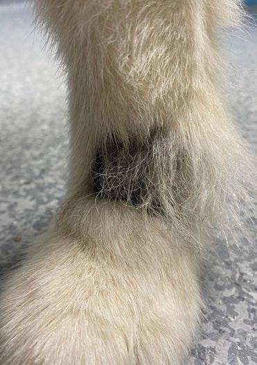
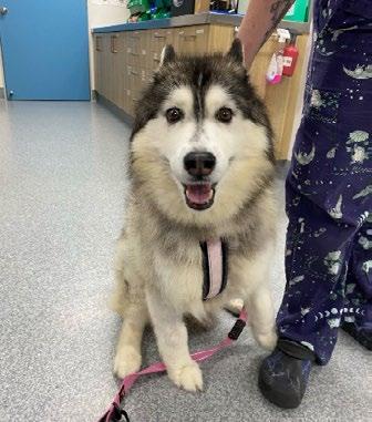
Snowy, a 10 year 11-month-old female neutered Husky presented for an acute progressively growing mass on the left dorsolateral metatarsus. The mass had been present for approximately 2 weeks before presentation and had become traumatised with intermittent bleeding.
Snowy has concurrent zinc responsive dermatoses, copper-associated hepatopathy and osteoarthritis. Medications include zinc tablets, Denamarin® (S-Adenosylmethionine and silybin) and synovan® (pentosan polysulphate and N-acetyl-D-glucosamine) injections.
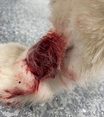
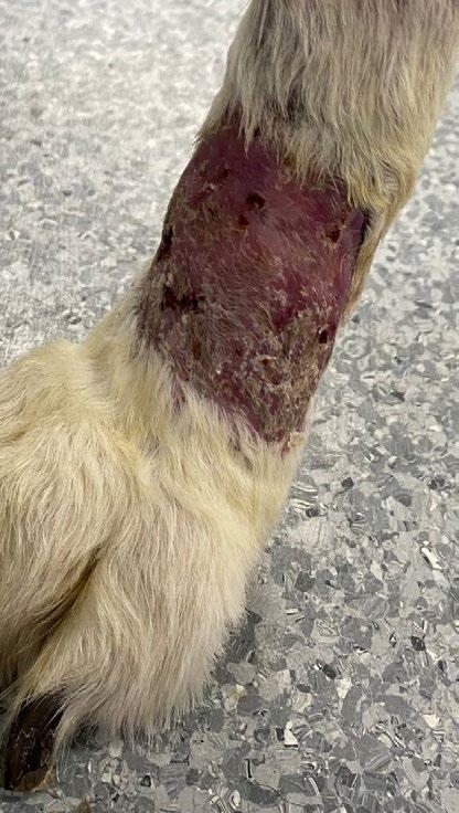
1. Snowy presented with an approximately 4x3cm raised well-defined dermal mass on the dorsolateral surface of the left metatarsus
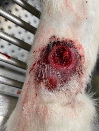
2. Appearance of the mass following 1 week of antibiotics
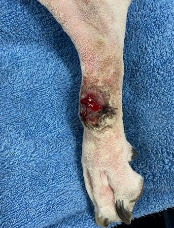
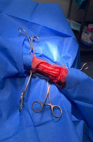
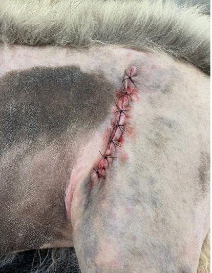
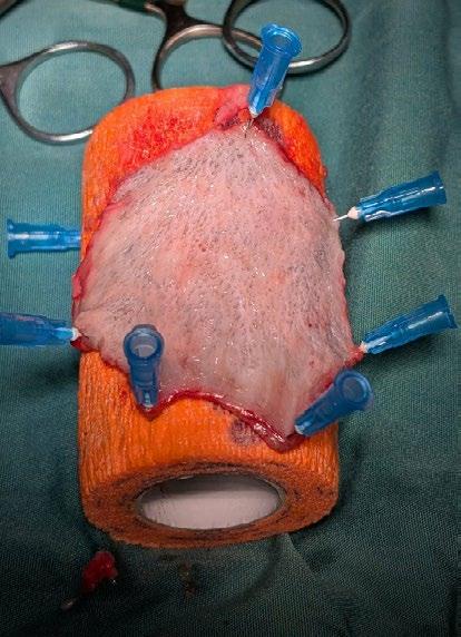
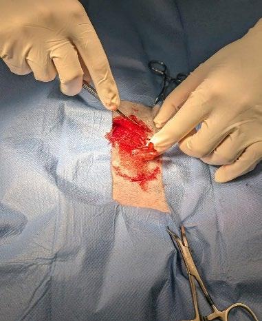
Figure 3. Appearance of the mass prior to excisional biopsy
Figure 4. The mass was excised with 1cm lateral margins
Figure 5. An impression in blood of the mass excision site was made with the sterile glove paper packet, and was used as a stencil to cut out the skin graft from the donor site (craniolateral thigh)
Figure 6. Closure of the donor site on the craniolateral thigh
Figure 7. The skin graft was secured to a sterile vetwrap roll with hypodermic needles to enable defatting of the graft (removal of subcutaneous tissue) with blunt and sharp dissection
Figure 8. The skin graft was meshed using a size #11 scalpel
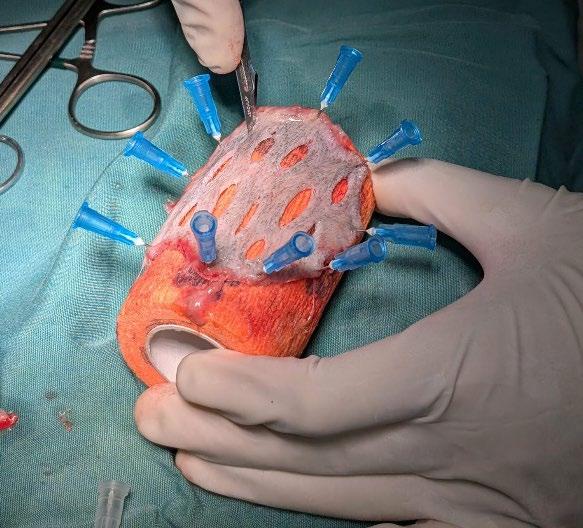
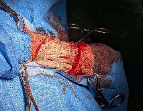
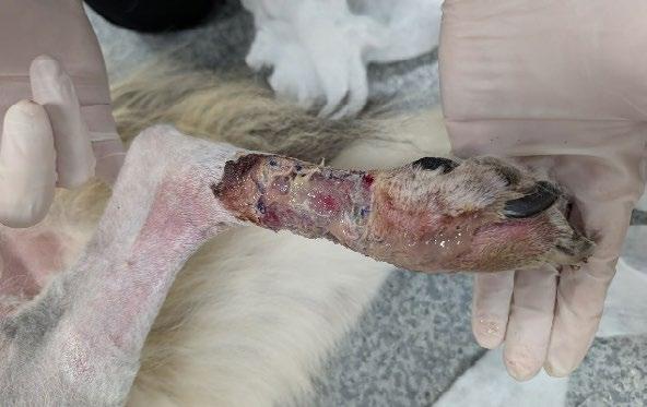
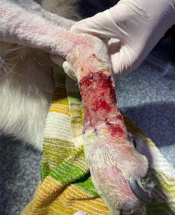
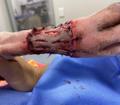
Figure 9. The graft was placed on the recipient bed
Figure 10. The skin graft was secured to the recipient bed with simple interrupted sutures around the margin of the graft. Two simple interrupted sutures were also placed to secure the mesh to the underlying recipient bed.
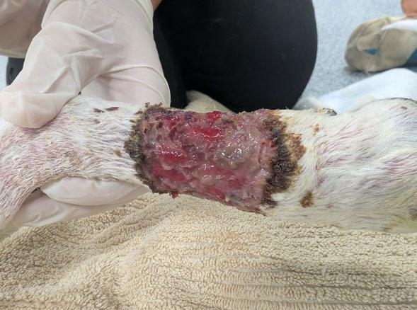
Figure 11. First bandage change at postoperative day 7. The surgical site was markedly infected with a marked volume of thick purulent discharge present.
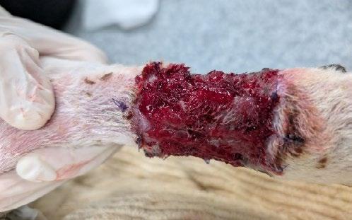
Figure 12. Appearance of the surgical site following lavage with Hartmann’s solution. There were no obvious signs of necrosis.
Figure 13. Appearance of the surgical site at post-operative day 9. There was evidence of granulation tissue developing. The volume of exudate was mildly reduced.
On clinical examination, Snowy’s vital parameters were within normal limits. There was an approximately 4x3cm raised well-defined dermal mass on the dorsolateral surface of the left metatarsus (Figure 1). The mass had an ulcerated bleeding surface with underlying friable tissue. There were no active signs of infection. The left popliteal lymph node was of normal size and texture. There was no left hindlimb lameness.
Snowy was started on a 7-day course of Noroclav® tablets (Amoxicillin clavulanic acid; 500mg by mouth twice daily for 7 days) prior to surgical biopsy of the mass (Figure 2)
Incisional biopsy of the mass was performed under general anaesthetic. Two wedge biopsies were taken from the craniolateral and caudolateral aspects of the mass using a size #10 scalpel. Each biopsy site was closed with simple cruciate suture (2-0 non-absorbable nylon). A fine needle aspirate was also taken of the popliteal lymph node and submitted for cytology.
Histopathology of the biopsy samples revealed a multilobulated densely cellular epithelial neoplasm with formation of cystic and ductal structures multifocally throughout. Cells showed moderate anisocytosis, anisokaryosis and nuclear pleomorphism. The mitotic count was 18 per 2.37mm2. The neoplasm was defined as a carcinoma, favouring apocrine adenocarcinoma due to the formation of occasional cystic spaces filled with intensely eosinophilic material surrounded by attenuated to cuboidal epithelium.
Cytology of the popliteal lymph node FNA sample revealed a heterogenous population of lymphoid cells with no obvious metastatic neoplastic cells.
Surgical excision of the mass was performed under general anaesthetic, with a free autograft obtained from the craniolateral thigh to allow closure of the surgical site. Other approaches that were considered included open wound management of the surgical wound bed with bandaging, and delayed grafting following granulation of the wound bed. We decided on a free autograft approach with immediate placement on the surgical wound bed as it was likely to allow more rapid wound healing (vs open wound management), and to avoid two general anaesthetics for Snowy given her co-morbidities.
The patient was placed in right lateral recumbency. The donor site (craniolateral thigh) and mass were clipped and prepared in a sterile manner (Figure 3). The
metatarsal mass was excised with 1cm lateral margins using a size #10 blade, with Metzenbaum scissors used to dissect the subcutaneous tissue of the deep margin (Figure 4)
A stencil of the mass excision site was made by placing the sterile glove paper on the excision site and obtaining a corresponding impression in blood. The stencil was placed on the donor site (craniolateral thigh) and traced using a size #10 blade to sharply dissect it from the donor site (Figure 5). The subcutaneous tissue was dissected using Metzenbaum scissors. The graft donor site was closed in 2 layers – simple continuous pattern (3-0 absorbable suture) to close the subcutaneous layer and simple cruciate pattern to close the skin (2-0 nonabsorbable nylon suture) (Figure 6)
The skin graft was stretched flat and secured to a sterile coadhesive bandage (Vetwrap®) roll using 22G hypodermic needles, with the subcutaneous fat facing the surgeon (Figure 7). The graft was kept moist with sterile saline during the defatting procedure. The subcutaneous tissue was sharply dissected from the dermis using a combination of a size #10 blade and Metzenbaum scissors. This was performed until the cobblestone appearance of the dermis was reached, and the exposed bulbs of the hair follicles could be appreciated.
The skin graft was meshed with a size #11 blade, with 1cm slits created at approximately 0.5-1cm apart (Figure 8) The edges of the graft were trimmed using Metzenbaum scissors.
The graft was placed over the recipient bed (Figure 9) Simple interrupted sutures were placed around the skin graft to secure it to the recipient bed using 3-0 absorbable suture. 2 tacking sutures were placed to attach the mesh to the underlying recipient bed (Figure 10)
A non-adherent petrolatum impregnated dressing (Jelonet®) was applied to the surgical site, followed by Allevyn® absorbent foam dressing, Soffban® soft padding bandage and conforming bandage. A splint was applied followed by further Soffban®, conforming, vetwrap® and Elastoplast® bandage.
Metacam® (Meloxicam) was administered as a subcutaneous injection in recovery.
Post-operative medications included Metacam® (Meloxicam) and Noroclav (amoxicillin- clavulanic acid), initially for a 7-day course post-operatively. Meloxicam was considered safe to use as Snowy’s chronic liver disease was mild, her liver enzymes were stable and she was asymptomatic at the time. Her owners were directed to administer the medication with food, and to stop if any vomiting or diarrhoea was observed. Snowy was rested throughout the post-operative bandaging period. This consisted of short 5-10 minute leashed walks only to toilet, avoiding jumping, running and excessive movement. A plastic boot was applied to the foot in case of moisture from the environment when walked outside.
Bandage changes were able to be performed without sedation.
The graft site was markedly infected at day 7 post-op. There was a marked volume of green mucopurulent discharge present over the graft site, and between the toes (Figure 11). The interdigital pressure sores were erythematous, moist and sore to touch. The surgical site and toes were lavaged with Hartmann’s solution using a 20 gauge needle attached to a 20mL syringe, as well as gentle debridement with gauze swabs soaked in Hartmann’s solution. Following lavage, there was bleeding present at the graft site, without any obvious regions of graft necrosis (Figure 12). There was evidence of hair viability at the meshed sites. The bandage was replaced in a similar fashion to previous, with padding between the toes and over the hock. The splint was not re-applied.
There was mild erythema and swelling present at the graft donor site.
A swab of the wound exudate was taken and sent for culture and sensitivity testing. Culture yielded a heavy growth of Staphylococcus pseudintermedius and Pseudomonas aeruginosa, the former being susceptible to amoxicillin clavulanic acid and enrofloxacin, and the latter being resistant to amoxicillin clavulanic acid, but susceptible to enrofloxacin. The amoxicillin-clavulanic acid course was extended for another 2 weeks, whilst a 2-week course of enrofloxacin (5mg/kg PO SID) was initiated. Metacam® (Meloxicam 0.1mg/kg PO SID) was continued for another 7 days.
The infection at the graft site appeared to show signs of improvement with improvement in the volume of exudate from the wound and pressure sores (Figure 13). Following wound lavage with Hartmann’s solution, there were no
Further evidence of granulation tissue development and reduced exudate production
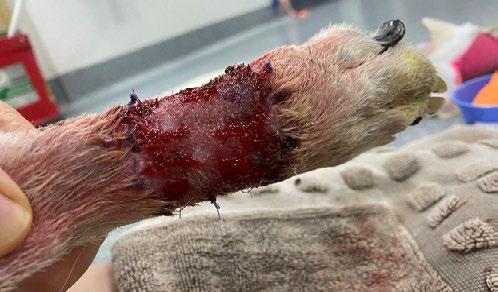
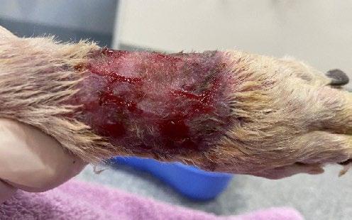
obvious necrotic regions present, and the graft had a pink to red colour. The interdigital sores were lavaged with dilute chlorhexidine solution (0.05%) and flamazine® (silver sulfadiazine) cream was applied. Flamazine® cream was also applied to the graft site. The petrolatum impregnated gauze dressing was not re-applied, instead Allevyn® (absorbent foam dressing) formed the primary layer.
Day 12
There was further improvement with mild exudate present at the graft site and pressure sores (Figure 14). There was evidence of granulation tissue present between the mesh slits. Flamazine® cream was applied to the graft site and pressure sores, and a bandage was applied with Allevyn® forming the primary layer.
Day 14
There was minimal wound exudate present. There was evidence of epithelialisation occurring and presence of viable hairs. The granulation tissue sites appeared to be smaller in size (Figure 15). The sutures around the graft site were removed. A melonin® dressing was applied as the primary layer. The sutures were removed from the graft donor site which had healed well.
Day 16
There was minimal wound exudate present. There was evidence of further epithelialisation and viable hairs, and shrinkage of the granulation tissue sites (Figure 16). A bandage was re-applied with Melonin® forming the primary layer.
Day 19
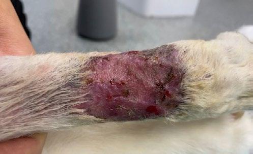
The majority of the wound had re-epithelialised with a pink colour (Figure 17). There were a few 2-3mm regions of granulation tissue present which bled after gentle wound lavage. A bandage was re-applied with Melonin® forming the primary layer.
Day 21
Further epithelialisation was present with few scabbed regions (Figure 18). A light bandage with Melonin® forming the primary layer was applied.
Day 23
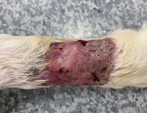
The wound bed had epithelialised with presence of viable hairs (Figure 19). The skin had a pink colouration and was dry. The bandage was removed. Petrolatum jelly (Vaseline®) was applied at home intermittently to moisturise the graft site for 7 days.
Histopathology revealed a moderately multilobulated unencapsulated but moderately circumscribed neoplasm spanning the dermis and subcutis, with an overlying extensively ulcerated epidermis. Tumour excision was complete with 10-12mm lateral margins and 0.51mm deep margins. The neoplasm was predominantly composed of solid lobules of cells with distinct cell margins, moderate to abundant vacuolated and sometimes clear cytoplasm. The central ovoid nuclei had finely stippled chromatin and indistinct nucleoli. There was moderate anisocytosis, anisokaryosis and nuclear pleomorphism. The mitotic count was 20 per 2.37 mm². There was multifocal cystic degeneration and in some areas of the mass, there were entrapped ductal structures lined by cuboidal epithelium (especially superficially).
The morphology of the neoplasm was unusual as there were areas of glandular differentiation as well as areas more suggestive of mesenchyme differentiation. The majority of neoplastic cells showed dual positive staining for cytokeratin and vimentin. The glandular elements present multifocally throughout the mass showed cytokeratin positive staining only.
The morphology of the tumour was suggestive of clear-cell adnexal carcinoma, or clear cell ductal carcinoma (subtype of adnexal carcinoma). There is little information in the literature on the behaviour and prognosis of these tumours, given their rarity. The majority of these tumours are reportedly slow-growing, however a proportion will recur, and those with a high mitotic count may metastasise to regional lymph nodes.
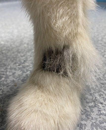
Snowy’s recheck at 134 days post-surgery revealed partial hair regrowth and no evidence of tumour recurrence (Figure 20). The regional popliteal lymph node was symmetrical in texture and size compared to the normal (right) hind limb. Snowy’s physical exam was within normal limits and she was ambulating well on the limb.
This was an interesting case of an unusual tumour present in a difficult location for surgical resection.
Biopsy of the mass was suggestive of an adnexal carcinoma. There was no evidence of metastasis to the local popliteal lymph node.
Tumours of the distal limb present a challenge as there is a paucity of loose skin available for wound closure via skin mobilisation or construction of local flaps. Furthermore, there is questionable integrity of the fascial planes of this region with thin to absent fascia
present. A fascial plane is a dense connective tissue layer that may be able to act as a physical barrier to neoplastic cells during surgical resection. As a result, it may be challenging to achieve adequate deep margins in some tumour sections in this region, and recurrence is a possibility. Histopathology of the mass revealed clean excision of a clear-cell adnexal carcinoma, or clear cell ductal carcinoma (subtype of adnexal carcinoma). The deep margin obtained was 0.5 -1 mm.
Tumour recurrence is possible despite the demonstration of clean margins, and similarly the presence of an incomplete surgical margin does not signify that tumour recurrence is inevitable (McSporran, 2009). There is little information available on the biologic behaviour of these tumours given their rarity. They have not been reported in humans, nor in animals other than dogs. A study of 26 cases (Schulman et al., 2005) found that there was no recurrence or evidence of metastasis in 22 dogs (85%) despite prominent atypia and pleiomorphism of neoplastic cells histologically. 1 (4%) had local recurrence twice, and 2 (8%) had metastasis to regional lymph nodes, with one of these dogs being free of recurrence or evidence of metastasis 17 months after excision of the affected lymph node. The findings suggested that surgical excision was curative in the large majority of cases, despite frequency of prominent atypia and pleiomorphism. Higher mitotic rates and lymphatic invasion may be predictive of metastasis, but further studies would need to be performed to investigate this. Due to the limited literature available, the role of adjuvant therapy (e.g. radiation therapy, chemotherapy) is not clear. Further staging and adjuvant therapy was not pursued in this case following consideration of Snowy’s comorbidities and the stress of veterinary visits, and Snowy is continued to be monitored every 3 months with a physical exam, including popliteal lymph node palpation.
A free full thickness mesh autograft was obtained from the craniolateral thigh to achieve closure of the surgical site. This consisted of a segment of epidermis and dermis that was completely removed from the vascular and nervous supply of the body and then placed to a recipient site on the same animal. The graft was meshed with numerous parallel slits in a staggered manner to allow the graft to expand in size, confer flexibility and conformity to the recipient site and allow for drainage of wound exudate, bleeding or serum oozing at the time of grafting. The disadvantage of meshed grafts is that excess granulation tissue may grow up through the slits and over the top of the graft.
The craniolateral thigh was chosen as the graft donor site as it had similar hair colour, texture, length and thickness to the hair surrounding the recipient site, and the patient did not have to be repositioned for graft placement and closure of the donor site. Another common site is the cranial lower lateral thoracic area due to the presence of fairly thin, well-haired skin and an abundance of loose skin, allowing easy closure of the donor site. Another consideration is the thickness of the dermis which creates a greater distance for diffusion of oxygen and nutrients during the early post-grafting stages. Regions where the dermis is thicker include the lateral neck and dorsal lumbar regions. The greatest density of hair follicles is also found over the dorsal lumbar region most commonly.
The graft was placed directly onto the surgical site wound bed following tumour excision. Successful engraftment is dependent on the establishment of arterial and venous connections with the recipient bed, which may be achieved by placing the graft on either a healthy granulation tissue bed, a surgically created acute wound or an acute natural wound that has been rendered surgically clean. The graft recipient site must be free from infection, debris and any excessively intense or chronic inflammatory process. Examples of tissues that lack adequate vascularity include chronically poorly vascularised tissue, irradiated tissue, avascular fat, bone, cartilage, tendon, nerves or wounds denuded of overlying connective tissue.
Post-operative care and bandaging is important in supporting wound healing, protecting against postoperative trauma to the graft and allowing immobilisation of the graft site in the early post-operative period. The primary dressing is typically comprised of a wide mesh non-adherent petrolatum impregnated cellulose acetate pad (e.g. Jelonet®), with an absorptive foam pad placed overlying. A splint is applied initially to facilitate immobilisation of the graft. The immediate post-operative bandage is left undisturbed generally for 24-48 hours, to facilitate graft adhesion, immobilisation and absorption of wound fluid, however some surgeons
prefer to leave it in place for 3-5 days. The frequency of bandage changes is variable and can vary from once daily to every 2-4 days. Changes should be performed for at least 2-3 weeks post-operatively, after which a light bandage can be considered for 10-14 days to prevent trauma. Noticeable hair regrowth is usually seen 2-3 weeks after grafting.
Some reflections from this case that can be applied in the future include:
– Considering the first bandage change at an earlier time frame as the graft became markedly infected in addition to the development of marked interdigital pressure sores. It may have been that the warm and humid weather had some contribution to this. It is generally understood that the immediate postoperative bandage is usually left undisturbed for at least 24-48 hours to facilitate graft adhesion, immobilisation and protection of the graft from trauma. Some surgeons prefer to leave the initial bandage in place for 3-5 days.
– Ensuring alignment of the hair follicles with the graft recipient bed for cosmesis. The orientation of the hair follicles was less obvious once the graft site had been clipped prior to surgery and once the subcutis had been dissected intra-operatively. A hypodermic needle of a different colour (gauge) could be used indicate the dorsal margin and orientate the graft on the vetwrap roll.
– Meshing of the graft allows a smaller graft to expand and therefore fill a larger defect. It is also important to note that trimming of the graft following meshing will mildly decrease the size of the graft overall.
– Infection is one the most common causes of graft failure and can be detrimental to graft survival. This is due to the enzymes that the bacteria produce, which can dissolve fibrin attachments and also potentially degrade elastin (as is the case in a Pseudomonas infection). Infection of the graft surface can occur when there is overgrowth of normal skin organisms on abnormal skin. It is important that any infection is treated promptly and appropriately (e.g. taking a swab for culture and sensitivity to select appropriate antibiotics), and that bandage changes are performed more frequently until the infection is resolved.
Thank you to Dr William Hawker for his surgical guidance and mentorship for this interesting case. Thank you to Dr Lucy Woolford for providing histopathological assessment and her time in discussing the case. Thank you to Dr Elizabeth Maher for the photos of the surgery. Thank you to Snowy’s family to their dedication and commitment to the surgery and regular post-surgical bandaging care.
McSporran, KD 2009, ‘Histologic Grade Predicts Recurrence for Marginally Excised Canine Subcutaneous Soft Tissue Sarcomas’, Veterinary pathology, vol. 46, no. 5, pp. 928–933.
Schulman, FY, Lipscomb, TP & Atkin, TJ 2005, ‘Canine cutaneous clear cell adnexal carcinoma: histopathology, immunohistochemistry, and biologic behavior of 26 cases’, Journal of veterinary diagnostic investigation, vol. 17, no. 5, pp. 403–411.
The author presents a thoughtful account of a challenging distal limb lumpectomy. The technical proficiency, careful planning and attentive postoperative management contributed to a successful clinical outcome.
Some things to consider in similar cases are the tumour margins and the benefits of delayed grafting. While immediate grafting following oncologic resection may be successful in selected cases, more conservative approaches delaying grafting to allow granulation is likely to reduce the risk of graft failure and allow revision surgery if tumour margins are incomplete on histopathological assessment.
Factors associated with an increased risk of graft failure include the distal limb location, limited soft tissue coverage, and the challenges of maintaining graft stability in a mobile patient, where strict rest and bandage management are critical. While this case has had an excellent outcome, these considerations highlight that such outcomes may not be consistently reproducible without similarly close management.
For general practitioners, this case may be most useful when considered within a framework of contextualised care. Decisions around timing of reconstruction often involve balancing oncologic considerations with patient-specific factors such as age, the potential need for multiple anaesthetics, and financial implications for the owner. In some circumstances, avoiding a second procedure may represent a clinically appropriate and pragmatic option that supports patient welfare and preserves access to treatment.
Quality online continuing education delivered by veterinary experts in short, manageable timeframes incorporating informal, social and collaborative learning with your peers.
Build in-demand skills in key clinical areas. Enjoy networking with peers long after the course has finished.
TimeOnline
4–6 weeks | 10–20 CPD points
TimeOnline courses offer short, focused online learning that still delivers depth. They are ideal when you want to build confidence in a specific area but cannot commit to a longer program, or when you wish to explore a topic before enrolling in Distance Education. Courses include expert presentations, case studies, quizzes, curated resources, and an active online discussion forum.
CVE members save 20-40% on enrolments
cve.edu.au/timeonline
TimeOnline OnDemand
90 days access | 5 CPD points
TimeOnline OnDemand provides flexible, self-directed learning without compromising quality.
These short courses allow you to work through practical, targeted content at your own pace and are ideal when tutor interaction is not required.
CVE members save 50% on enrolments
cve.edu.au/timeonline-ondemand
Winner - Best Visuals
Entitled to a a CVE$300 voucher
Dr David H Larratt BVSc IVAS
Sires on Ice Richmond Vet
312 Londonderry Rd, Richmond, NSW 2753
t. 02 4702 2074
Dr John Katakasi BVSc (Hons), CVA, CVHM
Adelaide Plains Veterinary Surgery
6 Gawler Rd, Two Wells, SA, 5501 t. 08 8520 3600
e. greyhoundtarsalscreening@gmail.com
w. greyhoundtarsalscreening.com.au
C&T No. 6129
Introduction
Tarsal injuries are common in sporting dog breeds and approximately 60% of all serious injuries in racing greyhounds are fractures of the right tarsus.
There are no veterinary canine specialists (surgical, imaging or sports medicine) researching preventative physical examination and imaging for the common injuries that continue to impact greyhound racing, worldwide.
A group of experienced greyhound veterinarians have convened to consolidate a strategy for injury prevention.
All 4 veterinarians have been On Track Veterinarians (OTVs), performed extensive orthopaedic surgery, supported rehoming organisations, refined skills in physical examination and extrapolated radiographic positioning and interpretation from the equine and human models. They have dedicated the greater part of their long careers to assist the welfare of racing greyhounds, and collaborate to elevate veterinary standards with the aim of injury prevention.
The Six Step Hock Check provides further practical guidance for the physical examination of the canine tarsus and supplements the earlier CVE publication, Save Your Dog’s Hock -Tips for Trainers (Yore, Katakasi and Larratt, 2024).
Central to their strategy is the provision of practical education and the development of the alliance between veterinarians and trainers.
Dr Peter Yore BVSc, IVAS
Belmont Veterinary Hospital
591 Pacific Highway, Belmont, NSW, 2280 t. 02 49459044
Dr John Newell BVSc
56 Burton Rd, Eleebana, NSW, 2282 t. 0418 225 026
Over the last three years, Dr John Katakasi and Dr David Larratt have presented on the principles of injury reduction for the tarsus and carpus (physical examination, radiography and protective strapping) at veterinary conferences, in New Zealand, Victoria and Ireland. They have also conducted injury reduction seminars for trainers in Western Australia and New South Wales.
The administrators of several racing jurisdictions are now advocating for these welfare initiatives and the support from trainers is very positive.
The 6 Step Hock Check, is an instructional manual that may:
1. Provide practical guidelines for all veterinarians who examine sporting dog breeds. This will assist the detection of early injuries, allow correlation with radiographic finding and provide management options.
2. Assist veterinarians in performing a ‘Clearance to Race Certificate’ for greyhounds returning from injury. This is to comply with a request from the Australian Greyhound Working and Sporting Dog Veterinarians (AGWSDV).
3. Educate Trainers to identify problems earlier. Greyhound industry administrators have identified that ‘ it is important for trainers to regularly assess their greyhounds for the presence of hock pain’ (GWIC Factsheet – Prevention Measures for Serious Injuries in Racing (2025)) ( www.gwic.nsw.gov.au/news/resource-library )
Authors’ views are not necessarily those of
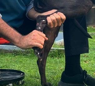
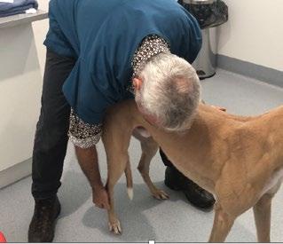
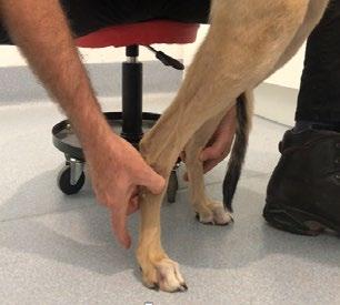
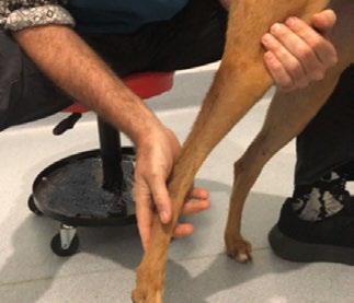
The 6 Step Hock Check is to be used in conjunction with the instructional video 'Examination Technique for Assessing Greyhounds Hocks for Fitness to Race' (2025) –a veterinary collaboration involving Drs Peter Yore, John Newell, John Katakasi and David Larratt. Available greyhoundtarsalscreening.com.au
The impact of universal anticlockwise racing is that over 90% of all greyhound hock fractures are right sided The right hind leg provides the propulsive drive, whilst also undergoing an internal twist of 25 degrees during the cornering phase of races.
The hardworking muscles of the right thigh and calf are prone to tightening due to fatigue. This contractive tightening increases the pull on the Achilles tendon, leading to a more upright hock position and increased vertical load on the front of the hock. CT imaging and radiography both confirm that the front of the hock is the most common site of early fractures.
The twisting may also lead to overstretching and laxity of the soft tissues at the front of the right hock. This soft tissue laxity may also occur in the left hock and both carpi.
The 6 Step Hock Check provides practical guidelines to detect and manage these early fatigue signs in both bone and soft tissues with the aim of preventing more serious injuries.
All 6 Steps are to be performed GENTLY with full awareness to detect pain and never to cause pain.
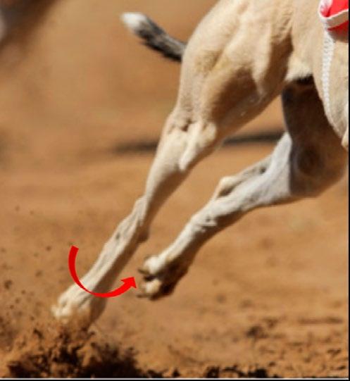
2. The right hind leg provides the propulsive drive, whilst also undergoing an internal twist of 25 degrees as it turns around the corners
Technique
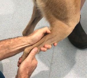
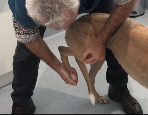
1. Stand over the dog to observe and compare both sides.
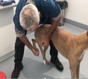
2. For the right side, cup the toes with your right hand and place your left hand above the stifle (Figure 3)
3. Push your hands together to flex the hock and assess for pain, range of motion and stiffness.
4. Stiffness is detected at maximum closure or compression when you feel a lack of recoil or bounce.
5. Perform several times, then repeat the test on the left side to compare the difference.
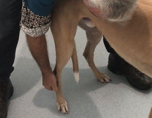
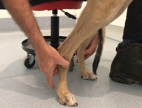
1. Stand over the dog and locate the main hinge joint (tibiotarsal joint)
2. Place fingers at the back of the hock and use your thumb on the front to press and bounce down the vertical lines of each tarsal bone and then follow to the underlying Metatarsal bone (as seen in figure 4).
3. It is Important to have the leg fully weight bearing for this test.
4. Repeat the test on the left side.
Findings
Pain may indicate presence of bone cracks at the front of the hock or soft tissue injury in the calf muscles, Achilles tendon or plantar ligaments at the back of the hock.
If there is a strong pain response, then gently perform the other 5 tests. If there is no obvious tear in calf muscles or Achilles tendon then radiography is required.
If there is stiffness with no pain, then the problem will likely be fatigue contraction in the plantar ligaments or calf and medial thigh muscles. To differentiate, perform steps D, E and F.
Findings
Pain may indicate presence of early bone cracks in the bones at the front of hock, particularly in the Central Tarsal Bone (CTB), Tarsal 3, Tarsal 4 and Metatarsals.
Mild pain at these points, may be secondary to fatigue contraction in the calf muscles or plantar ligaments. Steps D, E and F will assist localisation of the source of the pain.
If pain persists then radiography is required to check for bone crack and/or loss of bone density (demineralisation).
Technique
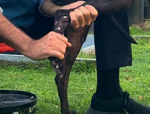
1. Sit on a low stool to palpate the front tendon of the right and left hocks simultaneously.
2. Place fingers at back and thumb at front of hock (as seen in figure 5).
3. Use thumb to feel thickened tissue at the front, just below the hinge joint. This 5 mm thickening is the fibrous ring anchoring the vertical tendonLong Digital Extensor (LDE).
4. With a very light touch, push against this thickening from the outside of the leg to the inside. Then place thumb on top and just below this thickening and repeat the push.
1. Sit on a low stool and pick up the right foot.
2. Place the left hand above the hock and hold the leg firmly to prevent movement. The right hand is placed below the hock and twists the metatarsal bones inwards and outwards like a throttle on a motorbike (as seen in figure 6).
3. Assess for pain, range of motion and stiffness.
4. To compare, repeat the test on the left hock.
Findings
There is laxity if you can push the LDE tendon 5 -15 mm further on the right compared to the left and this is referred to as subluxation of the LDE tendon. This tendon is firmly attached to the underlying joint capsule and intracapsular ligaments. All 3 structures are collectively referred to as the ‘ tarsal sleeve’ which provides elastic compressive support to the entire hock structure.
If tarsal sleeve laxity is detected, then strapping can be used to provide compressive protection to the underlying vulnerable hock bones. The benefits of hock strapping have been acknowledged by industry (GWIC Fact Sheet- Prevention Measures for Serious Injuries in Racing, 2025 )
Pain may indicate damage to the plantar ligaments or the bones at the back of the hock. If there is a pain response, radiography is required.
Restricted range of motion and stiffness may indicate fatigue contraction of the plantar ligaments or the associated Achilles tendon and calf muscles. To differentiate use Steps E and F.
Fatigue contraction of the plantar ligaments and calf muscles may also be common in other sporting breeds of dogs.
Technique
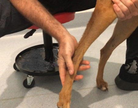
1. Sit on a low stool and pick up the right foot.
2. Place the left hand on the front of the stifle and hold the leg to prevent movement of the upper leg. The right hand is placed behind the hock and pushes forward (as seen in figure 7).
3. Push the hock forward to bring it into a vertical line with the stifle.
4. Assess for range of motion and elastic recoil or bounce.
5. To compare, repeat the test on the left hock
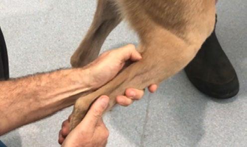
1. Sit on a low stool and pick up the right foot and hold at 90 degrees to the stifle.
2. Cradle the whole calf area with the left hand. While applying thumb pressure, bounce on the Achilles with the thumb, progressing, 2cm at a time, up the middle of the calf which is over the Superficial Digital Flexor muscle. (as seen in Figure 8). The SDF lies in the space between the two branches of the gastrocnemius muscle.
3. Feel for small ‘knots’ which are localised spasms of fatigued muscle known as ‘Myofascial Trigger Points’ and continue upwards to the back of the stifle joint. Repeat palpation of the two branches of the gastrocnemius muscle
4. Repeat the test on the left calf.
Findings
Mild pain and resistance when pushing to 180 degrees usually indicates fatigue contraction or fibre damage in the calf muscles or Achilles tendon. The most common cause in greyhounds is contraction of the Superficial Digital Flexor muscle which lies in the middle zone of the calf. With other sporting dog breeds, the lateral gastrocnemius muscle is more commonly affected. This can be confirmed by performing Step F.
Stiffness may be due to fatigue contraction in the medial thigh muscles especially impacting the gracilis muscle and the adductor muscle.
Pain and stiffness with this test may also indicate a problem with the cruciate ligament or meniscus of the stifle, though this is very rare in racing greyhounds.
Findings:
The trigger points in the Superficial Digital Flexor muscle can be released by massage and/or veterinary acupuncture or infusion of local anaesthetic. Once the muscle is relaxed, stretch to elongate the muscle fibres by performing the Calf Extension (Step E) and repeat several times. Once the trigger points are released there is usually also marked improvement in Hock Flexion (Step A) and a reduction in bone pain (Step B).
Many greyhounds suffering with contracted calf muscles will dramatically improve their race times once the fatigue contraction is effectively addressed.
Fatigue contraction of calf muscles occurs in other sporting breeds with the lateral belly of the gastrocnemius commonly involved.
This is not a replacement for local veterinary consultation, but may assist trainers to seek immediate veterinary attention and radiographic investigation to potentially avoid more serious injury.
The 6 Step Hock Check is to be used in conjunction with the instructional video 'Examination Technique for Assessing Greyhound Hocks for Fitness to Race' (2025) - a veterinary collaboration between Drs Peter Yore, John Newell, John Katakasi and David Larratt greyhoundtarsalscreening.com.au

Your clinic likely sterilizes surgical instruments and implants using one or more of the following methods:
Single-use blue wraps around some form of basket
Single-use paper or sealable plastic pouches
Reusable cloth drapes
Reusable rigid sterilization containers
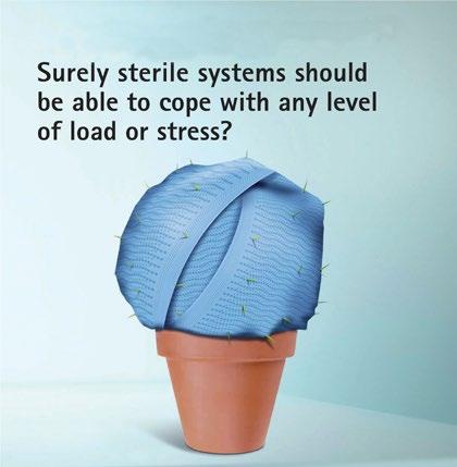
Single-use wraps (or blueys) are inexpensive and widely available. However, they are designed for one-time use and then discarded, contributing significantly to a practice’s waste stream, which ultimately ends up in landfills. Australians produce approximately 21.6 billion tonnes of landfill each year, with hospital single-use goods being a notable contributor.¹ Additionally, blueys cannot act as rigid barriers, and sharp instruments can perforate them, compromising kit sterility. If accidental perforation occurs, it’s unlikely you would even be aware of it. Wrapping a kit also takes time, which is something most nurses have in short supply.
Single-use paper or sealable plastic pouches are also inexpensive and commonly available. They allow small quantities of instruments to be contained and sterilized but also end up as landfill. Being non-rigid, they are also susceptible to accidental perforation.²
Reusable cloth drapes are relatively expensive and have been used for many decades. Although they are disposed of infrequently, they need to be washed and dried after each use. Consider the water, detergent, electricity, and nursing time required for each use.³
Reusable rigid sterilization containers are typically made of aluminium. While they require a larger upfront cost, their lifespan is incomparable, as rigid containers can last for more than a decade. If you calculate the processing cost per kit, a practice can see savings as soon as the second year of ownership, simply by comparing it to the cost of consumables that would have been used instead.⁴
References:
3. Exploring the Safety and Environmental Impact of Sterilization Techniques.
4. Sustainability | Reducing the Environmental Impact of Sterilization Packaging for Surgical Instruments in the Operating Room: A Comparative Life Cycle Assessment of Disposable versus Reusable Systems (mdpi.com)
Dr Alexander Teh*
BVetBiol DVM MVetStud (Pathology) MVetClinStud
DACVP (Anatomic Pathology)
Specialist Anatomic Pathologist, Vetnostics NSW
Adjunct Associate Lecturer, Sydney School of Veterinary Science, The University of Sydney
e. alexander.teh@sydney.edu.au
e. alex.teh@vetnostics.com.au
*corresponding author
Dr Kyle Song
BVSc, FANZCVS (Small Animal Surgery)
Specialist Small Animal Surgeon, Animal Referral Hospital Greater Western Sydney
C&T No. 6130
A 9-month-old Australian Bulldog presented with acute onset of paraparesis grade 2 (ambulatory paraparesis) with progression to grade 3 (non-ambulatory paresis). Magnetic resonance imaging (MRI) of the spinal column revealed an intradural extramedullary contrast enhancing soft tissue mass at the level of T13 and L1 (Figure 1).
The dog underwent surgery for debulking of the soft tissue mass (performed via dorsal laminectomy from T13 to L2), and the tumour was submitted to Vetnostics NSW for histopathological examination.
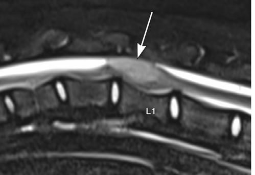
An MRI study of the thoracolumbar spine was acquired with multiple sequences (T2, T2 FS, T1 – pre/postcontrast, short tau inversion recovery (STIR), and T2 gradient echo (GRE) in a combination of sagittal, dorsal and transverse planes. The intraspinal mass is depicted in Figure 1
The mass consisted of a mixture of three cell types: (1) an epithelial population forming numerous tubular structures (Figure 2) and rarely, primitive glomeruloid structures (Figure 3); (2) small cuboidal to polygonal blastemal cells intermixed amongst the epithelial structures (Figure 2); and (3) a mesenchymal component consisting of dense arrangements of spindle cells (Figure 4)
This is a rare and unique tumour of the canine spinal cord typically found in young dogs between 5 months and 4 years of age with a median age of 14 months. Spinal cord nephroblastomas commonly develop between T10 and L3 segments of the spinal column. Whilst this is a rare tumour, comprising approximately 1% of all canine primary central nervous system (CNS) neoplasms, it is an important diagnostic possibilty to consider in this young age group, in particular if a soft tissue mass is found in the aforementioned spinal segments. Presenting clinical signs of this neoplasm often consist of progressive ataxia, paresis, or paralysis which can be either unilateral or bilateral.
The precise histogenesis of canine spinal cord nephroblastomas remains controversial and elusive. However, it is thought to arise from ectopic metanephric blastema which have been trapped between the dura mater and the developing spinal cord.
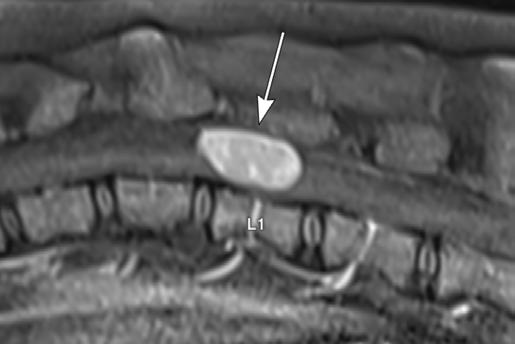
tau inversion recovery (STIR) imaging (left), and T1 post-contrast with fat saturation (right).
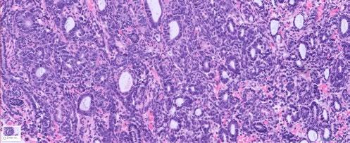
Figure 2. Photomicrograph of the spinal tumour showing an epithelial component forming numerous tubular structures intermixed with many smaller cuboidal to polygonal blastemal cells
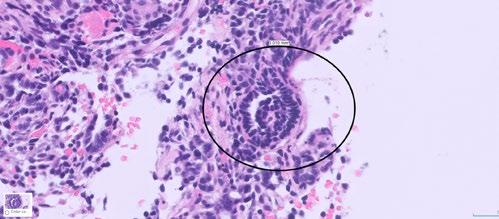
Figure 3. Photomicrograph of the spinal tumour showing a primitive glomeruloid structure (black circle)
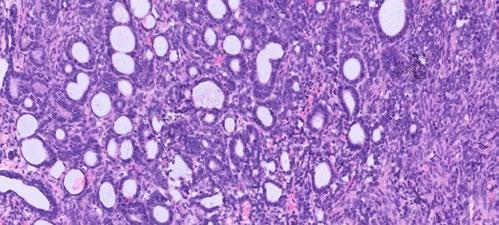
Figure 4. Photomicrograph of the spinal tumour showing the mesenchymal component of the neoplasm towards the right of the image consisting of dense arrangements of spindle cells
Following surgical debulking, the dog was referred for a course of radiation therapy. With radiation therapy, the owner has reported that the dog is currently readily ambulatory with no obvious ataxia or paraparesis.
Reference
Higgins, R.J., Bollen, A.W., Dickinson, P.J. and SisóLlonch, S. (2016). Tumors of the Nervous System. In Tumors in Domestic Animals, D.J. Meuten (Ed.). doi.org/10.1002/9781119181200.ch19
10 - 14 August 2026
8:30 AM - 4.00 PM
Venue
State Library Victoria | Melbourne
This exciting conference is led by the renowned Dr Linda Fleeman, the recipient of the CVE’s 2024 T G Hungerford Award recognising excellence in continuing veterinary education
For her profound and multifaceted contribution to veterinary medicine particularly to advancing the discipline of veterinary endocrinology.
Dr Fleeman is supported by a stellar panel of outstanding specialists, the conference theme being spectrum of care in the management of veterinary endocrine disease.


cve.edu.au/ endocrinology-conference
Madeleine Reichstein
Southern Tablelands Veterinary Hospital
105 Robinson St
Goulburn NSW 2580
e. southerntablelandsvets@gmail.com
C&T No. 6131
A 6-year-old entire female Corgi presented with lethargy and general malaise four weeks following oestrus. The owner reported no vaginal discharge. Physical examination revealed depression and mild abdominal discomfort. Rectal temperature was mildly elevated.
Blood tests revealed a mild neutrophilia with band neutrophils, a mild monocytosis, thrombocytopenia (revealed as clumped on smear review), mild hyperglobulinaemia, mild ALKP elevation, mild hypercholesterolemia and mild hyperchloraemia.
Abdominal ultrasonography identified a dilated tubular structure within the abdomen containing flocculent echogenic material, consistent with uterine pathology. The contralateral uterine horn was not grossly distended. Based on these findings, exploratory laparotomy and ovariohysterectomy were elected.
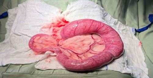
Surgical exploration confirmed marked distension of a single uterine horn containing purulent material, while the opposite horn appeared grossly normal. A diagnosis of unilateral pyometra was made. The patient recovered uneventfully following surgery and supportive medical therapy.
Discussion
Pyometra in intact bitches is hormonally mediated, typically associated with cystic endometrial hyperplasia, and usually affects both uterine horns due to the systemic effects of progesterone (Hagman, 2018). Clinical signs may include lethargy, anorexia, and vaginal discharge, although closed-cervix presentations without discharge are common.
Unilateral pyometra is rare. Recent reports describe isolated cases, often associated with congenital uterine anomalies such as segmental aplasia or unicornuate uterus (Johnston et al., 2001.) These malformations may predispose one horn to infection and retention of purulent material, resulting in a ‘unihorn’ pyometra. In cases without detectable malformations, unilateral
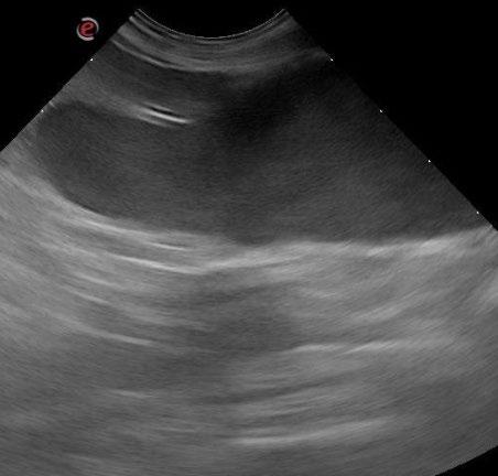
Figure 1. Abdominal ultrasound revealing a singular dilated hypoechoic tubular structure filled with flocculant material located in the caudal abdomen of the patient
2. Unilateral pyometra
pyometra may occur due to functional obstruction or localised disease.
In the present case, no gross uterine malformations were observed, suggesting either true unilateral involvement or functional obstruction of the contralateral horn. This case highlights the importance of including pyometra in the differential diagnosis for intact bitches presenting with systemic illness, even in the absence of vaginal discharge or when imaging suggests localised uterine involvement.
References
Hagman, R. (2018) Pyometra in small animals. Veterinary Clinics of North America: Small Animal Practice, 48(4), pp. 639–661.
Johnston, S.D., Root Kustritz, M.V. and Olson, P.N.S. (2001) Canine and Feline Theriogenology. Philadelphia: Saunders.
Authors’ views are not necessarily those
Unlimited access to webinar series
C&T No. 6121, Issue 322, Mar 2026
Charles Ley
Specialist in Veterinary Diagnostic Imaging

1 CPD point each EmergencyLIVE!
Practical, life-saving techniques + includes a lively Q&A
Managing Traumatic Brain & Spinal Injury
24 June 2026
Suzanne Tso
BCSc GCertSAECC FANZCVS (ECC)
cve.edu.au/emergencylive

WebinarPLUS
Dedicated Vet + Vet Nurse streams
Heads Up: Common Conditions of the Rabbit Skull
18 - 24 June 2026
Olivia Clarke
BSc BVMS MANZCVS (Unusual Pets, Avian)
cve.edu.au/webinarplus

WebinarLIVE!
1 Hour, World Experts + includes a lively Q&A FREE to the profession
Recognising Cushing’s Syndrome in Diabetic Animals
15 July 2026
Linda Fleeman
BVSc PhD MANZCVS
cve.edu.au/webinarlive
Swedish University of Agricultural Sciences
Uppsala, Sweden
e. charles.ley@slu.se
C&T No. 6132
Question What is your radiological diagnosis?
An 8-month-old female desexed Labrador Retriever cross presented for a 4–6-week duration of left forelimb lameness.
On clinical exam, there was mild pain on manipulation of both shoulder joints.
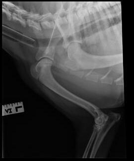
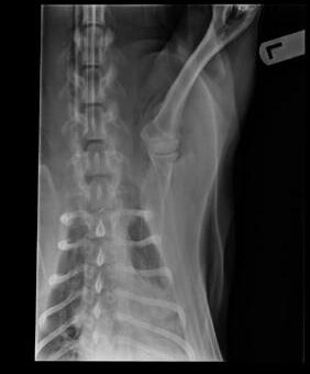
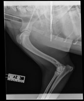
1 A, B, and C Lateral radiographs left and right shoulder and ventrodorsal view left shoulder
Authors’ views are not necessarily those of the
Concave contour
Sclerosis
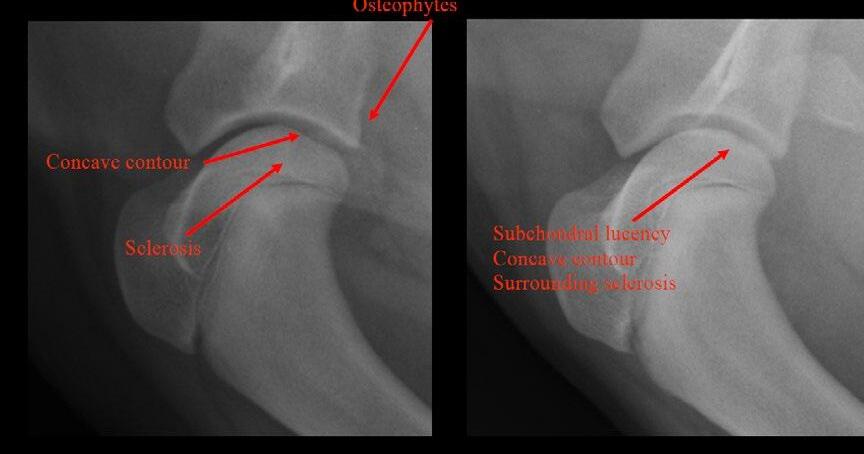
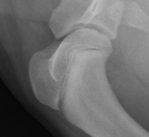
Subchondral lucency
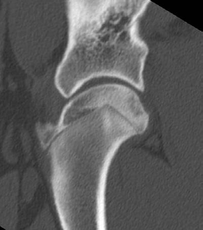
Concave contour
Surrounding sclerosis
Answer
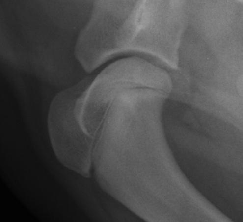
Almost the entire caudal aspect of the left front humeral head articular surface has a mildly concave contour (subchondral defect) and the joint space at this level is mildly wider than other regions of the joint. There is moderate to severe sclerosis of the caudal half of the left front humeral head epiphysis, with mildly lower opacity in the region surrounding the concave articular surface. The caudodistal margin of the left front scapula is a pointed, mildly rough shape, suggesting mild osteophyte formation on the margin of the shoulder joint.
Mild, small indistinct lucent region in the subchondral bone on the caudal aspect of the right front humeral head, with a small area of the adjacent articular surface that is flattened to mildly concave contour (subchondral
defect) and mild indistinct sclerosis surrounding the region of lucent subchondral bone.
The proximal physes of both front humeri and radii are open.
No abnormalities of the elbows are seen; however, this is not a complete set of elbow projections.
Osteochondrosis both front humeral heads with moderate changes left front and mild changes right front.
Osteoarthritis is suspected in the left front shoulder joint.
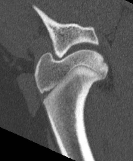
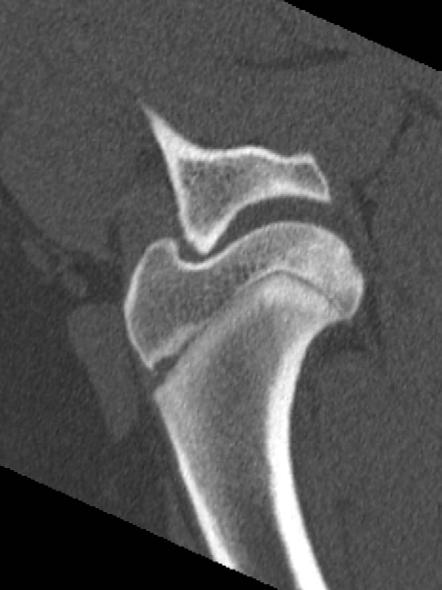
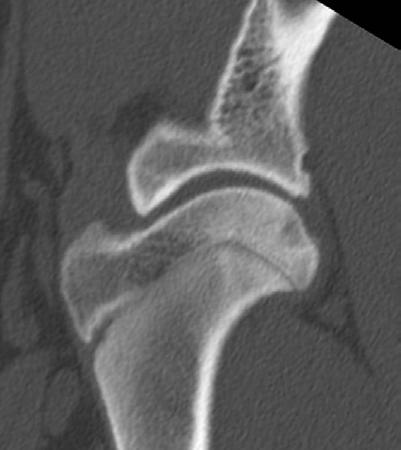
For surgical planning, CT of both shoulders was done:
Left Shoulder
An approximately 12 mm diameter region of the caudal aspect of the left humeral head is concave/flattened, and deep to this (approximately 5 mm deep) region there is a half-circle shaped region of heterogeneous
hypoattenuation in the subchondral bone and trabecular bone; surrounding this a moderate zone of poorly defined sclerosis. A poorly defined 1-2 mm sized hyperattenuating region is seen extending from the caudal margin of the subchondral defect into the joint space, and this is suspected to be a partially mineralised flap of articular cartilage. There is mild to moderate osteophyte formation on the caudal and lateral margin of the glenoid and mild osteophyte formation on the caudal margin of the humeral head. Moderate increase in the volume of the soft tissue attenuation synovial structures of the shoulder joint is seen.
Approximately 5 mm diameter, 2 mm deep concave subchondral defect on the caudal aspect of the articular surface of the right humeral head. The concave subchondral defect is surrounded by a moderate zone of poorly defined sclerosis. No osteophytes detected on joint margins.
Osteochondrosis of the caudal aspect of the humeral head both front legs. Changes are worst in the left front humeral head where osteochondrosis dissecans is suspected. Osteophytes on the joint margins of the left shoulder and increased synovial tissue/fluid volume suggest osteoarthritis. No osteophytes detected right shoulder.
A very large articular cartilage defect was identified within the left shoulder. The cartilage flap was removed and the defect debrided until bleeding subchondral bone was obtained. The right shoulder was then scoped. Luckily no cartilage flap could be identified and no intervention was performed. Unfortunately, a flap could form within the right shoulder at a later stage but this is unlikely.
Osteochondrosis occurs during growth and is a result of failure of normal endochondral ossification in focal areas of the articular-epiphyseal (growth) cartilage and subsequent necrosis of this cartilage.
Research indicates that the cause is premature interruption of the blood supply to small focal areas of the growth cartilage; without a blood supply, the growth cartilage in that region does not mineralise and becomes necrotic. This results in retention of necrotic cartilage within the articular ossification front of the epiphysis, seen as areas of lucency in the ossification front, which are often called subchondral defects. The necrotic growth cartilage results in a weakness in the
Authors’ views are not necessarily those of
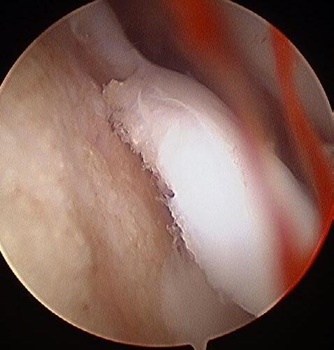
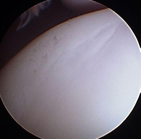
5A and 5B. Arthroscopy images of left and right shoulder
subchondral support for the articular cartilage; this can cause the articular cartilage to crack and cartilage flaps or fragments to develop.
Osteochondrosis latens is when microscopic changes of cartilage necrosis are seen in the growth cartilage and it is not possible to detect this stage in radiographs.
Osteochondrosis manifesta is when there is retention of necrotic cartilage within the ossification front and this stage is visible in radiographs as a subchondral defect.
Osteochondrosis dissecans is when fissures or cracks pass through the articular cartilage into the joint and cartilage flaps and fragments can develop.
It is not always possible to distinguish between osteochondrosis manifesta and dissecans in radiographs since cracks and fissures in the articular cartilage are not possible to detect in radiographs and flaps; fragments of cartilage that are not mineralised will also not be possible to define from the other soft tissue/fluid opacity structures of the joint. However, some cartilage flaps and fragments do have areas of mineralisation; these can be seen as separate structures in the joints space and, in these cases, a diagnosis of osteochondrosis dissecans can be given.
In this case, arthroscopy showed that osteochondrosis dissecans was present in the left shoulder and osteochondrosis manifesta in the right shoulder. Although in the radiographs the subchondral defect and sclerosis was more in the left shoulder than the right shoulder, it was not possible to say with certainty whether the articular cartilage was intact (osteochondrosis manifesta) or whether the articular cartilage was cracked, a flap or a separate fragment (o steochondrosis dissecans).
The osteophytes on the margin of the left shoulder developed as a result of osteoarthritis that had been initiated by the osteochondrosis; usually the osteochondrosis lesions have reached the stage of a dissecans when osteophytes start to be seen in the radiographs
C&T No. 6122, Issue 322, Mar 2026
Dr Moira van Dorsselaer BVSc
The Cat Clinic Hobart
150 New Town Rd
New Town TAS 7008
e. moira@catvethobart.com.au
t. +613 6227 8000
C&T No. 6133
Question
What's your diagnosis?
How Would You Treat This Case?
Raymond is a Male Neutered Domestic Shorthair cat who was found as a stray kitten at 4-5 weeks-of-age. Since then, he has lived indoors with a companion cat.
He presented at 9-months-of-age with what the owner thought was an abscess from the other cat with whom it play-fights.
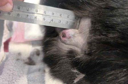
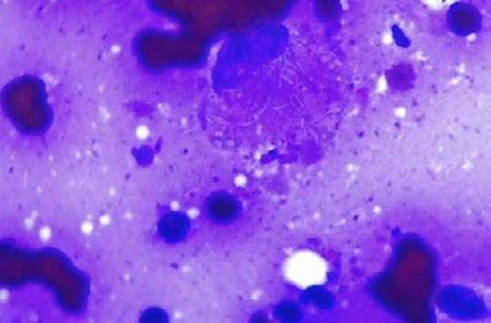
Answer
This case was a reminder of how powerful pattern recognition can be in clinical practice. Having encountered a similar presentation previously—I proceeded directly to cytology. Having seen cytology images shared by our
pathology lab, I quickly recognised the features of feline leprosy.
Interestingly, this presentation differed somewhat from the classic descriptions discussed in continuing education courses, where ‘pepper pot’ lesions are often emphasised. In contrast, the three cases I have encountered have all presented as firm, nodular masses which have subsequently become infected.
In this instance, the lesion progressed to partial tissue sloughing, resulting in an open, purulent wound. The owner had initially been undecided about intervention, but the progression of the lesion ultimately necessitated action. Surgical excision was performed and proved curative.
Adjunctive therapy with doxycycline was instituted post-operatively.
Feline leprosy is an uncommon but likely underdiagnosed condition in Australian cats, particularly those with outdoor access and hunting behaviour. It is seen across the country, especially in temperate regions, and in Tasmania is most commonly associated with Mycobacterium lepraemurium. Transmission is thought to occur via bite wounds sustained during hunting, with rodents acting as a reservoir. There is no strong evidence for cat-to-cat transmission and the zoonotic risk is considered extremely low.
Clinically, these cases most often present as firm, nodular cutaneous or subcutaneous masses, commonly affecting the head, limbs, or tail base. While they may initially be mistaken for abscesses, they frequently fail to behave like typical bite wounds and can progress to ulceration or secondary infection.
Cytology is often the most useful and rapid diagnostic test in practice. Fine needle aspirates typically reveal pyogranulomatous inflammation, with a predominance of macrophages and variable numbers of neutrophils.
A key diagnostic feature is the presence of numerous negative-staining (‘ghost’) rods within macrophages. On Romanowsky stains, these appear as faint or unstained rod-shaped organisms, often clustered in bundles, and are highly suggestive of a mycobacterial infection. Diagnosis can be confirmed with acid-fast staining, such as Ziehl–Neelsen, which demonstrates intracellular acidfast bacilli, although organisms may be less numerous in more chronic lesions.
Treatment depends on the extent of disease. In cases such as this, where a solitary lesion is present, complete surgical excision is often curative and remains the treatment of choice. The use of adjunctive antimicrobial therapy with doxycycline in this case was a reasonable
and practical option, particularly in a compounded form to aid administration. While doxycycline is not considered the most reliable sole therapy for feline leprosy, it can be a useful adjunct following surgery.
In contrast, more extensive, multifocal, or incompletely excised cases typically require prolonged combination antimicrobial therapy. Regimens most commonly include a macrolide such as clarithromycin in combination with rifampicin, with or without clofazimine. These protocols tend to be more effective against mycobacterial infections but require long treatment courses, often continuing for several months and for at least two months beyond complete clinical resolution.
The prognosis for feline leprosy is generally good, particularly for solitary lesions that can be completely excised. More complex cases may require prolonged therapy, and recurrence can occur if treatment is discontinued prematurely.
Dear Editor
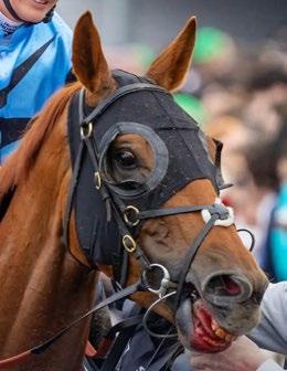
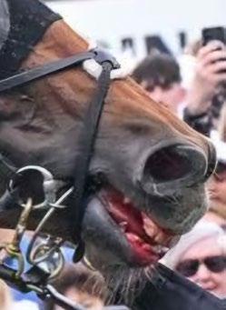
If ever the ethics of Australian horse racing and blind conformity were on world display, it was during the 2025 3,200 metres Melbourne Cup. The winning horse was struck repetitively whilst already trying his best, his tongue was tied tightly around the bottom jaw, the bottom lip appeared swollen, he displayed anxiety and his mouth was bleeding. It was a spectacle of absolute heroism on the horse’s behalf and shame on those who caused this animal pain and suffering. Racing Victoria veterinarians simply reported that the bloodied mouth was due to only a minor laceration requiring no medication, however photos and video tend to show otherwise (Figures 1 & 2)
Congratulations to Kayla Hamilton who has won two $CVE 300 vouchers for answering both C&T No. 6121 and No. 6122.
Read her answers here: cve.edu.au/wyd-6121 cve.edu.au/wyd-6122

The system itself exposed tainted practices of physical and mental abuse of ‘Half Yours’ and the dubious integrity of Racing Victoria.
We, the undersigned veterinary surgeons, understand that ‘tongue ties’ are applied with the intention to prevent dorsal displacement of the soft palate (DDSP) and/or to prevent the tongue displacing over the bit. We also understand this adverse animal welfare practice can cause severe physical and mental harm and has no place in contemporary equestrian sport. This is not a personal crusade but a legitimate pursuit for the welfare of horses and riders. Debate on the aforementioned issues will not fade soon.
Yours sincerely
Dr Peter Kerkenezov BM
Dr Victoria Lomax
Dr John McKenna
Dr Sam Beckett
Dr Doug English
Dr Eloise Koelmeyer
Dr Mary Calder
Dr Steven Roberts
Dr Peita Kerkenezov
Dr Adrian Bryant
Dr Bruno Ros
Dr Tim Mather
Dr
Nikki Whitehouse
Wagga Wagga Veterinary Hospital and Charles Sturt University
e. nikki.whitehouse@waggavet.com.au
C&T No. 6134
Jaffa was a 10-week-old male Border Collie who presented initially for being unwell. On presentation, he had painful, left-sided facial swelling and pyrexia. Initial haematology revealed a moderate non-regenerative anaemia and marked neutropenia (see Table 1.), which worsened on repeat testing. A faecal test for Parvovirus was negative. No significant bony changes were noted on skull radiographs. Due to financial limitations and the clinical suspicion of a possible bacterial infection, he was discharged with meloxicam and amoxicillin-clavulanate.
Five days later, Jaffa returned due to ongoing signs of systemic illness. At this time, he had:
– Dull mentation
– Tachycardia Heart rate: 160 bpm
– No respiratory signs, with clear lung fields
– Fever Temperature: 39.8°C
– Painful facial swelling over the left side, preventing palpation of the left mandibular lymph node
– Mild right-sided mandibular lymphadenomegaly
Jaffa was treated with fluid therapy and buprenorphine. Despite this, Jaffa’s condition deteriorated, with worsening mentation and poor pain control. Radiographs of the skull were reported to be normal for this age dog.
Due to worsening clinical state combined with financial limitations of the owner, a single IV dose of dexamethasone (0.2 mg/kg) was administered. Within one hour, Jaffa was bright, pain-free, and began eating
normally. This rapid response suggested an inflammatory or immune-mediated component to his illness. Empirical antimicrobial coverage was broadened with the addition of enrofloxacin (see note in discussion).
Haematocrit 0.231 0.373-0.617
Neutrophils 0.55 2.95-11.64 x 10⁹/L
Lymphocytes
Table 1.
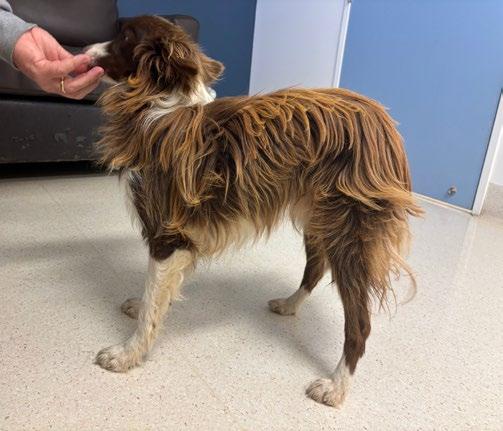
Figure 1. Jaffa side profile at 18 months-of-age
Given the severe neutropenia and suspicion of an underlying immunological disorder, a DNA sample was submitted for Trapped Neutrophil Syndrome (TNS) testing. Jaffa was discharged with amoxicillinclavulanate, enrofloxacin and prednisone (1mg/kg/day).
Within a week, Jaffa was described as ‘completely normal’ by his owners. Repeat haematology revealed neutrophil counts in the low-normal range. However, two weeks after tapering steroids, he re-presented with painful swelling on the opposite (right) side of his face. He was restarted on prednisone at the original dose. Genetic testing confirmed a diagnosis of Trapped Neutrophil Syndrome.
Jaffa is now 18-months-old. He remains bright and clinically well. He is small in stature and has pointed
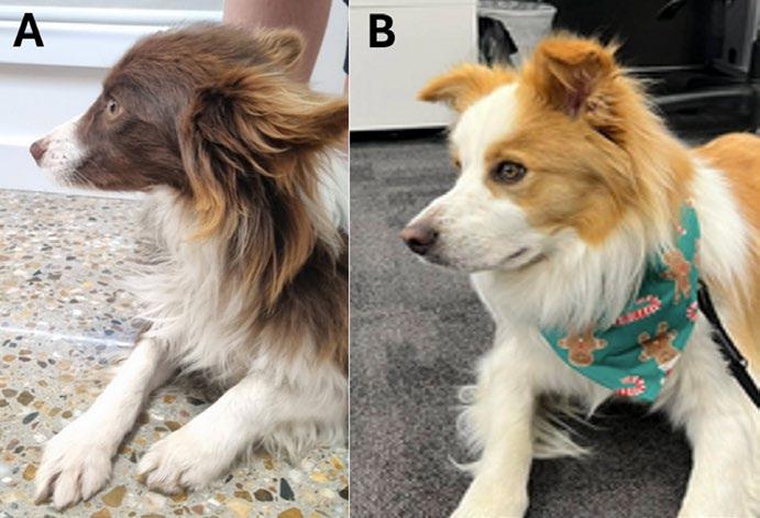
Figure 2
A. Jaffa at 18 months of age. Note his small stature and elongated nose and legs, characteristic of TNS.
B. Border Collie of similar age.
features, characteristic of TNS. He has a coarse, ‘mousey’ coat and weighs only 7kg. His owners have made several attempts to taper corticosteroids which have consistently resulted in clinical relapse. He is well managed on 0.3mg/kg/day prednisolone, which is increased to 1mg/kg/day if he becomes unwell.
Discussion
Trapped Neutrophil Syndrome (TNS) is an autosomal recessive hereditary disorder of Border Collies, caused by a mutation in the VPS13B gene. This syndrome is characterised by a persistent neutropenia, and hyperplasia of the myeloid cells in the bone marrow. This results in poor immune function, and dogs often suffer from frequent infections or complications of immune dysregulation.1 A study performed before reliable genetic testing became available in Australia suggested that the carrier state is present in 7-8% of Border Collies in Australia, making it a rare but widespread condition.2
Trapped Neutrophil Syndrome is usually identified prior to 9 months-of-age, with dogs presenting with pyrexia and lameness. Additional signs that may be present are lethargy, gastrointestinal signs, ataxia, and peripheral lymphadenopathy. Many dogs are smaller than their litter mates and display facial dimorphism (narrow, elongated skull).1,3 Jaffa was most uncomfortable on the sides of the face, which may have been associated with inflammation of the temporomandibular joint. He was poorly responsive to antibiotics, which confirms that bacterial causes of inflammation were unlikely.
Jaffa seems to have responded positively to corticosteroids and, interestingly, has had an increase
in his neutrophil count. We suspect this was due to steroids ability to enhance neutrophil release, although repeat testing has not been able to be performed to see if this was sustained. Sadly, there is no cure for TNS. At this stage, his owners increase the prednisone dose to 1mg/kg/day if Jaffa is unwell and return it to the lowest effective dose when he stabilises. We have not yet had any further secondary infections that have warranted further testing or antimicrobial treatment. At this stage, we have not yet trialled other immunomodulatory agents in Jaffa, given he is going well. It is unknown if there is a role for other immunosuppressants to be used for TNS. There is a single case report where azathioprine was successfully used in conjunction with steroids for the steroid-sparing effect.4 Further studies are needed to determine optimal treatment regimes.
In this case, antibiotic spectrum was broadened to include amoxicillin-clavulanate and enrofloxacin, given the lack of funding to search for a pathogen for culture and sensitivity. There are reports of cartilage damage with the use of enrofloxacin in young, growing animals, and a better option in this case may have been changing from amoxicillin-clavulanate to a cephalosporin to broaden the antimicrobial cover.5
Further testing may have involved blood cultures, arthrocentesis and possibly, bone marrow biopsy. However, in this case, the diagnosis could be obtained definitively and non-invasively with genetic testing. This is an example where by utilising the genetic testing tools that we have available, we were able to affordably and definitively obtain a diagnosis from this patient. It is important that we remember to check for rare
hereditary diseases when presented with a strange presentation in a pure breed animal. Similarly, genetic testing for this disease should be part of our standard recommendations for all Border Collie breeders to help eliminate the occurrence of this crippling and highly fatal disease.
Long term survival for dogs with TNS is not well reported, and in a case series of 12 dogs, 50% were alive at 1 year post diagnosis. We continue to be optimistic that Jaffa will have good and long-term quality of life.
Thank you to Alice Birkhead and Anita Helman for your contributions to this case
References
1. Suciu A, Starybrat D, Gil-Morales C, Matson H, Jepson R, Williams M, Lyraki M, McMahon L, Nerhagen S, Veitch A, Llewellyn E. Clinical findings, treatment and outcome of trapped neutrophil syndrome in Border Collies: 12 cases (2011-2022). J Small Anim Pract. 2024 Jul;65(7):560-568. doi: 10.1111/jsap.13726. Epub 2024 Apr 15. PMID: 38622907.
2. Shearman, J., Wilton, AN. A canine model of Cohen syndrome: Trapped Neutrophil Syndrome. BMC Genomics . 2011; 12:258 12, 258. 10.1186/1471-2164-12-258
3. Mizukami K, Shoubudani T, Nishimoto S, Kawamura R, Yabuki A, Yamato O. Trapped neutrophil syndrome in a Border Collie dog: clinical, clinico-pathologic, and molecular findings. J Vet Med Sci. 2012 Jun;74(6):797-800. doi: 10.1292/jvms.11-0472. Epub 2012 Jan 12. PMID: 22240985.
4. Wouda RM, King TJ, Mackay BM. Long-term Management of Trapped Neutrophil Syndrome in Two Border Collies. Aust Vet Prac. 2010; 40(2):58-63.
5. Papich MG. Saunders Handbook of Veterinary Drugs; Small and Large Animal. 3rd edition. 2011
Trapped neutrophil syndrome in Border Collies: clinical phenotype, diagnostics, pathophysiology and management
Overview
Trapped neutrophil syndrome (TNS) is an inherited disorder recognised predominantly in Border Collies and Border Collie crossbreeds, associated with homozygosity for a VPS13B mutation. Historically framed as a congenital neutrophil disorder with severe neutropenia and
immunodeficiency, TNS is now better appreciated as a broader syndrome in which pyrexia, lameness/ abnormal gait, and gastrointestinal signs are prominent presenting complaints, and in which immune-mediated inflammatory disease (polyarthritis and metaphyseal osteopathy) appears common. Importantly, neutropenia is not invariably present at first presentation, and some dogs show clinical patterns that lead clinicians toward immunosuppressive therapy—sometimes with dramatic initial benefit—yet with a persistent and clinically relevant risk of secondary infection.
TNS most often affects young dogs, commonly in the puppy to juvenile period. A contemporary multiinstitutional case series (2011–2022) described 12 affected dogs (10 Border Collies, two Border Collie crossbreeds) confirmed homozygous for the VPS13B mutation. The typical clinical scenario remains a young Border Collie presented for recurrent or unusual inflammatory disease, poor growth or systemic illness, often with litter history suggestive of autosomal recessive inheritance.
Phenotypic features can include a ‘dysmature’ appearance and fine-boned conformation (sometimes described colloquially as ‘ferret-like’), but this is variable and should be treated as a supporting clue rather than a defining criterion.
Across contemporary case descriptions, a recurring triad emerges:
1. Pyrexia
2. Abnormal gait or lameness (often shifting, multifocal, or episodic)
3. Gastrointestinal signs (diarrhoea ± haemorrhagic diarrhoea; sometimes vomiting, poor appetite, failure to thrive)
In the 12-case series, the most common signs at diagnosis were pyrexia, abnormal gait, and gastrointestinal signs. A representative case report described a 10-week-old Border Collie presented for haemorrhagic diarrhoea and pelvic limb lameness, with examination demonstrating pyrexia and pain/effusion in multiple joints.
On physical examination, many dogs display a systemic inflammatory picture (fever, lethargy) and clear orthopaedic pain. Joint effusions may be appreciable (carpi, tarsi, stifles, elbows) and gait abnormalities can be profound. Some dogs also show dermatological or respiratory infections, but in recent series a clearly identified septic focus was uncommon.
Authors’ views are not necessarily those of
Laboratory findings: neutropenia is important, but not obligatory
TNS is strongly associated with neutrophil abnormalities, but clinicians must avoid anchoring on neutropenia as a mandatory finding at presentation. In the 12-case series, segmented neutrophil counts at presentation were below reference interval in 7/12 dogs, within reference interval in 4/12 dogs, and above reference interval in 1/12 dogs.
This is a pivotal practical point: a young Border Collie may manifest ‘TNS-typical’ clinical disease while the neutrophil count at a single timepoint is not low. Repeated counts over time, context (prior antibiotics, stress leukograms, concurrent inflammation), and genetic confirmation become critical.
Additional haematological and biochemical changes are non-specific: inflammatory leukograms may be muted or variable; mild anaemia of chronic disease may occur; globulin changes can reflect chronic inflammation. Arthrocentesis findings often shape management decisions. In the case report, joint taps showed nondegenerate neutrophilic inflammation across multiple joints, while later in the disease course the dog developed findings consistent with septic arthritis.
Imaging findings: metaphyseal osteopathy / HOD-like lesions
A major shift in how TNS is recognised is the attention to concurrent bone and joint lesions. The 12-case series reported concurrent metaphyseal osteopathy and immune-mediated polyarthritis as common. The case report described capsular joint swelling and heterogeneous metaphyseal lucencies in multiple long bones (distal radius/ulna, femur, tibia). These lesions resemble metaphyseal osteopathy / hypertrophic osteodystrophy (HOD) patterns and support the view that a substantial proportion of cases have a prominent sterile inflammatory orthopaedic component.
The VPS13B association anchors TNS as a genetic syndrome, but the clinical phenotype suggests multiple interacting mechanisms:
1. Innate immune dysfunction (neutrophil-related): even if the absolute neutrophil count is intermittently normal, patients appear predisposed to infection and to atypical inflammatory responses. Functional neutrophil defects (chemotaxis, survival, trafficking, oxidative killing) are plausible contributors.
2. Immune dysregulation and sterile inflammation: the frequency of immune-mediated polyarthritis and metaphyseal osteopathy in contemporary cohorts suggests inflammatory dysregulation is integral to the syndrome, helping explain steroid responsiveness.
3. Secondary infection risk and “septic conversion”: patients may begin with lesions that look sterile/ inflammatory (nondegenerate neutrophilic arthritis) and later develop true septic complications. A documented case improved rapidly with prednisone yet later developed severe erosive polyarthritis and septic arthritis and died despite antibiotics, underscoring the need for ongoing infection surveillance.
approach
Genetic confirmation (VPS13B homozygosity) is the most specific diagnostic test and should be pursued early in any young Border Collie with compatible signs.
Management
often cannot wait for genetic results.
A pragmatic approach includes:
i. CBC/biochemistry with repeated neutrophil counts (trend is more informative than a single value)
ii. Arthrocentesis of multiple joints if effusion/pain is present: cytology and, importantly, culture where feasible—especially if systemic illness is marked or if immunosuppression is being considered
iii. Radiography of long bones for metaphyseal lesions when lameness/pyrexia is prominent
iv. Infectious disease evaluation tailored to geography and risk: faecal/parvoviral testing in haemorrhagic diarrhoea of young pups; blood cultures in persistent fever; imaging/culture of suspected deep infection sites
If a septic focus is identified, consider testing for concurrent canine cyclic neutropenia in rare cases with unusually severe or refractory infection patterns, as dual pathology has been reported.
Therapy: antimicrobials plus targeted immunomodulation, with vigilance
Therapy should be individualised to the dog’s dominant clinical problem at the time (infection vs sterile inflammation), with the understanding that many dogs likely have elements of both.
In contemporary series, all dogs received at least one antimicrobial agent. Empiric antibiotics are reasonable in febrile, systemically ill pups while diagnostic work-up proceeds, but culture-based refinement is ideal. Topical management for otitis/pyoderma and proactive dental care help reduce antibiotic cycling.
Steroids are frequently used in referral practice. In the 12-case series, 10/12 dogs received prednisone/ prednisolone (median starting dose 1 mg/kg/day; range 0.5–2.5 mg/kg/day). In a representative case, prednisone 1 mg/kg q12h produced dramatic improvement within 24 hours.
A balanced interpretation is:
i. Steroids may be appropriate when the syndrome is dominated by painful polyarthritis/metaphyseal osteopathy and no septic source is evident.
ii. Infection risk is real; relapse, fever, or systemic deterioration should prompt re-evaluation, repeat arthrocentesis, and culture as indicated.
iii. Taper to the lowest effective dose; prolonged highdose immunosuppression should be approached cautiously.
Supportive care (analgesia, GI support, nutrition) is essential. The role of haematopoietic stimulation (e.g., G-CSF) is not well defined in these recent summaries and is generally reserved for selected cases with severe, persistent neutropenia and recurrent infection, with careful monitoring.
Prognosis is variable. In the 12-case series, 9/12 dogs survived to 28 days and 6/12 survived beyond 6 months, supporting a nuanced message: long-term survival is possible, but relapse, chronic morbidity, and infectionrelated mortality remain substantial risks. A published case demonstrates potential for initial control of sterile inflammation followed by later lethal septic disease.
> Suspect TNS in young Border Collies with pyrexia, lameness/abnormal gait, and gastrointestinal signs, particularly if metaphyseal bone lesions or polyarthritis are present.
> Neutropenia may be absent at the first visit; do not exclude TNS on a single normal neutrophil count.
> Pursue VPS13B genetic testing early.
> Steroids are commonly used and can be dramatically effective for inflammatory pain/fever, but sepsis surveillance is mandatory; relapse should prompt repeat joint evaluation and culture.
> If a convincing septic focus is identified, consider whether concurrent cyclic neutropenia (rare) might explain unusual severity.
Georgina Milne
Young Veterinary Clinic
341 Boorowa Street
Young NSW 2594
e. admin@youngvetclinic.com.au
t. 02 6382 5155
C&T No. 6135
Read the original case report
C&T No. 6031
‘Winnie’ is a 6-month-old female entire dachshund cross dog who in February of 2024 was diagnosed with a nasal Aspergillus fumigatus infection of the left nostril. In communication with Dr Richard Malik we consequently treated ‘Winnie’ with oral therapy consisting of Posaconazole, Terbinafine and antibiotics based on culture and sensitivity testing which was published in a previous C&T.
Winnie’s treatment was continued for 6 months total. The discharge from the nostril resolved within 6 weeks of treatment and the sneezing reduced over the following
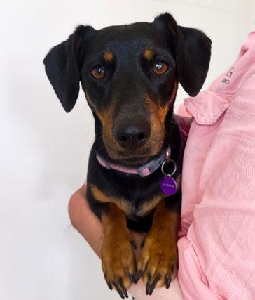
3 months to become steady at a few times a day. On the 6th of September 2024, seven days after finishing the medication, we performed a general anaesthesia and scoped the nostrils. There were no plaques observed at the time which was great. The turbinates were still atrophied in the left nostril and was slightly inflamed however, it had not progressed from the damage observed previously. There was also no sign of infection reoccurring. Winnie then went home to monitor and recheck as needed.
On the 20th of October 2025 we rechecked Winnie and are pleased to report that she is going well and has not had another episode of nasal discharge and owner is very happy with how she is at home. She still sneezes occasionally, however on clinical examination there were no abnormalities detected at the time of examination.
In this case we have had a successful response to treatment and demonstrated that one can consider oral therapy as a treatment option for nasal Aspergillus fumigatus infections in canines.
Winner
Entitled to a CVE$300 voucher
Ian Hodge DipAg BSc DipSci BVSc MANZCVS (Med Dairy Cattle)
FourLimb Vet Consulting Ltd
e. happycows@farmside.co.nz
C&T No. 6136
Ian was a 2024 Beef Production Distance Education participant.
Introduction
What follows is the first of a series of three articles about milking machines, mastitis and the involvement of dairy veterinarians in mastitis control.
The information is based on science and (many years of) experience and is designed to help get veterinarians involved in this area of work.
If anyone needs further information please feel free to get in touch.
Milking Machines
Ever since a successful milking machine was used to extract milk from cows using intermittent vacuum (circa 1917), milking machines have evolved into sophisticated conglomerations of electronics, rubber, steel, motors and pumps.
For some dairy vets that might present a barrier, but in actual fact milking machines are not complicated. Dairy vets should understand milking machines and, importantly, the risks they can potentially pose for mastitis.
A milking machine (herringbone or rotary style) consists of a vacuum pump which is driven by a motor. The pump sucks air from a system of pipes and canisters to create
a vacuum. To prevent the pipes and canisters from imploding, atmospheric air is allowed into the system at various points in a controlled way.
The machine has a main vacuum line (pipe) which takes air away from the cow to the outside. It also has a milk line (pipe) which carries milk (and vacuum) away from the cow to a holding tank (vat). The machine has a pulsation system which allows atmospheric air into the vacuum system intermittently to create a precise alternating vacuum and atmospheric pressure sequence. Milk is ‘sucked’ out of the cow and then drains into a receiving can. From there milk is ‘released’ from the vacuum system into the atmospheric air system and to the vat. The milk releaser/pump is a clever device which allows milk out but retains vacuum. Vacuum is distributed around the entire machine via a sanitary trap (junction). The machine also has its own cleaning system which distributes water and chemicals throughout the system after milking to wash and disinfect all components.
Milk is withdrawn from cow’s udders via rubber milking liners which sit in steel shells which in turn are connected to a ‘cluster bowl’. The cluster bowl contains milk and drains into the main milk line.
The internal space between the liner and the steel shell is called the pulsation chamber.
A vacuum continually exists inside the liner but intermittent admission of atmospheric air and vacuum into the pulsation chamber allows the liner to collapse and expand around the cow’s teat. Pulsation systems can be mechanical or electronic and can either cause clusters to milk two or four teats at one time. Pulsation creates four phases A, B, C and D.
The A phase is the liner opening, the B (milking) phase is the liner fully open, the C phase is the liner closing and the D (rest or squeeze) phase is the liner fully closed. When the liner is open milk is withdrawn from the cow. When the liner is closed the teat is ‘massaged’ or squeezed so that blood and lymph can return to normal levels. An ideal pulsation ratio is 60% opening and open, and 40% closing and closed. Of that ratio the milking phase should be at least 45% and the rest, or squeeze phase, should be at least 25%.
Some milking machines have automatic cluster remover systems. Sensors detect milk flow rates and when the set threshold is reached, vacuum is stopped at the cluster and enters a barrel which pulls (under negative pressure) a string attached to the clusters to pull the cluster off the cow.
Milking machines also have a teat disinfection system. This can be automated or manual. Automated systems
Pulsator
Sanitary
Milk receiver Tanker connection point
Delivery line
Bulk
Basic
(Courtesy of Google images)
Figure 2. The pulsation chamber (Courtesy of Google images)
Teat cup
Short milk tube
Milk claw
Long milk tube
Milking phase (liner opening or open)
Rest phase (liner closing or closed) Working vacuum 4kPa
Figure 3. Pulsation phases (Courtesy of Google images)
discharge teat-spray on to teats after milking. Manual systems require people to spray cows’ teats. Both types of systems carry pressurised teat-disinfection solution.
The vacuum in a milking machine is regulated so it stays within a narrow range. Vacuum regulation is controlled by either regulators or vacuum pump speed controllers. Regulators allow atmospheric air into the system to lower the vacuum. Vacuum pump speed controllers are complex electronic devices which have an internal set point which is the vacuum level that has been chosen for that particular milking machine.
Regulators are often located close to the vacuum pump in the main vacuum pipe close to a large, sealed tank called the interceptor which protects the vacuum pump from any liquids getting into it.
The vacuum pump discharges air to the outside via an exhaust system.
Whilst there are many makes of milking machines the principles are the same for all. At first, some machines can appear complex but sticking to basics and first principles will invariably solve the confusion.
In the next issue we will discuss mastitis and the role milking machines can play in changing incidence and prevalence.
Small Animal Medicine Specialist
1 Richardson Place, North Ryde NSW 2113 (02) 9889 0289
e. alam@sashvets.com
w. sashvets.com
C&T No. 6137
We predominantly use accelerated hydrogen peroxide (AHP) throughout the hospital for both feline / canine / human infectious disease aspects.
I haven’t appreciated marked aversion by cats to AHP as long as the area is left for 5 minutes. It is pretty irritant to airways (humans inclusive). The AHP wipes are great.
Note: take care not to have spray bottle on ‘mist’ mode to avoid inhalation of AHP fumes.
AHP spray is made up 1:16 and has a 90-day shelf-life.
Fluid pumps etc are wiped with Clinell wipes.
Tubes are cleaned thoroughly with detergent and water with a pipe cleaner/ bottle brush, then rinsed and flushed well to remove any detergent, then placed into a 0.5% Chlorhexidine solution for 10-15 minutes; they are then rinsed and hung to dry.
Any damaged tubes or tubes which are excessively contaminated or that have been used in an animal with a known infectious disease are discarded.

Specialist in Feline Internal Medicine
Director Cat Specialist Services
Clinical Lead Tutor Feline Distance Education Program e. drkorman@catspecialists.com.au
We use Oxivir AHP in our isolation ward. When we have known infectious patients in other parts of the hospital, we do a clean down with it, but we don't employ it as standard as cats seem to really hate the smell of it. Apparently in the States there is a version called ‘Rescue’ which has less odour but that is not available in Australia.
We wash down cages with detergent, allow to dry, then use Virkon™ then wipe out again.
Litter trays have a separate wash area—detergent, then Virkon™.
Bowls—washing machine or detergent hot handwash, Virkon™, then wash again.
Note: Virkon™ active ingredients include potassium peroxymonosulfate (an oxidizing agent), sodium dodecylbenzenesulfonate (a detergent), sulfamic acid (a cleaning agent), and inorganic buffers.
Tubes are cleaned with detergent and water and then gassed if reused (which we can use in bulk). If used in a known infectious patient, we bin them afterwards.
3B 3C
Figure 3A,B & C. ET tube drying rack made with pegboard from IKEA (https:// www.ikea.com/au/en/p/skadis-pegboardcombination-s69406366/)
Remove organic matter before applying a suitable disinfectant, ensuring adequate contact time, and following manufacturer guidelines for both cleaning and disinfection to optimise infection control.
– Clean thoroughly with detergent to remove organic material.
– Then, immerse in or wipe with a TGA-registered hospital-grade disinfectant (such as Virkon™, accelerated hydrogen peroxide, F10, or other approved agents), ensuring the recommended contact time (often 5–10 minutes).
– Rinse thoroughly with clean water, especially for items that contact mucous membranes, to remove disinfectant residue, and dry before reuse.
– Clean with detergent to remove food debris or biofilm.
– Disinfect using a suitable veterinary disinfectant at the recommended concentration, with proper contact time.
– Rinse carefully to avoid chemical residue that could be ingested by animals and air-dry completely.
– Handle soiled linen with gloves and minimise disturbance to avoid spread of pathogens.
– Launder at the hottest water temperature safe for the fabric, using detergent and, where possible, a disinfectant wash additive.
– If heavily contaminated, use an autoclave-capable or high-temperature wash/dry cycle.
– Ensure staff use PPE when handling potentially infectious laundry and follow safe disposal or cleaning protocols.
– Remove visible organic debris before applying any disinfectant, as this can otherwise reduce efficacy.
– Select a disinfectant proven effective against the pathogens of concern and safe for the intended material.
– Always follow label instructions for dilution, application, and contact time, and allow items to dry before reuse.
– Maintain high standards of staff hand hygiene and use of personal protective equipmentPPE) throughout cleaning processes.
Jen Nesbitt-Hawes
BVSc MVSt MANZCVS (Veterinary Behaviour)
Owner of Pet Perspective, a provider of behaviour resources for vets and trainers
e. jen@petperspective.com.au
C&T No. 6138
The CVE Behaviour conference in February 2026 was a fabulous opportunity to pick up behaviour hints and tips from some wonderful behaviour specialists. Here’s a challenge for every clinic.
People love photographs of themselves with their pets, whether these are selfies, or poses for a photographer. Unfortunately, in many cases, although the person in the photo has an expression of delight, the non-human animal is showing body language of discomfort. A quick search online will bring up images like this:
Figures 1-4. Pets looking uncomfortable during interactions
One of our key roles as pet professionals is to educate clients on understanding body language in their pets. This basic skill is essential for: effective socialisation, ethical training, advocating for pets, building a close bond with the animal and minimising bite risk/ risk of injury. A 2009 study demonstrated that ‘most dog
owners report having a good understanding of the emotional state of their dogs, when they seem to have low appreciation of the signals that dogs send in the earlier stages of emotional arousal’. This is dangerous, as people assume they know what their pet is feeling, when really they can’t pick up the early signs of discomfort. This is one of the reasons that people may report a bite ‘out of nowhere’.
Key signs that an animal is uncomfortable vary between species. Muscle tension, looking away from the person, or a lack of engagement are common. For dogs, some other obvious signs of discomfort that people should be taught to look for include:
– Licking the lips
– Yawning
– Closed mouth
In many photographs, the human is hugging or holding the animal. This is a behaviour enjoyed by social primates. Unfortunately, it is not a behaviour enjoyed by many other species, like dogs (tolerating is not enjoyment). It’s not all bad though – it IS possible to have photographs of both people and pets enjoying themselves. In general, there is willing engagement in the interaction, relaxed muscles, and a soft eye expression. Other signs to look for in dogs include:
– bouncy movement
– open mouth with gentle pant and upper lip drooping
– tongue lolling
As veterinarians, many of us provide photographs in a professional capacity, whether this is on a vet website, a speaker profile, or on social media. People look at these images and assume that our behaviour around animals is appropriate. We should lead by example and provide photographs where both parties in the photograph are enjoying themselves. To that end, I give you the photograph challenge –
– Find and review the photo(s) you’ve already taken, posing with a pet. Does the pet really look like it is enjoying itself?
– Take a photo of you with a pet following these rules: The pet is doing something they enjoy. You are not disturbing their enjoyment. You do NOT have to be touching (or even close to) the animal for it to be a great photo.
– Replace all your social media posts or pet professional portraits with photos where both/all species are having fun!
To relieve the feelings of any readers who discover that their furry friend was basically just tolerating their behaviour for a photo, I’ll share with you my own before and after:
Figures 5 & 6. Before and after.
Figures 7-9. Animals looking comfortable in the presence of people
Hopefully our veterinary galleries will start to contain more photos that show our clients not just that we like animals, but also that we take care to ensure that animals are as comfortable as possible with us.
References
Kerswell, K. J., Bennett, P. J., Butler, K. L., & Hemsworth, P. H. (2009). Self-reported comprehension ratings of dog behavior by puppy owners. Anthrozoös, 22(2), 183-193.
Lore Haug https://www.michaelsdogs.com/2015/02/24/what-pantingmeans-not-all-panting-is-the-same/
Perspective No. 166 published in Control & Therapy Issue 320, September 2025, pages 36-47.
Following publication of the above article, the Editorial Office has been notified of inaccuracies affecting the bibliography and one figure.
1. Bibliography Update
The bibliography in the originally published version contained some errors in the reference list. A revised and fully corrected bibliography has now been compiled and the updated reference list replaces the version previously published.
2. Figure Correction
An incorrect version of Figure 4 was included in the original publication. The corrected figure has now been provided and replaces the previous version. The correction does not affect the interpretation of the article
These corrections have been implemented in the online version of the article as of 1 June 2026. The Editorial Office apologises for any inconvenience caused to readers.
The International Cat Care Veterinary Society (formerly ISFM) is the veterinary membership division of pioneering cat welfare charity International Cat Care (iCatCare), bringing together a global community of veterinary professionals dedicated to improving the lives of cats worldwide. Trusted by vets and nurses, it provides professional development and CPD through access to expert knowledge resources, including the Journal of Feline Medicine and Surgery (JFMS). The iCatCare website is also a trusted resource of information and guidance for veterinary professionals, cat owners and caregivers.
Feline education partner with the CVE delivering the Feline Medicine Distance Education course. cve.edu.au/feline-medicine
Deborah Caunter
BSc Hons RVN ISFM CertFN RECOVER C&T No. 6139
Urethral obstruction remains a common but potentially fatal feline emergency, requiring rapid stabilisation and intensive ongoing care. In this case, Deborah Caunter follows a young male cat from initial presentation through emergency treatment, highlighting the severe metabolic and cardiac complications that can arise, including hyperkalaemia and dysrhythmias.
A key focus of this article is the critical role of veterinary nursing in achieving stabilisation and supporting recovery. From low-stress handling and timely analgesia to IV catheter placement, fluid therapy, and continuous monitoring, nursing interventions are central at every stage. Ongoing care—including urinary catheter management, pain scoring, and close monitoring for post-obstructive diuresis—demonstrates how attentive nursing can directly influence patient outcomes.
The case also reflects the realities of practice, including how financial constraints can impact treatment decisions, and extends into prevention through nurse-led multimodal environmental modification (MEMO) strategies.

Read the full article here

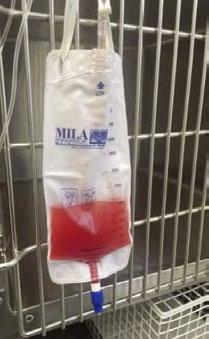
Welcome to Research Roundup where we bring you summaries of the latest feline research. Continuing our diabetes mellitus theme, we have a close look at the FreeStyle Libre 3 device, examine how common hypersomatotropism is in Australia and look back at some research on a novel insulin, glargine 300 U/ml, which is used increasingly frequently to treat diabetic cats.
cve.edu.au/rr-june-26
Elizabeth
BVSc (Hons) FANZCVS (small animal internal medicine)
The Pet Specialists
16 Myoora Road, Terrey Hills NSW 2084
e. info@tps.vet / elizabeth.thrift@tps.vet
t. 02 9452 2933
When a dog presents with a cough, recurrent pneumonia or vague swallowing issues, the immediate lines of thought often lead to primary airway disease, infection or even simple inhalation events. Yet, a growing body of evidence highlights a less obvious, but critically important, group of disorders that bridge the digestive and respiratory tracts: Aerodigestive Disorders (AeroDs). These conditions deserve a place on every general practitioner’s differential list because they frequently masquerade as respiratory disease, may go unrecognised in primary care and carry significant implications for patient outcomes.
Aerodigestive disorders in dogs include dysfunctions of swallowing, oesophageal motility, reflux (and gastrooesophageal or extra-oesophageal variants), and airway protection failures. These can lead to aspiration events, coughing, recurrent pneumonia and chronic airway inflammation, even when classical digestive symptoms (vomiting, regurgitation) are absent. The respiratory and alimentary tracts share protective mechanisms; when swallowing is compromised or reflux is unchecked, airway injury becomes a downstream problem.
Despite their importance, AeroDs remain underrecognised across veterinary practice. One canine study found that over 75% of dogs presenting with cough, but no overt digestive signs, had abnormalities on videofluoroscopic swallow study (VFSS). Without recognition, these cases may be mismanaged as recurrent infections or treated with empiric antibiotics rather than also addressing the underlying dysmotility or reflux.
Timely identification can break the cycle of aspiration, inflammation and chronic respiratory damage.
For your everyday practice, screening for AeroDs is about recognising red flags.
Night-time restlessness, intermittent excessive swallowing, or recurrent cough/aspiration pneumonia despite standard therapy should raise suspicion.
While any breed can be affected, brachycephalic dogs are over-represented. Anatomical predispositions (hiatal hernia, redundant oesophagus) and negative intrathoracic pressures driven by airway obstruction combine to increase risk. However, the alert vet will also recognise AeroDs in non-brachycephalic patients presenting with coughing or recurrent pneumonia that doesn’t respond as expected.
Clinical signs may include:
– Chronic or recurrent cough (especially if feeding or lying down)
– Nasal discharge
– Reverse sneezing, gagging or retching
– Excessive salivation, lip licking or head shaking
– Regurgitation or night-time restlessness
– Excessive or difficulty swallowing
– Weight loss or failure to thrive
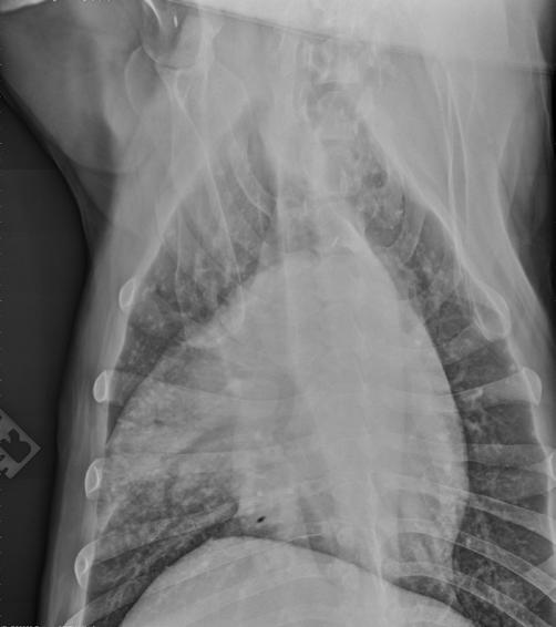
Initial assessment may be within your toolkit: thoracic radiography, basic screening bloods (CBC/biochemistry, TT4 +/- ACTH stimulation test) and, as always, a good clinical history. Increased oesophageal gas or consolidation in ventral lung lobes should raise concern. The gold standard diagnostic tool is VFSS, a dynamic imaging modality that visualises swallowing, reflux and even sometimes aspiration in real time. While VFSS may not be available in all practices, its use highlights the kind of cases that benefit from specialist referral. Recognising when to refer for advanced diagnostics or specialist collaboration is a key role of the vigilant GP clinician.
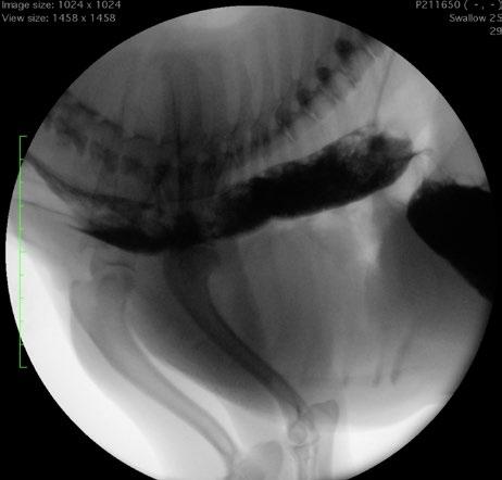
Once AeroD is identified, the management paradigm shifts from repetitive antibiotic cycles toward multidisciplinary care. Feeding modifications (elevated feeding, small frequent meals, feed consistency, dietary fat consideration), medical therapy (e.g., proton-pump inhibitors, pro-motility agents) and in some cases surgical correction of anatomical defects may form part of the plan. For your practice this means:
– Rather than repeating antibiotic courses for ‘aspiration pneumonia’, ask: Why is this happening?
– Monitor feeding behaviours, swallowing quality, nighttime signs or restlessness.
– Engage clients in understanding long-term management rather than just episodic treatment.
– Refer earlier when the history is suggestive of AeroD: e.g., recurrent aspiration, unexplained cough, or after first pneumonia episode when risk factors are present.
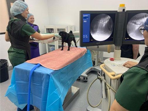
Aerodigestive disorders may sit at the intersection of respiratory and gastrointestinal medicine, but they are very much relevant to primary practice. Having them on your radar means that when a patient doesn’t ‘fit’ the standard respiratory or GI narrative, you pause and ask deeper questions. In doing so, you’re not just managing disease, you’re breaking the cycle of aspiration, protecting airway health and improving the quality of life for pets in a meaningful way.
Aerodigestive disorders refer to a spectrum of conditions in which dysfunction of the swallowing mechanism or failure of airway protection leads to respiratory disease. In these disorders, there is pathological interaction between the respiratory and gastrointestinal tracts – for example, aspiration of gastro-oesophageal contents into the airways can initiate or exacerbate pulmonary conditions. Clinical manifestations range from chronic coughing (with no overt gastrointestinal signs) to severe aspiration pneumonia. Historically, veterinarians tended to evaluate respiratory and digestive problems separately; however, growing evidence has highlighted that overlapping ‘aerodigestive’ conditions are more common and under-recognised than previously thought (Grobman and Reinero, 2023). Increased awareness of aerodigestive disorders is vital, as timely identification and management of both the airway and gastrointestinal components can significantly improve patient outcomes.
One of the presentations of an aerodigestive disorder in dogs is a chronic cough or other respiratory signs without concurrent vomiting, regurgitation, or other obvious gastrointestinal (GI) symptoms. In such cases, clinicians should maintain a high index of suspicion for
occult aspiration or reflux contributing to the respiratory issue. A study of 31 dogs that presented exclusively for chronic cough (with no GI signs) found that over 80% had abnormal swallowing or aspiration events detected on videofluoroscopic swallow studies (Grobman et al., 2019). Notably, many of these dogs had normal thoracic radiographs, yet still demonstrated significant swallowing abnormalities (Grobman et al., 2019). Similarly, a more recent study using videofluoroscopic swallow evaluations reported that 75% of dogs with chronic respiratory disease had one or more co-existing digestive tract abnormalities despite lacking any overt signs of regurgitation or vomiting (Howard et al., 2023).
It is also important to recognise that routine diagnostic imaging may not immediately reveal evidence of aspiration. Thoracic radiographs can appear unremarkable early on, particularly if only mild airway inflammation is present or if aspirated material has caused chemical pneumonitis without secondary infection. Radiographic changes may only become evident later, after progression to pneumonia. Aspirated gastric contents often affect the ventral lung lobes, especially the right middle lung lobe, which on lateral radiographs can be obscured by the cardiac silhouette, further contributing to missed diagnoses. Thus, a normal chest radiograph does not rule out an aerodigestive disorder. Clinicians should remain vigilant in chronically coughing dogs, particularly if signs worsen during eating or drinking or when recumbent.
Certain well-recognised conditions illustrate the link between swallowing dysfunction and respiratory disease. Dogs with megaoesophagus are prone to regurgitation and aspiration, frequently developing recurrent aspiration pneumonia. Dogs that have undergone unilateral arytenoid lateralisation for laryngeal paralysis have a permanently open glottis and are similarly at risk of inhaling food or liquid. Geriatric onset laryngeal paralysis and polyneuropathy (GOLPP) combines laryngeal dysfunction with oesophageal motility deficits, creating substantial aerodigestive risk. Even vomiting dogs may aspirate if consciousness or airway reflexes are impaired.
The central mechanism in canine aerodigestive disorders is aspiration of gastric or oesophageal contents into the airways. Gastro-oesophageal refluxate may contain acidic gastric juices, bile acids, and digestive enzymes. When inhaled, even in small volumes, these substances can provoke significant inflammation of the larynx, trachea, bronchi or lung parenchyma, leading to chronic cough or chemical pneumonitis. Repeated microaspiration may result in chronic bronchial remodelling or fibrosis.
Evidence for this pathway has been demonstrated experimentally. Elevated bile acids have been detected in bronchoalveolar lavage fluid and saliva of dogs with chronic respiratory disease compared with healthy controls, confirming reflux aspiration (Kouki et al., 2023). Subtle signs such as lip smacking, excessive swallowing, nocturnal restlessness, neck extension or gagging may indicate extra-oesophageal reflux and are frequently overlooked.
Normal anti-reflux defences include lower oesophageal sphincter (LES) tone, diaphragmatic support and positive abdominal pressure. Hiatal hernia disrupts these mechanisms. In sliding hiatal hernia, the stomach and LES intermittently move into the thorax, placing the sphincter in a negative-pressure environment that promotes reflux during inspiration. Obesity and pregnancy can further increase intra-abdominal pressure and predispose to reflux.
Aerodigestive disease results from failure of separation between airway and gastrointestinal tract, whether due to anatomical or functional defects. Addressing only respiratory consequences without controlling reflux is therefore unlikely to achieve lasting success.
Brachycephalic breeds are over-represented among dogs with aerodigestive disorders. These dogs commonly exhibit brachycephalic obstructive airway syndrome (BOAS) alongside oesophageal dysmotility and hiatal hernia. Computed tomography studies demonstrate that brachycephalic dogs have significantly larger oesophageal hiatuses than mesocephalic dogs, predisposing them to herniation (Conte et al., 2020). Gastro-oesophageal reflux is common, with 84% of brachycephalic dogs demonstrating acid reflux episodes on pH monitoring (Appelgrein et al., 2022).
Upper airway obstruction generates marked negative intrathoracic pressure during inspiration, which may draw the stomach cranially through the diaphragm and exacerbate herniation (Conte et al., 2020). Similar mechanisms operate in dogs with laryngeal paralysis, particularly those affected by GOLPP.
Chronic lower airway disease may also be perpetuated by occult aspiration, while respiratory compromise may secondarily impair swallowing. Although brachycephalic breeds are at highest risk, aerodigestive disease can occur in any breed (Howard et al., 2023).
Diagnosis often requires advanced investigations beyond routine radiography. Videofluoroscopic swallow study (VFSS) is the most informative test, allowing real-time
assessment of swallowing, aspiration and reflux. VFSS can reveal abnormalities undetected on static imaging (Howard et al., 2023). Diagnostic yield can be increased by gentle abdominal compression during fluoroscopy to provoke transient herniation (Reeve et al., 2017).
Upper gastrointestinal endoscopy enables direct visualisation of oesophageal inflammation and the gastro-oesophageal junction. Sliding hiatal hernia may be induced during endoscopy by brief airway occlusion or abdominal pressure (Broux et al., 2018). Oesophageal pH monitoring can quantify reflux episodes (Appelgrein et al., 2022). Bronchoscopy with BAL may identify aspiration markers such as bile acids (Kouki et al., 2023).
Diagnosis relies on careful history, targeted imaging and combined evidence from multiple modalities (Grobman and Reinero, 2023).
Management requires control of reflux, treatment of airway disease and prevention of aspiration.
Proton-pump inhibitors are first-line therapy. Prokinetic agents such as cisapride enhance LES tone and gastric emptying (Broux et al., 2018). Sucralfate protects inflamed oesophageal mucosa. Feeding modifications include small frequent meals, elevated feeding and avoidance of late-night meals.
Underlying airway disease should be addressed surgically where indicated. BOAS surgery reduces inspiratory effort and may resolve hiatal herniation (Poncet et al., 2006). Laryngeal paralysis surgery improves airflow but increases aspiration risk, requiring lifelong management strategies.
Surgical repair of hiatal hernia may involve phrenoplasty, oesophagopexy and gastropexy, with or without fundoplication. Dogs with persistent herniation often improve following surgery (Poncet et al., 2006).
Aspiration pneumonia requires prompt antibiotic therapy and supportive care. Long-term monitoring is essential.
Figure 2. Brachycephalic breeds are overrepresented among dogs with aerodigestive disorders.
Multimodal therapy yields best outcomes, with combined airway and gastrointestinal management significantly improving clinical signs (Poncet et al., 2006).
Aerodigestive disorders represent an important intersection between gastrointestinal and respiratory pathology. Chronic cough, recurrent pneumonia or unexplained respiratory disease should prompt investigation of swallowing and reflux disorders. Recognition of these relationships enables targeted diagnostics and integrated treatment, improving quality of life for affected dogs (Grobman and Reinero, 2023).
Appelgrein, C., Hosgood, G., Thompson, M. and Coiacetto, F. (2022) ‘Quantification of gastro-oesophageal regurgitation in brachycephalic dogs’, Journal of Veterinary Internal Medicine, 36(3), pp. 927–934. https://doi.org/10.1111/jvim.16400
Broux, O. et al. (2018) ‘Effects of manipulations to detect sliding hiatal hernia in dogs with brachycephalic airway obstructive syndrome’, Veterinary Surgery, 47(2), pp. 243–251. https://doi.org/10.1111/vsu.12702
Conte, A. et al. (2020) ‘Computed tomographic comparison of oesophageal hiatal size in brachycephalic and non-brachycephalic dogs’, Veterinary Surgery, 49(8), pp. 1509–1516. https://doi.org/10.1111/ vsu.13521
Grobman, M.E., Masseau, I. and Reinero, C.R. (2019) ‘Aerodigestive disorders in dogs evaluated for cough using respiratory fluoroscopy and videofluoroscopic swallow studies’, The Veterinary Journal, 251, 105344. https://doi.org/10.1016/j.tvjl.2019.105344
Grobman, M. and Reinero, C. (2023) ‘A One Health review of aerodigestive disease in dogs’, Journal of Veterinary Internal Medicine 37(3), pp. 817–834. https://doi.org/10.1111/jvim.16661
Howard, J. et al. (2023) ‘Videofluoroscopic swallow study abnormalities identify aerodigestive disorders in dogs with respiratory disease versus healthy controls’, Journal of Veterinary Internal Medicine, 37(3), pp. 1166–1178. https://doi.org/10.1111/jvim.16685
Kouki, S. et al. (2023) ‘Extra-oesophageal reflux and reflux aspiration in dogs with respiratory diseases and in healthy dogs’, Journal of Veterinary Internal Medicine, 37(1), pp. 268–276. https://doi.org/10.1111/jvim.16622
Poncet, C.M. et al. (2006) ‘Long-term results of upper respiratory syndrome surgery and gastrointestinal tract medical treatment in 51 brachycephalic dogs’, Journal of Small Animal Practice, 47(3), pp. 137–142. https://doi.org/10.1111/j.1748-5827.2006.00057.x
Reeve, E.J. et al. (2017) ‘Documenting the prevalence of hiatal hernia and oesophageal abnormalities in brachycephalic dogs using fluoroscopy’, Journal of Small Animal Practice, 58(12), pp. 703–708. https://doi. org/10.1111/jsap.12734
Cat Specialist Services
1-15 Lexington Rd, Underwood QLD 4119
t. 1300228377
Dr Nick Cleland
Specialist in Small Animal Surgery
– Never get frustrated in surgery and don’t take shortcuts—neither result in better outcomes and can often open the door to complications
– If you are having a bad run of cases or outcomes, remember that at some point in the future things will be OK
– Don’t rule out a post-renal azotaemia without imaging
Dr Brad Gavaghan
Specialist in Cardiology
– B-line assessment on T-Fast scans are helpful in emergencies, but are not a replacement for thoracic radiographs
– E xcessive/stressful handling can result in acute decompensation in feline cardiac patients
– Cage O₂ with a known inspired O₂ concentration is advised for congestive heart failure patients. Other forms of O₂ supplementation tend to be either dangerous or stressful for feline patients.
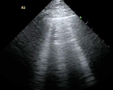
Figure 1. T Fast scans are not a replacement for radiology.
Andrea Harvey
Specialist in Feline Medicine
– Care about every patient as if they were your own pet. Not only will this help you to give the best patient care, but it will also inspire you to be empathetic to their owners; it will motivate you to read around every case, and enthuse you to keep learning. And this will give you enormous job satisfaction and a sense of fulfilment that will make you fall in love with being a vet; it will keep your morale up during the tough times and help you to continue striving to be the best vet you can be.
– Treat your veterinary team like a second family, and your nurses like gold dust. They will support you through challenging times and make you love going to work every day. If you can’t find this family-like quality in your veterinary team, look for another job; it isn’t failing to leave a practice that isn’t right for you—the right practice and the right veterinary family for you are out there but it may not be the first job you take.
– Stay in contact with your vet school friends and mentors—reach out if you’re struggling—they will understand and know how to help. Don’t forget to breathe and smell the roses—you’ve achieved your dream of becoming a vet, enjoy helping animals and their owners, and continuously learning more!
– Being a veterinarian is a continuous learning journey— you will never know everything and should not be expected to be a perfect clinician at the time of graduation. It will be a career of ups and downs and please do not let the ‘down moments’ dishearten you
– Keep in close contact with your peers in vet school— the network remains invaluable as everyone sees different types of cases throughout the start of the journey and it allows everyone to learn from each other, including the mistakes
– Always be honest in your communications with the clients. Clients will appreciate the honesty even if you say ‘I don’t know’ or if complications occur. Hiding information or sugarcoating it can sometimes compound issues.
A Surgeon's Standpoint
– Complications will occur eventually and are part and parcel of the job. Any surgeon who says they never have any complications has either not done enough surgery or is lying...
– Always read up before any surgery you are doing for the first time—we all have done surgery with textbooks open. It is a normal process. Lean on the mentors within your practice for advice and experience. Feel free to also give us a call at VSS to run through these cases if you have any.
Feline Medicine Resident
– Treat your nurses / team well, they can make your life much easier (or the other way around if they want...). Talk to your nurses and work with them.
– There’s no silly question; if in doubt, just ask
– You know more than you think you do
– Treat the animal (and sometimes the human) not just the numbers, understand what the client’s concerns ‘actually’ are and work out a plan with them
– Keep your own case log for interesting / challenging cases but try NOT to bring it home
Specialist in Feline Medicine
– Look at the patient as well as the tests
– Make sure you have a systematic physical examination and repeat the examination if needed. Cats don’t give up clues easily. Start your exam with the distant examination hands-off then examine the head, mouth and neck (thyroid). Palpate the abdomen (including kidneys /bladder in all cats) and conduct a thoracic auscultation—heart and lung sounds.
– Talk through cases with colleagues— there are no stupid questions!
– Have a Plan B (and C, D, E etc)
– Have a cheat sheet/book with common drug doses and treatment protocols. Best if small and pocket sized.
– You cannot express a male cat’s bladder awake (unless they have lower motor neurone disease). I would recommend not trying.
– The only reliable myotactic reflex in the cat is the patella reflex
– Weigh cats every time you see them and at least every 24 hours while in hospital
2. Weigh cats every time you see them and at least every 24 hours while in hospital
Rachel Korman Specialist in Feline Medicine
– Keep in touch with friends from uni—they will have similar experiences to you and it is really important to share the highs and the lows. Check in with each other.
– Develop ‘cheat sheets’—important information that you can refer to. Write everything into a book. It will become your safety blanket.
– Talk about your cases with others but remember there are many opinions
– If there are multiple different treatments for a disease it means there is no one perfect one
– Keep a record of your cases that have gone really well. Reflect back to this when you get tough cases. Don’t forget your tough cases—learn from them. They will happen.
– It takes years to develop a good bedside (or consult table side) manner. Don’t expect it instantly.
– Never try and talk someone out of euthanasia for their pet. People have all sorts of different background experiences and problems in their lives. It is impossible to put yourself in everyone’s shoes. Support them in their decision. Never rush a euthanasia.
– Auscultation time can also be thinking time
– Go slow to go fast with cats
Specialist Small Animal Internal Medicine
– Have someone either within or outside the practice you are happy chatting with about difficult cases, difficult clients,—YES we all have both even after 40 years of practice. Also about ‘Cool’ cases—we need someone who also gets excited about the stuff we get excited about!
– Have a couple of mentors—one who is a good friend you graduated with as you can absolutely relate to all of the same issues and also a ‘senior’ who has been there before but may have a bit more experience
– Go along to local branch meetings / conferences— meet your colleagues!
– Accept that our patients don’t always go to plan— money issues, client issues, patient issues…
– Having some interests outside work also helps
Dr Cindy Hung
Emergency & Hospital Vet
– Your entire team (reception, nurses, vets) are all there for the same reason. Help each other out and work as a team.
– Communication is important and is a skill that needs working on for everyone
– Accidents happen, big or small. Feeling bad about it is normal. Reflect on what lesson it teaches you and move on.
– Everyone has different stories. Listen to the client’s stories and work with them. Getting them on your side will make your job easier.
– Looking after yourself at work and outside of work is an intentional skill. Have plans for this. Don’t wait until you are already exhausted. This will look different for everyone and change over time for you.
Dr Richard Malik
Professor in Feline Medicine & Valentine Charlton Cat Specialist, Centre for Veterinary Education
– Don’t be too hard on yourself
– Find a few good mentors
– Read lots at night
– Use https://www.perplexity.ai/?focus=internet instead of Google – it gives a better-informed search—and you can see where the recommendations come from.

Dr Elizabeth Thomas
Veterinarian
and work mum
– Use a notebook and learn doses of just a few common drugs. Write down other doses as you come across them and become familiar.
– Try to get in a good routine for physical examination and history taking and use any spare time to listen to / palpate and ultrasound everything normal! For example, ultrasound the spays and castrates, just one organ in recovery, to get to know normal.
– Having someone safe to talk to if you are worried / make mistakes is so important. Maybe not the business owner or partner, someone that can be a neutral sounding board. Often just talking through an issue helps, or if you are feeling overwhelmed, they may offer to have difficult conversations with clients for you or alongside you. It’s always good to not feel isolated and share the burden.
Specialist Small Animal Internal Medicine
– It’s OK to say, ‘I don’t know’ as long as you follow that up with ‘but I will find out’; (what is that change on the radiograph? Or what do those blood results mean?). Many clients are happy that you are looking into something for them but make sure you get back to them.
– Make sure in your medical records you write what you think is wrong with the pet—assessment and plan are the parts that will help you and your colleagues (and specialists you might send the case to), even if you don’t have the diagnosis correct, we understand what you are thinking which helps so much.
Specialist Small Animal Surgery
Past President of the ANZCVS Surgery Chapter
– Your feet will hurt for the first few weeks—sit down while you can!
– I would invest in a few textbooks that are your own, so can sit at your desk or in your consult room (write your name and number on them!) like The 5-Minute Consult and a surgical and medical textbook. For surgery (I’m a surgeon), I recommend Tobias.
– Stick at it! The first couple of years tend to be the toughest, but with time and confidence you will find your way, and it can be a rewarding and fun career.
– Be honest with pet owners about your knowledge. Owners are generally very happy for you to say you need a bit of time to think/research/ask a more experienced colleague. Even as a specialist, I will frequently say to owners that I wish to discuss the case with the other surgeons. Never feel like you are alone, as the veterinary profession is a team sport. You can always call us at VSS/CSS re cases. We love helping new graduates!
– It helped me to remind myself that every day was different. One day that I felt completely out of my depth was quickly followed by a day where I felt I did a great job.
– Focus on the wins. You will encounter complications
and failures, but don’t let negative thoughts about cases consume you.
– https://www.wiley.com/learn/5mvc/pdf/blackwellsfive-minute-veterinary-consult.pdf
Head of School and Dean University of Sydney Professor of Veterinary Microbiology and Infectious Diseases
– Everyone in the team around you is a source of great learning and support—the owner, administration staff, vets and especially the nurses
– Treat everyone with the same high level of respect
– Each patient with a specific disease will become the poster child for learning and remembering new information about that clinical condition that will stay with you for years. Take the time to reflect on how this patient has or has not read the textbook on this condition. These cases as a new graduate will cement your memory.
– Being a veterinarian is as complex as learning an instrument. You rarely become good at playing without practice, dedication and accountability. But everyone will have tunes they are great at playing and others they will need accompaniment with and that is OK.

Visit cve.edu.au/vetcookbookii to find out about the vet cookbook, a veterinary collegial collaboration that is all about celebrating the wins!
Student discount available.

Coloured coded minimum volume extension tubing for use with Syringe drivers
Features
· Luer Lock fitting enables compatibility to all syringe drivers
· Pressure resistant up to 2 bar
· Made with PE (Polyethylene), does not contain PVC, DEHP or LATE X
· Small tube diameter (1.0 x 2.0 mm) to reduce residual volume
· Available in 4 colours and two lengths
· Low sorbing properties
· Excellent start-up characteristics
· Kink resistant