







Kiaora,
Welcome to the first edition of the NZSSA Inc. Supplyline for 2026. We start off another year with a range of information in Supplyline. Along with articles we have updates from both Toi Ohomai and the Open Polytechnic. There is news about the NZSSA Inc. Annual Conference in September 2026, it is our 50th Conference this year. What a huge achievement.
There is also information on the upcoming introduction to our new website. Antony and Alison have spent a lot of time and effort in getting it to this stage. You will also find adverts and business cards from companies who continue to Support the NZSSA Inc.
I would like to encourage you all to send in information and photos etc for each edition. It could be around a quality improvement project; or equipment upgrades or a special milestone for a staff member in your unit. It is a great way to share information with each other around New Zealand.
Ngã mihi
Aileen Derby Co-Editor NZSSA Inc. Supplyline

I hope everyone has had a great summer. I am looking forward to what the next 12 months will bring for the New Zealand Sterile Science Association.
Planning for this year’s conference is progressing well. We have some excellent speakers lined up, along with a strong trade presence. The conference will be held in Christchurch at the Te Pae Convention Centre. The conference dinner will take place at the Air Force Museum in Wigram, with an “Air Force – Prepare for Take-off” theme, which should make for an enjoyable evening.
I am pleased to report that we have now surpassed 900 members, demonstrating the continued growth and strength of our profession and association.
This year we will also be offering additional scholarships for both the annual conference and the World Congress so you ned to get applications in by the 30 March, providing more opportunities for members to participate and

This year’s SupplyLine cover carries a message that feels especially meaningful for our Sterile Sciences profession: flying high.
To me, this theme represents more than a visual concept, it reflects the pride, commitment, and momentum that continue to shape who we are. Every day, across the country, our teams are demonstrating what it looks like to lift our practice, elevate our standards, and achieve milestone after milestone.
“Flying high” is not about perfection. It’s about continuously rising, striving for better systems, safer processes, stronger capability, and a futurefocused mindset. It’s about aiming high because our work matters deeply and because the people who depend on us deserve nothing less.
This year, I have seen remarkable dedication: teams refining workflows, strengthening compliance practices, deepening technical knowledge, and supporting one another through change. These moments, often quiet and behind the scenes...are the true engines of progress in our profession.
As you read this issue, I hope you feel proud of how far we’ve come and energised for what lies ahead. Our profession continues to grow, evolve, and soar because of the passion and professionalism each of you brings to your role.
Let’s keep flying high...together
Donna Dador
Vice-President NZSSA Inc. Co-Editor NZSSA Inc. Supplyline
develop professionally.
From 16 January 2026, the AS/NZS 5369 standard has been adopted, which is a significant milestone for sterile Sciences across New Zealand and Australia.
We are also hoping to launch our new NZSSA website in April, which will provide a more user-friendly platform and improved access to information and resources for our members.
In addition, we will be starting NZSSA leaders’ meetings over the coming months. These will bring together leaders, educators, and other members to strengthen collaboration, share knowledge, and support the development of sterile sciences across New Zealand.
I look forward to another productive year for the association and thank all our members for their continued support.
Nga mihi Martin Bird
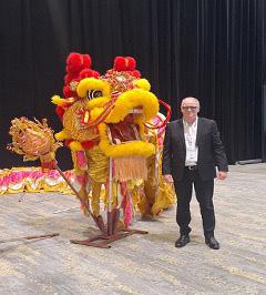
I would like to thank the NZSSA Executive for funding my attendance at the World Federation for Hospital Sterilisation Sciences (WFHSS) Conference held in Hong Kong from 4–6 December.
The conference was attended by over 1,300 delegates and 58 trade exhibitors, with 26 international speakers and more than 15 concurrent seminars delivered over two and a half days. It provided a valuable opportunity to share international knowledge, experience, and best practice in sterile science, while gaining insight into current global trends and scientific developments.
On Wednesday 3 December, I attended the WFHSS Forum, which functions in a similar way to an AGM. As members of the WFHSS, NZSSA has voting rights, allowing participation in discussion and decision-making at an international level. Conference registration and visits to the trade exhibition were held that evening, providing an opportunity to engage with suppliers and review emerging technologies.
The main conference commenced on Thursday and featured a wide range of informative and relevant presentations. Highlights included:
Anke van Rosmalen (Netherlands)
“A Centralized Endoscope Cleaning and Disinfection Department with Permanent Staff: Enhancing Quality, Safety, and Efficiency”
This presentation evaluated the benefits of a centralized Endoscope Cleaning and Disinfection (ECD) department. It highlighted the importance of permanent trained staff and well-established processes in improving reprocessing quality, efficiency, and patient safety.
Silvia Martinez (Argentina)
“Quantitative Monitoring of Residual Protein in Cannulated Medical Devices: A Multicenter Study”
This study examined the challenges associated with cleaning cannulated medical devices such as orthopaedic reamers, endoscope channels, and phacoemulsification handpieces. Due to their complex internal lumens, these devices are difficult to clean and inspect, increasing the risk of residual organic material. The study evaluated the effectiveness of a novel protein detection system (Chemdye® Pro1 Endo) designed to assess internal cleanliness, supporting improved verification processes and quality management systems.
Yongpeng Qin (China)
“How We Built CSSDGPT: A Generative AI Assistant Designed for Reprocessing Practitioners”
This presentation introduced CSSDGPT, a specialised generative AI model developed to provide accurate, instant answers to CSSD-related queries. The system aims to bridge knowledge gaps, reduce reliance on manual searches and expert consultation, and improve workflow efficiency across areas including sterilisation
science, education, quality and risk management, and departmental operations.
Teddy Lee (Hong Kong)
Hong Kong Experience in Control of Wet Pack Through a Scientific Approach”
This study focused on the critical importance of wet pack management in Sterile Services Departments (SSD). It explored the physics of steam and water, identified operational and environmental contributors to wet loads, and outlined evidence-based strategies such as equipment inspection, pack weight control, optimised pack configuration, material selection, drying cycle optimisation, water quality management, and post-cycle handling to maintain sterility and patient safety.
Samuel Law (Hong Kong)
“Performance Test for Sealing Capability of Rigid Containers”
This was the first clinical evaluation in Hong Kong assessing sealing integrity of rigid sterilisation containers in CSSDs. Conducted at Tuen Mun Hospital and Pok Oi Hospital, the study examined whether visual inspection alone is sufficient and validated alternative testing methods, including smoke testing, for both new and inuse containers.
Randalyn Harreld (USA)
“Empowering Growth from Within: Building an Internal Education Program to Advance the Sterile Processing Career Path”
This presentation outlined how structured education programs, protocols, and career pathways can support workforce retention, staff satisfaction, and long-term
sustainability within sterile processing departments, helping to address ongoing staffing shortages.
Martin Bird
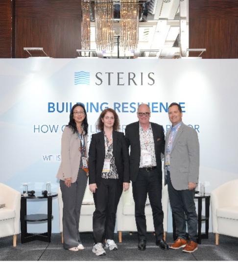
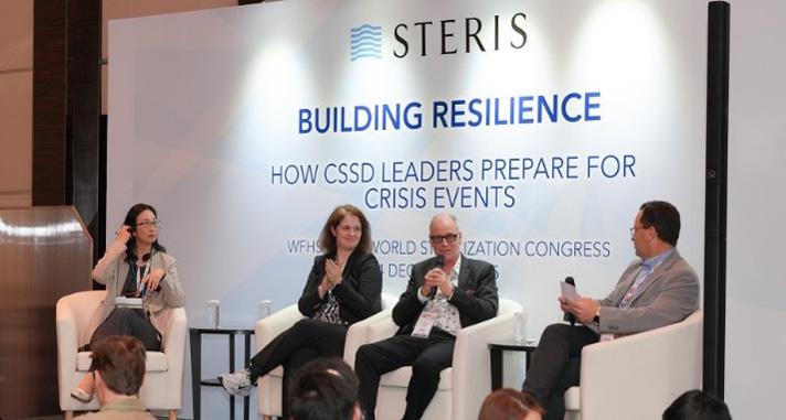
In addition, I was invited to participate in an expert panel titled “Building Resilience: How CSSD Leaders Prepare for Crisis Events.”
The panel included myself, Dr Marjorie Wall, and Pei-Tzu Lee, with Mark Munroe as moderator.
The discussion focused on how CSSD leaders prepare for and respond to crisis situations, including:
• Preparing for major infrastructure failures or service disruptions
• Planning for staffing shortages during crisis events
• Integrating CSSD preparedness into hospitalwide emergency planning
• Gaining executive and leadership support
• Identifying key internal and external partnerships essential during emergencies
Overall, attendance at the WFHSS Conference was an extremely valuable professional development opportunity. The knowledge gained and international perspectives shared will support continuous improvement, innovation, and resilience within sterile services in New Zealand. I am grateful to the NZSSA Executive for enabling my participation in this important international forum.
Pei-Tzu Lee, Dr Marjorie Wall, Martin Bird, Mark Munroe

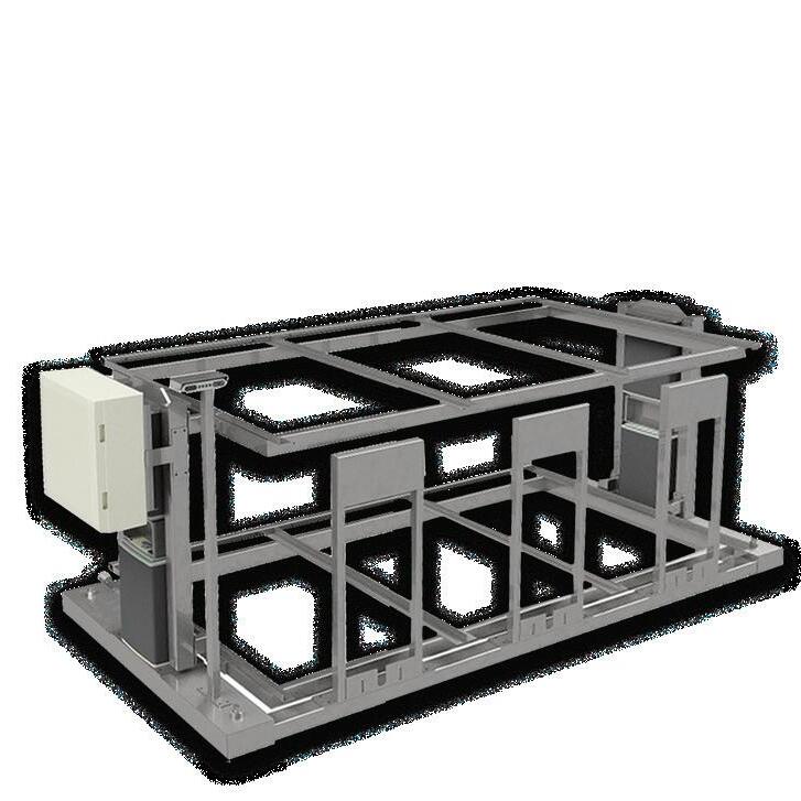
Minimise manual handling and support safer working practices in CSSDs.
Controlled loading enables precise allignment with washers and sterilisers.
Robust construction engineered for demanding sterile processing environments.
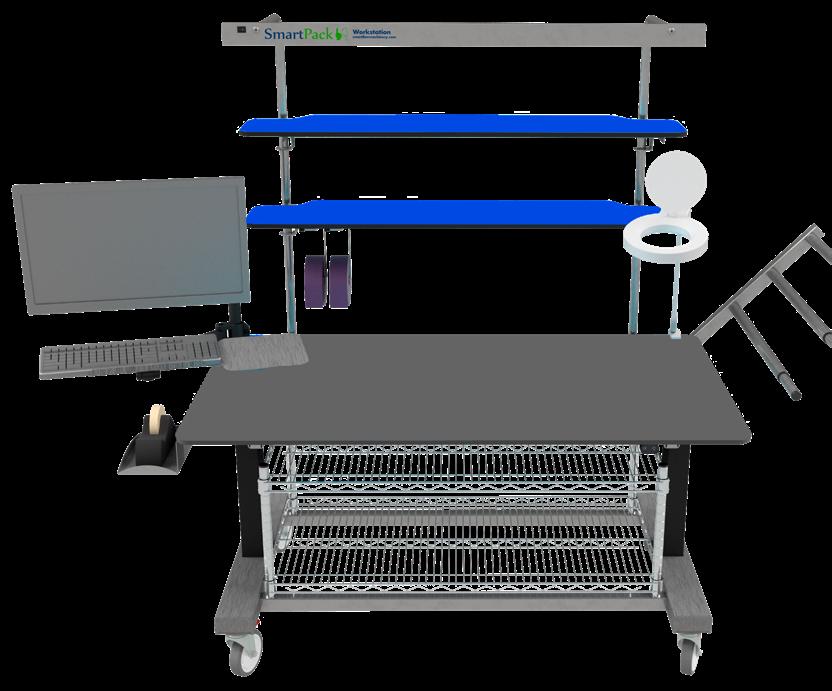
Optimised layouts for inspection, assembly and packing, ensure efficient workflow.
Ergonomic heights and configurations to reduce strain.
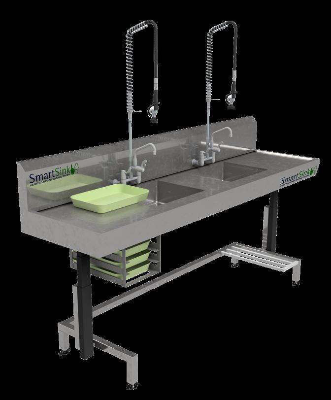
Precision-built stainless steel for CSSD compliance.
Purpose designed for CSSD decontamination workflows, supporting consistant outcomes.
Hygienic design for total confidence.
Engineered for high-volume use, chemical resistance and long-term durability.
PLEASE SPEAK TO YOUR HEALTHCARE MANAGER FOR FURTHER INFORMATION
ECOLAB HEALTHCARE NZ
4B Pukekiwiriki Place, East Tamaki, Auckland 2013 New Zealand
NZ: 0800 425 529 www.healthcare-nz.ecolab.com Global: www.ecolab.com
23-25 SEPTEMBER 2026 | TE PAE CHRISTCHURCH
Scan the QR Code to view the website and to register:

Hafiz Abdul Mannan*, Ashika Devi, Dr Farhan Rasheed, Anthony Valvoi, Ashwini Sharan
Author’s Affiliations
Allevia Hospital*, Auckland
Ameer ud Din Medical College, Lahore
Health New Zealand- Taranaki
Burwood Sterile Services, Christchurch
*First Author/ Corresponding Author
ABSTRACT
Background
Safe surgical practice requires on accurate sterile instrument processing within Central Sterile Supply Departments (CSSDs) in healthcare centers. Errors in sterilization, tray assembly, and distribution can lead to contamination risks, workflow disruptions, surgical delays, and compromised patient safety. Lean Six Sigma (LSS), a quality improvement procedure integrating Lean waste reduction principles and Six Sigma defect minimization strategies, has been extensively applied in healthcare settings. However, its application in surgical sterile instrument processing has not been comprehensively studied. This systematic review is aimed to evaluate the nature and outcomes of LSS interventions in sterile processing environments.
Methods
This systematic review was conducted in accordance with PRISMA guidelines. A comprehensive search of PubMed, Web of Science, and EMBASE databases was performed for studies published between January 2010 and January 2026. The PICO framework was used for study selection: Population—CSSDs/SPDs; Intervention—Lean Six Sigma implementation; Comparator—baseline or conventional methods; Outcomes—process performance indicators including defect reduction and turnaround time (TAT). Methodological quality was assessed by using the Joanna Briggs Institute (JBI) critical appraisal checklist.
Results
From 2,065 identified records, six studies met inclusion criteria. Studies were conducted across the USA, Switzerland, Iran, Brazil, and China, showing variations of data. LSS implementation consistently improved operational performance in CSSDs. First pass yield increased from 81.0% to 97.4% in one high-volume pediatric hospital, while error rates reduced from 2.2% to <0.10%. Error rates decreased by up to 50%, and packaging and foreign object errors were significantly
reduced. Hygiene compliance, workflow standardization, and staff satisfaction improved across multiple studies. Financial benefits were also observed, with reported cost savings ranging from $19,729 to 769,000 CNY. Overall study quality ranged from moderate to high (6/9 to 8/9 on JBI assessment).
Lean Six Sigma interventions resulted into consistent improvements of sterilization accuracy, error reduction, operational efficiency, and staff satisfaction related to surgical sterile instrument processing within CSSDs. Despite methodological heterogeneity and inclusion of non-randomized designs, the findings supported LSS as an effective framework for optimizing and managing high-risk perioperative workflows through accurate sterile instrument processing. Further high-quality comparative studies are demanded to improve evidence and outcome reporting.
Safe surgical practice and high quality patient care can become possible only with proper sterile instrument processing [1]. Nowadays, healthcare systems have been facing persistent pressures related to improvements in efficiency, reduction in errors and ensuring consistent compliance with standards alongside management of increased procedural volumes and limited resources. Patient safety during pre and post-operative procedures is maintained by Central Sterile Supply Departments (CSSDs) [2]. However, various drawbacks such as potential contamination, workflow disruptions, and increased operative times are associated with errors in sterilization, assembly and distribution of surgical instruments that may in turn cause harm to patient outcomes [3].
Complex operational challenges in healthcare have been resolved by quality improvement (QI) methodologies that opted from industrial engineering [4, 5]. Among these, Lean and Six Sigma are well reputed procedures
due their structured and data driven approaches due to less variation and waste. Lean principles mainly focus on identification and elimination of unnecessary steps in processes, facilitating workflows and avoiding delays in operations [6]. Six Sigma emphasizes on process variation and reduction of errors through improvement cycles as well as solid statistical analysis [7]. Thus, Lean Six Sigma (LSS), as integrated methodology, implicates the DefineMeasure-Analyze-Improve-Control (DMAIC) framework for improving the performance and quality of operations systematically [8].
Various challenges have been reported with traditional surgical sterile processing including incorrect set assembly, instrument misplacement, and delays in turnaround times that result into inefficiency and consequences related to surgical throughput and clinicians satisfactions [9, 10]. Additionally, increased risk preoperative complications and delayed operating room turnover times are major consequences of errors in sterile processing. All of these factors contribute to the poor clinical outcomes and department performance. Healthcare domains have been widely adopting the Lean Six Sigma, but the applications in surgical sterile instrument processing have less studied through research [9]. However, few studies have suggested Lean and Lean Six Sigma interventions as option of delivering meaningful improvements in sterilization processes [11, 12]. For instance, Lean methods have improved both quality and safety through targeted process mapping and error proofing strategies to reduce processing defects by 50% as compared to traditional strategies [13]. Additionally, wide applications of Lean Six Sigma in surgical sterile processing settings have reported to reduce the defect rates and increase the gains in first pass yield, emphasizing those systematic approaches can result into measurable operational benefits [14]. Despite these advantages, past evidences related to implications of Lean Six Sigma are limited [12, 15]. No comprehensive study that evaluated how Lean Six Sigma methodologies have been deployed in sterile processing, nor found the magnitude of their effects across diverse healthcare settings. Thus, there is immense need to address this gap for sterile services professionals, CSSD managers, and organizational leaders. Accordingly, this systematic review is aimed to identify the outcomes of Lean Six Sigma interventions in surgical sterile processing in CSSDs
This comprehensive review was undertaken to assess the outcomes after “Application of Lean Six Sigma in Surgical Sterile Instrument Processing within Central Sterile Services Departments (CSSDs)” in accordance with Preferred Reporting Items for Systematic Reviews and Meta-Analyses (PRISMA) guidelines to maintain transparency [16].
This systematic review was conducted by following PICO framework for screening and selection of papers [17].
Population (P): Central Sterile Services Departments (CSSDs), including healthcare centers where surgical instruments are sterilized.
Intervention (I): Adoption or Implementation of Lean Six Sigma
Comparator (C): Pre-implementation baseline performance, Traditional sterile processing methods and Non-Lean quality improvement approaches Outcomes (O): Process outcomes such as Reduction in instrument defects (missing/unusable instruments), Reduction in tray errors, and Decrease in turnaround time (TAT)
Three electronic databases such as PubMed, Web of Science and Embase were thoroughly searched from January 2010 to January 2026. The search strategy for Pubmed was “((“Lean Six Sigma”[Title/ Abstract] OR “Lean-Six Sigma”[Title/Abstract] OR “Lean”[Title/Abstract] OR “Six Sigma”[Title/Abstract]) AND (“sterile processing”[Title/Abstract] OR “central sterile supply”[Title/Abstract] OR “surgical instrument processing”[Title/Abstract] OR “instrument reprocessing”[Title/Abstract] OR “CSSD”[Title/Abstract] OR “SPD”[Title/Abstract])) and similar was used for other databases.
Inclusion Criteria
The inclusion criteria was used to identify eligible authentic studies were: 1). Those studies analyzing the Central Sterile Services Departments (CSSDs), Sterile Processing Departments (SPDs), or equivalent hospital sterilization units, 2). Implementation or evaluation of Lean and six sigma methodologies in surgical sterile instrument processing , 3). Studies comparing Lean Six Sigma with conventional methods, 4). Studies discussing outcomes such as reduction in errors, staff satisfaction rates, and accuracy rates 5). Study design must be Quasi-experimental studies, case–control studies, crosssectional studies and publications in English.
Exclusion Criteria
Those articles were excluded 1). Those not conducted in CSSDs/SPDs or hospital sterilization units, 2). Interventions unrelated to Lean Six sigma methodologies 3). Narrative reviews, Review based studies, Case reports, editorials, letters to the editor, conference abstracts without full text, 4). Studies discussing human outcomes rather than processing outcomes 5). Studies not reporting any relevant outcomes of interest (process, or clinical) and Non-English studies.
Research articles identified from databases: (n= 2065)
Records removed before the screening:
Removed as duplicated papers (n =1240)
Removed due to non-full text (n=548)
Research article screened (n = 277)
Excluded research papers** (n = 132)
Research articles sought for retrieval (n = 145)
Research articles not retrieved (n = 100)
Research articles assessed for eligibility (n = 45)
EXCLUDED RESEARCH PAPERS: 39
Figure no. 1: Flow chart of PRISMA-guidelined screening and selection of included studies evaluated using the JBI critical assessment checklist. Standardized critical appraisal questions or criterion are evaluated the potential for various biases that may arise in quantitative research. [18].
Two reviewers separately examined the included studies to accommodate those according to the inclusion criteria. Full-text publications with authentic research were ultimately considered for evaluation. The data extracted for each study was: study characteristics (authors, year of publication, country, study population, study area), or aspects of methodology applied, errors of system, findings, and conclusions. Any conflicts or differences between reviewers were settled by discussion with a third reviewer.
The Joanna Briggs Institute (JBI) critical appraisal checklist was used to assess methodological quality of included cohort studies, empirical studies and case studies for this systematic review. To address and minimize bias among included studies, the methodological bias was
The PRISMA guidelines were followed in the selection and screening of research articles pertaining to the study aim “Applications of Lean Six Sigma in in Surgical Sterile Instrument Processing within CSSDs” in recent study. Database searches generated a total of 2065 papers, of which 277 remained after duplicates and insufficient text were eliminated. After an initial screening of 277 research publications, 145 papers were searched for retrieval. There were only 45 publications that were analyzed for the eligibility requirements, and there were ultimately 6 research articles included in pooled analysis, as shown in Figure 1.
Table no. 1 : Characteristics of included studies in systematic review
Summary
The Lean Six Sigma methodology improved the sterilization accuracy and proved beneficial for manufacturing in the hospital setting to reduce missing and unusable instrumenta
tion.
Findings
Defects or Drivers Methodology applied
Study period
Study Area
Yield increased: 81.0% to 97.4% Defect rate reduced: 2.2% to < 0.10%
5s Lean Six Sigma Methodology (5S: Sort, set in order, shine, standardize, and sustain)
1. Staffing and training, 2. Equipment and SPD physical environment, 3. Governance structure 4. Inventory management, 5. standard workflows and communication
September 2020–August 2022
364-bed academic children’s hospital with a high surgical volume (~23,000 surgeries per year)
Study Design
Study Aims
Country
Author, year
Lean Six Sigma in Surgical instrument processing decreased the errors that are barrier to the highest quality and safety in surgical care.
Errors rate reduced: 3.0% to 1.5%
Errors of packaging: 0.66 to 0.24 errors per hundred cases
Errors of foreign objects: 0.17 to 0.02 errors per hundred cases
The application of the Lean Six Sigma methodology is proved cost-effective, significantly decreased the costs of poor quality and increased staff satisfaction.
Cost savings: $19,729
In CSR unit, staff performance improved after implications of Lean Six Sigma in hospitals of the province.
Staff satisfaction improved: 6.6 ± 2.2 pts to 7.0 ± 1.9 pts
personal and environment hygiene improved: 33.34% cleaning: 26.00% packaging: 68.06% cycle control: 21.8% handling: 13.50%
February 2010 to March 2013 1 Operator roles, 2 Alteration of the workspace, 3 Mistake-proofing, 4 Quality monitoring, 5 Staff training
To apply Lean Six Sigma Methodology for identification of unusable instruments to reduce waste and variations Quality improvement (QI) project
USA
Natarus et al., 2025 [14]
USA To evaluate the effectiveness of lean methods as quality improvement project in sterile processing to reduce errors Pre/ post intervention study
Blackmore et al., 2013 [3]
314,552 instruments July 2010 –December 2022
To evaluate the outcomes of Six-Sigma methodology in optimization of surgical instrument sterilization processes longitudinal observational study
Switzerland
Saporito et al., 2023 [19]
To assess the impacts of the Lean Six Sigma (LSS) management method on process outcomes in sterile instrument processing quasi-experimental study 28 CSR personnel 2021
Koushki et al., 2022 [20] Iran
The errors in sterilization were reduced and time data acquisition was optimized by lean design for a small sample of trays.
Error decreased: 7.83%
Accuracy of results: 90.8%
Hospitals in South Brazil 1. Expert Assessment, 2. Number of trays and their parts
Time Data Acquisition
Run model
Case study 18,378 instruments per day and 36 trays assembly
To apply Six sigma model for dimensioning the department’s capacity and predicting the expected outcomes of assemblers
Fogliatto et al., 2018 [21] Brazil
LSS-based precision instrument whole lifecycle management can successfully save hospital expenses, decrease wear rates, and raise surgeon and nurse satisfaction in sterile supply departments.
Staff satisfaction rates: 83.33% to 95.83% Cost saving: 769,000 CNY
Validate Results
Retrospective study 2023 to 2024
To apply the Lean Six Sigma (LSS) as a management tool that could enhance patient safety and medical quality through process improvements
Zhu et al., 2025 [22] China
Table no. 2: Quality Assessment of included studies in systematic review Study (Author, Year) Q1 Cause/Effect
Natarus et al., 2025 [14]
Blackmore et al., 2013 [3]
Saporito et al., 2023 [19]
Koushki et al., 2022 [20]
Fogliatto et al., 2018 [21]
Zhu et al., 2025 [22]
The methodological quality of the included studies was evaluated using a 9-point checklist. These nine points were cause–effect clarity, comparability of groups, treatment consistency, control group presence, follow-up, outcome measurement, and statistical appropriateness, as shown in Table 2, most studies have moderate-to-high or high quality, with total scores ranging from 6/9 to 8/9.
The included studies found increasing global application of Lean Six Sigma (LSS) within sterile processing environments across diverse healthcare systems, including the USA, Switzerland, Iran, Brazil, and China. The designs of included studies were quality improvement (QI) [14], quasi-experimental [3], longitudinal observational [20], case study [21], and retrospective analyses [22], representing diverse nature of sterile services research.
Natarus et al [14] ‘s study is a structured QI project, conducted in a high volume 364 bed academic children’s hospital (Chicago, USA), over 2 years from September 2020 to August 2022. The methodology used the 5S Lean Six Sigma framework (Sort, Set in Order, Shine, Standardize, and Sustain) to address staffing, physical environment, governance, inventory, and standardization of workflow. In the same way, Blackmore et al., [3] had a pre–post intervention study conducted from 2010 to 2013 that worked on workspace redesign, error proofing, staff training, and quality supervisions. Saporito et al., [19] used a longitudinal observational design with 12 years of follow up, studying Six Sigma optimization in 314,552 instruments. Koushki et al, [20] conducted a quasi-experimental study with 28 Central Sterile Room (CSR) pannels to see the impact of LSS on hygiene and procedural compliance. Fogliatto et al, [21] did a case study using Six Sigma modelling for the improvement of hospitals that handle 18.378 instruments a day. Finally, Zhu et al, [22] did a retrospective study using the complete DMAIC cycle to optimize the management of the instrument’s lifecycle. In general, most studies reviewed the structured Lean tools such as 5S, DMAIC, mistake-proofing, and redesign workflow which showed a methodological consistency despite the different healthcare environments.
Defects concerning to workforce shortages, insufficient training, disorganized workspaces, inventory mishandling, communication breakdowns, and a lack of standard operating procedures were prevalent in all of the included studies. Defects were also found to be significantly influenced by environmental factors in Sterile Processing Departments (SPDs), such as equipment limits and physical layout.
Natarus et al. [14] reported the primary drivers of unusable instruments such as staffing, governance structure, inventory control, and workflow variation. Blackmore et al. [3] explained major contributors to sterile processing errors such as operator ‘s negligence, packaging errors, and foreign object contamination. Improvements in sterilization were common and consistent among all studies. Natarus et al. [14] reported an increase in yield from 81.0% to 97.4%, with reduction in defect rates from 2.2% to less than 0.10%. Blackmore et al. [3] proved a reduction in overall error rates from 3.0% to 1.5%, with decrease in packaging errors significantly. Fogliatto et al. [21] reported a 7.83% reduction in sterilization errors and accomplished 90.8% model accuracy for process optimization.
Operational performance was more than the defect reduction. Koushki et al. [20] demonstrated significant improvements in hygiene and packaging compliance, particularly packaging performance (68.06% improvement). Zhu et al. [22] reported significant improvements in staff satisfaction (83.33% to 95.83%) alongside significant cost savings (769,000 CNY). Similarly, Saporito et al. [19] reported cost savings of $19,729 and improved staff satisfaction scores. Overall, qualitative synthesis reported that Lean Six Sigma is proved as a effective, and adaptable methodology for improving sterilization accuracy, reducing waste, and strengthening patient safety within surgical sterile instrument processing environments across diverse healthcare contexts.
This systematic review is aimed to synthesize evidences on the applications of Lean Six Sigma (LSS) methodologies in surgical sterile instrument processing (SSIP). The findings from included studies reported that LSS implementations have improved the operational efficiency, quality sterilization processes and reduced the defects and turnaround times. Hence, Lean Six Sigma is proved a valuable approach to optimize complex highrisk processes in perioperative services. The findings of this study reported the significant improvements in sterile instrument processing performance indicators after implications of LSS interventions. Likewise previous studies, this study reported that LSS have reduced the instrument processing cycle times by 20-50%, resulting into improved surgical instrument utilization and faster turner over. Furthermore, LSS decreased defect rates such as incomplete sterilization, handling errors and incorrect tray assembly. This methodology has improved workflow standardization and reliability, with decreased variability and increased adherence to process protocols along staff performance. For examples, another quasiexperimental study have implicated Define-MeasureAnalyze-Improve-Control (DMAIC) frameworks that results into significant reductions in error frequencies
and instrument turnaround times [23]. Additionally, these improvements are consistent throughout different follow ups, resulting into reliable process changes rather than unstable yields.
Furthermore, secondary outcomes of LSS methodologies are improved staff engagement and satisfaction, due to supporting nature of Lean Six Sigma activities (e.g., process mapping, root-cause analysis, Kaizen events). Few studies also reported cost savings after LSS implications due to reduced turnaround and overtime, however, economic gains varied in quality and consistency. The findings of this study are consistent with previous studies reporting the positive effects of Lean Six Sigma in sterile instrument processing in healthcare operations. Previous studies of LSS implications in clinical areas reported the improvements in efficiency, process reliability, and patient safety due to appropriate sterile instrument processing [10, 23]. The consistency of evidences emphasized the implication of Lean Six Sigma principles, despite their origination from manufacturing sector. These principles are also compatible with complex healthcare workflows followed by high variability and patient care demands [24].
Additionally, reductions of waste and improvements in process standardization by LSS applications are similar to findings of a study that reported optimization of instrument tray and reductions in operating room turnover time [24, 25]. LSS implications are also associated with reduced medication errors and laboratory turnaround time that would lead to analytical and operational challenges. However, process outcomes of LSS in sterile instrument processing are rarely reported by the previous studies that only evaluated the patient outcomes (e.g., mortality, morbidity) [24].
This study has several strengths. First, it addressed the sterile instrument processing as critical area of perioperative services that received less attention before. The review applied comprehensive database searches and solid inclusion criteria, resulting into synthesis of evidence from relevant included studies. Secondly, diverse outcomes have been studied such as process metrics, and quality indicators, enabling a complete assessment of Lean Six Sigma’s impact. Thirdly, this systematic review reported the consistent patterns of improvement across geographically and organizationally varied settings, suggesting generalizability of LSS benefits in sterile instrument processing contexts. The findings of this review supported the Lean Six Sigma as a viable framework for improving surgical sterile instrument processing. This review guided the healthcare organizations to adopt the LSS as part of broader quality improvement strategies due to increased workflow, optimize the resource utilization and reduced errors. Critical success factors of LSS implications staff training tools, and integration of data-driven decision making. Despite of several strengths, there are few limitations of this study. Firstly, all included studies were pre-post
intervention and quasi-experimental design. There were no randomized controlled trial that result into temporal biases and confounding factors that compromise the quality of research. Secondly, this study synthesized the evidences by qualitative analysis rather than quantitative analysis (meta-analysis) that resulted into difficult direct comparisons. Thirdly, this study has not discussed the economic analysis that reported as major drawback.
Conclusion
The integration of six global studies indicates that the LSS techniques, such as DMAIC, 5S, and mistakeproofing, directly address the primary operational issues peculiar to sterile processing settings, which can result in significant changes in the key CSSD core performance indicators. These are huge decrease in the defect rates (2.2 to a low of less than 0.10), high first-pass yield (81.0 to 97.4), high compliance levels (hygiene and packaging), high staff satisfaction, and direct savings amounted to 19,729 and up to 769,000 CNY. Lean Six Sigma interventions demonstrate consistent improvements in sterilization accuracy, defect reduction, operational efficiency, and staff satisfaction within surgical sterile instrument processing. Nevertheless, to enhance the evidence base, further high-quality comparative studies are warranted to strengthen evidence and standardize outcome reporting.
References
1. Zheng S, Jiang D, Liu P, Zhang H: Management quality of surgical instrument and influence of cleaning and sterilization on the surgical outcomes of the patient: a review Altern Ther Health Med 2023, 29(8):863-869.
2. Chakraverty R, Kundu AK: Good Practices and Standard Operating Procedures for Sterilization Across Wards. In: Hospital-Acquired Infections in Intensive Care Unit and their Management: The Indian Perspective. edn.: Springer; 2025: 115-127.
3. Blackmore CC, Bishop R, Luker S, Williams BL: Applying lean methods to improve quality and safety in surgical sterile instrument processing The Joint Commission Journal on Quality and Patient Safety 2013, 39(3):99-AP91.
4. Alavi J, Yasin MM: The role of quality improvement initiatives in healthcare operational environments: changes, challenges and responses International journal of health care quality assurance 2008, 21(2):133-145.
5. Zoutman DE, Ford BD: Quality improvement in hospitals: barriers and facilitators International journal of health care quality assurance 2017, 30(1):16-24.
6. Yaduvanshi D, Sharma A: Lean six sigma in health operations: challenges and opportunities—‘Nirvana for operational efficiency in hospitals in a resource limited settings’ Journal of Health Management 2017, 19(2):203213.
7. Nakhai B, Neves JS: The challenges of six sigma in improving service quality International Journal of Quality & Reliability Management 2009, 26(7):663-684.
8. Van den Heuvel J, Does RJ, De Koning H: Lean Six Sigma in a hospital International Journal of Six Sigma and Competitive Advantage 2006, 2(4):377388.
9. Samanta AK, G V, Gurumurthy A: Implementing lean six sigma in health care: a review of case studies International Journal of Lean Six Sigma 2023, 14(1):158-189.
10. Henrique DB, Godinho Filho M: A systematic literature review of empirical research in Lean and Six Sigma in healthcare. Total Quality Management & Business Excellence 2020, 31(3-4):429-449.
11. McDermott O, Antony J, Bhat S, Jayaraman R, Rosa A, Marolla G, Parida R: Lean six sigma in healthcare: a systematic literature review on motivations and benefits Processes 2022, 10(10):1910.
12. Fang L, Xiao K, Zhu H, Zhang M: Implementing ‘6S’Nursing Management in Sterilization and Supply Centers: Enhancing Surgical Instrument Quality and Work Efficiency Risk Management and Healthcare Policy 2025:10991108.
13. Farrokhi FR, Gunther M, Williams B, Blackmore CC: Application of lean methodology for improved quality and efficiency in operating room instrument availability The Journal for Healthcare Quality (JHQ) 2015, 37(5):277-286.
14. Natarus ME, Shaw A, Studer A, Williams C, Dominguez C, Mangual H, Olmstead J, Westmoreland K, Gill T, Wellington WZ: Optimization of a Sterile Processing Department Using Lean Six Sigma Methodology, Staffing
Enhancement, and Capital Investment The Joint Commission Journal on Quality and Patient Safety 2025, 51(1):33-45.
15. Sreekanth V, Kavilal E, Krishna S, Mohan N: Implementation of Six Sigma methodology in a medical equipment manufacturing company The TQM Journal 2025, 37(7):2041-2073.
16. Page MJ, McKenzie JE, Bossuyt PM, Boutron I, Hoffmann TC, Mulrow CD, Shamseer L, Tetzlaff JM, Akl EA, Brennan SE: The PRISMA 2020 statement: an updated guideline for reporting systematic reviews bmj 2021, 372
17. Schardt C, Adams MB, Owens T, Keitz S, Fontelo P: Utilization of the PICO framework to improve searching PubMed for clinical questions BMC medical informatics and decision making 2007, 7(1):16.
18. Munn Z, Barker TH, Moola S, Tufanaru C, Stern C, McArthur A, Stephenson M, Aromataris E: Methodological quality of case series studies: an introduction to the JBI critical appraisal tool JBI evidence synthesis 2020, 18(10):2127-2133.
19. Saporito A, Tassone C, Di Iorio A, Barbieri Saraceno M, Bressan A, Pini R, Mongelli F, La Regina D: Six Sigma can significantly reduce costs of poor quality of the surgical instruments sterilization process and improve surgeon and operating room personnel satisfaction Scientific Reports 2023, 13(1):14116.
20. Koushki A, Larti N, Fakhri M, Fatahi S: Investigating the effect of Lean Six Sigma method on the observance of performance standards in the central sterilization unit of the operating room Perioperative Care and Operating
Room Management 2022, 28:100269.
21. Fogliatto FS, Anzanello MJ, Tortorella GL, Schneider DS, Pereira CG, Schaan BD: A Six Sigma approach to analyze time-to-assembly variance of surgical trays in a sterile services department The Journal for Healthcare Quality (JHQ) 2018, 40(3):e46-e53.
22. Zhu X, Qin J, Zhou X, Chen H, Yan C, Bao R: Reducing Wear Rate of Precision Surgical Instruments through Lean Six Sigma: A single-center retrospective study medRxiv 2025:2025.2006. 2002.25328841.
23. Al Hammad ZJ, Al Mulhim SM, Al Raheb ZAH, Al Dossary FA, Al Bahrani EAA, Al Dossary WAL, Al Zaher AAR, Haji KS, Al Yami NM, Al Yasin ZA: Effectiveness Of Quality Indicators (EG, TAT, Error Rates) As Tools For Continuous Improvement In Clinical Laboratories: A Systematic Review Of The Evidence The Review of Diabetic Studies 2025:254-271.
24. Liu S, Fang T, Liu Y, Han Q: Application and analysis of monitoring indicators in CSSD in sentinel hospital Sterile Supply 2024, 3(4):246-251.
25. Steere L, Rousseau M, Durland L: Lean six sigma for intravenous therapy optimization: a hospital use of lean thinking to improve occlusion management Journal of the Association for Vascular Access 2018, 23(1):4250.

We are pleased to share that development of the new Level 4 Sterilising Technology programme at Open Polytechnic is progressing well. Course writing is now well underway, and we remain on track for a planned launch in the second half of this year.
The programme will consist of three courses covering key areas of sterile services practice: Foundations of Sterilising Practice; Decontamination and Instrument Reprocessing; and Sterilisation, Storage and Quality Systems.
The programme has been designed to support both ākonga (learners) who are currently working in sterile services environments and those who have arranged appropriate work experience. As a work-based programme, ākonga will be required to complete specific practical tasks within 120 hours of verified work experience within their workplace.
Closer to launch, we will be inviting expressions of interest from experienced sterile services professionals who may wish to contribute to the programme in adjunct marking roles.
We appreciate the ongoing interest and support from the sterile services community and look forward to sharing further updates as development continues.
Powder-Free
Gloves Means Protection” MAX, the newest was designed
Gloves Means Protection”
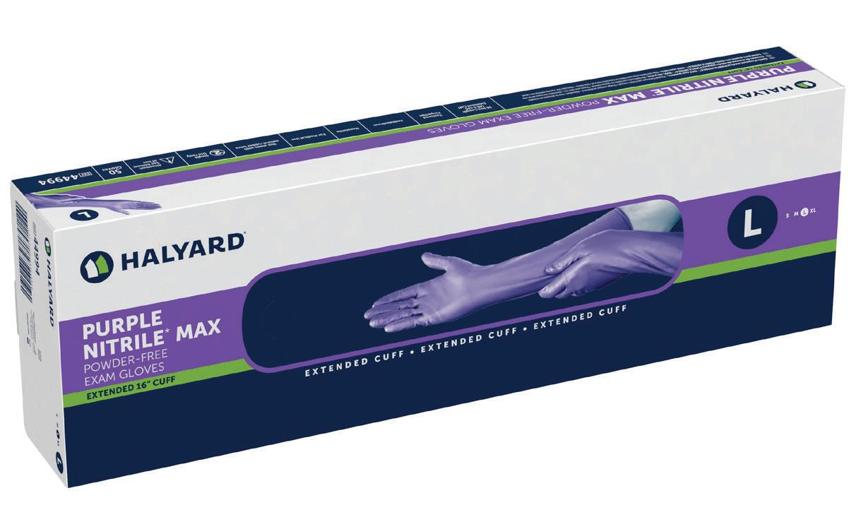
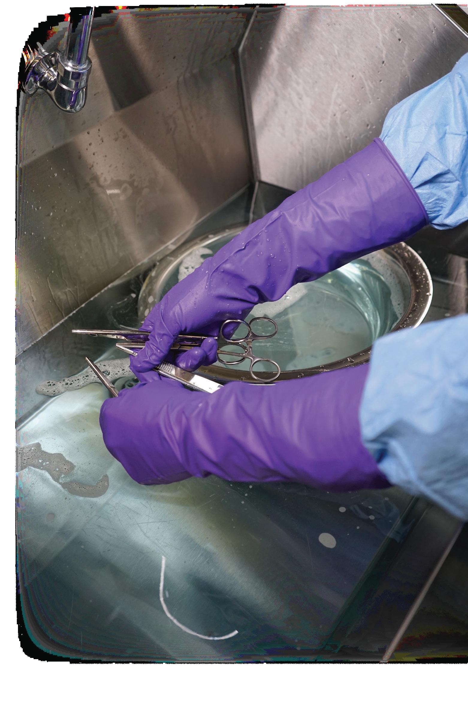
Once again, Halyard Health proves “Purple Means Protection” with the introduction of PURPLE NITRILE* MAX, the newest member of our singleuse exam glove family. It was designed specifically to meet the increased protection needs of the Central Sterilizing Services Department.
Once again, Halyard Health proves “Purple Means Protection” with the introduction of PURPLE NITRILE* MAX, the newest member of our singleuse exam glove family. It was designed specifically to meet the increased protection needs of the Central Sterilizing Services Department.
SD
tech these critical safety features:
tech these critical safety features:
Once again, Halyard Health proves “Purple Means Protection” with the introduction of PURPLE NITRILE* MAX, the newest member of our singleuse exam glove family. It was designed specifically to meet the increased protection needs of the Central Sterilizing Services Department.
Once again, Halyard Health proves “Purple Means Protection” with the introduction of PURPLE NITRILE* MAX, the newest member of our singleuse exam glove family. It was designed specifically to meet the increased protection needs of the Central Sterilizing Services Department.
MAX, the newest It was designed
Longer Length –a full 16 inches¹, providing the right level of protection from dangerous microorganisms, all the way to the midforearm.
tech these critical safety features:
Longer Length –a full 16 inches¹, providing the right level of protection from dangerous microorganisms, all the way to the midforearm.
tech these critical safety features:
Longer Length –a full 16 inches¹, providing the right level of protection from dangerous microorganisms, all the way to the midforearm.
Longer Length –a full 16 inches¹, providing the right level of protection from dangerous microorganisms, all the way to the midforearm.
Heavier Weight – At 20 grams², PURPLE NITRILE* MAX is more than twice the weight of PURPLE NITRILE-XTRA*, for added confidence when handling instruments and decontamination chemicals.
Heavier Weight – At 20 grams², PURPLE NITRILE* MAX is more than twice the weight of PURPLE NITRILE-XTRA*, for added confidence when handling instruments and decontamination chemicals.
Heavier Weight – At 20 grams², PURPLE NITRILE* MAX is more than twice the weight of PURPLE NITRILE-XTRA*, for added confidence when handling instruments and decontamination chemicals.
Heavier Weight – At 20 grams², PURPLE NITRILE* MAX is more than twice the weight of PURPLE NITRILE-XTRA*, for added confidence when handling instruments and decontamination chemicals.
Thicker Fingertips – Its increased fingertip thickness provides almost double the protection of any other exam glove in the Halyard portfolio.
Thicker Fingertips – Its increased fingertip thickness provides almost double the protection of any other exam glove in the Halyard portfolio.
Fully Textured – PURPLE NITRILE* MAX features fully textured fingertips and palm, designed for excellent wet and dry grip.
Thicker Fingertips – Its increased fingertip thickness provides almost double the protection of any other exam glove in the Halyard portfolio.
Fully Textured – PURPLE NITRILE* MAX features fully textured fingertips and palm, designed for excellent wet and dry grip.
Thicker Fingertips – Its increased fingertip thickness provides almost double the protection of any other exam glove in the Halyard portfolio.
Fully Textured – PURPLE NITRILE* MAX features fully
Fully Textured – PURPLE NITRILE* MAX features fully textured fingertips and palm, designed for excellent wet and dry grip.
A p p licat ion s close 3 0 M a rch 2 02 6 , 5 pm
Education
Course & administration fees are covered for the student
NZSSA Conference, Christchurch
Conference registration, return flights, 3 days
accommodation
FSRACA Congress, Gold Coast Australia
Conference registration, return flights, 5 days
accommodation
GENCA Congress, Melbourne Australia
Conference registration, return flights, 5 days
accommodation
WFHSS Congress, Serbia
Conference registration, return flights, 5 days
accommodation
For full details of the scholarships and application forms visit the Scholarship page: https://nzssa.co.nz/scholarship/
Journal of Hospital Infection 167 (2026) 116 123
Available online at www.sciencedirect.com
journal homepage: www.el sevier.com/l ocate/jhin
Journal of Hospital Infection 167 (2026) 116—123
M. Arvand a, b , K. Konrat a , D. Cserto ¨ a , A.M. Richter a,
*
a Unit for Hospital Hygiene, Infection Prevention and Control, Department Infectious Diseases, Robert Koch Institute, Berlin, Germany
b Institute for Medical Microbiology and Hygiene, Department of Infectious Diseases, University of Heidelberg, Heidelberg, Germany
A R T I C L E I N F O
Article history:
Received 10 July 2025
Accepted 11 October 2025
Available online 4 November 2025
Keywords: Biofilm
Disinfection
Endoscope reprocessing
Klebsiella pneumoniae
Disinfectant tolerance
Carbapenem-resistance

S U M M A R Y
Background: Klebsiella pneumoniae is a prominent cause of healthcare-associated infections. Carbapenem-resistant K. pneumoniae (CRKP) is considered a serious public health threat and has been increasingly linked to endoscopy-associated outbreaks. Biofilm formation in endoscope channels contributes to increased antimicrobial tolerance, potentially compromising disinfectant efficacy during reprocessing. This study evaluated the efficacy of peracetic acid (PAA), a commonly used disinfectant in endoscope reprocessing, on K. pneumoniae in planktonic and biofilm form.
Methods: Disinfectant efficacy was assessed using the suspension test EN 13727 and the Bead Assay for Biofilms. Five K. pneumoniae strains were tested: two CRKP associated with endoscopy outbreaks, two carbapenem-susceptible clinical strains and ATCC 13883.
Findings: All strains were susceptible to PAA in suspension test, with 0.001 0.01% PAA achieving 5 log 10 cfu reduction within 10 min. In contrast, biofilm-residing cells required higher concentrations (0.075 0.2% PAA, 10 min) for equivalent efficacy Clinical strains were notably more tolerant than ATCC 13883. Under standard endoscope reprocessing conditions (PAA 0.075%, 5 min, 37 ◦ C), biofilms of all strains exhibited tolerance, failing to meet the 5 log10 cfu reduction threshold. In contrast, intensified endoscope reprocessing conditions (0.15% PAA, 10 min, 37 ◦ C) resulted in successful biofilm disinfection.
Conclusions: Biofilms of K. pneumoniae, including CRKP, exhibit marked tolerance to standard concentrations of PAA used in endoscope reprocessing. These data suggest that routinely used disinfection conditions may not be sufficient when biofilm formation is likely Disinfection parameters in areas at high risk of biofilm contamination, e.g., endoscope reprocessing, and in the context of CRKP outbreaks should be reviewed and adjusted if necessary.
© 2025 The Author(s). Published by Elsevier Ltd on behalf of The Healthcare Infection Society This is an open access article under the CC BY license (http://creativecommons.org/licenses/by/4.0/).
* Corresponding author Address: Unit for Hospital hygiene, Infection Prevention and Control, Nordufer 20, Robert Koch Institute, Berlin, Germany
E-mail address: richteranj@rki.de (A.M. Richter).
Klebsiella pneumoniae is a prominent cause of healthcareassociated infections. The WHO Bacterial Priority Pathogens
https://doi.org/10.1016/j.jhin.2025.10.012 0195-6701/© 2025 The Author(s). Published by Elsevier Ltd on behalf of The Healthcare Infection Society This is an open access article under the CC BY license (http://creativecommons.org/licenses/by/4.0/).
List has identified carbapenem-resistant and third-generation cephalosporin-resistant K. pneumoniae as critical threats, placing them at the top of the priority list of pathogens of public health importance requiring research and development [1]. In September 2024, the global burden of bacterial antimicrobial resistance (AMR) estimates were published, identifying K. pneumoniae as one of just six organisms with an attributable burden estimated to exceed 100,000 AMR deaths in 2021 [2].
Recently, endoscope-associated outbreaks by carbapenemresistant K. pneumoniae (CRKP) have been increasingly reported, suggesting nosocomial transmission may occur via contaminated endoscopes [3 7]. Endoscopes are frequently employed in today’s medicine for diagnostic and therapeutic purposes. Due to their delicate construction and the materials used, many endoscopes cannot be sterilized and are therefore subjected to high-level disinfection, which should ensure an equally high level of safety However, flexible endoscopes in particular have been found to be frequently contaminated by pathogens despite compliant cleaning and disinfection per instructions for use (IFU) and clinical practice guidelines [8 11].
An important cause of persistent contamination of endoscopes despite reprocessing in accordance with guidelines is the formation of biofilms in the narrow endoscope channels. The moist environment inside the channels, in conjunction with potential damage caused by instrumentation and cleaning, creates an ideal setting for bacterial attachment and the formation of biofilms. These biofilms can resist cleaning and disinfection, thereby contributing to the transmission of pathogens [12 18]. Within biofilms, cells are encapsulated in a self-produced extracellular matrix consisting of proteins, polysaccharides and extracellular DNA that mediates surface adherence [19]. Biofilm-residing bacteria (including K. pneumoniae) have been shown to exhibit elevated levels of tolerance to disinfectants in comparison with their planktonic counterparts [5,16,20].
It is noteworthy that the currently available and used international standards for disinfectant efficacy testing do not cover the biofilm phenotype. In Europe, the efficacy testing of disinfectants approved for use in the medical field is carried out in accordance with European Standards. A comprehensive overview of these can be found in the superior standard EN 14885 [21]. The latter includes, for example, EN 13727 (quantitative suspension test for the evaluation of bactericidal activity in the medical area) or EN 14561 (quantitative carriertest for the evaluation of bactericidal activity for medical instruments), in which the efficacy of disinfectants is evaluated using planktonic cells [22,23]. In addition, the validation of the disinfection step in automated endoscope washerdisinfector (AEWD) devices, as specified in ISO 15883-4 [24], is also conducted using planktonic cells. Consequently, it is
Table I
Information on Klebsiella pneumoniae strains analysed in this study
therefore questionable whether the disinfection step of the endoscope reprocessing procedure, whose efficiency is validated using planktonic cells, is able to effectively inactivate biofilms of K. pneumoniae or other nosocomial pathogens.
Recently, we established a robust and versatile method for disinfectant efficacy testing of biofilm-residing micro-organisms, the Bead Assay for Biofilms [25]. This method has been shown to produce reliable and reproducible results and to be adaptable to a range of organisms, e.g., on Pseudomonas aeruginosa, K. pneumoniae, Salmonella enterica, and nontuberculous mycobacteria [5,25 28].
Using this assay to evaluate the disinfection efficacy, we previously reported on two CRKP strains involved in endoscopyrelated outbreaks in 2014 and 2022 in two different hospitals in Germany [5,16]. In both cases, the outbreak strain had been isolated from affected patients and from reprocessed endoscopes. The reprocessing of endoscopes had been performed in AEWDs using peracetic acid (PAA) as disinfectant and in accordance with the IFU and national guidelines [29]. In both cases, increased tolerance of the biofilm of the outbreak strain to PAA was observed, demonstrating a direct link between biofilm formation, tolerance to PAA as used for the high-level disinfection of endoscopes, and nosocomial transmission of CRKP
In the present study, we aimed to systematically analyse the susceptibility of biofilms of five K. pneumoniae strains and their planktonic counterparts to PAA under the same experimental conditions. The panel consisted of the two previously mentioned CRKP strains associated with endoscopy outbreaks in 2014 and 2022 [5,16], two clinical, non-outbreak, carbapenemsusceptible K. pneumoniae strains, and the K. pneumoniae ATCC 13883 as a type strain. We aimed to determine whether increased PAA tolerance correlates with carbapenemase production, as the above-mentioned outbreak-associated K. pneumoniae strains were both carbapenemase-producers. We assessed whether biofilms of clinical strains and the ATCC type strain exhibit comparable tolerance to PAA disinfection. In addition, we tested the efficacy of the PAA-based disinfectant product used in the AEWD during the 2022 outbreak and of the application conditions as defined by the manufacturer of the AEWD (‘standard disinfection conditions’ and ‘intensified disinfection conditions’) to achieve disinfection of biofilmresiding K. pneumoniae.
strains
The strains analysed in this study and the background information are listed in Table I. These included four clinical K. pneumoniae isolates, namely two carbapenem-resistant
Strain Background Year of isolation Place of isolation Source of isolation Carbapenemase Reference
ATCC 13883 Type strain Before 1990 Unknown Unknown
2202051 Outbreak strain 2022 Stralsund Endoscope
None
OXA-181 [16]
886/14 Outbreak strain 2014 Berlin Endoscope OXA-48 [5] 189445 Routine diagnostic 2022 Stralsund Urine
None
None 606527 Routine diagnostic 2021 Schwerin Swab (nasal/throat)
Disinfection of planktonic cells
Peracetic acid concentration
Figure 1. Efficacy of peracetic acid (PAA, Lerasept Spezial) on planktonic cell suspensions of Klebsiella pneumoniae evaluated with the quantitative suspension assay according to EN 13727 (no additional soiling). Bars represent mean with standard deviation of log10 cfu reduction after PAA treatment (10 min, 20 ◦ C) compared with untreated controls. Results of at least three individual experiments with three technical replicates each are presented. Red dashed line indicates the threshold of 5 log10 cfu reduction.
strains associated with endoscopy outbreaks (2202051 and 886/ 14), two carbapenem-susceptible strains that were not associated with endoscopy or outbreaks (189445 and 606527), and the ATCC 13883 type strain. Cells were stored at -80 ◦ C and recovered in Tryptic Soy Broth (TSB) prior to each experiment. Pre-cultures for biofilm cultivation and Tryptic Soy Agar (TSA) plates for colony-forming unit (cfu) enumeration were incubated at 37 ◦ C overnight under aerobic conditions.
Disinfection and neutralizatio n conditions
The efficacy of the PAA-containing products Lerasept® Spezial (Stockmeier Chemie, Bielefeld, Germany, in the following Lerasept Spezial) and Aperlan Poka-Yoke (Getinge Lancer, solution A and B, in the following Aperlan) was tested. Lerasept Spezial serves as our internal laboratory standard for PAA-based product and was used in all disinfection experiments. In addition, Aperlan was tested in one set of experiments, as it had been used for the disinfection step of endoscope reprocessing in the AEWD (Getinge ED-FLOW) during the 2022 outbreak [16]. Products were stored according to the manufacturer recommendations. The PAA concentration in both products is approx. 5%. For the purpose of disinfection experiments in suspension and in biofilms, bacteria were exposed to different concentrations of PAA diluted in hard water for 5 min (for AEWD standard conditions only) or 10 min (for all other experiments). This was followed by neutralization
of PAA with 1.65% sodium sulphite in phosphate-buffered saline (PBS) (0.1 M, pH 7) for 10 s. The disinfection was performed at 20 ◦ C or 37 ◦ C (for AEWD conditions only). Validation of neutralization and toxicity testing was conducted with the highest PAA concentration that was used for disinfectant experiments (0.2% PAA), in accordance with EN 13727 [22], as well as the preparation of hard water.
Disinfectant efficacy testing on planktonic cells and biofilms
Efficacy testing on planktonic cells was carried out in accordance with the quantitative suspension test EN 13727 [22] without organic load as described in Brunke et al. [5], wherein 1 mL of the bacterial suspension was mixed with 9 mL of disinfectant. After the appropriate exposure time, 1 mL was taken from this solution and added to 9 mL of neutralizer. Applied PAA concentrations ranged from 0.0001% to 0.01%.
Efficacy testing on biofilm-residing cells was performed using the Bead Assay for Biofilms [5,16,25]. Biofilms were cultivated on 4-mm porous glass beads (Sinterglas Pellets, ROBU Glasfilter Gera¨te GmbH, Hattert, Germany) in 24-well plates, each well containing one bead and 1 mL TSB with 105 cfu/mL K. pneumoniae Plates were incubated for 24 h at 37 ◦ C in a rotary shaker at 150 rpm for biofilm development. Thereafter, each bead was dipped in sterile H2O to remove non-adherent cells and transferred to a sterile microcentrifuge tube
Disinfection of biofilm cells
Peracetic acid concentration (%)
Figure 2. Efficacy of peracetic acid (PAA, Lerasept Spezial) on Klebsiella pneumoniae biofilms evaluated with the Bead Assay for Biofilms. Bars represent mean with standard deviation of log10 cfu reduction after PAA treatment (10 min, 20 ◦ C) compared with untreated controls. Results of at least three individual experiments with three technical replicates each are presented. Red dashed line indicates the threshold of 5 log10 cfu reduction.
containing 0.2 mL of the disinfectant (0.001 0.2% PAA) or hard water (negative control). After the indicated exposure time, PAA was neutralized with 1.8 mL neutralizer and tubes were sonicated in an ultrasonic bath (BactoSonic®, Bandelin, Berlin, Germany) at 40 kHz for 10 min using 200 Weff to detach biofilm cells from the glass beads. For cfu enumeration and calculation of cfu reduction, cells were serially diluted, 5 μL of each dilution was spot-plated on TSA plates. In addition, 1 mL of the undiluted suspensions was processed in TSA medium using the pour plate technique. If growth was observed here, data was included for enumeration of cfu counts, resulting in an assayspecific limit of detection of 1 cfu/mL in suspension tests and 2 cfu/mL in biofilm tests. All experiments were performed three times, with three technical replicates per condition (e. g., each PAA concentration). Successful disinfection was defined as a cfu reduction of ≥5 log10 cfu in accordance with European standards for the medical area.
Colony forming unit reduction was calculated as previously described [27]. First, the mean cfu/mL of the untreated controls (biofilm or suspension) was determined for each experiment. Then, the reduction factor was calculated for each disinfectant-treated sample by subtracting its cfu/mL of from the control mean. Finally, the individual values were averaged to obtain a mean value of the reduction factor in cfu log10 for
each test condition (i.e., PAA concentration). Statistical analysis including unpaired, two-tailed t-test, F test to compare variances, and data visualization were performed using PRISM 9.0 GraphPad Software (Version 9.1.0, La Jolla, CA, USA).
The efficacy testing on planktonic cells was carried out in accordance with EN 13727 (suspension test, however, without additional soiling) with the PAA-based product Lerasept Spezial in concentrations of 0.0001 0.01%. All tested K. pneumoniae strains showed full susceptibility to PAA in the suspension assay (Figure 1). The effective PAA concentration to achieve a 5 log10 cfu reduction was 0.001% for the ATCC 13883 strain, while higher PAA concentrations (0.005 0.01%) were required for the disinfection of the four clinical strains (0.005% for the carbapenem-resistant strains 2202051 and 886/14; 0.005 and 0.01% for the carbapenem-susceptible strains 189445 and 606524, respectively). An unpaired two-tailed t-test was applied to compare the effective PAA concentrations required for the disinfection of the carbapenem-resistant strains (2202051 and 886/14) with those of the carbapenemsusceptible strains (189445 and 606524) in suspension test. No statistically significant difference was found between the effective concentrations (P=0.169).
Standard AEWD conditions
Intensified AEWD conditions
Figure 3. Efficacy of peracetic acid (PAA, Lerasept Spezial) on Klebsiella pneumoniae biofilms evaluated with the Bead Assay for Biofilms. Biofilms were treated with PAA with standard automated endoscope washer-disinfector (AEWD) conditions (0.075%, 5 min, 37 ◦ C, left side) or intensified AEWD conditions (0.15%, 10 min, 37 ◦ C, right side). Graph depicts the mean cfu reduction and standard deviation of three individual experiments and individual cfu reduction values (symbols) with replicates of the same experiment having the same colour shade. Red dashed line indicates the threshold of 5 log10 cfu reduction. Asterisks indicate significant difference in cfu numbers analysed with unpaired, two-tailed t-test (P<0.0001).
The efficacy of PAA (Lerasept Spezial) to inactivate biofilmresiding cells was tested using the Bead Assay for Biofilms, which was originally developed in the Robert Koch Institute and has been adapted for testing various bacterial species including K. pneumoniae [5,16,25,27]. As expected, significantly higher PAA concentrations were required for the disinfection of biofilms of K. pneumoniae strains compared with the suspension test (Figure 2). Here, 0.075% PAA was sufficient to achieve a 5 log10 cfu reduction of ATCC 13883, while the effective concentrations for the clinical isolates were higher (0.1 0.15% PAA for carbapenem-resistant strains 2202051 and 886/14, and 0.15 0.2% PAA for carbapenem-susceptible strains 189445 and 606524). The current results for the outbreak strain 886/14 were consistent with previously reported data, although a different PAA-based disinfectant product and a different type of glass beads were used in the earlier study [5]. Equivalent to the suspension tests, an unpaired two-tailed t-test was used to compare the effective PAA concentrations for disinfecting biofilms of carbapenem-resistant strains with those of carbapenem-susceptible strains. Here, the effective concentration for carbapenem-susceptible strains was significantly higher than for carbapenem-resistant strains (P=0.001). This can be attributed to the higher concentrations required for 606524.
Supplementary Table S1 summarizes the effective PAA concentrations required for disinfection of the tested K. pneumoniae strains in planktonic and in biofilm form. In general, the required concentrations were 15- to 75-fold higher
for biofilm-residing cells compared with planktonic cells. A relatively high ratio between the effective concentrations for biofilm and suspension was observed for ATCC 13883, primarily due to the comparatively low concentration required for disinfection of suspensions (0.001%).
Remarkably, we found that cfu counts recovered from the glass beads of the untreated biofilms (negative control) were significantly higher for the four clinical strains compared with ATCC 13883 (7.31 7.61 log10 cfu/mL vs 6.76 log10 cfu/mL, P<0.0001). This finding suggests that the clinical strains might attach better to the substrate, but it is also possible that it is the result of higher recovery rates from the beads or decreased doubling times under the conditions tested (Supplementary Figure S1).
In the 2022 outbreak, endoscope reprocessing was carried out in an AEWD with the PAA-based disinfectant Aperlan [16]. According to the IFU of the AEWD, the following conditions can be used for the disinfection step: (i) standard conditions: 0.075% PAA, 5 min, 37 ◦ C, or (ii) intensified conditions: 0.15% PAA, 10 min, 37 ◦ C. The standard conditions are commonly used for routine reprocessing, while the intensified conditions are used in special cases, e.g., after use in patients colonized or infected by multi-drug-resistant organisms, Clostridioides difficile, or other relevant pathogens. We next tested whether the standard and intensified AEWD conditions are able to inactivate K. pneumoniae when embedded in biofilm. Three
K. pneumoniae strains (ATCC 13883, outbreak strain 2202051, and 606527, which showed the highest tolerance to PAA in biofilm form) were tested applying the PAA concentrations, exposure times and temperature as specified in the IFU.
Using standard AEWD conditions, all three strains showed tolerance to PAA (Lerasept Spezial), i.e., cfu reduction of 5 log10 was not achieved (Figure 3). The cfu reduction under standard conditions ranged from >1.15 to >3.59. Using intensified conditions (i.e., double PAA concentration and double exposure time), the required 5 log10 reduction in cfu was achieved for all three strains, resulting in successful biofilm disinfection (Figure 3). For all three strains, cfu reduction values after disinfection under standard conditions have been significantly different from those obtained after intensified conditions (P<0.0001).
The above experiments were conducted with Lerasept Spezial serving as PAA-based disinfectant, as this product is used as internal standard in our laboratory To test whether Aperlan, which was used for disinfection in the AEWD during the 2022 outbreak, would perform similarly in terms of disinfection efficacy, we compared both products in the next series of experiments. It is noteworthy that both products rely on PAA as the active substance, with comparable composition of H2O2 and acetic acid. Biofilms of the outbreak strain 2202051 and ATCC 13883 were tested with both products using the same conditions. The efficacy of both products, Lerasept Spezial and Aperlan, for disinfecting biofilms of the tested strains proved to be similar, with mean reduction values below 5 log10 under standard AEWD disinfection conditions (>3.59 and >2.84 for ATCC 13883; >1.84 and >1.46 for 2202051, respectively), and above 5 log10 under intensified AEWD conditions (>6.86 and >7.10 for ATCC 13883; >7.41 and >7.36 for 2202051, respectively) (Table II).
In the present study, the efficacy of PAA, which is commonly used as an active substance for the disinfection of endoscopes during endoscope reprocessing, on biofilms of different K. pneumoniae strains was investigated. Two of these strains were the causative agents of endoscopy associated outbreaks and were carbapenem-resistant. The other two clinical strains were not associated with outbreaks and were susceptible to carbapenems. Our data show that K. pneumoniae biofilms are highly tolerant to PAA disinfection compared with their planktonic counterparts. They suggest that the biofilmmediated tolerance to PAA is not a unique feature of endoscopy or outbreak-associated strains. This property could therefore also apply to other clinical K. pneumoniae strains,
regardless of their origin. Statistical analysis suggested that biofilms of carbapenem-susceptible strains exhibit a slightly higher tolerance to PAA compared with carbapenem-resistant strains. However, this could be due to the limited number of analysed strains. Further studies with a larger sample of clinical K. pneumoniae strains would help to test this hypothesis. In summary, our data show that the standard conditions specified by the manufacturer for use in an AEWD for disinfection with PAA do not ensure successful disinfection of K. pneumoniae biofilms. A successful disinfection was only achieved by applying the intensified treatment conditions of AEWD. These results potentially have clinical implications as they show that the concentrations and conditions used in practice for routine reprocessing of endoscopes are generally not high enough to inactivate K. pneumoniae when embedded in biofilm. Our results suggest that the use of intensified reprocessing conditions, i.e., higher disinfectant concentration and longer exposure time, could be a way to increase the success and safety of endoscope reprocessing. However, as the continuous application of intensified conditions may affect the material quality and surface integrity of endoscope components, this measure needs to be carefully considered and evaluated.
In this study, we found that ATCC 13883, which is a type strain of K pneumoniae, was more susceptible to PAA treatment than the clinical strains both in planktonic and biofilm forms. This observation is in line with our previous results obtained with Salmonella spp. [26] and with CRKP [5]. They demonstrate that the results of efficacy testing on type strains accustomed to laboratory conditions do not necessarily apply to clinical strains. It is noteworthy that, according to EN 13727, the standard reference organisms for disinfectant efficacy testing against bacteria are Escherichia coli, P aeruginosa, Staphylococcus aureus, Enterococcus hirae and Enterococcus faecium. Consequently, subsequent studies assessing the efficacy of disinfectants (or disinfection protocols) against biofilms should also include these organisms.
In summary, our results suggest that commonly used disinfectants may not be able to inactivate biofilm-residing K. pneumoniae to a sufficient level under routine application conditions, although the efficacy of these disinfectants has been validated in accordance with prevailing International Standards, which are, however, based on test results from planktonic cells and reference (type) strains.
The development of tolerance to commonly used disinfectants has previously been described in the literature. For example, Noel et al. observed a long-term adaptation of K. pneumoniae following sequential challenge with disinfectants [30]. Our results add to a growing body of studies showing increased tolerance of healthcare-associated bacteria
Table II
Mean log10 colony forming unit reduction (± standard deviation) of Klebsiella pneumoniae biofilms after treatment with the peracetic acid (PAA)-containing products Lerasept Spezial or Aperlan using standard or intensified conditions, respectively, as defined by the manufacturer of the automated endoscope washer-disinfector that was used in the outbreak 2022
Condition applied
Standard (0.075% PAA, 5 min, 37 ◦ C)
Intensified (0.15% PAA, 10 min, 37 ◦ C)
Lerasept Spezial Aperlan Lerasept Spezial Aperlan ATCC 13883 >3.59 (±0.95) >2.84 (±1.19) >6.86 (±0.06) >7.10 (±0.05)
Outbreak strain 2202051 >1.84 (±0.29) >1.46 (±0.13) >7.41 (±0.10) >7.36 (±0.04)
to common disinfectants when they are imbedded in biofilm communities, encapsulated and protected by an extracellular matrix [5,14,15,31,32].
As an oxidizing agent, PAA targets organic macromolecules, including proteins, lipids and DNA [33]. It has been shown that the activity of PAA is affected by the presence of organic matter [20,34] a consideration which is taken into account in International Standards for disinfectant efficacy testing by adding blood or bovine serum albumin to the test media. It can be hypothesized that the extracellular matrix components within biofilms may have a similar neutralizing effect on oxidizing disinfectants, thereby reducing their effectiveness.
Our results could also be relevant for other healthcareassociated pathogens known to form biofilms, e.g., on hospital surfaces or reusable medical devices. It is possible that increased tolerance to disinfectants also applies to biofilms of other healthcare-associated pathogens, as suggested in previous studies by other groups [15,32]. In this context, it would be interesting to investigate whether biofilms of other pathogens such as P aeruginosa, S aureus or Acinetobacter baumannii would also withstand PAA disinfection under standard reprocessing conditions for AEWD. Further studies are needed to address this question. Furthermore, earlier research has demonstrated that antimicrobial tolerance can be further enhanced in older biofilms [15]. In the present study, the development of biofilm growth was restricted to a 24-h period. Consequently, subsequent research should consider the impact of biofilm maturation on the effectiveness of established disinfection protocols by expanding the time for biofilm cultivation.
Our study also has some limitations. It was conducted under controlled laboratory conditions and focused on the disinfection step of endoscope reprocessing. Therefore, it cannot reflect the complexity of the entire reprocessing procedure, which includes many different steps such as cleaning, drying, and storage. Our biofilms consisted of a single species and a single strain, which may not reflect the real-world situation. Only a limited number of K. pneumoniae strains were tested, and findings may not be generalizable to all clinical isolates or other biofilm-forming pathogens. Finally, this study focused solely on two PAA products, with only one (Aperlan) being used in endoscope reprocessing and AEWD conditions of a single model, while the efficacy of alternative disinfectants (e.g., glutaraldehyde) or other application conditions warrants further investigation.
In conclusion, our study addresses a scenario where the risk of biofilm formation is high and inadequate disinfection may pose a risk to patient safety Cleaning and disinfection recommendations in areas where biofilm formation is likely, e.g., in the narrow and not easily accessible endoscope channels or in AEWD devices, need to be carefully reviewed and reconsidered to combat recurrent or persistent contamination by biofilms.
We would like to thank Sandra Amling, Helios Kliniken Schwerin, and Stephan Schaefer, MVZ Limbach VorpommernRugen, Stralsund, for providing the K. pneumoniae isolates 606527 and 189445, and Yvonne Pfeifer, Robert Koch Institute, for performing antimicrobial susceptibility testing.
Conflict of interest statement
None.
Funding sources
This research did not receive any specific grant from funding agencies in the public, commercial, or not-for-profit sectors.
Supplementary data to this article can be found online at https://doi.org/10.1016/j.jhin.2025.10.012.
[1] World Health Organization (WHO). WHO Bacterial Priority Pathogens List, 2024. Bacterial pathogens of public health importance to guide research, development and strategies to prevent and control antimicrobial resistance. 2024. Available at: https://www.who.int/publications/i/item/9789240093461 [last accessed June 2025].
[2] Collaborators Gbdar. Global burden of bacterial antimicrobial resistance 1990 2021: a systematic analysis with forecasts to 2050. Lancet 2024;404:1199 226
[3] Gastmeier P, Vonberg RP. Klebsiella spp. in endoscopy-associated infections: we may only be seeing the tip of the iceberg. Infection 2014;42:15 21
[4] Kola A, Piening B, Pape UF, Veltzke-Schlieker W, Kaase M, Geffers C, et al. An outbreak of carbapenem-resistant OXA-48producing Klebsiella pneumonia associated to duodenoscopy Antimicrob Resist Infect Control 2015;4:8
[5] Brunke MS, Konrat K, Schaudinn C, Piening B, Pfeifer Y, Becker L, et al. Tolerance of biofilm of a carbapenem-resistant Klebsiella pneumoniae involved in a duodenoscopy-associated outbreak to the disinfectant used in reprocessing. Antimicrob Resist Infect Control 2022;11:81
[6] Jørgensen SB, Bojer MS, Boll EJ, Martin Y, Helmersen K, Skogstad M, et al. Heat-resistant, extended-spectrum beta-lactamase-producing Klebsiella pneumoniae in endoscope-mediated outbreak. J Hosp Infect 2016;93:57 62.
[7] Marsh JW, Krauland MG, Nelson JS, Schlackman JL, Brooks AM, Pasculle AW, et al. Genomic epidemiology of an endoscopeassociated outbreak of Klebsiella pneumoniae carbapenemase (KPC)-producing K. pneumoniae PLoS One 2015;10:e0144310
[8] Goyal H, Larsen S, Perisetti A, Larsen NB, Ockert LK, Adamsen S, et al. Gastrointestinal endoscope contamination rates elevators are not only to blame: a systematic review and meta-analysis. Endosc Int Open 2022;10:E840 53
[9] Pineau L. Endoscope reprocessing: retrospective analysis of 90,311 samples. Endosc Int Open 2023;11:E247 57
[10] Rauwers AW, Voor In ’t Holt AF, Buijs JG, de Groot W, Erler NS, Bruno MJ, et al. Nationwide risk analysis of duodenoscope and linear echoendoscope contamination. Gastrointest Endosc 2020;92. 681-91.e1
[11] Rauwers AW, Voor In ’t Holt AF, Buijs JG, de Groot W, Hansen BE, Bruno MJ, et al. High prevalence rate of digestive tract bacteria in duodenoscopes: a nationwide study. Gut 2018;67:1637 45.
[12] Primo MGB, Tipple AFV, Costa DM, Guadagnin SVT, Azevedo AS, Lea˜o-Vasconcelos L, et al. Biofilm accumulation in new flexible gastroscope channels in clinical use. Infect Control Hosp Epidemiol 2022;43:174 80
[13] Moshkanbaryans L, Shah V, Tan LY, Jones MP, Vickery K, Alfa M, et al. Comparison of two endoscope channel cleaning approaches to remove cyclic build-up biofilm. J Hosp Infect 2024;150:91 5.
[14] Leeb-Zatorska B, Van den Nest M, Ebner J, Moser D, Spettel K, Bovier-Azula L, et al. Tolerance of Pseudomonas oleovorans
biofilms to disinfectants commonly used in endoscope reprocessing? Biofilm 2024;8:100221
[15] Akinbobola AB, Sherry L, McKay WG, Ramage G, Williams C. Tolerance of Pseudomonas aeruginosa in in-vitro biofilms to high-level peracetic acid disinfection. J Hosp Infect 2017;97:162 8
[16] Haak J, Klempien I, Hans JB, Schaefer S, Meyer-Bothling K, Gatermann S, et al. Endoscope-associated outbreak of OXA-181carbapenemase-producing Klebsiella pneumoniae and its implications for hygiene management. J Hosp Infect 2025;158:19 28
[17] Pajkos A, Vickery K, Cossart Y. Is biofilm accumulation on endoscope tubing a contributor to the failure of cleaning and decontamination? J Hosp Infect 2004;58:224 9
[18] Kovaleva J, Peters FT, van der Mei HC, Degener JE. Transmission of infection by flexible gastrointestinal endoscopy and bronchoscopy. Clin Microbiol Rev 2013;26:231 54.
[19] Flemming HC, Wingender J, Szewzyk U, Steinberg P, Rice SA, Kjelleberg S. Biofilms: an emergent form of bacterial life. Nat Rev Microbiol 2016;14:563 75
[20] Bridier A, Briandet R, Thomas V, Dubois-Brissonnet F. Resistance of bacterial biofilms to disinfectants: a review. Biofouling 2011;27:1017 32
[21] EN 14885:2022. Chemical disinfectants and antiseptics Application of European Standards for chemical disinfectants and antiseptics.
[22] EN 13727:2012+A2:2015. Chemical disinfectants and antiseptics Quantitative suspension test for the evaluation of bactericidal activity in the medical area Test method and requirements (phase 2, step 1).
[23] EN 14561:2006. Chemical disinfectants and antiseptics Quantitative carrier test for the evaluation of bactericidal activity for instruments used in the medical area Test method and requirements (phase 2, step 2).
[24] ISO 15883-4:2018. Washer-disinfectors Part 4: Requirements and tests for washer-disinfectors employing chemical disinfection for thermolabile endoscopes.
[25] Konrat K, Schwebke I, Laue M, Dittmann C, Levin K, Andrich R, et al. The Bead Assay for biofilms: a quick, easy and robust method for testing disinfectants. PLoS One 2016;11:e0157663.
[26] Osland AM, Oastler C, Konrat K, Nesse LL, Brook E, Richter AM, et al. Evaluation of disinfectant efficacy against biofilm-residing wild-type Salmonella from the porcine industry Antibiotics (Basel) 2023;12:1189
[27] Richter AM, Konrat K, Osland AM, Brook E, Oastler C, Vestby LK, et al. Evaluation of biofilm cultivation models for efficacy testing of disinfectants against Salmonella typhimurium biofilms. Microorganisms 2023;11:761
[28] Oschmann-Kadenbach AM, Schaudinn C, Borst L, Schwarz C, Konrat K, Arvand M, et al. Impact of Mycobacteroides abscessus colony morphology on biofilm formation and antimicrobial resistance. Int J Med Microbiol 2024;314:151603
[29] Commission on Hospital Hygiene and Infection Protection (KRINKO). Hygiene Requirements for the reprocessing of medical devices. 2012. Available at: https://www.rki.de/DE/Content/ Infekt/Krankenhaushygiene/Kommission/Downloads/Hygiene_ Requirements_Medical_Devices_2012.pdf [last accessed September 2025].
[30] Noel DJ, Keevil CW, Wilks SA. Development of disinfectant tolerance in Klebsiella pneumoniae J Hosp Infect 2025;155:248 53
[31] Cholley AC, Traore ´ O, Hennequin C, Aumeran C. Klebsiella pneumoniae survival and regrowth in endoscope channel biofilm exposed to glutaraldehyde and desiccation. Eur J Clin Microbiol Infect Dis 2020;39:1129 36
[32] Exner M, Tuschewitzki GJ, Scharnagel J. Influence of biofilms by chemical disinfectants and mechanical cleaning. Zentralbl Bakteriol Mikrobiol Hyg B Umwelthyg Krankenhaushyg Arbeitshyg Prav Med 1987;183:549 63
[33] McDonnell G, Russell AD. Antiseptics and disinfectants: activity, action, and resistance. Clin Microbiol Rev 1999;12:147 79.
[34] Lambert RJ, Johnston MD. The effect of interfering substances on the disinfection process: a mathematical model. J Appl Microbiol 2001;91:548 55
LOOKING FOR A SYSTEM THAT CAN MANAGE INSTRUMENT TRACKING, ENDOSCOPY TRACKING, INVENTORY MANAGEMENT & PREFERENCE CARDS SEAMLESSLY?



With the smart, easy to use design of our modules, information is only a click away. Integrates with PAS, e-MR, ERP, patient billing & sterilisation equipment.
Instrument tracking, developed around compliance with AS/NZS 5369 2023, provides a comprehensive suite of modules that provide guidance to staff when performing their duties, ensuring the right action at the right time.
The product journey board provides a view of products as they move through the mandatory processes within the SSD, highlighting fast-track & priority items.
Comprehensive reporting rounds off the system providing evidence for accreditation & audits

info@surgicalsystems co nz
www.precisionmedical.com.au 0508 787 633
As of 16 January 2026 AS 5369:2023 became a joint standard with New Zealand. This change also came with some amendments including to correct referenced clauses.
Now is the time to get the new version of the joint standard to keep current with industry best practice!


Ākonga (students) that completed their Certificate or Diploma in 2025, are celebrating in Rotorua on 18 March and 1 April!
Can’t be there to support? All graduation ceremonies will be live streamed. Connect in so the whole team can celebrate!
Semester 1 started on Monday 23 February and it is great to see our ākonga engaging early. Each course has a tutor to support ākonga throughout their journey along with the wider Toi Ohomai services. Remember to reach out if you would like to.
Key contacts:
Alison Stewart, Programme Coordinator; alison.stewart@toiohomai.ac.nz
Priscilla Hamilton, Administrator: priscilla.Hamilton@toiohomai.ac.nz

journal homep age: www.el sevier.com/l ocate/jhin
of Hospital Infection 167 (2026) 73—80
ev idence
T-C. Liu a , C-L. Peng b , P-H. Tseng b, c , W-K. Chang a, *
a Division of Gastroenterology, Department of Internal Medicine, Tri-Service General Hospital, National Defense Medical University, Taipei, Taiwan
b Division of Endoscopy, Department of Integrated Diagnostics and Therapeutics, National Taiwan University Hospital, National Taiwan University College of Medicine, Taipei, Taiwan
c Division of Gastroenterology and Hepatology, Department of Internal Medicine, National Taiwan University Hospital, National Taiwan University College of Medicine, Taipei, Taiwan
Article history:
Received 18 July 2025
Accepted 7 September 2025
Available online 11 October 2025
Keywords: Endoscopes
Elevator wire channel Automated endoscope reprocessor Detergent Cleaning
Background: The air/water, auxiliary water, and elevator wire channels of endoscopes are narrow (0.06 0.14 cm), long (129 345 cm), and cannot be brushed manually These ‘unbrushable’ channels depend solely on flushing during reprocessing, and are prone to residual debris and biofilm accumulation. This study evaluated the efficacy of cleaning of these channels in automated endoscope reprocessors (AERs), and assessed compliance with detergent instructions for use (IFUs).
Methods: Endoscopes were reprocessed in an AER using a standardized protocol with highlevel disinfection at 20 ◦ C using peracetic acid. The efficacy of cleaning was verified using the FlexiCheck system with stainless steel test coupons coated with standardized blood and polysaccharide soils. Three detergents one non-enzymatic (Product A) and two enzymatic (Products B and C; IFU: 25 60 ◦ C) were tested at 1:200 dilution for 1, 5 and 10 min under summer and winter conditions. A nationwide survey of 19 endoscopy centres assessed real-world AER cleaning practices and IFU compliance.
Results: In winter, low water temperatures impaired enzymatic activity, reducing the efficacy of cleaning. Product A achieved effective cleaning at ≥5 min across all temperatures. Product B showed temperature- and time-dependent efficacy, while Product C failed under all tested conditions. Survey data revealed substantial variability in detergent use, cleaning duration, and temperature control, with frequent non-compliance with IFUs. Only two centres (10.5%) performed in-AER cleaning verification.
Conclusions: Effective reprocessing of unbrushable channels requires detergent compatibility with AER settings, temperature monitoring, adequate cleaning time, and routine verification of the efficacy of cleaning.
© 2025 The Authors. Published by Elsevier Ltd on behalf of The Healthcare Infection Society This is an open access article under the CC BY-NC-ND license (http://creativecommons.org/licenses/by-nc-nd/4.0/).
* Corresponding author Address: Division of Gastroenterology, Department of Internal Medicine, Tri-Service General Hospital, National Defense Medical University No. 325, Chengong Rd, Sec. 2, Neihu, Taipei 114, Taiwan. E-mail address: weikuohome@hotmail.com (W-K. Chang).
https://doi.org/10.1016/j.jhin.2025.09.020 0195-6701/© 2025 The Authors. Published by Elsevier Ltd on behalf of The Healthcare Infection Society This is an open access article under the CC BY-NC-ND license (http://creativecommons.org/licenses/by-nc-nd/4.0/).
Flexible endoscopes are essential tools in gastrointestinal diagnostics and therapy. However, their complex internal channel systems pose significant challenges for effective reprocessing. Incomplete cleaning can leave residual organic debris, increasing the risk of microbial contamination and infection [1 3]. Instrument (biopsy) and suction channels (diameter 0.28 0.60 cm) are relatively wide and can be brushed manually (Table I) [4 8]. In contrast, the air/water, auxiliary water, and elevator wire channels are narrow (0.06 0.14 cm), long (129 345 cm), and structurally complex, and cannot be brushed manually [4 7,9]. These ‘unbrushable’ channels rely solely on flushing, and are susceptible to residual debris and biofilm accumulation [9 12]. The air/water and auxiliary water channels are particularly vulnerable to persistent contamination due to their narrow lumens and lack of brushability [11,12]. Duodenoscopes have been implicated in infection outbreaks because their internal mechanisms promote biofilm accumulation and debris retention, increasing the risk of transmitting carbapenem-resistant Enterobacterales [13,14]. To address these concerns and enhance infection prevention, duodenoscopes with disposable distal ends have now been adopted widely in clinical practice.
Effective channel cleaning by flushing within the automated endoscope reprocessor (AER) requires strict adherence to detergent instructions for use (IFUs), including dilution ratios, water temperatures, and contact times [15 17]. However, real-world practices often deviate from these parameters due to variability in detergent formulations, seasonal temperature fluctuations, and workflow constraints The Association for the Advancement of Medical Instrumentation (AAMI) and the Association of Perioperative Registered Nurses (AORN) recommend routine verification using objective tools. Simulated soil devices offer a standardized method to assess cleaning performance in the AER, particularly for unbrushable channels [13,18].
Despite the widespread reliance on AERs, few peerreviewed studies have assessed the cleaning performance of unbrushable endoscope channels under real-world conditions. This study evaluated the efficacy of cleaning of these channels
Table I
Channel dimensions of common gastrointestinal endoscopes
1. Instrument (biopsy) EGD scope, colonoscope, duodenoscope
2. Suction EGD scope, colonoscope, duodenoscope Yes
3. Air/water EGD scope, colonoscope, duodenoscope No
4. Auxiliary water EGD scope, colonoscope No
5. Elevator wire Duodenoscope No
using an AER, and assessed compliance with detergent manufacturer IFUs and AAMI ST91 guidelines in clinical practice [13,18,19].
This study was conducted in the endoscopy units of two tertiary medical centres in Taiwan: National Taiwan University Hospital and Tri-Service General Hospital. Both centres followed identical reprocessing protocols, and used the same model of AER for this study This study evaluated the effects of detergent type, water temperature, and cleaning time on the efficacy of cleaning of unbrushable endoscope channels using the AER. The study was approved by the Institutional Review Board of Tri-Service General Hospital (IRB No. 1-108-05-155, Taipei, Taiwan). No human subjects were enrolled, and no identifiable patient data were collected.
AER
Endoscopes were reprocessed using the AER in accordance with the reprocessing guidelines [19]. The OER-AW AER (Olympus Medical Systems, Tokyo, Japan) is equipped with two auxiliary water/elevator wire channel ports (yellow colour, Figure 1), allowing reprocessing of unbrushable channels. The FlexiCheck system was processed in parallel with a patientused duodenoscope (TJF-260V; Olympus Medical Systems) in the AER to evaluate the efficacy of cleaning of the elevator wire channel. In this set-up, one port was connected to the endoscopic elevator wire channel and the other to the FlexiCheck system for cleaning verification.
Endoscopes were reprocessed using the OER-AW AER (Olympus Medical Systems), following a standardized six-step cycle: (i) leak testing (1 min); (ii) cleaning with detergent (5 min, including ultrasonic cleaning and detergent flushing); (iii) rinsing (1 min); (iv) high-level disinfection (5 min at 20 ◦ C) with peracetic acid; (v) final rinsing (7 min); and (vi) alcohol flushing and air purge (3 min). Participating endoscopy centres
Accessible for manual brushing [4 7]
131 197 Accessible for manual brushing [4 7]
129 195 Unbrushable; biofilm formation if not maintained properly [9 12]
281 345 Unbrushable; debris retention if flushed inadequately [11,12]
154 Unbrushable; associated with CRE outbreaks [13,14]
EGD, oesophagogastroduodenoscopy; CRE, carbapenem-resistant Enterobacterales.
Note: The channel length measurements reflect those of commonly used gastrointestinal endoscopes. Paediatric, ultra-thin, echoendoscopes and enteroscopes were excluded due to their distinct specifications. For further details, refer to the Methods section.
Polysaccharide
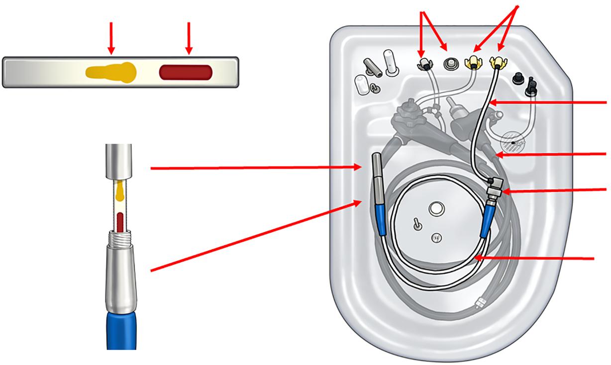
Auxiliary water/elevator wire channel connector Connecting tube
Endoscope
Luer lock
Flexible tube holder
Automated endoscope reprocessor (AER) Test system device
Figure 1. Verification of the efficacy of cleaning of endoscope channels using the FlexiCheck system. (A) Stainless steel test coupon coated with standardized blood and polysaccharide soils. (B) Test coupon placed into a flexible tube holder simulating a single-lumen endoscope channel. (C) Test system connected via Luer-lock fitting to the auxiliary water/elevator wire channel port (yellow colour) of an automated endoscope reprocessor, replicating clinical reprocessing conditions.
used filtered tap water for AERs. All detergents were applied at a 1:200 dilution, which is consistent with the preset configuration in the AER. A full-factorial design was used to evaluate three cleaning durations (1, 5 and 10 min) under two seasonal temperature conditions (winter and summer).
Commercial detergents used in the AER
Three commercially available detergents were tested: EndoRapid (Olympus Medical Systems), a non-enzymatic alkaline detergent (Product A); 3M Biofilm Removal Multi-Enzyme Cleaner (3M Company, St Paul, MN, USA), an enzymatic detergent containing protease, lipase, amylase and cellulase (Product B); and Enzyme Detergent PLUS (Enzyme Solutions Inc., Garrett, IN, USA), a low-foaming multi-enzyme formulation with demonstrated proteolytic and lipolytic activity (Product C).
Monitoring ambient and AER water temperatures
To simulate real-world variability, seasonal fluctuations in ambient and AER water temperatures were monitored during winter and summer. The OER-AW AER maintains a fixed disinfection temperature of 20 ◦ C using peracetic acid. Taiwan’s subtropical climate yields winter lows below 10 ◦ C and summer highs exceeding 35 ◦ C. In endoscopy centres without centralized water heating, low AER water temperatures may impair the activity of enzymatic detergent.
Hourly outdoor ambient and AER water temperatures were measured using a calibrated RC-4HC digital thermo-hygrometer (Elitech, Taiwan, China). The RC-4HC device was placed inside the AER and recorded all reprocessing phases continuously, including detergent cleaning, rinsing, high-level disinfection, and final drying. Thus, the temperature profiles reflect dynamic variations throughout the full AER cycle.
Verification of the efficacy of cleaning of endoscope channels
The efficacy of cleaning was evaluated using the FlexiCheck system (Healthmark Industries, Fraser, MI, USA) according to
AAMI ST91 and AORN guidelines [13,18]. Standardized stainless steel test coupons coated with blood and polysaccharide soils (Figure 1A) were placed into flexible tube holders (Figure 1B) simulating single-lumen endoscope channels. The flexible tube holders have an internal lumen diameter of 0.57 cm and a length of 108 cm. These were connected to the auxiliary water/elevator wire channel port of the AER via Luer-lock fittings (Figure 1C), and processed along with an endoscope through the full AER reprocessing cycle. After the AER reprocessing cycle, coupons were inspected visually Complete soil removal was classified as ‘pass’ and visible residue was classified as ‘fail’. This system enabled objective assessment of cleaning performance in the AER.
Survey of endoscopy centres in routine practice
A questionnaire-based survey was conducted at 19 endoscopy centres in Taiwan. All centres followed the reprocessing guidelines of the Digestive Endoscopic Society of Taiwan, and performed manual cleaning of patient-used endoscopes prior to AER placement [19,20]. The structured questionnaire included: (i) use of detergent and high-level disinfectant in the AER; (ii) cleaning and disinfection time, and temperature settings; and (iii) implementation of tools for in-AER verification of the efficacy of cleaning. Responses were collected through direct communication with reprocessing staff, and were verified by the study team.
Channel dimension measurements of common gastrointestinal endoscopes
The lengths of the instrument (biopsy), suction, and elevator wire channels were measured from the distal tip to the control section, excluding the segment from the control section to the connector The auxiliary water channel (also known as the water jet) was measured from the distal tip to the connector The inner diameters and length measurements of the channels reflect those of commonly used gastrointestinal endoscopes. Paediatric, ultra-thin, echoendoscopes and
enteroscopes were excluded due to their distinct specifications.
A full-factorial design was used to assess the effects of detergent type, cleaning duration, and seasonal temperature variation on the efficacy of cleaning of unbrushable channels. Visual inspection outcomes were classified as ‘pass’ or ‘fail’. No formal statistical tests were performed; results are summarized descriptively in tables and figures to illustrate comparative performance.
Channel dimensions of common gastrointestina l endoscopes
(Table I) For the EGD scope (EVIS LUCERA ELITE GIF-HQ290; Olympus Medical Systems), the instrument (biopsy), suction, and air/water channels have approximate lengths of 117 cm, 131 cm and 133 cm, respectively In comparison, the colonoscope (EVIS X1 Zoom CF-H290L; Olympus Medical Systems) has corresponding channel lengths of approximately 183 cm, 197 cm and 195 cm. The auxiliary water channel in the EGD scope measures approximately 281 cm, whereas in the colonoscope, it measures approximately 345 cm. The elevator wire channel extends from the distal tip to the control section. For example, in duodenoscopes (EVIS LUCERA TJF-Q260V; Olympus Medical Systems), the elevator wire channel length is approximately 154 cm.
The inner diameters of the instrument (biopsy) and suction channels in the GIF-HQ290 EGD scope are approximately 0.28 cm. In comparison, certain models, such as the GIFXTQ160 (Olympus Medical Systems), feature a larger biopsy channel with an inner diameter of up to 0.60 cm. The air/water and auxiliary water channels typically have inner diameters of approximately 0.14 cm. The elevator wire channel, as found in duodenoscopes such as the EVIS LUCERA TJF-Q260V, has a narrower inner diameter of approximately 0.06 cm.
Detergent dilution ratio: compatibili ty with AER setting of 1:200
All detergents were applied at a dilution of 1:200, which matched the preset configuration of the OER-AW AER (Olympus Medical Systems). This dilution complied with the IFUs for Product A (1:200), and fell within the recommended ranges for the enzymatic detergents: Product B (1:200 1:400) and Product C (1:125 1:250) (Table II).
Table II
Detergent temperature : IFU requirement vs AER setting at 20 ◦ C
The AER operated with a fixed disinfection temperature of 20 ◦ C using peracetic acid. However, this fixed temperature setting did not align with the IFU-recommended temperature ranges for enzymatic detergents Product B (40 60 ◦ C) and Product C (25 55 ◦ C) (Table II). Consequently, both enzymatic detergents operated below their optimal temperature ranges, potentially compromising the efficacy of cleaning.
Seasonal changes in ambient outdoor temperature affected the AER water temperature of the OER-AW AER (Olympus Medical Systems). During summer (Figure 2A), outdoor temperatures ranged from 24 ◦ C to 38 ◦ C; however, the AER maintained a cleaning water temperature near 20 ◦ C, which was below the IFU-recommended ranges for enzymatic detergents Product B (40 60 ◦ C) and Product C (25 55 ◦ C), which may have limited the efficacy of cleaning during warmer months.
In contrast, in winter, outdoor local temperatures ranged from 15 ◦ C to 22 ◦ C, during which the AER water temperature was close to 19 ◦ C in the morning (Figure 2B). This lower temperature was likely insufficient to activate enzymatic detergents within their IFU-specified ranges [Product B (40 60 ◦ C) and Product C (25 55 ◦ C)], further compromising the efficacy of cleaning under colder conditions.
Test coupons (N=5) were inspected visually after each AER reprocessing cycle under predefined cleaning durations (1, 5 and 10 min) and seasonal conditions (summer and winter) (Table III). The efficacy of cleaning of endoscope channels varied significantly with cleaning time and seasonal temperature.
Product A (non-enzymatic detergent) passed at all durations in summer. In contrast, in winter, it passed at 5 and 10 min but failed at 1 min, indicating inadequate detergent contact time under colder conditions. Product B (enzymatic detergent) showed clear time- and temperature-dependence: it passed at 5 and 10 min in summer, but only passed at 10 min in winter Product C failed consistently at all durations in both seasons, indicating limited efficacy regardless of time or temperature. These findings highlight the importance of adequate contact time and seasonal temperature monitoring for optimal AER cleaning performance.
Figure 2. Ambient outdoor and automated endoscope reprocessor (AER) water temperatures in summer (A) and winter (B). The AER temperature setting for peracetic acid is fixed at 20 ◦ C. During winter mornings, reduced AER water temperature may prevent enzymatic detergents from reaching their optimal activity range, potentially reducing the efficacy of cleaning.
Table III
Efficacy of cleaning of unbrushable endoscope channels in the automated endoscope reprocessor
Detergent product Detergent name
Product A Olympus EndoRapid (non-enzymatic) Summer Pass Pass Pass Winter Fail Pass Pass
Product B 3M Biofilm Removal Multi-Enzyme Cleaner Summer Fail Pass Pass Winter Fail Fail Pass
Product C ESI Medical Enzyme Detergent PLUS Summer Fail Fail Fail Winter Fail Fail Fail
Verification of the efficacy of cleaning using FlexiCheck system: pass, complete soil removal; fail, visible residue.
Endoscopy centre survey: cleaning practice s in the AER
Among the 19 endoscopy centres surveyed (Table IV), 11 centres (57.9%) used peracetic acid (5 min at 20 ◦ C), and eight centres (42.1%) used ortho-phthalaldehyde (5 min at 25 ◦ C) for high-level disinfection.
Fifteen endoscopy centres (78.9%) reported using detergents (Product A, B or C) in the AER. Notably, cleaning time settings and detergent use varied considerably between centres. Specifically, two centres (10.5%) used a 1-min cycle, three centres (15.8%) used a 3-min cycle, 10 centres (52.6%) used a 5min cycle, three centres (15.8%) used an 8-min cycle, and one centre (5.3%) adjusted the cleaning time based on contamination level and procedure type. Only two centres (10.5%) performed routine in-AER verification of the efficacy of cleaning using objective assessment tools in accordance with AAMI and AORN recommendations.
Discussion
To the authors’ knowledge, this is the first study to demonstrate that: (i) the efficacy of cleaning of endoscope channels is influenced significantly by detergent formulation, water temperature, and contact time within the AER; (ii) enzymatic
detergents (Products B and C) with recommended temperatures (25 60 ◦ C) did not achieve their IFU-recommended performance at the AER’s fixed 20 ◦ C setting (Figure 2, Table II); (iii) seasonal temperature fluctuations, particularly in facilities without centralized water heating, may impair enzymatic activity during winter, reducing the efficacy of cleaning (Table III); and (iv) only two endoscopy centres (10.5%) performed in-AER cleaning verification in accordance with AAMI and AORN guidelines (Table IV) [13,18].
These findings highlight substantial variability in AER cleaning practices, and underscore the need for standardized validation protocols and robust quality assurance measures in endoscope reprocessing workflows [21].
Verification of the efficacy of cleaning after manual cleaning or within the AER
Verification of the efficacy of cleaning after manual cleaning and before AER processing is essential to ensure the overall success of endoscope reprocessing. As recommended in the AAMI ST91 guidelines, tests such as protein, haemoglobin or ATP assays can detect residual contamination that is not visible to the naked eye, particularly within unbrushable channels. These tests serve as a critical quality checkpoint for identifying 8:00 9:00 10:00 11:00 12:00 13:00 14:00 15:00
Table IV
Cleaning and disinfection practices in the automated endoscope reprocessor at 19 endoscopy centres
Percentages are based on the total number of endoscopy centres surveyed (N=19).
inadequately cleaned endoscopes prior to AER placement, and helps reduce the likelihood of insufficient cleaning during automated reprocessing.
Although detergents are typically validated under ideal manual cleaning conditions [22], their performance may not accurately reflect real-world effectiveness in AER environments [21]. Regular in-AER testing of the efficacy of cleaning is recommended, particularly when introducing new detergents, modifying the reprocessing AER settings, or upgrading AER systems. Such verification ensures that workflow or system changes do not compromise cleaning outcomes, thereby maintaining consistent reprocessing quality between endoscopy centres [13].
Many enzymatic detergents were originally designed for manual soaking in heated water, and their efficacy decreases in AERs lacking active temperature control [23]. These detergents, which contain proteases, lipases and amylases, require optimal concentration, temperature and contact time to degrade proteinaceous and polysaccharide debris effectively [24 26]. Importantly, the detergent, disinfectant and AER products used may be manufactured by different companies. These manufacturers do not guarantee interoperability across product categories, and no validated assurance exists that combining detergents, disinfectants and AERs from different sources will yield effective or compatible reprocessing outcomes. Effective reprocessing requires thermal compatibility between cleaning agents and disinfectants. However, common high-level disinfectants, such as peracetic acid, orthophthalaldehyde and glutaraldehyde, have defined temperature ranges for optimal microbicidal action [1]. However, AERs, such as the OER-AW AER (Olympus Medical Systems), operate at a high-level disinfection temperature fixed at 20 ◦ C for peracetic acid, which falls below the IFU-recommended temperature ranges for enzymatic detergents such as Products B and C (typically 25 60 ◦ C). This study compared one non-enzymatic detergent (Product A) and two enzymatic detergents (Products B and C). Non-enzymatic agents depend on surfactants and alkaline chemistry, making them less sensitive to temperature variation and more compatible with fixed-temperature AER reprocessing cycles.
Outdoor temperatures below 20 ◦ C affected AER water temperature during the initial cleaning cycle, particularly in the early morning. The OER-AW AER (Olympus Medical Systems) lacks an integrated heating system for the cleaning phase. This AER is equipped with a heating mechanism for the high-level disinfection phase, which raises internal temperature gradually during subsequent steps, but lacks active heating during the cleaning phase. As such, the cleaning phase was conducted at a fixed temperature of 20 ◦ C, consistent with the nonadjustable default setting of the OER-AW AER (Olympus Medical Systems) and the preset used for the peracetic-acid-based high-level disinfection phase. Although enzymatic detergents generally recommend a temperature range of 25 60 ◦ C for optimal activity, the AER cleaning phase depends on the temperature of the facility water supply
This mismatch between detergent IFUs and AER operational settings may impair the efficacy of cleaning during colder months. Raising the cleaning temperature could reduce seasonal variability; however, this would require hardware modifications or centralized water heating infrastructure, which are not standard across all facilities.
The evaluated seasonal temperature ranges represent realworld variability, particularly in facilities lacking centralized water heating (Figure 2). Cold AER water during winter can reduce AER cleaning temperature, diminishing enzyme activity and impairing soil removal. Conversely, summer temperatures may improve cleaning performance, but excessively high heat may denature enzymes, reducing efficacy [27]. Thus, regular water temperature monitoring and appropriate adjustments should be integrated into quality assurance protocols.
The selected cleaning durations (1, 5 and 10 min) were based on manufacturer recommendations. Shorter durations (e.g. 1 min) simulate high-throughput scenarios, while longer
durations (5 10 min) support thorough removal of biofilms and embedded debris from complex lumens [28]. A 10-min cleaning cycle represents the upper operational limit in most AERs, helping to define a practical minimum duration that balances infection prevention with efficiency.
These results clearly demonstrate that 1-min cleaning cycles were consistently ineffective, both for enzymatic detergents under all seasonal conditions, and for the nonenzymatic detergent during winter The availability of 1-min cycles in AERs may create a false sense of the efficacy of cleaning, particularly in high-throughput settings where time constraints promote their use. Although preprogrammed in many AERs, the use of 1-min cycles should be critically reevaluated, especially during periods of high clinical demand. Validated longer cleaning durations (≥5 min) should be prioritized to ensure consistent and effective reprocessing.
Enhancing endoscope channe l cleaning during manual cleaning
Manual flushing remains a critical step in the cleaning of unbrushable endoscope channels. To ensure complete debris removal, each channel should be flushed with an appropriate detergent solution in accordance with the standard manual cleaning protocol. However, detergent selection, flushing volume, and duration are not standardized in current guidelines, resulting in variability in practice [29]. The primary objective of this study was to isolate the efficacy of cleaning of endoscope channels within the AER under controlled, standardized conditions. To achieve this, bedside flushing and manual cleaning were deliberately excluded from the experimental protocol. This design allowed for targeted assessment of detergent flushing performance in the AER using the FlexiCheck system. Automated flushing systems, such as the Scope Buddy Endoscope Flushing Aid (Medivators; Steris Healthcare, Mentor, OH, USA), used with an enzymatic detergent, can deliver targeted irrigation with adjustable time and pressure settings, thereby improving detergent penetration and debris removal. However, the overall effectiveness of manual flushing depends largely on consistent operator technique and strict adherence to established protocols [15]. Additionally, sonication, either in dedicated manual tanks or integrated into the AER detergent cycle, can enhance cleaning by generating ultrasonic waves that disrupt biofilms and dislodge embedded material [30]. Combining manual flushing, sonication and optimized AER settings may further improve debris removal from complex lumens, reducing the risk of residual contamination and biofilm persistence.
This study has several limitations. It was conducted in a controlled laboratory setting using simulated soils, which may not fully replicate the complexity of patient-derived contaminants. Only three detergents, three durations, and two temperature points were tested; intermediate and extreme variations were not assessed. Findings apply specifically to the OER-AW AER (Olympus Medical Systems) with peracetic acid at a fixed temperature at 20 ◦ C, and may not apply to other models or disinfectants.
The auxiliary water channel is found only in EGD scopes and colonoscopes. When reprocessing the EGD scopes or
colonoscopes, the same ports are used (yellow colour; Figure 1): one for the auxiliary water channel of these endoscope, and one for the FlexiCheck system. Therefore, the cleaning verification set-up is applicable to the auxiliary water channel through the same connection configuration.
As the endoscopic air/water channel includes a bifurcation where air and water pathways converge, this potentially alters fluid dynamics and reduces the efficacy of cleaning. This study specifically evaluated single-lumen, non-branching channels to ensure standardized testing conditions; therefore, the air/ water channel in the AER was not evaluated. Although this study focused on single-lumen, non-branching channels to ensure standardized conditions, the unique architecture of bifurcated channels may require distinct cleaning considerations.
Duodenoscopes with three-channel systems (air/water, biopsy/suction, and elevator wire channels) may alter detergent flow dynamics. In this study, the risk was minimized by isolating the elevator wire channel through a single-lumen connection. Nonetheless, in clinical practice, endoscope complexity may influence detergent distribution; this factor warrants further investigation.
The FlexiCheck flexible tube holder has an internal lumen diameter of 0.57 cm and a length of 108 cm. While the diameter is larger than that of typical unbrushable endoscope channels (0.06 0.14 cm), the length is comparable to clinical channels (129 345 cm; Table I). This dimensional similarity enables the FlexiCheck system to reasonably simulate flow dynamics and cleaning challenges encountered during detergent flushing. Microbiological outcomes were not evaluated, limiting conclusions on pathogen elimination. Based on the authors’ previous study, manual cleaning following bedside pre-cleaning and prior to AER processing is standard practice; all participating endoscopy centres in Taiwan reportedly comply with this protocol [20]. However, manual cleaning practices were not evaluated directly in the current study, which remains a limitation of this investigation. Finally, the two-centre design and small sample size may limit the generalizability of the findings.
In conclusion, reprocessing unbrushable endoscope channels remains a major infection control challenge. This study demonstrates that detergent selection, cleaning time, and water temperature are critical determinants of the efficacy of cleaning. Full-factorial testing and in-AER cleaning verification should be standard components of AER workflows. Implementing these measures will improve cleaning consistency, and reduce the risk of infection from residual contamination in high-risk channels.
CRediT authorship contribution statement
T-C. Liu: Writing original draft, Resources, Methodology, Funding acquisition, Data curation. C-L. Peng: Validation, Methodology, Data curation, Conceptualization. P-H. Tseng: Visualization, Validation, Methodology, Conceptualization. W-K. Chang: Writing review & editing, Validation, Supervision, Data curation, Conceptualization.
Conflict of interest statement
None declared.
Funding source
This study was supported by the Ministry of Defense Medical Affairs Bureau, Tri-Service General Hospital (Grant No. TSGH-D-114095).
[1] Rutala WA, Weber DJ. Disinfection and sterilization in health care facilities: what clinicians need to know. Clin Infect Dis 2004;39:702 9
[2] Muscarella LF. Risk of transmission of carbapenem-resistant Enterobacteriaceae and related ‘superbugs’ during gastrointestinal endoscopy. World J Gastrointest Endosc 2014;6:457 74
[3] Ofstead CL, Hopkins KM, Buro BL, Eiland JE, Wetzler HP. Challenges in achieving effective high-level disinfection in endoscope reprocessing. Am J Infect Control 2020;48:309 15
[4] Reprocessing manual. Instructions for Evis Lucera Elite duodenovideoscope Olympus TJF-Q290V. Tokyo: Olympus Medical Systems Corp; 2018.
[5] Reprocessing manual. Instructions for Evis Lucera Elite gastrointestinal videoscope, colonovideoscope and small intestinal videoscope. Tokyo: Olympus Medical Systems Corp.; 2022
[6] Reprocessing manual. Instructions for PENTAX Medical gastrointestinal videoscopes and duodenoscopes. Tokyo: PENTAX Medical; 2023
[7] Reprocessing summary and guide for Fujinon/Fujifilm flexible GI endoscopes. Tokyo: Fujifilm Medical Systems; 2012
[8] Anwardeen Ziaudeen SZ, Yeo CS, Yeo DXW. Validity of the efficacy of the spray flushing cleaning method in flexible endoscope reprocessing. World J Gastroenterol 2025;31:101743
[9] Ribeiro MM, de Oliveira AC. Analysis of the air/water channels of gastrointestinal endoscopies as a risk factor for the transmission of microorganisms among patients. Am J Infect Control 2012;40:913 6
[10] US Food and Drug Administration. Design of endoscopic retrograde cholangiopancreatography (ERCP) duodenoscopes may impede effective cleaning: FDA Safety Communication. Silver Spring, MD: US FDA; 2015.
[11] Alfa MJ, Singh H. Impact of wet storage and other factors on biofilm formation and contamination of patient-ready endoscopes: a narrative review. Gastrointest Endosc 2020;91:236 47
[12] Moshkanbaryans L, Shah V, Tan LY, Jones MP, Vickery K, Alfa M, et al. Comparison of two endoscope channel cleaning approaches to remove cyclic build-up biofilm. J Hosp Infect 2024;150:91 5
[13] Association for the Advancement of Medical Instrumentation. Flexible and semi-rigid endoscope processing in health care facilities. Arlington, VA: AAMI; 2021.
[14] Thaker AM, Muthusamy VR, Sedarat A, Watson RR, Kochman ML, Ross AS, et al. Duodenoscope reprocessing practice patterns in U. S. endoscopy centers: a survey study. Gastrointest Endosc 2018;88:316 322e2.
[15] Beilenhoff U, Biering H, Blum R, Brljak J, Cimbro M, Dumonceau JM, et al. Reprocessing of flexible endoscopes and endoscopic accessories used in gastrointestinal endoscopy: Position Statement of the European Society of Gastrointestinal
Endoscopy (ESGE) and European Society of Gastroenterology Nurses and Associates (ESGENA) update 2018. Endoscopy 2018;50:1205 34
[16] Herve R, Keevil CW. Current limitations about the cleaning of luminal endoscopes. J Hosp Infect 2013;83:22 9
[17] Gonzalez JA, Vanzieleghem T, Dumazy A, Meuris C, Mutsers J, Christiaens G, et al. On-site comparison of an enzymatic detergent and a non-enzymatic detergent-disinfectant for routine manual cleaning of flexible endoscopes. Endosc Int Open 2019;7: E412 20
[18] Croke L. Guideline for processing flexible endoscopes. AORN J 2022;116:P5 7
[19] Chang WK, Peng CL, Chen YW, Sun CK, Chen CC, Liu TC, et al. Recommendations and guidelines for endoscope reprocessing: current position statement of Digestive Endoscopic Society of Taiwan. J Microbiol Immunol Infect 2024;57:211 24
[20] Chang WK, Liu TC, Liu TL, Peng CL, Wang HP. Enhanced manual cleaning efficacy of duodenoscope in endoscopy units: results of a multicenter comprehensive quality control program. Am J Infect Control 2019;47:1233 9
[21] Hu R, Yi L, Zou T, Hu J, Chen Y, Pan W. Current management status of cleaning and disinfection for gastrointestinal endoscopy: a meta-analysis. Sci Rep 2024;14:27238.
[22] Hicks J, Mutowo M. Streamlining endoscopy cleaning: the impact of a new detergent on time and water use. J Mark Access Health Policy 2025;13:23
[23] Noubam-Tchatat C, Badrikian L, Traore O, Aumeran C. Evaluation of two detergent-disinfectants and a detergent on a Klebsiella pneumoniae biofilm formed within Tygon tubes. J Hosp Infect 2023;140:1 7
[24] Ren W, Sheng X, Huang X, Zhi F, Cai W. Evaluation of detergents and contact time on biofilm removal from flexible endoscopes. Am J Infect Control 2013;41:e89 92
[25] da Costa Luciano C, Olson N, Tipple AF, Alfa M. Evaluation of the ability of different detergents and disinfectants to remove and kill organisms in traditional biofilm. Am J Infect Control 2016;44: e243 9.
[26] Vickery K, Pajkos A, Cossart Y. Removal of biofilm from endoscopes: evaluation of detergent efficiency. Am J Infect Control 2004;32:170 6
[27] Zuhlsdorf B, Winkler A, Dietze B, Floss H, Martiny H. Gastroscope processing in washer-disinfectors at three different temperatures. J Hosp Infect 2003;55:276 82
[28] Pajkos A, Vickery K, Cossart Y. Is biofilm accumulation on endoscope tubing a contributor to the failure of cleaning and decontamination? J Hosp Infect 2004;58:224 9.
[29] Kovaleva J. Endoscope drying and its pitfalls. J Hosp Infect 2017;97:319 28
[30] Kreeft D, Arkenbout EA, Henselmans PWJ, van Furth WR, Breedveld P. Review of techniques to achieve optical surface cleanliness and their potential application to surgical endoscopes. Surg Innov 2017;24:509 27











Date: Thursday 4 September 2025
Time: 1330 - 1400
Venue: Energy Event Centre, Rotorua and Microsoft TEAMS
Executive Present: Martin Bird, John Barnacott, Jenny Carston, Carla Coetzee, Donna Dador, Aileen Derby, Antony Owens, Charanjeet Kaur Sidhu, Alison Stewart, Shelagh Thomas
Full list of attendees: Refer to Appendix A
Welcome / Opening: Attendees were welcomed by the NZSSA President Martin Bird
1. Request for additional proxy voting forms – Carla Coetzee: No forms provided
2. Apologies – Carla Coetzee: Shelley Morrison
3. Minutes from last Annual General Meeting – Carla Coetzee
NZSSA AGM Minutes 2024 were circulated to members in the AGM pack prior to the meeting. The motion was put to those present that the minutes be accepted.
Moved by: Maureen Scott
Seconded: Shelagh Thomas
4. Matters arising from minutes of last meeting – Carla Coetzee
No matters arising from the minutes.
5. NZSSA Annual report 2025 – Martin Bird
The Annual Report containing information on the Association for 2025 including President, Secretary and Treasurer and Performance reports, was circulated with the AGM pack to members prior to the meeting. The meeting was asked if anyone wanted to discuss the content. No discussion was requested. It was moved that the report be accepted as written.
Moved by: Martin Bird
Seconded: Antony Owens
6. Treasurer’s report – Alison Stewart
The following content was discussed:
Maureen Scott asked if members will have to pay for registration in the future. The executive did not rule this out, as the NZSSA is in the process of becoming a self-regulatory body, but this is a process and paying for registration is not an option in the near future.
Anthony Valvoi asked why technicians with a level 3 qualification are not allowed to be registered. Executive members explained that those who obtained their level 3 qualification before 2019 are allowed to register but after 2019 the level 3 qualification is more relevant to non-hospital based environments and therefor a more technical level 4 qualification is needed and acknowledged for registration. NZ3208 targeted for low processing environments, rather than AS5369:2023.
No further discussion raised. It was moved that the report be accepted as written.
Moved by: Martin Bird
Seconded: Amandeep Kang
7. Auditor for 2025/2026 – Alison Stewart
Accounting for Charities has been engaged as the auditors for the Association performance report since 2021. A motion was raised to retain the audit services of Accounting for Charities for the coming financial year.
Moved by: Alison Stewart
Seconded: Jenny Northover
8. New Executive Member – Martin Bird
Shelley Morrison was endorsed as a new member of the executive in the light of a previous member stepping down from the position earlier this year. A question was raised how executive members are selected and it was explained that after election (which was last year), if someone steps down, a call is made for expressions of interest via the NZSSA website. Applicants are then interviewed by a subcommittee voted by the executive and the new member chosen. It was moved that the aforementioned member be ratified as an NZSSA Executive member.
Moved by: Alison Stewart
Seconded: Donna Dador
9. Compulsory Registration – Martin Bird & Shelagh Thomas
All Sterile Sciences Technicians (L3 prior to 2019 and L4 after 20198) has to be registered by the end of January 2026. This will require a professional portfolio to be kept by the technician, and evidence of education and hours to be provided. This is all in preparation of the NZSSA and Sterile Sciences becoming a self-regulating body and profession. The registration guidelines and forms are available on the website.
A question was raised by Jey Vellupillai if research and presentation / teaching time can contribute to the education hours needed to be registered and how much it would account for. The executive agreed this should be taken into account, will have to discuss this and give feedback in due course.
10. The NZSSA new logo – Aileen Derby
The logo had to be changed to include the Māori version of “The NZ Sterile Sciences Association” and “Inc.” to meet legislation.
11. NZSSA Constitution & Rules – Aileen Derby
The association’s constitution has been reviewed. Drafts were completed and sent for legal review with suggested amendments made. The latest version was posted on the website and a request was placed in Supplyline and on the Website that members were to inform Aileen if they had any comments or concerns, and there were none. It was assumed that all members are happy with this. A copy of the new constitution and rules is available to look at. The process of re-registration (which can be done online) can start after ratification. The meeting was again reminded by Alison that the NZSSA is an association under the registry but we are registered as a charity as we have goals that we are held to such as providing for education opportunities via scholarships. It was moved that the new constitution & rules be ratified.
Moved by: Maureen Scott
Seconded: Anthony Valvoi
12. Remits
No remits submitted.
13. General Business
· Martin Bird announced that Shelagh Thomas will now be a Lifetime Member of the NZSSA. He handed her a bouquet of flowers and thanked her for her service and all she has done for the profession in NZ. It was moved that her Lifetime membership be approved.
Moved by: Martin Bird
Seconded: Alison Stewart
· Hawkes Bay Hospital CSSD Educator Amandeep Kang asked about the competency checks that will accompany registration. What will the format be and would it be separate from each hospital’s?
Shelagh Thomas explained that this is still a work in progress and that there is a difference between the workplace’s performance appraisals and the association’s checklist to deem a technician competent. Amandeep also asked if a Scope of Practice, and if a generic one will be available. Again Shelagh answered, alongside Alison that scopes can be workplace specific and role specific.
· No other general business was raised.
Meeting Closed: 1400hrs
Aileen Derby
Michael Consador
Akanesi Tu’iono Moana Ruffell
Alison Stewart Monique Ethier
Alyss Campbell Nadine Wells
Amandeep Kang Paul Martillana
Amber Tredinnick Roka Cooper
Anthony Owens Sajna Janish
Anthony Valvoi
Sandra Paku
Ashwini Sharan Sharleen Allan
Carla Coetzee Shelagh Thomas
Carley Field
Steven Grant
Charanjeet Sidhu Victoria Copland
Christian Apacible
Dee Davis
Donna Dador
Edward Isla
Elsie Nivo
Fiona Stewart-Webster
Gemma Clavano
Gita Nadan
Janine Warren
Jenny Carston
Jenny Northover
Jey Vellupillai
Jill Haig
John Barnacott
Karen Steen
Karli Robin
Kelsey Smith
Kerry Harriman
Kimberley Saunders
Leighton White
Luke Rabjohns
Mandy Labuschagne
Margaret Bridge
Marina Nasmith
Martin Bird
Maureen Scott
Meryka Potgieter
Kia Ora Everyone,
We’re pleased to share an update on the progress of the new NZSSA website (see below for a snapshot of the home page).
Firstly, our apologies for the delay in going live. We had hoped to launch prior to the NZSSA Conference in 2025; however, we have been working through several technical matters behind the scenes. In particular, we have focused on ensuring the new membership software meets the Association’s requirements and is straightforward and user-friendly for all members.
We are now delighted to confirm that the new website will go live on 1 April 2026.
One requirement we were unable to work around is the need for all members to re-register on the new system. Please see the relevant information below:
Individual Member Registrations
To make this process as seamless as possible, you will be issued a monthly coupon code aligned with your original registration month. This will:
• Enable your registration on the new system
• Apply a discount aligning you to your scheduled month of renewal
• Set up automatic annual renewal going forward
This will streamline the process for future renewals.
Registrations via Purchase Order (PO)
• Managers or Team Leads will be contacted directly to provide updated member information.
• Organisations will continue to be invoiced as usual
• Members will be manually imported into the system by the webmaster.
One of the key improvements to the new website is the ability to reset your username and password independently, without needing to contact support.
We hope you enjoy the new, modern, and refreshed look. The site has been designed for ease of navigation and provides improved access to members-only professional development resources.
Please keep an eye out for an email from the NZSSA Treasurer with details about your coupon code. We also recommend updating your contact details to a personal email address rather than a work email to ensure continued access.
Thank you all for your patience and support during this transition.
Cheers, Alison
& Antony

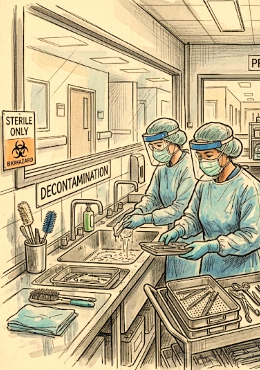

Take the stage! Winners will be announced at the NZSSA conference, and finalist videos will be featured live during the event!
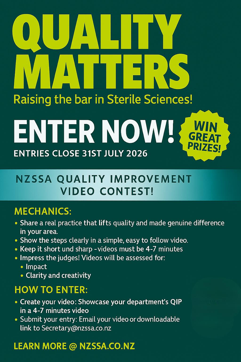
Martin Bird (President)
CSSD Manager
Dunedin Hospital
Dunedin
martin.bird@southendhb.govt.nz
Carla Coetzee (Secretary)
SSD
Health NZ Hawkes Bay
Hastings
Email: secretary@nzssa.co.nz
Aileen Derby
CSSD Manager
Counties Manukau District
Auckland aileen.derby@tewhatuora.govt.nz
Charanjeet Kaur Sidhu
CSSD Quality Lead Clinical Counties Manukau District
Auckland
Charanjeet.Sidhu@TeWhatuOra.govt.nz
Jenny Carston
Team Leader CSU
Tauranga Hospital
Tauranga
Jenny.Carston@bopdhb.govt.nz
Shelley Morrison
CSSD
University of Otago Faculty of Dentistry Dunedin 9054
shelley.vivian@otago.ac.nz
General Enquiries
Donna Belle Dador (Vice President)
National Sterile Services Adviser Southern Cross Healthcare Christchurch donna.dador@southerncrosshospitals.co.nz
Shelagh Thomas CSSD
Hutt Valley Capital, Coast and Hutt Valley shelagh.thomas@huttvalleydhb.org.nz
Antony Owens
Team Leader
Southern Cross Healthcare
Southern Cross Gillies Hospital Auckland antony.owens@schl.co.nz
John Barnacott
Manager Sterile Services Unit
Mid Central Palmerston North Hospital Palmerston North John.Barnacott@midcentraldhb.govt.nz
Alison Stewart (Treasurer)
NZSSA Treasurer
28 Brighton Street, Island Bay Wellington 6023
Email: accounts@nzssa.co.nz
secretary@nzssa.co.nz
Overseas qualification and membership inquiries accounts@nzssa..co.nz
Complaints complaints@nzssa.co.nz
Registration Assessors –
Charanjeet Sidhu
Maureen Scott
Donna Dador
Shelagh Thomas
Charanjeet.Sidhu@TeWhatuOra.govt.nz Maureen.Scott@waikatodhb.health.nz donna.dador@southerncrosshospitals.co.nz shelagh.thomas@huttvalleydhb.org.nz