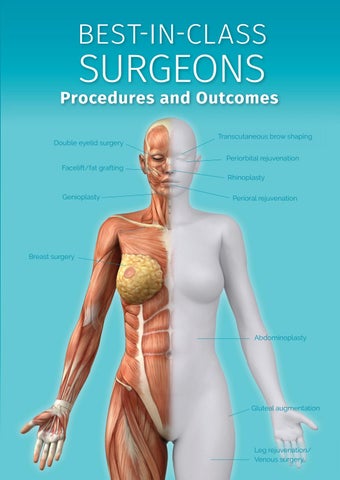
BEST-IN-CLASS
SURGEONS
Procedures and Outcomes Double eyelid surgery
Transcutaneous brow shaping Periorbital rejuvenation
Facelift/fat grafting Rhinoplasty Genioplasty
Perioral rejuvenation
Breast surgery
Abdominoplasty
Gluteal augmentation
Leg rejuvenation/ Venous surgery
BEST-IN-CLASS
SURGEONS procedures and outcomes
www.beyondblack.org
CONTENTS 4 FOREWORD
6 The Surgeon’s Learning Curve Dr. Lorne King Rosenfield
pain = maximum gain” 24 “Maximum Fact or fallacy in Facelifting? Mr. Barry M. Jones
First published in 2019 by BB PUBLICATIONS © B.B. Publications 2019 All rights reserved. No part of this publication may be reproduced, stored in or introduced into a retrieval system, or transmitted in any form or by any means (electronic, mechanical, photocopying, recording or otherwise) without the prior written permission of both the copyright owner and the publisher of this book. www.beyondblack.org www.bbpublications.org ISBN 978-1-905904-77-8 Designed by www.squareandcircus.co.uk
32
Framing the Eyes
40
Facial Rejuvenation with M.A.D.E.
52
Using a Sculptor’s Eye to Customise The Facelift
Dr. Mario Pelle Ceravolo
Dr. Andrew Jacono
Dr. T. Gerald O’Daniel
70 Facelift and Fat Grafting Dr. Jesper Sorensen
80 Perioral Rejuvenation Dr. Jesper Sorensen and Dr. Sameira Perren
94
Asian Cosmetic Surgeries Dr Shim Ching
102
Defy Aging with Volume Restoration
116
The Natural Facelift
124
Genioplasty
130
Transaxillary Endoscopic Breast Augmentation
138
The ‘True Form’ tummy tuck
150
Gluteal Augmentation with Autologous Fat Grafting
164 180
Dr. William Lao
Dr. P. Craig Hobar
Dr. P. Craig Hobar
Dr. P. Craig Hobar
Dr. Christopher Patronella
Dr. Adel Quttainah
The Whiteley Protocol® for Veins Prof. Mark S Whiteley
Regenerative Aesthetics: A Global Revolution Dr. Mariam Awada
BEST-IN-CLASS SURGEONS: PROCEDURES AND OUTCOMES 3
Foreword The pursuit of eternal youth and beauty has fuelled a massive boom in plastic surgery and considerable advances in the field of elective procedures technologies generally. The emergence of social media “influencers” who shape the aesthetic trends of the day through existing and paid-for followers, on the other hand, has created a new “monster” phenomenon: the pursuit of unattainable perfection. We use the word “monster” advisedly: when very young individuals start altering their appearance in order to emulate the heavily photoshopped pictures of idealised models, it is time for plastic surgeons to remember they are physicians too. Traditionally and historically, people have turned to plastic surgery for two distinct reasons: reconstruction of some genetic or acquired defect, or rejuvenation. Increasingly, plastic surgery is being harnessed to beautify ever younger women and men, but at what future cost? We have, over the years, worked with some of the most reputable surgeons in the world and their disquiet at social media-led procedures is become more and more vocal. This book, as has been the case with previous editions, seeks to demystify some of the procedures that are available, caution against others that have evolved from trends and educate on what the possibilities and limitations of surgery are. The plastic surgeon may be an aesthete, an artist even, but he is above all a finely skilled technician, not a magician. His skill and experience can make all the different between a well-executed, long-lasting result and a poor one, but he only has the patient’s genetic material to work with.
4 SURGERY HEADER
Halting the visible passage of time and reversing the signs of decay is possible up to a point. Is it desirable? Yes, without a doubt. We all want to look better for our age, younger if at all possible, more desirable physically. There is an added subtlety to this: individuals who can afford to look after themselves, whose teeth are in good condition, whose hair is well-kept and whose face and body are impervious to sagging here and there are by implication successful. The age of maintenance requires a considerable financial investment because plastic surgery at its best is not cheap and neither is elective dental care. Non-invasive procedures that keep the skin in top condition require regular updates. It is wise to plan for this judiciously and smartly: which plastic surgeon is properly accredited (as opposed to wellmarketed), old enough to be experienced, but young enough to accompany you through years of “tweaks”, innovative without being radical at the expense of your face/body, artistic without imposing his own beauty ideal on you, and a professional of integrity who would tell you the truth as opposed to try and sell you a “kiss me quick” procedure that fits all. This edition contains some interesting essays and a number of chapters outlining the contributing surgeons’ philosophies and approaches. Some are divergent. It is incumbent upon everyone seeking surgery to look for different opinions, rather than to try and validate their own. We have invited surgeons who have contributed to previous editions, but we have also democratised the selection process. There are a handful of “celebrity” surgeons in the world, but a number of discreet super talents in the profession too. Plastic surgery evolves incrementally rather than in revolutionary leaps. Just as is the case in our books, surgeons, more and/or less prominent, observe, critic and perfect their own and each others’ techniques all the time. The publisher merely seeks to profile some of the best-inclass, in their own words.
BEST-IN-CLASS SURGEONS: PROCEDURES AND OUTCOMES 5
6 SURGERY HEADER
The Surgeon’s Learning Curve Dr. Lorne King Rosenfield
BEST-IN-CLASS SURGEONS: PROCEDURES AND OUTCOMES 7
“There are three constants in life… change, choice, and principles.”
J
ames Covey, the late author of the bestselling 7 Habits of Highly Effective People, coined the above phrase – one that stands as a fitting epithet for aesthetic plastic surgeons. Over the last 30 years, I have been a diligent, evangelical and, most importantly, practicing disciple of this belief. I say “belief” because beneath the very evidenced, methodical and factual nature of our surgeries - our science - there should also be a honing, a harnessing of the more philosophical, ruminative, and introspective aspect of our procedures - our art. However, if we conflate these two seemingly opposing ideas, we arrive at what another great thinker, K. Anders Ericsson, calls “deliberate practice” - an effective philosophy best baked into our surgical routines: “With deliberate practice, however, the goal is not just to reach your potential but to build upon it, to make things possible that were not possible before. This requires challenging homeostasis—getting out of your comfort zone—and forcing your brain or your body to adapt.” For aesthetic plastic surgeons, it is this process of perennially challenging our approach to our surgical techniques that may be called our “Learning Curve”. Ideally, this curve does not plateau, lulling us into a routine once the first summit is reached. And neither does it look anything like a smooth arc: it’s more like a never-ending coil, drawn by trained, habituated steps (our techniques), sporadically interrupted by critical backward steps (our complications) that can inspire us to novel steps forward (our corrections). Within this “coil” the surgeon should always be assessing which steps in the technique are worth keeping, which should be modified and which should be eliminated. It is this deliberate pruning that should strive to deliver no more (in complications) and no less (in results). The purpose of this introduction is to set the tone for this entire tome, which boasts surgeons who have all been deliberately practicing to achieve their own mastery in aesthetic surgery. In the same vein, I will lay the foundation of the many steps, the successful maneuvers and the failed outcomes, that I have experienced. Lastly, I will articulate the strategic choices I made as a result
8 DR. LORNE KING ROSENFIELD
of those experiences; choices that represent my evolving practices in both the clinic and the operating room, as well as, ultimately, the guiding principles upon which I now operate.
In The Beginning
When a plastic surgeon starts out in practice, their primary motive is to studiously follow the learned surgical steps drilled into them over years of training in an effort to deliver an acceptable result without major complication. During this initial phase, one rightfully looks upon the idea of contributing to the advancement of a particular surgical technique as a distant goal, at least until one gains significant experience. But for many more mature surgeons, that same goal may gradually evaporate as the surgeon gets ever more comfortable with “habit”. This steady-state defines what we call the “standard of care” in the community. And, frankly, this “standard” represents a pretty low bar. Instead, the surgeons in this book demonstrate the power of deliberate practice to deliver, initially, incremental technical improvements along their unique learning curves and, ultimately, better results with fewer complications.
My Learning Curve
Several technical principles have emerged during my personal learning curve. As with all worthwhile principles, these may be applied universally across all aesthetic surgeries: 1. Age-induced lax tissue behaves predictably everywhere: a. Aged tissue need not be further loosened. Surgeons need not undermine or otherwise mobilize tissue in order to tighten or excise it (eg: skin recruitment by discontinuous dissection in an abdominoplasty). b. Aging affects all tissue layers. You must tighten not only the skin but the underlying fascial structures as well (eg: canthal tightening in a lower blepharoplasty). c. Aged tissue “left behind” will predictably relapse: that is, it is no better in quality than the tissue excised (eg: central neck skin in a facelift).
BEST-IN-CLASS SURGEONS: PROCEDURES AND OUTCOMES 9
2. Vector “analysis” should be applied to all lax tissues: a. vectors of excision should follow the vectors of excess: a.i. Horizontal excess is best removed by a horizontal ‘draw’ (in which the tissue is tightened by ‘drawing’ the tissue in a given direction) or through a vertical incision and conversely, a.ii. Vertical excess is best removed by a vertical draw or through a horizontal incision. a.iii.The vector of draw should be as close as possible to the excess for maximal correction. 3. Safety should always trump Results. One’s respect for the safety of the result should never be compromised by the often-blinding pursuit of the aesthetics of the outcome. 4. Simplicity should always trump Complexity. In our pursuit of “perfection”, we should strive to forge our techniques into simpler not more complex procedures. These principles will now be animated by the demonstration of their practical application within a few aesthetic surgeries. I. Blepharoplasty: The Deliberate Practice
The Evolutionary Steps to the Pinch Blepharoplasty Challenges: 1. Residual Skin: Lingering ‘crepey’ skin was noted, most often in those with prodigious fat herniation or skin wrinkling. This scenario makes cogent sense considering how little skin is excised, albeit prudently, from the bulky, relatively denervated, skin-muscle flap. 2. Scleral Show: Mild but enduring scleral show (ie: visible exaggeration of the white part of the eye) was frequently evident, often preceded by weeks of overly optimistic eyelid taping and massage. This outcome can also be predicted if one concedes the unintended consequences of the heavy skin-muscle flap. Solutions: These discomfiting observations led to my seeking an alternative. 10 DR. LORNE KING ROSENFIELD
Initially, the primary procedural aspiration was defined: I sought a modified procedure that would at once both ensure optimal correction of the eyelid deformities and also secure normal eyelid posture. The guiding principles within Glenn Jelks’ work became my motivating change agents. The solution became clear when our decades-old approach to the upper eyelid blepharoplasty was considered: surgeons already measure the extent of eyelid excess with a temporary skin “pinch” while observing the effect on the eyelash and brow posture. Why not apply the same metric to the lower eyelid - but this time using an actual skin pinch? Thus the “pinch blepharoplasty” was born. The procedure was so reliably effective and safe that it fully usurped the skin-muscle flap approach. With the clear goal in mind, two seminal technical tweaks were made: 1. As originally reported, at first I did not invoke the stitch canthopexy (ie: using stitches to move the lower lid to a slightly higher position on the face) in patients who did not demonstrate obvious laxity or scleral show. However, before long I once again began to witness, albeit not as severe or as often, scleral show. As such, I began to integrate the canthopexy in all blepharoplasty patients. This resulted in minimal (approaching zero) instances of even mild show. (The strategy makes sense when considering that any patient who presents with aging changes of their skin will inevitably have similar laxity of their tarso-ligamentous sling) 2. Early in my experience, I found a subset of patients, particularly the most wrinkled, who despite the pinch excision were still left with some residual skin. These unsatisfying results led to the secondstage “re-pinch” procedure. Initially unplanned, this appended step soon became a planned stage at 6 months+post-op. 3. However, despite its efficacy, this strategy necessitated a second intervention. It usually mandated a repeat stitch canthopexy to insure a definitively robust re-pinch. So, a new tack was taken to endeavor to remove more if not all of the redundant skin at the first sitting by gradually moving the pinch lower onto the eyelid, ever closer BEST-IN-CLASS SURGEONS: PROCEDURES AND OUTCOMES 11
to the excess. This advance has indeed displaced the second stage pinch, whilst delivering as innocuous a scar as before. At this point in its evolution, this approach - armed with a maximally applied stitch canthopexy and in turn, pinch skinplasty - safely offers a complete treatment of not only the most wrinkled but even patients displaying festoons (ie: puffy skin bags around the lower eyelid).
Principles:
This technique is girded by three principles: 1. The first guiding premise is articulated above: that one need not widely undermine tissue in order to recruit, excise and otherwise treat its excess. Instead, excess can and is most efficiently removed by either drawing the tissue in the same vector of its excess and/or excising it through an incision perpendicular to the excess. In the case of the lower eyelid wrinkling and the pinch skinplasty, both of these maneuvers are employed: the vertical excess is effectively captured by its naturally facile recruitment vertically and excised through a conveniently well-hidden incision horizontally. The smoothing of the lower eyelid wrinkling may be analogized to the making of a rumpled bed.
Demonstration of the pinch skinplasty - effectively “making the bed� - at the lower eyelid
12 DR. LORNE KING ROSENFIELD
2. The second underlying precept - and corollary of the first – is that the closer one gets to the excess, the more efficacious the pinch. This effect is similar to the more productive method of rolling up a living room carpet when one approaches it from the end closest to the excess.
3. The third principle is that the inherent support within a structure must be strengthened before a load strains it. In the instance of the lower eyelid, before the “weight” of any surgery should test this retinacular sling, it must first be tightened. Hence the efficacy of the stitch canthopexy in girding the eyelid prior to the pinch skinplasty. This maneuver is analogous to the tightening of a clothesline before the placement of the wet garments.
Illustrating the principle that the closer one is to the excess, the more effective the repair
Illustrating how the stitch canthopexy girds the eyelid before the pinchplasty
BEST-IN-CLASS SURGEONS: PROCEDURES AND OUTCOMES 13
Illustrating a 1 year postoperative result with an upper and lower blepharoplasty on this 49 year old female. She underwent a lower eyelid pinchplasty and stitch canthopexy.
II. THE FACELIFT: THE DELIBERATE PRACTICE The Evolutionary steps to the Pinch Rhytidoplasty Challenges: 1. Recurrent Neck Bands: Despite the diligent manipulation of the SMAS (superficial muscular aponeurotic system) with an extensive open neckplasty there was an unsatisfyingly persistent incidence of recurrent neck bands due to: a. the dueling vectors of the mid-line and postauricular repairs, encouraging both the early and late reappearance of bands, made more frequent the greater the tensions applied. b. the platysma muscle and SMAS harboring aginginduced impotent collagen - thus predicting the folly of its aggressive manipulation at combating recurrence. 2. Iatrogenic Neck Deformities: After great effort to correct the platysma bands, the result can still be marred by surgery-induced... a. worsening of the very same bands: the mid-line platysma plication can induce vertical shortening and thus a virtual “bowstringing�. b. scarification of the otherwise smooth neck: the extensive dissection can result in hematoma or seroma, either of which, upon resolution, can leave stubborn skin irregularities in their wake.
14 DR. LORNE KING ROSENFIELD
Solutions Initially, I sought a procedure that could preferably build upon the “traditional” SMASectomy technique. The proven work of Dan Baker became my trusted change agent. Baker taught that the general goal of SMAS tightening in the face should be to treat the obviously redundant, mobile SMAS and anchor its lateral margin to the relatively fixed, immobile SMAS. So why not apply this same principle, so effective in the face, to the neck? The same fascial imbrication/plication could simply be advanced into the neck to treat the redundant platysma/ SMAS tissues. In so doing, the repair is accomplished from this solely lateral approach, obviating the need for the traditional submental incision and central neck manipulation. Thus the entirely lateral SMAS “pinch rhytidoplasty” was conceived. With the clear goal in mind, a few seminal technical tweaks were made: 1. In order for this entirely lateral approach to deliver the broadest of repairs, from the mid-face/ malar tissues to the low neck bands and skin, the dissection gradually crept both superiorly on the face - well above the malar (the ‘high-SMAS’, a la Fritz Barton) - and inferiorly in the neck - nearing the clavicle. To properly expose and safely repair this low in the neck, a short segment of the incision was jogged to run along the hairline before diving back into the scalp. 2. With the entire result essentially resting on the adequacy of the lateral treatment, over time there was an evolution towards both a more robust SMAS plication/imbrication and an equally diligent placement of sutures to approximate a virtually “watertight” repair. 3. That said, most recently, in an effort to improve the efficacy and longevity of the neck repair, discontinuous dissection of the neck skin (similar to that performed with under-the-trunk tissues a la Lockwood) is being performed to recruit as much redundant skin and platysma as possible.
BEST-IN-CLASS SURGEONS: PROCEDURES AND OUTCOMES 15
Principles There are a few basic tenets of this technique that gird its success: 1. Skin-SMAS Assessment & Manipulation: As noted, it is not necessary to undermine either the eyelid skin flap or the abdominal cutaneous flap for either to be mobilized and tightened effectively. Similarly, one need not undermine/mobilize the skin and SMAS in order to tighten either - permitting the surgeon to circumvent an extensive undermining of the cheek flap and now, also, the formal, central neck dissection. The vector principle (that the vector of draw is ideally perpendicular to the vector of excess) is reflected in the hockey stick shaped design of the SMASectomy for maximal correction: the upper half is drawn parallel to the naso-labial and marionette folds and the lower half parallel to the skin and platysma bands. The eponymous “pinch” is applied to the preoperatively and to the SMAS intraoperatively:
Demonstrating the preoperative “pinch” of the skin for measuring the extent of skin excess and proving its translation into a full neck “correction”
16 DR. LORNE KING ROSENFIELD
skin
a. Preoperatively, this tenet can be tested and the result visualized by the “pinching” of the excess skin translating into a full “correction” of the check, jowl and neck. In my experience, this validation is consistent regardless of the extent of the excess. Additionally, this maneuver delivers an estimate of the extent of the future skin excision.
b. Intra-operatively, this principal is applied similarly with the “pinching” of the excess SMAS between two forceps to both measure and mark its excess and demonstrate its correction of the stigmata of facial aging. This expanded principle is the simple but effective maneuver of the “pinch test”, conducted between two forceps, which simultaneously demonstrates the full extent of excess, the ideal point of fixation and proper correction of the stigmata of facial aging. 2. SMAS Tightening and Repair: As Baker has taught, the general goal of SMAS tightening should be to remove the excess, mobile SMAS and anchor its lateral margin to the immobile SMAS. A particularly unyielding portion of this same tissue (residing in the preauricular area called Lore’s fascia) was originally described by Labbe to deliver an impressive suspension-induced correction of the jawline and neck. Identifying and utilizing this similarly tenacious tissue along the entire length of the facial repair is the raison d’etre of the pinch rhytidoplasty. A
B
A
B
Demonstrating design of SMAS repair Demonstrating SMAS repair deep in the lateral neck
BEST-IN-CLASS SURGEONS: PROCEDURES AND OUTCOMES 17
A
B
Demonstrating hairline jog in incision to allow for necessarily deeper neck exposure
Illustrating result at 1 year postoperatively in this 69 year old female after a lateral pinch rhytidoplasty which obviates a central neck dissection.
18 DR. LORNE KING ROSENFIELD
A
B
C
D
III ABDOMINOPLASTY: THE DELIBERATE PRACTICE The Evolutionary Steps to the High Tension Abdominoplasty Challenges 1. Skin Flap Necrosis: The most dreaded local complication of the abdominoplasty is the feared or worse, unexpected, flap ischemia and skin death. This outcome was an inherent attribute of the traditional procedure simply because the skin flap created remains, despite all efforts to the contrary, a random flap. 2. Residual Excess Skin: Regardless of how much dissection or skin resection, the traditional technique will routinely yield redundant skin at the trunk, particularly below the incision. This occurs as a result of the global nature of the excess and the relatively near-sighted goals of the typical abdominal procedure. 3. Unpredictable Scar Topography: The more excess skin, the more potentially wayward the scar. With the traditional abdominoplasty, this was primarily due to the relatively untreated redundant skin below the incision which would result in a high-riding scar and elongated pubis. Solutions: The above outcomes, uniquely ranging from simply annoying to – frankly – devastating, led me to forge a new path beyond traditional abdominoplasty. My goal was to modify the approach to realize maximum extraction of excess skin, whilst maintaining the greatest vascular safety and ensuring a predictable, easily hidden scar. At this point, the work of Ted Lockwood became my redeeming change agent. From here the following technical modifications were gradually incepted: 1. The traditional wide flap elevation and mobilization was replaced by discontinuous dissection (using Lockwood’s specialized instruments) as soon as it became clear that the latter just as effectively delivered the excess skin for excision. 2. Rather than focusing only on the pannus above incision, I began to treat more deliberately the excess below, again with the application of BEST-IN-CLASS SURGEONS: PROCEDURES AND OUTCOMES 19
Lockwood’s discontinuous dissectors clear to the knees. This maneuver is analogous to “lifting up the pants” of excess skin around the pubis and thighs. 3. My usual hip to hip, more or less horizontal incision graduated to be more obliquely upward in order to more effectively treat the excess at both below, at the thighs, and above, at the upper abdominal zones. 4. Instead of designing the incision to rest in the usual sub-pannus crease, the markings were deliberately made lower, within the patient’s underwear, to insure a properly hidden scar. 5. However, this change in the pattern of the incision both laterally (with the more oblique cant drawing more of the horizontally excessive upper abdominal skin) and centrally (with the lower incision purposely taking less infraumbilical skin), the original umbilical site was most often closed as a small vertical scar in the lower abdomen.
6. With this more comprehensive skin resection I began to harness the newly-described superficial fascial system (SFS) to insure both a secure deep fascial and tension-free skin closure.To combat the rare, but still real, possibility of necrosis - despite the perforator-saving discontinuous dissection liposuction of the undermined, central skin flap was eventually replaced by a planned second stage global liposuction (6 months+ post-operatively).
Principles 1. As with the facelift and blepharoplasty, the more the procedure follows the vectors of excess, the more efficient the treatment of the redundant skin. So in the case of the abdominoplasty (particularly in the MWL – Massive Weight Loss - patient), the excess is primarily horizontal in the central and mid-upper abdomen and primarily vertical in the lower abdomen (which explains the central horizontal and lateral oblique components of the High Tension Abdominoplasty).
20 DR. LORNE KING ROSENFIELD
Demonstrating how the vectors of the excision should follow the vectors of excess.
2. As a corollary of the above: when drawing the markings, the longer the incision is designed, the more the redundant skin will be removed. This is particularly relevant in the MWL patient where the incision can and should be as far postero-lateral as necessary. When this principle is respected, an almost total body lift can be realized from an entirely supine/anterior approach, with gratifying improvement even in the buttock ptosis (ie: sagging).
BEST-IN-CLASS SURGEONS: PROCEDURES AND OUTCOMES 21
A
B
Illustrating how the marking can be checked and the result demonstrated.
3. The abdominal repair should not only be about the excision of the obvious excess skin above the pubis - the pannus - but also beneath it: any excess pubis and inner/outer thigh redundant skin, and even the ptotic lateral buttock. Only then can a comprehensive repair be realized.
Conclusion: Hopefully this chapter has successfully demonstrated how, through a myriad of deliberate, incremental changes, the learning curve becomes crucial to achieving a reliable surgical technique. The phenomenon is similar to the “flywheel” concept offered by Jim Collins, author of From Good to Great. He describes how the path to ‘better’ is never due to one single key event or moment. Rather, like a flywheel, it is realized through repetitive efforts that eventually builds enough momentum to create seemingly effortless, unstoppable momentum. In that sense, it is very much like our most sound surgical techniques: consistently reliable in their rendering of an aesthetic result while thwarting a complicated recovery.
22 DR. LORNE KING ROSENFIELD
A
Demonstrating results at 14 months in this 45 year old MWL patient after a High Tension Abdominoplasty, with correction both above the incision (including central superior excess) and below the incision (including the lateral thigh excess) B
C
D
E
In addition to the more obvious benefits for the surgeon, this introspective approach also facilitates a far more useful informed consent for the patient. The surgeon can more assuredly tell the patient not only what aesthetic results they may expect, but also what complications they may experience in their hands with their technique. That kind of consent, compared to the generic litany, is truly ‘informed’! Similarly, only if and when the steps taken to improve a procedure have been thoroughly explained can the author legitimately be said to have given informed consent to the reader (so that either a fellow surgeon or prospective patient may choose to respectively attempt or undergo an improved procedure themselves). Hopefully that goal has been accomplished in this expository chapter revealing the learning curve for some of my favorite techniques.
BEST-IN-CLASS SURGEONS: PROCEDURES AND OUTCOMES 23
24 SURGERY HEADER
“Maximum pain = maximum gain”
Fact or fallacy in Facelifting? Mr. Barry M. Jones
BEST-IN-CLASS SURGEONS: PROCEDURES AND OUTCOMES 25
About Mr. Barry M. Jones Barry M. Jones is one of the greatest plastic surgeons of a generation. He is known as “Mr Facelift” and operates from London, UK, but has a stellar reputation internationally, both among his peers and long-term patients. He has written extensively on facelifting procedures, primarily for academic publications, as well as in his own book – Facial Revujenation Surgery (Mosby Elsevier). His insight into the ever-evolving field of rejuvenation and elective procedures in general is both instructive and a footnote to the societal changes underpinning a proliferation of online review forums and the media-fuelled obsession with surgical enhancement.
26 MR. BARRY M. JONES
“Maximum pain = maximum gain” FACT OR FALLACY IN FACELIFTING?
I
t is correct to assert that the more aggressive and extensive the procedure, the greater the bruising, swelling and discomfort and the longer the recovery time but it is incorrect to assume that this will necessarily equate to a better outcome. To some extent, the choice of procedure depends on the surgeon’s philosophy on one hand, and on the particular patient and what they hope to achieve on the other. The majority of patients want to look as well as they can, but not different and absolutely not as though they have been operated on. For many, recovery time is of paramount importance and, for most, a 6-8 weeks is not practical, however good the potential outcome may be. As anatomical knowledge improved during the 1960s, 70s and early 80s, many new procedures were described which addressed not just the skin but the deeper tissues of the face also. This facilitated a more natural, reliable outcome which was also more durable than skin based techniques. Based on knowledge from Craniofacial Surgery came the subperiosteal facelift (also known as the “mask lift”) which has the ability to change features rather than just improve them. There is no doubt that the trend for this type of facelift has faded, with only a few patients wanting to alter their features dramatically. The majority of patients want to look as well as they can - more refreshed is the word most often used. Some would argue that because it is possible to carry out radical surgical procedures, for instance removing the submandibular gland and part of the anterior digastric muscle in the neck, then we should do that because it gives a better outcome. Many experienced surgeons, however, would take the view that the potential gain is not justified by the potential risk.
BEST-IN-CLASS SURGEONS: PROCEDURES AND OUTCOMES 27
The impression that I have gained from patients over many years is that the majority seek restoration rather than dramatic change, with a speedy, complication free recovery and, of course, a pleasing and durable result. Defining durability in surgery is not a straightforward as it might appear. If, for example, a hernia returns 5 years after surgery, the operation has clearly failed. With aesthetic surgery procedures, you can rejuvenate the anatomy, but not the genetic code. The patient will continue to age, because we are all programmed to do that, but on top of a new starting point.In general, a wellexecuted facelift should give a continuing benefit for 5-10 years and, in many cases, even after this time, patients will look better than if they did before the procedure. This depends on supporting deep tissues in the face, fat and muscle, rather just skin by manipulating a thin but strong anatomical layer known as SMAS.
Explaining the 21st century rejuvenation facelift approach I have many subperiosteal facelifts in the past, mostly endoscopically (key hole) but, for the reasons outlined above, I do these rarely now, mostly on request and only if I feel the patients would be better served, i.e. if they wanted to alter a facial feature (for example, change the shape of the eyes). The results are very durable, but recovery time can be quite long.
Safe surgery is all about knowing your anatomy. The majority of facelifts I perform are SMAS based, most frequently what is known as a SMASectomy. I discuss a variety of different approaches since anatomy is unique to the individual. For some, a technique known as a Volumetric Facelift may produce the most genuine rejuvenating effect. This involves scars hidden around the ear, as most facelifts do but also an incision inside the mouth where the upper lip joins the gum. This approach enables us to elevate the cheeks at a subperiosteal level (against the bone) restoring volume and creating the more heart shaped contour of youth to the face as well as correcting jowls and the neck contour.
28 MR. BARRY M. JONES
A combination of changes surrounding surgical techniques, based on scientific analysis, most of which I have published in the scientific literature, has radically reduced swelling and bruising reducing recovery time (now 7 -10 days on average) and the incidence of complications such as haematoma. These changes include the infiltration of a considerable volume of fluid in subcutaneous tissues before the surgery starts. The fluid injected instead consists of two local anaesthetics, a steroid and Hyalase enzyme. Traditionally, surgeons have injected adrenalin because it reduces bleeding, however, there is a rebound as the adrenaline wears off which actually increases the risk of bleeding.
Incisions marked (anterior)
“True rejuvenation is restoration, not change” If not well done in the first place, secondary facelift is more challenging although by no means impossible. Other than nerve damage, there are few irreversible errors. The complexity or ease of secondary surgery depends on the skill and gentleness of the primary surgeon – the gentler the surgical technique, the less scarring is caused and the easier a second operation will be.
Incisions marked (posterior)
Too many surgical interventions or poor technique will create skin tension and result in the dreaded lateral sweep. Rejuvenation v. beautification and the case against seeking over-perfection based on trends
Can you make a person more beautiful vs younger? Yes, although beauty is in the eye of the beholder. In any case, and even allowing for commonly accepted metrics, beautification is not rejuvenation, it represents change. In terms of beautification alone, very few patients request this specifically.
BEST-IN-CLASS SURGEONS: PROCEDURES AND OUTCOMES 29
Only surgery can create this type of change. If you operate on bones, on the skeleton, then you’ve changed the face. If you look at the profile of the face, the balance between the nose and the jaw is very important aesthetically.
Temporal incision
Subperiosteal infiltration with needle bevel against bone
You might, for example, make someone more beautiful by improving the skeletal balance of their profile, by moving the chin forward and adjusting the nose. Profiloplasty (rhinoplasty and genioplasty done together) can address this without it being massively invasive. Genioplasty moves the chin but it doesn’t move the teeth, whereas orthognathic surgery will move the whole jaw and therefore the teeth, which requires pre and post operative orthodontics lasting approximately 2 years and a much bigger surgical procedure. Whilst, in a sense, a disguise in that it will not correct an abnormal bite, in terms of balancing profile, genioplasty is a very effective procedure. Generally speaking, the public’s tolerance of complications has decreased as expectations have increased in the last decade or so. Willingness to express displeasure particularly (anonymously) online is more common, whether justified or not. As a result surgeons generally are becoming more and more cautious both in patient selection and the procedures that they offer. Online defamation is a big topic in our profession at the moment precisely because it’s so difficult to deal with. Physicians can do little or nothing by way of responding because they are bound by patient confidentiality. I personally don’t use social media for any purpose.
Extent of subcutaneous dissection - TPF held in silk stay suture
30 MR. BARRY M. JONES
On a general note, surgeons should behave responsibly and not feed into the narrative of the unattainable ideal, itself fuelled by unrealistic expectations, selfies and social media.
The perennial question: how long will my facelift last and how many can I have in a lifetime? If it lasts as long as it should, the question doesn’t arise. If done carefully, and if the surgery is gentle/ doesn’t cause much subcutaneous scarring, there should be no need to have multiple facelifts. Scar tissue is also created through radiofrequency type treatments that claim to tighten the skin. Such treatments promise a magic bullet that nothing can, in fact, deliver. So, if a facelift lasts ten years, and if one had their first facelift at 50, one would probably not want to have one after the age of 80. Therefore, the honest answer is, if it’s done carefully – and the surgery is gentle and therefore does not cause undue subcutaneous scarring – there isn’t any physical limit as such. Conversely, I wouldn’t dream of doing a facelift for someone in their twenties or mid thirties because it wouldn’t work. In terms of facial rejuvenation procedures, the clue is in the name. You don’t need rejuvenation if you 24 or 34. Young people are being encouraged to pursue beautification, surgical or non surgical, by magazine articles, advertisements and social media. They should be cautious and carefully consider both their motivation and expectation. I would, for instance, be very concerned about the motivation of a 24 year old who did not have some kind of congenital deformity, who wanted to change the shape of their eyes. Body dysmorphic disorder (BDD) is a well recognised condition in which individuals seek to change aspects of their appearance, surgically or otherwise, when the feature of concern in their mind greatly exceeds its appearance in reality. Surgery exacerbates the condition rather than improving it and such individuals require expert psychological help.
BEST-IN-CLASS SURGEONS: PROCEDURES AND OUTCOMES 31
32 SURGERY HEADER
Framing the Eyes Dr. Mario Pelle Ceravolo
BEST-IN-CLASS SURGEONS: PROCEDURES AND OUTCOMES 33
About Dr Ceravolo
Mario Pelle Ceravolo is one of the most illustrious plastic surgeons of his generation, universally lauded by his peers. Dr Pelle Ceravolo trained at both the New York Medical College and the Medical School of the University of Rio de Janeiro where he became an assistant of the legendary Ivo Pitanguy. A past President of AICPE ( the Italian Association for Aesthetic Plastic Surgery) and a member of all the most prestigious international plastic surgery societies, he is also a Visiting ISAPS Professor. He has published a vast number of scientific papers, including a book titled “Aesthetic surgery of the midface and neck” and co-authoring “Aesthetic surgery of the head and body” with Prof. Pitanguy.
34 MARIO PELLE CERAVOLO
Framing the eyes
T
he truism that the eyes are the most expressive feature of the face was described by Cicero (106-43 B.C.) thus: 'Ut imago est animi voltus sic indices oculi' (The face is a picture of the mind as the eyes are its interpreter). Centuries later, plastic surgeons are waging an epic and constantly evolving battle to keep this most important physical asset as youthful and beauteous as ever. The procedure for rejuvenating the eyes, blepharoplasty, is well-known and documented, with many surgeons staking their professional reputation on it. Plastic surgery patients often give much less thought to the brow, which of course frames the eyes and as such, is an integral part of the rejuvenation or enhancement process. There are a number of surgical techniques for correcting age-related brow sagging and the horizontal forehead lines. The endoscopic
browlift has, to some extent, declined in popularity with the advent of Botox, however the latter addresses the lines, not the sagging. In fact, excessive administration of Botox has an adverse effect and can result in droopy eyebrows. Dr Pelle Ceravolo describes a more straightforward and precise method to lift and shape the eyebrows, which he has named the Transcutaneous Brow Shaping (TBS). The height of the eyebrow, he posits, is less crucial from an aesthetic point of view than is the relationship between the inside start point and outer end of the arch. Although various surgical procedures are effective in raising the brow, TBS is the only one that enables precise shaping of the brow and correction of minor asymmetries. Scar visibility is low and patients express a high level of satisfaction with the aesthetic results, however TBS requires accurate planning, preservation of volume and most of all, surgical skill.
BEST-IN-CLASS SURGEONS: PROCEDURES AND OUTCOMES 35
While several surgeons have sought to define the ideal shape and position of the eyebrows, the brow characteristics must also be compatible with age, gender, the size of the face, and other facial features. Conversely, there is consensus regarding the optimal relation between the position of the eyebrow and the bony rim above the eyes. For women, the brow should arch above this bony rim; for men, the brow should arch along the rim. In addition, the relationship between the different portions of the arch is vital to brow aesthetics. The inside end of the arch should start approximately above the inside of the eye, ascend gradually towards the middle, and continue upward above the arch and peak between eye and temple. In every case, the inside end of the arch should be lower than the highest mid-point of the eyebrow arch. While aesthetics are not seriously compromised if the optimum position is not achieved with absolute precision, if either end of the arch coincides with the middle third, the appearance is one of advanced age and/or tiredness. Conventional and endoscopic techniques for browlift that involve distant incision (in the hairline above the forehead or in the temple area) may be effective in raising the brow, but do not allow for the precise shaping of it. The TBS technique, which can be carried out in conjunction with blepharoplasty and/ or a facelift, or indeed on its own, requires the evaluation of several parameters: an assessment of the anatomy and function of the upper and lower eyelids, with meticulous attention paid to the presence of sagging; the amount of skin in the upper lid, and the prominence of the eyeball. Photographs are taken with the patients’ eyes opened and closed to evaluate static and dynamic brow malposition. The brows are pulled and repositioned manually in front of a mirror to provide the patient with an approximation of the postoperative result. In addition, 36 MARIO PELLE CERAVOLO
patients are shown images of various brow shapes and expected results are discussed. Markings for TBS are made with the patient standing. The planned brow is indicated by lifting the brow manually to the desired position, holding a pen at this level, letting the brow drop back into its original position, and marking the planned brow position on the skin. This maneuver is repeated along the upper brow contour to create a series of points that are connected to indicate the incision lines. The lower incision closely follows the hairline of the brow and occasionally is placed 1 mm within the hairline, removing some stray hair follicles above the body of the brow. This step is carried out (frequently) repeatedly to make sure that the final scar stays exactly at the brow’s upper border. To create the desired shape of the postoperative brow, the markings were amended as needed. (The thickest portion of the brow, nearest to the nose, is rarely marked) The marking rarely extends to the portion of the brow nearest to the nose because it is unusual for this region to require lifting. Moreover, a scar in this area is likely to be highly noticeable because the skin there is thicker. For patients who undergo upper blepharoplasty at the same time, preoperative markings of excess skin on the upper eyelid are adjusted based on experience that excision of 2 mm of forehead skin lifts the upper eyelid by 1 mm. To determine the precise amount of excess skin of the upper lid, the eyebrow is held in its planned position with the surgeon’s non-dominant hand while markings on the eyelid are made. This adjustment is vital to avoiding hyper-correction. Two to three days before surgery, 5 U of Botox type A (Vistabex, Allergan, Irvine, CA) are
injected into the frontalis (a series of muscles that cover the front of the skull) above the lateral two-thirds of each brow for 2 reasons: (1) to decrease the activity of this muscle, in turn reducing the tension on the wound postoperatively; (2) to allow for a slight lowering of the eyebrows, due to the muscle paralysis, in the early post-operative period. Lowering the eyebrows helps patients adjust to the dramatic change in their facial aesthetics by allowing the brows to rise more progressively over time. Typically, the incision begins 1 to 1.5 cm away from the inner end of the brow arch and continues towards the temple end of it. To compensate for some dropping of the brow postoperatively, the upper incision is usually overcorrected 1 to 1.5 mm higher than the markings.
Only the skin is excised; the deep dermis and subcutaneous fat are retained to preserve volume and avoid a contour depression at the scar. A very limited surgical manipulation is performed below the eyebrow on the surface of the orbicularis oculi (the muscle that opens and closes the eyelids) to increase brow mobility and avoid tension on the suture line. Meticulous care is taken to preserve the hair bulbs. In patients with hooded eyes (excess of orbicularis oculi) who are NOT undergoing blepharoplasty, the eyelid muscle is suspended with 2-3 sutures to the frontal muscles in order to decrease the volume of the upper eyelid. For patients with limited skin and muscle excesses, TBS can be useful to improve the upper eyelid, especially when this suspension BEST-IN-CLASS SURGEONS: PROCEDURES AND OUTCOMES 37
of the orbicularis oculi to the frontalis is performed. Dr Pelle Ceravolo routinely performs TBS concurrently with upper blepharoplasty in divergence from other surgeons who perform upper blepharoplasty in a subsequent surgical session. With regard to surgeons who find anecdotally that this approach yields unsightly scars, Dr Pelle Ceravolo maintains that an incorrect surgical technique is responsible for unfavorable results. The most common mistakes associated with TBS are: (1) incorrect placement of incisions, (2) removal of volume from the eyebrow; and (3) inappropriate suturing techniques. An incision that is placed 1 to 2 mm from the hairline to preserve the integrity of the cranial hair will produce a visible scar. The most common feature of an unsightly scar is a localized (below) above the eyebrow depression which results from overzealous resection of underlying tissue. For TBS, it is recommended that only the superficial layer of the dermis is removed. By preserving the deep layer of the dermis and subcutaneous fat, a contour depression at the scar can be avoided. The results of these precautions are not noticeable during surgery but become obvious after several weeks, when swelling has subsided. (Surgical procedures of the forehead and eye areas must accommodate the opposite tensions of the frontal/ forehead muscles and the ocular muscles.) An additional advantage of injecting Botox preoperatively is the temporary decrease of activity of the frontal muscles during wound healing. The incision for TBS is almost linear and seemingly easy to close, but the importance of appropriate suturing should not be underestimated. (A fast, over-and-over, continuous suture can produce a depressed, visible scar.) Visible scars are more likely to develop in patients with weak hair bulbs, which may be damaged during the surgical procedure. If hairs along the upper brow contour are lost, a noticeable scar will result 2 to 3 mm from
38 MARIO PELLE CERAVOLO
the eyebrow. To remedy this, pre- and postoperative application of a lotion formulated with minoxidil (eg, Rogaine or Theroxidil) is recommended to reinforce the hair bulbs. However, risks associated with other types of browlift procedures (e.g. endoscopic browlift), performed with excision in the subcutaneous/deep plane of forehead or scalp area, can be more serious and include, among others, alopecia (hair loss) or at worst, injury to the frontal branch of the facial nerve.
Limitations of TBS
Many browlift procedures concurrently improve wrinkles of the forehead, between the eyebrows and in the region of the temples. TBS cannot be performed to treat wrinkling in these areas unless the skin excision is much larger than usual. The ideal candidate for TBS is middle-aged with light, thin skin; brow hairs that are thick and of high density; and a horizontally extended, poorly shaped brow contour. TBS would not be recommended for patients with vertically narrow brows, thin and sparse brow hair, or a short horizontal extension. In part, this is because Dr Pelle Ceravolo prefers to limit his incision to the length of the brow.
CONCLUSION
In conclusion, while most browlift procedures are associated with patient satisfaction rates that exceed 95%, this high level of satisfaction may be based more on the decreased visibility of forehead wrinkles than on the new position and shape of the eyebrows. The success of other procedures notwithstanding, it is virtually impossible to precisely shape the eyebrow and correct brow asymmetry with distant approaches (e.g.endoscopic browlift). Subtle variations in the sharpness of the arch or in the height of each portion of the brow can be achieved only with TBS. TBS allows for dramatic and precise improvements to the brow and is the technique of choice to treat sagging or asymmetric brows. ď‚ž
BEST-IN-CLASS SURGEONS: PROCEDURES AND OUTCOMES 39
40 SURGERY HEADER
Facial Rejuvenation with M.A.D.E. Dr. Andrew Jacono
BEST-IN-CLASS SURGEONS: PROCEDURES AND OUTCOMES 41
About Dr. Jacono Andrew Jacono, M.D., FACS is part of a globally elite group of facial plastic surgeons known to be pioneers in their field. A true innovator with over 17 years of experience, Dr. Jacono has presented clinical research and showcased his live surgical techniques in front of peer audiences at over 100 plastic surgery meetings and symposiums around the world.
42 DR. ANDREW JACONO
D
r. Jacono is known for his original, advanced approaches to non-surgical and minimally invasive facial rejuvenation with a focus on facelifts and is pioneer of the M.A.D.E. (Minimal Access Deep Plane Extended) facelift, a minimally invasive hybrid facelift with flawless, natural appearing results and a rapid recovery. He has performed over 5,000 surgeries to rejuvenate the faces of his clients which include international socialites, models, actresses, CEOs and everyday men & women looking to enhance their appearance. In addition to his aesthetic work, Dr. Jacono volunteers for numerous charity organizations aimed at helping children throughout the world with limited medical and financial resources receive surgeries. The majority of his medical missions have been to South & Central America and Southeast Asia to perform pro-bono plastic surgery on children born with facial deformities including cleft lip and palate, microtia, and facial vascular tumors. To date, Dr. Jacono has completed surgery on more than 500 children while on over 20 surgical missions in the past 13 years. He believes that his extensive background in reconstructive surgery of the face gives him a greater understanding of facial anatomy which he incorporates into his innovative techniques that allows him to create natural appearing rejuvenation and beauty.
BEST-IN-CLASS SURGEONS: PROCEDURES AND OUTCOMES 43
In an age of unregulated self-promotion and rampant internet marketing, it is often difficult for prospective consumers to identify a skilled surgeon that will both listen to their goals and guide them honestly & skilfully to their best personal outcome. Some of the reasons for this are: • The prospective patient has a general lack of understanding regarding how to judge true surgical skill, as surgical techniques are complicated to understand and most doctors will only show their best before & afters, so often times patients do not end up in the best hands. And • The majority of surgeons are only skilled in one surgical approach to any given procedure regardless of the patient’s individual needs. This “one size fits all” approach often leaves patients looking like they have undergone plastic surgery because the approach was not customized to their individual needs. The above may explain some of the odd, even distorted facelift results we see on individuals both in the public and private domain. The vast majority of plastic surgeons and in this case, those that offer facelifts, perform primarily what they have been taught, never deviating from their comfort zone. They adopt a superficial technique that lifts the surface of the skin which often appears tight and add fat grafting to the cheeks creating an overfilled unnatural appearance. Looking for a superior facelift result for his patients, Dr. Jacono began adapting his techniques early on with the goal of offering a customizable deep plane facelift that appeared totally natural and non-surgerized. A deep plane facelift avoids the problem of the tight overfilled look of many modern facelifts. The main tenet of the deep plane facelift is that it lifts only under the muscle layer, leaving the skin attached to the muscle layers, so it can never look tight. It also lifts the cheeks by releasing the tethering points of the face, called retaining ligaments. When the drooping cheek fat pads are lifted vertically, the natural volume over the cheek bones is restored so that addition of fat or filler is unnecessary. This recreates the heart shaped face of youth and maintains the patient’s facial identity. In fact, Dr. Jacono asks all patients who he will be operating on to bring him photos of their 44 DR. ANDREW JACONO
BEST-IN-CLASS SURGEONS: PROCEDURES AND OUTCOMES 45
46 DR. ANDREW JACONO
face through the decades of their life (20s, 30s and 40s) that he hangs up in his operating suite on Park Avenue during surgery. He uses these photos as a guide during the surgery so that he only restores what the patient once had and not create a distorted face unidentifiable by the patient. The “deep plane” is the term used to describe the anatomic plane that exists between the SMAS-platysma complex (which is muscle and fascia) and the deeper layer of muscles responsible for facial expression. The deep plane facelift focuses on release and movement of muscle and fat layers instead of skin pull and removal. Because all of the lift comes from the deep tissues, it is impossible for the surface of the face and skin to appear tight. The result is a smooth and rejuvenated appearance, and to his patients’ delight, their friends, family and colleagues will often comment that they look great, but plastic surgery is never suspected. Dr. Jacono built on the deep plane facelift with his M.A.D.E. (Minimal Access Deep Plane Extended) facelift technique which gives superior, longer lasting results (10 – 12 years) with fewer complications. He discovered that by combining the optimal features of the deep plane facelift and those of the short-scar minimal access cranial suspension lift, the output was a more significant vertical motion of the mid-face and jaw line with a more natural-looking result. The incisions are completely hidden which is important to his female patients who are athletic and often wear their hair back in pony tails, and his male patients who wear short haircuts or shave their heads because of hair loss. Dr Jacono also further developed the deep plane facelift; his modifications include extending the deep plane surgery into the neck, lifting the platysma muscle that drops in the neck creating vertical neck bands. One of the common failings of a superficial facelift is that the neck is undertreated or the neck starts to hang a few years after surgery. With his modification he finds that not only can he create greater neck rejuvenation, but that it lasts longer, minimizing the need for additional tucks. Dr. Jacono’s ground-breaking findings were published in the November 2011 issue of the globally respected Aesthetic Surgery Journal.
BEST-IN-CLASS SURGEONS: PROCEDURES AND OUTCOMES 47
Since the 2011 publishing of the above study, Dr. Jacono has continued to innovate and refine his techniques, but still to this day, the majority of facelift surgeons have not adopted a deep plane approach mainly because the procedure requires infinitely more skill and experience than a traditional facelift. If you are looking to undergo a facelift and seeking to restore a natural, youthful heart-shaped face, I hope that you have learned that there are options out there, such as a deep plane facelift, that will not have you looking like a plastic surgery victim, but finding the right doctor is key. For an aesthetic facial procedure, you should select a boardcertified facial plastic surgeon with impressive before and after results. Do your research to understand if the surgeon performs a deep plane or traditional facelift, ask to see at least 30 - 40 sets of before & afters – those that specialize in facelift surgery will have and be happy to show you as many before & afters as you have time to see. Digital morphing using 3D technology is an important tool when talking to perspective doctors about your surgical goals as it will create a digital image of your “after� result. Anaesthesia is something else to discuss when consulting with a doctor and today there are three options: general, twilight and local anaesthesia. Many patients are concerned about having a general anaesthetic for an elective surgical procedure and Dr. Jacono agrees, so does not perform general anaesthesia for facelifting surgery. Dr. Jacono performs 70% of procedures under twilight anaesthesia and the other 30% under local anaesthesia, with just injections to numb the face like would be done at the dentist. For those who do not want to be awake for their surgery, twilight anaesthesia uses medication to put you in a deep sleep where you do not hear, see or feel any of the surgery, and you wake up feeling well rested. Anyone who has had a colonoscopy has experienced twilight anaesthesia. Dr. Jacono believes in using injectables and facial laser resurfacing as a means by which to stave off a facelift but in a metered fashion. As we age our facial skin becomes more lax, requiring an increasing amount of injectable material to lift what is dropping. The results are the dreaded overfilled caricature-esque faces we see on television and on the streets. This is when tightening is needed to reposition what
48 DR. ANDREW JACONO
BEST-IN-CLASS SURGEONS: PROCEDURES AND OUTCOMES 49
50 DR. ANDREW JACONO
has fallen. Interestingly, patients don’t realize that a less invasive facelift is likely to be less expensive vs. noninvasive treatments in the long run. A decade of injectable and lasers can often cost 2 to 3 times that of surgery. There is no perfect age or perfect time to undergo a non-surgical facelift or minimally invasive surgical facelift. It simply depends on how much improvement you are looking for given the downtime and costs associated. Dr. Jacono says it also depends on how high his clients hold the bar. He regularly performs facelifts on patients in their forties that want to look ten years their junior for personal or professional reasons. Others wait until they are in their sixties and even seventies. It is a very personal journey. Remember, the goal of any plastic surgery procedure is not to look like you’ve had plastic surgery, but only like a younger, more well-rested version of yourself and today, more than ever, this can be achieved.
BEST-IN-CLASS SURGEONS: PROCEDURES AND OUTCOMES 51
Using a Sculptor’s Eye to Customise The Facelift Dr. T. Gerald O’Daniel
52 JERRY O’DANIEL
Introduction
E
very human face has unique characteristics, bestowed at birth, that differentiate us from one another. Each face also ages uniquely - due to intrinsic factors such as genetics, wellness, and lifestyle, and extrinsic factors such as environmental exposures. The effects of these factors, however, are common to us all - that is, skin ageing and changes in the facial ‘shape’. Studies have suggested that as we age we become more comfortable with our appearance, but may also feel that our appearance doesn’t reflect the ‘person on the inside’. The majority of people considering facial procedures say that they don’t want to appear as a different person, but want to rejuvenate their appearance to reflect this inner identity (Dove study). Given our unique characteristics and different ageing experiences, it reasonably follows that there is no universal, optimal technique or procedure for patients seeking facial restoration. We surgeons must analyse the root causes of observed facial changes, and customise our surgical and nonsurgical procedures to accord with the patient’s intrinsic identity.
Evaluating the Ageing Face with the Eye of a Sculptor
Face-lift surgeries to this day, unfortunately, rely upon procedures that tighten the skin. The traditional approach to the face is two-dimensional, such as a tailor might treat fabric. By contrast, a sculptor must think three dimensionally to create a work of art. Some principles of sculptural design can be applied to facial rejuvenation procedures. The three basic elements of sculptural design are mass, space and surface. Mass refers to the sculpture’s bulk, the physical material contained within its surfaces. Looking at the face, we can analogize sculptural mass to facial features such as the eyes, cheeks, lips and so on. Space is the relationship between discrete parts of both sculpture and face forming hollows and regions, and delineating edges. The final element, surface, produces quite different visual effects depending on its color and texture. BEST-IN-CLASS SURGEONS: PROCEDURES AND OUTCOMES 53
Similarly, the quality of the skin is a profoundly important indicator of youth and vitality. The worse one’s skin color and texture, regardless of other factors, the more aged and unhealthier one appears. Leonardo da Vinci taught us that the shape of the face has a greater influence upon the perception of youth than its individual features - and that the wrinkles in the skin have the least effect. (Figure 1) By studying basic lessons from the great masters of sculpture, a surgeon can design and perform customised procedures with natural looking results.
Figure 1: Leonardo da Vinci 500 years ago emphasized the greater importance of shape on the perception of youth over features of the face such as the eyes and mouth or wrinkles in the skin.
The Central Facial Triangle
Contemporary facial rejuvenation also requires an understanding of the impact of ageing upon the structural elements of the face - skin, fat, muscle and bone. Restoring specific elements of the face begins with a review of the sum of changes in individual elements on the overall shape of the face. The face can be horizontally divided into three sections. In the youthful face, the middle third is characterised by full cheek volume that enhances the eyes, and the lower third by lean jowls and inconspicuous lines around the mouth and nose. This creates a natural triangle, with its base at the midface and apex at the chin. (Figure 2a).
54 DR. T. GERALD O’DANIEL
Over time there is central volume loss, with marked deflation of the cheek and deepening of the folds around the nose and mouth. The lower third becomes more prominent as loose skin and the jowl fat pads descend and accumulate below the jawline. Deepening of the folds between the chin and jowl create ‘marionette’ lines. These changes effectively flip the facial triangle - with the base now extending from jowl to jowl and central features appearing to fall around the apex above. (Figure 2b). First and foremost, any facial procedure should prioritize the reversion of this facial triangle. (Figure 2c) Ageing affects the upper third of the face too - in particular the eyebrow shape and position, volume loss in the temporal forehead, and volume changes in both upper and lower eyelids. Finally the neck will droop, due to a number of factors: laxity of the skin; loss of platysmal support; ptosis; enlargement of the deep structures of the central neck; and, often, an accumulation of fat in the central area.
Figure 2: A) The central triangle of youth with high cheeks and a narrower lower face in this 18-year-old patient. B) The same patient at 58 years of age shows the perception of a reversed central facial triangle with aging as the cheeks deflate and jowls descend. C) One year after surgical correction of the facial volume with fat grafting and elevation of the sagging jowls with a high SMAS facelift restores the triangle of youth. Procedure performed by T. Gerald O’Daniel, MD, FACS.
(Courtesy of T. Gerald O’Daniel, MD, FACS, O’Daniel Plastic Surgery Studios, Louisville, KY)
BEST-IN-CLASS SURGEONS: PROCEDURES AND OUTCOMES 55
Figure 4: Qualitative changes, which include surfacing, produce quite different visual effects in this patient study. A) 23-year-old. B) The same person at 67 years of age. C) The same patient 2.5 years after extended deep plane face and neck lift, micro-fat grafting with skin restoration with nano-fat grafting and CO2 laser resurfacing. Procedure performed by T. Gerald O’Daniel, MD, FACS. (Courtesy of T. Gerald O’Daniel, MD, FACS, O’Daniel Plastic Surgery Studios, Louisville, KY)
Figure 3: The quantitative changes in mass and space can be seen in this patient study. A) Photograph at 18 years of age. B) Same person at 62 years of age demonstrates quantitative changes in the face with loss of cheek fat, descent and accumulation of fat in the jowls and neck. C) The same patient at 65 years of age three years after volume restoration and high SMAS facelift. Procedure performed by T. Gerald O’Daniel, MD, FACS. (Courtesy of T. Gerald O’Daniel, MD, FACS, O’Daniel Plastic Surgery Studios, Louisville, KY)
56 DR. T. GERALD O’DANIEL
Quantitative and Qualitative Changes
A personalized evaluation starts with a comparison of photographs taken the day of the consultation with those from the patient’s twenties and thirties. Just as the sculptor studies a model to create a human form, we can compare photographs to understand facial transformation over time. Before designing a procedure, we describe these changes both quantitatively and qualitatively. (Figure 4) Quantitative changes refer to the fat, bone and muscles. They affect the shape of the face secondary to volume changes from the combination of loss and accumulation of facial fat in different areas, as well as progressive bone loss in the facial skeleton. In addition, the laxity of the skin and facial muscles along with the descent of the facial fat compartments affect the facial shape. Therefore restoration is designed to recreate the sculptural shape of the face in its youthful past. (Figure 3)
Qualitative changes refer to the skin and its health. Genetics, medical conditions and lifestyle can affect color, clarity and the degree of imperfections and wrinkles. Over time there will be an increasingly blemished appearance with color changes, enlargement of pores and the development of fine and deep wrinkles. In this respect, the skin may be thought of as the veneer upon the underlying, structural material, and its diminished quality undesirably signifies one’s particular ageing. To effectively restore an appearance of youth and viability this surface quality must be addressed. (Figure 4 )
Sagging or Deflation?
To properly evaluate the ageing face, an observer must differentiate between the components of the face that are sagging and those that are deflated. We divide the face into two areas, delineated by the central face. This is the area between the lateral canthi and includes the midface, periorbital, and perioral regions. The lateral face - that is, the side of the face from the lateral canthus to the oral commissures - will experience sagging of the skin and fat pads along the lines of the retaining ligaments. Retaining Ligaments
Retaining Ligaments
Orbital-malar ligaments
Zygomaticus cutaneous ligament
Zygomaticus cutaneous ligament
Upper masseteric cutaneous ligament Masseteric cutaneous Ligament
Orbicularis oculi muscle
Zygomaticus muscle
Figure : The zygomatic cutaneous and masseteric cutaneous ligaments form the vertical line that
delineates theligaments fixed SMAS andlower the mobile SMAS. The In the zygomatic accumulation-dominant (heavy) face Retaining face: cutaneous we see significant accumulation of fat in front of these ligaments. and masseteric cutaneous ligaments form the vertical line that delineates the fixed SMAS and the mobile SMAS. In the accumulation-dominant (heavy) face we see significant accumulation of fat in front of these ligaments.
Figure : The zygomatic cutaneous and orbital-malar ligaments form the oblique line across the midface. In th we see significant ligaments loss of fat under upper these ligaments create the indention of the midface. Retaining face:thatThe zygomatic
cutaneous and orbital-malar ligaments form the oblique line across the midface. In the depletion-dominant (hollow) face we see significant loss of fat under these ligaments that create the indentation of the midface.
BEST-IN-CLASS SURGEONS: PROCEDURES AND OUTCOMES 57
Orbicularis oculi muscle Depressor angui oris
The fat compartments of the central face
The fat compartments of the lower face
Over the last 25 years, comparing patients’ pictures from their youth to their present age, I have observed deflation in the central facial elements, and sagging (with varying degrees of accumulation) in the lateral components of the face. Although all faces are affected by a combination of the two, there seems to be a dominant process for each individual. Therefore, we divide patients between those with central facial, depletion-dominant faces, and those with lateral descent and accumulation-dominant faces. (Figure 5) In the depletion-dominant (i.e. hollow) face we primarily see a loss of the deep central facial fat pads, which lie below the facial musculature, and a descent of the superficial fat pads that sit above the facial musculature. Additionally there is a decrease in bony volume around Figure 5: A) This is a typical volumedepletion face in a 57-year-old gentleman. B) This an example of an accumulation-dominant face.
58 DR. T. GERALD O’DANIEL
Figure 6: A patient with a typical volume-depletion face A) An 18-year-old man with central facial fullness. B) The same gentleman at age 56 years old, with significant central facial depletion.
the pyriform aperture, with rotation of the nasal base and elongation of the upper lip. The thinning and elongation of the upper is accompanied by an inversion of the lower lip, due to loss of fat and loss of dental support. Characteristic lines are drawn, particularly along the orbital retaining ligaments and along the infraorbital rim, where there is loss of the superficial orbital fat pad and descent of the soft tissue. When evaluating the lateral face in depletion-dominant face patients there will be associated sagging of the skin and jowl fat pads that contributes to the reversal of the central facial triangle with the increased width of the inner jowl distance. (Figure 6) Here the surgical emphasis should be on centrofacial restoration and management of the descending lower face. (Figure 7)
Figure 7: A patient with a typical volume-depletion face
In the accumulation-dominant (i.e. sagging) face there is still a loss of central facial volume; however, the defining aspects of age are going to be accumulation and increased width in the lower third of the face, between the jowls.
Procedure performed by T. Gerald O’Daniel, MD, FACS. (Courtesy of T. Gerald O’Daniel, MD, FACS, O’Daniel Plastic Surgery Studios, Louisville, KY)
A) An 18-year-old man with central facial fullness. B) The same gentleman at age 56 years old with significant central facial depletion. C) The same patient one year after centrofacial volume restoration with fat grafting and high lamellar SMAS facelift.
BEST-IN-CLASS SURGEONS: PROCEDURES AND OUTCOMES 59
Figure 8: A) A patient at 27. B) The same gentleman at 65. C) The accumulation-dominant face showing the enlargement and descent of the buccal fat pad (blue) and the jowls fat compartments (green, yellow and red) in front of the retaining ligaments. Procedure performed by T. Gerald O’Daniel, MD, FACS. (Courtesy of T. Gerald O’Daniel, MD, FACS, O’Daniel Plastic Surgery Studios, Louisville, KY)
The fixed SMAS lateral to the facial retaining ligaments remain stable over time, however anterior to this line of fixation which include the zygomaticus osteocutaneous ligaments, masseteric cutaneous ligaments and the mandibular cutaneous ligaments we see an increase in the volume of the superior and inferior jowl fat pad, as well as descent and increase in the size of the buccal fat pad. (Figure 8) To optimally restore the accumulation dominant face, the main objective of surgery must be to sculpt and reposition both the jowl fat fads and lower face. (Figure 9)
Figure 9: The progression of ageing is shown with the eventually accumulation of volume in the lower face. Restoration of facial shape is accomplished with an extended deep plane facelift with fat grafting to the central face, upper and lower blepharoplasty and volume reduction of the neck with removal of deep fat and reduction of the digastric muscle and submandibular glands. Procedure performed by T. Gerald O’Daniel, MD, FACS. (Courtesy of T. Gerald O’Daniel, MD, FACS, O’Daniel Plastic Surgery Studios, Louisville, KY)
60 DR. T. GERALD O’DANIEL
Evaluating the Neck
In addition, the face will see ageing by the continued sagging of the skin and fat into the neck with accumulation and sagging of the neck components. Optimizing a rejuvenation procedure for the neck requires an examination of volume changes, platysmal changes and skin changes. Volume is evaluated at two levels: one is the fullness created by the subcutaneous fat just below the skin but above the platysma muscle. The second level is the often overlooked volume provided by deep fat, the digastric muscles and the submandibular glands in the deeper part of the central neck - all of which contribute to an obtuse neck angle. This oversight may become obvious following a failed attempt at liposuction of the neck, in an attempt to create a pleasing shapely youthful neck. (Figure 10) The paired platysma muscles naturally create bands in the neck over time. These bands are either dynamic bands, which are enhanced with contraction, and adynamic redundant bands which sag without movement on contraction. Cervical skin changes may include quantity, presence of horizontal wrinkling, colour, texture, and the amount of sagging in the central and lower neck.
Figure 10: A, B, C) This is a 48 year is who had previously undergone liposuction of the subcutaneous fat in the neck in an attempt to create a more aesthetically balance neck. (liposuction performed by an unknown doctor). D, E, F) These one year postoperative photosshow the creation of a beautiful, elongated neck with a well-defined jawline after the reduction of the volume in the deep neck after partial removal of deep fat, digastric muscles and submandibular glands. Secondary procedure performed by T. Gerald O’Daniel, MD, FACS. (Courtesy of T. Gerald O’Daniel, MD, FACS, O’Daniel Plastic Surgery Studios, Louisville, KY)
BEST-IN-CLASS SURGEONS: PROCEDURES AND OUTCOMES 61
Specific Site Procedures
Having determined the nature and degree of ageing in the face - of volume, laxity and skin quality - we design a custom procedural plan for each patient. When addressing volume changes I use procedures that will replace the loss of volume, procedures that will reduce the volume that has accumulated, and reposition the volume that has been affected by inferior descent. In both the volume-depleted and volume-accumulated dominant faces I use autologous fat, harvested from the patient’s trunk or hips, to replenish the centro-facial fat compartment. The fat is prepared by accurately placing micro-fat grafts to recreate the harmonious curves and reflections of the periorbital (around the eye), midface and peri-oral (around the mouth) areas. Sharp needle infiltration is performed on the most marked wrinkles to diminish their depth. The laxity (sagging) of the facial fat compartments and skin is managed through the use of a SMAS based procedure. The SMAS is an acronym for superficial musculoaponeurotic system and was first described in 1976 by Mitz and Peyronie. Since that time numerous operations have been described which utilize the SMAS as the most important component of face lifting surgery. The options include plication in a SMAS plication, SMASectomy, high and low SMAS lifts and extended deep plane facelifts. The skin only type facelifts are well described as creating a “tight, pulled” look that distorts the face, created deformed ears and displace hairlines and poor scars.
Figure 11: A) A typical facelift incision at 6 days post-op showing the placement of sutures. B) The same patient, one year post-facelift; the incisions are barely perceptible and there is no distortion of the ears and hairline. Procedure performed by T. Gerald O’Daniel, MD, FACS. (Courtesy of T. Gerald O’Daniel, MD, FACS, O’Daniel Plastic Surgery Studios, Louisville, KY)
62 DR. T. GERALD O’DANIEL
Only the operations performed under the SMAS allow the surgeon the latitude of manipulation to make an operation truly individualised.The sub-SMAS procedure is designed to reposition the lower eye fat pad (bags under the eyes), re-drape the skin in that area, redefine the chin area and lower cheek, elevate the corner of the mouth and redefine the youthful triangle in the chin area of the face. By affixing all of the tissue structures’ weight onto the deep structures, we are able to reduce tension on the skin. This allows the incision to heal cleanly, without distortion of the ears or alteration of the hairline. (Figure 11)
Based on the characterization of the face as either volume-depleted (i.e. hollow) or volume-accumulation (i.e. heavy) dominant I will choose either a “high” lamellar SMAS procedure or an extended deep plane procedure, respectively. In the volume-depleted face, the high SMAS technique allows me to elevate and preserve the dense lateral fixed SMAS, which can be precisely repositioned to areas of depletion in thinner faces and improve their volume. (Figure 12)
Figure 12: A, B, C) A 51 year old patient with volume-depletion dominant face before facial restoration. D, E, F) The same patient 5 years after “high” SMAS facelift and neck lift with micro-fat grafting to the central face. Procedure performed by T. Gerald O’Daniel, MD, FACS. (Courtesy of T. Gerald O’Daniel, MD, FACS, O’Daniel Plastic Surgery Studios, Louisville, KY)
BEST-IN-CLASS SURGEONS: PROCEDURES AND OUTCOMES 63
In accumulation-dominant faces, an extended deep plane facelift provides a more direct approach to the descended mid and lower face. In these heavy lower faces, the inferior and superior jowl fat pads have increased in size and descended below the cervical facial interface. A closer point of access and fixation enables more reliable repositioning in order to create a crisp, clean jawline (e.g. by lifting the sagging fat around the mouth). In instances of significant accumulation, I utilize micro-liposuction to reduce the size of the fat compartments. (Figure 13 )
Figure 13: A, B) These photographs are from the patient’s early twenties and demonstrate the central triangle of youth. B, C) At age 55 the jowls, buccal fat and neck have accumulated volume reversing the central facial triangle E, F) One year after deep plane facelift, buccal fat removal, and micro-liposuction of the jowls. In addition, we reduced the deep neck structures including the submandibular glands, digastric muscles and deep fat. Procedure performed by T. Gerald O’Daniel, MD, FACS. (Courtesy of T. Gerald O’Daniel, MD, FACS, O’Daniel Plastic Surgery Studios, Louisville, KY)
64 DR. T. GERALD O’DANIEL
Both the high SMAS technique and extended deep plane technique offers the opportunity to explore the buccal (pertaining to the mouth) space to manage the buccal fat pad as necessary for both facial types. In thin faces I can reposition a bulging buccal fat pad to restore midface fullness. (Figure 14)
Figure 14: This is a thin face with bulging buccal fat pads. A) Frontal view of patient at twenty years old. B) Front view of the same person at 45 years old preoperatively with the buccal fat pad bulge noticeable (white arrow). C) 18 months post-facelift with buccal fat pad repositioning, micro-fat grafting, open neck with submandibular gland reduction and full face CO2 fractional laser resurfacing. D) Patient in her twenties without an obvious cheek fullness. E) Pre-operative photograph showing the bulging fat pad (white arrow). F) The 18 months post-operative photograph shows a smooth, full cheek after lifting the buccal fat pad onto the cheek. G) The lateral view shows the enlarged submandibular gland (white arrow). H) 18 months post-operative after reduction of the submandibular gland. I) The flexion view shows correction of neck fullness. Procedure performed by T. Gerald O’Daniel, MD, FACS. (Courtesy of T. Gerald O’Daniel, MD, FACS, O’Daniel Plastic Surgery Studios, Louisville, KY)
BEST-IN-CLASS SURGEONS: PROCEDURES AND OUTCOMES 65
In heavier faces, I will routinely reduce the buccal fat pad in order to narrow the lower face - thus reversing the triangle of youth. (Figure 15) Cervical changes, also defined by volume and laxity, likewise need to be managed in order to complement the facelift procedure. Improving the neck is the highest priority for all the patients I see for facial rejuvenation procedures. It is therefore of utmost importance that an artistically contoured neck be achieved to fully convey the ideal appearance of youth, health, and attractiveness. (Figure 16)
Figure 15: A, D) Photographs from this patient's youth show the lack of fullness in the lower face. B, E) The same person at age 69 after previous standard facelift with recurrent jowls and prominent buccal fat pad (see arrow). C, F) The same patient 8 months after deep plane facelift with micro-liposuction of the jowl fat pads and reduction of the buccal fat pads. Procedure performed by T. Gerald O’Daniel, MD, FACS. (Courtesy of T. Gerald O’Daniel, MD, FACS, O’Daniel Plastic Surgery Studios, Louisville, KY)
66 DR. T. GERALD O’DANIEL
Figure 16: An artistically contoured neck and jawline must be achieved to fully convey the ideal appearance of youth, health, and attractiveness. A and D) Photos from this patients’ youth demonstrate a beautiful neck and jaw line. B and E) By the age of 60’s the impact of aging and life obscure the jawline and neck of her youth. C, G and F) These photographs are 2 years after deep lane facelift with sculpting of the deep neck volume with reduction of the deep fat, submandibular glands and digastric muscles. Procedure performed by T. Gerald O’Daniel, MD, FACS. (Courtesy of T. Gerald O’Daniel, MD, FACS, O’Daniel Plastic Surgery Studios, Louisville, KY)
BEST-IN-CLASS SURGEONS: PROCEDURES AND OUTCOMES 67
Figure 17: A) This photo from this patient's youth demonstrate a full, obtuse neck angle. B, D) At the age of 65 the fullness of the lower face and neck has significantly progressed. C, E,) These photographs from 2 years after a deep plane facelift with sculpting of the deep neck volume with reduction of the deep fat, submandibular glands and digastric muscles. Procedure performed by T. Gerald O’Daniel, MD, FACS. (Courtesy of T. Gerald O’Daniel, MD, FACS, O’Daniel Plastic Surgery Studios, Louisville, KY)
Figure 18: A, B) This patient's photographs are from her twenties. C, D) Pre-operative photograph at age 56 shows the changes that have occurred in the lower face and neck. There is ptosis of the submandibular glands without a heavy central neck. E, F) The patient, one year after “high” lamellar facelift with reduction of the submandibular glands through the facelift incision without opening the neck. She also had micro-fat grafting and blepharoplasty. Procedure performed by T. Gerald O’Daniel, MD, FACS. (Courtesy of T. Gerald O’Daniel, MD, FACS, O’Daniel Plastic Surgery Studios, Louisville, KY)
68 DR. T. GERALD O’DANIEL
Figure 19: Severe surface irregularities can be treated with combination treatments such as nano-fat injections and resurfacing. A) A 67-year-old with heavy wrinkles and severe texture changes to the perioral area. B) Six months after micro-fat and nano-fat grafting with simultaneous fractionated CO2 laser resurfacing. Procedure performed by T. Gerald O’Daniel, MD, FACS. (Courtesy of T. Gerald O’Daniel, MD, FACS, O’Daniel Plastic Surgery Studios, Louisville, KY)
When there is a sagging of tissues and fat towards the lower face, resulting in a jowly, bottom-heavy contour, I sculpt the neck through an incision below the chin. Although surgery of the deep neck is often criticized as being too difficult for the average surgeon to perform, it is often imperative in creating the optimal beautiful neck that the discerning client expects. Exploration of the central neck enables direct evaluation of the deep structures that can impact its appearance. I can then create the ideal contour by reducing and sculpting the deep fat, the digastric muscles and the submandibular glands. Many patients, who may have had a poor neck throughout their life, will require release of the fascial thickening that bridges the cervicomental angle. Through this same incision, enhancements can be performed for patients with excessively weak chins. (Figure 17) In patients who do not present heavy lower facial profiles, the sagging neck/protruding
glands can be addressed by reducing and suspending the neck glands through a lateral dissection. (Figure 18) Management of the platysma muscle depends on both its operation and the degree of redundancy. I most cases, whether in a central approach or lateral approach to the neck rejuvenation, I perform a complete myotomy low in the neck to create elongation and prevent recurrence of platysmal bands. We use a full spectrum of treatments to improve skin quality, starting with the peri-operative program directed by our aesthetician team. During surgery we deliver stromal vascular fraction (SVF) cells - derived from adipose tissue - into the dermis by injecting a nano-fat preparation. There is compelling evidence that these injections can initiate regeneration of damaged cells within the different layers of the skin. I can also utilize fractionated CO2 laser resurfacing for patients with significant sun damage. (Figure 19) BEST-IN-CLASS SURGEONS: PROCEDURES AND OUTCOMES 69
Facelift and Fat Grafting Dr. Jesper Sorensen
70
JESPER SORENSEN
A
facelift combined with autologous fat grafting and therapeutic skin restoration is effective in providing a comprehensive, long-lasting and natural facial rejuvenation. The procedure will elevate and reposition ptotic soft-tissue, restore volume and contours throughout the face, correct lax skin and improve on the definition of neck and jawline. As part of the facial ageing process, there are three key physiological events which occur in all individuals from early mid-life onwards. They include: (1) volume loss due to soft-tissue atrophy, deflation of facial fat compartments and bone resorption, (2) laxity, ptosis, and gravitational descent, (3) dermal thinning, rhytids, uneven skin tone, texture and pigmentation. The degree to which these events occur, will depend on a range of individual circumstances, including general health, diet, sun-exposure and environmental factors. Ageing may present in a localised anatomical region or develop evenly throughout the face (pan-facial ageing). The time frame may be gradual, presenting with subtle changes over an extended period of time or accelerated over a shorter period, as a result of specific life events (e.g. menopause, undue stress, facial trauma, disease or medication). In order to achieve a balanced refreshment of the face, it is essential to address all manifestations of ageing simultaneously. The majority of patients will benefit from tissue elevation and repositioning (facelift), volume restoration (fat grafting) and optimisation of their skin health. A combination of treatments, each of which targets the relevant component of the ageing process, will provide synergistic benefits beyond what can be achieved from a single procedure. At the Sorensen Clinic, we provide highly specialised plastic surgery to the face, with an emphasis on natural facial aesthetics. This chapter outlines our treatment regimens.
Facial anatomy
A convenient method for assessing facial ageing is to divide the face into the upper third (forehead and brows), middle third (mid-face and nose), and lower third (chin, jawline, and neck). A facelift will address laxity and gravitational changes in the lower two thirds of the face (below the corner of the eye), while fat grafting has the potential to restore volume and contours evenly throughout the face. The perioral area (mouth and lips) and the periorbit (eye and eyelids) are considered separate anatomical regions, each of which requires specialised treatment to address ageing.
Facial examination
During initial clinical examination, the entire face is evaluated (i.e. hairlines, forehead, brow, temple, periorbit, mid-face, cheek, auricular region, perioral area, chin, jawline and neck). The relationship between the craniofacial architecture and soft-tissue is considered, followed by an in-depth soft-tissue assessment. Special emphasis is placed on mapping the deep and superficial volume distribution throughout the face. Dynamic examination of muscle function is important in order to assess facial movements and underlying asymmetries. Signs of gravitational ageing are noted (e.g. deepening of nasolabial folds, laxity, lower facial heaviness) as well as the passive mobility of soft-tissues. Facial skin is assessed for skin type, texture, pigmentation, colour, line-formation and areas of redundant skin or tissue laxity. If indicated, patients will also consult one of the clinic’s skin specialists during their appointment. We take face and neck photographs from five different angles. In addition, we encourage patients to bring photographs of themselves at a younger age (preferably non-smiling images,
BEST-IN-CLASS SURGEONS: PROCEDURES AND OUTCOMES 71
dating back 10-15 years) for comparison and to evaluate the age-related changes. In cases of structural or functional asymmetries, deficiencies, bone reabsorption etc., a full-face three-dimensional CT scan (Cone Beam CT), combined with three-dimensional photography (Laser Surface Scanning) may be used to aid the diagnostic process. Evaluating the patient’s photographs in consultation enables a discussion of anatomical characteristics, age-related changes, as well as the patient’s preferences with regards to aesthetic or rejuvenating treatments.
When to consult
For patients from mid-life onwards seeking facial rejuvenation, a thorough and informative consultation is a good place to start. This will provide a clear overview of relevant treatment options. A significant proportion of new patients requesting consultation will have had previous aesthetic treatments elsewhere. In order to properly assess facial movement and contours (especially in the area around the eyes), we generally recommend that they postpone their consultation until the effects of botulinum toxin and hyaluronic acid filler have worn off, usually a minimum of 3-4 months following the last treatment. Patients who have undergone ‘thread-lifts’ or ‘skin-tightening’ treatments are generally advised to delay consultation for 6 months post-treatment. For those who have had previous facial surgery elsewhere and require a secondary procedure (or revision), it is generally recommended to wait for 9-12 months following their last procedure. However, individuals in this category can also be evaluated on an ad-hoc basis, depending on their specific needs and concerns.
72
DR. JESPER SORENSEN
Facial ageing
In order to understand soft-tissue changes in the ageing face and how it is surgically corrected, it is important to look at the anatomical arrangement. The facial soft-tissue is organised in two separate layers. An outer mobile layer and an inner fixed layer. The flexible outer layer consists of subcutaneous fat, superficial mimetic muscles, superficial fascia and skin. The deep and fixed softtissue layer comprises of structures associated with the deep facial fascia, deep connective tissues, deep fat compartments, nerves and fixed structures such as the parotid and mastoid glands as well as the deeply situated facial muscles. The interconnections between the mobile and fixed soft-tissue are usually referred to as the retaining ligament system. This system is an arrangement of fibrous attachments that preserve the normal position of the superficial tissue (for example fixation of the malar fat pad in a youthful position over the mid-face and cheekbones). Age-related gravitational descent is usually only evident with regards to the mobile soft-tissue layer (that slides downwards with age). The deep soft-tissue foundation largely preserves its position in relation to the craniofacial skeleton throughout life. There are two main reasons for the gravitational changes: (1) The deep soft-tissue layer tends to lose volume due to deflation of facial fat compartments and both layers will gradually become thinner. This changes the topography of the foundation, diminishes the volumetric projection and makes the mobile outer layer more prone to descent. (2) The ligamental support system, which provides the fibrous anchoring to the superficial mobile tissue and keeps it in place, weakens with age and loses elasticity. It should be noted, that a well-developed craniofacial bone structure, for example high cheek-
bones and a strong jawline, will to some extent compensate for both volume loss and the decline in ligamentous support. This may either delay agerelated changes or make them less apparent.
The SMAS allows the mobile soft-tissue layer to move as a single anatomical unit and separates the deep and superficial adipose tissue of the face.
From the fifth decade onwards, ageing in the lower part of the face and neck is dominated by loss of ligamental support, leading to ptosis, descent, jowling, fat accumulation and skin laxity. Ageing in the mid-face, periorbit and upper cheeks is dominated by deflation and volume loss.
During a facelift procedure the SMAS is released, mobilised and elevated. This manoeuvre repositions the ptotic tissue back to the cheekbones, restores facial contours and improves soft-tissue distribution in the mid-face, cheek, neck and jawline.
A combined facelift and fat grafting procedure, will address both these changes, by repositioning the superficial soft-tissue layer to its original position, while fat grafting will restore the volume and shape of the deep soft-tissue and craniofacial foundation.
Using the SMAS as a vehicle for lifting the sagging facial tissues, provides a range of functional and aesthetic benefits. The inelastic fibrous tissue of the SMAS layer is able to provide significant and sustained support to all associated tissues. It will remove tension from the overlying skin, allowing both the dermis and subcutaneous fat layer to redrape naturally over the restored softtissue foundation, providing a soft and healthy appearance.
Treatments
The combined facial rejuvenation treatment includes: soft-tissue repositioning, volume restoration and skin health optimisation. Each component can be individually adjusted to ensure the desired outcome and provide an overall rejuvenation. It is relevant to distinguish between a correction of the structural changes of ageing and the enhancement of facial features. Both objectives can be achieved within the framework of the combined procedures, as required. The procedure will also allow for corrections of facial asymmetries.
1) SMAS Facelift
Below the mobile soft-tissue layer, which descends with age, there is a superficial fascia (a fibrous network of connective tissue) which has a close relationship with both the skin and superficial muscles, commonly referred to as the superficial musculoaponeurotic system, or SMAS.
Restoring facial contours
Analysis of the soft-tissue distribution in the face will determine the appropriate SMAS technique to be used. Do the gravitational changes only relate to heaviness, jowls and skin laxity in the lower face and neck? Is there also a component of midfacial ageing, sunken malar contours, hollowness under the eyes, prominent nasolabial folds and lowering of the oral commissures? When age-related changes are predominantly evident in the lower third of the face, a traditional low SMAS facelift is sufficient to provide correction of facial contours. When pan-facial changes affect the entire face, a high SMAS facelift is indicated. The terminology high and low refers to where the SMAS modification is performed in relation to the cheekbones (i.e. above or below). The three principle methods of surgical modification of the SMAS layer are: SMAS-
BEST-IN-CLASS SURGEONS: PROCEDURES AND OUTCOMES 73
plication, SMASectomy and SMAS-flaps, all of which can be performed in multiple variations. Both SMAS-flap and SMASectomy procedures will effectively elevate and restore facial contours throughout the face. The surgical modification chosen, will depend on the individual patient requirements. Both techniques combine equally well with fat grafting.
SMAS-flap
A SMAS-flap is a versatile method of SMAS modification and provides opportunities for both rejuvenation and enhancement of facial features. The technique is also applicable to a variety of anatomical problems, including facial asymmetries. A flap is formed by two incisions in the SMAS layer (a transverse and a vertical incision). The flap is then released by separating the SMAS layer (subSMAS dissection) from the underlying foundation of the fixed deep soft-tissue. This flexible anatomical unit (consisting of fascia, associated fat and muscle) is advanced vertically, depending on individual anatomical requirements. Once an appropriate restoration of facial contours has been achieved, the SMASflap is anchored with several layers of internal dissolvable sutures. Raising the flap above the zygomatic arch (highSMAS facelift) will elevate and restore soft tissue distribution throughout the face, including mid-face, cheek, jawline and neck, resulting in a balanced and natural facial rejuvenation. In addition, the lift will preserve the natural layered anatomy between the deep and superficial softtissue layers.
SMASectomy
A SMASectomy is a different method for SMAS modification, where a section of the sagging SMAS layer is resected to provide elevation. It is an effective technique for rejuvenation and lower
74
DR. JESPER SORENSEN
facial contouring in individuals with redundant cheek tissue, cervicofacial laxity or a naturally wide face. A strip of SMAS is surgically removed together with the associated subcutaneous fat layer, usually along a line perpendicular to the nasolabial fold (lateral SMASectomy). The SMASectomy is performed along the border of the fixed superficial fascia (in the upper cheek) and the mobile superficial fascia (in the lower cheek). This enables a repositioning of the mobile SMAS to the fixed SMAS, producing a long-lasting lift of both fascia, associated fat and muscle. Depending on the degree of SMAS laxity present, the strip of resected tissue can be adjusted as required (a wider resection is associated with more elevation). This procedure restores soft-tissue distribution to the mid-face, cheeks, perioral area, jawline and neck. As with the SMAS-flap procedure, the SMASectomy will also preserve the natural anatomy between deep and superficial layers, but due to removal of tissue, is associated with a reduction in cheek volume (slimmer facial contours).
Skin redraping
All facelift procedures are initiated by a fine surgical incision in front and around the ears, followed by a subcutaneous skin undermining. Releasing the skin from the underlying softtissue will create two separate layers. Each of these layers can be independently modified and advanced along its own vector. The benefit of treating individual soft-tissue layers separately, is that restoration of facial contours can be performed without tension to the skin. The SMAS layer and associated soft-tissues often need to be repositioned in an upward curve, reversing the natural effects of gravity, while the skin (which ages at a different rate to the SMAS layer) is treated along a more oblique vector.
Neck and jawline
Changes to the neck and lower jawline are the most obvious and earliest signs of facial ageing and usually one of the main objectives for patients seeking facial rejuvenation. The neck tissues consist of skin, subcutaneous fat, platysma muscle, deep fat (sub-platysma fat), deeper neck muscles and sub-mandibular glands. As the SMAS layer is an extension of the platysma muscle, all SMAS facelifts will provide improvements to both jawline definition and neck contours. A sub-SMAS dissection is performed in the neck. The platysma muscle is released, modified as a flap and transposed to the mastoid fascia behind the ears (lateral platysmaplasty), which further improves neck contours. Patients with heavy necks or pronounced platysma bands may benefit from additional surgical modifications. This includes fat removal, muscle tightening and skin adjustment.
2) Restoration of facial volume
The loss of fullness in the face is a normal component of ageing. Significant changes in the thickness of the facial soft-tissues will usually become noticeable in the fourth and fifth decades of life. For this reason, a conservative volume restoration should always be included as part of a facial rejuvenation procedure. Fat transfer treatment will replenish the lost volume and improve on soft-tissue distribution throughout the face. It complements the SMAS facelift procedure as it provides structural support to soft tissues, counteracts gravitational descent and improves on overall skin quality.
Autologous Fat Grafting
When transplanting living fat cells, it is important to have the aim of the treatment in mind. Will the focus be on (1) restoring youthful soft contours as
an adjunct to the SMAS facelift, (2) compensating for localised volume deficiencies, or (3) enhancing facial features and proportions? The key to a successful outcome is to harvest fat from donor sites which match the recipient sites. This treatment strategy can be referred to as ‘differential fat grafting’. Fat can be harvested from areas with homogeneous soft fat to restore smooth contours, or from areas with dense fibrous connective tissue to provide deep augmentation of the craniofacial architecture. The fat yield may be liquified particles suitable for thin mobile tissues, or dense larger particles, appropriate for restoring volume in deflated fat compartments. Fat may also be harvested in small fat-clusters (pearl fat) suitable for restoring volume within the periorbit, or attached to layers of dermis (lipodermal grafts), indicated for restoring localised jawline deficiencies. Autologous fat grafting is a specialised surgical procedure, far beyond the simple addition of volume. It involves a variety of different harvesting and processing techniques and methods of fat redistribution. The treatment utilises a sequence of multi-layered fat placement, for a nuanced and three-dimensional restoration of volume throughout the face.
Superficial layers
Restoring a healthy soft foundation to the ageing skin is especially important in the delicate tissue surrounding the mouth (perioral region) and eyes (periorbit). However, almost all skin in the face can potentially benefit from superficial fat grafting, depending on individual requirements. The treatment may also be indicated for patients with surface depressions, trauma or acne scars. A liquified emulsion of autologous micro-fat particles (small-sized fat particles) is distributed in the subdermal tissue layer, stimulating collagen and elastin fibres, revitalising the dermis, but without adding significant volume.
BEST-IN-CLASS SURGEONS: PROCEDURES AND OUTCOMES 75
The smoother appearance of the skin surface is noticeable within weeks of treatment. It is difficult to ascertain as to whether the outcome is related to neo-angiogenesis (improved microcirculation), new fat cells under the dermis, or a genuine stem-cell induced regenerative effect on the treated tissue (or a combination of all the above). It is however obvious that superficial microfat grafting in combination with medical skin treatments is a potent combination for skin restoration, associated with improved texture, flexibility and dermal thickness.
structures and works synergistically with SMAS facelift procedures.
Intermediate layers
Deep layers
The intermediate soft-tissue layer corresponds with the majority of facial mimetic muscles and fat compartments. Volume loss in this layer is normally due to the combined effect of fat atrophy and gravitational descent of soft-tissues. Some gender-related variations in facial volume are noted during mid-life. In most women, thinning of soft-tissue layers due to fat atrophy will be most evident in the upper two thirds of the face (e.g. brow, temple, upper/lower periorbit, mid-face and cheeks), as well as the central perioral area. Men however, will experience less fat atrophy in the upper part of the face, as most deflation will take place in the mid-face, especially the undereye area. Both men and women are equally prone to developing flatter contours in the mid-face as a result of gravitational descent of the outer mobile soft-tissue layer. Autologous fat placement requires careful pre-surgical mapping and analysis, in order to establish the relevant treatment areas and the amount of fat required. Fat grafting is performed with a multi-layered distribution of small-tomedium-sized particles. In general, the denser structural fat-yield is distributed in the deep layers, while smaller particles are placed closer to the surface. The treatment will revolumise fat compartments, support and elevate adjacent soft-tissue
76 DR. JESPER SORENSEN
It should be noted, that patients who undergo the combined procedure of a SMAS facelift and fat grafting, will experience a more physiologically correct reversal of their age-related changes. The procedure considers both volume loss due to fat atrophy and the elevation and repositioning of ptotic tissue. For this reason, less fat grafting is required to restore facial contours when the procedure is combined with a facelift.
The facial skeleton has a profound effect on an individual’s facial shape and appearance, as it provides the structural foundation for all soft tissue. The craniofacial skeleton undergoes bone resorption with ageing. Areas with a strong predisposition include the mid-face skeleton (Maxilla), the lower orbital rim and the frontal area of the Mandible. In order to compensate for the changes in facial contours caused by resorption and deep softtissue volume loss, supraperiosteal fat grafting (treatment on the bone) is performed with dense fibrous fat. An early sign of craniofacial ageing, is the weakening of the infraorbital area. This will typically be associated with reduced orbital projection, gravitational descent in the upper mid-face and formation of a hollow V-shaped-triangle in the under-eye area. Supraperiosteal fat transfer can effectively restore structural support, reverse agerelated changes to the lower lid shape and provide soft aesthetic contours in the upper cheek area. Another common sign of craniofacial ageing, is the deepening of nasolabial folds. This typically relates to a combination of gravitational descent of mid-facial soft-tissue and loss of maxillary bone projection (pyriform area of the Maxilla). A supraperiosteal fat transfer combined with a facelift will address these concerns.
Deep fat grafting may also be used to subtly enhance natural features. A well-defined craniofacial architecture is associated with health and vitality. For this reason, it is not uncommon for patients with naturally receding or flat contours to request a conservative augmentation of the key projecting areas of their facial skeleton. This includes: enhanced cheek contours, improved chin projection and a strong uninterrupted jawline. It is worth noting that augmentation in the deeper layers will tend to provide increased angularity and definition, while treatment closer to the surface will result is smoothness and softer contours. Many patients find that a combination of both is desirable.
Fat integration and outcome
Fat grafting is transplantation of delicate living cells without inherent blood supply, similar to nerve grafts, bone grafts or hair transplants. As with any procedure involving transplantation of living cells, it is important that the transplanted tissue gets the best possible conditions at the recipient site in order for the fat cells to integrate. After approximately 2-3 weeks the transplanted cells will have established a viable microcirculation of their own. There is a natural upper limit as to how many transplanted cells the soft-tissue can accommodate in one treatment. Thin tissues such as the tear trough, periorbit or temple, may only have a suitable recipient area of a few millimetres, consequently they will accommodate less cells as compared to the central cheek area, which has a deeper and more generous recipient area. Generally, a 30-40% integration rate for the transplanted fat cells can be expected for healthy individuals who are treated conservatively. The remaining cells will be gradually reabsorbed by the tissue. It is a common misconception that fat grafting needs to be over-corrected in order to achieve a certain volumetric outcome. The better treatment strategy is always to aim for staged
conservative treatments, as this will result in an even and uniform integration. Any medical condition associated with compromised microcirculation, impaired lymphatic drainage, internal scarring, endocrine or metabolic dysfunction, will decrease the level of cell integration. Individuals who have undergone prolonged use of dermal fillers, received radio-frequency, ultrasonic or highenergy ‘skin-tightening’ treatments, can be treated successfully, but will require a staged approach. The outcome of fat grafting will last for many years; however, the treatment can be repeated as often as required. Many patients choose to have further treatments as they continue to age and also to maintain the outcome of their facial rejuvenation.
3) Skin Restoration
Restoration of skin health is the third component of the combined facial rejuvenation procedure. The patient’s facial skin is assessed as part of their initial consultation. This includes determining skin type, thickness, texture, elasticity, pigmentation, line-formation and areas of specific concern (e.g. acne, rosacea, melasma, scars). The assessment is often performed by one of the clinic’s skin specialists in addition to the pre-surgical consultation. An individualised skin treatment protocol is formulated for each patient, alongside the surgical treatment plan. If required, skin can be optimised prior to surgery, which is often the case for individuals with thin and compromised skin, extensive sun-damage or previous facial surgeries. Some patients will benefit from a deep exfoliation treatment (chemical peel) during surgery. Postsurgery, all patients receive treatment suitable for their individual skin concerns. The best results are always obtained by combining treatments that stimulate and optimise the skin on both a structural and cellular level.
BEST-IN-CLASS SURGEONS: PROCEDURES AND OUTCOMES 77
Therapeutic skin care
A personalised skin care regime is beneficial for every patient seeking facial rejuvenation. Depending on the initial status of the skin and the degree of treatment required, patients will usually begin to see improvements in approximately two skin cycles (equivalent to 10-12 weeks) from the start of treatment. Active ingredients of therapeutic skin care include: retinoids, antioxidants, growth factors and hydroquinone. The first step is to improve and stabilise the quality of the skin: (a) promote the skin’s barrier function and improve tolerance to active ingredients, (b) upregulate the skin cycle and improve skin texture, (c) stimulate protein synthesis (collagen and elastin) to improve the properties and thickness of the dermal layer and (d) correct uneven pigmentation The second step is to support the skin during the post-surgery phase and to expedite recovery. The third step is to maintain the surgical results with a personalised maintenance regime. This can be combined with periods of intensified correction and restoration in order to keep the skin looking revitalised, radiant and healthy.
Chemical Peels
Chemical peels (medium to full-strength) may be indicated for deep exfoliation, cellular stimulation and renewal. Peels are effective in addressing pronounced hyperpigmentation, acne scarring, thickened and sun-damaged skin as well as resistant lines and wrinkles. Commonly used active ingredients include: up to 30% TCA (Trichloroacetic Acid), Salicylic Acid (17%), Lactic Acid (5%) and high-dose retinoids (up to 6%)
78
DR. JESPER SORENSEN
Microneedling
Medical microneedling is a valuable adjunct to facelift and fat grafting surgery, as it provides a fractional stimulation of the dermis and epidermis. The treatment induces new collagen production, reorganises existing collagen fibres, improves elasticity and increases dermal thickness. As an additional benefit for post-operative patients, microneedling stimulates dermal recovery at a cellular level and reduces the appearance of surgical scars. Microneedling produces similar results to what can be achieved following a fractional laser, but with less redness and no downtime. An important benefit is the absence of thermal energy. This is important to avoid the risk of scarring and preserve the subdermal capillary network. Furthermore, microneedling will not adversely affect the natural subcutaneous fat layer. The treatment works synergistically, both with therapeutic skin care and superficial autologous fat grafting, in order to significantly improve skin texture and skin health.
Post operative recovery
The recovery for facial aesthetic surgery is generally straightforward. There is little difference in the level of post-operative discomfort between a procedure where a stand-alone SMAS facelift is performed as compared with a procedure with fat grafting. We find that the only difference relates to the level of bruising and swelling. At the Sorensen Clinic we inform our patients that when a facelift is performed alone, it generally takes 2 weeks for patients to return to normal social activities. A SMAS facelift combined with fat grafting is usually associated with 3-4 weeks recovery for the bruising and yellow discoloration to fully dissipate. It is especially the lower circumference (undereye area) of the orbit that retains bruising. In most cases this area can be concealed with well-applied makeup.
All patients are reviewed and discharged from the hospital the following morning after surgery. They will return at one week to remove sutures in front of the ear, after ten to twelve days the last sutures are removed behind the ear and a general check-up is performed. Patients return for follow-up at one month, three months and six months for their final evaluation of outcome. Depending on the patient’s skin optimisation regime, additional appointments may be scheduled with the clinic skin specialist. Patients are always welcome to return for additional surgical follow-up, as and when required.
Complementary procedures
The eyes are the most important aesthetic and functional component of the face and should always be considered when planning a facial rejuvenation. Common concerns are related to puffy or sagging skin/muscle and the formation of eye bags. Restoration of eyelid tissue, using a tissue-sparing upper blepharoplasty and transconjunctival lower blepharoplasty, will provide a significant refreshment with relatively little additional surgery.
Conclusion
In order to achieve a uniform, harmonious and balanced facial rejuvenation, it is important to consider all key events of the ageing process and address them appropriately. The approach described in this chapter combines three well-established treatment modalities for reversing facial ageing: (1) soft-tissue elevation and repositioning to counter gravitational descent and restore facial shape, (2) restoration of volume throughout the face, with an emphasis on areas of deflation and hollowness, (3) optimisation of skin health, to improve dermal thickness, texture, tone and pigmentation. When combined, these synergistic treatments will deliver a natural and physiologically correct reversal of the age-related changes. They will enhance each others effectiveness, providing an overall refreshment of the face with significant aesthetic benefits for the patient. ď‚ž
For patients with a naturally low descending brow or asymmetric brow position, an endoscopic brow lift will enable an invisible and discreet restoration of brow position. Â A defined jawline is considered a desirable feature for both men and women. Improvements to chin and jawline contours can be achieved with fibrous fat grafting as described above, restoring up to 2-3 mm projection. If more is required (for example for treating a hereditary receding chin), a conservativesized chin implant will offer a significant restoration of projection, usually 5-10 mm.
BEST-IN-CLASS SURGEONS: PROCEDURES AND OUTCOMES 79
80 SURGERY HEADER
Perioral Rejuvenation Dr. Jesper Sorensen Dr. Sameira Perren
BEST-IN-CLASS SURGEONS: PROCEDURES AND OUTCOMES 81
A
ny successful facial rejuvenation must to some degree consider the status of the mouth and lips. If the perioral soft-tissues appears youthful and refreshed, the overall appearance of the face will be enhanced considerably. The mouth and surrounding soft-tissues undergo significant physiological and structural changes with age and is one of the main aesthetic concerns of women from early mid-life onwards. Common clinical presentations include: vertical line formation, uneven pigmentation, volume loss, changes to the craniofacial architecture, elongation of the upper lip, gravitational descent with subsequent deepening of natural folds (nasolabial and marionette) and buccal fat herniation. Although lip enhancement with non-autologous dermal fillers, is one of the most commonly performed nonsurgical aesthetic procedures, these treatments will only address an isolated component of the perioral changes and the status of the surrounding tissue is often overlooked. In order to achieve a comprehensive, natural, proportionate and lasting improvement, it is important to consider and treat the deeper layers alongside the lips and superficial skin. A combination of treatments (each of which addresses a relevant component of the ageing process), will provide synergistic benefits and beyond what can be achieved from a single procedure. At the Sorensen Clinic we analyse and discuss the perioral region with all patients seeking facial rejuvenation. We have identified seven key physiological components of the perioral ageing process, all of which need to be considered and addressed (if indicated), to achieve a uniform and proportionate rejuvenation of the entire perioral region. This chapter outlines our treatment regimens.
82
DR. JESPER SORENSEN + DR. SAMEIRA PERREN
Perioral anatomy
The perioral area is demarcated by the nasal base superiorly, the nasolabial folds and marionette lines laterally and the lowest part of the chin inferiorly. Systematic analysis and treatment require consideration of each anatomical layer. This includes (1) superficial layer, (2) muscle layers, (3) fat compartments and (4) craniofacial foundation.
Examination and evaluation
Clinical examination of the perioral region is performed circumferentially, considering vertical height, lateral width and anterior projection. The skeletal relationship between the maxilla and mandible is considered followed by an in-depth soft-tissue assessment. Dynamic examination is important in order to assess muscle function, facial movements and underlying asymmetries. Dental examination is also performed to ascertain class of occlusion, tooth loss and any associated bone loss. Finally, facial skin is assessed for skin type, texture, colour and elasticity, perioral lines etc. We perform photography of all patients in 5 angles (frontal view, 45-degree side-view (right/left) and 90-degree profile-view (right/left). We encourage our patients to bring photographs of themselves at a younger age for comparison and to evaluate the age-related changes.
ANATOMY OF THE INTERMEDIATE LAYERS OF THE MOUTH AND LIPS The right half of the face illustrates the superficial perioral musculature. The left facial half has the Depressor Anguli Oris muscle removed, exposing the deeper perioral fat compartments, which corresponds with the buccal fat. Surgical reduction of buccal fat may be indicated for patients who have developed fat herniation or lower facial heaviness. The Masseter muscle, although located outside the perioral region, does affect the proportions of the central lower face. Injections of botulinum toxin into the muscle are effective for the treatment of bruxism symptoms and will also slim the lower face.
In more complex cases of structural asymmetry, deficiencies or pronounced bone reabsorption, a threedimensional CT scan (Cone Beam CT Scan), combined with three-dimensional photography (3D Laser Surface Scanning) is used in the diagnostic process. These imaging tools have the ability to detect even minute differences in bone and soft-tissue between the two sides. Evaluating the patient’s photographs in consultation, enables us to discuss the anatomical characteristics, current age-related changes, potential future age-related changes, as well as the patient’s preferences with regards to rejuvenating treatments.
BEST-IN-CLASS SURGEONS: PROCEDURES AND OUTCOMES 83
Ageing of the perioral region
The majority of age-related changes in the perioral region fall into seven physiological categories. Systematically considering and treating each of these appropriately (if and when required), will ensure a proportionate, uniform and natural rejuvenation.
A
B
The seven categories: (1) atrophy and volume loss in the central perioral area, including the mouth and lips; (2) localised fat accumulation along the corners of the mouth, including the buccal fat and superficial fat in the perioral mound; (3) bone reabsorption and structural changes to the craniofacial architecture, including the maxillary bone, jaw and chin; (4) elongation, thinning and gravitational descent of the upper lip; (5) deflation and gravitational descent of soft-tissue from the midface and cheeks (i.e. deepening of the nasolabial folds, marionette lines, formation of jowls, and development of lower facial heaviness); (6) changes in muscle function, movements and the position of the oral commissure; (7) changes to skin texture, elasticity and colour, including dermal thinning and vertical line formation. As perioral ageing rarely exists as an isolated event in a healthy individual, a combination of treatments, each of which addresses a relevant component of the facial ageing process, will provide synergistic benefits beyond what can be achieved from a single procedure.
PERIORAL AGEING Three representative images illustrating the ageing timeline and associated physiological changes. A) 20-year-old female with well-defined cutaneous (white) and pink parts of the lip. Raised vermillion border. Distinct philtral columns and cupid’s bow. No lines or folds at rest. Smooth skin texture and even pigmentation. C
B) 40-year-old female with moderate lower facial heaviness and buccal fat herniation. Superficial fat atrophy around the mouth. Skin texture changes, decrease in elasticity and dermal thickness, minor perioral rhytids at rest. Flattening of lip contours and shape, decrease in the height of the vermillion border. C) 70-year-old female, presenting pan-facial ageing with craniofacial changes (bone resorption to the front part of the mandible). Gravitational descent of soft-tissue, deepened nasolabial folds and jowling. Reduced support for oral commissures which appear downturned at rest. Elongation in the cutaneous white part of the lip, vertical lines, sundamage, epidermal thinning and submucosal atrophy. Courtesy of the Sorensen Clinic, Plastic and Reconstructive Surgery.
84
DR. JESPER SORENSEN + DR. SAMEIRA PERREN
THE SEVEN KEY COMPONENTS OF PERIORAL AGEING (1) Atrophy, deflation and volume loss (central zone) (2) Fat accumulation (peripheral zone) (3) Bone resorption (4) Elongation of the upper lip (5) Gravitational descent, skin laxity, deepening of folds (6) Oral muscle changes, function and position of mouth (7) D ermal thinning, skin texture, lines and pigmentation
Treatments
When treating the perioral region, it is relevant to distinguish between (a) rejuvenation to address the structural changes of ageing, and (b) enhancement of the natural features of the mouth, lips, chin etc. A combination of surgical and non-surgical procedures are generally highly effective and often preferable in the more advanced stages of perioral ageing. Non-surgical treatments alone may be sufficient in earlier stages. The list below describes various treatment options available at the Sorensen Clinic, some of which will be combined depending on anatomical characteristics, physiological changes and individual requirements.
SURGICAL PROCEDURES Facelift
Facelift surgeries which address the mid-face (such as the high SMAS facelift), will benefit the outer perimeter of the perioral region by elevating and repositioning the tissue of the cheeks, therefore improving on the nasolabial lines, marionette lines, jowls and jawline.
The area surrounding the mouth and lips consists of multiple layers of tissue (i.e. muscle, fat and ligaments), some of which are adherent to the underlying craniofacial architecture and therefore do not respond to lifting procedures. This central region requires a specialised treatment approach (see below).
Facelift surgery and its benefits to ageing soft-tissue, will be covered in detail in another chapter in this book.
Fat transfer
Autologous fat transfer is key to volume restoration and rejuvenation of the central area around the mouth, both superficially and in the deeper layers. When discussing fat transfer, it is important to distinguish between various types of fat and the aim of the treatment. Will the focus be on softening superficial contours, improving features, or compensating for a natural or acquired tissue deficiency?
Superficial layers: Restoring a healthy and soft foundation for the perioral skin in the sensitive cutaneous part of the upper and lower lip, will increase dermal thickness and improve on skin texture and fine lines. For this purpose, a refined liquified emulsion of autologous micro-fat particles is distributed within the thin subdermal layers, stimulating collagen and elastin fibres, restoring the properties of the dermis, but without adding significant volume. Fat grafting to the sub-mucosal layer of the lips (‘wet’ portion) is performed to restore lost volume and requires under-correction and conservatism. Soft small-sized fat particles are infiltrated with a fine cannula, in a way that reverses age-related atrophy and optimizes lip shape. BEST-IN-CLASS SURGEONS: PROCEDURES AND OUTCOMES 85
For a more nuanced re-shaping of the lips (i.e. increasing lip projection or the vermilion border contour), non-autologous hyaluronicacid dermal filler may be indicated. A combination of the surgical and non-surgical treatments are often aesthetically beneficial.
a dense structural fat (which include fibrous connective tissue components). Treatment of the deep layers is effective in elevating the mouth and lip area, restoring more youthful perioral contours.
Intermediate layers: Volume restoration in the intermediate layers, is usually performed above and below the Orbicularis Oris muscle, which creates the foundation for lip shape and movements. Intermediate fat placement is ideal in restoring the foundation of the lip itself.
Fat accumulation in the lower cheeks, next to the mouth is a common (and sometimes early) manifestation of perioral ageing.
A soft structural fat transfer in the submuscular compartment (small-to-mid sized particles), will ensure a conservative lip lift, but without enlarging the actual size of the lips. Multi-layered fat transfer may also be indicated to support and elevate the lower angle of the mouth or to blend deep creases or natural folds (i.e. nasolabial line and the pre-jowl sulcus). Deep layers: Loss of craniofacial skeletal support to the overlying soft tissue, is associated with a deficient mouth/lip projection. In order to compensate for deep volume deflation caused by maxillary or mandibular bone reabsorption, supraperiosteal fat transfer to the premaxillary (or premandibular) space is performed with
BUCCAL FAT REMOVAL Modifying the deep fat of the face from inside the mouth, is a gentle, precise and tissue-sparing procedure for facial contouring and rejuvenation. The surgery is effective for improving facial proportions, soft-tissue asymmetries and age-related lower facial heaviness. Buccal fat removal (bichectomy) is often performed in combination with other surgical and non-surgical treatments to optimise contours of the cheeks and lower face. Surgery performed by Jesper Sorensen, MD, PhD. Courtesy of the Sorensen Clinic, Plastic and Reconstructive Surgery.
86
DR. JESPER SORENSEN + DR. SAMEIRA PERREN
Perioral fat reduction
Localised perioral fullness is typically caused by two separate physiological and anatomical events which can occur simultaneously: (1) an increase in subcutaneous fat deposition (the perioral mound), and (2) a downward volumeshift within the deep buccal-fat compartment (buccal fat herniation). Micro-liposuction: A conservative reduction of the superficial fat deposits of the perioral mound, is performed with micro-liposuction. The treatment is performed with fine-cannulas attached to a light vacuum and is effective in addressing localised fat accumulation, providing slimmer contours of the front portion of the lower face. Buccal fat removal: Modifying the deep fat of the face from inside the mouth, is a gentle, precise and tissuesparing procedure for lower facial contouring and rejuvenation. The surgery is effective for
improving facial proportions, asymmetries and age-related lower facial heaviness. Modification of the buccal fat pad will highlight the natural definition of the underlying craniofacial architecture, providing a more defined jawline and cheekbones.
Facial implants
The maxillary and mandibular bone forms the upper and lower framework of the mouth and provides the deep foundation for the perioral soft-tissues. It is the point of muscular attachment, essential for the shape and movement of the mouth. A well-defined craniofacial architecture is aesthetically desirable and will deliver resistance to age-related changes in the perioral region. Facial implants are used to balance facial contours, compensate for age-related bone reabsorption and augment areas of poorly defined tissue. Depending on individual patient requirements, we use (1) anatomical silicone implants; (2) porous high-density polyethylene implants (Medpor); or (3) customised three-dimensional implants (PEEK implants) manufactured from the patient’s CT Scans. Chin implants are especially relevant for perioral rejuvenation. They improve facial proportions, strengthen the jawline and counteract age-related gravitational descent.
Lip lift
The upper lip is prone to elongation and thinning with age. This relates to a combination of gravitational descent, orbicularis muscle atrophy, deep fat atrophy and maxillary bone reabsorption. Subtle and gentle adjustments may be indicated to improve on the shape, balance and proportion of lips and perioral tissues. Surgical procedures include: (1) a lip lift
(along the nasal base) to shorten and elevate the upper lip; (2) mucosal advancement (V-Y–plasties) are occasionally indicated to improve on the shape of flattened or inverted lips. A lip lift is always performed in combination with a multi-layered autologous fat transfer (see above), to compensate for age-related volume loss and to blend the nasolabial creases with the cheek and mid face.
Non-surgical procedures
Non-surgical procedures are often indicated for treating the earlier stages of facial ageing, while a combination of surgical and nonsurgical procedures is effective (and often preferable) in the more advanced stages, due to the synergistic benefits achieved. All anatomical layers of the perioral region can be addressed (i.e. superficial, intermediate and deep tissue layers). Superficial layer treatments will address the epidermis and dermis, targeting age-related changes to colour (dyspigmentation), skin texture, elasticity, dermal thickness and fine vertical line formation. The overall aim is to optimise and restore skin health. Effective treatments include, therapeutic skin care, microneedling, chemical peels and injectable hyaluronic acid. Intermediate layer treatments are directed at the subcutaneous tissue, fat compartments and the musculature. The aim is to restore volume and improve the function and position of the oral muscles. Treatments include, hyaluronic acid fillers and neuromodulation with botulinum toxin. Deep layer treatments target the skeletal foundation of the perioral region, enhancing the contours, shape, and definition of the chin and jawline. Treatments consist of hyaluronic acid fillers injected deep onto the periosteum. BEST-IN-CLASS SURGEONS: PROCEDURES AND OUTCOMES 87
Skin Care
A personalised skin care regime using active therapeutic ingredients, is beneficial for everyone and can be adapted to treat specific concerns. The aim is (1) to upregulate the skin cycle which naturally slows with age, (2) stimulate protein synthesis (i.e. collagen and elastin) to improve properties and thickness of the skin, (3) manage uneven pigmentation for a smooth and even skin tone, (4) promote the skin’s protective barrier function and (5) prevent premature ageing in the sunsensitive lip-area.
TCA SKIN PEEL Medical-strength peels utilising 15-30% Trichloroacetic Acid (TCA) are particularly effective for perioral rejuvenation. A TCA peel is indicated for deep skin restoration and treatment of specific skin disorders that require full exfoliation and complete skin cell renewal. Both men and women can benefit medically and aesthetically from chemical peels. A peel can be used as a single treatment or may be repeated annually, or as often as required, as part of a personalised skin restoration and optimisation programme. Facial TCA peel performed by Dr Sameira Perren. Courtesy of the Sorensen Clinic, Plastic and Reconstructive Surgery.
88 DR. JESPER SORENSEN + DR. SAMEIRA PERREN
Active ingredients may include: retinoids, hydroquinone, antioxidants (e.g. vitamin C) and peptides. For patients undergoing facial rejuvenation surgery, we generally find it beneficial to optimise skin health pre and post procedure, in order to expedite recovery and further enhance the aesthetic outcome. Chemical Peels Chemical peeling is an effective method for skin rejuvenation. A medical strength peel formulation is applied to the surface of the skin, causing exfoliation of the epidermal layer, stimulating cell proliferation and protein synthesis. The treatment exposes a new layer of healthy skin, resulting in a smoother texture and a more even skin tone.
The strength of the peel affects the depth of the treatment and the degree of exfoliation. Active ingredients include: salicylic, lactic and glycolic acids; TCA (trichloroacetic acid) in variable concentrations (up to 15-30%); and highdose retinol (up to 6%).
A
Medium-strength peels are often preferable around the mouth. They penetrate the full level of the epidermis including the basal layer (Stratum Basale). These peels will resurface sun-damaged skin, improve on texture roughness, fine vertical lines, and most pigmentation disorders. Full-strength peels penetrate past the papillary dermis into the reticular dermis. These deeper treatments are effective in improving pronounced hyperpigmentation, perioral scarring, thickened and sun-damaged skin as well as resistant lines and wrinkles.
B
Microneedling Microneedling provides fractional stimulation of the dermis and epidermis, which activates a cascade of skinregenerative processes, resulting in cellular proliferation (such as fibroblasts and keratinocytes) and the formation of new collagen and elastin. The cutaneous part of the upper and lower lip is especially receptive to treatment, but all areas of the perioral region may be included as required. Within weeks the skin becomes smoother with improved thickness, vascularity and texture. Microneedling is indicated for a variety of chronic and age-related skin conditions, including sun-damage, vertical perioral lines and perioral scars. The treatment provides synergistic benefits when combined with micro fat grafting and therapeutic skin care containing high-dose retinol. Microneedling is suitable for all skin types and colour. Hyaluronic Acid Fillers Non-autologous hyaluronic acid fillers are a temporary, but effective treatment (1) to improve the general quality of the perioral skin, (2) treat fine rhytids, (3) enhance the definition and contour of the lips, (4) provide support and elevation of the oral commissure, (5) to subtly augment the chin and jawline.
PERIORAL REJUVENATION A) 52-year-old female before surgery. Patients concerns included premature lower facial ageing, asymmetric buccal fat herniation, skin laxity and uneven pigmentation B) Same patient following a SMAS facelift, neck lift, micro fat grafting and buccal fat reduction. Non-surgical treatment with therapeutic skin care, microneedling and subtle hyaluronic acid injection to upper and lower lip. Surgical procedures performed by Dr Jesper Sorensen. Skin restoration and non-surgical treatments performed by Dr Sameira Perren. Courtesy of the Sorensen Clinic, Plastic and Reconstructive Surgery.
BEST-IN-CLASS SURGEONS: PROCEDURES AND OUTCOMES 89
There is a selection of injectable hyaluronic acids, varying in concentration, cross-linking and molecular weight. The choice will relate to the specific requirements of the area to be treated. Improving skin quality Concentrated hyaluronic acid injected into the deep dermis acts as an intensive moisturiser, hydrating and improving the skin’s quality. Static rhytids Fine superficial perioral lines and rhytids may be softened by injection of hyaluronic acid filler directly into the dermis, providing a smoother skin surface. Lip rejuvenation To counter age-related lip inversion and atrophy, a conservative use of hyaluronic acid fillers will provide full and well-defined lips. To optimise lip contours, injections are performed superficially along the vermillion borders, whilst treating the body of the lip (tubercles) will restore volume and projection.
REJUVENATION OF THE LIPS Age-related atrophy and deflation will typically occur in the projecting areas of the lip (tubercles) and along the lip outline (vermilion border), resulting in thinner, flatter and inverted lips. Restoration of lip volume and contours can be performed with (1) autologous micro-fat grafting; (2) non-autologous hyaluronic-acid filler; or (3) a combination of both. The markings indicate a typical treatment strategy, emphasizing both tubercles and the vermilion border. Courtesy of the Sorensen Clinic, Plastic and Reconstructive Surgery.
90 DR. JESPER SORENSEN + DR. SAMEIRA PERREN
Volume restoration Hyaluronic acid fillers will provide targeted volume replenishment to the intermediate soft-tissue layers of the perioral region (i.e. elevating and supporting the oral commissure and softening nasolabial folds, marionette lines and the mental crease). Chin and jawline definition Central projection can be added to the chin with deep injections of hyaluronic acid onto the periosteum. Areas of localised atrophy or deficiency (e.g. pre-jowl sulcus) can be filled to provide a smoother and more defined jawline.
A
CHIN AUGMENTATION A chin implant can enhance the contours, shape and balance of the lower face by increasing the projection of the chin and strengthening the jawline. A) A young female before surgery (45-degree sideview). Patients concerns related to a hereditary lack of chin projection and a weak profile. B) Same patient following placement of a small customised chin implant (adding 5 mm central projection) and micro fat grafting to the superficial soft-tissue layers of the perioral area.
B
A well-defined chin provides structural support to the overlying facial soft tissues, will counter gravitational changes and the appearance of ageing. Surgery performed by Jesper Sorensen, MD, PhD. Courtesy of the Sorensen Clinic, Plastic and Reconstructive Surgery.
BEST-IN-CLASS SURGEONS: PROCEDURES AND OUTCOMES 91
Botulinum Toxin Botulinum toxin is indicated for functional modification of the perioral musculature. The muscle relaxing injections have several benefits, (1) softening of rhytids; (2) smoother chin contours: (3) counter age-related changes to the shape of the mouth; (4) reduce lower facial heaviness caused by hypertrophic muscles. Softening of rhytids and perioral lines Thinning of the oral soft-tissues and loss of elasticity can make movements of the Orbicularis Oris muscle increasingly apparent with age. Conservative injection of botulinum toxin in the vermillion border can help soften the appearance of fine perioral lines. Smoothing the chin The mentalis muscle activity will often become more prominent with age, due to age-related bone reabsorption and muscle hypertrophy, resulting in a textured “cobblestoned� appearance in the central chin area. Injection of botulinum toxin into the muscle and its subsequent relaxation will smoothing surface contours, reduce the chin crease and create a subtle lengthening effect. Countering a downturned mouth Injection of botulinum toxin into the lower part of the Depressor Anguli Oris muscle and the upper part of the Platysma muscle, will counteract the age-related downward pull and provide a subtle elevation of the oral commissure. Slimming of the lower face Although the Masseter muscles are located outside the immediate perioral area, they will impact on the lower facial proportions and should therefore be considered when analysing and treating the perioral region. Masseter muscles can be prone to hypertrophy as a result of bruxism or may be naturally large. Injection of botulinum toxin will provide relief from bruxism symptoms and result in a slimmer more defined appearance.
92 DR. JESPER SORENSEN + DR. SAMEIRA PERREN
Conservative and natural results
A healthy and attractive perioral region has contours, shape and proportions which are in harmony with the rest of the face and appropriate to the individual. We have described a comprehensive and anatomically targeted approach to restoration of the entire perioral region. We find that this individualised and multivectored process, significantly increases the aesthetic outcome, both when used as a stand-alone treatment to the perioral region or as part of a full facial rejuvenation. Our perioral treatments are of particular benefit to those individuals who wish to appear rejuvenated and refreshed, while maintaining a natural balance between their anatomical structures and without evident signs of surgical or non-surgical intervention. ď‚ž
BEST-IN-CLASS SURGEONS: PROCEDURES AND OUTCOMES 93
94 SURGERY HEADER
Asian cosmetic surgeries:
Observations and refinements to 3 procedures Dr Shim Ching
BEST-IN-CLASS SURGEONS: PROCEDURES AND OUTCOMES 95
About Dr Ching Dr Shim Ching is the founder of Asia Pacific Plastic Surgery in Honolulu, Hawaii. Since 2010, he has offered his services in the Middle East through the Quttainah Medical Center in Kuwait City. Dr Ching's approach to plastic surgery can be defined by his adherence to a principle of continual innovation within his field. This ensures that his patients consistently reap the benefits of a surgeon who readily adopts the most cutting edge advancements in both surgical techniques and technological improvements. As a board certified plastic surgeon, Dr Ching is highly regarded within the cosmetic surgery community for his expertise in working with Asian ethnicity patients. While every patient can pose unique challenges for surgeons, it is generally accepted that certain procedures can be greatly improved by adapting them to a patient's physiology as defined by his or her ethnicity. In this chapter, Dr Ching outlines a series of technical refinements he has made to three separate procedures when working specifically with Asian ethnicity patients, ranging from the common (i) eyelid surgery, to (ii) rhinoplasty, to (iii) endoscopic breast augmentation.
Dr Ching with his team in Asia
96 DR. SHIM CHING
ASIAN COSMETIC FACIAL SURGERY Double Eyelid Surgery
T
he most frequently performed cosmetic surgery in Asia is the creation of an eyelid fold, commonly referred to as 'double eyelid surgery'. The surgical technique can be incisional, which involves an incision along the proposed eyelid crease to open up the eyelid and subsequently create the fold with sutures. Although the scar is usually minimal, this type of surgery has a long recovery period with significant swelling of the eyes. Non-incisional double eyelid surgery uses tiny incisions, half a millimeter in length, to create the fold with sutures. Because the incisions are so small, and the eyelid is not surgically opened, the patient's recovery time is much shorter and, in the best cases, the eyelids can look quite normal in a week or less. Generally speaking, this will be the ideal procedure in most patients. A complex interlocking configuration of the suture is used to greatly minimize the chance that the fold will become undone with time. In addition, this method also allows the correction of mild ptosis (drooping of the eyelid) with the same suture that is used to create the eyelid fold. Specifically, this is achieved by tightening one of the muscles of the upper eyelid. Very often, it makes sense to offer patients a medial epicanthoplasty concurrently alongside nonincisional double eyelid surgery. In certain patients, the presence of medial epicanthus, which is a small amount of skin excess in the corner of the eye, makes the horizontal dimension of the eye seem shorter. By lengthening the inner corner of the eye through a medial epicanthoplasty, the eye will appear longer and larger. This procedure can also improve the shape of eyelid folds that are created with a double eyelid surgery. These two procedures, medial epicanthoplasty and double eyelid surgery, are often performed at the same time in order to achieve the best results.
Medial epicanthoplasty (inner eye corner lengthening) and double eyelid fold surgery, non incisional
BEST-IN-CLASS SURGEONS: PROCEDURES AND OUTCOMES 97
Asian Rhinoplasty
Asian rhinoplasty differs from nasal surgery in Caucasian patients in that Asian noses usually need to be made more prominent at the bridge and tip, while in Caucasian patients we would usually make the nose less prominent. In that sense, Asian rhinoplasty can be considered an augmentation rhinoplasty, where we are using either implants or cartilage to reshape the nose into a more prominent shape. The usual method of raising the bridge of the nose is to use a silicone implant. This provides a very stable and reliable method of raising the bridge of the nose. Other methods include using various forms of cartilage grafts harvested from either the nose, ear or rib. As the tip is often deficient and requires refinement in Asians, reshaping the nasal tip is a critical component of Asian rhinoplasty. In most instances, as there is insufficient cartilage in the tip of the nose, cartilage grafts are necessary. The most advanced form of tip surgery is the use of an asymmetrical septal extension graft, which extends the septum so that the tip of the nose is moved downwards and away from the face. The majority of Asian noses require this surgical maneuver to attain appropriate results. When done correctly, this results in a longer nose and a more projected tip. A common concern in Asian noses is how wide the nose appears, especially when smiling. To address this, alar base narrowing can be employed to narrow the lower third of the nose by removing the skin from the inside of the nostrils or from the side of the nose. These surgical maneuvers can reduce the width of the nose and the size of the nostrils.
98 DR. SHIM CHING
Asian Rhinoplasty with Asymmetrical Septal Extension Graft, Dorsal Silicone Implant, Tip Reshaping and Alar Base Narrowing
BEST-IN-CLASS SURGEONS: PROCEDURES AND OUTCOMES 99
Scarless Asian Breast Augmentation using Transaxillary (Armpit) Endoscopic Placement of Submuscular Silicone Implants. Left Inspira Full Profile 385cc, Right Inspira Full Profile 450cc
100 DR. SHIM CHING
Endoscopic Breast Augmentation
In the search for an ideal technique of breast augmentation for my patients, I have focused my efforts in developing a method of placing breast implants endoscopically through an armpit incision. An armpit incision is generally the least noticeable of all surgical scars when compared to incisions on the breast itself. This is particularly true when operating on Asian women, who have naturally occurring darker wrinkles in their armpits. As such, it is very easy to hide scars in this region.An endoscopic armpit incision usually heals with an invisible or minimal scar, as the incision can be placed in a prominent crease in the armpit. Incisions placed in a preexisting crease heal exceptionally well with very minimal scarring. Endoscopic surgery as used in my technique is performed under complete control and with extreme precision. An endoscope utilizes a video camera connected to a surgical telescope, which allows the surgeon to see inside the body. As a result, I have refined my technique to use as short an incision as possible to minimize patient scarring. Using specialized tools, this approach allows me to perform the surgery just as I would if the incision was on the breast, but with very limited visible scarring. Because this technique is so precise and controlled, blood loss is very minimal and the trauma to the breast is minimized. Thus swelling and bruising is minimal after surgery and recovery time is reduced. In patients with drooping of the breast, implants can be placed in either a subfascial position (over the muscle) or in an extended dual plane pocket (below the muscle). Both approaches can avoid the need for a breast lift, which involves removing skin and breast tissue from the patient. By avoiding a breast lift, a patient can avoid unnecessary additional scarring. Revision surgery can also be done for women who have had prior surgery with an armpit scar and who do not wish to have surgery through an incision on their breasts. Capsulectomy to correct hardening of the implants (capsular contracture), replacement of implants, and/ or repositioning of implants can all be done through an armpit incision when an endoscope is used. ď‚ž
BEST-IN-CLASS SURGEONS: PROCEDURES AND OUTCOMES 101
102 SURGERY HEADER
Defy Aging with Volume Restoration Periorbital Rejuvenation with Fat Grafting Fat Grafting to the Face and Breasts Transaxillary Breast Augmentation Dr. William Lao
BEST-IN-CLASS SURGEONS: PROCEDURES AND OUTCOMES 103
About Dr. Lao Dr. William Lao is a New York based plastic surgeon who is board certified in both the United States and in Taiwan. He received his Doctorate of Medicine from Johns Hopkins University. After six years of a plastic surgery residency at the Medical College of Wisconsin, he further pursued a specialized fellowship training in Aesthetic Surgery at the New York University/MEETH program under the guidance of Dr. Sherrell J. Aston. Dr. Lao has a wide range of skill sets and vast experience performing microsurgery and aesthetic surgery. He is also one of few to be board-certified in plastic surgery in both Asia and North America, and has actively practiced on both continents. Dr. Lao currently practices in New York City, the world’s most diverse and trendy capital. In a city that constantly reinvents itself, Dr Lao’s aesthetic continues to be at the cutting edge of innovation. In this chapter, Dr. William Lao explores the relationship between beauty and aging, and his particular surgical approach to reverse the ravages of time on the face and breasts across different ethnic groups.
104 DR. WILLIAM LAO
INTRODUCTION “Study the science of art. Study the art of science. Develop your senses - especially learn how to see. Realize that everything connects to everything else.” ― Leonardo da Vinci
U
nlike other fields in medicine, plastic surgery is not organ-based. The word “plastic”, derived from the Greek word “plastikos”, means to “mold”. The central idea of plastic surgery is to alter or restore the form and function of the entire human body. Thus, unlike other surgical disciplines, plastic surgeons have no one area of anatomical focus but work to change the shapes of all body parts. Under this guiding principle, aesthetic surgery has flourished as a branch of plastic surgery, using various techniques developed to enhance patients’ appearance and beauty. Though many say that beauty cannot be objectively defined, existing as it does in the eyes of the beholder; there are actually classic proportions, often distorted by the aging process, that plastic surgeons use as guidelines in cosmetic surgeries.
PERIORBITAL (EYE AREA) AGING
Three changes occur in the course of normal aging across all ethnic groups: skin thinning, tissue drooping, and volume depletion. These changes happen in concert and to different degrees in every person. As skin thins out and looses its elasticity, it loses the luster and taughtness of youth. With gravity, the soft tissues on the face, breasts and body descend, resulting in a saggy appearance in all respective body parts. Additionally, with continual volume depletion in the face and breasts, the face tends to hollow out while the breasts deflate and sag. The most common tell-tale sign of aging is often around the eyes. Periorbital aging is, however, a complex process involving the three components mentioned above. The loosening of eyelid skin and muscle, combined with changes in the bony orbit leads to a wider eye socket which in turn gives the appearance of protruding fat pads, the so called “baggy lower eyelids”. BEST-IN-CLASS SURGEONS: PROCEDURES AND OUTCOMES 105
Fig. 1
The before and after photos of the patient in Case 1 shown on the left demonstrate that simply by improving the baggy lower eyelids, we can change the overall appearance. (Fig.1) This, however, is not the only periorbital aging manifestation in patients. Fat atrophy also occurs at the same time in the lower eyelids. When the same fat atrophy occurs in the upper eyelid, it leads to an exaggerated sunken appearance between the upper eyelid and brow. This can also be seen in patients with overdone eyelid surgeries where fat is resected. Traditional eyelid excision procedures only remove excess loose skin and fat; they do not address the volume loss that often occurs in the upper eyelid depression and the lower eyelid tear trough area. Furthermore, they can lead to an exaggerated, hollowed appearance, emphasizing the “operated on look”. Case 2 below illustrates the aging changes described above. This patient, in her late 40s, has skin excess in both her upper and lower eyelids. Her lower eyelid has fat herniation with a “baggy eyelid” appearance. She also has severe volume depletion in the area just below the baggy lower eyelid, with a deepened lower eyelid tear trough. Just superior to her upper eyelids she appears to be extremely hollow, a direct consequence of the fat
Fig. 2
106 DR. WILLIAM LAO
atrophy. Together these changes give her a prematurely aged and tired look. (Fig. 2) In order to properly reverse all aging changes around the eyes, I combine the concept of “eyelid lift” and “volume filling” at the same time. Treating for volume loss in the periorbital area is just as important as addressing the excess droopy skin and herniated baggy fat.
Periorbital Rejuvenation with Fat Grafting
Current trends in plastic surgery have made fat grafting the main autologous tool for facial volumization. Specifically by filling in the periorbital depressions, my approach shows significant restoration of the youthful transition between the anatomical units of the eyelid, the brow and the cheek. The post-operative photo of the patient in Case 2 below illustrates the importance of restoring volume combined with traditional eyelid skin excision. Comparing the before and after photos, the patient’s multiple upper eyelid skin folds have disappeared using the excision method; the protruding baggy lower eyelids are evened out using excess fat extraction; and both the deepened lower eyelid tear trough and prominent upper eyelid depressions are beautifully filled in using autologous fat grafting (Fig. 3). Better than man-made fillers, fat grafting can have permanent effects compared to all currently approved commercial fillers that last at most 1 to 2 years.
Common areas for fat grafting to the face Locations of hidden eyelid incision Location of hidden facelift incision
Fig. 3
BEST-IN-CLASS SURGEONS: PROCEDURES AND OUTCOMES 107
Fig. 4
Case 3 on the left shows close up profile photos of another patient, with similar upper eyelid depression, before and after fat grafting to the upper eyelids. The volume changes are three-dimensional as seen in the profile view. (Fig. 4) There are numerous technical difficulties associated with using autologous fat as filler in the eyelid area. Eyelid skin is the body’s thinnest and if fat is not well positioned or injected, the result can be visible contour deformities and uneven texture. Another issue is the unpredictable fat resorption. If either too much or too little is injected, the results are unsatisfactory for different reasons. The advantages of using autologous fat, however, are significant as explained earlier. Using fat as filler requires performing liposuction first, a procedure that can reduce unwanted fat elsewhere. The best analogy here is “killing two birds with one stone�. Fat also has the potential of permanency compared to the short-lived improvement achieved by current commercial injectables. In order to maximize the benefits and minimize the complications of fat grafting, I use a specialized fat injection device to precisely deliver liposuctioned fat in mini parcels to the visible depressions around the eye. This fat injection gun can deliver as tiny a particle as 1/240 th of a cc per trigger click, thus minimizing overcompensating and contour deformity while maximizing fat graft survival and longevity. The photo below shows the miniature fat parcels that this injection gun accurately delivers. (Fig. 5) Fig. 5
108 DR. WILLIAM LAO
Steps to Comprehensive Eyelid Rejuvenation
Every individual has a different aging profile; every patient should be carefully evaluated and every procedure customized to fit. The upper and lower eyelids are separately examined during the preoperative consultation and decision making is done only after thorough communication with the patient:
Upper Eyelid and Brow Complex 1. The upper eyelid is routinely evaluated for three factors: 1) volume deficiency or hollowness, 2) excess skin and 3) protruding fat pads. Each aging change is considered an independent factor and addressed specifically in surgery. 2. Under either local or general anesthesia, the excess skin is measured with a caliber to exact millimeter accuracy and excised with the incision hidden in the natural eyelid folding crease. The eyelid incision is then re-approximated with a fine pull-out 6-0 suture, leaving no suture marks. 3. If there are excess protruding fat pockets, often occurring in the medial portion of the upper eyelid, the excess fat will be removed under direct vision through the same incision. 4. For patients with a hollowed upper eyelid, autologous fat is liposuctioned from a predetermined area, i.e. lower abdomen or medial thigh. This part of the procedure can also be performed under either local or general anesthesia with minimal discomfort. 5. Fat is filtered and purified, then loaded to the fat injection device. 6. Through small needle holes, fat cells are injected accurately to the hollowed area
of the upper eyelid without any visible incision. The entire procedure is often completed within an hour. Lower Eyelid and Cheek Complex 1. The lower eyelid is also routinely evaluated for the three aging factors: 1) the presence of tear trough depression or deflated anterior cheek, 2) protruding baggy fat pads and 3) excess skin. 2. A flattened anterior cheek relative to the lower eyelid (lower eyelid looks more protruding than the cheek when examined from the profile view) or a visible tear trough requires fat grafting. This will not only fill the hollowed depression under the baggy eyelid but also extend onto the anterior cheek to give a youthful “apple cheek� appearance. 3. Fat is harvested and processed the same way as the upper eyelid. The precise fat injection gun is also used in the lower eyelid with only needle holes and no visible scars. 4. If the patient has excess skin in the lower eyelid with visible wrinkles then skin excision is done with a well hidden incision just below the eyelash line. 5. Through the same incision, excess fat pads are removed from each fat compartment to resolve the baggy appearance. The skin incision is closed with a fine running suture that is removed between 5 to 7 days after surgery. 6. Alternatively, if no excess skin is present, the baggy fat pads of the lower eyelids can also be removed with an incision hidden from the inner surface of the eyelid conjunctiva without leaving any visible scar. The entire procedure lasts less than an hour.
BEST-IN-CLASS SURGEONS: PROCEDURES AND OUTCOMES 109
Fat injection to the Face
Excess body fat can also be used to remedy other areas presenting facial hollowness. In fact, using the same fat delivery system, I routinely combine facelift surgery with fat grafting. Facelift surgery lifts the droopy facial soft tissue, especially the jowls, but the addition of fat grafting magnifies the lifting effect by filling out the usual facial depressions, i.e. nasolabial folds, the glabella “11� lines, temple hollows, lateral cheek depression, and a shortened chin. Case 4 below illustrates this principle. The patient is in her early 60s. Besides the skin excess seen in her face and neck, her entire face is severely volume depleted. Looking at her frontal pre-surgical photo, you can almost see the outline of her facial skeleton resulting from the advanced global facial fat atrophy. (Fig. 6) Fig. 6
Fig. 7
110 DR. WILLIAM LAO
In order to reverse her complex aging changes, a composite method had to be applied. Face and neck lifts only could get rid of her loose skin and give her a taut face but also would leave her with a skeletonized appearance. By adding fat grafting to the procedure I was able to restore composite youth to her face. (Fig. 7) Notice her rejuvenation is three-dimensional as evidenced from her frontal and profile photos. Another subset of patients, in their late 30s or early 40s, experience premature aging due to fat atrophy alone. Because of their relatively young age, no excess skin and facial drooping are apparent, thus removing the need for the more aggressive procedures such as face and neck lifts. Fat grafting can work magic in these patients. Using small needle holes to inject fat, the facial rejuvenation is scar-less and natural. Before and after photos below of the Case 5 patient described above perfectly illustrate this particular subset of patients. (Fig. 8) She is in her late 30s, her skin is still elastic, with minimal to no facial jowling. However, she presents with severe fat atrophy in multiple areas of her face. Fat was taken and processed from her abdomen, and grafted to her upper eyelids, lower eyelids, anterior cheek, lower cheek and chin areas. Not only was I able to give her a more youthful appearance but also improved her facial proportions. Fig. 8
BEST-IN-CLASS SURGEONS: PROCEDURES AND OUTCOMES 111
THE AGING BREASTS
Breast aging is a less commonly used term in plastic surgery as people tend to focus primarily on facial aging. But female breasts undergo similar changes, with the process being even more complex. Female breasts are subject to hormonal changes from menstrual cycles and breast feeding periods, in addition to constant gravitational forces. The pregnancy process likely alters breasts in the most dramatic and permanent way. The areola and nipple darken in color and breast feeding enlarges the nipple significantly. During feeding breasts increase tremendously in volume in a short period of time and the skin is passively stretched. After breast feeding ends, the breast glands atrophy but the stretched out skin has already lost its elasticity and cannot return to its original form, resulting in a deflated look with
excess skin. Some breast fat and gland atrophy also occurs along the way and worsens the already deflated appearance. The end result is smaller, deflated and saggy breasts with darker nippleareolar complex and stretch marks. Most post partum females experience different degrees of deflation severity, especially in the upper pole of the breasts. This phenomenon is usually worse in patients with larger breast sizes prior to pregnancy, i.e. > C cup. In addition to deflation some patients experience droopiness of the breast skin envelope at the same time. Patients with severely sagging breasts (nipples situated below the level of the inferior breast fold) would most likely need an additional breast lift procedure called mastopexy. For simplicity, this chapter will focus on patients with deflation only and minor breast sagging, whose nipple is still situated at, or above the inferior breast fold.
Areas where fat graft is commonly harvested from (Abdomen, Waist, Lateral and Medial Thighs) Fat is grafted evenly to entire breast mount Common locations for breast implant insertion (Armpit, Nipple, Lower Breast Fold) Breast implant can sit either above or below the Pectoralis Major Muscle
112 DR. WILLIAM LAO
Fig. 9
Choices in Volumizing the Aging Breast
For patients presenting with deflation and minor ptosis, the primary goal is volume restoration. Volume can come from two primary sources: fat or implants, or a combination of both. Fat grafting to the breasts has similar advantages to facial fat grafting: the source is an abundant and natural substance that is basically transplanted to a different part of the body. One disadvantage is the unpredictability of fat absorption. However, for the fat cells that survived the absorption process, they can continue to exist permanently and act as a natural filling substance that is soft to the touch. The patient in Case 6 demonstrates the volume change after only fat grafting to the breasts. Her breasts were severely deflated after breast feeding three children and the effect from volume restoration is obvious. (Fig. 9) Because fat is soft, it only provides the volume to fill the breasts but it lacks the structural support that coherent breast implants have. Thus, another disadvantage of using fat to fill a deflated breast is that fat can restore volume but is less effective in restoring the original perkiness we see in young breasts.
BEST-IN-CLASS SURGEONS: PROCEDURES AND OUTCOMES 113
Fig. 10
Transaxillary Breast Augmentation
There are many surgical approaches to performing breast augmentation with implants. All breast implants today share a common soft silicone shell, but are either filled with silicone gel or saline (salt water). There are advantages and disadvantages for either material. The implant placement can be either above or below the Pectoralis Major muscle. To insert these implants a surgeon can choose three incision points: through the axilla, under the breast, or through the nipple. I perform augmentation surgery using all three incisions. However, I specialize in endoscopic breast augmentation through the axilla. For patients who want to avoid any chance of having noticeable postsurgical scar on or around their breasts this is the best option. This unique approach involves a small incision made around the top of the armpit as the insertion point for the breast implant. There are already natural creases in the axilla and the breast implant incision is hidden within one of these natural creases. Given that I routinely use an endoscope during the procedure, the surgical field is magnified and blood vessels are seen clearly under direct vision, eliminating bleeding problems in the traditional axillary method. This approach can be used for both saline and silicone implants. I am one of a handful plastic surgeons who is specialized in, and capable of performing this procedure in New York City.
114 DR. WILLIAM LAO
Cases on this page show the before and after photos of ladies, of different races and skin colors, who underwent breast augmentation using silicone implants placed through the axilla. Using the endoscope, the breast pocket was precisely created to fit, allowing the round implants to fall into a natural tear drop position. The underarm photos also show how well hidden their scars are, blending in with the rest of the natural axillary creases. (Fig. 10)
Conclusion
Through various case examples, I have demonstrated the effect of aging on volume loss in patients’ faces and breasts. Given that aging is a complex process, a combination of the effect of gravity and volume depletion, the best solution often takes a combined approach of surgical lifting with volume filling. Out of all filling techniques, fat grafting is the most versatile and can be applied through simple injections around the eyes, face, and in deflated breasts to restore volume and a youthful appearance.
“We are what we repeatedly do. Excellence then, is not a single act, but a habit.” - Aristotle
BEST-IN-CLASS SURGEONS: PROCEDURES AND OUTCOMES 115
116 SURGERY HEADER
The Natural Facelift: Full Layer Midface Rejuvenation in Facelift Procedures Dr. P. Craig Hobar blush aesthetics and wellness
BEST-IN-CLASS SURGEONS: PROCEDURES AND OUTCOMES 117
About Dr. Hobar Dr. Craig Hobar is a Dallas based surgeon with 28 years’ experience as a board certified plastic surgeon. He has operated on thousands of patients and received local and national recognition for his work. Dr. Hobar and his wife, Robin, are also the founders of LEAP Global Missions (leapmissions.org). LEAP has performed life changing surgeries, for more than 9000 patients from more than 20 countries, around the world. Having personally led more than 100 missions, Dr. Hobar has received numerous recognitions for his work: the In Chul Song Humanitarian Award; the American Society of Plastic Surgery Humanitarian Award; the Frist Humanitarian Award; and The Agape Clinic Humanitarian Award.
118 DR. P. CRAIG HOBAR
W
e are in an extraordinary period of longer, healthier and more productive lives. The average life expectancy of a 60-year-old woman is more than 27 years. That means that, potentially, nearly one-third of her life may lie ahead of her. There are many things that can be done nonoperatively to maintain a youthful look. Proper nutrition, physical activity, hormone therapy, sun protection, and avoiding nicotine are essential to promote long term vitality. Skincare regimens, laser procedures and chemical peels are all powerful agents to improve the skin’s surface appearance. But none of these address progressive stretching and sagging of muscles and skin, which are caused by gravity, loss of elasticity and genetic predisposition. Adding volume with fillers can help to a limited degree - but aesthetic procedures are ameliorative at best, and cannot substitute for surgery. No one wants to look ‘over-filled’. I think it is fair to say that more than 90% of our patients are referred by aesthetic professionals, or by friends and family that have come through our practice. There is no ideal age to undergo an operation, and a patient does not have to wait until they are 60 to have one. The most common time for women to have a facelift is in their fifties and sixties, but those in their forties with advanced aging may also benefit. Healthy people can certainly have it done in their seventies too. There is a trend of earlier age surgeries, for several reasons: the visual changes are less dramatic; healing is significantly improved; and the patient gets to benefit from the procedure for a longer period, often through the prime of her (or his) life. Indeed, the younger the patient the more likely they are to benefit from a partial facelift.
BEST-IN-CLASS SURGEONS: PROCEDURES AND OUTCOMES 119
A well performed procedure should not be self-evident, but result in an amazing, credible form of rejuvenation. A facelift does not wear off over time, and the patient should always look better after having it done. The key is to understand the anatomy of each individual and tailor the operation to match what is best for that patient. The Procedure Where I differ from most surgeons is in the subperiosteal midface lift, which I have been performing over nearly 30 years. The procedure focuses on manipulating the facial muscles, as they provide a crucial foundation for superficial layers. Skin does not look taut when the underlying muscles are adequately tightened. The midface is the region between the eyes and the jawline. It consists of muscles, fat, fascia (supporting tissue) and skin. It is much larger and heavier than the lower eyelids, thus it is affected more severely by gravity. Many of the effects of aging in the lower eyelids are actually a result of midface descent. The crisp, youthful comma shape of the lower eyelid becomes a loose v-shape, and the fat of the lower eyelid starts herniating out of its pockets - creating what is commonly known as ‘bags’. I make incisions inside the mouth, in front of the upper teeth, and in the temporal hair bearing region. This way I am able to lift all the layers of the midface at once.
120 DR. P. CRAIG HOBAR
BEST-IN-CLASS SURGEONS: PROCEDURES AND OUTCOMES 121
If the lower eyelid is addressed alone without addressing the midface at a minimum, the visual results simply won’t be as good. Worse, the lower eyelid may be pulled downward - a medical condition called ectropion. Elevating the entire midface will simultaneously reshape the youthful comma, and can provide a supporting ledge for the lower eyelid. The midface portion of the lift will also rejuvenate the nasolabial fold. These are the natural lines that run from the nostril down to the corner of the mouth, commonly known as the ‘laugh-lines’ or ‘smilelines’. The remainder of the procedure is performed similarly to other surgeons, but with a particular emphasis on tightening the muscles of the face and neck. This will reveal extra skin that can be removed without causing a pulled or unnatural look. I make incisions at the base of the sideburn, both to keep natural look and to hide the eventual scar. I continue the incision into the crease at the front of the ear, down to the tragus - the small, rearward projection. I continue behind the tragus, to avoid a visible scar on the face, and follow the crease downwards. Going around the earlobe, behind the ear, I complete the incision across the posterior hairline. This pattern is very well hidden, and the face should heal excellently. I may also make an incision in the submental region (under the chin) which allows me to tighten the neck muscles in the center of the neck, and remove any extra fatty tissue in that region. Some surgeons will use IV sedation for their facelifts, but I feel general anesthesia is the safest, most controlled way to perform this procedure. Post Operation The first evening I like to have the patient stay in the surgery center, in a private room, with an experienced nurse caring for them. It is vital to prevent high blood pressure, as this can lead to bleeding under the skin and further complications.
122 DR. P. CRAIG HOBAR
The next morning, when head dressing and neck drain come off, and following a positive examination the patient can return home. At this point, very little medical care is required. We usually remove the eyelid sutures at day 4 or 5, and the remainder of the sutures at day 7. Most patients should have at least 3 weeks of protected social time, where they can be active but are not forced into uncomfortable social situations while they still have noticeable swelling and bruising. Being physically active is also very good for the healing process, both physically and emotionally, and I encourage my patients to do so even returning to light exercise after 10 to 14 days. The results of the surgery are long lasting: the desire for future work is often due to continued ageing, rather than any ‘wearing out’ of the procedure itself. There are many things that can be done to maintain results, including sun protection and regular skincare. Sun protection is the most important thing one can do to prevent facial aging. The most beneficial products and procedures include retinols, glycolic acids, growth factors, photofacial treatments, and superficial to intermediate resurfacing (HaloR) among others. These things are not necessary but may make a complementary difference, particularly in patients who have some degree of sun damage. ď‚ž
BEST-IN-CLASS SURGEONS: PROCEDURES AND OUTCOMES 123
124 SURGERY HEADER
Genioplasty:
The Importance of the Chin in Facial Aesthetics Dr. P. Craig Hobar blush aesthetics and wellness
BEST-IN-CLASS SURGEONS: PROCEDURES AND OUTCOMES 125
F
aces that are considered beautiful frequently have a symphony of characteristics to create that beauty.
The face can be divided into three horizontal sections. The forehead is the upper third; the middle third comprises the midpoint between the eyebrows (glabella) to the base of the nose; and the lower third from the base of the nose to the bottom of the chin. When one measures a well formed face, the vertical lengths of these sections are equally proportionate. Moreover, the ratio of nasal length to the total length of the middle face is, ideally, 0.67. The nasal length should also be equal to the chin length: the distance measured from the space between the lips, with the teeth and lips gently closed, to the bottom of the chin. Simultaneously, the chin projection should approach a line drawn from halfway down the nose and extending past the front of the upper lip.
126 DR. P. CRAIG HOBAR
An excess chin vertical (i.e. too long a chin) creates facial imbalance, and frequently results in difficulty closing the lip. This forces the muscle responsible for closing the lips (the mentalis muscle) to work harder and wrinkle over time (a phenomenon called ‘mentalis strain’). On the other hand, an inadequate chin vertical is just as aesthetically disruptive. The most common problem with the chin, however, is inadequate forward projection. This not only affects the face, but also predisposes one to early aging of the neck - the dreaded double chin. A mild to moderate deficiency in chin projection, alone, can be corrected with a chin implant. A more serious deficiency, which may include improper vertical alignment, might call for genioplasty instead. The Procedures Both procedures, the chin implant and genioplasty, are done through a relatively small incision in the inside of the mouth and take about the same amount of time - 30 to 45 minutes. Chin implants are usually silicone, but can be made from other substances. Generally, they are well tolerated, but are susceptible to infection. There is always a risk of displacement: for this reason, I will fix mine with a single screw. An implant that is placed over the roots of the teeth can erode into them, so it is very important to affix it at the lowest, thickest rim of bone. Regardless of placement, there will be some re-absorption into the bone over time.
BEST-IN-CLASS SURGEONS: PROCEDURES AND OUTCOMES 127
128 DR. P. CRAIG HOBAR
A genioplasty separates the bottom of the chin from the rest of the jawbone, allowing it to be adjusted horizontally and vertically. The free segment is fixed in place with a special titanium plate and 4 screws. The operation in no way affects the teeth, bite, or any other function of the lower jaw. The plate is immediately stable, so there is no limitation to activity or eating afterwards. The genioplasty has many advantages over the chin implant: • It allows vertical adjustment of the chin, in either direction. • Genioplasty retains the natural appearance in the front of the chin, as the bone conforms to the overlying muscle and skin (which may be effaced by a chin implant). • It may also result in some rejuvenation in the neck. There are four muscles attached to the back of the chin: if it is advanced forward, these muscles are also made taut. • By permanently improving the bony structures of the chin and jaw, genioplasty provides lifelong support to the soft tissues of the neck and lower face. • It avoids the risk of bone erosion that can occur with a chin implant.
BEST-IN-CLASS SURGEONS: PROCEDURES AND OUTCOMES 129
130 SURGERY HEADER
Transaxillary Endoscopic Breast Augmentation:
The Natural Breast Augmentation Dr. P. Craig Hobar blush aesthetics and wellness
BEST-IN-CLASS SURGEONS: PROCEDURES AND OUTCOMES 131
B
reast augmentation is an artistic procedure that should not only seek to increase the volume of a patient’s breasts, but to look natural and provide the best shape for a patient’s figure. It should also be done in a manner that not only makes the patient look better now, but, that hopefully, will continue to do so through years and life changes. A balanced figure is a beautiful figure. Excessively large implants not only look unnatural, but also promise subsequent disappointment by aggravating the effects of gravity and stretching the supporting skin. Today, approximately 88% of inserted implants are silicone and 12% are saline. There has been a big shift to silicone following their reapproval in 2006, when evidence showed no correlation between silicone breast implants and autoimmune diseases. Nevertheless, each woman should familiarize herself with the options and make the selection with her surgeon’s advice. For the most natural looking breast augmentation, I feel that the implant should be placed under the muscle. This provides endogenous tissue coverage, supports the weight of the implant, and also enables more accurate mammograms if and when they are required.
132 DR. P. CRAIG HOBAR
The Procedure My standard technique is the transaxillary endoscopic procedure. This technique is defined by the incision in the axilla (armpit). The axilla has both natural creases and very thin skin, properties which can make for an inconspicuous scar. Moreover, it provides easy access to the plane behind the pectoralis major (the bulge at the front of the armpit). This important muscle will cover between a half to two thirds of the implant. Another defining procedural aspect is dissecting out the breast pocket under endoscopic magnification and visualization. I was fortunate to train under John Tebbets, who pioneered the axillary approach before the endoscope was used. In my opinion, the introduction of this tool essentially perfected the procedure. I will separate the pectoralis major and minor muscles, and introduce the endoscope. With the operative field magnified on a high definition monitor, I complete the dissection. This is a very precise and controlled operation, given the requisite practice and experience. If I have any doubts about the implants themselves, I will insert temporary sizers to make an evaluation. Most of the time, through my experience and discussions with the patient, the ideal dimensions are already known. Either way, I will sit the patient up (operating tables can accommodate this) and verify the implants’ size, shape and position. Surgeons who are unfamiliar with the transaxillary endoscopic procedure are rightfully hesitant to adopt it. With an experienced practitioner, the axillary approach has no downsides. Both results and healing should be just the same as for an inframammary (bottom of the breast) approach. However, I believe the axillary approach offers a considerable advantage because of the absence of a scar on the breast.
BEST-IN-CLASS SURGEONS: PROCEDURES AND OUTCOMES 133
134 DR. P. CRAIG HOBAR
Post Operation I want the patient walking around after surgery: there will be some soreness, which is treated with muscle relaxants and “as necessary” pain medicine. Patients will also get antibiotics intra-operatively and post-operatively. I am not worried about the patient lifting objects, particularly their children, when they feel confident to do so. Even light exercise in the gym can be resumed after 10 days or so. The pectoral muscles will remain sore from stretching, but not weakened or injured – remember, there are no incisions into them. My concerns are more for comfort than for any risk of damage. Not all surgeons have this philosophy, so please check with your surgeon and follow his or her advice. Following a check up 3 to 5 days after surgery, the patient should commence massaging. Every implant naturally develops a layer of scar tissue, a capsule, around it. Massaging will help ensure that this capsule stays thin and flexible. I encourage this to become a daily habit, as a lifelong endeavor - at least as long as the implants remain in. It only needs to done for a minute or so - firm, circular movements of 360 degrees around the breast - and is easily planned around other routines such as bathing or brushing one’s teeth. Most women can have the surgery done towards the weekend and be back to work by Monday. There are usually good results right away. Over the next 6 to 12 weeks, the tissue supporting the implants softens, giving the breasts an even more natural feel and appearance.
BEST-IN-CLASS SURGEONS: PROCEDURES AND OUTCOMES 135
I continue to see the patient during this time, and encourage them to see me once a year thereafter. The majority of breast augmentations go smoothly, and when problems do occur they are usually easy to take care of. Capsular contracture is caused by thickening of the scar tissue around an implant. Affected women may notice some hardening of the breast, and possibly some discomfort from internal pulling on the surrounding nerves. Submuscular placement of the implant, which I always do, is the best method of prevention. The near constant muscle movement (the pectoralis major is attached to your arm) serves as a perpetual, internal massager of the capsule. In some cases capsular contracture will improve with Advolair (an asthma medicine that is believed to work on the connective tissue). More severe cases may require reoperation. A hematoma is a collection of blood around an implant that occurs in less than 1% of patients. It is usually triggered by a sudden increase in blood pressure, causing a blood vessel to open up and bleed. This will usually require reoperation to drain the collection of blood. Untreated, a hematoma will significantly impede healing
136 DR. P. CRAIG HOBAR
and increase the risk of capsular contracture. There is almost never any active bleeding at the time of surgery, and involves only draining the hematoma - I usually place one for between 24 to 72 hours - washing out the dissection and replacing the implant. Another uncommon problem is ‘bottoming out’, where the inferior shelf of tissue does not support the weight of the implant and it drops. In minor cases this can be tolerable, but severe bottoming out may require reoperation. An infected implant is extremely rare, and a great many measures are taken to prevent one. Intravenous antibiotics at the time of the surgery, soaking the implant in antibiotic solution, and irrigating the pocket with antibiotic solution all minimise the risk of infection. In a worst case scenario, the implant may have to be temporarily removed to allow treatment. Leakage can occur in any implant, with an estimated incidence rate of 1 to 2% per year. There is a misconception that implants must be replaced after a certain amount of time, often quoted as a decade. If an implant remains in place for a decade and has had no problems, this is a measure of both its quality and tolerance by the body. The overwhelming consensus is to replace only for an identifiable reason, not solely age. BEST-IN-CLASS SURGEONS: PROCEDURES AND OUTCOMES 137
138 SURGERY HEADER
The ‘True Form’ tummy tuck A new approach to Abdominoplasty
Dr. Christopher Patronella
BEST-IN-CLASS SURGEONS: PROCEDURES AND OUTCOMES 139
About Dr. Patronella Dr. Chris Patronella is a director at The Aesthetic Center for Surgery, the largest private cosmetic plastic surgery practice in Texas. Patients travel from around the globe to his surgery facility, where he’s renowned for blending artistry, surgical precision, advanced technology and research in a caring environment. Dr. Patronella is also the Clinical Professor of Surgery, Division of Plastic Surgery at The University of Texas Medical Branch in Galveston, Texas, USA. Dr. Patronella’s innovative body contouring and safety strategies have made him an in-demand author and a speaker at plastic surgery conferences around the world. He has received numerous awards for his exemplary results and customer care, including: • Voted one of “The Ultimate 100 Global Aesthetic Leaders,” 2019 • Winner: “Male Plastic Surgeon of 2018-South U.S.” award • Winner: Best Aesthetic Practice in Texas award, 2016
140 DR. CHRISTOPHER PATRONELLA
T
he sensual beauty of a svelte abdomen is a soughtafter feature that has catapulted abdominoplasty, also known as a ‘tummy tuck,’ into becoming one of the world’s most popular aesthetic procedures. Between 2005 and 2015, the number of abdominoplasties performed by board-certified plastic surgeons increased by over 100% in the United States, moving it into the top 5 most requested cosmetic procedures in the country. Leading the charge for its demand are women opting for plastic surgery to reclaim the physiques they had prior to pregnancy and childbirth. Pregnancy, though a cherished event for many, commonly leaves behind residual effects that diet and exercise can’t repair: stretched out skin and navels, weakened abdominal wall muscle tissue,’ abdominal hernias, and stubborn fat that creates bulkier than desired proportions. Weight loss and aging, too, often take a toll on the abdomen, making tummy tuck surgery an appealing choice for women and men seeking to transform their bodies’ contours beyond the results a fitness regime can realistically deliver. Traditional abdominoplasty techniques have focused on removing excess skin and pulling the abdomen taut. But Dr. Christopher Patronella, a board-certified plastic surgeon based in Houston, Texas who is widely considered to be one of the USA’s foremost specialists in this area of cosmetic surgery, observed early on in his career that these textbook surgical methods left the abdomen with an artificial board-like appearance, devoid of the three-dimensional details that can be seen on an attractive, youthful abdomen. His patients confirmed this, expressing during their initial consultations with him a desire to avoid the uniformly flat abdominal results they had viewed online and in person. Many specified this post tummy tuck goal: to feel comfortable wearing a two-piece swimsuit with carefree abandon, having confidence that their results would appear completely natural, revealing no signs of plastic surgery. With the objective to create more alluring, authenticlooking abdominoplasty results, Dr. Patronella, over the course of 15 years, gradually incorporated technical refinements to perfect tummy tuck results. His efforts culminated in his proprietary “True Form Tummy Tuck” procedure. Comprised of 10 key elements, the combined techniques redefine the native anatomical contours of the abdomen and avoid some of the most common BEST-IN-CLASS SURGEONS: PROCEDURES AND OUTCOMES 141
pitfalls that can be seen in abdominoplasty outcomes, including an overly high scar and a surgical-looking navel, unnatural transitions between the abdomen and its adjacent regions, and an inconsistent skin tone between the upper and lower abdomen. To further improve patients’ aesthetic results, Dr. Patronella implemented a Scar Recovery Program. With this regimen, which begins six weeks after their tummy tuck procedure and lasts for 6 to 12 months, patients apply a series of topical products selected by Dr. Patronella to mitigate the appearance of the abdominoplasty scar. To produce optimal results, patients frequently combine the Scar Recovery Program with a series of scar-diminishing laser treatments in Dr. Patronella’s medical spa.
AD-PTS This diagram demonstrates the sequencing of the anatomy-defining Progressive Tension Sutures Dr. Patronella incorporates in his True Form Tummy Tuck procedure. He first redefines the linea alba contour. He then restores the linea semilunaris and advances the lateral skin flap from medial to lateral.
In his initial endeavor to perfect tummy tuck results, Dr. Patronella used the Progressive Tension Suture (PTS), originally developed by Drs. Pollock and Pollock to reduce seroma formation, pockets of fluid buildup visible on the surface of the skin, and excessive tension upon the incision line. Dr. Patronella adapted this technique into a tool to create anatomy-defining stitches that outline the attractive abdominal contours he sought to reproduce via surgery. In the ensuing 5 years after his inclusion of PTS in his abdominoplasty procedures, Dr. Patronella integrated numerous additional technical modifications. The results of his efforts have met with consistently enthusiastic reviews from his patients, with many proclaiming their abdomens look “even better” after abdominoplasty than they did prior to having children. Over the course of nearly 20 years from 2000 to 2019, Dr. Patronella has performed more than 1600 abdominoplasties. In a survey of his tummy tuck patients, 97% reported a high satisfaction with the natural-looking appearance of their results. In addition to the exceptional beauty and authenticity of their results, patients appreciate the advances Dr. Patronella has implemented to give them a more comfortable recovery experience. During the abdominoplasty procedure, Dr. Patronella injects a
142 DR. CHRISTOPHER PATRONELLA
long-acting anesthetic called Exparel directly into the muscle tissue which is the source of most pain during the recovery period. In published studies Dr. Patronella and his group conducted, Exparel, which lasts for three days, was shown to reduce patients’ pain by approximately 50%. As a result, they typically require less oral pain medication, and they can resume their normal daily activities more easily. “It makes those first few days after surgery so much easier,” said Dr. Patronella. Furthermore, Dr. Patronella does not require, unlike many plastic surgeons, patients to wear the cumbersome, oft painful drain tubes in the initial weeks following surgery. With older abdominoplasty methods, drains have been necessary to prevent the development of fluid accumulations known as seromas. The aforementioned Progressive Tension Sutures that Dr. Patronella employs have the added benefit of eliminating the need for drains because they close the spaces where seromas could potentially form. “My patients feel better, they have less pain, and an easier recovery,” Dr. Patronella noted. BEST-IN-CLASS SURGEONS: PROCEDURES AND OUTCOMES 143
What are the key components of an abdominoplasty consultation?
To determine if a person is a good candidate for the True Form Tummy Tuck, Dr. Patronella evaluates 4 components: the degree of laxity throughout the abdominal region; excess subcutaneous fat (i.e.: the layer of fat directly beneath the skin); possible diastasis or herniating fat (in which fat protrudes between the rectus abdominis muscle - between one’s ‘abs’), and, lastly, intra-abdominal fat between the organs in the torso. If a significant level of intra-abdominal fat is present, Dr. Patronella advises the patient they must lose weight prior to surgery in order to optimize their safety during the operation and their aesthetic outcome.
144 DR. CHRISTOPHER PATRONELLA
The degree and location of skin laxity, diastasis and excess fat determines the type of abdominoplasty Dr. Patronella recommends: mini, modified, or full. Some patients request liposuction alone to improve the abdomen’s appearance, but this option is typically best for individuals who have relatively good skin tone and a moderate amount of excess fat. In cases where more substantial skin laxity, fat, and diastasis are present, liposuction alone, in fact, would likely increase skin laxity. This is one of the leading reasons patients consult with Dr. Patronella for revision body contouring surgery: to correct the poor aesthetic results that liposuction, performed in lieu of a tummy tuck, yielded.
True Form Tummy Tuck Objectives
Each of Dr. Patronella’s improvements to the ‘traditional’ abdominoplasty procedure loosely correspond to one or more of 9 aesthetic goals that the majority of prospective and actual patients consider to be important when assessing the success of an operation:
RECTUS DIASTASIS This drawing illustrates the abdomen prior to, during, and after pregnancy. During pregnancy, the abdominal muscle wall stretches apart to make room for the growing baby. This is referred to as "diastasis," meaning "separation," of the rectus abdominis muscles, commonly referred to as the 'ab' muscles. These muscles act as a type of compression garment, pressing the intestines in. Once they've been separated, they can't be brought back together by exercise, which is a contributing factor to the tummy bulge women commonly retain after childbirth. Eliminating this separation can only be achieved via surgical repair. In Dr. Patronella's True Form Tummy Tuck procedure, he sutures this muscle wall back together and removex lax skin to restore a toned appearance to the abdomen.
1) Low scar positioning. This facilitates concealment in a modest undergarment or swimsuit, achieving the goal of many patients: to wear a bikini with greater confidence. 2) Balanced repair of muscle wall separation Repairing the separation of the muscles in both the upper and lower abdomen creates a more congruent appearance. 3) Variance of abdominal fat thickness This replicates the depth nuances that a naturallybeautiful abdomen possesses. BEST-IN-CLASS SURGEONS: PROCEDURES AND OUTCOMES 145
4) Consistent skin tone throughout the abdominal region – Repair of skin laxity and diastasis in both the upper and lower abdomen creates a more congruent aesthetic. If correction of these elements is concentrated on the lower abdomen and neglected in the upper region, a taut lower abdomen will often result, providing a stark contrast with the looser skin and unrepaired diastasis of the upper abdomen. 5) 3-D contouring Soft definition along the sides and upper middle of the abdomen is a signature feature of an exquisite abdomen. 6) Accompanying rejuvenating treatment of the mons pubis Lax skin and excess fat is removed from the hair-bearing pubic region of the lower abdomen to create a smooth transition between this region and the area above it. 7) Smooth transitioning at the incision line By thinning fat either above or below
146 DR. CHRISTOPHER PATRONELLA
the scar site, Dr. Patronella removes the potential for disparity of skin thickness between these two areas. 8) Attentive treatment of the entire torso unit To ensure overall aesthetic harmony, the proportions and skin tone of regions adjacent to the abdomen, particularly the hip and waist, are blended to complement the abdomen’s appearance. This prevents artificial-looking contrasts such as a welltoned, contoured abdomen next to a thick waistline or bulging mons pubis. 9) A deeply-contoured, vertical bellybutton (umbilicus). Small though it is, the navel is a key feature that can make the difference between natural-looking results and those that appear surgically-created. Because the belly button is often left stretched out by pregnancy, its firmness and depth are restored.
What are the 10 technical refinements that Dr. Patronella has made to the abdominoplasty procedure? Dr. Patronella combines these 10 key elements to achieve the elegant, beautiful abdominoplasty results for which he’s known: 1) Strategic low position of the incision Dr. Patronella uses a precise system of measurements to calculate ideal placement of the incision. The subsequent scar closely corresponds with the waistband position of many women’s’ underwear and swimsuit bottom styles. As part of this step, the surgeon secures the tissues with sutures to help prevent migration of the scar upwards. 2) Full mobilization of the abdominal flap The abdominal flap is fully mobilized. In layman’s terms, this means the layer of skin that covers the patient’s torso is completely ‘opened’, and cut free from the rest of the torso, which allows for greater precision in performing the technical refinements of the abdominoplasty. Full mobilization permits direct access to both subcutaneous and deeper peritoneal (intra-abdominal fat), which can be trimmed during the course of surgery, potentially reducing the need for liposuction in the central part of the abdomen. 3) Complete diastasis repair During pregnancy, the rectus abdominus muscle wall separates to accommodate the growing baby. The medical term for this is “diastasis,” and it’s one of the main culprits behind the stubborn tummy “pooch’ many women strive to flatten after childbirth. This protrusion commonly results because the abdominus rectus muscles provide compression which helps to hold the organs in. While diet and exercise can improve muscle tone, they cannot bring the separated muscles back together.
Dr. Patronella repairs the weakened muscle wall during the tummy tuck procedure, helping to eliminate the tummy bulge. 4) Thinning of Sub-Scarpa’s fat. Thinning of the fat pad along the external oblique muscle deepens the waistline, producing slimmer proportions that complement the newly rejuvenated abdomen. Removing fat along the linea alba in the mid-upper abdomen and on the sides of the lower abdomen to highlight the linea semilunaris lines accentuates the natural definition found in these areas. This step lays the foundation for the application of the Progressive Tension Sutures. 5) Anatomy-Defining Progressive Tension Sutures Dr. Patronella meticulously places Progressive Tension Sutures to give the abdomen soft, 3-dimensional character. As the sutures are progressively completed, the abdominal flap is advanced in a manner which creates added waist definition and reduces the potential for “dog ears,” redundant skin at the outer edges of the flap. 6) Vertical closure of the umbilical defect (if required). In the event that the low incision point for the surgery results in aesthetic defects with the patient’s belly button, this can be repaired directly during the course of the main procedure. 7) Mons Pubis Rejuvenation It is common for the mons to accumulate unwanted fat and skin laxity. Treatment of this area through skin and fat excision reduces its fullness, ensuring a smooth transition between the abdomen and mons. 8) Customization of the abdominal skin flap. Dr. Patronella prefers to remove the excess skin of the abdominal flap after completing the above refinements in contrast to traditional abdominoplasty methods, where it is performed at the BEST-IN-CLASS SURGEONS: PROCEDURES AND OUTCOMES 147
beginning of the procedure. With the latter option, greater risk exists for too much skin to be removed, which can necessitate the flap being pulled tighter than is ideal at the incision line and a scar that migrates upwards as a result. Performing this step in the sequence Dr. Patronella recommends, allows for more accurate assessment of the degree of laxity. He is able to more precisely customize the flap, shaping it along the incision line to create a perfect, tensionfree fit. 9) Soft tissue equalization at the incision line. Because the tissue above the incision line is typically thicker than that below it, removal of fat equalizes the difference. This eliminates the all-too-common ‘stepoff’,’ where a cliff-like effect is created from thicker tissue above the incision
148 DR. CHRISTOPHER PATRONELLA
which protrudes beyond the thinner skin beneath the incision. 10) Deeply-contoured navel In plastic surgery studies analyzing the characteristics of attractive navels, a vertical orientation and inward contour were the most common denominators. To achieve this objective, Dr. Patronella implements a meticulous multi-step process. This includes first repairing any umbilical hernia that may be present. A generous removal of fat in a vertical fashion at the navel site and shortening of the umbilical stalk, if necessary, enhances the appearance of depth. Because excess abdominal skin has been removed, the navel is repositioned and secured using a technique which further deepens the belly button and conceals the sutures.
CONSTANT CONTACT PATIENT SURVEY – 2015 75 CONSECUTIVE RESPONSES Before True Form Tummy Tuck Procedure
After True Form Tummy Tuck Procedure
Comfort with intimacy
30%
90%
Comfort being seen without clothes by significant other
15%
95%
AFTER TRUE FORM TUMMY TUCK PROCEDURE
RESPONDED “GOOD” OR “EXCELLENT”
Comfort and confidence wearing a 2-piece swimsuit
82%
Scar concealment in 2-piece swimsuit
91%
Naturalness of Tummy
96%
Comfort and confidence exposing abdomen
88%
Appearance of abdomen as good 95% or better than before pregnancy
Outcomes: Professional Acclaim
Dr. Patronella’s True Form Tummy Tuck method has met with acclaim in the professional plastic surgery community. It was spotlighted in both the esteemed Aesthetic Surgery Journal and the International Society of Plastic Surgery’s periodical in a special report on some of the most exciting abdominoplasty advances around the globe. Dr. Patronella has been invited to share his innovative techniques to plastic surgeons at conferences throughout the world including in South America, Central America, Canada, and throughout the United States. His prolific contributions to advance the art and science of plastic surgery reflect the philosophy that drives him: “As plastic surgeons, we should be compelled to continually set the bar higher and provide better results.” With the plentiful information resources and photo galleries available online to prospective patients, their expectations and feedback have helped spur this challenge, “and that’s a good thing,” says Dr. Patronella. “This external challenge complements the internal competition I have with myself. I welcome it.”
“I can’t thank you enough for giving me my body back” - Patient quote
BEST-IN-CLASS SURGEONS: PROCEDURES AND OUTCOMES 149
150 SURGERY HEADER
Gluteal Augmentation with Autologous Fat Grafting Dr. Adel Quttainah
BEST-IN-CLASS SURGEONS: PROCEDURES AND OUTCOMES 151
About Dr Quttainah Dr. Adel Quttainah is the owner and Chief Medical Officer of the Quttainah Medical Centre in Kuwait. He is a board-certified cosmetic and reconstructive Plastic Surgeon, a member of the Royal College of Physicians and Surgeons Canada, the American Society of Plastic Surgeons and the Kuwait Society of Plastic Surgeons.
152 DR. ADEL QUTTAINAH
Gluteal Aesthetics and the Concept of Beauty
T
he concept of beauty is difficult to define objectively because everyone has a unique and individual understanding of what it is to be beautiful. It is fair to say that the characteristics of what one might find appealing in the buttock region can vary wildly; however, most agree that a round shape, firmness and projection are largely considered to be important attributes of an attractive female posterior. This curve, projection and shape is what differentiates the male from female shape – i.e: it is exclusive to the female body.
Female body contouring involves liposuction and fat grafting to obtain ideal results and optimal gluteal aesthetics. This is a natural-appearing, typical result case of a female 31 years old by Dr. Quttainah. He strives to obtain a shape considered to be the “ideal” in buttock aesthetics.
BEST-IN-CLASS SURGEONS: PROCEDURES AND OUTCOMES 153
Historically, feminine hips and buttocks have long been associated with the notions of both fertility and sensuality. Examples of this abound in the mother Goddess worship of Upper Paleolithic cultures, where the so-called 'Venus' artifacts almost exclusively feature a traditional 'hourglass' shape and rounded proportions that seem to have symbolized the ideals of youth and health. Recent studies in evolutionary biology suggest a correlation between this hourglass figure and reproductive potential assisting in mate selection. In contemporary society, over the last 20 years, this trend seems to have continued. With the inundation of social media posts of celebrities accentuating their natural or surgically enhanced bodies (which may or may not have been completely 'Photoshopped'), Western society has persisted in its idealization of the so-called 'perfectly-shaped' figure. Sadly, many women (in particular) continue to strive for a shape which can only be obtained through extreme surgical body shaping. Unrealistic images found across social media have led to dangerously disproportionate patient expectations with regards to the reality of safe and reasonable results. Fine, responsible surgeons will continue to base their results on normal proportions and concepts of beauty – i.e: those that will not go out of style with the next trend. When evaluating elements of feminine attractiveness such as the gluteal crease, the trochanteric depression and appropriate projection significantly affect one's perception of “beauty” in the overall context of the female shape. Liposuction alone can drastically improve the female body contour; however, if the gluteal region remains disproportionate, the result will not be as marked. The concept of gluteal liposculpture involves not only liposuction of the surrounding areas, but also fat grafting to the buttock and gluteal recess, including some surrounding areas, to obtain the ideal results and beautiful gluteal aesthetics. Millard has taught us that to have a successful outcome in surgery, we must understand what is considered to be the “ideal” normal in buttock aesthetics. The gluteus maximus muscle takes its origin from the lateral margin of the sacrum, coccyx, and sacral tuberous ligament. It continues upward in a curved fashion to the posterior superior iliac spine (the bilateral dimples in the parasacral zone) Up to that point, it takes origin from a ridge inferior to the crest. The muscle then inserts into the ileotibial tract and the greater trochanter. Contributors to buttock aesthetics are the gluteus medius and minimus, pelvic anatomy, as well as the volume and distribution of fat and skin quality. 154 DR. ADEL QUTTAINAH
The ideal buttock proportion includes a ‘V’ zone that is totally devoid of fat (the area that is between the two sacral dimples and the beginning of intragluteal crease) According to Mendieta, the height of that zone should be at least 1/3 –1/4 of the height of the intragluteal crease line. A well sculpted ‘V’ zone will define the origin and take of the gluteal muscles. The inner gluteal crease should end up at its most inferior end, with a downward 45 degree slope angle with the inferior gluteal fold. This should end at the midline of the buttock. This will create a diamond shaped space in relation to the legs. Lastly, in the gluteal recess, and lateral midbuttock region there should be slight fullness, and no evidence of any depression. In the lateral view, there should be an S shaped curve from the lower back to the superior posterior thigh. There should be deep inward concavity in the lower back and “V” zone (sacral area) followed by convexity and fullness of the central buttock.
25 year old female lateral thigh and flank lipodystrophy with liposuction of approx 800cc flank and 300cc from lateral thigh. Injection of 700 cc to buttock and 220 cc to gluteal recess with 50cc lateral inferior gluteal fold.
BEST-IN-CLASS SURGEONS: PROCEDURES AND OUTCOMES 155
Liposuction with fat grafting to the gluteal region has significantly increased in rate, demand, and volume in the last few decades. Although it is frequently promoted as a technique to increase the size of the buttock region, purely for cosmetic reasons, this is not necessarily the case. The etiology of buttock contour defects can be genetic, traumatic or acquired. Gluteal fat transfer is effective for patients who desire body sculpting, as well as augmentation of the deflated buttock for massive weight loss patients, for those with irregularities or deformities of the buttocks, or for patients with contour deformities due to trauma, infection, or botched previous surgeries. Fat grafting has developed as the technique of choice for most surgeons since the first reported attempts to contour the buttock region were documented by Bartels et al. in 1969. Dr. Quttainah initially used buttock implants: eventually, however, he moved exclusively to fat grafting due to the increased risk of complications when using implants. Specifically, those included: migration; capsular contracture; infection; seroma around the implant; high percentage of wound dehiscence; implant visibility and palpability; and the fact that implants cannot fill the lateral thigh or lateral buttock areas. Refinements in liposuction, fat preparation, and injection technique have contributed to the success of gluteal transfer, allowing for longer lasting and more attractive results. Statistics show that gluteal augmentation has skyrocketed in popularity in recent time, especially in the last 10 years. 2015 was dubbed ‘The Year of the Rear' by the American Society of Plastic Surgeons (ASPS) as this type of procedure dominated in terms of year-over-year growth in the number of surgeries performed. According to the American Society for Aesthetic Plastic Surgery (ASAPS) national plastic surgery statistics from 2017 to 2018, there was a 19% increase in the number of gluteal augmentation with fat grafting procedures. In 2015 nearly 320,000 buttock or butt lift procedures were performed world wide, a 30% increase from 2014. And this trend has continued, according to the International Society of Aesthetic Plastic Surgeons (ISAPS), as buttock augmentation procedures by fat transfer increased 11% (year-over-year) in 2017. Dr. Quttainah has personally performed over 3000 gluteal augmentation cases in the last 14 years and, as such, is well-qualified to attest to a significant increase in requests
156 DR. ADEL QUTTAINAH
for buttock augmentation. Body sculpting - including abdominoplasty, liposuction, breast surgery - are frequently performed together with gluteal fat transfers by Dr. Quttainah. Although some patients may desire to have the over-exaggerated look of a extremely large buttock, Dr. Quttainah discourages this type of fat transfer and continues to provide natural, proportional results that are customized for each and every patient.
Before and after female case of Dr. Quttainah: 32 year old, liposuction of flank, lateral thigh and “v zone” to accentuate take off of gluteal muscle. These refinements allow for more attractive results with Dr. Quttainah’s technique of fat grafting.
BEST-IN-CLASS SURGEONS: PROCEDURES AND OUTCOMES 157
Pre-operative process
Proper patient selection is essential to secure an optimal outcome. The ideal candidate should have excess fat available in one or multiple areas that can be harvested from liposuction to ensure that there is enough fat to allow for effective contouring and augmentation. Commonly, fat is taken from the abdomen, flanks, back, arms, thighs. This is, in effect, a gluteal liposculpture procedure rather than merely augmenting the buttocks with fat. Liposuction of this region accents the upper contour of the gluteal region. The appropriate amount of fat harvested should be between approximately 300-1000cc from each side of the abdomen to be injected into the gluteal region. This, of course, is dependant on the choice of the patient regarding the preferred size of buttock post-op. Patients should ideally have good gluteal musculature, quality skin tone and not have had massive weight loss; however, these patients who fall outside of the ideal can still be treated with satisfactory results. Patients in Dr Quttainah's practice are assessed pre-operatively by an anesthetist, and their preoperative tests include blood work and an ECG, in addition to the typical pre-operative workup. Smokers are requested to stop smoking 2 weeks before and after the procedure. In order to reduce the potential for complications, risk factors such as hematological disorders and/or a history of DVT or pulmonary embolisms usually need to be eliminated. Patients on anticoagulation or OCP medication are evaluated separately. Extremely obese patients are not considered for surgery. All patients are seen by Dr Quttainah's in-house dietitian, who consults with the patients throughout the procedure and follows up with them afterwards.A complete physical exam is also performed pre-operatively. The surgical plan is discussed and agreed with the patient ahead of the operation. This includes a full and frank discussion on expectations and likely results. Pre-operative photos are usually taken to fully assess the surgery and its outcome. Pre-operative marking is completed while the patient is standing. The aim is to obtain a curve that is narrowest at the waist before gradually widening towards the hips and lateral thigh. At this point, the areas of liposuction are marked, as well as the areas to be reinjected. The flank area is divided into two zones: the upper zone is defined by (i) the 12th rib superiorly, (ii) the iliac crest inferiorly, and (iii) the paralumbar region on the medial side. The lower flank 158 DR. ADEL QUTTAINAH
28 year old female lateral thigh and flank lipodystrophy with liposuction of approx 800 cc flank and 300 cc from lateral thigh. Injection of 700 cc to buttock and 220 cc to gluteal recess with 50 cc lateral inferior gluteal fold.
area is marked by (i) the iliac crest superiorly, and (ii) the beginning of the gluteal recess depression inferiorly. Other areas to be marked are the gluteal recess; the parasacral area (the “V” zone); the midline lower back dimple at the level of L5-S; and the parasacral dimples. The buttock itself is divided into four quadrants. The inferior gluteal fold and its extent laterally is also marked. Additional areas of liposuction, the superior posterior thigh, the lateral thigh, and the medial thigh are also marked. At the area of the inferior medial gluteal quadrant, a note is made of the angle between the lower inner gluteal line and the gluteal fold. The superior aspect of the buttock is also marked. Markings for any other procedures to be performed during the surgery, such as abdominoplasty or breast surgery, are also made at this time. BEST-IN-CLASS SURGEONS: PROCEDURES AND OUTCOMES 159
Intra-operative process
The gluteal liposculpture is performed under general anesthetic with endotracheal intubation. Unlike most surgeons, Dr. Quttainah performs the procedure with the patient positioned in the lateral decubitus position, exposing one half of the buttock region, which allows for more precise sculpting. The patient is prepared with povidone-iodine solution for antiseptic purposes. Antibiotic phrophylaxis is 1g IV Rocephin. The fat harvesting and re-injection is done from the same position as the main procedure. Liposuction can be performed from the flank, postero-lateral thigh, abdomen, back and arm areas. In addition, fat re-injection into the buttock is done from this position, as the surgeon completes one side fully before re-positioning the patient to continue on the opposite side. While most surgeons keep their patients in the prone position throughout this procedure, Dr. Quttainah prefers the lateral decubitus position for optimal results.
Another before and after result of Dr. Quttainah: a female, 29 year old patient. Liposuction was performed with the patient in the lateral decubitus position and re-injection into the buttock and gluteal recess was also done in this position. This technique is performed by a limited number of surgeons; however, Dr. Quttainah believes this allows for more precise sculpting of the region in order to obtain optimal results.
160 DR. ADEL QUTTAINAH
Liposuction during the procedure is performed using the tumescent technique for infiltration. The solution includes 1 litre of warm Ringers Lactate with one amp of 1:1000 epinephrine, with 20 ml of 1% Xylocaine. Epinephrine effects are at full effect after approximately 10 minutes. Incision entry points include the gluteal recess, superior intergluteal line, inferior gluteal fold and the use of any stretch mark or surgical scar if available. For liposuction, Dr. Quttainah uses cannula sixe 3 -5 with a Mercedes tip. Occasionally he uses the power assist Micro Aire System in fibrous or secondary liposuction areas. Liposuction begins with the flank area. Dr. Quttainah thoroughly removes fat from the upper flank area. In the lower flank area there is a more conservative approach as this is the area of transition between the hips and narrow waist. The fat is also completely removed from the lower back and the “V” zone to accentuate the 'take off' of the gluteal muscle superiorly. Any excess in the lower and upper dorsal rolls is addressed at this time. Particular attention is given to the posterial lateral thighs and the medial thigh posterioraly. If there is excess fat in the lower inferior gluteal quadrant, it is removed to affect a more acute angle between the inferior intergluteal line and the inferior gluteal fold. Occasionally, if there is skin redundancy in the region, an excisional procedure is required to remove it. Surgeons occasionally attempt to empty all of the subcutaneous fat in the midline between the paralumbar muscles as far inferiorly as an imaginary line between the sacral dimples (as described by A. Hoyos ). Dr. Quttainah avoids liposuction of the so-called “banana roll” (crescent-shaped rolls of fat that gather under the buttocks) region in fear of worsening any buttock ptosis (ie: sagging). If multiple procedures such as breast surgery, abdominoplasty or brachioplasty are included, the gluteal liposculpture is performed first, and then the other surgeries are completed. Dr. Quttaianh prefers drain free surgery unless absolutely necessary. Drains can be bothersome for patients and Dr. Quttainah finds that with his technique they are usually not required. Proper fat preparation is important to allow for the maximum survival and long term success of the fat transfer process. In particular, cell viability is essential to ensuring lasting results . With that in mind, Dr. Quttainah has developed a specific technique that gives his patients long-lasting
BEST-IN-CLASS SURGEONS: PROCEDURES AND OUTCOMES 161
results from their autologous fat transfer. The fat harvested is cleansed with normal saline, then sieved gently to present a homogeneous form. The average time for preparation from harvesting to re-injection is approximately 60 minutes, which helps to prevent any jeopardizing of the fat (because the longer the fat cells are exposed, the higher risk of cell destruction). The fat is prepared in multiple 10cc syringes and is injected via a 3mm diameter cannula. Dr. Quttainah has performed thousands of surgeries and finds no benefit in centrifuging or adding anything to the fat.
Fat Grafting
Once the fat tissue is harvested, Dr. Quttainah uses a specialized technique of insertion to give long lasting, smooth augmentation results. As mentioned, re-injection of the fat is done in the lateral decubitus position. 10cc syringes are used with the prepared purified fat, and the injection is performed with a 25 cm canulla , 3mm in diameter, through a single side hole. Fat is injected mostly through the entry point at the gluteal recess or through any previous scars or stretch marks present in the region. Every attempt is made to hide any insertion sites as much as possible to ensure that they will be virtually undetectable after healing. Fat is injected using a fanning technique of small droplets to multiple layers, starting just above the fascia of the gluteal muscle to immediately under the skin. Using that approach, the fat is layered from deep to superficial levels. Dr. Quttainah never injects the fat in, or under, the muscle. He believes that by avoiding this, one can eliminate the risk of a fat embolism. The amount injected into the buttock is dependent on what the patient requires or prefers, however on average, Dr. Quttainah uses a range of 300800cc throughout each buttock. The majority of the fat is injected into the superior quadrants of the buttock region. This, however, depends on the regional fat deficiencies in each patient. Fat is also injected into the gluteal recess if needed and up to 200cc can be injected into this region on both sides. Dr. Quttainah also injects into the lower, outer quadrants to effect a lift to the buttock and a shortening of the inferior gluteal line. All of Dr. Quttainah's patients have pneumatic compression garments on their legs intraoperatively.
162 DR. ADEL QUTTAINAH
An example case of liposuction by Dr. Quttainah, with a fat transfer to the buttock. Dr. Quttainah uses a range of 300cc – 800cc grafted into each buttock. The majority of fat is injected into the superior quadrants of the buttock region and the gluteal recess.
Post-operative
Patients are given analgesia after the procedure to assist in keeping them comfortable as well as an antibiotic, Zinnat 500mg, for 5 days. Patients who have had multiple procedures, or who are obese, may be started on a course of Low Molecular Weight Heparin post-operatively to decrease the risk of deep vein thrombosis. Immediately after the procedure, a full compression garment is placed on the patient that covers all areas liposuctioned and grafted. The compression garment is to be worn for approximately 4 weeks after surgery and should be removed daily for approximately 2-3 hours. Patients are allowed to shower 2 days post-operatively. Therapeutic lymphatic drainage massage is recommended to the areas of liposuction to assist in the reduction of swelling and the mobilization of fluid from the treated areas. This can be started around 7 days post-operatively. Compression TED stockings are also applied until fully mobilizing in order to additionally decrease the risk of deep vein thrombosis. Sutures are removed after 7 days, at which point Dr. Quttainah's on-site dietitian will follow up with patients to provide them with a bespoke diet program to help them maintain and maximize the results of their procedure. ď‚ž
All images copyright of the Quttainah Medical Centre References Conde-Green, A et all. Fat Grafting for Gluteal Augmentation: A Systematic Review of the Literature and MetaAnalysis. Plastic and Reconstructive Surgery . Vol138, No.3. 2016:437-446. Cardenas-Camarena, L. et al. Buttocks Fat Grafting: 14 Years of Evolution and Experience. Plastic and Reconstructive Surgery. 2011:Vol128: 545-555. Chopan, M. Autogenous Fat Grafting to the Breast and Gluteal Regions: Safety Profile Including Risks and Complications. Vol 143. No. 6. 2019:1625-1632. Hoyos, A. High Definition Body Sculpting. Springer 2014.
BEST-IN-CLASS SURGEONS: PROCEDURES AND OUTCOMES 163
164 SURGERY HEADER
Leg Rejuvenation
The Whiteley ® Protocol for Veins Prof. Mark S Whiteley Consultant Venous Surgeon The Whiteley Clinic Guildford, London, Bristol
BEST-IN-CLASS SURGEONS: PROCEDURES AND OUTCOMES 165
Background
V
aricose veins are one of the most misunderstood but also one of the most common afflictions of humans. It is hard to get the public interested in varicose veins as the perceived wisdom is that they are "only cosmetic" and that they "always come back". Moreover, I have found that virtually none of the celebrities I have ever treated will own up to having varicose veins as it is thought to be an old person's problem. None of these commonly held ideas are true. In this chapter I hope to explain that varicose veins are only the outward sign of a more problematic condition which involves not only the veins in the legs but also often involves veins of the pelvis. Far from been cosmetic only (Figure 1), varicose veins left untreated can lead to deterioration resulting in skin damage, bleeding, clots or even leg ulcers (Figure 2).
Figure 1: Varicose veins which bulge on sitting and standing. Often thought to be “cosmetic only�, if left untreated, 1 in 20 will deteriorate every year to swollen ankles, and then on to skin damage and even venous leg ulcers.
Moreover, with the new techniques that we have introduced at The Whiteley Clinic over the last 20 years or so, recurrence rates after treatment in expert hands is now very low, provided that an accurate scan has been performed by an experienced technologist first, and a set protocol of investigation and treatment is followed. The Whiteley ProtocolÂŽ has been shown to give these benefits to patients in the long term.
The circulation, arteries and veins
Most people are aware that varicose veins are caused when "valves fail" (Figure 3). However, when questioned most people don't really understand what this means. Moreover, many people get confused between "circulation", and the role of arteries and veins. In the most basic understanding, what most people think when they say "circulation" is the heart pumping blood around the body through arteries. This is a relatively simple process as the heart gives great force to the blood, pushing it to head, hands and feet and everything in between.
Figure 2: An example of skin damage at the ankle caused by leaving varicose veins untreated for years.
166 PROF. MARK S WHITELEY
It does not matter if you are lying, sitting, standing or even hanging upside down, arterial blood will get to way it is going unless there is a blockage in the artery. This is the advantage of having a high-pressure system.
Veins are tubes that allow blood to flow back from the head, hands and feet as well as all the areas of the body in between. Head and arms aren't really a problem as these tend to be at the level of the heart or above. Therefore, gravity works with the venous blood in the head and arms, helping it to flow back to the heart. The problem for the venous system is how blood gets back from the feet and legs which are below the heart. When you are lying flat, there is enough pressure in venous blood for it to flow back to the heart. Therefore, varicose veins disappear when lying flat.
Figure 3: Diagram of normal valves in the veins and valve failure. The diagram shows a vein section as if cut in half to see the inside. A = Normal venous valves open to let blood pass upwards when pumped under pressure from below. B = When the muscles relax and blood starts to fall back down with gravity, the valves close to stop the blood from refluxing back down the vein. The vein is said to be “competent”. C= If the valves have failed, venous blood falls back down the vein under the influence of gravity. This is called “venous reflux” and the vein is said to be “incompetent”.
As soon as you sit up or stand, the heart is above the ankles and the venous blood cannot flow back. In fact, there is only enough pressure to get it from the foot to the lower leg. Movement of the foot and leg pumps the venous blood from foot, calf and thigh up to the heart. However, a pump only works if once the blood is pumped up, valves close behind it stopping it falling back down when the muscles relax. In the leg veins, there are little valves every 8 to 10 cm. The deep veins inside the muscle are larger and transport most of the blood. The more superficial veins that lie under the skin take blood from skin and subcutaneous fat into the deep veins (Figure 4). Provided all the valves are working, and the patient is moving normally, blood is pumped back to the heart without a problem.
Figure 4: Diagram of normal venous flow from the foot to the heart when the valves are working and leg muscles can pump the blood with normal movement. (reproduced with permission from “Understanding Venous Reflux: The Cause of Varicose Veins and Venous Leg Ulcers” ISBN: 978-1908586001).
BEST-IN-CLASS SURGEONS: PROCEDURES AND OUTCOMES 167
Valve failure and varicose: Figure 5: Diagram of how varicose veins can form when valves fail in the superficial veins. Please note there are hundreds of different patterns of venous reflux and this only shows a simple example for clarity. ((reproduced with permission from “Understanding Venous Reflux: The Cause of Varicose Veins and Venous Leg Ulcers” ISBN: 978-1908586001).
The commonest cause of varicose veins is when the valves fail (Figure 5). It is not known why the valves fail in some people and many researchers have investigated whether it is a problem with the valves or the vein wall that supports them. However, what is known is that this valve failure tends to run in families. When the valves fail, blood that has been pumped up the veins can fall straight back down again. More importantly, blood that has gone up the normal veins can also fall back down the abnormal ones, increasing this back flow or "venous reflux". It is this venous blood refluxing back down the veins that causes the vein walls to stretch giving the appearance of varicose veins and causing inflammation at the ankle (Figure 6).
Figure 6: Venous reflux of blood falling down incompetent veins causes impact and hence inflammation at the ankle (reproduced with permission from “Understanding Venous Reflux: The Cause of Varicose Veins and Venous Leg Ulcers” ISBN: 978-1908586001).
Figure 7: Varicose veins dilate to act as shock absorbers to reduce this inflammation. Hence any treatment that takes varicose veins away without also treating the underlying venous reflux only makes the inflammation worse! (reproduced with permission from “Understanding Venous Reflux: The Cause of Varicose Veins and Venous Leg Ulcers” ISBN: 978-1908586001).
168 PROF. MARK S WHITELEY
Although most patients are worried about the bulging veins, these varicose veins act as shock absorbers reducing the amount of inflammation at the ankle. People who have failed valves in their veins and therefore have venous reflux, but who do not dilate the veins, end up with venous reflux rushing down straight veins and causing inflammation at the ankle (Figure 7). Over the years this inflammation causes aching of the legs followed by swelling of the ankles, venous flares around the ankles, venous eczema and red patches around the ankles, eventually leading to brown skin staining called "haemosiderin" and eventually venous leg ulcers. Therefore, patients who have the valve failure without any varicose veins are more likely to deteriorate quicker than those patients who see bulging varicose veins on the surface. However, it is always hard to convince patients with bulging varicose veins that they are the lucky ones, as they know that they have a problem. Patients who have tired or aching legs, swelling of the ankles proceeding to skin damage and leg ulcers often have no visible varicose veins. There are several complex medical terms for this including chronic venous incompetence or superficial venous reflux. Therefore, this is why I introduced the term "hidden varicose veins" in 2011 in a book that I wrote on venous reflux (“Understanding Venous Reflux: The Cause of Varicose Veins and Venous Leg Ulcers).
Which veins are involved in varicose veins?
Traditionally it has always been taught that there are two main superficial veins in the legs the "long saphenous vein" and the "short saphenous vein" (Figure 8). It was always thought that varicose veins occur when the valves in one or both fails. Therefore, treatment always used to be aimed at these veins. We will come back to treatment later. In the 1980s, a new technique called venous duplex ultrasound scanning was invented and by the 1990s this had become sophisticated enough to be useful in venous surgery. In basic terms, the duplex ultrasound allows two things to happen at once. Firstly, the veins can be seen by
Figure 8: Diagram showing the Great and Small Saphenous Veins in the leg (previously called the “Long” and “Short” Saphenous Veins) (reproduced with permission from “Leg Ulcer Treatment Revolution” ISBN: 978-1908586056)
BEST-IN-CLASS SURGEONS: PROCEDURES AND OUTCOMES 169
Figure 9: Duplex ultrasound images showing blood flowing in the veins when pumped from below by the muscle pump, and then refluxing back down the incompetent vein in which the valves have failed (red). The patient is standing, but by convention, the scan is showed horizontal. The head is to the left and feet to the right. Red blood flow (reflux) on this scan is left to right (head to feet).
a black-and-white ultrasound picture. Secondly, by using Doppler technology, blood flow can be seen inside the veins. In normal veins, blood can be seen to flow up the veins of movement with no venous reflux. In veins that had lost their valves, blood can be seen to flow up the veins of movement and then reflux down the veins when movement stops (Figure 9).
Over the last 25 years, the technology has improved immensely, and the education of specialists called vascular technologists has kept pace, meaning we are now able to understand the venous system more than ever before. In 2001, the long saphenous and short saphenous veins were renamed the "great saphenous vein" and the "small saphenous vein" respectively to stop any confusion. Before this renaming, some people would write LSV for the long saphenous vein, but others will also write LSV for the lesser saphenous vein, another name for the short saphenous vein. Therefore, an international consensus in Rome decided that everyone should use the same names. This was agreed by the American Societies in 2004. 170 PROF. MARK S WHITELEY
However, duplex ultrasonography soon showed that rather than the valves failing at the top of the vein due to "pressure", the valves started fading at the bottom of the vein. Previously it had always been thought that pregnancy, being overweight, constipated or anything else that caused increased pressure in the abdomen would make the valves fail. Duplex ultrasound scan showed that this is totally incorrect. Valves to start failing for unknown reasons from the bottom of the leg upwards.
Figure 10: Diagram showing how perforator vein reflux and pelvic vein reflux can cause leg varicose veins (reproduced with permission from “Understanding Venous Reflux: The Cause of Varicose Veins and Venous Leg Ulcers� ISBN: 978-1908586001).
Moreover, it was also found that it is quite uncommon for only the great and/or small saphenous vein to be the cause of varicose veins. Most patients have a variety of different refluxing veins causing their varicose veins or "hidden varicose veins". These can be the great saphenous vein, small saphenous vein, a vein in the thigh called the anterior accessory saphenous vein, any one or more of 150 different perforating veins in the thigh or calf (Figure 10) and any one or more of the four main veins in the pelvis (Figure 11). Both men and women have internal iliac veins in their pelvis. Men then have to testicular veins and women have to ovarian veins. All of these have valves and when they fail, they can cause varicose veins in the pelvis which can extend into the legs. As you will start to understand, there are a huge number of different possible patterns of venous reflux that will cause varicose veins or "hidden varicose veins".
Figure 11: The 4 pelvic veins that can be involved in the development of leg varicose veins in females. 1 in 6 females with leg varicose veins have a major contribution arising from the pelvic varicose veins. (ROv = Right Ovarian Vein; LOv = Left Ovarian Vein; RIIV = Right Internal Iliac Vein; LIIV = Left Internal Iliac Vein)
BEST-IN-CLASS SURGEONS: PROCEDURES AND OUTCOMES 171
Unfortunately, most doctors still think that varicose veins come from one of the two main veins in the legs. When they do their own scan, they only check these two veins and not surprisingly, failed to find or treat refluxing the incompetent perforating veins or pelvic veins. This is one of the major causes of why veins come back. The national guidelines published by the National Institute for Health and Care Excellence (NICE clinical guidelines 168) state that everyone with varicose veins should be investigated with venous duplex ultrasonography. It also states that patient should be seen by a team of practitioners. A doctor doing their own scan therefore does not satisfy this criterion and I hope from the above description you can see why such a practice is likely to be inadequate.
Treatment of varicose veins:
Figure 12: The way that normal leg veins pump venous blood up from the foot to the heart can be understood by imaging emptying a bath using a bucket (reproduced with permission from the book “Leg Ulcer Treatment Revolution” ISBN: 978-1908586056)
Ideally, we would like to be able to make the valves work again so that we could get veins back to normal. Unfortunately, 40 years of research has shown that this is not possible. Therefore, we must stop the venous reflux in the veins that have failed valves, as these are making the normal veins work even harder. Many people say, "if you take the vein away or block it, where does the blood go?" This would make sense if we were talking about arteries. If we were to block an artery, we would have to think about how blood would get from the heart to the target area if the artery was blocked. This is not the case in veins. In veins, blood has already had to be pumped up the working veins with good valves to be able to fall back down the veins which have valves that are not working. The easiest way to understand this, and the model that I have published in the book "Leg Ulcer Treatment Revolution" to explain this is to think of the veins like a person lifting blood in a bucket (Figure 12).
Figure 13: Holes in the bucket are how incompetent varicose veins and venous reflux affect the pump (reproduced with permission from the book “Leg Ulcer Treatment Revolution” ISBN: 978-1908586056)
172 PROF. MARK S WHITELEY
If all the valves are working, it is exactly like lifting blood up in a bucket. Having a varicose vein is like having a hole in the bucket. Although the working valves are lifting most of the blood up, blood is refluxing out of the hole. Therefore, the person must work harder to lift the same amount of blood (Figure 13).
It is simple to understand that the treatment this would be to close the whole and stop the blood falling out of it. Similarly, if we can identify which of the veins are refluxing with venous duplex ultrasonography, we simply must block them permanently to stop the blood refluxing and make sure all the venous blood gets back to the heart. As we have seen above, provided we use trained vascular technologists who specialise in venous duplex ultrasonography, we are able to find all the veins that are refluxing. Therefore, all we must do now is know how to treat them. Right back to Roman times, there are reports that varicose veins were treated by simply pulling out the bulging veins. As you can see from the above description, this does not get to the underlying refluxing veins and not surprisingly all the veins come back again if you only treat the visible bulging veins. In the 1890s, a man called Trendelenburg examined dead bodies and realised the connection between valves that were not working in the underlying truncal veins and how veins below these areas were bulging as varicose veins. He therefore invented the "Trendelenburg tie" where the incompetent vein was tied surgically. Over the next 80 years, this progressed to tying the veins and stripping them out. Most people and indeed most doctors and nurses would think that if you remove something from the body it would be gone. This is indeed true for organs such as gallbladders, wombs, kidneys et cetera. However, those same people would also expect that after surgery to remove these organs, the connective tissue such as skin, fat, muscle would all heal. Indeed, they would also know that if you then cut back through scar a year later, the scar would bleed showing that all the veins had reconnected. This really should have made surgeons understand that when you remove the vein, you are not removing an organ you are in fact merely damaging connective tissue. Not surprisingly, it goes back again. This is part of the normal healing process. The difficulty is that when a vein grows back, it never grows new valves. Therefore, when a vein is stripped out, BEST-IN-CLASS SURGEONS: PROCEDURES AND OUTCOMES 173
over time the same vein will grow back in most people, and it will be incompetent. Not only is stripping painful, it does not even work in the medium or long term. We prove this by following up patients who had had stripping, publishing the results in both 2007 and 2014. Tying a vein with a stitch that dissolves and stripping it out will only give temporary relief from venous reflux. It is true that if this sort of surgery is performed, veins will almost always come back!
Endovenous ablation:
At the end of the 1990s, a company in America called VNUS Technologies were trying to develop a technique to make valves that had failed work again. They were putting a catheter inside veins that would then heat the vein wall. The idea was to try and make the vein wall contract, bringing the valve leaflets closer together so they might work again. The trouble is that when the heated the vein wall, the vein would shrink right away and would close completely. In some cases, this would be by clot and would reopen. In other cases, it was permanently closed. Hence rather than try to restore the veins, they decided to permanently close them. This was the first endovenous technique and was called "VNUS Closure" (Figure 14). I introduced this into the UK in March 1999. Using this
Figure 14: The original VNUS Closure® catheter. The catheter was introduced into the vein closed and passed up the vein and into position. Under ultrasound control, the end was opened inside the vein (“endovenous”). The electrodes could then pass electrical current through the vein wall between the different electrodes, alternating direction at radiofrequency rates – ie: millions of times per second. This generated heat in the vein wall, shrinking the protein and so constricting the vein and killing the cells so it would never come back again.
174 PROF. MARK S WHITELEY
technique, we used ultrasound to pass the catheter into the vein with failed valves, passing it right the very top of the vein. The vein was emptied of blood, the catheter opened and then radiofrequency current was used to heat the vein wall. Over time we find out exactly how much heat was needed to each different size of vein to cause permanent destruction of the vein wall and permanent closure to the vein would never open again. In 2004, I published my understanding that permanent closure of the vein required complete destruction of the whole of the vein wall. If the vein wall was only partially damaged, it would cause clot inside the vein which stimulated the vein to grow back again as part of the healing process. Over the next 15 years, we have progressed from radiofrequency through different sorts of laser and are now using microwave. All these techniques use heat to complete the ablate the vein wall. Just like cooking a chicken in an oven, the amount of energy and time taken will depend upon how big the vein is that needs treatment. Therefore, it is essential to modify the treatment depending on the size of the vein in each individual patient. Because the vein must be heated, local anaesthetic is injected around the vein not only to make the treatment as painless as possible, but also to stop heat damage to surrounding tissue. This technique is called "tumescence". Over the last decade, some doctors have moved on to try and use techniques that ablate veins but without using any heat. These "non-thermal" methods include glue, mechanochemical ablation (where a rotating wire damages the vein wall while sclerotherapy is pumped into the damaged area) or foam sclerotherapy. The
advantage of these techniques is that they do not need tumescence and therefore are quicker and do not have the discomfort of local anaesthetic injection. However, they have their own drawbacks. Currently glue is more expensive and can cause a mild and temporary allergic reaction in about 1 in 10 people. Mechanochemical ablation requires the doctor to be quite coordinated as they must decide how fast to pull catheter back whilst it is being activated, and how much sclerosant they need to inject as they do so. However, both techniques are very effective in the right hands. Foam sclerotherapy, however, is very disappointing in large veins particularly with thick walls. It seems to only have a good result in the longer term in small veins within walls. Therefore, my personal view is that foam sclerotherapy is more of an adjunct to be used in combination with other treatments rather than by itself unless the veins are very small. Most doctors who treat varicose veins will be aware of the above and most should be able to offer these treatments. However, to get the best results with the lowest recurrence rates in the long term, we have found other treatments to be needed in addition to these.
High Intensity (HIFU):
Focused
Ultrasound
Technology moves on in all areas of medicine and, as I am writing this chapter, a new technology has emerged that may revolutionise varicose vein surgery in the future. High Intensity Focused Ultrasound (HIFU) is a completely non-invasive method of heating tissue deep to the skin by focusing ultrasound through the skin itself BEST-IN-CLASS SURGEONS: PROCEDURES AND OUTCOMES 175
(Figure 15). The tissue at the point of focus is heated high enough to denature proteins and hence ablate any living tissue. HIFU has been used to get rid of excess fat, tighten skin and treat tumours in thyroid and breast. However, a company called Theraclion (Paris, France) has produced a device called “Sonovein”. Sonovein uses normal greyscale ultrasound to locate the vein to be treated, and then uses this image to target the HIFU beam precisely on the vein wall. This technique has been called “Echotherapy” by Theraclion. Figure 15: Echopulse High Intensity Focused Ultrasound (HIFU) treatment of varicose veins using Sonovein by Theraclion. Completely non-invasive, ultrasound passes through the skin, and is concentrated on the target vein deep to the skin. The target vein is permanently closed whilst the skin and surrounding tissue is undamaged.
176 PROF. MARK S WHITELEY
In May 2019, I performed the first Echotherapy cases in the UK and have found successful ablation so far in each vein that has been treated. Although very early days, Echotherapy using HIFU certainly has the potential to revolutionise the treatment of varicose veins from being minimally invasive to being non-invasive.
Incompetent perforating veins:
Many doctors, if not most doctors, ignore incompetent perforating veins. There has been research published in the past suggesting that if varicose veins were treated properly, incompetent perforating veins would correct themselves and become competent. This has subsequently been discredited by our own research. However, many doctors like to believe it is true as incompetent perforating veins are small, difficult to diagnose on duplex ultrasound scanning, and difficult to treat. Doctors who do their own ultrasound scan rarely spend the time necessary to check for all the incompetent perforating veins. Those of us that work in teams with specialist vascular technologists find that 40% of patients with primary varicose veins and 63% of patients with recurrent varicose veins have incompetent perforating veins as part of their underlying venous reflux. Judy Holdstock and I invented a technique called TRansLuminal Occlusion of Perforating veins (TRLOP) in 2001 (Figure 16). Using a duplex ultrasound, a needle can be passed into the incompetent perforating vein under local anaesthetic only. Down this needle, a laser fibre, radiofrequency catheter, catheter for glue or even a mechanical Ablation catheter can be passed to then ablate the incompetent perforator. Of note, these veins are also the first veins that we started targeting with HIFU (see above). As such, with practice, incompetent perforating veins can treated easily.
Figure 16: Diagram demonstrating how incompetent perforating veins are closed with TRansLuminal Occlusion of Perforators technique invented in 2001 by Mark Whiteley and Judy Holdstock (diagram by Mr Barrie Price, venous surgeon).
BEST-IN-CLASS SURGEONS: PROCEDURES AND OUTCOMES 177
Pelvic vein reflux
In 2000 we realised that many patients with recurrent varicose veins having been treated elsewhere and coming to see us for a second opinion at The Whiteley Clinic, had their varicose veins emerging from the pelvis. Indeed, subsequent research that we published in 2009 has shown that approximately 1 in 6 women with varicose veins of the legs have their venous reflux coming from varicose veins in the pelvis (Figure 11).
Although some doctors use MRI, CT or venography, it is totally illogical to use these tests. Each of these are performed lying flat and, as most people will know, varicose veins do not show when you lie flat! Therefore, Judy Holdstock of The Whiteley Clinic invented the transvaginal venous duplex ultrasound scan using the Holdstock Protocol. This investigation has now been shown to be the gold standard test for pelvic varicose veins and pelvic congestion syndrome.
In 2018, we also found the same thing happens in 1 in 30 men.
This investigation is performed with the patient at 45°, allowing the pelvic varicose veins to dilate and be visible on ultrasound. It also allows the expert vascular technologist to check for any compression of the veins higher up in the abdomen or pelvis.
Varicose veins in the pelvis are commonly known as "pelvic congestion syndrome". Some women, and occasionally men, get symptoms from these pelvic varicose veins. Symptoms include as irritable bowel, irritable bladder, aching or "dragging" in the pelvis, discomfort after sexual intercourse (in women), varicose veins and the testicle (in men), low backache, hip pain, haemorrhoids, vulvar or vaginal varicose veins. Indeed, research has shown that one in three women going to gynaecologists with chronic pelvic pain have pelvic congestion syndrome from pelvic varicose veins. Sadly, as this condition is not widely recognised, it is rarely diagnosed by most gynaecologists. Interestingly, in varicose veins practice, we have found that many patients who do have leg varicose veins arising from the pelvis, do not have any pelvic symptoms at all. Therefore, anyone who is thought to have varicose veins coming from the pelvis, or who has any of the symptoms in the pelvis, needs to have an investigation to check exactly what is going on. Failure to do so increases the risk of recurrence after leg varicose veins surgery.
178 PROF. MARK S WHITELEY
As most doctors who treat varicose veins do not look for nor treat pelvic vein reflux, it is not surprising that so many women still believe that "varicose veins always come back after treatment".
Summary and "take home messages":
We have come a very long way from thinking that varicose veins are simple bulging veins in the legs whose only impact is cosmetic. We now know that the bulging varicose veins are a sign of venous reflux. It is been shown that left untreated, almost 1 in 20 patients every year will deteriorate through the different classes of venous disease – from varicose veins to swollen ankles, then skin damage at the ankle and finally venous leg ulcers. This has also led us to the understanding that most venous leg ulcers are caused by varicose veins or "hidden varicose veins" and are therefore curable by varicose vein surgery. It is estimated that the NHS in the
UK wastes £1-3 billion a year on leg ulcer dressings in patients who would be curable if they were referred to a venous surgeon. We now understand that varicose veins of the legs are only one manifestation of venous reflux disease. Not only does this encompass varicose veins, "hidden varicose veins" and all the leg problems noted above but also pelvic congestion syndrome, which includes vulvar and vaginal varicose veins, haemorrhoids as well as chronic pelvic pain and other symptoms in or around the pelvis. Investigation of varicose veins or other venous disorders requires venous duplex ultrasonography performed by a properly trained specialist who can scan leg veins, perforating veins and pelvic veins. To perform a thorough venous duplex ultrasound sufficient enough to get an accurate diagnosis, takes an expert 15 to 20 minutes per leg. Doctors performing their own “quick scan” of just the main truncal veins and taking only a couple of minutes to do so, will not be finding all the sources of reflux in their patients and therefore will not be able to offer complete treatment. Complete treatment of varicose veins or other venous conditions requires ablation of all reflux whether it be in leg veins, perforating veins or pelvic veins. The precise treatment must be tailored to the individual patient. Treatment schedules depend upon which combination of these different possible veins are refluxing, and what size and position each of these veins is. As every patient is different and each has different sizes of veins and patterns of venous reflux, every patient needs different combinations of treatments.
all this knowledge. Under The Whiteley Protocol®, patients undergo venous duplex ultrasonography of their leg veins by a specially trained vascular technologist who specialises in venous disease. If reflux is found to arise from the pelvis, then a transvaginal venous duplex ultrasound scan is performed using the Holdstock Protocol in women, and pelvic venous duplex and MRI in men. Once the pattern of venous reflux has been ascertained and the sizes of the individual veins have been measured, the patients can then be advised what combination of endovenous techniques will give the best long-term results. Our long-term audit has shown that by using The Whiteley Protocol®, the recurrence rate in the long term is 3.3% per year which is the same as the risk of developing new varicose veins in a patient from a family with varicose veins. This is the lowest possible risk of recurrence after varicose vein surgery and certainly shows that the old idea that "varicose veins always come back" is erroneous. Doctors who continue to perform their own quick scans missing incompetent perforating veins and ignoring pelvic vein reflux, and treating patients with either stripping or endovenous surgery to the truncal veins only and not treating incompetent perforating veins and pelvic vein reflux, will have recurrence rates in the region of 10 times higher than those that we have found possible to achieve.
Over the years we have developed "The Whiteley Protocol®" which encompasses
BEST-IN-CLASS SURGEONS: PROCEDURES AND OUTCOMES 179
Regenerative Aesthetics:
A Global Revolution Dr. Mariam Awada
180 SURGERY HEADER
Regenerative Medicine: A Global Revolution
A
global regenerative era is transforming surgical aesthetic and reconstructive treatments to personalized, natural, and minimally invasive nonsurgical regenerative medicine. In all aspects of medicine around the world, self-healing regenerative and anti-aging cellular-based techniques are becoming alternatives to drugs, chemicals, and more invasive surgical procedures. New approaches use stem cell-based advanced fat grafting to enhance, refill, and replace areas where volume has been lost with time or tissues have lost their elasticity and quality. Modern proven anti-aging modalities use a patient’s own blood and tissues to obtain a concentrated sources of innate growth factors, cytokines, and stem cells to heal, repair, regenerate, and reverse aging. We now have specialized equipment and labs devoted to the safety and efficacy of isolating one’s natural biological chemicals and factors that direct and moderate the entire anti-aging, healing, and regenerative ability of humans. Advancements in technology now enable simple methods to obtain a patient’s own anti-aging and selfregenerative powers: platelet-rich plasma (PRP), platelet-rich fibrin (PRF), adipose-derived stem cell (ADSC), stromal vascular fraction (SVF), growth factors, cytokines, peptides, antioxidant factors, and other powerful natural regenerative factors. In treatments of cancer, trauma, and reconstruction, our ultimate goal is to regenerate our own tissues in a fashion similar to a salamander. A salamander can replace a disembodied limb after it has been removed hundreds of times with complete perfection - naturally and without any assistance. This is the optimal elixir for reconstruction: turning on the biocellular switch for exact self-repair and regeneration. A key to the process has been the identification (and isolation) of stem cells, along with the growth factors involved in the miracle of creation.
BEST-IN-CLASS SURGEONS: PROCEDURES AND OUTCOMES 181
The Science of Regenerative Medicine
Regeneration is the natural process of the human body to repair, produce, and regenerate new tissues and preserve the function of the tissue. Regenerative medicine is the science of human cells, tissues, medication, biologics, and biomaterial devices working on innate cells to restore or improve normal form and function. Plastic Surgeons have been pioneers in using regenerative medicine techniques to help patients heal damaged tissues or form new tissues. The clinical applications are endless: from heart attacks, stroke, and cancer to other applications across all medical disciplines. Plastic surgeons use regenerative medicine techniques in wounds, scars and burn care, birth defects, trauma, reconstructive surgery, aesthetic surgery, and anti-aging therapies. Specialized tissue can now be enhanced to regenerate itself, with examples extending to epidermal skin layer; dermal skin layer; soft tissue matrix; nerves; blood vessels; fat; bone; cartilage, and muscle.
REGENERATIVE AESTHETIC MEDICINE “The future of aesthetics is regenerative medicine – the harnessing of the body’s often dormant natural mechanisms to stimulate healing and maintain youthful function. This is the ultimate organic elixir.”
Anti-Aging Regenerative Aesthetics
The emerging importance of regenerative aesthetics is based on its aims to recreate innate youth and enhance the health deep within the cell - not just by superficially improving the looks of things. Current regenerative aesthetic approaches utilize one’s own soft tissue regeneration processes by harnessing the power of stem cells, biological signals, and tissue engineering. These processes: • s timulate and increase activity of innate regenerative cells and capacity • enhance the local cellular architecture • deliver factors needed to repair, replenish, and renew cells • support transplanted cells such as fat grafts • recruit helper stem cells that are specific to the unique tissue and can regenerate the same exact type of tissue (muscle, bone, cartilage, fat,etc.) To better understand the benefit of regenerative aesthetics one must first understand the aging process and causes which impact tissues and their innate ability to regenerate or fail regenerating. 182 DR. MARIAM AWADA
The Aging Process
“When reactive oxygen species’ accumulation exceeds the detoxifying ability of the cell, the resulting oxidative stress induces damage, senescence and apoptosis.” Senescence, or cell arrest, and apoptosis cause physical, progressive, age-related decline. Oxidant stress is at the forefront of the aging process, causing injury to cellular proteins and DNA. During aging the DNA accumulates damage and this leads to: • diminished regenerations of normal protein creation and recycling • w eakened cell functions • l imited intercellular communication and strength • inability to repair accumulated damage to collagen, elastin and cells • inability to remove damaged tissues and accumulated toxins In a fetus prior to two months gestation, scarless healing occurs. As we age, our endogenous stem cell numbers dramatically decrease and the function of our stem cells becomes diminished and less capable of repairing damaged tissues. Since stem cells are key players in the role of tissue health and intimately tied to the wellness of tissues, a major goal in regenerative medicine is to identify how we can prevent stem cell dysfunction and tap into the power of the regenerative capacity of both stem cells and normal tissue.
History of Regenerative Aesthetics
“Twenty years ago, I began clinical research and applications with biology, chemistry, and stem cells derived from pre-adipocytes known as pre-fat cells. My BEST-IN-CLASS SURGEONS: PROCEDURES AND OUTCOMES 183
bio-cellular research aimed to define therapies directed to assist “undifferentiated” pre-adipocytes to become cells that carry out tissue repair and regeneration. Using different growth factors the pre-fat cells could be directed to mature into specific tissue such as bone, cartilage, and skin by using different growth factors. I have been thrilled to help real people clinically to benefit from their own abilities to self-repair. PRP and ADSC fat transfer have been a key modality over the past 10 years for many aesthetic regenerative treatments in my practice. My patients are thrilled with their natural results and improvements.” Since the plastic surgery community discovered that stem cells are in fat tissue, the invention of liposuction made obtaining stem cell simpler and minimally invasive. . Plastic surgeons have led the way in defining the optimal methods to harvest, isolate, and transfer adipose derived stem cells (ADSC) as well as fat cells. PRP and ADSC create a microenvironment conducive to enhancing innate regenerative capability and successfully restoring tissue volume and structural needs lost with age.
Key Regenerative Tools and Components
“Now, I can tap into one’s natural powerful healing and use natural fillers to deliver cutting edge regenerative anti-aging aesthetics head to toe. I employ personalized and customized plans using multiple methods to maximize natural repair including energy-based devices, collagen induction therapy, PRP, fat injections, stem cell injections, growth factors and vitamins as well as dietary guidelines. My patients have been ecstatic with their results.” The global medical community is excited for the tremendous potential of regenerative aesthetic procedures. Over the past decade, immense discoveries have led to identification of key factors involved in the body’s regenerative and aging process. Advancements clinical environment requires determination of the best and most effective ways to obtain, and utilize biological substances in clinical studies, monitor for untoward effects and obtain FDA regulatory approval. Over the past two decades, tissue engineering and regenerative medicine have evolved from what many considered a theoretical science to what is now a clinical reality. Tissue engineering now combines biomaterials, 184 DR. MARIAM AWADA
growth factors, PRP, scaffolds, and stem cells to repair damaged tissues. The extracellular matrix (ECM) provides residence and regulation of the stem cell pool. The ECM fills the void between cells and is composed of collagen, elastin, water mucopolysaccharides, and integrin receptors as well as other factors. The ECM provides architecture, strength and growth factors which enable cellular adhesion, migration, growth, and remodeling of fibers.
Growth Factors and Cytokines
Growth factors have the potential to speed up the differentiation of stem cells. approval. The growth factors that exist in the granules of the platelets (PRP) are essential to promote a differentiation of these stem cells. These are cytokines which is the method cells speak to each other or bio-signaling. Different growth factors facilitate different cascade of events which lead to creation of different types of tissue or healing responses.
Platelet Rich Plasma
PRP is used as a frontline regenerative aesthetic treatment by delivering powerful natural growth factor concentrates directly to areas where regeneration is desired. PRP boosts the patient’s natural ability to produce collagen, blood vessels, elastin, hyaluronic acid and extracellular matrix component, which are the foundation of healthy tissue. PRP repairs the components that are damaged with age by both intrinsic and extrinsic aging factors. The use of platelet rich plasma (PRP) for cosmetic surgery and regenerative aesthetics has significantly expanded in recent years. Platelets are critical to the repair response and advocated for an ever increasing array of aesthetic regeneration of: • All skin problems (wrinkles, skin texture and tone, acne, melasma, pigmentation, rosacea, skin quality and disease) • Hair for both women and men • Scars (surgical, acne, trauma, burns, etc.) • Fat grafts survival in fat transfer PRP is also the ideal adjunct to amplify and optimize the result of other treatments including: micro-needling, collagen induction therapy, laser treatments, chemical peels, facials, or surgical tissue transfer.
BEST-IN-CLASS SURGEONS: PROCEDURES AND OUTCOMES 185
Platelet rich plasma is a rich source of various growth factors including: vascular endothelial growth factor, epidermal growth factor, and platelet derived growth factor. These are all bio-cues, which are involved in signaling between cells to enable cellular biological changes such as: • Proliferation (increase in number), • Differentiation (specialization of a particular cell type) • Chemotaxis (calling out to other cells in the body for help) and • Tissue morphogenesis (development into functional tissue)
Scaffolds
Scaffolds provide the stimulations and the structure to guide and shape regeneration on a controlled fashion. They support the connectivity and proliferation of collagen, elastin, and healthy tissue matrix. They also facilitate collagen deposition and blood vessel formation as well as the ability of the tissue to create elastic fibers. A few common scaffolds include calcium hydroxyapetite fillers and nonpermanent resorbable tissue threads. Tissue threads are the key ingredients to the nonsurgical instant facelift. They are designed to support the lifted tissues and serve as a foundation while stimulating the body to rebuild tissue in a new lifted position. As the body performs this the threads gradually dissolve over a period of a couple of years until the tissues have enough strength to support the new facial shape and position. Routinely, PRP and stem cells are also used to optimize the nonsurgical facelift effect.
Fillers
Fillers serve as a source of hyaluronic acid and other similar substances in natural skin and tissue. Fillers are frequently combined with PRP to create a matrix that gives your body a boost and substrate to continue its natural regeneration and maintain. When combined with PRP, fillers have been demonstrated to last longer and perform much better. Further many studies demonstrate the permanent benefit and improvement in tissue quality, texture, tone, and volume.
Adipose Tissue Adipose tissue, an abundant and easily accessed tissue, is a potential source of stromal/stem cells for regenerative therapeutic applications. Like bone marrow-derived mesenchymal stem cells, adipose-derived stromal/stem 186 DR. MARIAM AWADA
cells display properties which promote regeneration. The adipose cells exert these actions, in part, through their secretion of paracrine growth factors. The adipose tissue restores and refills lost volume with age. Although research remains under way, many additional therapeutic benefits have been identified since adipose also has many stem cells which facilitate regeneration.
Stem Cells
Stem cell therapy is being widely touted as a magic bullet for various clinical applications. A stem cell can be defined as a cell that is capable of renewing tissue for the lifetime of an organism. Because our stem cells age along with us, those who store them now will preserve their current youth, health and vitality for future medical applications — cosmetic or clinical. As a result, there are increasing numbers of men and women seeking out stem cell banking options in what is being called “bioinsurance” for their future health and wellbeing needs. Pluri-potent stem cells (such as embryonic cells) generate all body cells including cartilage, bone, nerve, blood vessels, muscle, ligament, and more. Embryonic stem cells possess an innate self replicating capacity. These cells can differentiate infinitely in our bodies. We can inject these cells into any area where the skin, tissue or wound can be healed, and the cells will differentiate. To summarize: • Multi-potent cells generate multiple cells specific to the organ. • A dult stem cells are tissue specific to one tissue type. o Hair follicle stem cells are present in the bulge region for hair regrowth. • Epidermal stem cells rejuvenate the epidermal outer layer of skin.
Adipose Derived Stem Cells (ADSCs)
Adipose-derived stem cells (ADSCs) are a powerful therapeutic tool in cosmetic surgery and regenerative medicine due to their robust nature and ease of isolation from subcutaneous fat. ADSCs have also been demonstrated to be therapeutic in an increasing number of regenerative medicine applications including the treatment of myocardial ischemia, inflammatory disorders, and neurodegenerative disease. ADSCs are being used in the treatment of wrinkles touted as the ‘stem cell face lift’ and for soft tissue restoration. Different fat graft sizes are used in different areas of the body including: BEST-IN-CLASS SURGEONS: PROCEDURES AND OUTCOMES 187
• Macrofat (2mm+) is used in large volumes for breast, buttocks, and body. • Microfat (1.6mm) is used to refill small compartments of the face. • Nanofat (less than 1mm) is injected into the skin.
Stromal Vascular Fractionate
More recently, a ground breaking discovery has been the identification and isolation of multiple powerful stem cells in fat. These special fat derived stem cells and isolated tissues known as SVF have the potential to transform into the specific cell lineage and replace the soft tissue defect - whether skin, cartilage, bone, or soft tissue - in an even more powerful manner than induced stem cells from bone or ADSC. The SVF is very concentrated and has the highest density of stem cells. The SVF includes tissue fibroblasts, preadipocytes, pericytes, and endothelial cells, which are capable of signaling and transforming into the specific tissues. SVF is not yet FDA approved due to the need for further research into defining its safety profile. Wharton’s Jelly Wharton’s Jelly is derived from the tissue near the umbilical cord. It is processed and yields a high concentration of cytokines, growth factors, hyaluronic acid, and mesenchymal stem cells. Of all mesenchymal stem cell sources, it has the highest number of viable mesenchymal stem cells, making it one of the best sources of regenerative products. Moreover, it is the youngest biological source of mesenchymal stem cells which can become cartilage, fat, or bone better than any other source available. These cells can differentiate into any cell since they are very early in the cellular creation. Soon after as the cell divides, it becomes more specialized. The young early Mesenchymal stem cells can become anything.
188 DR. MARIAM AWADA
Regenerative Aesthetic Treatments
REGENERATIVE HAIR PROCEDURES The power of PRP to regenerate new hair is impressive. The most important aspect of the procedure - regardless of whether I am treating male or female pattern baldness - is that my patient’s self esteem is greatly improved. The ideal combination for a successful treatment is the by combining the use of PRP and stem cells to achieve substantial and amazing clinical results.
BEST-IN-CLASS SURGEONS: PROCEDURES AND OUTCOMES 189
Severe active acne with scarring treated with 3 sessions of PRP and laser.
Skin fine wrinkles, discoloration, texture, tone, and clarity after PRP.
REGENERATIVE SKIN PROCEDURES For skin, a powerful combination of lasers - to stimulate a person’s natural healing signals - followed by an infusion of regenerative factors and stem cells - to address a countless number of aesthetic skin concerns - provides outstanding results for skin problems. I get the 'wow factor' with PRP treatments for acne scars, melasma, wrinkles, stretch marks, burns, and cellulite that previously never had an effective treatment option.
190 DR. MARIAM AWADA
Arm Stretch Mark PRP treatment
BODY STRETCH MARKS - REGENERATIVE AESTHETICS PROCEDURE Stretch marks (or stria) are a common concern for women who have had children or massive weight fluctuations. A stretch mark is an area of the skin where the elastic fibers were overstretched and torn. Use of micro-needling with PRP and lipo-filling has been the most promising treatment which supports repair of torn elastic fibers and minimization of stretch marks. The best results can be obtained with the stimulation of the tissues with pro-fractional laser skin treatments and microneedling , resulting in maximum stretch mark repair and disappearance.
BEST-IN-CLASS SURGEONS: PROCEDURES AND OUTCOMES 191
REGENERATIVE FACIAL AESTHETIC PROCEDURES As an expert in surgical and nonsurgical aesthetic regenerative medicine, I use fat transfers daily for multiple conditions to achieve natural and beautiful contour enhancements.
Second and third degree burns after 3 PRP treatments 6 months.
NON-SURGICAL FACIAL AESTHETIC TOOLS Non-surgical face-lifts, pan-facial rejuvenation and other modern aesthetic techniques are being employed routinely around the world and are often referred to collectively as the “liquid face-lift.� These non-surgical liquid facelifts have no 'downtime' (i.e: no recovery time), no scars, and yield immediate, long-term, and natural-looking results. Ther is no cutting involved. The tissue is restored using a needle and does not require any surgical intervention. When performing minimally invasive facial rejuvenation, the tools of the trade are numerous: dermal fillers, neurotoxins, collagen-stimulating lifting threads, plateletrich plasma (PRP), micro-needling, fat transfer, lasers, peels and more. Simply stated one does not need surgery to achieve the best facial rejuvenation treatment outcomes. Minimally invasive facial soft tissue augmentation has become widely popular, and it is now one of the most common regenerative aesthetic procedures. I have noticed an increasing trend towards non-surgical and regenerative natural treatments. For these patients, the ability to address tissue loss, aging, poor quality skin, or fix a deformity with local anesthesia, lower financial expense, and less downtime are appealing. Great results can be achieved using autologous fat injections or facial fat transfer to restore lost volume that naturally occurs with age.
192 DR. MARIAM AWADA
FAT CONTRIBUTIONS TO FACIAL AGING Facial aging is, in part, characterized by how the discrete anatomic fat compartments diminish with age. Knowledge of this anatomy will lead to better understanding and greater precision in the preoperative analysis and surgical treatment of the aging face. Facial revolumization is the use of nano fat enriched with stem cells, and PRP used to fill facial fat volume loss. I always take notes on my patients preoperatively and then meticulously treat various specific fat compartments in the face - from temple to chin - to provide detailed facial rejuvenation.
Loss of Fat in various compartments with Age
Facial Lipo-filling Treatment
FACIAL LIPO-FILLING TREATMENT AREAS Locations to treat using fat transfer include: • deep forehead creases • brow drooping • hollowing of temples • lower eyelid dark hollows
Deep forehead creases fat lipofilling and PRP for skin repair.
• cheek asymmetry, deflation, sagging • n asal rhinoplasty reshaping and contouring • d eep nasolabial folds and smile line creases • w rinkles around mouth and corners of lips • natural permanent lip enlargement • j owl re-lifting and smoothening of jaw line • c hin and jaw enlargement, shaping, and contouring • n eck wrinkles, deep creases and skin quality support • deep acne scars • facial scars
Deep forehead crease, frown lines, lower eyelids, cheeks, smile lines, jaw line and jowls, lips, and PRP to skin.
BEST-IN-CLASS SURGEONS: PROCEDURES AND OUTCOMES 193
Brow shaping and lift, filling deep frown lines and temple hollows, smoothing outer and lower eye dark circles.
Brow and eyelid lift, cheek volume and shaping, deep smile line and hollow temple restoration, lips, perioral, jawline, and chin lipofilling. PRP skin rejuvenation and tightening.
Cheek, smile lines, corner of mouth, jowls, and jaw line contouring.
194 DR. MARIAM AWADA
Deep forehead crease repair; central brow and nose deep crease refill; cheek, lower lid, and temple volumization; lip and corner of mouth shaping; and jowl lifting.
Balancing and volumization of upper and outer thirds of lower lips.
BEST-IN-CLASS SURGEONS: PROCEDURES AND OUTCOMES 195
AESTHETIC BREAST FAT TRANSFER PROCEDURES This is an exciting time for fat transfer breast augmentation. Improved techniques allow for high fat-survival rate, and minimal interference with mammograms. Women with small or poorly shaped breasts may want to increase their breast size but don’t want to use implants. Some women only desire a cup size breast augmentation for natural breasts. Since breasts are comprised of fatty tissue, fat transfers achieve the most natural-looking results Breast aesthetic treatments use fat transfers for breast augmentation, asymmetry correction, shape change, implant removal with natural fat replacement, and volume restoration with a breast lift. Since new and more advanced tools and instruments have been developed to obtain fat using approaches which significantly increase its overall quality, this has led to a better graft take in this type of breast lift procedures. Proper handling and placing of the fat is critical to the fat cells being able to survive. This is akin to a farmer planting seeds: the quality of soil and amount of seeds are both important aspects in the success of the seed providing new growth.
BREAST AUGMENTATION WITH FAT TRANSFER Correction of asymmetric shape, different sizes, uneven nipple position and flat upper half and shape.
196 DR. MARIAM AWADA
Correction of: tubular shape; uneven and outer displaced nipple position; small disproportionate volume; lack of upper pole fullness and cleavage.
BEST-IN-CLASS SURGEONS: PROCEDURES AND OUTCOMES 197
Enhanced shape, nipple position, sagging, cleavage, and size.
198 DR. MARIAM AWADA
Correction of inward and low displaced nipple position, sagging tissues, empty wide cleavage, and small size.
BEST-IN-CLASS SURGEONS: PROCEDURES AND OUTCOMES 199
Removal of distorting unnatural implant and fat transfer with lift.
200 DR. MARIAM AWADA
REGENERATIVE AESTHETIC GLUTEAL PROCEDURES: Buttock Augmentation with Fat Transfer A flat, undefined posterior may prevent a patient from having the voluptuous curves she (or he) may desire. For many women, a shapely, prominent backside is impossible to obtain through diet and exercise. Fat transfer naturally improves the size and shape of the buttocks, enhancing a patient's appearance as well as their confidence. A Brazilian butt lift, also known as a BBL is successfully performed with shaping of certain areas, typically the hips, outer half and lower buttock – combined with a general balancing of the sides. Fat transfer to the buttocks has proven to be very successful and safe when performed by an experienced board certified plastic surgeon. The ultimate achievement of an hour glass figure is now possible with fat transferred from the waist to achieve an aesthetic waist-to-hip ratio.
Liposuction of flank and back with fat transfer to buttocks.
Liposuction of flank and back with fat transfer to buttocks.
BEST-IN-CLASS SURGEONS: PROCEDURES AND OUTCOMES 201
Regenerative Aesthetics Genital Modalities It is very rewarding to impact patients' sexual health and total wellness. As women grow older, the effects of childbirth and menopausal issues can lead to vaginal laxity, urine leakage, difficulty achieving orgasms, and vaginal dryness. These and other gynecological health concerns often combine with the normal aging process to produce a range of emotional and physical challenges for women. In recent years, the evolution of devices and non-surgical sexual wellness solutions has dramatically changed the treatment landscape once limited to surgery, medications or physical therapy. Men and women want to feel better sexually, and aesthetic physicians are at the frontlines, offering a growing array of non-surgical sexual wellness treatments. A safe, nonsurgical device, ThermiVaÂŽ, fulfills an unmet need in the areas of feminine health and wellness. It tightens and stimulates genital tissue regeneration to resolve sexual dysfunction, such as dryness and painful sex, loose vulvar skin and vaginal tissues, and leaky bladder. Frequently it is combined with PRP injections, P and O shots for the ultimate improvement in sexual function.
Generational Pre-juvenation Aesthetics
Powerful forces within our own bodies are being harnessed to regenerate and restore. Translational research is bringing new and exciting technologies to bear, which seem to have been taken out of science fiction, whilst proven and popular techniques are refined and optimized. The sky may be the limit regarding regenerative aesthetics. We have evolved to a time of natural healing and antiaging that is at the highest level of sophistication. In the near future, count on a steady increase in the use of regenerative therapies, devices and products that result in a more obvious and accelerated biologic response.
202 DR. MARIAM AWADA
While still on the fringes, evidence suggests that tissue banking; genetics-based therapies; and the expansive use of growth factors and stem cells will become the first line of therapy. Aesthetic medicine has become more accepted by the average consumer. The term ‘pre-juvenation’ has been coined to describe the increasing use of minimally invasive therapies which tap into natural repair and regenerative capabilities. Newer generations are now interested in preventative anti-aging, pre-juvenation as opposed to rejuvenation. For the first time, powerful preventative antiaging treatments to prevent and slow the appearance of aging are available to younger generations. The prejuvenation aesthetic era has arrived. ď‚ž
BEST-IN-CLASS SURGEONS: PROCEDURES AND OUTCOMES 203
BEST-IN-CLASS
SURGEONS Procedures and Outcomes
This new title in the BB Publications’ plastic/elective surgery series matches different procedures to some of the best-in-class surgeons globally. Each signature procedure is described and illustrated in an accessible format. The book, like all the titles in the series, is geared to the educated consumer, or the consumer who wants to make an informed decision. There has been a massive rise in plastic surgery over the past five years alone and the indications are that its appeal will continue to grow exponentially. However, entrusting one’s face or body to a surgeon is not a decision one should take lightly or based on summary online research because surgery results are often irreversible. If you are considering plastic or any elective surgery, the easy to digest chapters in our annual series are an absolute must. If you are a young surgeon, the chapters offer a wealth of experience in a condensed and easy-to-navigate format.
Praise for Dissecting the Facelift, the previous title in BB’s Plastic Surgery series: “The book is great - as usual. You always do it so well!” Dr Rod Rohrich “Thank you very much for all your work. The result is good - it is an impressive publication. I believe that it will be a success, especially among our colleagues: aesthetic surgeons.” Dr Michel Pfulg
www.beyondblack.org