
Medicine


CAMPBELL MEDICINE
As the first and only osteopathic medical school established in the state of North Carolina, Campbell University School of Osteopathic Medicine is cultivating the next generation of physicians.
ACADEMICS
We emphasize intellectual achievement, compassion and mind-body-spirit centered patient care.
STUDENT EXPERIENCE
We are a diverse community bound together by common interests and strong relationships.
campbell.edu
The development of research programs serves as part of our mission. Our experienced faculty drive efforts towards finding solutions to the most current medical problems while advancing medicine and science. At Campbell Medicine, our research includes biomedical, translational, clinical research, educational research and more.
Dear Readers,
It is with great pleasure that I introduce the second issue of the Campbell University School of Osteopathic Medicine’s premier medical academic journal, the Journal of Campbell Medicine (JCAM). As the Dean of CUSOM, I have followed the journal's development with keen interest since its inception, and I am thoroughly impressed by the scholarly rigor and clinical relevance demonstrated in this latest edition.
The selection of articles in this issue reflects a thoughtful curation of relevant research across multiple medical disciplines. It includes innovative approaches in medical practice and education methodologies that resonate strongly with our institution's commitment to both clinical and pedagogical excellence. I take pride in witnessing Campbell University students collaborate so effectively with established providers in our community to further best practices in medicine. By featuring contributions from both established investigators and promising early-career researchers, we are fostering an inclusive scholarly network that is essential for addressing complex health challenges in our community and beyond. The Journal of Campbell Medicine will quickly establish itself as a valuable resource for both researchers and clinicians.
To the authors and editorial team, please accept my sincere congratulations on this outstanding achievement! I look forward to the continued growth and influence of JCAM in advancing medical knowledge and improving patient care. Our institution remains committed to supporting your important work through contributions and peer review participation from our faculty. We look forward to many issues to come!
Sincerely,

David L. Tolentino, DO, FACOI, FACP
Interim Dean, Jerry M. Wallace School of Osteopathic Medicine
Hallie Dunning
Editor-in-Chief
h_dunning1208@email.campbell.edu
Reach out for:
Questions regarding the Editorial Policy, partnerships, or professional inquiries.
Helen Paglia
Editor & Review Board Director
h_paglia0103@email.campbell.edu
Reach out for:
Connecting with a principal investigator or acquiring a role on the Review Board.
Ariela Christian
Editor & Submissions Director
abchristian1128@email.campbell.edu
Reach out for:
Guidance regarding the submission process or acquiring a role on the Editorial Team.
ASSOCIATE EDITORS
Arti Bhalani
Emily Carletto
Vedavalli Govindan
Abgail Heims
Tanner Jefferies
Brandon Wood-Potter
Additional thanks to the CUSOM Research Club and the JCAM editors who remained available upon request.
PRESIDENT
J. Bradley Creed, PhD
DEAN
David Tolentino, DO
ASSOCIATE DEANS
Michael Mahalik, PhD
Eric Gish, DO
Terri Hamrick, PhD
Victoria Kaprielian, MD
Robin King-Thiele, DO
James Powers, DO
Robert Terreberry, PhD
Daniel Marlowe, PhD
Mark Hammond, PhD
CONTRIBUTORS
Billy Liggett
Evan Budrovich
Bennett Scarborough
Clarissa Angeloff
ALUMNI
We welcome alumni of any Campbell University health program to contribute to future publications.
DESIGN/PUBLISHER
Adam Fish
Vincent Benbenek
John Trump
Marketing & Communications at Campbell University School of Osteopathic Medicine
medicine@campbell.edu
Journal of Campbell Medicine
Spring 2025
Volume 01 Issue 02
A Case Report on the Complexities in Assessment and Treatment of ChildhoodOnset Schizophrenia in a 12-year-old Girl
Chan, V., Smith, B., Hafeez, H.
SNAPSHOT:
This case study examines a 12-year-old female with childhoodonset schizophrenia and the importance of thorough evaluation and early intervention for this complex disorder.
A Rare Form of Acute Coronary Syndrome but Common Manifestation of Fibromuscular Dysplasia
Willis, J., Ogden, J., Mbongo, C., Walker, W.
SNAPSHOT:
This case describes a 34-year-old postpartum woman with recurrent spontaneous coronary artery dissection, which presents with NSTEMI-like symptoms and requires careful identification and management.
Application of Ketamine in TBI-Related
Dunning, H., Bajwa, W.
SNAPSHOT:
This review explores ketamine's potential for treating traumatic brain injury-associated PTSD and depression, highlighting proposed mechanisms and benefits in the acute and chronic phases of head trauma.
Coyne, M., Monaco, M., Chan, E., Lee, A.
SNAPSHOT:
This report details a simple, cost-effective trainer for teaching medical students surgical knot-tying techniques, addressing the need for affordable teaching tools while building foundational skills for more complex procedures.
*Medical
Student, Campbell University School of Osteopathic Medicine
**Attending
Physician, Psychiatry, Cape Fear Valley Medical Center
ABSTRACT: This is a case study of a patient who presented with childhood-onset schizophrenia at age 12. Schizophrenia is a complex and disabling medical disorder with typical onset in late adolescence or early adulthood. Childhood-onset schizophrenia begins before the age of 13. The diagnosis of childhood-onset schizophrenia is often missed or delayed due to the variability in presentation. This case study details the timeline and thorough medical workup of a 12-year-old female who initially presented for altered mental status and tremor. Over the course of the patient’s evaluation, the patient’s irregular behavior included mumbling, staring, panic attacks, and nonverbal episodes. By describing the extensive workup for medical diagnoses and eventual psychiatric medication regimen, this case report highlights the comprehensive approach necessary for early diagnosis of childhood onset schizophrenia and initiation of therapy.
KEYWORDS: childhood-onset, schizophrenia, psychosis
Schizophrenia is a complex and disabling mental health disorder that is associated with substantial negative functional outcomes. Schizophrenia affects approximately 24 million people worldwide [1]. The typical onset of schizophrenia is in late adolescence or early adulthood with a median age of onset of 25 years old [2]. Its etiology is multifactorial, primarily involving genetic and environmental factors. Untreated schizophrenia leads to increased risk of long-term morbidity and mortality. A high excess mortality risk has been found across all age groups with schizophrenia, with life expectancy approximately 20 years less than the general population [3]. Childhood-onset schizophrenia (COS) begins before the age of 13 and is rare compared to early or adult-onset schizophrenia. COS has a prevalence of 1 in 40,000 people in the United States, while the prevalence of adult onset schizophrenia has a range of 0.25% to 0.64% [4-6]. However, studies suggest that COS presents with more severe symptoms and increased risk of long-term functional limitations compared to adult onset schizophrenia. Patients diagnosed with COS experience predominantly negative symptoms, such as avolition, anhedonia, or blunted affect. In contrast, positive symptoms describe abnormal behaviors or experiences, such as hallucinations or delusions. Patients with COS additionally experience higher risk of treatment resistance and poorer outcomes [7]. They may also have more premorbid deficits including learning disabilities or language delays. Participants in the National Institute of Mental Health study of COS demonstrated a marked decline in IQ scores approximately two years prior to the onset of frank psychosis. The study found that this decline in IQ of patients with COS reflected an inability to acquire new information and abilities [8]. The diagnosis of COS is often missed or delayed due to the variability in presentation, co-occurring neurodevelopmental
disorders, or in the setting of normal childhood fantasies such as imaginary friends. It can additionally be difficult to distinguish between COS and seronegative encephalitis as both can present with childhood psychosis and an unremarkable medical workup. Early diagnosis of COS is essential for improving functional outcomes and quality of life. This case report highlights the comprehensive approach necessary for early diagnosis of COS and initiation of treatment.
This is a case report of a 12-year-old female patient with no prior medical or psychiatric history who presented to the emergency department with new-onset irregular behavior. This report includes information from the patient’s clinical records. A written, informed consent was obtained from the patient’s legal guardian for this case report. Identifiable information has been altered to preserve patient confidentiality.
The patient, a 12-year-old African-American female with no prior medical history, was admitted to the Pediatric Intensive Care Unit after presenting to the emergency department with altered mental status (AMS), tremor, and ataxia. The patient’s mother had noticed a sudden onset of tremor in the patient’s hands bilaterally and reported that the patient had experienced abdominal pain, nausea, vomiting, and intermittent periods of inconsolable crying. On exam, the patient exhibited expressive aphasia, decreased strength in both lower extremities, tremors in her right-sided extremities, and abnormal coordination. Regarding the patient’s developmental history, she was born full-term via vaginal delivery. Her mother denied in-utero exposure to substances or withdrawal at birth, birth complications, or head injuries or seizures during childhood. In addition, the patient reached developmental milestones on time. The patient underwent an
extensive medical workup during this initial hospitalization. A computed tomography (CT) of the head showed no acute abnormality and an abdominal and pelvic CT was negative for a teratoma. Electrocardiogram showed sinus tachycardia with left ventricular hypertrophy. A throat culture, urine sample, urine drug screen, thyroid labs, complete blood counts (CBCs), and comprehensive metabolic panels (CMPs) were obtained with unremarkable results. An electroencephalogram (EEG) was performed with no abnormalities appreciated. A lumbar puncture with cerebrospinal fluid analysis was completed to rule out paraneoplastic conditions (Table 1). Additional labs were drawn for suspected lithium and lead toxicity. Rabies testing was also considered, but ultimately not recommended by the North Carolina State Microbiology lab. By the fifth day of her hospitalization, the patient completed a total of 2 mg/kg intravenous immunoglobulin (IVIG) in addition to prednisone 50 mg, resolving her tremors. After seven days, she was discharged home with high dose steroids.
Approximately one month later, the patient returned to the ED with tachycardia and AMS, described as mumbling and staring. She was subsequently admitted to the pediatric unit, where she required chemical sedation for episodes of thrashing her arms. During this admission, the patient received further workup including magnetic resonance imaging (MRI) of the brain, testing for anti-NMDA receptor antibody, urine drug screen, systemic lupus erythematosus (SLE) labs, rheumatoid factor, pheochromocytoma labs, and West Nile labs. The patient’s condition waxed and waned during a 3-day course of high dose steroids. Due to her unremarkable labs and imaging, seronegative encephalitis was considered. However, pediatric neurologists following her case recommended against further IVIG or steroids, ultimately agreeing that there did not appear to be a medical diagnosis. During this second hospitalization, it was revealed that that patient’s father had schizophrenia and was controlled on a long-acting injectable. Additionally, the patient’s sister had been diagnosed with schizophrenia at age 17. The patient had also experienced the sudden loss of a close relative that year. During her hospitalization, the patient received lorazepam and chlorpromazine as needed. These medications were shortly discontinued and the patient was started on gabapentin and hydroxyzine hydrochloride. Within a couple days, gabapentin was discontinued and the patient was started on quetiapine. Towards the end of her hospitalization, the patient was switched from quetiapine to risperidone due to reported efficacy of risperidone in other family members. However, the patient continued to have changing levels of alertness, orientation, paranoia, and anxiety. On the 12th day of her hospitalization, the patient was transferred to an inpatient psychiatric facility where she stayed for 5 days receiving further treatment. During this first psychiatric hospitalization, the patient was started on escitalopram 10 mg daily for panic attacks and risperidone 0.5 mg nightly for unspecified psychosis.
Approximately two months later, the patient returned to the ED with complaints of irregular behavior. Her parents reported that she had been receiving escitalopram and risperidone appropriately. However, her parents at bedside said that the patient would intermittently be in a “catatonic state,” stating that she would go through episodes of insomnia and exhibit signs of confusion or blankness. They endorsed that the patient also intermittently refused to eat because she believed her food was contaminated. In addition, the patient had begun to exhibit aggressive behavior towards her family, biting her mother and younger sibling. The patient was subsequently admitted to the hospital for a third time. The patient’s continued catatonia was treated with lorazepam 1.5 mg three times a day and the patient’s risperidone was increased to 0.5 mg twice daily for aggressive behavior. In addition, her escitalopram was continued. Over the course of three days, the patient’s catatonia had partly improved and she was shortly transferred to another inpatient psychiatric facility.
The following month, the patient and her mother presented to the outpatient psychiatric setting to establish care. It was reported that her primary care physician decreased her risperidone prescription from 0.5 mg to 0.25 mg daily.
The patient denied suicidal or homicidal ideation following this decrease in dosage, but reported auditory hallucinations of multiple voices in her head that she called her “friends,” in addition to a brief episode of catatonic symptoms. She denied other positive symptoms such as visual hallucinations, thought insertion, or thought broadcasting, but endorsed occasional ideas of reference. The patient also endorsed occasional panic attacks. During this encounter, the patient’s risperidone dose was increased to her original dose of 0.5 mg. One week later, the patient presented to the ED with altered mental status and was transferred to a psychiatric hospital where risperidone was discontinued. Olanzapine 15 mg once daily at night and hydroxyzine hydrochloride 25 mg once daily at night were started.
Approximately one month later, the patient returned for a follow-up visit with outpatient psychiatry, reporting good efficacy from her current medication regimen. The patient was continued on olanzapine 15 mg once daily at night for psychosis and hydroxyzine hydrochloride 25 mg as needed for anxiety and sleep. She denied adverse side effects of her medication regimen other than an increase in weight of 8.2 kg. However, the patient otherwise endorsed efficacy and did not require hospitalization while on olanzapine. The patient reported good sleep, improved appetite, and progress in academic performance. The patient had previously been on risperidone, however experienced a brief episode of catatonic symptoms after her primary care physician decreased her dose. After a discussion of risks versus benefits with the patient and her family, the decision was made to continue olanzapine while continuing to monitor for changes in weight.
This 12-year-old female patient underwent three medical hospitalizations, two psychiatric hospitalizations, multiple ED visits, and an extensive medical workup which ultimately led to the diagnosis and treatment of childhood-onset schizophrenia (COS) (Figure 1). COS is defined by the onset of positive and negative symptoms before the age of 13 years of age, lasting for six or more months, and causing marked changes in areas of functioning including work, interpersonal relations, or self-care [9]. This patient had auditory hallucinations, grossly disorganized and catatonic behavior, episodes of speech paucity, and flattening of affect. She also experienced a decline in academic functioning, self-care, and interpersonal relations. She had periods of insomnia, food refusal, and aggression. Notably, the patient had a family history of schizophrenia in first-degree relatives and a biological sibling with early-onset schizophrenia at the age of 17. Twin studies have indicated that schizophrenia is highly heritable, with earlier onset of illness being associated with a higher genetic predisposition [10, 11]. More recently, attention has turned to the role of epigenetics in schizophrenia. In a systematic overview of the genetic heterogeneity of COS, inherited and de novo cytogenetic abnormalities were identified in 16 autosomes and two sex chromosomes [12].
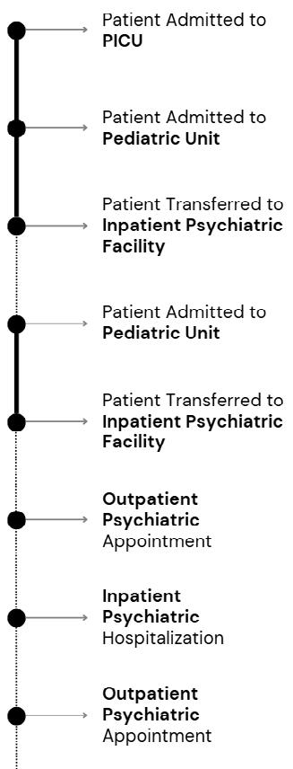
In addition to highlighting the heritability of schizophrenia, the above case underscores the necessary extensive medical workup for the differential diagnosis of COS. The patient was tested for multiple medical diseases including a seizure disorder, encephalitis, autoimmune disorders such as systemic lupus erythematosus, metabolic disorders, central nervous system tumors, and demyelinating disease. Results were ultimately unrevealing for organic causes and diagnosis of COS was subsequently established based on observations of the progression of symptoms and signs. Despite requiring multiple hospitalizations before reaching the diagnosis of COS, this patient’s diagnosis was more prompt than average, with the average diagnosis of COS from onset of symptoms being two years [13]. As discussed before, this delay is commonly due to variability in presentation, onset of symptoms in the presence of co-occurring neurodevelopmental disorders, or in the setting of childhood fantasies considered a part of normal child development. The limitations of this case report includes its focus on a rare case. Due to the rarity of this disorder, this report may not be generalizable to other cases of COS.
Upon reaching a diagnosis of COS, controversy remains over starting antipsychotics for pediatric patients. However, the use of antipsychotics are generally recommended for severe cases based on evidence that suggests that early initiation improves outcomes, particularly positive symptoms [14]. In this patient’s case, she was started on multiple psychotropic medications in the hospital, including quetiapine, risperidone, escitalopram, hydroxyzine hydrochloride, lorazepam, and gabapentin. During her first psychiatric hospitalization, her medication regimen was adjusted to escitalopram 10 mg daily for panic attacks and risperidone 0.5 mg nightly for unspecified psychosis. This regimen was shortly changed to olanzapine 15 mg once nightly and hydroxyzine hydrochloride 25 mg once nightly, resulting in overall stabilization of the patient, including improvement in the patient’s academics.
The treatment of COS should involve a comprehensive approach including, but not limited to, pharmacological, psychological, social, and educational interventions involving both the patient and the family. Although this disorder is rare, further research is required to formulate a consistent diagnostic curriculum for accurate and prompt diagnosis of COS. Due to the innate variability of the disorder, accurate diagnosis of COS relies on a combination of standardized diagnostic tools in addition to perceptive clinician observations and interviews. Identifying individuals affected by this disorder earlier in the course of its pathogenesis is crucial to effective treatment and monitoring to enhance functional outcomes and quality of life of patients. Treatment of COS should be initiated promptly once a diagnosis is made as such a disabling mental disorder can have a devastating impact during this critical time of development. With early intervention, appropriate treatment, and a supportive network of care, individuals with COS can make significant progress and lead fulfilling lives.
DECLARATIONS OF INTEREST
The authors declare no competing interests.
ACKNOWLEDGEMENT
The authors thank Dr. Elizabeth Shaffer and Dr. Mandeep Kaur for their conceptualization and support of this article.
REFERENCES
[1] Institute of Health Metrics and Evaluation. (2021). Global Health Data Exchange (GHDx). Retrieved May 14, 2022, from https:// vizhub.healthdata.org/gbd-results/
[2] Solmi, M., Radua, J., Olivola, M., Croce, E., Soardo, L., Salazar de Pablo, G., Il Shin, J., Kirkbride, J. B., Jones, P., Kim, J. H., Kim, J. Y., Carvalho, A. F., Seeman, M. V., Correll, C. U., & Fusar-Poli, P. (2022). Age at onset of mental disorders worldwide: largescale meta-analysis of 192 epidemiological studies. Molecular psychiatry, 27(1), 281–295. https://doi.org/10.1038/s41380-02101161-7
[3] Charlson, F. J., Baxter, A. J., Dua, T., Degenhardt, L., Whiteford, H. A., & Vos, T. (2015). Excess mortality from mental, neurological and substance use disorders in the Global Burden of Disease Study 2010. Epidemiology and psychiatric sciences, 24(2), 121–140. https://doi.org/10.1017/S2045796014000687
[4] Kessler, R. C., Birnbaum, H., Demler, O., Falloon, I. R., Gagnon, E., Guyer, M., Howes, M. J., Kendler, K. S., Shi, L., Walters, E., & Wu, E. Q. (2005). The prevalence and correlates of nonaffective psychosis in the National Comorbidity Survey Replication (NCS-R). Biological psychiatry, 58(8), 668–676. https://doi. org/10.1016/j.biopsych.2005.04.034
[5] Wu, E. Q., Shi, L., Birnbaum, H., Hudson, T., & Kessler, R. (2006). Annual prevalence of diagnosed schizophrenia in the USA: a claims data analysis approach. Psychological medicine, 36(11), 1535–1540. https://doi.org/10.1017/S0033291706008191
[6] Desai, P. R., Lawson, K. A., Barner, J. C., & Rascati, K. L. (2013). Estimating the direct and indirect costs for community-dwelling patients with schizophrenia. Journal of Pharmaceutical Health Services Research, 4(4), 187–194. https://doi.org/10.1111/ jphs.12027
[7] Ordóñez, A. E., Luscher, Z. I., & Gogtay, N. (2016). Neuroimaging findings from childhood onset schizophrenia patients and their non-psychotic siblings. Schizophrenia research , 173(3), 124–131. https://doi.org/10.1016/j.schres.2015.03.003
[8] Bedwell, J. S., Keller, B., Smith, A. K., Hamburger, S., Kumra, S., & Rapoport, J. L. (1999). Why does postpsychotic IQ decline in childhood-onset schizophrenia?. The American journal of psychiatry, 156(12), 1996–1997. https://doi.org/10.1176/ ajp.156.12.1996
[9] Sauermilch, W. S., Ivey, M. L., Rasmussen, E. E., & Najera, C. J. (2025). Examining the Authenticity of Autistic Portrayals in US Adult and Children’s Television Shows Using Medical and Social Models of Disability. Journal of autism and developmental disorders, 55(2), 524–539. https://doi.org/10.1007/s10803-02306215-z
[10] Hsu, C. W., Lee, S. Y., & Wang, L. J. (2019). Gender differences in the prevalence, comorbidities and antipsychotic prescription of early-onset schizophrenia: a nationwide population-based study in Taiwan. European child & adolescent psychiatry, 28(6), 759–767. https://doi.org/10.1007/s00787-018-1242-9
[11] Hilker, R., Helenius, D., Fagerlund, B., Skytthe, A., Christensen, K., Werge, T. M., Nordentoft, M., & Glenthøj, B. (2017). Is an Early Age at Illness Onset in Schizophrenia Associated With Increased Genetic Susceptibility? Analysis of Data From the Nationwide Danish Twin Register. EBioMedicine, 18, 320–326. https://doi. org/10.1016/j.ebiom.2017.04.002
[12] Fernandez, A., Drozd, M. M., Thümmler, S., Dor, E., Capovilla, M., Askenazy, F., & Bardoni, B. (2019). Childhood-Onset Schizophrenia: A Systematic Overview of Its Genetic Heterogeneity From Classical Studies to the Genomic Era. Frontiers in genetics, 10, 1137. https://doi.org/10.3389/ fgene.2019.01137
[13] Slomiak, S., Matalon, D. R., & Roth, L. (2017). Very early-onset schizophrenia in a six-year-old boy. American Journal of Psychiatry Residents’ Journal , 12(2), 9–11. https://doi.org/10.1176/ appi.ajp-rj.2017.120204
[14] C. Correll, C. Arango, B. Fagerlund, S. Galderisi, M. J. Kas, S. Leucht. (2024). Identification and treatment of individuals with childhood-onset and early-onset schizophrenia. European Neuropsychopharmacology, 82, pp. 57-71. https://doi. org/10.1016/j.euroneuro.2024.02.005
Jamison Willis*, Dr. Jeffrey R. M. Ogden**, Dr. Caroline Mbongo**, Dr. William P. Walker***
*Medical Student, Campbell University School of Osteopathic Medicine
**Resident Physician, Family Medicine, Conway Medical Center
***Attending Physician, Conway Medical Center
ABSTRACT: Spontaneous dissection of the coronary artery (SCAD) is an uncommon presentation of acute coronary syndrome (ACS). Little research has been done on this specific presentation of ACS. While rare, SCAD is most often observed in patients diagnosed with fibromuscular dysplasia, and presents with similar symptoms to a non-ST-elevation myocardial infarction (NSTEMI): substernal chest pain, troponin elevation, and non-specific electrocardiogram findings. Initial diagnostic approach is similar, with a heart catheterization commonly indicated. However, it is managed differently, through the use of anticoagulation, blood pressure control, and, if indicated, statin therapy. This article is a description of a 34-year-old woman who had two spontaneous coronary artery dissections in a time span of 11 days, both of which occurred within a month of childbirth.
KEYWORDS: SCAD, fibromuscular dysplasia, chest pain
In the setting of fibromuscular dysplasia (FMD) the most common arterial involvement is the renal artery, with pathologic changes such as the “string of beads,” in 75-80% of patients. In approximately 75% of patients, the carotid and vertebral arteries are involved. Many patients present with symptoms of hypertension, headache, pulsatile tinnitus, and/or flank pain. A rare manifestation of FMD is a spontaneous dissection of the coronary artery (SCAD) [1]. SCAD is the underlying pathology in anywhere from 0.1% to 4.9% of acute coronary syndrome (ACS) [2]. However, the incidence of FMD within the group of patients diagnosed with SCAD could be as high as 86% [3]. To establish the diagnosis of FMD-associated SCAD, there must be characteristic findings of FMD on computed tomography (CT) or computed tomography angiography (CTA) in another region of the body. The classic “string of beads” appearance is often absent in coronary arteries or is inconclusive if beading was present before the dissection [3]. This appearance is caused by abnormal fibrous cells replacing normal arterial cells. Leading to areas of overly stiff and to elastic portions of the arterial wall, causing a beaded appearance.
SCAD is diagnosed conclusively at the time of coronary angiography [2]. Most commonly, it is described as a long region of narrowing during catheterization. The most common locations of SCAD are the left anterior descending artery (LAD), left circumflex artery (LCX), and right coronary artery. Thus, patients commonly present with unstable angina or myocardial infarction [3]. While many SCADs are due to FMD, other causes such as Ehlers-Danlos, Takayasu’s arteritis, and cocaine abuse must be considered depending on the clinical presentation. Those who are pregnant or have recently given birth are also at
increased risk of SCAD and should be considered in any female of childbearing age.
In the treatment of SCAD, conservative approaches are preferred, as the majority of cases in patients will heal without invasive intervention [2]. However, there have been limited studies to support or refute this conclusion. It is important to note that the components of conservative management in SCAD may differ from other causes of ACS, especially regarding the use of a statin. In treating an NSTEMI or other cause of ACS, a statin is started empirically, as plaque formation is considered a major cause [4]. When treating SCAD, statin use is controversial. Tweet et al. showed that recurrence was higher in groups given a statin [5]. However, the authors note possible confounding variables, and there have been limited follow up to support or refute this conclusion. Further management includes the addition of birth control in women of childbearing age, due to elevated recurrence rates in subsequent pregnancies [6].
The patient is a 34-year-old female who presented to the emergency department approximately 20 minutes after the onset of chest pain requiring two sublingual nitroglycerin tablets. Her past medical history was significant for LCX stent placement three years prior (see Table 1), hypertension, hyperlipidemia, and obesity. Additionally, she has a procedural history of Roux-en-Y gastric bypass and vaginal birth four weeks prior to this presentation. Her pain started at rest while she was holding her 29-day-old daughter and was described as initially sharp progressing to dull, non-radiating, substernal chest pain. The patient reported a similar episode of chest pain 11 days prior, at which time she presented to the emergency department. ECG showed sinus rhythm without evidence of
acute injury pattern or acute dysrhythmia. The patient had serial troponin readings of 1022.8 ng/L at 0 hours (N 10-27.1 ng/L) and 5335.8 at 2 hours during that visit.
The patient had undergone a left heart catheterization (LHC) at her previous hospitalization that showed SCAD of the LAD. She was also diagnosed with FMD of the bilateral carotid arteries via CTA of the brain/head/neck (Figure 1). Upon discharge from that hospitalization three days later, she was prescribed clopidogrel, aspirin, amlodipine, metoprolol, and sublingual nitroglycerin. She was instructed to continue taking her atorvastatin previously prescribed for hyperlipidemia following a stent placement in her LCX three years prior. It was determined that due to the previous plaqueinduced myocardial infarction, the statin should be continued. A timeline of her first hospitalization is available in Table 1, represented by the negative days. She was also instructed to follow up with outpatient cardiology for a comprehensive screening for fibromuscular dysplasia. Her second presentation to the emergency department occurred one day prior to her scheduled outpatient follow-up.
Confounding this admission was the continued possibility of the patient leaving due to lack of childcare, and an ongoing hurricane that prevented her family from reaching the hospital. With the help of a social worker, the father of the patient’s children was able to be present. The patient was then
admitted for observation. Her existing medication regimen was continued, with the addition of scheduled isosorbide dinitrate. The patient was kept NPO in anticipation of another LHC. Her troponins rose to 16,844.6 ng/L that evening. Following an uneventful night, the patient underwent her LHC that showed a second SCAD, now of the LCX. As part of the cardiac workup, an echocardiogram was completed that was unremarkable with an ejection fraction of 55-60%. With troponins down-trending but a new SCAD, it was decided to keep the patient on her current regimen and observe for one more night. The following morning, the patient was consulted regarding treatment options, including medical management, percutaneous intervention, and the possibility of a coronary artery bypass graft. All options included a referral to a vascular surgeon at a tertiary care center. The patient decided to continue medical management and enroll in a cardiac rehab program.
During her tertiary care appointment, she was advised to continue conservative management and placed on progestinonly birth control to prevent pregnancy. At her three-month follow-up with her cardiologist, the patient was stable with no further complaints of chest pain. At this point, she was attending a weight loss clinic where she had started taking a GLP-1 agonist to aid in weight loss and help mitigate her controllable risk factors. She also had a positive outlook while enjoying the newest member of her family.
days
catheterization showing SCAD of LAD, diagnosis of carotid FMD on CT head and neck -9 days
Discharge from hospital with conservative management
0 days (1 day prior to scheduled cardiology appointment) 2nd presentation to ED for chest pain
+2 days
+3 days
+3 months
Decision to maintain patient on conservative management with follow up appointment at tertiary care center.
Discharge
PT doing well at cardiology follow up, on birth control, and with no further reports of chest pain. Tertiary care center decided to continue conservative management.
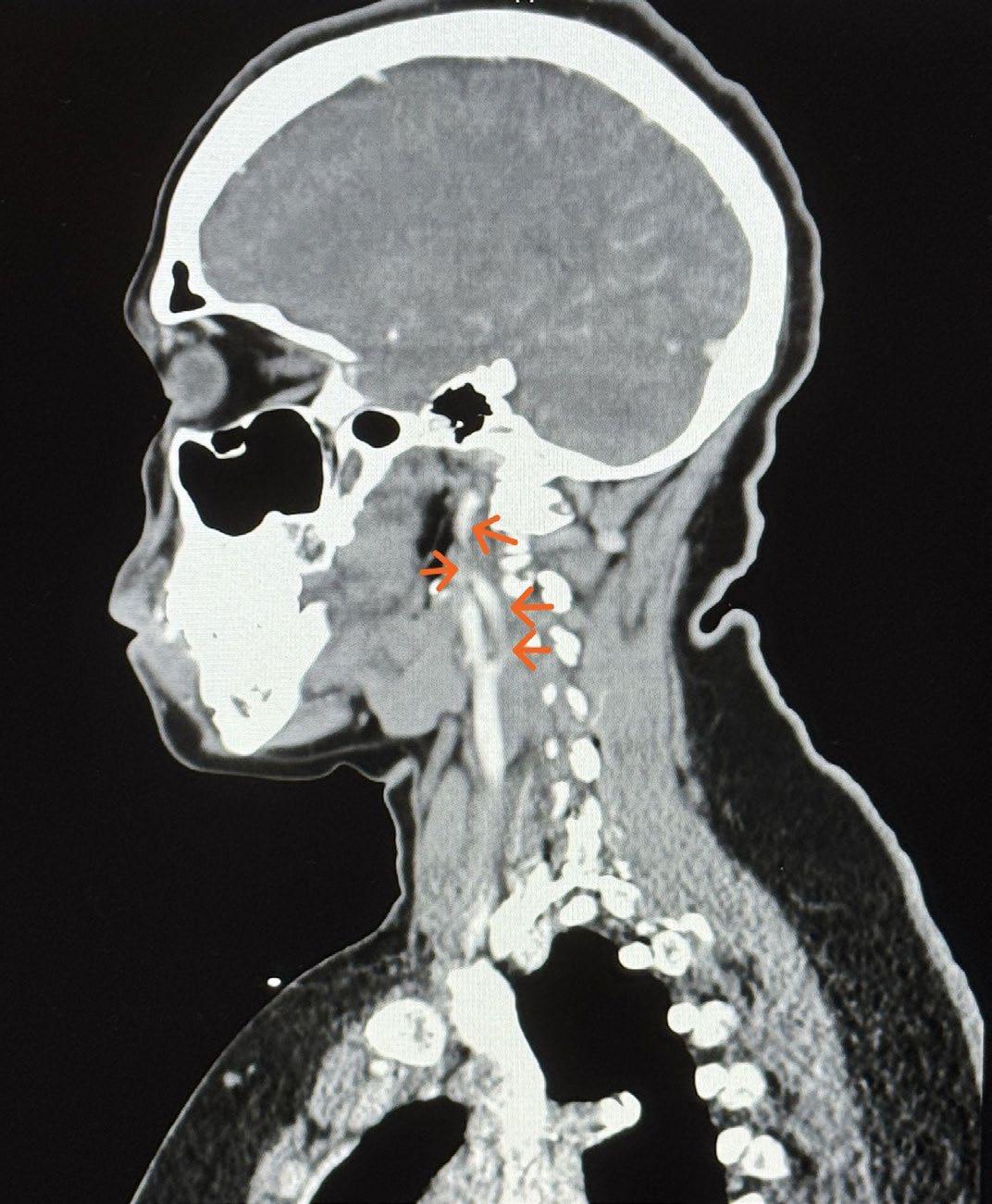
Pregnant and postpartum women are at higher risk for SCAD compared to their counterparts, especially in the third trimester and early postpartum. The etiology is unclear, but has been hypothesized to be hormone-related or due to the increased hemodynamic stress of pregnancy [6]. SCAD should be considered in those who present with an NSTEMI without obvious cardiac risk factors. The patient’s previous stent placement and hyperlipidemia led to an initial clinical suspicion of a plaque-induced etiology during her first hospitalization.
When discharging any patient with SCAD home from the hospital, therapy should include aspirin, a beta-blocker, clopidogrel for 1-12 months, an anti-anginal such as nitrates or a calcium channel blocker, and a statin if the patient is in a hyperlipidemic state. In females of childbearing age, contraception should be discussed with the patient. In the limited studies on this subject, many noted to have low statistical power, there is a risk of recurrence in subsequent pregnancies. In one study it was reported that 8% of women had another episode of SCAD during a subsequent pregnancy or postpartum period [7]. If contraception is utilized, caution should be exercised with regard to estrogen-containing products. While it is poorly understood why combined contraceptives can trigger SCAD, the mechanism is thought to be secondary to increased arterial wall stress brought on by estrogen itself [8].
This case underscores the importance of considering FMD and SCAD in young women, especially in the postpartum period, where hormonal changes and increased hemodynamic stress can trigger vascular events. Diagnosis requires thorough imaging and clinical evaluation, as SCAD may be misdiagnosed as plaque-related ACS. Conservative management is generally preferred, with careful consideration of statins. This case highlights the need for comprehensive follow-up, screening for other vascular involvement, and patient education on contraception, particularly in women of reproductive age.
DECLARATIONS OF INTEREST
The author(s) declare no competing interests.
ACKNOWLEDGEMENT
We want to thank Conway Medical Center for the use of its facilities. We would also like to thank our patient for allowing us to use her medical records and unique presentation to allow us and others to learn.
[1] Olin, J. W., Froehlich, J., Gu, X., Bacharach, J. M., Eagle, K., Gray, B. H., Jaff, M. R., Kim, E. S. H., Mace, P., Matsumoto, A. H., McBane, R. D., Kline-Rogers, E., White, C. J., & Gornik, H. L. (2012). The United States Registry for fibromuscular dysplasia. Circulation, 125(25), 3182–3190. https://doi.org/10.1161/circulationaha.112.091223
[2] Nishiguchi, T., Tanaka, A., Ozaki, Y., Taruya, A., Fukuda, S., Taguchi, H., Iwaguro, T., Ueno, S., Okumoto, Y., & Akasaka, T. (2013). Prevalence of spontaneous coronary artery dissection in patients with acute coronary syndrome. European Heart Journal: Acute Cardiovascular Care, 5(3), 263–270. https://doi. org/10.1177/2048872613504310
[3] Michelis, K. C., Olin, J. W., Kadian-Dodov, D., d’Escamard, V., & Kovacic, J. C. (2014). Coronary Artery Manifestations of Fibromuscular Dysplasia. Journal of the American College of Cardiology, 64(10), 1033–1046. https://doi.org/10.1016/j. jacc.2014.07.014
[4] Singh, A., & Grossman, S. A. (2023, July 10). Acute Coronary Syndrome. National Library of Medicine; StatPearls Publishing. https://www.ncbi.nlm.nih.gov/books/NBK459157/
[5] Tweet, M. S., Hayes, S. N., Pitta, S. R., Simari, R. D., Lerman, A., Lennon, R. J., Gersh, B. J., Khambatta, S., Best, P. J. M., Rihal, C. S., & Gulati, R. (2012). Clinical Features, Management, and Prognosis of Spontaneous Coronary Artery Dissection. Circulation, 126(5), 579–588. https://doi.org/10.1161/circulationaha.112.105718
[6] Krishnamurthy, M. (2004). Spontaneous coronary artery dissection in the postpartum period: Association with antiphospholipid antibody. Heart, 90(9). https://doi.org/10.1136/ hrt.2004.038869
[7] Chan, N., Premawardhana, D., Al-Hussaini, A., Wood, A., Bountziouka, V., Kotecha, D., Swahn, E., Palmefors, H., Pagonis, C., Lawesson, S. S., Kądziela, J., Garcia-Guimarães, M., Alfonso, F., Escaned, J., Macaya, F., Santás, M., Cerrato, E., Maas, A. H. E. M., Hlinomaz, O., … Adlam, D. (2022). Pregnancy and spontaneous coronary artery dissection: Lessons from survivors and nonsurvivors. Circulation, 146(1), 69–72. https://doi.org/10.1161/ circulationaha.122.059635
[8] Pan, A. L., Fergusson, D., Hong, R., & Badawi, R. A. (2012). Spontaneous coronary artery dissection following topical hormone replacement therapy. Case Reports in Cardiology, 2012 , 1–3. https://doi.org/10.1155/2012/524508
Hallie Dunning*, Dr. Waheed Bajwa**
* Medical Student, Campbell University School of Osteopathic Medicine
**Attending Physician, Cary Behavioral Health
ABSTRACT: This review investigates ketamine’s therapeutic efficacy in traumatic brain injury (TBI) and associated conditions including post-traumatic stress disorder (PTSD) and depression. A comprehensive literature was performed to examine the intersection of TBI pathophysiology and ketamine intervention across both acute and chronic disease phases. Results demonstrate ketamine’s ability to maintain hemodynamic stability during acute head trauma while providing neuroprotection through attenuation of cortical spreading depolarization, reduction of inflammatory biomarkers, and other proposed mechanisms. The neurobiological relationships between TBI, PTSD, and depression are explored, highlighting potential shared pathways amenable to ketamine intervention. While current evidence supports ketamine’s application in this context, further investigation is warranted. Future research should prioritize largescale randomized controlled trials with diverse patient populations to establish evidence-based treatment protocols guided by objective physiological parameters. This work contributes to the emerging understanding of ketamine as a multifaceted therapeutic agent in both physical brain trauma and its psychological sequelae.
KEYWORDS: traumatic brain injury, ketamine, PTSD, depression
Traumatic brain injury (TBI) has significant epidemiological impacts, necessitating a better understanding of its mechanism and prevention. This review examines the neurobiological underpinnings of TBI and how these mechanisms may elucidate the therapeutic potential of ketamine in treating both acute TBI and its associated psychiatric complications, particularly posttraumatic stress disorder (PTSD) and depression. Originally developed as an anesthetic, ketamine has emerged as a promising treatment option due to its unique pharmacological properties and is currently in use for treatment-resistant depression (TRD). Beyond treatment of existing disease, ketamine’s effects on intracranial pressure, cortical spreading depolarization, and modulation of inflammatory markers in TBI may reveal mechanisms for prevention of chronic outcomes such as PTSD and depression. Understanding the intersection of these conditions may particularly benefit military populations, where TBI and PTSD frequently co-occur. The review evaluates established benefits and potential risks of ketamine therapy, while highlighting promising directions for future research in this critical field.
A comprehensive literature search was conducted using PubMed that focused on articles published between 2020 and 2025. The search strategy employed the following terms: “traumatic brain injury,” “ketamine,” “depression,” and “PTSD.” Boolean operators combined these terms to ensure the capture of relevant intersections between these conditions and treatments.
The initial search yielded approximately 62 articles, screened first by title and abstract. Inclusion criteria encompassed peer-reviewed articles in English, including clinical trials, systematic reviews, meta-analyses, and highquality observational studies. Articles were selected based on their relevance to neurobiological mechanisms, treatment outcomes, and novel therapeutic approaches. Studies investigating the comorbidity of these conditions and the therapeutic potential of ketamine received special attention. After applying these criteria, 30 articles were selected for full-text review. Six articles were excluded, as they were already cited in one of the selected systematic reviews. Three additional articles were excluded, as their primary focus was on an intervention other than ketamine.
Final analysis included 21 articles that directly addressed the intersection of TBI, PTSD, depression, and ketamine treatment, or provided context to the pathophysiology of their intersection. Data extraction focused on neurobiological findings, treatment outcomes, and emerging therapeutic approaches.
Traumatic brain injury (TBI) represents a major global health challenge affecting over 60 million people annually, with 5 million cases classified as severe [18]. As a leading cause of neurological impairment worldwide, TBI results in significant cell death and neural dysfunction [16], affecting all age groups through various causes such as falls and traffic accidents [2]. The CDC has documented an increasing trend in TBI-related hospital visits and deaths [2], with 2014 data showing 2.87 million TBI-related emergency visits, hospitalizations, and
deaths in the US alone [12]. Known as a “silent epidemic,” TBI poses a substantial socioeconomic burden exceeding $200 billion globally [1], while creating significant financial strain on healthcare systems and families through lengthy rehabilitation processes [2].
The condition is particularly prevalent in military contexts, gaining prominence during the Iraq and Afghanistan conflicts, where blast exposures from mortars, artillery shells, and improvised explosive devices were common [7]. Military personnel face additional risks from subclinical blast exposures in both combat and non-combat settings [7]. The impact of TBI extends beyond immediate physical trauma, as patients face a high risk of poor clinical outcomes due to secondary brain injuries [3]. Long-term consequences include persistent neurobehavioral deficits [5], and among veterans, TBI frequently correlates with chronic mental health issues such as anxiety, depression, impulsivity, insomnia, and suicidality [7]. Given the lifelong disability burden owing to complex pathophysiology and limited treatment options, there is an urgent need for new therapeutic approaches [2, 18].
Traumatic brain injury (TBI) follows a biphasic pathophysiological pattern. The primary phase involves direct mechanical damage to brain tissue, membranes, and blood vessels [2], which can be focal (e.g., contusions) or diffuse (e.g., diffuse axonal injury), ranging from microscopic changes to visible lesions [12]. The secondary phase involves a complex cascade of events including hemorrhage, reduced blood flow, inflammation, and cellular death [2]. During this phase, neurochemical changes occur, including glutamate release, ionic imbalances (potassium efflux, sodium/calcium influx), metabolic disruption, and neuroinflammation [12]. Alqahtani et al. [2] reveals that at the molecular level, immune cells such as microglia and astrocytes become activated, releasing inflammatory cytokines and chemokines. In particular, inducible nitric oxide synthase (NOS) is released from neurons and glial cells, causing increased production of nitric oxide (NO) and its resultant leukocyte accumulation. The transcription factor NF-κB controls NOS production and thus plays a role in this immune process. Additionally, persistent elevation of glutamate levels contribute to neuronal damage through AMPA and NMDA receptor activation [2].
The severity of TBI is classified as mild, moderate, or severe based on the Glasgow Coma Scale, consciousness level, and presence of amnesia [12]. Severe cases require intensive care to prevent secondary complications from hypoxia, low blood pressure, and inflammation [3]. As described by Acero et al. [1], TBI shares pathological features with stroke, including immune dysfunction, endocrine disruption, and neurovascular damage. Long-term consequences can include seizures, post-traumatic epilepsy (PTE), and structural changes such as dendritic fragmentation and reduced spine density. The brain’s natural plasticity mechanisms, including brain-derived neurotrophic factor (BDNF) upregulation, aid recovery [1]. Encouragingly, the numerous pathways involved in TBI pathophysiology provide
many potential treatment targets, several of which will be highlighted in this review.
Ketamine was developed in 1962 as a safer alternative to phencyclidine, which caused post-surgical complications [15, 17]. Rueda Carrillo et al. [15] chronicles the history of ketamine further, describing its 1970 FDA approval as Ketalar after successful clinical trials. Though widely used in Vietnam as a battlefield anesthetic, its use declined due to recreational abuse in the 1980s, leading to Schedule III classification. It regained medical attention in the 1990s for treating pain and psychiatric conditions. Over the past two decades, evidence emerged about the efficacy of ketamine to treat multiple neurological conditions, including traumatic brain injury (TBI), migraines, and refractory status epilepticus (RSE) [15]. Today, it serves as both an anesthetic and novel treatment for various psychiatric conditions, including TRD, with recognized anti-inflammatory properties [17].
As a non-competitive N-methyl-d-aspartate receptor (NMDA) receptor inhibitor, ketamine affects multiple neurotransmitter systems to produce amnesia, pain relief, and potential neuroprotection [8, 15]. Derived from phencyclidine, ketamine exists in two forms: S(+) and R(-) [15], and is available as S-ketamine or a racemic mixture [8]. Studies show (R)- and (S)-ketamine work differently: (R)-ketamine activates TrkB receptors while (S)-ketamine uses mTOR/ERK pathways [17]. The S-form has stronger anesthetic effects due to higher NMDA receptor binding [3, 15], which is responsible for its dissociative anesthetic properties [8, 14]. NMDA receptors normally respond to glutamate, glycine, and magnesium, and thus ketamine’s NMDA binding reduces channel activity and protects against glutamate toxicity through CREB regulation [15]. Additionally, ketamine acts on opioid receptors, enhances GABA, blocks muscarinic receptors, and influences monoamine reuptake and HCN channels [15].
According to Rueda Carrillo, et al. [15], ketamine can be given through multiple routes, including IV (100% bioavailability), IM (90%), intranasal (50%), oral (17%), and rectal (25%), with rapid crossing of the blood-brain barrier. The liver then converts ketamine to norketamine through P450 3A4, which is then eliminated via urine (90%) and feces (10%). Ketamine’s side effects are dose-dependent and self-limiting, including: excess saliva, heightened reflexes, muscle twitches, dizziness, nausea, vomiting, increased heart rate and blood pressure, and urinary issues. Additionally, patients may experience temporary hallucinations and confusion after anesthesia, treatable with benzodiazepines [15]. These side effects are largely avoided in sub-anesthetic doses or by the utilization of (R)-ketamine, which has a lower side effect profile [17].
Historically, ketamine was avoided in TBI treatment due to concerns about increased intracranial pressure (ICP), cerebral blood flow, and metabolic rate [14, 15]. However, recent research allayed these concerns, leading to renewed interest in ketamine for TBI treatment [15]. Ketamine demonstrates remarkable utility in TBI treatment through its ability to provide effective sedation and pain relief while preserving crucial respiratory and cardiac functions [3]. Unlike other anesthetic agents, ketamine does not induce dilation of the vascular bed and thus does not induce hypotension, nor does it have a negative chronotropic impact on the heart [8]. This makes ketamine ideal for anesthetizing patients with TBI, as perfusion pressure is maintained [8]. Its exceptional hemodynamic properties have led to increased adoption in trauma care [14], with both the S-enantiomer and racemic formulations maintaining stable airway protection with minimal respiratory depression [3]. This stability makes ketamine particularly beneficial for TBI patients with hypotension, where its ability to maintain stable blood pressure makes it a preferred choice [8, 16], particularly in resource-limited settings [3]. Additionally, ketamine’s main mechanism of NMDA receptor blockade provides both anesthetic effects and potential neuroprotection against harmful brain activity patterns with few complications [2, 3, 16].
Initial concerns about ketamine raising intracranial pressure (ICP) were dispelled through extensive research, including systematic reviews of 228 patients (127 non-trauma, 101 TBI) providing Oxford level 2b/Grade C evidence [15]. Further studies, including a review of 11 clinical trials, showed that ketamine maintained stable circulatory parameters and did not have a significant effect on ICP, with any ICP increases being temporary or procedure-related while maintaining stable mean arterial pressure (MAP) and cerebral perfusion pressure (CPP) levels across both adult and pediatric populations [8, 14, 15, 16]. Use in combat settings supports the efficacy of ketamine for moderate to severe pain management without significant increase in ICP or mortality, especially in patients at risk of shock or respiratory distress [20]. Analysis of five randomized trials comparing ketamine to other sedatives revealed no significant difference in adverse events between ketamine and opioids, though evidence quality was low and ketaminetreated patients typically had more severe injuries than those receiving opioids [3,13]. However, a multinational, multicenter trial found that ketamine-exposed patients showed no difference in survival or disability despite having more severe injuries [14]. Contrary to initial concerns, ketamine leads to vasoconstriction through inhibition of NO synthesis, which promotes stabilization of blood pressure and hippocampal cell growth post-TBI [2].
Spreading depolarization (SD), characterized by waves of neuronal and glial depolarization caused by the loss of
ion gradients, is a byproduct of delayed cerebral ischemia in acute trauma and plays a crucial role in traumatic brain injury pathophysiology [1, 5, 15]. SD can be measured through electroencephalogram (EEG) or internal electrodes as altered electrical activity patterns [8], moving at 3 mm/min through gray matter and causing sodium and calcium ion influx that exceeds the sodium pump’s capacity [19]. This results in water influx, cell swelling, and the release of neurotransmitters like glutamate [19]. Brain regions show varying susceptibility, with the primary sensory cortex being highly vulnerable while the hypothalamus and brainstem show resistance [4].
During peritrauma, ketamine significantly reduces spreading depolarization via NMDA receptor blockade, effects that can be monitored via EEG, suggesting neuroprotective potential in treating TBI [3, 5, 8, 15, 16, 19]. Ketamine affects brain activity in a dose-dependent manner (0.5-2.0 mg/kg), with EEG showing decreased alpha and increased theta wave activity [15]. S(+) ketamine and racemic forms have a stronger effect on EEG readings than R(-) ketamine [15]. Ketamine’s ability to reduce SD is likely key to its neuroprotective potential when considering the sequelae of TBI.
Biomarker analysis and in vivo studies suggest that ketamine may suppress brain injury markers and attenuate chronic damage, enhancing functional recovery [1, 14]. The TXA for TBI trial, a large multicenter study, further investigated these effects by measuring TBI biomarkers (GFAP, MAP2, UCHL1) to compare ketamine-exposed and unexposed groups [14]. Ketamine showed suppressive effects on biomarkers, with lower GFAP and MAP2 levels overall and significantly reduced GFAP at 48 hours post-injury in intracranial hemorrhage (ICH) patients [14]. Additionally, while not TBI-specific, the metabolites of the kynurenine pathway (KP) are produced in TBI and may be normalized by ketamine [12].
Several additional potential treatment targets were demonstrated in animal models. A study by Tang, et al. found that the protein TFEB protects brain cells by clearing damaged cells through autophagy, and that esketamine activates TFEB via AMPK/mTOR and Nrf2 pathways. Researchers found that an optimal dose of 4 mg/kg esketamine led to reduced swelling and cell damage while boosting protective cellular activity [18]. Additional research in mice showed that combining ketamine with perampanel (an anti-epileptic agent) reduced inflammatory markers and improved recovery, likely due to the synergistic effect of ketamine’s NMDA blockade and perampanel’s AMPA receptor targeting [2]. Other rat models showed an increase in local cerebral glucose utilization and cerebral blood flow, particularly in limbic structures, after treatment with ketamine [15]. As is evident, ketamine’s interaction with a wide variety of pathways provides many avenues of exploration into its therapeutic potential.
Over 50 years of clinical experience and extensive research have demonstrated ketamine’s favorable safety profile in TBI patients [8]. The Brain Trauma Foundation has not made specific recommendations, but ketamine offers multiple benefits: analgesic, sedative, and anti-seizure effects, plus improved hemodynamics and airway function [15]. These properties make it particularly valuable for sedation and pain management in TBI patients, who often experience seizures, hemorrhagic shock, and lung trauma [15]. While ketamine can affect various physiological parameters in TBI patients, current evidence regarding its long-term impact on functional outcomes, serious adverse events, mortality, agitation, and quality of life in critically ill patients with severe acute brain injury remains limited [3, 16]. This paper will now consider ketamine’s utility in the long-term outcomes of TBI.
Nearly 10% of trauma survivors develop chronic symptoms of posttraumatic stress disorder (PTSD), a debilitating psychiatric condition that manifests through intrusive memories, avoidance behaviors, hypervigilance, and negative changes in mood and cognition [9, 21]. Brain imaging reveals PTSD’s impact on regions controlling emotions, cognition, and memory [21]. Additionally, extensive interdisciplinary research has shown that chronic PTSD creates a substantial stress burden across multiple body systems [6]. A striking feature shared by veterans of the Iraq and Afghanistan conflicts is the overlap between a history of blast-related mild TBI (mTBI) and clinical symptoms consistent with a diagnosis of post-traumatic stress disorder (PTSD) [7]. Understanding the physiological mechanisms underlying TBI-associated PTSD is a critical component to addressing this health dilemma.
A key mechanism underlying TBI’s cognitive effects, including PTSD, involves inflammation-driven tryptophan breakdown into kynurenine metabolites, which can be either neurotoxic or neuroprotective [12]. Research shows that elevated levels of neurotoxic kynurenines are associated with poorer TBI outcomes, suggesting that disrupted kynurenine metabolism could explain post-TBI psychiatric conditions and potentially serve as a biomarker [12]. This inflammatory cascade triggers hippocampal neuronal death, with both TBI and PTSD showing increased IL-6 and BAX protein levels [11]. Both TBI and PTSD involve disrupted autonomic function, with TBI causing sympathetic storming (an exaggerated stress response with elevated plasma catecholamines due to lost cortical control) while PTSD develops through nerve growth factor (NGF)-induced sympathetic growth and elevated brain norepinephrine levels. [11].
Structural brain changes are also a key feature of PTSD development. Research demonstrates that PTSD is associated with cortical thinning in the hippocampus, amygdala, insula, ventromedial prefrontal cortex (vmPFC), and anterior cingulate cortex (ACC) [9, 21]. These affected regions control emotion, sensory processing, executive function, threat processing, and emotional memory [9, 21]. As highlighted by Huggins et al. [9], recent research also sheds light on the cerebellum’s unexpected role in PTSD, extending beyond motor control to influence cognition, emotion, and fear learning. A comprehensive study of 4,215 adults using ENIGMA-PGC PTSD data demonstrated significantly reduced cerebellar volume in PTSD patients, particularly in the posterior cerebellum, vermis, and flocculonodular regions. The authors note that these reductions were more pronounced with increasing symptom severity and persisted after controlling for depression and alcohol use. While these cerebellar changes may contribute to symptoms like hypervigilance and concentration problems, their small effect size limits their use as clinical biomarkers of PTSD [9]. Rather, the constellation of structural changes documented in PTSD may prove more useful in diagnosis and treatment targeting.
The relationship between TBI and psychiatric conditions involves complex mechanisms, including external stressors, functional impairments, and neuropathological changes marked by inflammation and brain alterations [12]. While PTSD often presents with depression and anxiety symptoms, research shows that TBI itself independently increases psychiatric disorder risk [12]. The disruption of emotional brain circuits, particularly in the vulnerable basal forebrain and hippocampus regions (notably susceptible to TBI-induced cognitive insult per their anatomical location), contributes to heightened depression and anxiety [12]. TBI can cause persistent pro-inflammatory states in astrocytes and microglia, affecting neuronal function and blood-brain barrier permeability [1]. Increased neurotoxic KP metabolites post-TBI may worsen secondary injury and raise psychiatric risk [12]. Additionally, epigenetic mechanisms, including DNA methylation, histone modification, and RNA regulation, appear to influence psychological disorder vulnerability [1]. This is evidenced by the correlation between BDNF promoter methylation and PTSD in veterans, helping to explain how factors like hormones and inflammation contribute to transgenerational mental illness risk [1].
As emphasized by Lipov, Special Operations Forces (SOF) personnel face heightened TBI and PTSD risks from combat deployments and specialized activities [11]. The authors emphasize that traditional treatments have proven inadequate for their complex symptoms, with research focusing more on PTSD than TBI interventions. Current therapies show recovery rates of approximately 40% in intent-to-treat analyses, with high dropout rates (30-40%). The overlapping symptoms of TBI and PTSD create diagnostic challenges, as both conditions affect similar brain regions (the amygdala, hippocampus, and prefrontal cortex) and disrupt neural connectivity patterns
[11]. Continued identification of the features shared by TBI and PTSD may narrow the focus for treatment targets, as well as timing of key interventions.
Depression, within the context of major depressive disorder (MDD) and bipolar disorder (BD), affects more than 300 million people worldwide and is a leading cause of ill health and disability [10]. While many pharmacological agents are available for depression, up to 30% of patients will fail to respond to two or more antidepressants and meet criteria for TRD [10]. The rate of treatment resistance also increases when depression co-occurs with other psychiatric disorders, notably PTSD [10]. Meta-analyses of PTSD diagnoses demonstrate elevated risks for various conditions (overall OR=2.00), including depression (OR=2.14), bipolar disorder (OR=1.85), and anxiety (OR=1.84) [12]. A substantial study of over 10,000 TBI patients revealed that 27-38% developed major depression or dysthymia, with increased PTSD rates observed in both civilian and military populations [12].
As an anesthetic agent working on multiple CNS receptor systems, ketamine has gained renewed interest in neurological conditions, including TBI, PTSD, and depression [5, 15]. Postrecovery, ketamine helps manage the neurological and psychiatric symptoms affecting 44-50% of mild TBI patients [5]. Research shows promising initial results from pilot trials of intravenous ketamine for PTSD, particularly given its high comorbidity with MDD and theoretical benefits to PTSD pathophysiology [6]. Ketamine demonstrates rapid-onset antidepressant effects and antisuicidal potential, particularly in treatment-resistant MDD [6, 17]. A study performed by Johnson et al. demonstrated a reduction in both depressive symptoms and PTSD symptoms with ketamine infusions, and notes that comorbid PTSD does not reduce ketamine’s antidepressant effects [10]. Evidence also suggests ketamine’s potential for treating PTSD and chronic pain in TBI patients, as these conditions often co-occur [5]. While ketamine has not been formally tested in TBI patients, the 2019 FDA approval of intranasal S-ketamine as an efficacious adjunct treatment for depression will likely increase the available study population [5, 17]. Given that ketamine achieves anesthetic effects not through sedation, but through dissociation or disconnection between mind and body, ketamine-assisted psychotherapy may also be of utility [6].
Research reveals complex mechanisms underlying ketamine’s therapeutic effects. NMDA receptor activation typically increases depressive and anxious intrusive memories through amygdala activation and long-term potentiation, but ketamine can ameliorate these processes [6]. The drug upregulates BDNF, leading to enhanced synapse formation and dendritic spine growth, particularly in the prefrontal cortex
(PFC), helping to repair stress-damaged neural connections [6]. Clinical studies using doses from 0.2 to 1 mg/kg administered either once or thrice weekly demonstrated rapid improvement in both PTSD and MDD symptoms across civilian and military populations [6]. At a molecular level, ketamine promotes neurogenesis and addresses post-blast TBI complications through BDNF upregulation and NMDA antagonism, while working alongside SGB to reduce neuron death by blocking BAX expression and lowering inflammatory markers [11]. The drug’s interaction with astrocytes, which express various neurotransmitter receptors and maintain close neuronal coupling, appears crucial to its mechanism [1].
Additionally, ketamine induces antidepressive effects through histone deacetylase 5 (HDAC5) phosphorylation and nuclear export in rat hippocampal neurons, which mediates upregulation of BDNF activity. These rapid antidepressant effects correlate with increased astrocyte size and branching in the hippocampus’s CA1 subregion—an area vital for learning and memory [1]. As further described by Meier et al. [12], the hippocampus’s high NMDA receptor density makes it vulnerable to quinolinic acid (QuinA) damage. The authors reveal that the kynurenine pathway produces two key compounds: neuroprotective kynurenic acid (KynA) from astrocytes and neurotoxic QuinA from inflammation. After TBI, these compounds cross the blood-brain barrier, with high QuinA levels leading to worse outcomes, especially in patients with previous concussions. These metabolites show potential as recovery markers, suggesting that targeting the kynurenine pathway may be more effective than general anti-inflammatory treatments for TBI-related depression [12].
Acero et al. [1] brings attention to studies which show ketamine’s effect on gut bacteria composition. Specific bacterial strains may influence brain function through vagus nerve signaling, and gut microbes can alter brain inflammation through metabolites. Ketamine reduced histological damage after dextran sodium sulfate (DSS) treatment, a chemical method to induce epithelial damage in the ceca of mice. It also reduced local necroptosis, an inflammatory cell death, as well as TNF levels via NMDAR antagonism [1]. These effects are promising given the emerging role of the gut-brain axis in neurocognitive conditions.
Research reveals (R)-ketamine as a promising treatment option for depression and addiction, with advantages over (S)-ketamine including minimal dissociation and reduced side effects [17]. Ketamine’s metabolite, (2R,6R)-hydroxynorketamine [(2R,6R)-HNK], maintains rapid antidepressant effects while providing improved safety, reduced abuse potential, and the benefit of oral administration [7]. Studies of blast-exposed rats treated with (2R,6R)-HNK demonstrated positive outcomes for both depression and PTSD [7]. Clinical trials employing (R)-ketamine for treatment-resistant depression have shown promising results, with 43% of patients achieving 24-hour remission and effects lasting two weeks after a single dose [17]. (R)-ketamine also has milder cognitive and behavioral
effects, along with longer-lasting anti-depressive effects (7 days compared to 3 days for (S)-ketamine) [17]. Additionally, (R)-ketamine has demonstrated potential for treating substance use disorder (SUD), reducing morphine withdrawal, and preventing ethanol tolerance in animals [17]. The drug’s interaction with the opioid system has been demonstrated through naltrexone administration, which partially attenuates its antidepressant effects [6].
Research on ketamine use in military populations reveals mixed results. While prehospital ketamine administration following combat injury lowered first-year PTSD rates compared to opioids alone, the benefit was limited to patients without TBI [13]. Interestingly, as noted by Johnson et al. [10], veterans with TRD and PTSD demonstrate lower remission rates (43% less) than civilians, with PTSD complicating 35% of depression cases. In two-week ketamine infusion trials using 0.5 - 0.75 mg/kg over four doses, patients showed promising results: 78% reported improvement, with 52% achieving significant response rates, although many remained above PTSD diagnostic thresholds. A smaller study of veterans and first responders (n=18) demonstrated 67% improvement. While ketamine’s efficacy matches traditional treatments (d=0.73), it offers faster relief than typical 12-week protocols [10].
Recent investigation by Lipov et al. [11] describe the potential of combining ketamine infusion (KI) with cervical sympathetic block (CSB) in treating Special Operations Forces (SOF). This approach leverages nerve growth factor modulation and norepinephrine reduction to enhance memory and decrease inflammation. The authors describe a protocol consisting of ultrasound-guided bupivacaine administration at C6 and C4 vertebrae, followed by five 45-minute ketamine sessions starting at 0.5 mg/kg. Treatment progress is monitored through standardized assessments of PTSD, depression, anxiety, and suicide risk. One notable combat veteran case study demonstrated remarkable one-year outcomes, including reductions in depression (93.55%), anxiety (86.67%), PTSD (73.81%), suicidal thoughts (75%), and impulsivity (100%) [11].
Research into ketamine’s applications for TBI, PTSD, and related comorbid conditions remains ongoing. While ketamine shows therapeutic promise, clinicians must exercise caution due to its side effects, especially given the unknown sensitivity of TBI patients [5]. Administration of sedatives in early post-TBI stages may hinder recovery, and existing TBI-related deficits could alter patients’ responses to ketamine [5]. Some studies found that ketamine can induce oxidative stress and elevate the risk of arrhythmia [11]. Additionally, (S)-ketamine showed abuse potential, with potential behavioral changes and brain cell damage with repeated use [17]. For these reasons, it is important to consider preexisting conditions, particularly cardiac conditions or substance abuse disorder, when identifying ketamine treatment candidates.
Ketamine’s applications in neurological and psychiatric conditions show evolving promise across multiple domains. In TBI treatment, it offers key advantages: maintaining stable hemodynamics and ventilation during acute resuscitation in low-resource settings, benefiting patients with hemodynamic instability and shock through its sympathomimetic properties, and proving especially useful for patients with respiratory complications. The drug serves effectively for refractory convulsive status, provides non-opioid analgesia, enables neurological evaluation during conscious sedation, and maintains gut motility unlike opioids. The data suggests reconsideration of any existing restrictions on ketamine’s use in the acute trauma setting. Understanding ketamine’s exact physiologic mechanism on brain ICP and reperfusion in acute injury can illuminate the appropriate clinical circumstances for its use and predict downstream impact on TBI sequelae, such as PTSD and depression. Furthermore, randomized double-blind trials have demonstrated ketamine’s efficacy in independently treating PTSD and depression symptoms in TRD patients. While the treatment landscape remains challenging, emerging research combining neurobiological insights with novel treatment approaches offers new hope.
While evidence-based treatments for TBI and PTSD exist, their effectiveness faces limitations from various barriers, comorbidities, and trauma-related symptoms [6]. The landscape of TBI drug development proves challenging, with over 40 failed phase II/III trials in recent decades [1]. However, an increase in awareness and recognition of the mechanisms underlying TBI has sparked an encouraging wave of exploration. Several therapeutic targets highlighted in this paper warrant continued investigation:
▶ The promotion of neuroplasticity through TrkB and mTOR pathways [1]
▶ Inhibition of NOS induction and reduction of cellular damage via NF-κB modulation [2]
▶ Ketamine’s impact on ICH-related biomarkers such as GFAP and the ability to track these biomarkers in predicting outcomes [14]
▶ Ketamine’s neuroprotective properties via SD suppression [3, 19]
▶ The potential of ketamine in cerebellar modulation, which has recently emerged as a key predictor of PTSD severity [9]
▶ Combined ketamine infusion and cervical sympathetic block that effectively treated military personnel with treatment-resistant PTSD and TBI comorbidity by enhancing brain circulation and promoting hippocampal growth [11]
▶ The potential of (2R,6R)-HNK in treating blast-related neurobehavioral syndromes in veterans [7]
▶ The ability of (R,S)-ketamine and (S)-ketamine to enhance standard antidepressant effects, in comparison with (R)ketamine’s enhanced cognitive benefits and milder side effect profile [17]
▶ Expanding the limited evidence of ketamine in treating substance abuse disorder, carefully weighing its ability to modulate glutamatergic systems and enhance neuroplasticity with its potential for abuse [17]
Additional investigations are underway:
▶ Current clinical trials, such as KIND and KETA, are investigating protective effects against delayed cerebral ischemia [15]. Research also continues into GABA A receptor potentiators and non-NMDA glutamate modulators [17].
▶ Lysine acetyltransferase (KAT) enzyme activation and Kynurenine‐3‐monooxygenase (KMO) inhibition demonstrate neuroprotective potential in TBI. LAT1 inhibition reduced depression symptoms in mouse models. Ongoing trials are investigating KAT activation, KMO inhibition, and LAT1 inhibition for major depressive disorder and TBI-related sleep disorders [12].
▶ Researchers are exploring numerous innovative approaches to improve outcomes, including epigenetic analysis and 3D biomimetic environments to identify neuroprotective effects [1].
Future research demands robust placebo-controlled trials across diverse populations, with priorities focusing on optimizing treatment protocols, examining therapeutic effects, and conducting comparative studies. Key research areas include treatment parameter optimization, therapeutic approach comparison, investigation of sex differences in TBI and PTSD outcomes, and implementation of advanced analytical methods like non-negative matrix factorization (NMF). Additionally, combining ketamine with structured psychotherapy shows promise, as the drug’s dissociative effects may create a window of psychological plasticity that enhances trauma processing. This integrated approach could particularly benefit treatment-resistant PTSD patients, though establishing standardized protocols requires more rigorous clinical trials to determine optimal psychotherapy integration. Comprehensive longitudinal studies across diverse patient populations with varying TBI severities will be crucial for validating these approaches.
This review has several important limitations. Primarily, preference for ketamine in certain cases of acute trauma is recently renewed, and use of ketamine in the treatment of neuropsychological conditions is a novel application. Thus, the body of evidence is currently limited and faces methodological constraints, such as lack of standardization. Additionally, publication bias likely favors positive results. While selection criteria were applied to identify those studies that are most
recent and comprehensive, there remains a lack of longitudinal and high-powered studies on the topic. These factors, along with the lack of standardization tools, make direct comparisons difficult. Several studies fail to adequately control for essential variables such as baseline severity, treatment history, and comorbidities. Others introduced confounding variables such as concurrent application of other treatments.
Importantly, discussion of adverse events was limited to the application of ketamine in acute brain injury, PTSD, and depression, and is therefore not comprehensive. Applications that involve higher doses or longer infusion duration of ketamine can have additional and more severe adverse outcomes. Results should not be interpreted as generalizable to all conditions or patient populations. Additionally, this review’s generalizability is hampered by a focus mainly on male patients in developed countries. Significant research gaps remain, particularly in female participant representation and overall patient diversity. Strengthening the evidence base will require large-scale randomized controlled trials with comprehensive long-term data across diverse injury severities and demographic populations.
In conclusion, ketamine represents a promising therapeutic avenue for both acute TBI treatment and associated chronic conditions, such as PTSD and depression. The evolution of ketamine research dispelled historical concerns about intracranial pressure and revealed its potential benefits in trauma care. Its unique pharmacological profile enables stable hemodynamics and respiratory function while providing neuroprotection through NMDA receptor modulation and a variety of proposed mechanisms. Particularly noteworthy is the emergence of (R)-ketamine and its metabolites, which offer improved safety profiles and sustained therapeutic effects. While challenges remain in establishing standardized protocols and understanding long-term outcomes, the integration of ketamine within multimodal treatment approaches, particularly in military personnel with comorbid TBI and PTSD, shows remarkable promise. Future research directions focusing on optimized treatment parameters, diverse population studies, and combination therapies will be crucial in fully realizing ketamine’s therapeutic potential in TBI and its psychiatric sequelae.
The authors declare no competing interests.
[1] Acero, V. P., Cribas, E. S., Browne, K. D., Rivellini, O., Burrell, J. C., O’Donnell, J. C., Das, S., & Cullen, D. K. (2023). Bedside to bench: the outlook for psychedelic research. Frontiers in pharmacology, 14, 1240295. https://doi.org/10.3389/ fphar.2023.1240295
[2] Alqahtani, F., Assiri, M. A., Mohany, M., Imran, I., Javaid, S., Rasool, M. F., Shakeel, W., Sivandzade, F., Alanazi, A. Z., AlRejaie, S. S., Alshammari, M. A., Alasmari, F., Alanazi, M. M., & Alamri, F. F. (2020). Coadministration of Ketamine and Perampanel Improves Behavioral Function and Reduces Inflammation in Acute Traumatic Brain Injury Mouse Model. BioMed research international, 2020, 3193725. https://doi. org/10.1155/2020/3193725
[3] Andreasen, T. H., Madsen, F. A., Barbateskovic, M., Lindschou, J., Gluud, C., & Møller, K. (2024). Ketamine for Critically Ill Patients with Severe Acute Brain Injury: A Systematic Review with Metaanalysis and Trial Sequential Analysis of Randomized Clinical Trials. Neurocritical care, 10.1007/s12028-024-02075-2. Advance online publication. https://doi.org/10.1007/s12028-024-02075-2
[4] Andrew, R. D., Hartings, J. A., Ayata, C., Brennan, K. C., DawsonScully, K. D., Farkas, E., Herreras, O., Kirov, S. A., Müller, M., OllenBittle, N., Reiffurth, C., Revah, O., Robertson, R. M., Shuttleworth, C. W., Ullah, G., & Dreier, J. P. (2022). The Critical Role of Spreading Depolarizations in Early Brain Injury: Consensus and Contention. Neurocritical care, 37(Suppl 1), 83–101. https://doi. org/10.1007/s12028-021-01431-w
[5] Browne, C. A., Wulf, H. A., Jacobson, M. L., Oyola, M. G., Wu, T. J., & Lucki, I. (2022). Long-term increase in sensitivity to ketamine’s behavioral effects in mice exposed to mild blast induced traumatic brain injury. Experimental neurology, 350, 113963. https://doi.org/10.1016/j.expneurol.2021.113963
[6] Burback, L., Brémault-Phillips, S., Nijdam, M. J., McFarlane, A., & Vermetten, E. (2024). Treatment of Posttraumatic Stress Disorder: A State-of-the-art Review. Current neuropharmacology, 22 (4), 557–635. https://doi.org/10.2174/1570159X21666230428091433
[7] Garcia, G. P., Perez, G. M., Gasperi, R., Sosa, M. A. G., OteroPagan, A., Abutarboush, R., Kawoos, U., Statz, J. K., Patterson, J., Zhu, C. W., Hof, P. R., Cook, D. G., Ahlers, S. T., & Elder, G. A. (2023). (2R,6R)-Hydroxynorketamine Treatment of Rats Exposed to Repetitive Low-Level Blast Injury. Neurotrauma reports, 4 (1), 197–217. https://doi.org/10.1089/neur.2022.0088
[8] Gregers, M. C. T., Mikkelsen, S., Lindvig, K. P., & Brøchner, A. C. (2020). Ketamine as an Anesthetic for Patients with Acute Brain Injury: A Systematic Review. Neurocritical care, 33(1), 273–282. https://doi.org/10.1007/s12028-020-00975-7
[9] Huggins, A. A., Baird, C. L., Briggs, M., Laskowitz, S., Hussain, A., Fouda, S., Haswell, C., Sun, D., Salminen, L. E., Jahanshad, N., Thomopoulos, S. I., Veltman, D. J., Frijling, J. L., Olff, M., van Zuiden, M., Koch, S. B. J., Nawjin, L., Wang, L., Zhu, Y., Li, G., … Morey, R. (2024). Smaller total and subregional cerebellar volumes in posttraumatic stress disorder: a mega-analysis by the ENIGMA-PGC PTSD workgroup. Molecular psychiatry, 29 (3), 611–623. https://doi.org/10.1038/s41380-023-02352-0
[10] Johnson, D. E., Rodrigues, N. B., Weisz, S., Chisamore, N., Kaczmarek, E. S., Chen-Li, D. C. J., Doyle, Z., Richardson, J. D., Mansur, R. B., McIntyre, R. S., & Rosenblat, J. D. (2025). Examining the impact of comorbid posttraumatic stress disorder on ketamine’s real-world effectiveness in treatment-resistant depression. European neuropsychopharmacology : the journal of the European College of Neuropsychopharmacology, 91, 69–77. https://doi.org/10.1016/j.euroneuro.2024.11.008
[11] Lipov, E., Sethi, Z., Nandra, G., & Frueh, C. (2023). Efficacy of combined subanesthetic ketamine infusion and cervical sympathetic blockade as a symptomatic treatment of PTSD/ TBI in a special forces patient with a 1-year follow-up: A case report. Heliyon, 9 (4), e14891. https://doi.org/10.1016/j. heliyon.2023.e14891
[12] Meier, T. B., & Savitz, J. (2022). The Kynurenine Pathway in Traumatic Brain Injury: Implications for Psychiatric Outcomes. Biological psychiatry, 91 (5), 449–458. https://doi. org/10.1016/j.biopsych.2021.05.021
[13] Melcer, T., Walker, G. J., Dye, J. L., Walrath, B., MacGregor, A. J., Perez, K., & Galarneau, M. R. (2022). Is Prehospital Ketamine Associated With a Change in the Prognosis of PTSD?. Military medicine, usac014. Advance online publication. https://doi. org/10.1093/milmed/usac014
[14] Peters, A. J., Khan, S. A., Koike, S., Rowell, S., & Schreiber, M. (2023). Outcomes and physiologic responses associated with ketamine administration after traumatic brain injury in the United States and Canada: a retrospective analysis. Journal of trauma and injury, 36 (4), 354–361. https://doi.org/10.20408/ jti.2023.0034
[15] Rueda Carrillo, L., Garcia, K. A., Yalcin, N., & Shah, M. (2022). Ketamine and Its Emergence in the Field of Neurology. Cureus, 14 (7), e27389. https://doi.org/10.7759/ cureus.27389
[16] Sameer, M., & Abbas, D. A. (2024). Clinical outcomes of ketamine in patients with traumatic brain injury: A systematic review. International journal of critical illness and injury science, 14 (3), 160–175. https://doi.org/10.4103/ijciis.ijciis_36_24
[17] Shafique, H., Demers, J. C., Biesiada, J., Golani, L. K., Cerne, R., Smith, J. L., Szostak, M., & Witkin, J. M. (2024). (R)-(-)Ketamine: The Promise of a Novel Treatment for Psychiatric and Neurological Disorders. International journal of molecular sciences, 25 (12), 6804. https://doi.org/10.3390/ijms25126804
[18] Tang, Y., Liu, Y., Zhou, H., Lu, H., Zhang, Y., Hua, J., & Liao, X. (2023). Esketamine is neuroprotective against traumatic brain injury through its modulation of autophagy and oxidative stress via AMPK/mTOR-dependent TFEB nuclear translocation. Experimental neurology, 366, 114436. https://doi. org/10.1016/j.expneurol.2023.114436
[19] Telles, J. P. M., Welling, L. C., Coelho, A. C. S. D. S., Rabelo, N. N., Teixeira, M. J., & Figueiredo, E. G. (2021). Cortical spreading depolarization and ketamine: a short systematic review. Neurophysiologie clinique = Clinical neurophysiology, 51 (2), 145–151. https://doi.org/10.1016/j.neucli.2021.01.004
[20] Torres, A. C., Bebarta, V. S., April, M. D., Maddry, J. K., Herson, P. S., Bebarta, E. K., & Schauer, S. (2020). Ketamine Administration in Prehospital Combat Injured Patients With Traumatic Brain Injury: A 10-Year Report of Survival. Cureus, 12 (7), e9248. https://doi. org/10.7759/cureus.9248
[21] Yang, J., Huggins, A. A., Sun, D., Baird, C. L., Haswell, C. C., Frijling, J. L., Olff, M., van Zuiden, M., Koch, S. B. J., Nawijn, L., Veltman, D. J., Suarez-Jimenez, B., Zhu, X., Neria, Y., Hudson, A. R., Mueller, S. C., Baker, J. T., Lebois, L. A. M., Kaufman, M. L., Qi, R., … Sotiras, A. (2024). Examining the association between posttraumatic stress disorder and disruptions in cortical networks identified using data-driven methods. Neuropsychopharmacology : official publication of the American College of Neuropsychopharmacology, 49 (3), 609–619. https://doi.org/10.1038/s41386-023-01763-5
Matthew Coyne*, Margaret Monaco*, Elizabeth Chan*, Amanda Lee**
*Medical
**Director
Student, Campbell University School of Osteopathic Medicine
of Simulation Education & Physician Assistant, Campbell University Clinic
ABSTRACT: The acquisition of surgical skills during the preclinical years of medical school often involves kinesthetic learning through the use of simple models, allowing students to practice in a controlled and safe environment. Learning to tie surgical square knots using one and two-handed techniques requires guided instruction and consistent practice. Mastery of these techniques provides students with the confidence to progress to more complex surgical skills later in their medical education. While a variety of resources are available across different platforms to support clinical skills departments in teaching knot-tying, there remains a need for affordable modalities that reduce departmental costs while helping students develop foundational surgical skills. In this report, we describe a simple and costeffective task trainer that can be used in a clinical skills setting to teach ambidextrous one- and two-handed knot-tying techniques to medical students.
KEYWORDS: education, suture, surgical, trainer
Knot-tying is among the first procedural skills a student must master before moving on to more advanced surgical skills like suturing, instrument ties, laparoscopic, and robotic-assisted procedures. Surgical skills are best acquired through deliberate, repetitious, kinesthetic training with frequent assessment and feedback [1,2]. The best teaching modalities allow trainees to physically feel and interact with the instrumentation, tools, and materials that will become future staples of their careers [1]. Early in training, the use of large-scale models improves visualization for both learners and instructors, facilitating more accurate feedback and accelerating skill development [3]. Currently, medical students are taught ambidextrous knot-tying through a range of instructional modalities, such as live demonstrations, educational videos, simulation clinics, suturing workshops, and hybrid models incorporating both in-person and virtual learning [4-8]. A common practice in medical simulation is to have students learn simple knottying using readily available string or yarn in tandem with an instructor [5,9]. While many instructional methods exist for knot-tying, affordable and effective models are still needed for under-resourced clinical skills departments and their students. Further, medical trainees are exposed to different knots throughout their careers, but most programs today prefer teaching the square knot [3]. Square knots are widely regarded as a superior surgical technique due to their high tensile strength and secure anchoring of suture material [3,10-11]. The skills learned from this simple knot can be built upon in future training [3].
Simple, inexpensive, portable models that allow students to practice these critical knot ties during idle time are ideal, offering valuable opportunities for skill development before clerkship and residency [4,12-13]. Students who are able to repeatedly and deliberately practice procedural skills build confidence in their abilities [3]. This confidence translates to better performance during clinical rotations, better retention of knowledge, increased student engagement, and ultimately, successfully execution of procedures on real patients [3]. To provide increased opportunities for practice, we present an ambidextrous task trainer that utilizes affordable, durable, reusable, everyday household items to teach basic one- and two-handed square knot-tying in a clinical skills setting.
The trainer consists of one self-adhering wall hook, two 32-inch strands of 1/8-inch 550 paracord, and one 2.55-inch non-load-bearing carabiner. The materials used for the task trainer are shown in Figure 1. All materials were purchased from a dollar store. Spools of paracord were cut to 32 inch strands using standard household scissors. Any frayed ends of paracord were cauterized using a butane lighter.

FIGURE 1. General description and images of materials used to create the trainer. A) Neon green colored, 1/8 inch, 550 paracord. B) White colored, 1/8 inch, 550 paracord. C) Adhesive wall hooks. D) Non-load bearing, 2.55 inch carabiner. To aid instruction, two contrasting colors of paracord were selected and joined using a sheet-bend knot [16]. The resulting two-stranded, multicolored paracord was then looped through the carabiner, enabling learners to grasp both strands, apply tension, and practice knottying (See Figure 2 and Figure 3). Figure 4 further demonstrates the trainer in use during a knot-tying session, specifically for practicing the traditional two-handed “4” method [1,5,7].
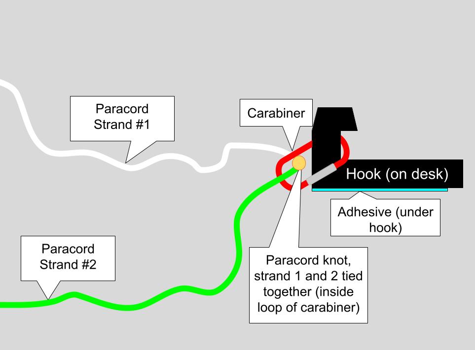
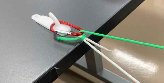
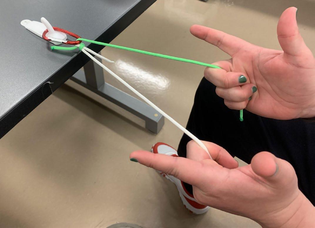
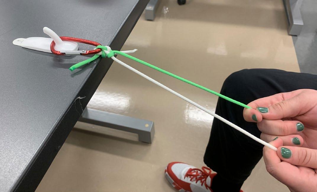
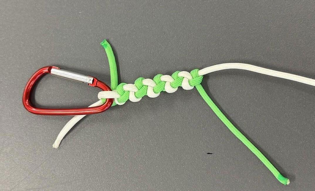
We successfully created a simple, inexpensive task trainer for knot-tying practice. The materials are easily accessible, affordable, suitable for bulk order, and allow for a rudimentary introduction to the critical skill of tying surgical knots using one, and two-handed techniques. This can help build a foundation for more advanced surgical skills later in a learner’s medical career.
The selection criteria for the materials used was convenience and low cost. Still, there are multiple benefits that a simple wall-hook, a non-load-bearing carabiner, and two distinctly-colored paracord strings offer for a medical student or comparable learner. For example, regarding the wall hook, if a learner pulls too much tension on the paracord strings, then the learner will dislodge the wall hook from its adhesive housing. Dislodged hooks are easily remounted to the table using another adhesive strip. This creates a low-stakes teaching opportunity, helping students calibrate the appropriate amount of tension when tying knots – an essential skill that translates well to suturing on silicone-based pads [17]. Additionally, the carabiner provides visual feedback for tension control. If the learner applies insufficient tension, the carabiner will sag, causing the paracord to droop. This makes it more
difficult to tie consistent, uniform, and secure knots [3,18]. Further, paracord was selected for its strength, durability, and variety of color options. Using two paracord strands of contrasting colors allows students to better visualize the alternating pattern of left- and right-handed throws required to form a proper square knot [1,4,5]. The added thickness of the paracord also provides a better grip, enabling learners to focus on developing muscle memory for knot-tying techniques. To our knowledge, no existing models or skills trainers specifically utilize affordable multicolored paracord for this purpose.
While this model certainly will not mimic the tissue endfeel of skin, it is not meant to take the place of silicone-based suture pads. Our model serves as a rudimentary introduction to knot-tying only. Other models should be considered for surgical accuracy and proper skin tissue end-feel [19].
The cost per task trainer is shown in Table 1. The initial cost of the first task trainer accounts for the necessary purchase of scissors and a butane lighter. The first trainer will cost $3.84 to produce. Each subsequent trainer will cost $1.34 to produce. Producing the task trainers in this manner is costeffective and can be done easily at home or within a clinical skills simulation environment.
This task trainer is well-suited for any preclinical instructional setting focused on teaching medical students square knot-tying techniques. It can be used by instructors for in-person demonstrations; incorporated into didactic lectures followed by in-person practice; paired with instructional videos for remote, hybrid, or in-person learning; or be easily assembled by students for at-home practice [1,4-6].
This task trainer offers a low-fidelity simulation for knottying. One limitation is the use of paracord, which differs considerably from standard suture material. This makes paracord more approachable for beginners but sacrifices realism in favor of easier learning. Additionally, this task trainer requires a flat surface for setup, particularly when using adhesive wall hooks. This angle may not be representative of all knot-tying instances.
Future work could explore the impact of this task trainer on student proficiency compared to other commercially available knot-tying task trainers. A crossover study may also assess student satisfaction and long-term retention of knottying skills using this trainer. Additional considerations include evaluating students’ perceptions of how effectively this model complements other knot-tying instructional methods. While this model could support the foundational hand movements for knot-tying, its direct applicability to actual suturing techniques remains uncertain and should be explored further.
We developed a task trainer that enables the practice of both one- and two-handed square knots at a highly affordable price of $3.84 for the first trainer and $1.34 for each subsequent trainer. It is ideal for novice learners. Given its simplicity and low cost, if utilized for in-person learning, we recommend that students be allowed to take carabiners and paracord home following instructional sessions to encourage continued practice.
DECLARATIONS OF INTEREST
The authors declare no competing interests.
ACKNOWLEDGEMENT
The authors would like to recognize and thank Dusty Barbour, EMT-P, for his invaluable contributions to this work.
[1] Kim, E. H., Chern, H., Huang, E., & Palmer, B. (2013). How to teach knot tying: A kinesthetic approach. MedEdPORTAL https://doi. org/10.15766/mep_2374-8265.9328
[2] Nagaraj, M. B., Namazi, B., Sankaranarayanan, G., & Scott, D. J. (2023). Developing artificial intelligence models for medical student suturing and knot-tying video-based assessment and coaching. Surgical endoscopy, 37(1), 402–411. https://doi. org/10.1007/s00464-022-09509-y
[3] Pender, C., Kiselov, V., Yu, Q., Mooney, J., Greiffenstein, P., & Paige, J. T. (2017). All for knots: evaluating the effectiveness of a proficiency-driven, simulation-based knot tying and suturing curriculum for medical students during their third-year surgery clerkship. American journal of surgery, 213(2), 362–370. https:// doi.org/10.1016/j.amjsurg.2016.06.028
[4] Arja, S. B., Arja, S. B., & Fatteh, S. (2019). The hybrid model of clinical skills teaching and the learning theories behind it. Journal of advances in medical education & professionalism, 7(3), 111–117. https://doi.org/10.30476/JAMP.2019.74838
[5] Ebrahim, S., Kinoo, S. M., Naidoo, M., & Van Wyk, J. M. (2024). The use of household items to support online surgical knot-tying skills training: a mixed methods study. BMC medical education, 24 (1), 605. https://doi.org/10.1186/s12909-024-05549-1
[6] Kumins, N. H., Qin, V. L., Driscoll, E. C., Morrow, K. L., Kashyap, V. S., Ning, A. Y., Tucker, N. J., King, A. H., Quereshy, H. A., Dash, S., Grobaty, L., & Zhou, G. (2021). Computer-based video training is effective in teaching basic surgical skills to novices without faculty involvement using a self-directed, sequential and incremental program. American journal of surgery, 221 (4), 780–787. https://doi.org/10.1016/j.amjsurg.2020.08.011
[7] Morris, M., Caskey, R., Mitchell, M., & Sawaya, D. (2012). Surgical skills training restructured for the 21st century. The Journal of surgical research, 177(1), 33–36. https://doi.org/10.1016/j. jss.2012.03.060
[8] Tytherleigh, M. G., Bhatti, T. S., Watkins, R. M., & Wilkins, D. C. (2001). The assessment of surgical skills and a simple knot-tying exercise. Annals of the Royal College of Surgeons of England, 83 (1), 69–73.
[9] Hammoud, M. M., Nuthalapaty, F. S., Goepfert, A. R., Casey, P. M., Emmons, S., Espey, E. L., Kaczmarczyk, J. M., Katz, N. T., Neutens, J. J., Peskin, E. G., & Association of Professors of Gynecology and Obstetrics Undergraduate Medical Education Committee (2008). To the point: medical education review of the role of simulators in surgical training. American journal of obstetrics and gynecology, 199 (4), 338–343. https://doi.org/10.1016/j.ajog.2008.05.002
[10] Avoine, X., Lussier, B., Brailovski, V., Inaekyan, K., & Beauchamp, G. (2016). Evaluation of the effect of 4 types of knots on the mechanical properties of 4 types of suture material used in small animal practice. Canadian journal of veterinary research = Revue canadienne de recherche veterinaire, 80 (2), 162–170.
[11] Marturello, D. M., McFadden, M. S., Bennett, R. A., Ragetly, G. R., & Horn, G. (2014). Knot security and tensile strength of suture materials. Veterinary surgery : VS, 43 (1), 73–79. https://doi. org/10.1111/j.1532-950X.2013.12076.x
[12] Pourak, K., Zugris, N., Palmon, I., Monovoukas, D., & Waits, S. (2023). Nodo-Tie: an innovative, 3-D printed simulator for surgical knot-tying skills development. Surgery open science, 16, 221–225. https://doi.org/10.1016/j.sopen.2023.11.007
[13] Pourak, K., Zugris, N., Palmon, I., Monovoukas, D., & Waits, S. (2024). Innovating medical education: Development of an affordable, 3-D printed knot-tying simulator. The clinical teacher, 21 (5), e13770. https://doi.org/10.1111/tct.13770
[14] McMillan, R., Redlich, P. N., Treat, R., Goldblatt, M. I., Carver, T., Dodgion, C. M., Peschman, J. R., Davis, C. S., Alizadegan, S., Grushka, J., Olson, L., Krausert, T., Lewis, B., & Malinowski, M. J. (2020). Incoming residents’ knot-tying and suturing skills: Are medical school boot camps sufficient?. American journal of surgery, 220 (3), 616–619. https://doi.org/10.1016/j. amjsurg.2020.01.031
[15] Dasci, S., Schrem, H., Oldhafer, F., Beetz, O., Kleine-Döpke, D., Vondran, F., Beneke, J., Sarisin, A., & Ramackers, W. (2023). Learning surgical knot tying and suturing technique - effects of different forms of training in a controlled randomized trial with dental students. GMS journal for medical education, 40 (4), Doc48. https://doi.org/10.3205/zma001630
[16] Petit P. (2013). Why knot?: How to tie more than sixty ingenious, useful, beautiful, lifesaving, and secure knots! ABRAMS.
[17] Gershuni, V., Woodhouse, J., & Brunt, L. M. (2013). Retention of suturing and knot-tying skills in senior medical students after proficiency-based training: Results of a prospective, randomized trial. Surgery, 154 (4), 823–830. https://doi.org/10.1016/j. surg.2013.07.016
[18] Romero, P., Nickel, F., Mantel, M., Frongia, G., Rossler, A., Kowalewski, K. F., Müller-Stich, B. P., & Günther, P. (2017). Intracorporal knot tying techniques - which is the right one?. Journal of pediatric surgery, 52 (4), 633–638. https://doi. org/10.1016/j.jpedsurg.2016.11.049
[19] Shaharan, S., & Neary, P. (2014). Evaluation of surgical training in the era of simulation. World journal of gastrointestinal endoscopy, 6 (9), 436–447. https://doi.org/10.4253/wjge.v6.i9.436
Jerry
M. Wallace School of Osteopathic Medicine

of CAMPBELL MEDICINE