
Of Mice and Motion: An Analysis of the Correlation between Local Field Potential, Membrane Potential, and Movement in Mice
Tai Chu-Shore
Senior Thesis | 2026

AnAnalysisoftheCorrelationbetweenLocalFieldPotential, MembranePotential,andMovementinMice
TaiChu-Shore
HanLab,BostonUniversity
XueHan
Introduction
Thepurposeofthisstudywastoexaminetheeffectof movementonmice’sneuralmembranepotential(Vm),neural synchrony,andlocalfieldpotential(LFP).Inaddition,we examinedtherelationshipbetweencorrelatedneuronalmembrane potentialsandtheLFP.Wefocusedonhigh-frequencygamma oscillationsintheLFPbecausewehypothesizedthattheywould bemorecorrelatedwiththeparvalbuminneuronsthatwerecorded. Ourresultsshowthatneuralactivityduringmovementismore disorganizedthanduringrest,withthecorrelationbetweenspikes andbetweenmembranepotentialsdecreasing.Inaddition,we foundthatwhenthereishighcorrelationbetweenspikesinthe recordedneurons,theLFP’sfrequencycompositionchanges significantly.
TheVmistheelectricalpotentialdifferenceacrossa neuron’smembraneandreflectswhatishappeninginasingle neuron.AkeycomponentoftheVmisspiking,theelectrical pulsesthatenablecommunicationbetweenneurons.However, therearealsomoresubtlechangesinVmthatdonotgeneratea largeenoughelectricalpotentialdifferencetobecomeaspike. Theserhythmicfluctuationsarecalledsubthresholdmembrane potentialoscillations.Whiletheseoscillationsdonotdirectlysend signalstootherneurons,theycontributetothenetwork-wide frequencyofthebrainandregulatethetimingofspikes.1,2
TheLFPrepresentstheactivityofhundredsofthousandsof neuronscapturedbyarecordingelectrode,anditsoscillationsare believedtoreflectthesynchronizationofspikingactivity.Using thismethodtorecordbrainactivityoffersmanybenefitscompared toalternativerecordingmethods,suchassliceelectrophysiology, patchclamping,andcalciumimaging.Forexample,slice
1 Boehmeretal.,2000
2 Chiang&Durand,2023
electrophysiologyrequiresthatthepatient’sbrainberemovedand maintainedexvivo,whiletheLFPcanbemeasuredinvivo.LFP alsoprovidesamorerobustelectricalsignalfromalargersampling ofneuronsthanpatchclamping,whichonlymeasuresneuronal activityatthesingleneuronlevelusingafragileglasspipette. Additionally,calciumimagingistypicallysampledatarateof ~20-50Hzandisonlyanindirectindicatorofelectricalneural activity,whereasLFPrecordingscanachievesamplingratesofup to30kHzandreportbulkneuralelectricalactivity.3 However,it cannotcapturesubthresholdmembraneoscillations,andthe contributionofindividualneuronstotheLFPisunknown.This limitationcanbeovercomewhentheLFPisexaminedin conjunctionwithneuralVm.Thus,therecordingofboththeLFP andVmofseveralneuronsallowsdirectcomparisonofsingle-cell membranedynamicstopopulation-levellocalfieldpotentials.
TheLFPconsistsofdifferentfrequencybands:delta oscillations(0.5-4Hz),thetaoscillations(4-10Hz),alpha oscillations(10-20Hz),betaoscillations(20-40Hz),andgamma oscillations(40-100Hz).Thesefrequencybandsareshowntobe associatedwithdifferentmodesofactivity,suchasthethetaband beingassociatedwithlearningandmemory,whereasthegamma bandisassociatedwithattentionandmovement.4 Abnormal oscillationscanbeindicativeofseveralneurologicaldisorders, includingParkinson’sdiseaseandepilepsy.5
ThecoherencebetweenLFPandneuronspikeshasbeen showntoincreaseduringperiodsofrest,anddecreaseduring periodsofmovement.6 Furthermore,deltaoscillationsinVm, whichareshowntoorganizespikesandLFPoscillationsatbeta
3 Fristonetal.,2015
4 Chiang&Durand,2023
5 Asadietal.,2022
6 Mollazadehetal.,2009
frequencies,arecoupledwithmovement.7 Whilethesepatterns havebeenexplored,fewstudieshaveexaminedthesubthreshold membranepotentialdynamicsorthetemporalvariabilityin couplingtoLFP.Similarly,thecorrelationbetweengroupsof neurons’membranepotentialsandtheLFPhasnotbeenstudied, whichcouldshowtheeffectthatindividualneuronshaveon population-widerhythms.
Theneuronsexaminedinthisstudywereparvalbumin(PV) interneuronslocatedinthesensorimotorcortex.WestudiedPV interneuronsbecausethistypeofneuronhasthehighestspiking rateamongcorticalneurons,andwechosetoimageinthe sensorimotorcortextobestassesstheeffectofmovementon spiking,Vm,andtheLFP.Duetoitsuniquelyhighspikingrate, PVinterneuronsarebelievedtoberesponsiblefordrivingthe gammaoscillations.8 Astheirspikingisinhibitory,itpreventsother neuronsfromspiking,andtheirdysfunctionisafactorinmany neurologicaldisorders,suchasepilepsyandAlzheimer’s disease.9,10
WerecordeddatafromtransgenicPV-Cremicethatexpress anenzymecalledCrerecombinaseselectivelyinparvalbumin(PV) interneurons.Weinjectedthesemicewithaviruscarryinga Cre-dependentversionofVoltron,ageneticallyencodedvoltage indicator.BecauseVoltron2isdesignedtobeexpressedonlyin cellsthatcontainCre,itsexpressionwasrestrictedtoPV interneurons,allowingustoselectivelyimagethiscelltype. 11 Voltronbindsasyntheticfluorescentdyeinjectedduringsurgery, anditsfluorescencevariesdependingonaneuron’sVm.Voltronis
7 Shroffetal,2023
8 Deleuzeetal.,2019
9 Tsengetal.,2022
a“negative-going”voltageindicator,meaningthatitsfluorescence isinverselyproportionaltoVm,withhigherfluorescence indicatinglowerVm.Bymeasuringthesefluorescencechanges optically,werecordneuronalactivity.Thistechniqueiscalled voltageimaging.12 However,thismethodoftenresultsinimages withdecreasedcontrastduetobackgroundfluorescencefrom out-of-focusneuronsandautofluorescence,whichlessensthe rangeofinformationintherecordingandsignalquality.To minimizetheeffectsofthisnoise,weusedtargetedillumination confocalmicroscopy,whichutilizestargetedpatternsoflightto highlightonlycellsofinterest,aswellasaslittoexclude out-of-focuslightinthebackground.13 Theprecisemeasurements oftheVoltronsensorcapturedthespikingofeachneuroninthe fieldofview,aswellastheirsubthresholdmembranepotential oscillations.14 Inthisstudy,wetestedthehypothesisthatthe correlationbetweentherecordedneuronalmembranepotentials contributestothelocalfieldpotentialsignal.Whilethishypothesis iswidelybelievedtobetrue,ithasnotbeenproven.Wefocused ongammaoscillations,aswehypothesizedthattheyshouldbe morestronglyassociatedwiththeparvalbuminneuronsthatwe recorded.
Methods
AnimalPreparationandVoltageImaging
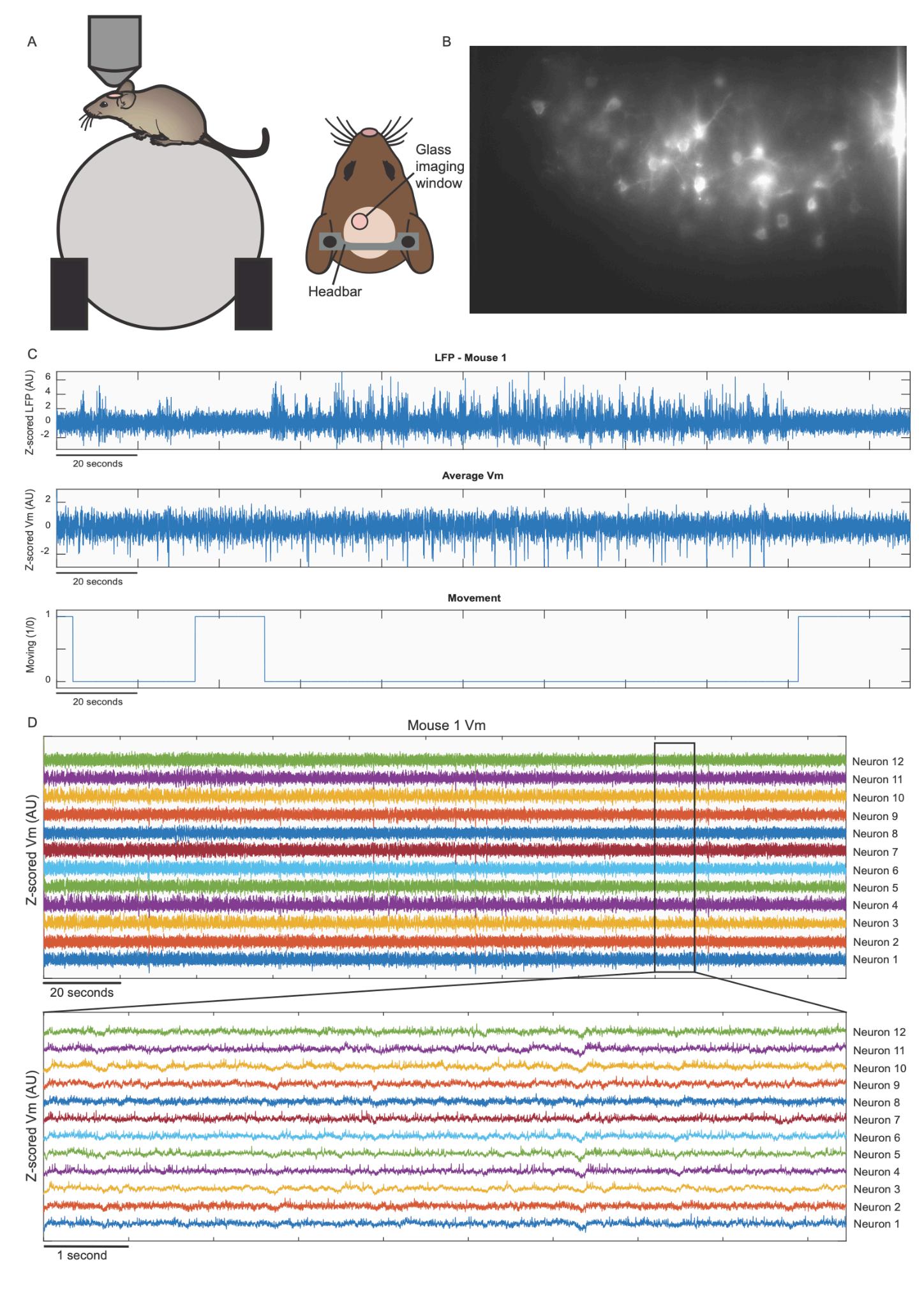
Mouse graph
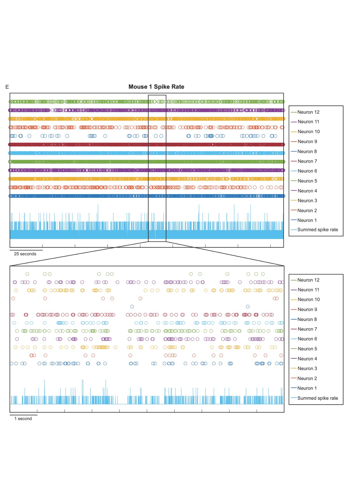
result was plotted at the bottom of the graph. The full length of the recording is represented, and a secondary graph is provided to show changes at a smaller scale.
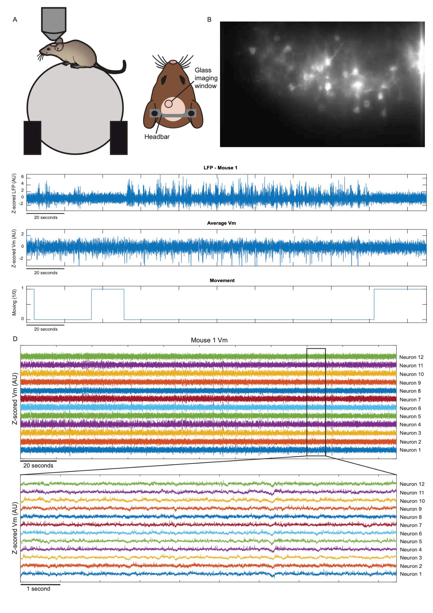
methodinwhichthemeanandstandarddeviationofasetof values,forexample,theVmfluorescenceacrossallframes,are firstcomputed,andeachvalueisthenremappedandexpressedin termsofhowmanystandarddeviationsitliesaboveorbelowthe mean.TheLFPwasalsonormalizedanddownsampledto800Hz tomatchtheVmrecordings,andthespikesintheVmrecordings wereidentifiedandmarkedautomatically(Fig.3).Thespeedtrace wasresampledtomatchtheVmrecordings’samplingfrequency andwasseparatedinto100–millisecond–longchunks.Foreach chunk,ifthemousemovedlessthan5percentofthetime,itwas
markedasaperiodofrest,butifitwasmovingforlonger,itwas markedasaperiodofmovement(Fig.4).
SpikeRateandRasterAnalysis
Weautomaticallymarkedthespikesofeachneuronacross thelengthoftherecordingusingcustomMATLABcode.To calculatethespikerateandplothowitchangedovertime,we summedthenumberofspikesataspecifictime(Fig.3).We repeatedthisprocessacrossmice.Wecreatedaspikerasterasa binarytrace,whereeachframewasmarkedwitha1ifitcontained thepeakofaspikeand0otherwise.Becausespikesoccurina singleframeat800Hz,spiketrainswereconvolvedwitha Gaussiankernelspanning±25mstoestimatespikeprobability aroundeachevent.Pairwisespike-spikecorrelationswerethen calculatedacrossneuronsusingtheseconvolvedtraces.Totest whetherspike-spikecorrelationsdifferedduringmovementand rest,therasterwasseparatedbybehavioralstate,andthe convolutionandcorrelationprocedureswererepeatedforeach condition.
Time-basedAnalysis
Throughoutthisstudy,wecalculatedthePearson correlationcoefficienttofindthecorrelationbetweentwosetsof data.Itmeasuresthelinearcorrelationbetweenthedatasets, resultinginanumberbetween-1and1thatrepresentsthestrength anddirectionofthecorrelation.Forexample,apositivenumber representspositivecorrelation.TheequationtofindthePearson correlationcoefficientis:
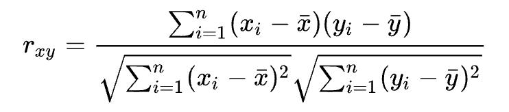
Inwhichnrepresentsthesamplesize,andxi andyi representelementioftwoseparatedatasets.15
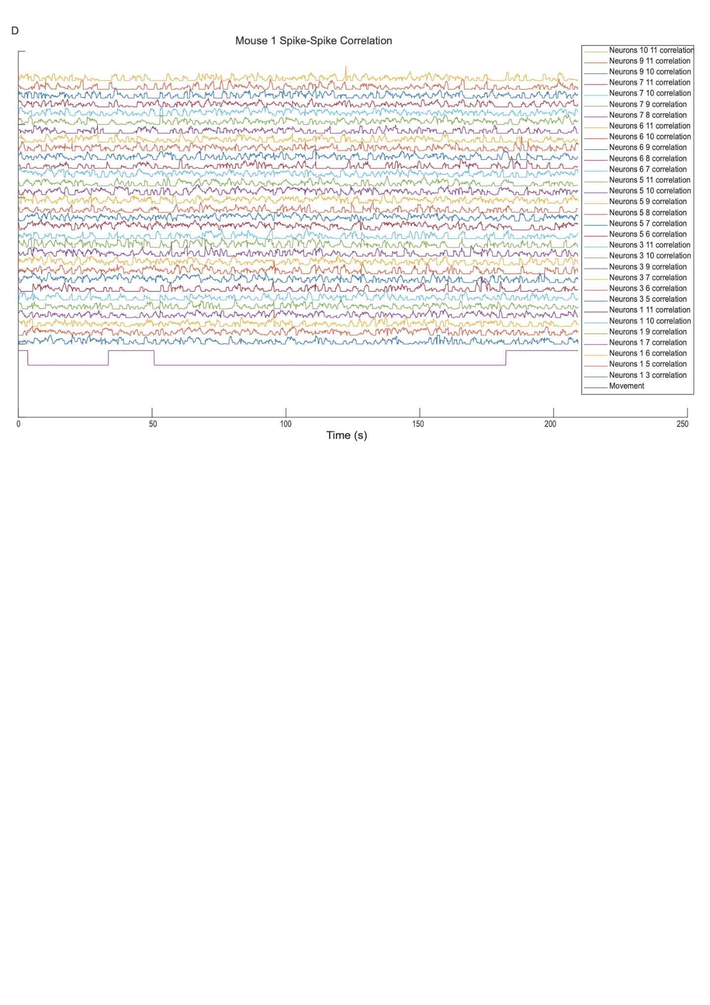
Toassessthecorrelationofspikingactivitybetween neurons,wecalculatedtheaverageVmactivityofallneurons whenoneneuronspiked(seeninFig.8).Todothis,weseparated theactivityofeachneuron100msbeforeandafterthemarked neuron’sspike.Spikeswithin100msofthebeginningandendof therecordingwerenotused.Wethenaveragedeachneuron’s activityovertime,resultinginthespike-triggeredaverageVm.We comparedhowthespike-spikecorrelationbetweenpairsof neuronschangedovertime,andmarkedanyneuronpairsthatwere morecorrelatedthanaveragewhencomparedtoallpossible neuronpairs(seeninFig.5,Fig.6).Thisprocesswasrepeatedfor allpairsofneuronsacrossall5mice.Todeterminewhetherthe spike-spikecorrelationbetweenneuronswasdependentonthe mousemoving,werepeatedthecorrelationcalculationduring movementandrestperiodsseparatelybyonlyusingspikesfrom theserespectiveperiods(seeninFig.5andFig.6).
Tofurtherinvestigatetherelationshipbetweenpairsof neurons,wecomputedthecorrelationbetweentheVmtracesof twoneurons.Foreachneuron,wecalculateditscorrelationwith eachotherneuronbygroupingeachVmrecordinginto 800–millisecond–longsections,thenrunningacorrelationtestfor eachchunk(seeninFig.7).Wethenmarked100-msboutsofhigh andlowVm-Vmsynchrony.PeriodswheretheaverageVm-Vm synchronyacrossallneuronpairsexceededthe80thpercentileof
15 Pearson Correlation Coefficient - an Overview | Scie800–millisecond–longnceDirect Topics,n.d.
correlationvalueswereclassifiedashighVm-Vmsynchrony. PeriodswheretheaverageVm-Vmsynchronyfellbelowthe20th percentileofcorrelationvalueswereclassifiedaslowVm-Vm synchrony.
SpectralAnalysisTechniques
Everysignaliscomposedofsinewavesatdifferent frequencies,andtheamplitudeofeachsinewaveisthepowerof thatfrequency.Throughoutthisstudy,weusedseveralmethodsto calculatethefrequenciesmakinguptheVmandLFPandtheir correspondingpower.Thefirstmethodweusedwasthecontinuous wavelettransform,whichseparatesthefrequenciesbyaltering waveletstomatchthesignalataspecifictime.Thus,theresultof thisoperationgivesthepowerofeachfrequencycomposingthe originalsignalovertime.Theequationofthecontinuouswavelet transformis:
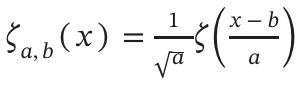
Inwhichthemotherwaveletisgivenby: Inthisequation,aisthescalingfactor,andbisthe translationfactor,orthetimeshift.16
Anothermethodtocalculatethepowerofindividual frequenciesistheFouriertransform.TheFouriertransformis similartothecontinuouswavelettransform,butitdoesnotretain timeinformation.Instead,itisanoverallmeasurementofhow prominentaspectralcomponentis.TheequationoftheFourier transformis:

16 Continuous Wavelet Transform - an Overview | ScienceDirect Topics,n.d.
Frequency-basedAnalysis
Toinvestigatehowdifferentspectralcomponentsofthe LFPactivitywerecorrelatedwithaspecificneuron’sspiking activity,wecalculatedthespike-triggeredaverageLFPacross neuronspermouse.WeseparatedtheactivityoftheLFP100ms beforeandafterthemarkedneuronspiked,averageditacrosstime, andthenperformedacontinuouswavelettransformontheresult. Weseparatedtheresultintothefrequencybandsthatcomposethe LFP—thedelta(0.5-4Hz),theta(4-10Hz),alpha(10-20Hz),beta (20-40Hz),andgammabands(40-100Hz)—thenaveragedeach bandateachpointintime,resultinginasingulartraceper frequencyband.Werepeatedthisprocessforeachneuronper mouse.
TocomparehowindividualspikesaffectedtheLFP activity,wecomparedthespike-triggeredaverageLFPtothe LFP’stypicalactivity(seeninFig.12).Wecompiled1000random 201–millisecond–longchunksofLFPandaveragedthemacross timetocalculatethetypicalLFPactivity,andaveragedacrossall 201–millisecond–longLFPwindowscenteredaroundspikesto calculatethespike-triggeredLFP.Wethenperformedacontinuous wavelettransformontheresults,andrepeatedthisprocessforeach mouse.
Tofindtherelativepowerofeachfrequencybandinthe LFP,aswellashowtheychangedovertime,weperformeda continuouswavelettransformonthefullLFPrecording,then averagedthepoweracrossallspectralcomponentswithineach frequencybandofinterest(delta,theta,alpha,beta,andgamma) foreachpointintime.Wethensummedthepowerofallfrequency
bandsperpointintime,andcalculatedeachfrequencyband’s percentcontributiontothetotalpowerateachpointintime.We repeatedthisprocessforeachmouse.
TotesthowsynchronizedspikingactivityaffectedtheLFP, weanalyzedtheLFPduringperiodsofhighandlowspike-spike synchronyseparately.Highandlowspike-spikesynchronyperiods weredefinedaswhentheaveragespike-spikecorrelationacrossall neuronpairswasabovethe90thpercentileorbelowthe10th percentile,respectively.Weperformedacontinuouswavelet transformontheresultingtracesandcalculatedthepowerofeach frequencybandovertimeasbefore.Wethenperformeda Wilcoxonrank-sumtesttocomparetheLFP’sactivityduring periodsofhighandlowspike-spikecorrelation.
Periodsofhighandlowdelta,theta,andgammapowerin theLFPwerefurtheranalyzedrelativetotheaverageVmactivity inthesebandsbecausedeltaandthetabandsareassociatedwith coordinatingmovement,andbecauseParvalbuminneuronsare thoughttodrivegammabandactivity.Toanalyzewhetherthe delta,theta,andgammabandactivityoftherecordedneuronswas reflectiveoftheLFP’sdelta,theta,andgammabandpower,we separatedtheVmofallneuronsduringperiodsofhighandlow LFPdeltapower.PeriodswereconsideredhighLFPdeltapowerif theyexceededthe90thpercentileandconsideredlowLFPdelta poweriftheyfellbelowthe10thpercentile.Werepeatedthis processforthethetaandgammabandsoftheLFP.Weperformeda FouriertransformontheVmandtheLFPtracesduringthesehigh andlowpowerperiods.Thistestcomputedthedelta,theta,and gammapoweroftheVmandLFP(seeninFig.13).
Movement-basedAnalysis
ToanalyzehowmovementaffectedtheLFP’sactivity,we separatedtheLFPofeachmouseintoperiodsofmovementand
rest.Usingacontinuouswavelettransformfunction,wecalculated thepowerofeachfrequencybandduringmovementandduring rest,thenaveragedeachfrequencyband’spowerovertime(seenin Fig.9).Wethensummedtheconvolvedspikerasterofeach neuronandaverageditacrosstime.Theresultshowedtheaverage probabilitythataspikewasoccurringataspecifictime.We chunkedtheresultingaveragespikerastertrace,aswellasthe traceofthepowerofeachfrequencyband,into 100–millisecond–longchunks.Wethentestedthecorrelationin activitybetweenthetwodatasetsandperformedaWilcoxon signed-ranktestontheseparatedLFPtraces(seeninFig.11).
ToinvestigatehowtheVmchangedaccordingtochanges inmovementandrest,wemarkedandseparatedtheVmtracesof allneuronsaccordingtowhetherornotthemousewasmoving.We performedaFouriertransformontheresultingtraces,which revealedthedifferingpowerofthefrequencybandsduringperiods ofrestandmovement(seeninFig.13).
Results
CorrelationofNeuralSpikinginNetworksofNeuronsis DependentonMouseBehavior
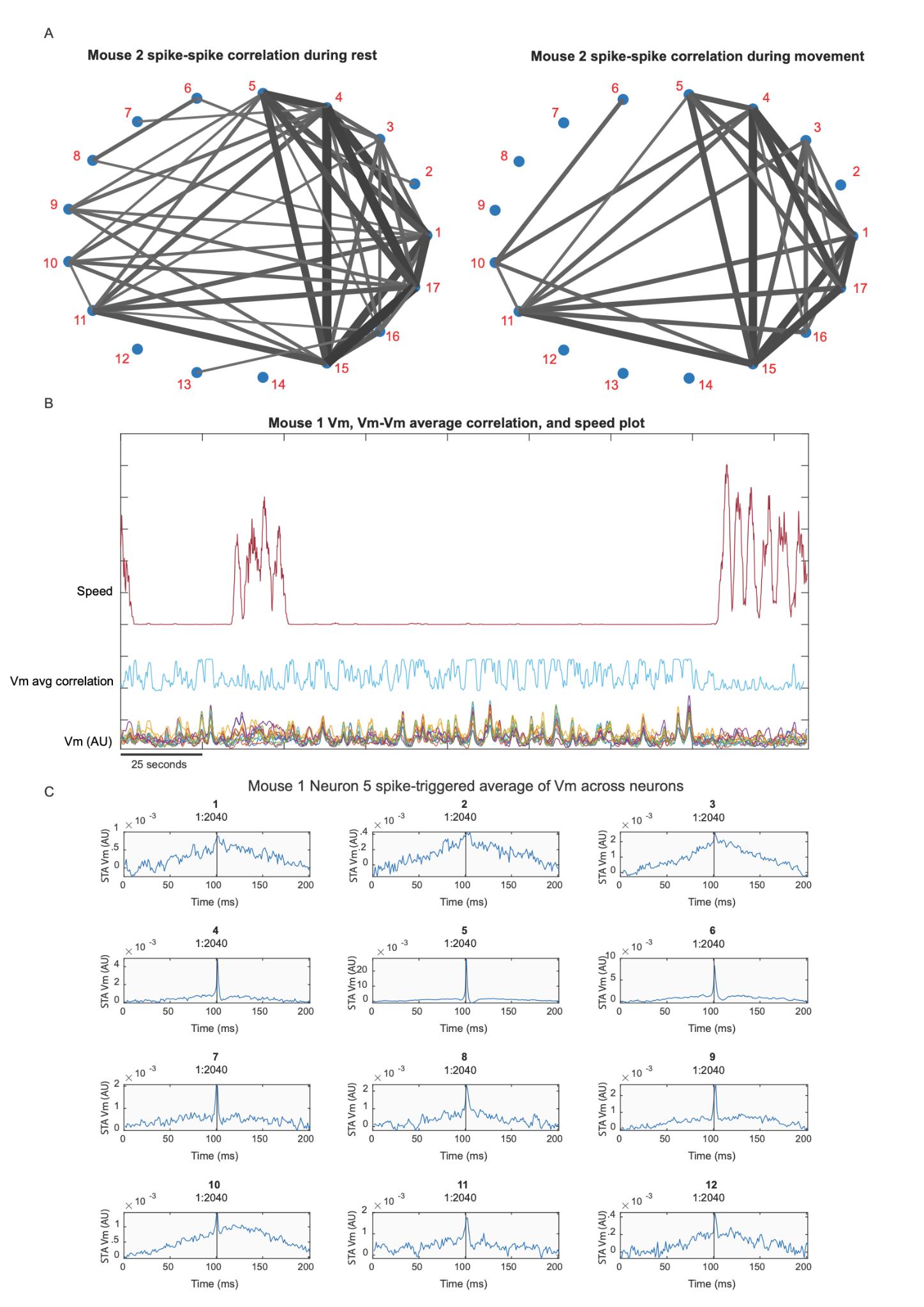
Figure state
The of values pr A = periods For correlation correlation according Neurons (Fig. Fig. ). weanalyzedourfindingsinconjunctionwithamarkedfieldof view,wefoundthattheseneuronpairswereoftenfoundtobein closephysicalproximity.
changesarereflectiveofthespike-spikecorrelation changes—decreasingduringmovementandincreasingduring rest—validatesthathypothesis.
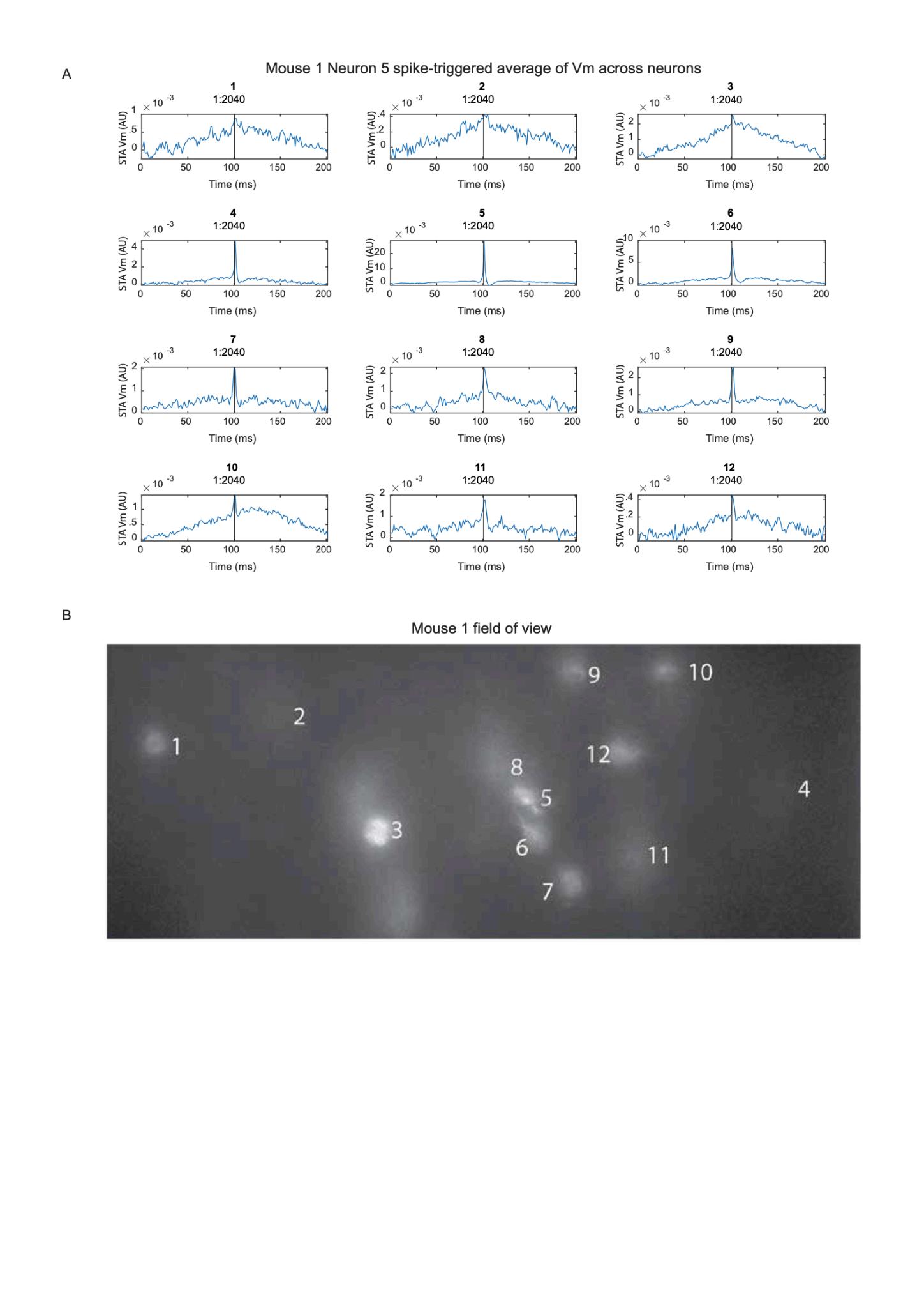
recorded neuron is marked with the number used to label it for all testing.
Severalneurons’spikingdoesnotappeartohaveany correlationwithNeuron5’sspiking,suchasNeurons1,2,and3. Otherneurons’spikingappearstohaveaslightcorrelationwith Neuron5’sspiking,suchasNeurons10,11,and12.However,the peakoccurringatthetimethatNeuron5spikedineachofthese tracesdoesnotrepresentalargechangefromtheirstandard activity.Someneurons’spikingdidshowastrongcorrelationwith Neuron5’sspiking—namely,Neurons4and6(Fig.8A).Whenwe analyzedtheseresultsinconjunctionwithourmarkedfieldof view,wefoundthattheseneuronswerelocatedincloseproximity. However,thisrelationshipwasnotalwaysfoundtobe true—Neurons4and5’sactivitywasmorecorrelatedthan Neurons5and8’sactivity,thoughtheywerefurtherapart(Fig. 8B). SingleNeuronElectricalBehaviorsofTensofNeurons CorrelatetoButCannotFullyExplainLocalFieldPotential Characteristics
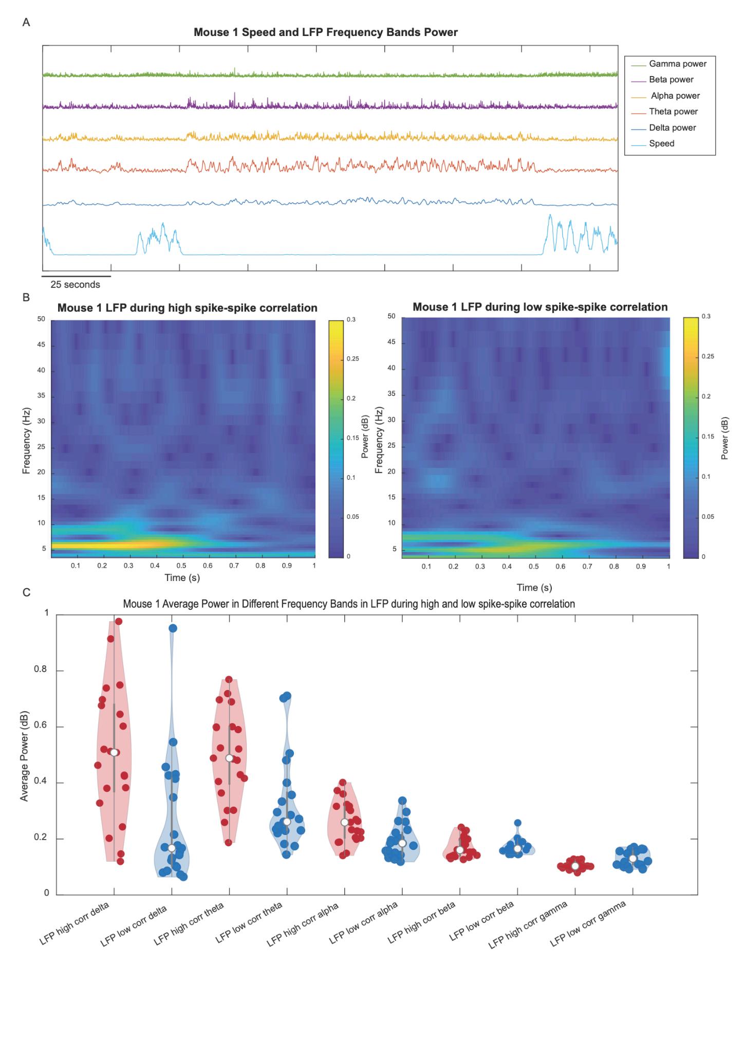
full recording in Mouse 1. Changes in the power frequency bands in the LFP are plotted alongside in Mouse 1.
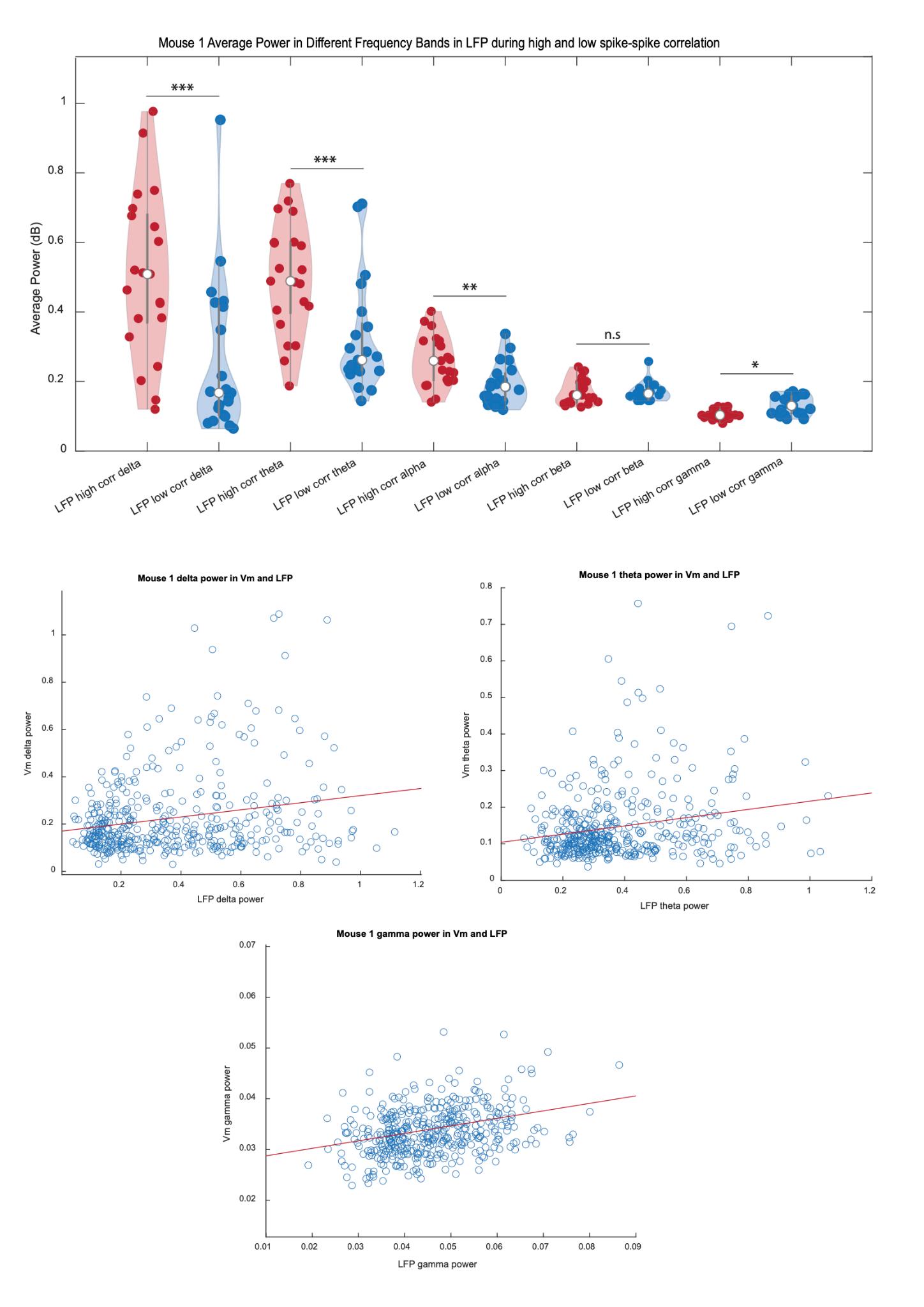
Figure gamma-band spike–spike Comparison and spike-spike corr bands periods marked for 0.005, averaged delta band wellasthealphaband(p=2.00e-3)andthegammaband(p= 8.9e-3).Therewasnosignificantdifferenceinthepowerofthe
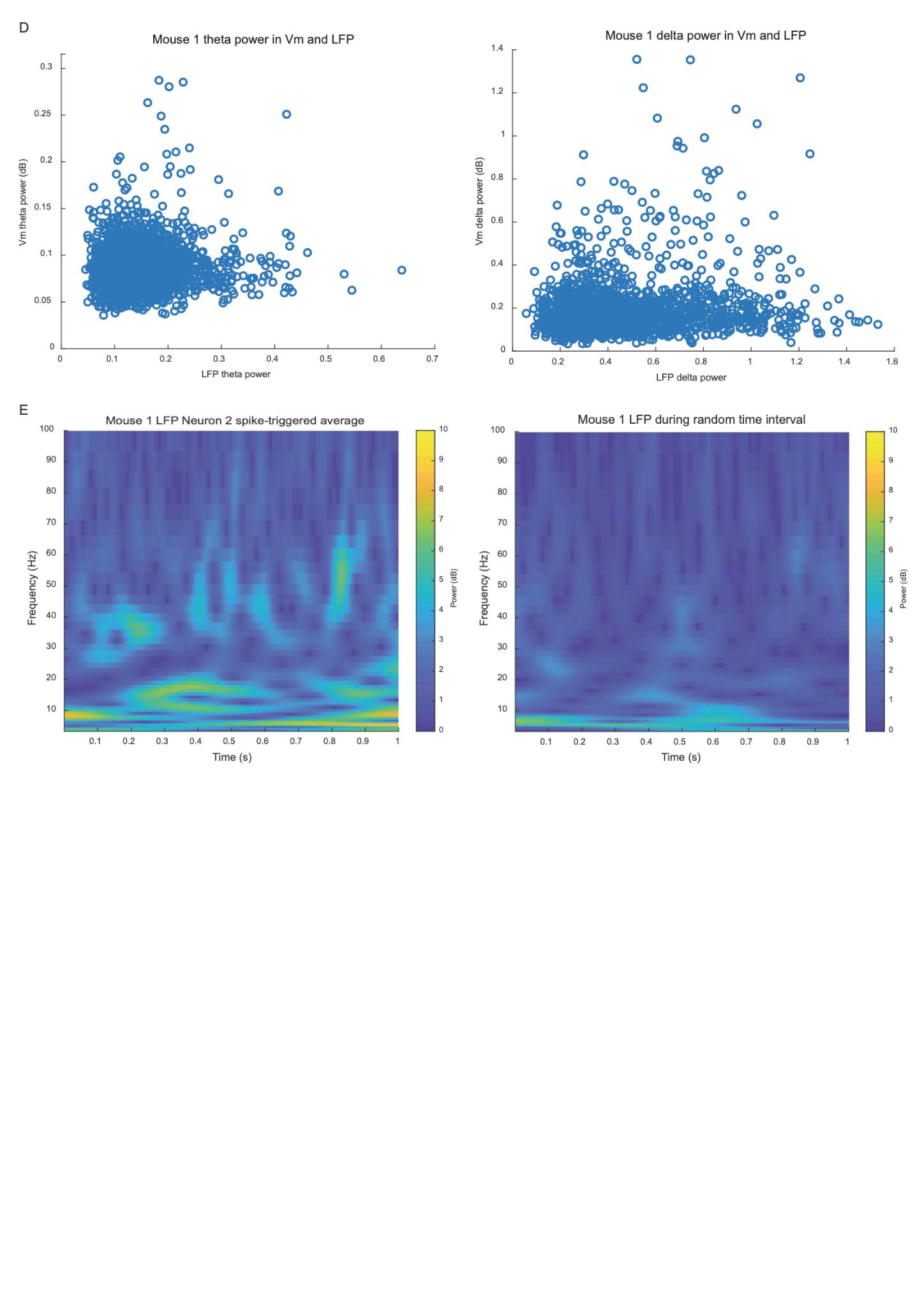
LFPplot.Thespike-triggeredaverageLFPalsoshowshigher powerinthealphabandandgammaband.Thereappearstobean
Figure 13: Vm delta, theta, and gamma power do not predict the corresponding LFP band power.
Each dot represents a point in time, and its position reflects the Vm and LFP chosen frequency band’s power at that time—either the delta, theta, or gamma band. In each plot, the x-value represents the power of the chosen. The trendline is depicted in red. Outliers are not represented.
Therewasnotfoundtobeanycorrelationbetweenthe powerofthethetabandintheVmandthepowerofthethetaband intheLFP.Therewasalsonocorrelationbetweenthepowerofthe deltabandintheVmandthepowerofthedeltabandintheLFP,
noranycorrelationbetweenthepowerofthegammabandinthe VmandthepowerofthegammabandintheLFP.Theequationof thetrendlineinthedeltabandcomparisonplotisy=0.1478x+ 0.02727(R2 =0.023).Theequationofthetrendlineinthetheta bandfrequencyplotisy=0.1125x+0.1039(R2 =0.045).The equationofthetrendlineinthegammabandfrequencyplotisy= 0.1504x+0.1698(R2 =0.046).Thepowerofeachfrequencyband intheLFPwasshowntohavemorevariancethanthepowerofthe samefrequencybandintheVm(Fig.13).
Discussion
PhysicalproximityappearedtohaveaneffectonVm-Vm synchrony,asneuronsthatwereclosetogethertypicallyhadhigher correlationinactivitythanneuronslocatedfurtherapart.This likelyreflectsthatneuronsincloseproximityaremore interconnectedandarelikelyusedtocompletethesameorsimilar tasks,resultinginsimilarspikingactivityduringbothmovement andrest.
Themouse’smovementsystematicallyalteredtheactivity ofbothindividualneuronsandtheLFP.Forexample,thepowerof theLFPfrequencybandsfluctuatedaccordingtochangesin movement.Thesechangesinactivityarelikelyduetothefactthat LFPfrequencybandsaretypicallylinkedtodifferentmodesof activity—forexample,thedeltabandhasbeenshowntobemost powerfulduringrestingperiods.19 Onaverage,theneuronal synchrony—bothspike-spikeandVm-Vm—decreasedduring movement,asdidthecouplingbetweenneuronsandtheLFP.This likelyreflectsthattheneuronsareinvolvedinvariedtasksduring movement,andtheyareengagedinsynchronizedrestingstate networkswhennotmoving.20 However,someVm-Vmcorrelation levelsremainedstableduringmovementandrest.Itispossiblethat
theseneuronsareinvolvedinprocessesnotrelatedtomovement, likesensoryprocessing.Furthermore,someneuronsshowed increasedcorrelationwitheachother.Thismaybebecausethese neuronsareinvolvedinthesameorsimilaractivities,duetotheir physicalproximity.Futurestudiescouldfocusonthisconnection andcreatespatialmapsofcorrelationduringmovementandrest, explicitlyanalyzinghowlocationrelatestosynchronization.
TheobservedVm-Vmsynchronywastypicallyreflective ofthecalculatedVm-LFPsynchrony,suggestingthatlocal membranepotentialcoordinationcontributestothelarger mesoscopicfielddynamics.However,ourstudyonlyfocusedona smallsetofPVneuronsinthesensorimotorcortex;thus,the correlationbetweentheVmandLFPwasratherweak,andwe wereunabletofullypredicttheLFPusingonlytheneuralVm dynamicswehadavailable.Futureresearchcouldstudythe relationshipbetweenVm-VmsynchronyandVm-LFPsynchrony acrossalargerareaordifferentregionsofthebrain,analyzinghow itisinfluencedbylocation,ifatall.
Wedidnotobservesignificantcouplingbetweenthetheta ordeltabandsinVmandthecorrespondingLFPbandpower. GiventhattheLFPreflectslarge-scalesynapticanddendritic currentsacrossdiversecellpopulations,PVinterneurons' subthresholddynamicsmaynotbethedominantcontributorsto low-frequencyLFPoscillations.Alternatively,low-frequencyLFP rhythmsmaybedrivenbyinputsfromfarawaycircuitsnot capturedinourfieldofview.Furtherresearchcouldidentifywhy therewasnocorrelation,andinwhatregionsofthebrainthis relationshipisdifferent.Additionally,wedidnotobserve significantcouplingbetweenthegammabandinVmandLFP,but wedidseebriefincreasesintheLFPgammabandpower surroundingPVspikes,aswepredicted.BecausePVinterneurons, thefocusofthisstudy,areabletospikesorapidly,theyarethought
toberesponsibleforthegammaband.Thelackofcorrelation betweenneuralVmandLFPcouldbeduetothelowelectrical poweroftheVmsubthresholdoscillationscomparedtothehigh electricalpowerofneuralspiking.Itispossiblethatonlyneural spikesarepowerfulenoughtonoticeablyinfluencethebulklocal electricfield.
Spike-triggeredanalysesrevealedsignificantmodulationof LFPspectralpowercomparedtobaselineLFPactivity.Whilethe spikingofthesmallnumberofneuronswerecordedisnotlikelyto befullyresponsibleforthischange,itdoesrevealsome system-widesynchrony.Futurestudiescouldexaminethis phenomenon,studyingwhetherthissynchronyiscausedbyinput orifthereisconsistentsynchronizationbetweenvariousgroupsof neurons.Furthermore,ourresultscouldbeusedtoidentifythe standardpowerofLFPfrequencybandsduringmovementandrest. Identifyingthishealthyneuralactivitycouldhelpinformand improveneuromodulationtherapiesforneurodegenerative movementdisorders,suchasParkinson’sDisease,inwhichthe frequencybandsassociatedwithmovementareamplifiedand attenuatedtotreatsymptoms.
Thisstudyhadseverallimitations,includingthesmall samplesizeofneurons(4to17neuronspermouse)duetothe physicallimitationsofvoltageimagingasanexperimental technique.Thissmallsamplesizelimitsourabilitytodraw conclusionsregardingtherelationshipbetweenVmandLFP, becausetheLFPisthoughttoreflecttheactivityofthousandsof otherneuronsinadditiontotheoneswerecorded.Additionally,we onlystudiedneuronslocatedinthesensorimotorcortex.Therefore, wecannotdrawconclusionsregardinghowtherelationship betweentheVmandLFPchangesdependingonbrainregion,or howmovementaffectstheserelationshipsoutsidethesensorimotor cortex.
Futureworkcouldhelpaddresstheselimitations.For example,iftheexactlocationoftheelectrodeisrecorded,the effectofdistancefromtheelectrodeonVm-LFPcorrelationcould bestudied.Electricfieldsdissipaterapidlywithdistance;therefore, itisbelievedthatLFPwouldbedominatedbytheelectrical activityofneuronsclosesttotheelectrode.Inaddition,performing spike-sorting,astandardelectrophysiologicaltechniquefor distinguishingneuralactivityfrommultichannelelectrodeLFP data,tocorrelateLFPwithlocalneuralactivitydirectlywould improveouranalysisoftheeffectofindividualneuronsonthe LFP.Futurestudiescouldalsoinvestigatehowtheaverage Vm-LFPcorrelationchangesaccordingtochangesinbrainregion andcelltypebyperformingthewindowimplantationsurgeryata differentbraintargetandusingadifferentCre-mouseline.While ourstudyonlysortedeachmouse’sactivityintomovementand rest,futureworkcouldalsotesttherelationshipbetweenLFPand neuralactivityduringspecifictasks,allowingformore sophisticateddataanalysis.
008069 PV[cre] Strain Details.(n.d.). https://www.jax.org/strain/008069
Abdelfattah,A.S.,Kawashima,T.,Singh,A.,Novak,O.,Liu,H.,Shuai, Y.,Huang,Y.-C.,Campagnola,L.,Seeman,S.C.,Yu,J.,Zheng, J.,Grimm,J.B.,Patel,R.,Friedrich,J.,Mensh,B.D.,Paninski, L.,Macklin,J.J.,Murphy,G.J.,Podgorski,K.,…Schreiter,E. R.(2019).Brightandphotostablechemigeneticindicatorsfor extendedinvivovoltageimaging. Science, 365(6454),699–704. https://doi.org/10.1126/science.aav6416
Asadi,A.,MadadiAsl,M.,Vahabie,A.-H.,&Valizadeh,A.(2022).The OriginofAbnormalBetaOscillationsintheParkinsonian CorticobasalGangliaCircuits. Parkinson’s Disease, 2022, 7524066.https://doi.org/10.1155/2022/7524066
Biswal,B.,ZerrinYetkin,F.,Haughton,V.M.,&Hyde,J.S.(1995).
Functionalconnectivityinthemotorcortexofrestinghuman brainusingecho-planarmri Magnetic Resonance in Medicine, 34(4),537–541.https://doi.org/10.1002/mrm.1910340409
Boehmer,G.,Greffrath,W.,Martin,E.,&Hermann,S.(2000).
Subthresholdoscillationofthemembranepotentialin magnocellularneuronesoftheratsupraopticnucleus. The Journal of Physiology, 526(1),115–128. https://doi.org/10.1111/j.1469-7793.2000.t01-1-00115.x Chiang,C.-C.,&Durand,D.M.(2023).SubthresholdOscillatingWaves inNeuralTissuePropagatebyVolumeConductionandGenerate Interference. Brain Sciences, 13(1),74. https://doi.org/10.3390/brainsci13010074
Continuous Wavelet Transform An overview | ScienceDirect Topics. (n.d.).
https://wwwsciencedirectcom/topics/mathematics/continuous-w avelet-transform
Deleuze,C.,Bhumbra,G.S.,Pazienti,A.,Lourenço,J.,Mailhes,C., Aguirre,A.,Beato,M.,&Bacci,A.(2019).Strongpreference forautapticself-connectivityofneocorticalPVinterneurons
facilitatestheirtuningtoγ-oscillations. PLoS Biology, 17(9), e3000419.https://doi.org/10.1371/journal.pbio.3000419
Fourier Transform An overview | ScienceDirect Topics.(n.d.). https://www.sciencedirect.com/topics/neuroscience/fourier-transf orm
Friston,K.J.,Bastos,A.M.,Pinotsis,D.,&Litvak,V.(2015).LFPand oscillations Whatdotheytellus? Current Opinion in Neurobiology, 31,1–6
https://doi.org/https://doi.org/10.1016/j.conb.2014.05.004 Lampl,I.,&Yarom,Y.(1997).Subthresholdoscillationsandresonant behavior:Twomanifestationsofthesamemechanism. Neuroscience, 78(2),325–341. https://doi.org/10.1016/S0306-4522(96)00588-X
Lu,H.,Zuo,Y.,Gu,H.,Waltz,J.A.,Zhan,W.,Scholl,C.A.,Rea,W., Yang,Y.,&Stein,E.A.(2007).Synchronizeddeltaoscillations correlatewiththeresting-statefunctionalMRIsignal.
Proceedings of the National Academy of Sciences of the United States of America, 104(46),18265–18269. https://doi.org/10.1073/pnas.0705791104
Mollazadeh,M.,Aggarwal,V.,Thakor,N.V.,Law,A.J.,Davidson,A., &Schieber,M H (2009) CoherencybetweenSpikeandLFP ActivityinM1duringHandMovements. International IEEE/EMBS Conference on Neural Engineering : [Proceedings]. International IEEE EMBS Conference on Neural Engineering, 2009,506–509.https://doi.org/10.1109/NER.2009.5109344
Onorato,I.,Tzanou,A.,Schneider,M.,Uran,C.,Broggini,A.C.,& Vinck,M.(2025).DistinctrolesofPVandSstinterneuronsin visuallyinducedgammaoscillations. Cell Reports, 44(3), 115385.https://doi.org/10.1016/j.celrep.2025.115385
Pearson Correlation Coefficient An overview | ScienceDirect Topics. (n.d.).
https://www.sciencedirect.com/topics/computer-science/pearsoncorrelation-coefficient
Reyes-Chapero,R M,Tapia,D,Ortega,A,Laville,A,Padilla-Orozco, M.,Fuentes-Serrano,A.,Serrano-Reyes,M.,Bargas,J.,&
Galarraga,E.(2025).Corticalparvalbumin-expressing interneuronssamplenetworkoscillationsintheirsynaptic activity. Neuroscience, 573,25–41.
https://doi.org/10.1016/j.neuroscience.2025.03.021
Shroff,S.N.,Lowet,E.,Sridhar,S.,Gritton,H.J.,Abumuaileq,M., Tseng,H.-A.,Cheung,C.,Zhou,S.L.,Kondabolu,K.,&Han, X.(2023).Striatalcholinergicinterneuronmembranevoltage trackslocomotorrhythmsinmice Nature Communications, 14(1),3802.https://doi.org/10.1038/s41467-023-39497-z
Tseng,H.,Mount,R.A.,Lowet,E.,Gritton,H.J.,Cheung,C.,&Han, X.(2022).MembraneVoltageDynamicsofParvalbumin InterneuronsOrchestrateHippocampalThetaRhythmicity. bioRxiv.https://doi.org/10.1101/2022.11.14.516448
Xiao,S.,Cunningham,W.J.,Kondabolu,K.,Lowet,E.,Moya,M.V., Mount,R.A.,Ravasio,C.,Bortz,E.,Shaw,D.,Economo,M.N., Han,X.,&Mertz,J.(2024).Large-scaledeeptissuevoltage imagingwithtargeted-illuminationconfocalmicroscopy. Nature Methods, 21(6),1094–1102.
https://doi.org/10.1038/s41592-024-02275-w