Trust our nationally renowned rehabilitation experts and specialized care to guide the way.
Your patient just had a stroke, spinal cord injury, or traumatic brain injury. What happens next is critical to their recovery. Finding the best choice for rehabilitation—where specialized, multidisciplinary experts guide their care with state-of-the-art techniques, innovative research, and advanced technology—will make all the difference in their tomorrows.
At Casa Colina Hospital and Centers for Healthcare, we have been providing a comprehensive continuum of rehabilitative care for nearly a century. We use proven pathways and the latest evidence-based science to help each patient in their recovery.
Our Continuum of Care
º Inpatient rehabilitation, medical-surgical and ICU beds, surgical suites
º Residential rehabilitation beds and da y treatment program
º Outpatient rehabilitation centers
º Long-term residential facilities
Outdoor Adventures & Wheelchair Sports program
BRAIN INJURY
vol. 22 issue 2 professional
5 Editor in Chief Message
Neuromodulatory Interventions in Patients with Disorders of Consciousness after Severe Brain Injury: What is the State of the Evidence?
Patricia Grady-Dominguez, PhD • Lauren Teague
Jennifer Weaver, PhD, OTR/L
Traversing the Landscape of Neuroendocrine Deficits Following Traumatic Brain Injury: The Role of a Neurorehabilitation Specialist
Chantel T. Debert, MD, MSc, FRCPC
Family and Caregiver Brain Injury Education –Leveraging Model Systems Knowledge Translation Center Resources
Tracy Shannon, PsyD • Cynthia Beaulieu, PhD, ABPP
Non-invasive Neuromonitoring in Traumatic Brain Injury: Current Insights and Future Trends
Sebastián Vásquez-García, MD • Chiara Robba, PhD
Engagement in Brain Injury Rehabilitation
Anthony H. Lequerica, PhD • Michael Williams, PhD Irene Ward, DPT
Moving the Field Toward Health Equity in Traumatic Brain Injury
Monique R. Pappadis, PhD • Chinedu K. Onwudebe, BS
Anthony H. Lequerica, PhD • Angelle M. Sander, PhD, FACRM
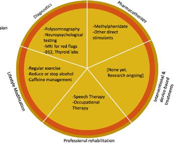
Multidisciplinary Concussion Care: Delivering the Whole Pizza
David L. Brody, MD, PhD
Considerations in the Neuropsychological Assessment of Spanish-speaking Adults
Giselle Leal, PsyD
NORTH AMERICAN BRAIN INJURY SOCIETY
CHAIRMAN Mariusz Ziejewski, PhD
VICE CHAIR Debra Braunling-McMorrow, PhD
IMMEDIATE PAST CHAIR Ronald C. Savage, EdD
TREASURER Bruce H. Stern, Esq.
SECRETARY Brian Greenwald, MD
FAMILY LIAISON Skye MacQueen
EXECUTIVE DIRECTOR/ADMINISTRATION Margaret J. Roberts
EXECUTIVE DIRECTOR/OPERATIONS J. Charles Haynes, JD
MARKETING MANAGER Megan Bell-Johnston
BRAIN INJURY PROFESSIONAL
PUBLISHER J. Charles Haynes, JD
CO-EDITOR IN CHIEF Beth Slomine, PhD - USA
CO-EDITOR IN CHIEF Nathan Zasler, MD - USA
ASSOCIATE EDITOR Juan Arango-Lasprilla, PhD – Spain
TECHNOLOGY EDITOR Stephen K. Trapp, PhD - USA
EDITOR EMERITUS Debra Braunling-McMorrow, PhD - USA
EDITOR EMERITUS Ronald C. Savage, EdD - USA
DESIGN AND LAYOUT Kristin Odom
ADVERTISING SALES Megan Bell-Johnston
EDITORIAL ADVISORY BOARD
Nada Andelic, MD - Norway
Philippe Azouvi, MD, PhD - France
Mark Bayley, MD - Canada
Lucia Braga, PhD - Brazil
Ross Bullock, MD, PhD - USA
Fofi Constantinidou, PhD, CCC-SLP, CBIS - USA
Gordana Devecerski, MD, PhD - Serbia
Sung Ho Jang, MD - Republic of Korea
Cindy Ivanhoe, MD - USA
Inga Koerte, MD, PhD - USA
Brad Kurowski, MD, MS - USA
Jianan Li, MD, PhD - China
Christine MacDonell, FACRM - USA
Calixto Machado, MD, PhD - Cuba
Barbara O’Connell, OTR, MBA - Ireland
Lisandro Olmos, MD - Argentina
Caroline Schnakers, PhD - USA
Lynne Turner-Stokes, MD - England
Olli Tenovuo, MD, PhD - Finland
Asha Vas, PhD, OTR - USA
Walter Videtta, MD – Argentina
Thomas Watanabe, MD – USA
Alan Weintraub, MD - USA
Sabahat Wasti, MD - Abu Dhabi, UAE
Gavin Williams, PhD, FACP - Australia
Hal Wortzel, MD - USA
Mariusz Ziejewski, PhD - USA
EDITORIAL INQUIRIES
Managing Editor
Brain Injury Professional PO Box 131401, Houston, TX 77219-1401 Tel 713.526.6900 Email: mbell@hdipub.com Website: www.nabis.org
ADVERTISING INQUIRIES
Megan Bell-Johnston
Brain Injury Professional
HDI Publishers PO Box 131401, Houston, TX 77219-1401
Tel 713.526.6900 Email: mbell@internationalbrain.org
Raising the bar for inpatient and day rehabilitation services
Children’s Healthcare of Atlanta is Commission on Accreditation of Rehabilitation Facilities (CARF)-accredited for pediatric rehabilitation services.
We offer:
• An expansive Inpatient Rehabilitation Program
– A spinal cord system of care, brain injury and pediatric specialty programs that have received CARF specialty recognition
– A team of brain injury board-certified pediatric physiatrists
– Comprehensive care for young patients from birth to age 21
– Therapy seven days a week
– 28 private patient rooms
• A Day Rehabilitation Program to assist patients during recovery
• Technology-assisted therapy through our Center for Advanced Technology and Robotic Rehabilitation
• A full-service hospital with emergency services
Learn more or make a referral:
Nathan D. Zasler, MD, DABPM&R, FAAPM&R, FACRM, BIM-C, CBIST
Editor Bio
Nathan Zasler, MD, is an internationally respected physician specialist in acquired brain injury (ABI) care and rehabilitation. He is CEO and Medical Director of the Concussion Care Centre of Virginia, an outpatient neurorehabilitation practice, as well as, the Medical Director of Tree of Life, a living assistance and transitional neurorehabilitation program for persons with acquired brain injury in Richmond, Virginia. He is board certified in Physical Medicine and Rehabilitation and fellowship trained in brain injury, as well as, Brain Injury Medicine certified.
Dr. Zasler is an Affiliate Professor of PM&R at VCU in Richmond, Virginia, as well as, a Visiting Professor of PM&R at the University of Virginia, Charlottesville, Virginia.
Dr. Zasler has lectured and written extensively on neurorehabilitation issues in ABI. He is active in national and international organizations dealing with acquired brain injury and neurodisability, serving in numerous consultant and board member roles.
editor from the
In this issue of Brain Injury Professional we are revisiting a few popular previously published articles that we felt were worth disseminating one more time to our readers given the important albeit diverse topics discussed therein.
The first article by Grady–Dominguez et al. addresses treatment of persons with disorders of consciousness after severe brain injury with neuro modulatory interventions and examines the current evidence space for same. Interestingly, the authors include discussion of "sensory stimulation" in the context of the review and also discussed more traditional neuromodulatory techniques familiar with many clinicians who are involved with DOC treatment including tDCS, rTMS, NILT as well as transcranial focused ultrasound. The topic of transcutaneous vagal nerve stimulation (tVNS) was not included in readers are encouraged to review more recent literature on this topic.
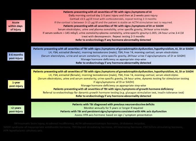
The second article by Dr. Debert addresses the important topic of neuroendocrine dysfunction following TBI and the role of the neuro rehabilitation physician in screening for these disorders. In that context a very helpful figure is provided looking at times post-injury and guideline recommendations for what labs are important to assess relative to endocrinological function. Important points are also made in this article regarding the various manifestations of neuroendocrine dysfunction following TBI and the importance of staying cognizant of these during not only the acute but also post-acute rehabilitation course.
Shannon and Beaulieu discuss MSKTC resources for family and caregiver education and the third article in this issue. This free web-based resource should be made readily available to caregivers and families to facilitate education being provided by the treatment team the authors review the various formats of educational materials available through the MSKTC including fact sheets, videos, slide presentations and links to other resources.
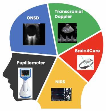
The fourth article on noninvasive neuro monitoring by Vasquez- Garcia and Robba discusses some of the cutting edge techniques being used in various settings for assessing suspected or known TBI using noninvasive technologies some older some newer. They discuss techniques including TCD, optic nerve sheath diameter assessment via ultrasound, automated pupillometry, NIRS as well as a device for assessing ICP waveforms in the neurocritical care called the Brai4Care.
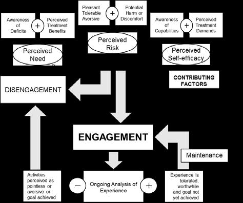
A very important discussion addressing the issue of engagement in brain injury rehabilitation follows as the fifth article in this series by Lequerica et al. This is a topic that is often underappreciated and under addressed in the context of rehabilitation outcomes assessment and optimization of rehabilitation efforts more generally. The authors do a very nice job of addressing engagement barriers, consequences, and considerations in this very "engaging" review of the topic. In that context they also provide a review of various assessment measures for therapy engagement.
The sixth article in this issue by Papppadis et al addresses equity issues in TBI health care. The authors address the National Institute of Minority Health and Health Disparities framework model. The model encompasses 5 key domains of influence including biological, behavioral, physical/ built environment, sociocultural environment, and healthcare system domains within the context of individual, interpersonal, community and societal influences. The authors also pointed out the need for more research in the areas of sociocultural environment and health system domains in the context of health equity in this patient population.
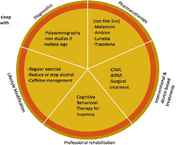
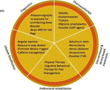
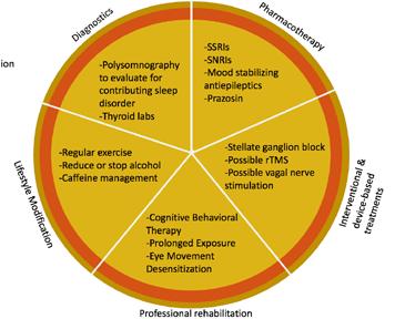
Dr. Brody provides any commentary on the importance of multidisciplinary concussion care as the seventh article in this issue. His discussion leads off with the caveat that specialized care is critical for those patients who do not make rapid and complete recovery within the first 1 to 3 weeks postinjury. He then reviews 3 basic principles in the overall summary of the most important things to consider when initially evaluating the patient and then follows this with a discussion of 5 basic domains of intervention to consider in this patient population including diagnostics, pharmacotherapy, interventional and device based treatments, professional rehabilitative therapies, and lifestyle modifications. This article is a great primer for those not familiar with post-concussive care.
The eighth article in this issue by Dr. Leal addresses neuropsychological assessment of Spanishspeaking adults. This is obviously an important topic given the number of Spanish-speaking individuals who sustained TBI not only in the United States but internationally. The author points out the need to not use this population as a homogeneous ethnic minority group and points out some of the differences across Hispanic populations that should be taken into consideration in addition to concepts of acculturation, normative data for this group, and cultural considerations and testing as well as issues of how best to manage language dominance issues and bilingual patients.
We hope that readers will find revisiting these articles helpful and encourage all readers to consider the information included in your everyday practice.
Neuromodulatory Interventions in Patients with Disorders of Consciousness after Severe Brain Injury: What is the State of the Evidence?
Patients with disorders of consciousness (DoC) are characterized by a continuum of clinical states (Table 1)1,2 and emerging research suggests that neuromodulatory interventions may lead to improved neurobehavioral function.3,4 In the context of brain injury rehabilitation, neuromodulation has been defined as “the alteration of nerve activity through targeted delivery of stimulation provided to modulate dysfunctional as well as functional neural pathways to support neural repair and neural alterations necessary
for sustained recovery of functional skills valued by the patient” (p. 368).5 Neuromodulatory interventions may aid in reconfiguring neural networks, improve the structure and function of viable networks after injury, engage dormant networks, and create new neural connections. For patients with DoC, these benefits may contribute to increases in neurobehavioral function including arousal and awareness, subsequently leading to an improved state of consciousness.
Table 1. Characterizing States of Consciousness in DoC, adapted from Giacino et al. and Thibaut et al.1,2
Clinical States Sleep/Wake Cycles
Motor Func6on
Comatose Absent Reflexive and postural responses
Unresponsive Wakefulness Syndrome (UWS)
Minimally Conscious State Minus (MCS-)
Minimally Conscious State Plus (MCS+)
Emerged from Minimally Conscious State (eMCS)
Present Withdrawal from painful/noxious s<muli; some non-purposeful movement
Present Localized response to painful/noxious s<muli, occasional automa<c and/or purposeful movement
Present Localized response to painful/noxious s<muli, occasional automa<c and/or purposeful movement
Present Func<onal object use2
Auditory Func6on Visual Func6on Communica6on
None
Startle, brief orienta<on to sound
Localiza<on to sound, inconsistent response to command
Localiza<on to sound, command following1
Localiza<on to sound, command following2
None
Startle, brief visual fixa<on
Sustained visual fixa<on
Sustained visual fixa<on
Sustained visual fixa<on
None
None
None
Intelligible vocaliza<on and/or gestural communica<on of yes/no responses regardless of accuracy1
Func<onal communica<on, confusion is oJen present2
1 For MCS+, only one of either command following, intelligible vocaliza<on, or consistent (even if inaccurate) verbal or gestu ral yes/no responses must be present.
2For eMCS, either func<onal object use or func<onal communica<on must be present; command following may be present but is not currently required to meet criteria for eMCS.
Table 2. Current Evidence For Neuromodulatory IntervenDons in DoC.
Interven6ons Brief Descrip6on
Interven6ons Brief Descrip6on
Unimodal
Sensory S6mula6on
Unimodal Sensory S6mula6on
Mul6modal Sensory S6mula6on
Mul6modal Sensory S6mula6on
Median Nerve S6mula6on*
Median Nerve S6mula6on*
Transcranial
Applica<on of s<muli to the auditory, tac<le, visual, gustatory, olfactory, propriocep<ve, or ves<bular senses.
Applica<on of s<muli to the auditory, tac<le, visual, gustatory, olfactory, propriocep<ve, or ves<bular senses.
Applica<on of at least two types of sensory s<mula<on.
Applica<on of at least two types of sensory s<mula<on.
Electrical s<mula<on of the median nerve at the wrist.
Electrical s<mula<on of the median nerve at the wrist.
Direct Current S6mula6on*
Transcranial Direct Current S6mula6on*
Repe66ve
Repe66ve Transcranial Magne6c S6mula6on*
Transcranial Magne6c S6mula6on*
Near Infrared Laser Therapy*
Near Infrared Laser Therapy*
Applica<on of low, constant current using scalp electrodes.
Applica<on of low, constant current using scalp electrodes.
Applica<on of alterna<ng magne<c fields to up- or down-regulate nerve cells in the brain.
Applica<on of alterna<ng magne<c fields to up- or down-regulate nerve cells in the brain.
Applica<on of low-level nearinfrared laser to the scalp.
Applica<on of low-level nearinfrared laser to the scalp.
Considera6ons
Considera6ons
Sensory S6mula6on
Sensory S6mula6on
Low cost and uses readily available materials. Most research has focused on familiar auditory s<muli (e.g., music or storytelling). Only one study demonstrated moderate evidence; this study used familiar voices telling structured stories. Only familiar voices telling structured stories showed moderate evidence; all other modali<es had low evidence.
Low cost and uses readily available materials. Most research has focused on familiar auditory s<muli (e.g., music or storytelling). Only one study demonstrated moderate evidence; this study used familiar voices telling structured stories. Only familiar voices telling structured stories showed moderate evidence; all other modali<es had low evidence.
Low cost and uses readily available materials. Interven<ons including personally relevant s<muli and/or family involvement may be more effec<ve.
Low cost and uses readily available materials. Interven<ons including personally relevant s<muli and/or family involvement may be more effec<ve.
Peripheral Nerve S6mula6on
Peripheral Nerve S6mula6on
Low cost, safe, and generally available in inpa<ent rehabilita<on seUngs.
Non-Invasive Brain S6mula6on
Low cost, safe, and generally available in inpa<ent rehabilita<on seUngs.
Non-Invasive Brain S6mula6on
Low cost, safe, and some<mes available in inpa<ent rehabilita<on seUngs. May be more effec<ve for pa<ents in MCS+ or MCS- compared to UWS.
Low cost, safe, and some<mes available in inpa<ent rehabilita<on seUngs. May be more effec<ve for pa<ents in MCS+ or MCS- compared to UWS.
Level of Evidence
Level of Evidence
Low to moderate
Low to moderate
Strong
Strong
Moderate
Moderate
Moderate
Moderate
High cost and limited availability of rTMS units. Low
High cost and limited availability of rTMS units. Low
Sensory Stimulation
Low to moderate evidence supports the use of unimodal sensory stimulation. There is moderate evidence for the use of structured, familiar storytelling and low evidence for the use of unstructured storytelling and music.3 All studies examining unimodal stimulation Table 2. Current Evidence For Neuromodulatory IntervenDons in
High cost and limited availability of near infrared laser units. Safety has not been established. Single study does not adequately describe protocol. Low
High cost and limited availability of near infrared laser units. Safety has not been established. Single study does not adequately describe protocol.
In this article, we draw upon two recent literature reviews to briefly summarize the current evidence and clinical utility of six noninvasive neuromodulatory interventions for patients with DoC (Table 2). Murtaugh and colleagues (2024)4 conducted an umbrella review of systematic reviews for allied health interventions (i.e., music, occupational, physical, and speech therapy). To include additional information about emerging stimulation interventions that are less available in clinical practice, we also include evidence from a systematic review conducted by Weaver and colleagues (2022).3 Non-invasive brain stimulation techniques based on medical devices (noted with an ‘*’ in Table 2) are regulated in the United States (US) by the Food and Drug Administration (FDA) and companies marketing these devices are required to comply with regulatory requirements before they can legally sell their devices in the US. US based clinicians considering the purchase of a device should have the company confirm the FDA approval status for use in brain injury rehabilitation.
Sensory stimulation is provided to individuals with DoC to increase arousal and awareness. Research has largely examined two types of sensory stimulation interventions: unimodal and multimodal (i.e., interventions where more than one of the visual, auditory, tactile, olfactory, gustation, vestibular, and/or proprioception senses are addressed). Protocols typically involve providing 2 to 5 minutes of stimulation several times per day.4
Low
have, to date, focused on auditory stimuli including structured and unstructured storytelling, familiar voices, and music (within and outside the context of music therapy). These studies have low methodological quality, limiting the ability to provide evidence for their efficacy. Systematic reviews of music therapy interventions indicate promise for improving arousal and awareness, but current research is largely exploratory.4
Strong evidence supports the use of multimodal sensory stimulation to improve neurobehavioral function in patients with DoC.3,6 Approaches include a combination of at least two types of sensory stimuli, including storytelling (auditory), familiar music (auditory), footbaths (tactile), massage (tactile), positioning (vestibular/ proprioceptive), and other types of stimulation. Some studies used structured protocols, while others used stimuli tailored to the patient’s preferences. Two studies showed that patients had better recovery in neurobehavioral function when sensory stimulation was delivered by family members compared to delivered by clinical staff.
Clinical Takeaway
Sensory stimulation is a low-technology intervention that can be delivered by clinicians, staff members, and/or family members at the bedside.3 Strong evidence supports the delivery of familiar, multimodal stimuli provided by family members. Moderate to low evidence supports unimodal sensory stimulation. Most research has focused on auditory and tactile sensory stimulation. Significant heterogeneity exists in the research for sensory stimulation protocols – currently, no specific protocol has emerged as superior. Clinicians and family members should consider applying this lowrisk intervention to patients with DoC to increase neurobehavioral function.
Median Nerve Stimulation
Peripheral nerve stimulation has been studied as it can increase bilateral cerebral blood flow, directly stimulate the brainstem and cerebral cortex, and enhance the secretion of neurotransmitters in patients with DoC.7 Most research has focused on stimulation of the right median nerve at the wrist, a simple, inexpensive, and safe approach to peripheral nerve stimulation.
Emerging research suggests that median nerve stimulation may have a positive impact on improving state of consciousness.4 However, as with other interventions, significant heterogeneity in dosing and frequency prevents conclusive evaluation of this intervention. Individual patient responses vary significantly across studies. No research has determined which patients (i.e., UWS or MCS) are most likely to benefit from this intervention.
Clinical Takeaway
Median nerve stimulation, like other non-invasive neuromodulatory interventions, shows some promise for increasing arousal and awareness in patients with DoC. More research is necessary to establish appropriate dosing and determine which patients are most likely to respond to this therapy. Advantages to be considered include that, relative to other interventions, median nerve stimulation is safe and inexpensive.
Non-Invasive Brain Stimulation
Non-invasive brain stimulation can be used to induce electrical currents in the brain via the delivery of electrical stimuli or magnetic pulses. These methods vary in cost and availability to clinicians for use with patients in DoC. Evidence for using these devices to treat patients with DoC is just beginning to emerge, and we include it to highlight potential future clinical applications.
Transcranial Direct Current Stimulation
Transcranial Direct Current Stimulation (tDCS) is a technique that involves delivering low, constant current to the brain using electrodes placed on the scalp. Depending on the parameters applied, it may increase viable synaptic connections (anodal tDCS) or decrease undesirable connections (cathodal tDCS).3 tDCS units are relatively inexpensive, portable, and can be used for multiple patients. This intervention has gained attention in recent years for its potential therapeutic benefits in patients with DoC.
Moderate evidence supports the use of tDCS on the dorsolateral prefrontal cortex. Studies included in the Weaver review ranged in frequency from a single session to 20 sessions over four weeks.3 Patients in the MCS showed gains in neurobehavioral outcomes, suggesting a potential benefit for enhancing neurobehavioral function. Results were mixed for patients with UWS; two studies showed benefits for these patients while two did not. Weaver and colleagues also identified a single study examining tDCS stimulating the primary motor cortex; this study found no benefit from the intervention.3
Repetitive Transcranial Magnetic Stimulation
Repetitive transcranial magnetic stimulation (rTMS) uses alternating magnetic fields to up- or down-regulate nerve cells in the brain.3 rTMS has been applied to many neurological conditions, and
Educational Resources for Persons Caring for Individuals with Disorders of Consciousness
Sidebar: Educational Resources for Persons Caring for Individu Consciousness
In collaboration with Brainline.org, the Family Education workgroup of the American Congress of Rehabilitation Medicine Brain Injury Special Interest Group Disorders of Consciousness Task Force has created a comprehensive web-based education and resource guide for family caregivers of persons with severe brain injury. All of the resources and website links included on this “Disorders of Consciousness Hub” (www.brainline.org/dchub) have been reviewed and vetted by brain injury experts to ensure accuracy. Informed by consumer input at every stage of development, the DoC hub is easyto-navigate by caregivers on their own to support education, answering questions, and advocacy about their loved one's needs. This fully customizable resource can also be used by professionals as a tool to aid implementation of best practices for providing individualized education and training to family caregivers.
In collaboration with Brainline.org, the Family Education of Rehabilitation Medicine Brain Injury Special Interest Task Force has created a comprehensive web-based education caregivers of persons with severe brain injury. All of the this “Disorders of Consciousness Hub” (https://urldefense.com/v3/__https://www.brainline.org/dchub__;! BQKhk!TS7biUM91GT7OXUYq9LDxkLJFCIyqSajwqwzZCqCq959ItD8slHmcoLIOGRuUqIOJfJz1iV p3rA5uG4gi_ZYzA$) have been reviewed and vetted by brain Informed by consumer input at every stage of development, caregivers on their own to support education, answering loved one's needs. This fully customizable resource can also to aid implementation of best practices for providing individualized family caregivers.
recent evidence has examined its efficacy in increasing arousal and awareness in patients with DoC. rTMS units are large and more expensive than tDCS devices, limiting their availability for use with this population. While randomized placebo-controlled clinical trials of rTMS are underway, only one low-quality study was identified by Weaver and colleagues, and this report indicated no clinical benefit. While the currently published evidence of clinical efficacy is limited, an in-press article in Journal of Head Trauma Rehabilitation,8 is a seminal report of rTMS-related seizure risk indicating low likelihood that rTMS elevates baseline seizure risk for the majority of patients with DoC. This evidence and emerging evidence of efficacy from rigorous trials should be considered by researchers studying the clinical benefits of rTMS in isolation and when combined with other interventions provided to patients with DoC.
Near Infrared Laser Therapy and Focused Shockwaves
Near infrared laser therapy may increase the availability of adenosine triphosphate in the brain, leading to improved cellular respiration and oxygenation.3 Focused shockwaves are also thought to produce biologic responses including anti-inflammatory actions and improved cellular function. One small study, included in the Weaver review, compared these two approaches and reported that both groups experienced statistically significant increases in neurobehavioral function. Both approaches require costly, specialized equipment and trained personnel and, at this time, these
approaches are not readily available for use in rehabilitation for patients in DoC. These techniques may improve neurobehavioral function, but the current evidence is low due to the small sample size and lack of control group.
Clinical Takeaway
Evidence supporting clinical use of tDCS, rTMS, near-infrared laser therapy, and focused shockwaves is slowly emerging. tDCS applied to the dorsolateral prefrontal cortex shows moderate evidence for improvements in arousal and awareness in patients in the minimally conscious state. The other approaches currently have limited evidentiary support and are largely unavailable in current clinical settings. Notably, at this time the FDA has not approved clinical use of these devices in the United States to treat patients in DoC. Further research is needed to establish safety, clinical benefits, optimal protocols, understand long-term effects, for patients in both the minimally conscious state and those with unresponsive wakefulness syndrome.
Concluding Remarks
The American Congress of Rehabilitation Medicine and the American Academy of Neurology (ACRM/AAN) published joint clinical practice guidelines for the evaluation and treatment of patients with prolonged DoC.9 They noted that existing treatments for DoC generally lack strong evidentiary support, leading to uncertainty in clinical decision-making for these patients. While the neuromodulatory interventions reviewed in this article present some benefits and/or merit further study for enhancing neurobehavioral recovery, there are no clinical practice guidelines for their use. For both existing and emerging treatments, variability in study methodologies and patient responses to treatments pose substantive challenges to providing clinical guidance. Given the paucity of clear guidance, clinicians should engage in transparent communication and shared decision-making with family caregivers while selecting neuromodulatory interventions. Continued research efforts should focus on establishing safety, clinical benefits, optimal protocols, understanding long-term effects, for both existing and emerging treatments for patients in the minimally conscious state and with unresponsive wakefulness syndrome.
References
1. Giacino JT, Ashwal S, Childs N, et al. The minimally conscious state: Definition and diagnostic criteria. Neurology. 2002;58(3):349-353. doi:10.1212/WNL.58.3.349
2. Thibaut A, Bodien YG, Laureys S, Giacino JT. Minimally conscious state “plus”: diagnostic criteria and relation to functional recovery. J Neurol. 2020;267(5):1245-1254. doi:10.1007/s00415-019-09628-y
3. Weaver JA, Watters K, Cogan AM. Interventions facilitating recovery of consciousness following traumatic brain injury: A systematic review. OTJR: Occupation, Participation and Health. Published online September 1, 2022:153944922211177. doi:10.1177/15394492221117779
4. Murtaugh B, Morrissey AM, Fager S, Knight HE, Rushing J, Weaver J. Music, occupational, physical, and speech therapy interventions for patients in disorders of consciousness: An umbrella review. Schnakers C, Zasler ND, eds. NRE. 2024;54(1):109-127. doi:10.3233/NRE-230149
5. Bender Pape TL, Herrold AA, Guernon A, Aaronson A, Rosenow JM. Neuromodulatory interventions for traumatic brain injury. Journal of Head Trauma Rehabilitation. 2020;35(6):365-370. doi:10.1097/ HTR.0000000000000643
6. Padilla R, Domina A. Effectiveness of sensory stimulation to improve arousal and alertness of people in a coma or persistent vegitative state after traumatic brain injury: A systematic review. The American Journal of Occupational Therapy. 2016;70(3):7003180030p1-7003180030p8. doi:10.5014/ajot.2016.021022
7. Wang P, Cao W, Zhou H, et al. Efficacy of median nerve electrical stimulation on the recovery of patients with consciousness disorders: a systematic review and meta-analysis. J Int Med Res. 2022;50(12):030006052211344. doi:10.1177/03000605221134467
8. Ripley D Krese K Rosenow J Patil V Schuele S Pacheco M Roth E Kletzel S Livengood S Aaronson A Herrold A Blabas B Bhaumik R Guernon A Burress Kestner C Walsh E Bhaumik D Bender Pape T (in press) Seizure risk associated with the use of transcranial magnetic stimulation for coma recovery in individuals with disordered consciousness after severe traumatic brain injury, J Head Trauma Rehabilitation. (PMID: 39293071)
9. Giacino JT, Katz DI, Schiff ND, et al. Practice guideline update recommendations summary: Disorders of consciousness: Report of the Guideline Development, Dissemination, and Implementation Subcommittee of the American Academy of Neurology; the American Congress of Rehabilitation Medicine; and the National Institute on Disability, Independent Living, and Rehabilitation Research. Neurology. 2018;91(10):450-460. doi:10.1212/WNL.0000000000005926
Author Bios
Patricia Grady-Dominguez, PhD, a postdoctoral fellow in the Meaningful Measurement for Rehabilitation Research Lab at Colorado State University, holds a Ph.D. in Occupation and Rehabilitation Science. Her research focuses on improving precision of rehabilitation outcome measures and improving utility of assessment tools for evaluating recovery, particularly for severe traumatic brain injury and pediatrics. With expertise in advanced psychometric models, including Rasch analysis, she has contributed to developing and validating instruments used to measure rehabilitation outcomes ranging from body functions to participation.
Lauren Teague is a Doctor of Occupational Therapy student at Colorado State University. She is originally from Indiana, where she graduated from Purdue University with a Bachelor of Science in Psychological and Brain Sciences. Lauren is a Graduate Research Assistant in the Meaningful Measurement in Rehabilitation Research Lab (METEOR Lab) at Colorado State University. Her research interests include rehabilitation measures, practitionercaregiver communication, and neurological disorders.
Jennifer Weaver, PhD, OTR/L, is an Assistant Professor in the Department of Occupational Therapy at Colorado State University (CSU), Director of the Meaningful Measurement in Rehabilitation Research Lab, and Director of Implementation Research for the Translational Neurological Lab located at the CSU Spur campus. She has over 10 years of experience as an occupational therapist and was a certified brain injury specialist. She is the project lead on advancing outcome measures and implementing evidence-based measurement practices in rehabilitation for patients with disorders of consciousness.
Resources for the Familiar Auditory Sensory Training Intervention
Familiar Auditory Sensory Training (FAST) is a subcortical and cortical neuromodulation treatment for people with severely impaired attention systems. Repeated exposure to personal linguistic FAST stimuli (FAST-L) in people with disorders of consciousness after traumatic brain injury is known to induce attention system changes and improve attention skills.
Sidebar:
To listen to the patient’s and caregiver’s perspective about the FAST benefits, please go to this URL: https://news.feinberg.northwestern. edu/2015/01/22/pape-coma-voices/
To listen to the patient’s and caregiver’s perspective
For a checklist on how to create the FAST stories as well as articles reporting the evidentiary basis for providing the FAST, please go to this URL or scan the QR code: https://arch. library.northwestern.edu/ concern/generic_works/ wp988k41f?locale=en
Traversing the Landscape of Neuroendocrine Deficits Following Traumatic Brain Injury: The Role of a Neurorehabilitation Specialist
Chantel T. Debert, MD, MSc, FRCPC
Worldwide, 69 million individuals are estimated to suffer a traumatic brain injury (TBI) annually.1 Following TBI, debilitating symptoms can occur, lasting months to years, and can be permanent. Neuroendocrine deficits are a prominent contributor to disability after TBI, occurring in approximately 28-32% of patients acutely and chronically.2; 3 Previous recommendation suggested only patients presenting with intracranial bleeds and/or meeting the criteria for moderate or severe TBI should be screened for hypothalamic pituitary axis (HPA) deficits, however 3; 4; 5 we now know neuroendocrine insufficiencies can arise with any severity of TBI, including mild TBI and sport-related concussions.6; 7; 8; 9; 10; 11 If left untreated, neuroendocrine deficits can increase morbidity and mortality, impede participation in rehabilitation and have long-term consequences on recovery. Therefore, it is essential neurorehabilitation specialists have a good understanding of HPA deficits following TBI, and to appropriately screen and treat neuroendocrine insufficiencies that can occur during a vulnerable and critical time of TBI recovery.
Screening for Acute Neuroendocrine Dysfunction Following TBI
Most acute neuroendocrine dysfunctions following TBI are transient and will resolve within 6 months following injury.12 However, patients presenting with signs of electrolyte abnormalities or adrenal insufficiency should be screened. The most common acute neuroendocrine dysfunction includes injury to the posterior pituitary causing alterations in antidiuretic hormone (ADH), also called arginine vasopressin (AVP). Early in recovery, reported within 7-20 days following TBI, arginine vasopressin (AVP) deficiency (formerly known as diabetes insipidus) occurs in 22-26% of patients and reflects an underproduction of antidiuretic hormone (ADH) from the posterior pituitary, leading to signs of polyuria, nocturia and polydipsia.12 Diagnostic testing for AVP deficiency includes serum sodium > 145 mEq/l, urine osmolality>plasma osmolality, urinespecific gravity<1.005 and polyuria>3-4 l/day. AVP deficiency is most often transient but can persist in approximately 6.9% beyond 6 months.12 The syndrome of inappropriate
antidiuretic hormone secretion (SIADH) can also occur acutely, within the first three weeks of injury, in approximately 12% of patients with TBI and is the most common reason for hyponatremia during this phase.12
Importantly, during the acute phase after TBI, adrenal insufficiency (AI) can be life threatening and needs to be identified and treated when present. AI occurs when adrenocorticotropic hormone (ACTH) is unable to be released from the pituitary. Patients with all severities of TBI presenting with some or all of the following symptoms should be screened for AI: weakness, nausea, weight loss, loss of appetite, muscle and joint pain, dizziness, orthostatic hypotension, hypoglycemia, hyponatremia, eosinophilia and anemia. Diagnostic tests consistent with AI include morning cortisol <3.5 μg/dl; if the value is between 3-15 μg/dl a stimulation test is required. It is important to note, transient AI due to acute phase illness response should be considered, particularly in patients with moderate-severe TBI admitted to hospital, as this may not require treatment. In one study, repeat morning cortisol testing within 24 hours and then daily for the first 9 days following severe TBI revealed that 53% of patients had transient AI; they were deemed not to require treatment.13 Acute phase illness responses may lead to diagnostic uncertainty of AI in the acute period following TBI, with provocative testing being inaccurate within the first 6 weeks following injury. Importantly, acute AI does not predict chronic HPA dysfunction.13; 14; 15
Neurorehabilitation guidelines for TBI suggest an initial time of rest (24-72 hours) and then proceeding with focused rehabilitation as tolerated.16; 17; 18 Identification of neuroendocrine deficiencies is important, as symptoms of HPA deficits such as weakness, muscle and joint pain, dizziness, orthostatic hypotension and nausea can delay the onset of rehabilitation and limit critical therapies that focus on return to activities of daily living (dressing, feeding, eating, toileting and hygiene), work, school or sport. Important treatments such as vestibular therapy, physiotherapy and occupation therapy that target improvement of symptoms and return to function may be limited or non-existent because the patient is unable to tolerate the rehabilitation.
Legend: TBI traumatic brain injury, AI adrenal insufficiency, ACTH adrenocorticotropic hormone, DI diabetes insipidus (now known as arginine vasopressin deficiency), SIADH syndrome of inappropriate antidiuretic hormone, TSH thyroid stimulating hormone, LH luteinizing hormone, FSH follicle stimulating hormone, HPA hypothalamic pituatary axis.
The rehabilitation specialist can play an integral role in identifying neuroendocrine dysfunction in the acute and subacute phases of recovery. For example, patients unable to participate in therapies due to frequent voiding, exercise intolerance, dizziness or orthostatic hypotension should be evaluated for hormone deficiencies involving ADH. Similarly, patients with TBI presenting with additional symptoms of muscle pain, global weakness weight loss, loss of appetite and nausea evaluation of AI should be considered. For the rehabilitation specialist, awareness of common acute/subacute neuroendocrine deficits is essential; insufficiencies left untreated can delay initiation of rehabilitation and have significant impact on the patient’s recovery.
Screening for Neuroendocrine Dysfunction in the Chronic phase Following TBI
Patients with all severities of TBI can present with persistent or permanent symptoms that consist of headache, dizziness, fatigue, sleep disruptions, cognitive dysfunction, vision changes, sensory deficits, mood/behavioural changes, language impairments and motor dysfunction. These symptoms can be non-specific especially in mild TBI and can have significant overlap with symptoms of HPA deficits making diagnosis of neuroendocrine insufficiencies difficult. Therefore, screening HPA hormones in patients with chronic symptoms, even if non-specific during recovery, is recommended. If left untreated, persistence of neuroendocrine deficits can occur well into the chronic phase of TBI, hindering recovery and causing deleterious health consequences. Of neuroendocrine deficiencies, growth hormone deficiency (GHD) after TBI is the most common chronic hormone deficit with variable prevalence cited in the literature. 13; 19; 20; 21 The large variation in prevalence most likely reflects timing and method of testing, age, and injury severity.
Patients with GHD may present with significant fatigue,22; 23 poor sleep,22 cognitive dysfunction,22 decreased exercise tolerance,24 reduced muscle mass and strength,24; 25 dyslipidemia,26 anxiety,27 depression,23; 27 and osteoporosis.28 Timing of screening and treating GHD can be controversial. Some endocrinology centres will screen >3 months but treat between 6-12 months. Whereas many will wait until one year post-injury to screen and treat, as studies have shown recovery of GHD up to one year following TBI.29 Though previous guidelines recommended assessment with serum IGF-1,31 studies have found IGF-1 lacks specificity and sensitivity in patients with TBI and GHD.32 Specifically, Lithgow et al. evaluated 60 participants with persistent symptoms one year post-TBI and found there was no correlation between IGF-1 and dynamic testing results, suggesting that IGF-1 had no utility in diagnosing GHD in patients with TBI. Therefore, when GHD is suspected, we recommend referring to endocrinology for dynamic testing, such as glucagon stimulation testing or insulin tolerance testing, for a definitive diagnosis.
Deficits in other HPA hormones also occur. Damage of the anterior pituitary following TBI can alter levels of circulating sex steroids leading to gonadotropin deficiencies (GD). Acutely, 40-80% of the patients with TBI may display low gonadotropin levels,20; 32 but the prevalence declines to 2-32% in the chronic phase post-injury.33 Patients may present with loss of hair, sexual dysfunction, fatigue, decline in muscle mass, infertility, galactorrhea, cognitive changes, disrupted menses and breast atrophy. Diagnostic testing of sex hormones acutely following TBI is not recommended, as most patients recover. Previously published guidelines agree that patients should be tested for gonadotrophic dysfunction, including estradiol, progesterone, luteinizing hormone and follicular stimulating hormone at 3-6 months and 12 months post-injury. 3; 34; 35
Hypothyroidism can also occur following TBI, though it is reported to be less frequent than other HPA hormone deficiency. At 1-year post-injury the reported prevalence of hypothyroidism is 4.1-6.2% with patients presenting with fatigue, cold intolerance, weight gain, altered menses, hyperlipidemia, hyponatremia, low mood and constipation.2; 4 (See FIGURE 1 for chronic phase screening details.)
The neurorehabilitation specialist plays an important role in identifying and treating neuroendocrine deficiencies in the chronic phase following TBI. As the most common medical provider during this phase of recovery, rehabilitation specialists require a clear understanding of signs and symptoms of chronic HPA deficiencies, guidelines for evaluation, and when to refer and principles of treatment. Brain injury rehabilitation often requires a multidisciplinary team collaboratively working together to improve symptoms and function. Identification of HPA deficits may be easiest to determine when patients are engaged with a multidisciplinary neurorehabilitation team, as there is increased interaction with a variety of care providers, and subtle signs and symptoms are more evident. Neuroendocrine deficiency during the chronic phase of TBI can delay rehabilitation, slow recovery and hinder patients’ ability to reach optimum function. Symptoms following TBI can be non-specific (fatigue, sleep disruption, poor attention and memory, decrease mood and increased anxiety) and overlap with those presenting with neuroendocrine deficits. Implementing regular evaluation of screening questions and diagnostic testing during this phase of recovery is important ( FIGURE 1).
Conclusion
Neuroendocrine deficits can occur following all severities of TBI and the absence of intracranial pathology on structural neuroimaging (computer tomography or magnetic resonance brain imaging) does not eliminate the possibility of HPA deficits. Neuroendocrine deficiency after TBI is more common than once thought and can occur in all types of TBI, mild TBI and sport-related concussion included. In the acute phase, screening for AVP deficiency, SIADH and adrenal insufficiency in individuals with signs and symptoms suggestive of these deficiencies is recommended. However, particularly in moderate/severe TBI, a stress response and commonly used medications such as propofol may contribute to transient changes in HPA function and need to be considered. In patients with TBI admitted to the hospital we recommend morning cortisol assessments for day 1-5 and then at again at 2-weeks postinjury; if AI is diagnosed, repeat provocative testing at 2-3 months is recommended. It is not recommended to test for gonadotropin or growth hormone deficiency in the acute phase as there is limited evidence that treatment is beneficial.
When and whom to screen is controversial in the chronic phase. For example, Tanriverdi et al. recommended screening at 6 months and 12-months in patients with complicated mild TBI (evidence of intracranial bleed on neuroimaging) and moderate/severe TBI.3 However, Glynn et al recommend similar hormone screening at 3-6 months and 12 months but in moderate/severe TBI only.36 More recently, Mahajan et al. recommended screening at 3-6 months, 12 months, in patients with TBI admitted to hospital for >48 hours or symptomatic patients not admitted or those admitted for < 48 hours.37 We recommend screening for adrenal, gonadal and thyroid deficits at 3-6 months and 12 months post-injury after all severities of TBI, including mild TBI and sport-related concussion with clinical suspicion of hormone deficiency. If any hormone deficits are identified, referral to endocrinology is recommended. At 1-year post-injury, in all severities of TBI, if patients present with signs and symptoms of growth hormone deficiency, we recommend a
referral to endocrinology for dynamic testing. Opinions differ, but one group suggests if hormone deficits are identified at 12-months post-injury, annual repeat assessment for up to 5 years should be completed (Figure 1).36 All patients with TBI presenting with signs and symptoms of HPA deficits should be screened at the appropriate timepoints after injury to improve symptom burden and enhance recovery. If deficiencies are identified, it is important hormone replacement is not delayed as optimization of a patient’s wellbeing will enhance rehabilitation participation and subsequently improve symptoms and function. Neurorehabilitation teams members such as physiotherapists, occupational therapists, social workers, recreational therapists, nurses, and physical medicine and rehabilitation physicians provide care for patients with TBI at all stages of recovery. Identification and treatment of neuroendocrine deficits during TBI rehabilitation can provide the neurorehabilitation specialist with another important tool to optimize patient’s health.
References
1. DEWAN, M. C. et al. Estimating the global incidence of traumatic brain injury. J Neurosurg, v. 130, n. 4, p. 1080-1097, Apr 27 2018. ISSN 1933-0693. Disponível em: < https://www.ncbi.nlm.nih.gov/ pubmed/29701556 >.
2. EMELIFEONWU, J. A. et al. Prevalence of Anterior Pituitary Dysfunction Twelve Months or More following Traumatic Brain Injury in Adults: A Systematic Review and Meta-Analysis. J Neurotrauma, v. 37, n. 2, p. 217226, Jan 15 2020. ISSN 1557-9042. Disponível em: < https://www.ncbi.nlm.nih.gov/pubmed/31111791 >.
3. TANRIVERDI, F. et al. Pituitary dysfunction after traumatic brain injury: a clinical and pathophysiological approach. Endocr Rev, v. 36, n. 3, p. 305-42, Jun 2015. ISSN 1945-7189. Disponível em: < https://www.ncbi. nlm.nih.gov/pubmed/25950715 >.
4. LAUZIER, F. et al. Clinical outcomes, predictors, and prevalence of anterior pituitary disorders following traumatic brain injury: a systematic review. Crit Care Med, v. 42, n. 3, p. 712-21, Mar 2014. ISSN 1530-0293. Disponível em: < https://www.ncbi.nlm.nih.gov/pubmed/24247474 >.
5. SCHNEIDER, H. J. et al. Hypothalamopituitary dysfunction following traumatic brain injury and aneurysmal subarachnoid hemorrhage: a systematic review. JAMA, v. 298, n. 12, p. 1429-38, Sep 26 2007. ISSN 15383598. Disponível em: < https://www.ncbi.nlm.nih.gov/pubmed/17895459 >.
6. KELESTIMUR, F. et al. Boxing as a sport activity associated with isolated GH deficiency. J Endocrinol Invest, v. 27, n. 11, p. RC28-32, Dec 2004. ISSN 0391-4097. Disponível em: < https://www.ncbi.nlm.nih.gov/ pubmed/15754728 >.
7. TANRIVERDI, F. et al. Hypopituitarism due to sports related head trauma and the effects of growth hormone replacement in retired amateur boxers. Pituitary, v. 13, n. 2, p. 111-4, Jun 2010. ISSN 1573-7403. Disponível em: < https://www.ncbi.nlm.nih.gov/pubmed/19847653 >.
8. TANRIVERDI F, DE BELLIS A, BATTAGLIA M, BELLASTELLA G, BIZZARRO A, SINISI AA, BELLASTELLA A, UNLUHIZARCI K, SELCUKLU A, CASANUEVA FF, KELESTIMUR F. Investigation of antihypothalamus and antipituitary antibodies in amateur boxers: is chronic repetitive head trauma-induced pituitary dysfunction associated with autoimmunity? Eur J Endocrinol, v. 162, n. 5, p. 861-7, May 2010. ISSN 1479-683X. Disponível em: < https://www.ncbi.nlm.nih.gov/pubmed/20176736 >.
9. LANGELIER, D. M.; KLINE, G. A.; DEBERT, C. T. Neuroendocrine Dysfunction in a Young Athlete With Concussion: A Case Report. Clin J Sport Med, v. 27, n. 6, p. e78-e79, Nov 2017. ISSN 1536-3724. Disponível em: < https://www.ncbi.nlm.nih.gov/pubmed/28114247 >.
10. LITHGOW, K. et al. IGF-1 Level for Diagnosis of Growth Hormone Deficiency Following Traumatic Brain Injury. Canadian Journal of Diabetes, v. 41, n. 5, p. S35-S36, 2017. ISSN 1499-2671.
11. MERCIER, L. J. et al. Growth hormone deficiency testing and treatment following mild traumatic brain injury. Sci Rep, v. 11, n. 1, p. 8534, Apr 20 2021. ISSN 2045-2322. Disponível em: < https://www.ncbi.nlm.nih. gov/pubmed/33879807 >.
12. AGHA, A. et al. Neuroendocrine dysfunction in the acute phase of traumatic brain injury. Clin Endocrinol (Oxf), v. 60, n. 5, p. 584-91, May 2004. ISSN 0300-0664. Disponível em: < https://www.ncbi.nlm.nih.gov/ pubmed/15104561 >.
13. COHAN, P. et al. Acute secondary adrenal insufficiency after traumatic brain injury: a prospective study. Crit Care Med, v. 33, n. 10, p. 2358-66, Oct 2005. ISSN 0090-3493. Disponível em: < https://www.ncbi.nlm. nih.gov/pubmed/16215393 >.
14. KLOSE, M. et al. Acute and long-term pituitary insufficiency in traumatic brain injury: a prospective single-centre study. Clin Endocrinol (Oxf), v. 67, n. 4, p. 598-606, Oct 2007. ISSN 0300-0664. Disponível em: < https://www.ncbi.nlm.nih.gov/pubmed/17880406 >.
15. BENSALAH, M. et al. Cortisol evaluation during the acute phase of traumatic brain injury-A prospective study. Clin Endocrinol (Oxf), v. 88, n. 5, p. 627-636, May 2018. ISSN 1365-2265. Disponível em: < https:// www.ncbi.nlm.nih.gov/pubmed/29405355 >.
16. SCHNEIDER, K. J. et al. Targeted interventions and their effect on recovery in children, adolescents and adults who have sustained a sport-related concussion: a systematic review. Br J Sports Med, v. 57, n. 12, p. 771-779, Jun 2023. ISSN 1473-0480. Disponível em: < https://www.ncbi.nlm.nih.gov/pubmed/37316188 >.
17. LEDDY, J. J. et al. Rest and exercise early after sport-related concussion: a systematic review and metaanalysis. Br J Sports Med, v. 57, n. 12, p. 762-770, Jun 2023. ISSN 1473-0480. Disponível em: < https://www. ncbi.nlm.nih.gov/pubmed/37316185 >.
18. LENDRAITIENĖ, E. et al. The impact of physical therapy in patients with severe traumatic brain injury during acute and post-acute rehabilitation according to coma duration. J Phys Ther Sci, v. 28, n. 7, p. 2048-54, Jul 2016. ISSN 0915-5287. Disponível em: < https://www.ncbi.nlm.nih.gov/pubmed/27512262 >.
19. TANRIVERDI, F. et al. A five year prospective investigation of anterior pituitary function after traumatic brain injury: is hypopituitarism long-term after head trauma associated with autoimmunity? J Neurotrauma, v. 30, n. 16, p. 1426-33, Aug 15 2013. ISSN 1557-9042. Disponível em: < https://www.ncbi.nlm.nih.gov/ pubmed/23470214 >.
20. KGOSIDIALWA, O. et al. Growth Hormone Deficiency Following Traumatic Brain Injury. Int J Mol Sci, v. 20, n. 13, Jul 06 2019. ISSN 1422-0067. Disponível em: < https://www.ncbi.nlm.nih.gov/pubmed/31284550 >.
21. WAGNER, A. K. et al. Persistent hypogonadism influences estradiol synthesis, cognition and outcome in males after severe TBI. Brain Inj, v. 26, n. 10, p. 1226-42, 2012. ISSN 1362-301X. Disponível em: < https:// www.ncbi.nlm.nih.gov/pubmed/22571223 >.
22. BROD, M. et al. Impact of adult growth hormone deficiency on daily functioning and well-being. BMC Res Notes, v. 7, p. 813, Nov 18 2014. ISSN 1756-0500. Disponível em: < https://www.ncbi.nlm.nih.gov/ pubmed/25406443 >.
22. BROD, M. et al. Impact of adult growth hormone deficiency on daily functioning and well-being. BMC Res Notes, v. 7, p. 813, Nov 18 2014. ISSN 1756-0500. Disponível em: < https://www.ncbi.nlm.nih.gov/ pubmed/25406443 >.
23. BROD M, BECK JF, HØJBJERRE L, BUSHNELL DM, ADALSTEINSSON JE, WILKINSON L, RASMUSSEN MH. Assessing the Impact of Growth Hormone Deficiency (GHD) in Adults: Interpreting Change of the Treatment-Related Impact Measure-Adult Growth Hormone Deficiency (TRIM-AGHD). Pharmacoecon Open, v. 3, n. 1, p. 71-80, Mar 2019. ISSN 2509-4254. Disponível em: < https://www.ncbi.nlm.nih.gov/ pubmed/29797004 >.
23. BROD M, BECK JF, HØJBJERRE L, BUSHNELL DM, ADALSTEINSSON JE, WILKINSON L, RASMUSSEN MH. Assessing the Impact of Growth Hormone Deficiency (GHD) in Adults: Interpreting Change of the Treatment-Related Impact Measure-Adult Growth Hormone Deficiency (TRIM-AGHD). Pharmacoecon Open, v. 3, n. 1, p. 71-80, Mar 2019. ISSN 2509-4254. Disponível em: < https://www.ncbi.nlm.nih.gov/ pubmed/29797004 >.
24. DÍEZ, J. J.; SANGIAO-ALVARELLOS, S.; CORDIDO, F. Treatment with Growth Hormone for Adults with Growth Hormone Deficiency Syndrome: Benefits and Risks. Int J Mol Sci, v. 19, n. 3, Mar 17 2018. ISSN 14220067. Disponível em: < https://www.ncbi.nlm.nih.gov/pubmed/29562611 >.
24. DÍEZ, J. J.; SANGIAO-ALVARELLOS, S.; CORDIDO, F. Treatment with Growth Hormone for Adults with Growth Hormone Deficiency Syndrome: Benefits and Risks. Int J Mol Sci, v. 19, n. 3, Mar 17 2018. ISSN 14220067. Disponível em: < https://www.ncbi.nlm.nih.gov/pubmed/29562611 >.
25. CUNEO, R. C. et al. Skeletal muscle performance in adults with growth hormone deficiency. Horm Res, v. 33 Suppl 4, p. 55-60, 1990. ISSN 0301-0163. Disponível em: < https://www.ncbi.nlm.nih.gov/ pubmed/2245969 >.
25. CUNEO, R. C. et al. Skeletal muscle performance in adults with growth hormone deficiency. Horm Res, v. 33 Suppl 4, p. 55-60, 1990. ISSN 0301-0163. Disponível em: < https://www.ncbi.nlm.nih.gov/ pubmed/2245969 >.
26. TWICKLER, T. B. et al. Adult-onset growth hormone deficiency: Relation of postprandial dyslipidemia to premature atherosclerosis. J Clin Endocrinol Metab, v. 88, n. 6, p. 2479-88, Jun 2003. ISSN 0021-972X. Disponível em: < https://www.ncbi.nlm.nih.gov/pubmed/12788843 >.
26. TWICKLER, T. B. et al. Adult-onset growth hormone deficiency: Relation of postprandial dyslipidemia to premature atherosclerosis. J Clin Endocrinol Metab, v. 88, n. 6, p. 2479-88, Jun 2003. ISSN 0021-972X. Disponível em: < https://www.ncbi.nlm.nih.gov/pubmed/12788843 >.
27. BROD, M. et al. Assessing the impact of growth hormone deficiency and treatment in adults: development of a new disease-specific measure. J Clin Endocrinol Metab, v. 99, n. 4, p. 1204-12, Apr 2014. ISSN 1945-7197. Disponível em: < https://www.ncbi.nlm.nih.gov/pubmed/24438372 >.
27. BROD, M. et al. Assessing the impact of growth hormone deficiency and treatment in adults: development of a new disease-specific measure. J Clin Endocrinol Metab, v. 99, n. 4, p. 1204-12, Apr 2014. ISSN 1945-7197. Disponível em: < https://www.ncbi.nlm.nih.gov/pubmed/24438372 >.
28. HOLMES, S. J. et al. Reduced bone mineral density in patients with adult onset growth hormone deficiency. J Clin Endocrinol Metab, v. 78, n. 3, p. 669-74, Mar 1994. ISSN 0021-972X. Disponível em: < https://www.ncbi.nlm.nih.gov/pubmed/8126140 >.
28. HOLMES, S. J. et al. Reduced bone mineral density in patients with adult onset growth hormone deficiency. J Clin Endocrinol Metab, v. 78, n. 3, p. 669-74, Mar 1994. ISSN 0021-972X. Disponível em: < https://www.ncbi.nlm.nih.gov/pubmed/8126140 >.
29. WEXLER, T. L. Neuroendocrine Disruptions Following Head Injury. Curr Neurol Neurosci Rep, v. 23, n. 5, p. 213-224, May 2023. ISSN 1534-6293. Disponível em: < https://www.ncbi.nlm.nih.gov/pubmed/37148402 >.
29. WEXLER, T. L. Neuroendocrine Disruptions Following Head Injury. Curr Neurol Neurosci Rep, v. 23, n. 5, p. 213-224, May 2023. ISSN 1534-6293. Disponível em: < https://www.ncbi.nlm.nih.gov/pubmed/37148402 >.
30. TANRIVERDI, F.; UNLUHIZARCI, K.; KELESTIMUR, F. Pituitary function in subjects with mild traumatic brain injury: a review of literature and proposal of a screening strategy. Pituitary, v. 13, n. 2, p. 146-53, Jun 2010. ISSN 1573-7403. Disponível em: < https://www.ncbi.nlm.nih.gov/pubmed/20037793 >.
30. TANRIVERDI, F.; UNLUHIZARCI, K.; KELESTIMUR, F. Pituitary function in subjects with mild traumatic brain injury: a review of literature and proposal of a screening strategy. Pituitary, v. 13, n. 2, p. 146-53, Jun 2010. ISSN 1573-7403. Disponível em: < https://www.ncbi.nlm.nih.gov/pubmed/20037793 >.
31. LITHGOW, K. et al. Utility of serum IGF-1 for diagnosis of growth hormone deficiency following traumatic brain injury and sport-related concussion. BMC Endocr Disord, v. 18, n. 1, p. 20, Apr 02 2018. ISSN 1472-6823. Disponível em: < https://www.ncbi.nlm.nih.gov/pubmed/29609574 >.
31. LITHGOW, K. et al. Utility of serum IGF-1 for diagnosis of growth hormone deficiency following traumatic brain injury and sport-related concussion. BMC Endocr Disord, v. 18, n. 1, p. 20, Apr 02 2018. ISSN 1472-6823. Disponível em: < https://www.ncbi.nlm.nih.gov/pubmed/29609574 >.
32. TANRIVERDI, F. et al. High risk of hypopituitarism after traumatic brain injury: a prospective investigation of anterior pituitary function in the acute phase and 12 months after trauma. J Clin Endocrinol Metab, v. 91, n. 6, p. 2105-11, Jun 2006. ISSN 0021-972X. Disponível em: < https://www.ncbi.nlm.nih.gov/ pubmed/16522687 >.
32. TANRIVERDI, F. et al. High risk of hypopituitarism after traumatic brain injury: a prospective investigation of anterior pituitary function in the acute phase and 12 months after trauma. J Clin Endocrinol Metab, v. 91, n. 6, p. 2105-11, Jun 2006. ISSN 0021-972X. Disponível em: < https://www.ncbi.nlm.nih.gov/ pubmed/16522687 >.
33. HOHL, A. et al. Hypogonadism after traumatic brain injury. Arq Bras Endocrinol Metabol, v. 53, n. 8, p. 908-14, Nov 2009. ISSN 1677-9487. Disponível em: < https://www.ncbi.nlm.nih.gov/pubmed/20126842 >.
33. HOHL, A. et al. Hypogonadism after traumatic brain injury. Arq Bras Endocrinol Metabol, v. 53, n. 8, p. 908-14, Nov 2009. ISSN 1677-9487. Disponível em: < https://www.ncbi.nlm.nih.gov/pubmed/20126842 >.
34. TAN, C. L. et al. The screening and management of pituitary dysfunction following traumatic brain injury in adults: British Neurotrauma Group guidance. J Neurol Neurosurg Psychiatry, v. 88, n. 11, p. 971-981, Nov 2017. ISSN 1468-330X. Disponível em: < https://www.ncbi.nlm.nih.gov/pubmed/28860331 >.
34. TAN, C. L. et al. The screening and management of pituitary dysfunction following traumatic brain injury in adults: British Neurotrauma Group guidance. J Neurol Neurosurg Psychiatry, v. 88, n. 11, p. 971-981, Nov 2017. ISSN 1468-330X. Disponível em: < https://www.ncbi.nlm.nih.gov/pubmed/28860331 >.
35. GLYNN, N.; AGHA, A. Which patient requires neuroendocrine assessment following traumatic brain injury, when and how? Clin Endocrinol (Oxf), v. 78, n. 1, p. 17-20, Jan 2013. ISSN 1365-2265. Disponível em: < https://www.ncbi.nlm.nih.gov/pubmed/22891644 >.
35. GLYNN, N.; AGHA, A. Which patient requires neuroendocrine assessment following traumatic brain injury, when and how? Clin Endocrinol (Oxf), v. 78, n. 1, p. 17-20, Jan 2013. ISSN 1365-2265. Disponível em: < https://www.ncbi.nlm.nih.gov/pubmed/22891644 >.
36. MAHAJAN, C.; PRABHAKAR, H.; BILOTTA, F. Endocrine Dysfunction After Traumatic Brain Injury: An Ignored Clinical Syndrome? Neurocrit Care, Feb 14 2023. ISSN 1556-0961. Disponível em: < https://www. ncbi.nlm.nih.gov/pubmed/36788181 >.
36. MAHAJAN, C.; PRABHAKAR, H.; BILOTTA, F. Endocrine Dysfunction After Traumatic Brain Injury: An Ignored Clinical Syndrome? Neurocrit Care, Feb 14 2023. ISSN 1556-0961. Disponível em: < https://www. ncbi.nlm.nih.gov/pubmed/36788181 >.
Author Bios
Author Bios
Dr. Chantel T. Debert is an associate professor and clinician scientist in the Department of Clinical Neurosciences, division of physical medicine and rehabilitation and member of the Hotchkiss brain Institute at the University of Calgary. She is the lead of the Calgary brain injury program and research lead of the Canadian association of physical medicine and rehabilitation.
Dr. Chantel T. Debert is an associate professor and clinician scientist in the Department of Clinical Neurosciences, division of physical medicine and rehabilitation and member of the Hotchkiss brain Institute at the University of Calgary. She is the lead of the Calgary brain injury program and research lead of the Canadian association of physical medicine and rehabilitation.
Clinically, she sees patients across the age spectrum from adolescents to elderly with concussion and brain injury. Dr. Debert’s research interests include exploring the pathophysiology of concussion through a variety of imaging and fluid biomarkers and techniques, with a specific interest in hormones. She is also interested in evaluating novel treatments for patients struggling with symptoms following concussion, such as neuromodulation, exercise, nutraceuticals and pharmacological interventions.
Clinically, she sees patients across the age spectrum from adolescents to elderly with concussion and brain injury. Dr. Debert’s research interests include exploring the pathophysiology of concussion through a variety of imaging and fluid biomarkers and techniques, with a specific interest in hormones. She is also interested in evaluating novel treatments for patients struggling with symptoms following concussion, such as neuromodulation, exercise, nutraceuticals and pharmacological interventions.
SPECIALTY CARE FOR COMPLEX PEDIATRIC PATIENTS.
• Traumatic/acquired brain Injury
• Spinal cord Injury
• Seizure disorder
• Behavioral dysregulation
• Complex neurological conditions
Leveraging more than 30 years of brain and spinal cord injury rehabilitation with today’s medical advancements, Nexus is mending minds.
Ready to get your pediatric patient on the path to recovery? Let’s talk.
713.351.6633
nexushealthsystems.com
Family and Caregiver Brain Injury Education –Leveraging Model Systems Knowledge Translation Center Resources
Tracy Shannon, PsyD
Cynthia Beaulieu, PhD, ABPP
If you work with persons with traumatic brain injury (TBI) long enough, you will undoubtedly come across the following words, “I know you don’t have a crystal ball, but…” When working with persons with moderate to severe TBI and their families, practitioners are commonly asked to provide prognosis regarding potential recovery trajectories and functional outcomes. While this may not seem all that daunting of a task, many seasoned practitioners have identified common pitfalls when answering these questions. For example, practitioners may be perceived as overly harsh when trying to emphasize severity of injury and possible functional limitations or perceived as providing “false hope” when describing potential positive outcomes. If practitioners attempt to avoid the question all together, families then are charged with the responsibility of finding and digesting relevant research while also trying to “put out the fires” associated with having a loved one in the hospital (e.g., maintaining the household, identifying financial implications of the hospital stay, and communicating/updating employers, family members and friends about the progress and setbacks, etc.).
As neuropsychologists and rehabilitation psychologists, we are often consulted to evaluate and treat persons with TBI. Responsibilities can include: devising behavioral treatment plans to target maladaptive behavior, providing support for families as well as persons adjusting to disability following TBI, completion of capacity evaluations when discrepancies emerge between persons with TBI, their families and/or the treatment team regarding the treatment plan, and assisting with alignment of the treatment team, persons with TBI, and their families when needed. One of the most important roles involves providing information about brain injury, and strategies to support patients throughout their recovery. When discussing information about recovery or strategies to manage sequelae from TBI, practitioners often provide current evidencebased information to answer the practical and common questions such as, “Will he ever be independent?
Will she be able to go back to work?, Will they drive again?, and How long will recovery take?” In some cases, the way information is provided can be just as, if not more, important as what information is provided.
Earlier in my career, I asked the mother of a patient with TBI what we, as a team, could do to improve our TBI rehabilitation program. She responded with very specific and helpful feedback. Specifically, she commented that the entire experience was very overwhelming. She noted that she often forgot information and facts almost as quickly as she asked the questions. She felt that getting handouts and handbooks was fine, but again, found the information too overwhelming. She said the last thing she wanted to do was spend the night reading when she went home at the end of the day. She then said “it would have been great if someone could have sat down with me and presented the information in an “old school” power point format.” She went on to discuss her personal learning style and that having the information presented in a more “academic way” would have been a better fit for her personally. This was a turning point for me as I realized the need to provide specific and concrete information to families in a way that fits with their learning style. Before this, I primarily provided verbal information, but after getting this valuable feedback, I realized that I needed to change how I provide critical information to better meet the needs of individual caregivers and families.
Fortunately, the Model Systems Knowledge Translation Center (MSKTC) is a free and widely available web-based resource that has answers to frequently asked questions (FAQ) associated with TBI for caregivers, persons with TBI, and practitioners. Information on the MSKTC website summarizes 30 years of longitudinal research from over 15 Model Systems programs targeting TBI, as well as spinal cord injury and burn. Model Systems Centers were established in 1987 and are sponsored by the National Institute on Disability, Independent Living and Rehabilitation Research (NIDILRR).
Model System centers conduct research with persons across the continuum of recovery following TBI. Research findings are then used to develop and refine practical informational resources for persons with TBI and their families at various time points postinjury. MSKTC offers information in a variety of forms such as fact sheets, videos, PowerPoints and links to associated research articles to allow families and persons with TBI to access information in multiple ways. Resources cover topics such as functional outcomes, behavioral and cognitive changes, impact on relationships (e.g., return to intimacy of TBI, parenting after TBI, etc.).
Essentially, using MSKTC resources for families of persons with TBI is completing a necessary loop in which families can learn from those who came before them and whose loved ones have contributed to existing research. That said, the TBI Model Systems data has some important limitations. For example, because of the nature of the sites and patient populations involved, information primarily applies to those ages 16 and higher and does not address other forms of acquired brain injury (e.g., stroke, anoxic injuries, etc.). Additionally, in some cases applying large-scale research findings to specific individuals can be challenging and may not be relevant depending on the nature of an individual’s injury and other characteristics. Despite limitations, for rehabilitation practitioners, TBI Model Systems outcome studies can be a valuable tool to teach caregivers and families about their loved one’s injury. In our disorders of consciousness (DOC) program, we frequently use MSKTC resources to help caregivers understand a confusing and unclear phase of recovery following severe TBI. We initially start by learning about the person with TBI and their family, as pre-injury characteristics are an important consideration for treatment and education. Next, we provide a basic overview of the injury itself, define common terms, and answer questions about the goals and purpose of inpatient rehabilitation, along with introducing the roles of team members. Throughout their admission, we provide information regarding TBI recovery and ways that caregivers can best support the person with TBI. Areas we address depends on the individual’s recovery and presenting concerns, and the questions and areas of interest of the family. In many cases, various MSKTC resources are used to help reinforce important concepts.
Thanks to the feedback I described above, I have modified my role to include an optional educational session (or in some cases more than one session) with family, potential caregivers, and anyone else the family would like to be present. The presentation includes functional outcome data based on scholarly articles that used the TBI Model Systems’ national database (Hammond, et al, 2019; Whyte et al, 2013). The session also includes reviewing and operationalizing the different terms that were being discussed daily by different team members (e.g., coma, minimally conscious state,
Table 1 Considera/ons for Family Educa/on
Table 1 Considera/ons for Family Educa/on
References
1. Hammond, Giacino, Richardson, Sherer, Zafonte, Whyte, Arciniegas, & Tang (2019). Disorders of Consciousness due to Traumatic Brain Injury. Journal of NeuroTruama 36: 1136-46. DOI: 10.1089/ neu.2018.5954
2. Facts About the Vegetative and Minimally Conscious States After Severe Brain Injury was developed by Sherer M, Vaccaro M, Whyte J, Giacino JT, Childs N, Eifert B, Katz, DI, Long DF, Novak P, Cho S, & Yablon SA and the Consciousness Consortium in 2007.
3. TBIMS National Database: •Title: Traumatic Brain Injury Model Systems National Database •Author: Traumatic Brain Injury Model Systems Program •Distributor: Traumatic Brain Injury Model Systems National Data and Statistical Center •Persistent identifier: DOI10.17605/OSF.IO/A4XZB •Date: 2020 •URL: http://www.tbindsc.org •Version: https://osf.io/a4xzb/
4. TBIMS Annual Presentation: Traumatic Brain Injury Model Systems National Data and Statistical Center, 2020 Traumatic Brain Injury Model Systems Annual Presentation [PDF File]. Retrieved from https://www.tbindsc.org
5. Whyte J, Nakase-Richardson R, Hammond FM, et al. Functional outcomes in traumatic disorders of consciousness: 5-year outcomes from the National Institute on Disability and Rehabilitation Research Traumatic Brain Injury Model Systems. Arch Phys Med Rehabil 2013;94(10):1855–60. nonresponsive wakeful state, emergence, CRS, FIMS, PTA). For this discussion, the MSKTC’s Facts About the Vegetative and Minimally Conscious State After Severe Brain Injury is particularly helpful as it discusses: differences between levels of consciousness, different levels of care, financial implications of severe injury, need for guardianship, and other relevant topics. (Sheer et. al 2007). Not all families find this style helpful and while families know an optional education session is available, it is not a mandatory aspect of the rehabilitation stay. As repetition and review can be helpful, I am also available to provide education more than once or to additional caregivers. Additional considerations are included in table 1.
Author Bios
Tracy Shannon, PsyD, is a board certified neuropsychologist and rehabilitation psychologist at The Ohio State University Wexner Meical Center (OSWUMC). Dr. Shannon is primarily based out of Dodd Hall Inpatient Rehabilitation Center, where she works on the inpatient brain injury unit and general rehabilitation unit. Dr. Shannon earned her PsyD at Antioch University of New England. She completed her postdoctoral fellowship at Hurley Medical Center, in Flint Michigan.
Cynthia Beaulieu, PhD, ABPP, is a board-certified clinical neuropsychologist and associate professor in the Department of Physical Medicine and Rehabilitation at The Ohio State University College of Medicine. Dr. Beaulieu joined the faculty at Ohio State in 2019 after working over 30 years in the private rehabilitation industry where she engaged in patient care, program development, hospital leadership, and research in traumatic brain injury rehabilitation. She is one of three PIs on the CARE-4-TBI NIHNINDS-funded study.
Encourage the family to invite addi4onal family members and/or caregivers, if possible Find a 4me that works best for the family
Encourage the family to invite addi4onal family members and/or caregivers, if possible Find a 4me that works best for the family
Schedule the ini4al mee4ng for about 1-2 hours and plan to review current stage of recovery, outcomes, limits of exis4ng research, etc.
Schedule the ini4al mee4ng for about 1-2 hours and plan to review current stage of recovery, outcomes, limits of exis4ng research, etc.
Create a space for ques4ons and give lots of opportuni4es for aEendees to ask ques4ons Focus on transla4ng the available research and informa4on in family-friendly language to make it more accessible, manageable, and relatable
Create a space for ques4ons and give lots of opportuni4es for aEendees to ask ques4ons Focus on transla4ng the available research and informa4on in family-friendly language to make it more accessible, manageable, and relatable
Understand that some families may desire less educa4on and informa4on and some may desire more
Understand that some families may desire less educa4on and informa4on and some may desire more
Non-invasive Neuromonitoring in Traumatic Brain Injury: Current Insights and Future Trends
Sebastián Vásquez-García, MD1-3
Chiara Robba, PhD4
Introduction
Traumatic brain injury (TBI) represents a critical public health burden, with almost 50 million–60 million people having a TBI each year, and costing the global economy around US$400 billion annually1. The management of TBI patients is based on the prevention of secondary brain damage, and therefore a subsequent cascade of events following primary injury2. This necessitates a profound understanding of physiological derangements that take place across TBI severity spectrum, is influenced by the type of injuries (e.g cerebral edema, hematomas, contusions),and is associated extracranial injuries, patients comorbidities, among others. As such, multimodal neuromonitoring (MMN) is now a recommended practice parameter in order to understand physiological changes occurring after TBI2,3, and research efforts are directed toward the development of management protocols based on individualized precision medicine. Despite invasive methods are considered the gold standard in TBI patients, noninvasive MMN (NIMMN) offers the advantage of being widely available, particularly in low resource settings, is safe, and can be used when invasive tools are contraindicated (e.g coagulopathy)4. Here, we provide an overview of the current and most frequently used modalities for NIMMN in TBI, with some considerations about the near future in this field (figure 1).
Transcranial Doppler (TCD): More Than a Noisy Signal
Transcranial Doppler was first introduced in 1982 by Aaslid et al. to record flow velocity in the basal cerebral arteries5. Implementation of transcranial color-coded duplex sonography allows the assessment of brain anatomy, real-time monitoring of essential (flow velocity (FV) and pulsatility index (PI))6, as well as “advanced” TCDderived parameters such as cerebral autoregulation7
Applications of TCD in TBI include the assessment of increased intracranial pressure (ICP)/intracranial hypertension (IH) assessment, which has been suggested using different methods: PI >1.3, diastolic FV (FVd) <20 cm/s, and a formula-derived from flow velocities for ICP estimation7, 8, which showed a high negative predictive value (NPV) in ruling out intracranial hypertension, even in patients in whom craniotomy was carried out8. Most importantly, the trends rather than the absolute values of these metrics are of greater importance for therapeutic decision-making processes in such patients.
Near Infrared
Other potential applications of TCD in TBI are vasospasm screening (evaluated through increased FV and a Lindegaard ratio >3)6,9, carbon dioxide vasoreactivity monitoring4,11, static cerebral autoregulation (CA) assessment using the transient hyperemic response test10, and dynamic CA using different indices11, that consider a surrogate of cerebral blood flow (TCD measured FV) and their correlation with mean arterial pressure11,12
Optic Nerve Sheath Diameter (ONSD) by Ultrasound: Eyes are Still Windows into the Brain
By applying path length 1970’s to measure brain oxygenation regional hypoxia hospital mortality pressure brain during increases hypertension. patient’s skin measurements, Finally, NIRS epidural or
The optic nerve is surrounded by the meningeal space, where cerebrospinal fluid (CSF) is contained. Therefore, when the pressure in the CSF increases, this causes an enlargement of the elastic membrane which surrounds the optic nerve. On this basis, ONSD has emerged as a useful tool in NI-MMN in TBI patients. In fact, Ultrasound based ONSD assessment may be considered, when technically-appropriate done, a reliable method for detecting high ICP13,14
To date, many cutoffs for ONSD-estimated high ICP have been proposed13,15, 17; however, there is still no widely defined value for that purpose.
is the most reliable variable in pupillometry for this purpose. By monitoring the trend of the results, it allows the prediction of ICP increments, even before other non-invasive methods, and a serial cumulative abnormal-NPi is a predictor of poor prognosis in TBI patients according to the recently published ORANGE trial.24,25,26,27
Near Infrared Spectroscopy (NIRS): Let’s Track Hemoglobin
By applying Beer-Lambert law, which states that the concentration of the substance and the path length is directly proportional to the absorbance of the light, NIRS has been used since 1970’s to measure oxyhemoglobin28, being one of the most easily available tools for regional brain oxygenation monitoring. Indeed, there appears to be a close relationship between regional hypoxia (regional O2 saturation (rSO2) <60%)) during the initial stages of TBI and inhospital mortality and functional outcome29. NIRS values are also correlated with partial pressure brain tissue oxygen (PtiO2) values, and low rSO2 values have been demonstrated during increases in ICP29, reflecting a disturbed brain oxygenation secondary to intracranial hypertension. However, NIRS has also important disadvantages, such as extracranial noise30, patient’s skin-device poor adherence due to sweating or movement, and the focal nature of measurements, which limits the assessment of a globally or more widely spread dysoxic insult.
According to the available evidence14, and from information analyzed by authors’ own work, an ONSD value >5.8 mm (either as a mean value or in a single eye) can be suggestive of high ICP in the appropriate settings, again, with more importance given to trends rather than absolute values. Finally, although there are some interesting protocols for ONSD measurement related to technique16,17, training in ONSD measurement and standardization of cutoffs is an urgent need for the proper management of TBI patients worldwide19
Automated Pupillometry (AP): Is it ready for forecasting in TBI?
Finally, NIRS can be used as a triage tool in TBI patients31, when intracranial bleeding (e.g epidural or subdural hematomas) is suspected32, by identifying higher light absorbance in the affected side due to higher hemoglobin concentrations, and with the ability to detect a hematoma volume of 3.5 ml, with up to 3.5 cm of depth from the scalp, with a negative predictive value reaches 93.9% when evaluated under this conditions33
Brain4Care®: Do skull pulsations really exist?
applying Beer-Lambert law, which states that the concentration of the substance and the is directly proportional to the absorbance of the light, NIRS has been used since measure oxyhemoglobin28, being one of the most easily available tools for regional oxygenation monitoring. Indeed, there appears to be a close relationship between hypoxia (regional O 2 saturation (rSO2) <60%)) during the initial stages of TBI and inmortality and functional outcome29. NIRS values are also correlated with partial brain tissue oxygen (PtiO2) values, and low rSO2 values have been demonstrated increases in ICP29, reflecting a disturbed brain oxygenation secondary to intracranial hypertension. However, NIRS has also important disadvantages, such as extracranial noise30, skin-device poor adherence due to sweating or movement, and the focal nature of measurements, which limits the assessment of a globally or more widely spread dysoxic insult. NIRS can be used as a triage tool in TBI patients31, when intracranial bleeding (e.g subdural hematomas) is suspected32, by identifying higher light absorbance in the
The pupillary response involves different processes that lead to a qualitatively visible result: the change in pupil size, mediated by two pathways that are coupled; constriction due to parasympathetic activation and dilatation due to sympathetic activation, both influenced by different physiological stimuli.18,19 For this reason, the use of a tool that allows an objective evaluation of pathological pupillary changes is of utmost importance, given the challenge represented by the subjectivity of the results of the neurological examination from the naked-human eye perspective.19
The pupillometer is a tool that assesses dynamic variables of the pupillary response for each eye, such as maximum and minimum pupil size, maximum constriction velocity, dilation velocity, percentage of constriction and latency, as well as the neurological pupillary index (NPi) which is a proprietary variable that uses an algorithm that brings together all the aforementioned variables, comparing them with a mean derived from results in healthy individuals, and whose normal value is >3, and with a value <3 or a difference >0.7 between each eye indicative of abnormal pupillary responses.21,23 The role of NPi in TBI stands out in the detection of ICP changes, with an inversely proportional relationship, and it
Sergio Mascarenhas, a brazilian professor of physics, underwent a ventriculo-peritoneal shunt procedure for normal pressure hydrocephalus (NPH) in 2006, when he realized that more efforts in the diagnosis of NPH were needed34. Since then, and with the work of one of his mentees, Gustavo Frigieri, they progressively demonstrated the presence of skull microexpansions related to ICP changes35, which were transmitted to the cranial vault, with subsequent acquisition of ICP waveform (ICPW)34,35 by a thiara-like device, named Brain4Care®34
Although a deep explanation about ICPW is out of the scope of this paper, it is crucial to recall that P2 component results from changes in compliance of the cerebrovascular bed (as changes in cerebral blood volume (CBV))36. With increasing CBV, P2 is increased in comparison with short extension of P1, given that the latter is correlated with pulse amplitude of systolic pressure37. Based on these principles, a P2>P1 pattern is suggestive of a loss of intracranial compliance (ICC) (from the “arterial side”), and thus, P2:P1 ratio, and time-to-peak (TTP), are the analyzed components by the Brain4Care® technology, with P2:P1=1 being a sort of “warning” of risk of ICC loss; and P2:P1= >1 suggesting “in progress” or actual loss of ICC35. Even though the evidence in this field has been increasing over the past years, more studies are needed for considering this promising tool as standard of care for NI-MMN in the TBI population.
Figure 1. Most frequently used modalities for NI-MMN in TBI.
Figure 1. Most frequently used modalities for NI-MMN in TBI.
Is there a role for simultaneous multiparameter monitoring using NI-MMN methods?
The management of TBI is highly complex, and information extracted from simultaneously-monitored physiological variables that are correlated with each other at the same time, may reflect an interplay of the different phenomena affecting patient’s clinical condition, which may translate into better management and precision medicine approaches38
Different comprehensive softwares, like ICM+® (Cambridge Enterprise, Cambridge, UK), collect different digital signals and translate them in clinically relevant data obtained in real time at the bedside, allowing the integration of multiple signals and the acquisition of complex information that can drive changes in therapeutic strategies39, or give information about patient’s outcome38,39. Some NI-MMN modalities have been analyzed using these softwares, particularly TCD40-43, and, although NIRS signals have been included in those analysis44,45, it would be very interesting to see in the near future the inclusion of Brain4Care® derived-signals in softwares like ICM+®, and see how well they behave in comparison with invasive ICP-derived signals.
Conclusions
NI-MMN in TBI follows the common premise of “the more you look, the more you find”. The proper use of each modality, orientedtoward a physiological-based interpretation, and advantages of their noninvasive nature, will allow a more precise diagnosis and management of TBI patients across the whole spectrum of severity. Inclusion of novel NI-MMN modalities into actual multiparameteranalysis systems may be considered as revolutionary and practicechanging strategies in the near future.
References
1. Maas AIR, Menon DK, Manley GT, Abrams M, Åkerlund C, Andelic N, Aries M, et al; InTBIR Participants and Investigators. Traumatic brain injury: progress and challenges in prevention, clinical care, and research. Lancet Neurol. 2022 Nov;21(11):1004-1060. doi: 10.1016/S1474-4422(22)00309-X.
2. Meyfroidt G, Bouzat P, Casaer MP, Chesnut R, Hamada SR, Helbok R, Hutchinson P, Maas AIR, Manley G, Menon DK, Newcombe VFJ, Oddo M, Robba C, Shutter L, Smith M, Steyerberg EW, Stocchetti N, Taccone FS, Wilson L, Zanier ER, Citerio G. Management of moderate to severe traumatic brain injury: an update for the intensivist. Intensive Care Med. 2022 Jun;48(6):649-666. doi: 10.1007/s00134-022-06702-4. Epub 2022 May 20. Erratum in: Intensive Care Med. 2022 Jul;48(7):989-991.
3. Foreman B, Kapinos G, Wainwright MS, Ngwenya LB, O’Phelan KH, LaRovere KL, Kirschen MP, Appavu B, Lazaridis C, Alkhachroum A, Maciel CB, Amorim E, Chang JJ, Gilmore EJ, Rosenthal ES, Park S. Practice Standards for the Use of Multimodality Neuromonitoring: A Delphi Consensus Process. Crit Care Med. 2023 Aug 18. doi: 10.1097/CCM.0000000000006016.
4. Rasulo, F.A., Togni, T. & Romagnoli, S. Essential Noninvasive Multimodality Neuromonitoring for the Critically Ill Patient. Crit Care 24, 100 (2020). https://doi.org/10.1186/s13054-020-2781-2.
5. Aaslid R, Markwalder TM, Nornes H. Noninvasive transcranial Doppler ultrasound recording of flow velocity in basal cerebral arteries. J Neurosurg. 1982;57:769–74.
6. Robba C, Taccone FS. How I use Transcranial Doppler. Crit Care. 2019 Dec 23;23(1):420. doi: 10.1186/ s13054-019-2700-6.
7. Cardim D, Robba C, Donnelly J, Bohdanowicz M, Schmidt B, Damian M, Varsos GV, Liu X, Cabeleira M, Frigieri G, Cabella B, Smielewski P, Mascarenhas S, Czosnyka M. Prospective Study on Noninvasive Assessment of Intracranial Pressure in Traumatic Brain-Injured Patients: Comparison of Four Methods. J Neurotrauma. 2016 Apr 15;33(8):792-802. doi: 10.1089/neu.2015.4134.
8. Rasulo, F.A., Calza, S., Robba, C. et al. Transcranial Doppler as a screening test to exclude intracranial hypertension in brain-injured patients: the IMPRESSIT-2 prospective multicenter international study. Crit Care 26, 110 (2022). https://doi.org/10.1186/s13054-022-03978-2.
9. Ha J et al. Cerebral Vasospasm After Traumatic Subarachnoid Hemorrhage and Its Risk Factor: Combined Periodic Follow Up of Transcranial Doppler and CT Angiography. J Neurointensive Care. 2023;6(1):49-56.
10. Smielewski P, Czosnyka M, Kirkpatrick P, Pickard JD. Evaluation of the transient hyperemic response test in head-injured patients. J Neurosurg. 1997 May;86(5):773-8. doi: 10.3171/jns.1997.86.5.0773.
11. Rivera-Lara L, Zorrilla-Vaca A, Geocadin RG, Healy RJ, Ziai W, Mirski MA. Cerebral Autoregulation-oriented Therapy at the Bedside: A Comprehensive Review. Anesthesiology. 2017 Jun;126(6):1187-1199. doi: 10.1097/ ALN.0000000000001625.
12. Depreitere B, Citerio G, Smith M, Adelson PD, Aries MJ, et al. Cerebrovascular Autoregulation Monitoring in the Management of Adult Severe Traumatic Brain Injury: A Delphi Consensus of Clinicians. Neurocrit Care. 2021 Jun;34(3):731-738. doi: 10.1007/s12028-020-01185-x.
13. Robba C, Santori G, Czosnyka M, et al. Optic nerve sheath diameter measured sonographically as noninvasive estimator of intracranial pressure: a systematic review and meta-analysis. Intensive Care Med. 2018 Aug;44(8):1284–94; doi: 10.1007/s00134-018-5305-7.
14. Aletreby W, Alharthy A, Brindley PG, et al. Optic Nerve Sheath Diameter Ultrasound for Raised Intracranial Pressure: A Literature Review and Meta-analysis of its Diagnostic Accuracy. J Ultrasound Med. 2022 Mar;41(3):585–95; doi: 10.1002/jum.15732.
15. Du J, Deng Y, Li H, et al. Ratio of Optic Nerve Sheath Diameter to Eyeball Transverse Diameter by Ultrasound Can Predict Intracranial Hypertension in Traumatic Brain Injury Patients: A Prospective Study. Neurocrit Care. 2020 Apr;32(2):478–85; doi:10.1007/s12028-019-00762-z.
16. Aspide R, Bertolini G, Albini Riccioli L, Mazzatenta D, Palandri G, Biasucci DG. A Proposal for a New Protocol for Sonographic Assessment of the Optic Nerve Sheath Diameter: The CLOSED Protocol. Neurocrit Care. 2020 Feb;32(1):327-332. doi: 10.1007/s12028-019-00853-x.
17. Hirzallah MI, Lochner P, Hafeez MU, et al. Quality assessment of optic nerve sheath diameter ultrasonography: Scoping literature review and Delphi protocol. J Neuroimaging. 2022 Sep;32(5):808–24; doi: 10.1111/jon.13018.
18. Mathôt S. Pupillometry: Psychology, Physiology, and Function. J Cogn. 2018;1(1):16, doi:10.5334/joc.18.
19. Olson DM, Stutzman S, Saju C, Wilson M, Zhao W, Aiyagari V. Interrater Reliability of Pupillary Assessments. Neurocrit Care. 2016 Apr;24(2):251–7; doi: 10.1007/s12028-015-0182-1.
20. Boulter JH, Shields MM, Meister MR, Murtha G, Curry BP, Dengler BA. The Expanding Role of Quantitative Pupillometry in the Evaluation and Management of Traumatic Brain Injury. Front Neurol. 2021;12:685313; doi: 10.3389/fneur.2021.685313.
21. Jahns FP, Miroz JP, Messerer M, et al. Quantitative pupillometry for the monitoring of intracranial hypertension in patients with severe traumatic brain injury. Critical Care. 2019 May 2;23(1):155l doi: 10.1186/s13054-019-2436-3.
22. Singer KE, Wallen TE, Jalbert T, et al. Efficacy of Noninvasive Technologies in Triaging Traumatic Brain Injury and Correlating With Intracranial Pressure: A Prospective Study. J Surg Res. 2021 Jun;262:27–37; doi: 10.1016/j.jss.2020.12.042.
23. McNett M, Moran C, Janki C,et al. Correlations Between Hourly Pupillometer Readings and Intracranial Pressure Values. J Neurosci Nurs. 2017 Aug;49(4):229–34; doi: 10.1097/JNN.0000000000000290.
24. Stevens AR, Su Z, Toman E, Belli A, et al. Optical pupillometry in traumatic brain injury: neurological pupil index and its relationship with intracranial pressure through significant event analysis. Brain Inj. 2019;33(8):1032–8; doi: 10.1080/02699052.2019.1605621.
25. Robba C, Pozzebon S, Moro B, et al. Multimodal noninvasive assessment of intracranial hypertension: an observational study. Crit Care. 2020 Jun 26;24(1):379; doi: 10.1186/s13054-020-03105-z.
26. Al-Obaidi SZ, Atem FD, Stutzman SE, et al. Impact of Increased Intracranial Pressure on Pupillometry: A Replication Study. Crit Care Explor. 2019 Oct 30;1(10):e0054; doi: 10.1097/CCE.0000000000000054.
27. Oddo M, Taccone F, Galimberti S, Rebora P, Citerio G; Orange Study Group. Outcome Prognostication of Acute Brain Injury using the Neurological Pupil Index (ORANGE) study: protocol for a prospective, observational, multicentre, international cohort study. BMJ Open. 2021;11(5):e046948; doi:10.1136/ bmjopen-2020-046948.
28. Lindblad C, Raj R, Zeiler FA, Thelin EP. Current state of high-fidelity multimodal monitoring in traumatic brain injury. Acta Neurochir (Wien). 2022 Dec;164(12):3091-3100. doi: 10.1007/s00701-022-05383-8.
29. Mathieu F, Khellaf A, Ku JC, Donnelly J, Thelin EP, Zeiler FA. Continuous Near-infrared Spectroscopy Monitoring in Adult Traumatic Brain Injury: A Systematic Review. J Neurosurg Anesthesiol. 2020 Oct;32(4):288-299. doi: 10.1097/ANA.0000000000000620.
30. Skrifvars MB, Sekhon M, Åneman EA. Monitoring and modifying brain oxygenation in patients at risk of hypoxic ischaemic brain injury after cardiac arrest. Crit Care. 2021 Aug 31;25(1):312. doi: 10.1186/s13054021-03678-3.
31. Leon-Carrion J, Dominguez-Roldan JM, Leon-Dominguez U, Murillo-Cabezas F. The Infrascanner, a handheld device for screening in situ for the presence of brain haematomas. Brain Inj. 2010;24(10):1193201. doi: 10.3109/02699052.2010.506636.
32. Kontojannis V, Hostettler I, Brogan RJ, Raza M, Harper-Payne A, Kareem H, Boutelle M, Wilson M. Detection of intracranial hematomas in the emergency department using near infrared spectroscopy. Brain Inj. 2019;33(7):875-883. doi: 10.1080/02699052.2019.1610796.
33. Correa M, Cardona S, Fernández-Londoño L, Griswold D, Olaya S, Sánchez D, Rubiano A. Implementation of the Infrascanner in the Detection of Post-traumatic Intracranial Bleeding: A Narrative Review. Brain Disorders. 2021 (5). 100026. 10.1016/j.dscb.2021.100026.
34. Brasil S, Godoy DA, Hawryluk GWJ. A Point-of-Care Noninvasive Technique for Surrogate ICP Waveforms Application in Neurocritical Care. Neurocrit Care. 2023 Jul 12. doi: 10.1007/s12028-023-01786-2.
35. Mascarenhas S, Vilela GH, Carlotti C, Damiano LE, Seluque W, Colli B, Tanaka K, Wang CC, Nonaka KO. The new ICP minimally invasive method shows that the Monro-Kellie doctrine is not valid. Acta Neurochir Suppl. 2012;114:117-20. doi: 10.1007/978-3-7091-0956-4_21.
36. Czosnyka M, Czosnyka Z. Origin of intracranial pressure pulse waveform. Acta Neurochir (Wien). 2020 Aug;162(8):1815-1817. doi: 10.1007/s00701-020-04424-4.
37. Carrera E, Kim DJ, Castellani G, Zweifel C, Czosnyka Z, Kasparowicz M, Smielewski P, Pickard JD, Czosnyka M. What shapes pulse amplitude of intracranial pressure? J Neurotrauma. 2010 Feb;27(2):317-24. doi: 10.1089/neu.2009.0951.
38. Tas J, Czosnyka M, van der Horst ICC, Park S, van Heugten C, Sekhon M, Robba C, Menon DK, Zeiler FA and Aries MJH (2022) Cerebral multimodality monitoring in adult neurocritical care patients with acute brain injury: A narrative review. Front. Physiol. 13:1071161. doi: 10.3389/fphys.2022.1071161.
39. Uryga A, Ziółkowski A, Kazimierska A, Pudełko A, Mataczyński C, Lang EW, Czosnyka M, Kasprowicz M; CENTER-TBI High-Resolution ICU (HR ICU) Sub-Study Participants and Investigators; CENTER-TBI HighResolution Sub-Study Participants and Investigators. Analysis of intracranial pressure pulse waveform in traumatic brain injury patients: a CENTER-TBI study. J Neurosurg. 2022 Dec 23;139(1):201-211. doi: 10.3171/2022.10.JNS221523.
40. Czosnyka M, Brady K, Reinhard M, Smielewski P, Steiner LA. Monitoring of cerebrovascular autoregulation: facts, myths, and missing links. Neurocrit Care. 2009;10(3):373-86. doi: 10.1007/s12028008-9175-7.
41. Cardim D, Schmidt B, Robba C, Donnelly J, Puppo C, Czosnyka M, Smielewski P. Transcranial Doppler Monitoring of Intracranial Pressure Plateau Waves. Neurocrit Care. 2017 Jun;26(3):330-338. doi: 10.1007/ s12028-016-0356-5.
42. Cardim D, Robba C, Bohdanowicz M, Donnelly J, Cabella B, Liu X, Cabeleira M, Smielewski P, Schmidt B, Czosnyka M. Non-invasive Monitoring of Intracranial Pressure Using Transcranial Doppler Ultrasonography: Is It Possible? Neurocrit Care. 2016 Dec;25(3):473-491. doi: 10.1007/s12028-016-0258-6.
43. Varsos GV, Kolias AG, Smielewski P, Brady KM, Varsos VG, Hutchinson PJ, Pickard JD, Czosnyka M. A noninvasive estimation of cerebral perfusion pressure using critical closing pressure. J Neurosurg. 2015 Sep;123(3):638-48. doi: 10.3171/2014.10.JNS14613.
44. Smielewski, P., Czosnyka, M., Zweifel, C. et al. Multicentre experience of using ICM+ for investigations of cerebrovascular dynamics with near-infrared spectroscopy. Crit Care 14 (Suppl 1), P348 (2010). https://doi. org/10.1186/cc8580.
45. Rivera-Lara L, Geocadin R, Zorrilla-Vaca A, Healy R, Radzik BR, Palmisano C, Mirski M, Ziai WC, Hogue C. Validation of Near-Infrared Spectroscopy for Monitoring Cerebral Autoregulation in Comatose Patients. Neurocrit Care. 2017 Dec;27(3):362-369.
Author Bio
Sebastián Vásquez-García is a Neurologist and Neurointensivist working at Clínica del Country, in Bogotá, Colombia. He was trained in standard-level applications of multiparameter monitoring in brain injury at the Brain Physics Lab at Addenbrooke’s Hospital, in Cambridge, UK, under the supervision of Prof Peter Smielewski. His research interests are mainly on Multimodal Neuromonitoring, Neuroprognostication and Brain Death.
Chiara Robba is a Consultant and Associate Professor in Neuro and General Intensive Care at Policlinico San Martino, Genova. She worked for many years at Addenbrookes Hospital, in Cambridge, and she got a PhD in Neuroscience under the supervision of Prof Marek Czosnyka. She is currently Chair of the Neuro Intensive Care section of the ESICM. Her research interests are mainly in Neuromonitoring, autoregulation and mechanical ventilation.
1. Neurosciences and Intensive Care Department, Clínica del Country, Bogotá, Colombia.
2. Neurocritical Care Fellowship, Meditech Foundation, Cali, Colombia and University of Cambridge, Cambridge, United Kingdom.
3. Universidad del Rosario, Bogotá, Colombia.
4. Department of Anesthesia and Intensive Care, Policlinico San Martino, Genova, Italy.
Traumatic brain injury (TBI) represents a critical public health burden, with almost 50 million–60 million people having a TBI each year, and costing the global economy around US $400 billion annually1.
Engagement in Brain Injury Rehabilitation
Anthony H. Lequerica, PhD • Michael Williams, PhD
Irene Ward, DPT
During acute hospitalization after brain injury, individuals receive critical, life-saving care aimed at maximizing health. Once medically stable, patients may be transferred to a rehabilitation facility where there is a greater focus on functional gains to maximize independence and prepare patients for community reintegration. The rehabilitation setting typically requires greater effort and volition on the part of the brain injury survivor as they progress through intensive rehabilitation therapies that may involve physical therapy, occupational therapy, and speech-language pathology, therapeutic recreation, rehabilitation psychology and neuropsychology services, and other therapeutic interventions delivered by the rehabilitation staff. With lengths of stay becoming shorter over the years, therapeutic engagement is an important consideration to maximize the patient’s ability to benefit from services during their rehabilitation stay. Engagement can continue to hold prominence as individuals transition to outpatient therapies or other rehabilitation programs in the community. In this article, we will review some of the prior research regarding barriers and facilitators of engagement and present an updated conceptual model that may provide a useful visual reference for understanding engagement as a phenomenon within the context of brain injury rehabilitation.
Engagement in rehabilitation has been defined previously as a “deliberate effort and commitment to working toward the goals of rehabilitation interventions, typically demonstrated through active, effortful participation in therapies and cooperation with treatment providers” (p.416)1. Therapeutic engagement in rehabilitation has been demonstrated to be an important predictor of functional outcomes, with poorer engagement found to be associated with lower functional gains and longer length of stay 2,3
Presented here is an updated model (see Figure 1) from one that was previously published 1. In some ways, this model has been simplified.
However, some contributing factors have been added for consideration as contextual issues that can influence various elements in the model. Some of these issues are affected by brain injury sequelae (i.e., post-traumatic amnesia, cognitive deficits, disinhibition, agitation, emotional distress, and pain) that can pose barriers to engagement in rehabilitation and negatively influence outcomes.
The duration of post-traumatic amnesia, or the acute period of confusion following a traumatic brain injury is an indicator of injury severity that has been inversely linked to therapy engagement 2,4 . Disinhibition and agitated behaviors that are often observed during the early phase of recovery have also been shown to be associated with engagement in rehabilitation, and an indirect effect of agitation on rehabilitation progress has been demonstrated with engagement as the mediator 5 .
Even after individuals emerge from the acute period of confusion, residual cognitive deficits can pose barriers to engagement. Williams and colleagues (2021) demonstrated positive relationships between engagement and various cognitive domains 6. Notably, executive functioning, delayed
Even after individuals emerge from the acute period of confusion, residual cognitive deficits can pose barriers to engagement. Williams and colleagues (2021) demonstrated positive relationships between engagement and various cognitive domains6. Notably, executive functioning, delayed memory, and processing speed had the strongest bivariate relationships with therapy engagement.
Table 1. Engagement Barriers, Consequences, and Considerations.
Barriers Possible Consequences
Impaired Awareness
Agitation, Disinhibition
Attention Impairment
Reduced Arousal
Not understanding the need for treatment
Behaviors that may be disruptive, violent, or otherwise counterproductive
High distractibility, lack of focus on tasks
Reduced English Proficiency
Diverse Cultural Practices or Beliefs
Depression, Adjustment Disorders, Denial
Pain or Fear of Pain
Low Education or Health Literacy
Memory Dysfunction
Low energy, motivation, or capacity to initiate or sustain participation.
Difficulty understanding task instructions
High level of participation in treatment and decisions regarding treatment options may be a foreign concept.
Low motivation, reduced energy. Hopelessness can impede a focus toward future gains / goal-setting
Restrict voluntary mobility/range of motion
Difficulty understanding rationale for therapies or mechanism of action
Poor follow-through, or difficulty remembering to follow home exercise recommendations
Executive functioning is a prominent cognitive domain with respect to engagement as it captures self-regulatory behaviors and cognitive abilities. Relatedly, impaired self-awareness (or awareness of deficits) is an important factor in recovery affecting therapeutic engagement. Toglia & Kirk (2000) comprehensively detailed awareness of deficits after TBI, which highlighted the metacognitive/ executive functioning aspects of self-awareness 7 .
Considerations
Contextualized activities with education to draw a direct connection between therapy activities and real-world goals relevant to the patient.
Prompting, redirection, modeling calm behavior, treatment sessions conducted in a low stimulation environment, consultation with other disciplines for behavior management (Psychology/Physiatry/Psychiatry/Neurology)
Environmental modifications, reducing light, glare, commotion, connecting therapy activities to real-world goals, setting a time goal for maintaining attention on a therapeutic activity (e.g., let's get to 10 minutes this time).
Physician involvement to identify causes (poor sleep quality, medication side-effects, disorders of consciousness) and potential treatments to promote alertness. Use positioning (being upright when possible), schedule shorter therapy sessions with breaks in between. Night nurses to monitor sleep or create environment for restful sleep, sleep specialist consultation as indicated
Access to interpreter services and training in the effective use of interpreters to achieve goals of treatment delivery, use visual cues and demonstration
Culturally humble approaches respectful of individual values (scheduling therapies outside of religious observance) paired with education. Family training or trusted community members (clergy, healers) may help bridge gaps in understanding and world view regarding healing and recovery.
Involvement of rehabilitation psychologist
Pain management through physician and psychologist for nonpharmacological strategies. Therapist should consider medication schedules when scheduling therapy and use therapeutic treatments and modalities to address pain
Education to make direct connections between therapy activities and patient goals. Introducing the concept of evidence-based interventions and reliance on research demonstrating effectiveness.
Cognitive rehabilitation approaches with compensatory strategies, (i.e., external memory aids, smart phone reminders, establishing a routine or schedule) or family education to reinforce adherence in the home.
patient
In addition to pharmacological interventions, modification to the therapy environment can play an important role in minimizing distraction or creating a therapy space tailored to the needs of the brain injury survivor. See Table 1 for barriers to engagement in the recovery and rehabilitation process as well as considerations for addressing these barriers.
Arnould and colleagues (2016) expanded the conceptual understanding of awareness deficits to include other problematic behavior changes (i.e., impulsivity and apathy) while maintaining connection to executive control and adding neuroanatomical correlates pertinent for individuals after brain injury 8. Impaired self-awareness is likely to affect the perceived need depicted in the conceptual model. However, even when awareness of deficits is not impaired, it may not always be intuitive how therapy activities will be of benefit. Not understanding how therapeutic activities will be beneficial has been cited as a barrier to rehabilitation in exercise-based interventions 9. Shared decision making (SDM) is a dynamic process that “involves patient engagement through patient education, which arms the patients with the necessary knowledge needed to make informed decisions regarding their health” or potential benefits to treatment 10
10
BRAIN INJURY professional
Rehabilitation therapists may need to employ creative ways such as the use of decision aids or include patient’s families to explain the evidence to patients with limited health literacy or cognitive capacity 10. Rehabilitation therapists working with individuals with brain injury reported frequently tailoring their therapy tasks to be meaningful to patients and in line with their goals in order to enhance engagement 11. Therapy activities that are decontextualized or removed from a real-world context, may require therapists to take extra time to help patients understand the connection between a therapeutic exercise and the anticipated benefit to everyday functioning. In addition to executive dysfunction, poor memory functioning can present as lack of follow through with unsupervised, independent practice. The bivariate relationship between processing speed and therapy engagement found in previous research6 may reflect an overarching attentional component whereby a lack of engagement may arise from high distractibility or reduced arousal.
Emotional status is connected to engagement and requires careful consideration. Apathy is inversely related to therapy engagement6,12 However, the cognitive drivers for low engagement could also affect level of apathy in other life areas. Individuals with brain injury may have a complex combination of anosognosia (lack of awareness of deficits as described earlier) and denial (lack of acceptance or acknowledgement), the latter of which represents a psychological coping mechanism 13,14. Denial of illness is an important predictor of engagement 15. A rehabilitation psychologist is often charged with assisting rehabilitation patients as they begin the process of adjustment to disability. As awareness of deficits increases, emotional distress may also increase. Findings have been mixed with regard to the relationship between rehabilitation engagement and self-reported psychological symptoms of depression and anxiety. Although Ramanathan-Elion and colleagues (2016) found an inverse bivariate relationship, depression was not a relevant predictor of engagement in the full model controlling for other facilitators and barriers 15. Individual differences in the manifestation of depressive symptoms is likely an important consideration as common concomitants of depression such as apathy and reduced motivation have been shown to be associated with engagement 12,16 . Further study is needed in this area regarding potential self-referent cognitions regarding self-esteem and adjustment to disability that may affect the perceived self-efficacy factors in the proposed model of engagement.
In addition to pharmacological interventions, modification to the therapy environment can play an important role in minimizing distraction or creating a therapy space tailored to the needs of the brain injury survivor. See Table 1 for barriers to engagement in the recovery and rehabilitation process as well as considerations for addressing these barriers.
General anxiety was not related to engagement in a study by Williams and colleagues 6 although pain-related anxiety was inversely linked with level of therapy engagement in the same study sample 17. The fear-avoidance model of pain suggests a person will avoid activities that cause pain, which can include physical rehabilitation activities 18. Moderate to large associations between pain-related fear (trigger for avoidance) and disability has been demonstrated for people with acute or chronic pain 19 .
Emotional status is connected to engagement and requires careful consideration. Apathy is inversely related to therapy engagement6,12 However, the cognitive drivers for low engagement could also affect level of apathy in other life areas. Individuals with brain injury may have a complex combination of anosognosia (lack of awareness of deficits as described earlier) and denial (lack of acceptance or acknowledgement), the latter of which represents a psychological coping mechanism 13,14. Denial of illness is an important predictor of engagement 15. A rehabilitation psychologist is often charged with assisting rehabilitation patients as they begin the process of adjustment to disability. As awareness of deficits increases, emotional distress may also increase. Findings have been mixed with regard to the relationship between rehabilitation engagement and self-reported psychological symptoms of depression and anxiety. Although Ramanathan-Elion and colleagues (2016) found an inverse bivariate relationship, depression was not a relevant predictor of engagement in the full model controlling for other facilitators and barriers 15. Individual differences in the manifestation of depressive symptoms is likely an important consideration as common concomitants of depression such as apathy and reduced motivation have been shown to be associated with engagement 12,16 . Further study is needed in this area regarding potential self-referent cognitions regarding self-esteem and adjustment to disability that may affect the perceived self-efficacy factors in the proposed model of engagement. General anxiety was not related to engagement in a study by Williams and colleagues 6 although pain-related anxiety was
Physical pain may hamper engagement in rehabilitation activities by limiting range of motion or task persistence in the moment. Managing pain through pharmacological and non-pharmacological means should be included in a comprehensive plan that includes addressing any associated anticipatory pain-related anxiety that may interfere with rehabilitation therapies. The conceptual model component of perceived risk may include a fear of pain or discomfort as well as a fear of personal injury resulting from active participation in therapy exercises. Other contextual factors in the model such as trust in the therapist can play an important role in rehabilitation engagement on a number of levels such as minimizing the fear associated with perceived risks and providing a general openness to cooperation
Rehabilitation therapists may need to employ creative ways such as the use of decision aids or include patient’s families to explain the evidence to patients with limited health literacy or cognitive capacity 10. Rehabilitation therapists working with individuals with brain injury reported frequently tailoring their therapy tasks to be meaningful to patients and in line with their goals in order to enhance engagement 11. Therapy activities that are decontextualized or removed from a real-world context, may require therapists to take extra time to help patients understand the connection between a therapeutic exercise and the anticipated benefit to everyday functioning. In addition to executive dysfunction, poor memory functioning can present as lack of follow through with unsupervised, independent practice. The bivariate relationship between processing speed and therapy engagement found in previous research6 may reflect an overarching attentional component whereby a lack of engagement may arise from high distractibility or reduced arousal.
Rehabilitation therapists often walk a fine line between challenging individuals toward advancement, while not overwhelming them, and also supporting a sense of self-efficacy even in the face of perceived failures by providing encouragement and validation.
In the model, engagement is shown to occur when effort is applied which may be affected by one’s capacity to initiate. Once the patient is engaged in the therapeutic activity, the ongoing analysis of experience may lead to a reassessment of the outcome expectancies which will persist as long as there is energy to participate, the experience is tolerated, and the goal has not yet been achieved. Individual differences may be observed with regard to the degree of importance one places on each of the factors depicted in the model. Rehabilitation therapists often encounter a wide variety of individuals with brain injury sequelae that may differentially affect the perceptions and outcome expectancies. For some individuals with greater cognitive impairment, ability to follow commands or behavioral disturbance may play a greater role in engagement. Another important consideration is that culturally and linguistically diverse individuals may have different ideas and values regarding health care, independence, and personal control that can affect motivation to participate 20,21. For example, the concept of active participation in one’s own treatment, while an important ingredient in the rehabilitation setting, may be a foreign concept to many individuals 22. Culturally humble approaches within a patientcentered framework are important as a step toward minimizing racial/ethnic disparities in functional outcomes.
Table 2.
Measures of Motivation and Engagement in Rehabilitation
Physical pain may hamper engagement in rehabilitation activities by limiting range of motion or task persistence in the moment. Managing pain through pharmacological and non-pharmacological means should be included in a comprehensive plan that includes addressing any associated anticipatory pain-related anxiety that may interfere with rehabilitation therapies. The conceptual model component of perceived risk may include a fear of pain or discomfort as well as a fear of personal injury resulting from active participation in therapy exercises. Other contextual factors in the model such as trust in the therapist can play an important role in rehabilitation engagement on a number of levels such as minimizing the fear associated with perceived risks and providing a general openness to cooperation
Measures of Engagement and Participation in Therapy Abbreviation
Rehabilitation therapists often walk a fine line between challenging individuals toward advancement, while not overwhelming them, and also supporting a sense of self-efficacy even in the face of perceived failures by providing encouragement and validation.
In the original engagement model proposed by Lequerica and Kortte (2010), engagement was described as a complex concept that involved one’s interaction with their environment1. This concept of engagement as a process was reflected in review by Bright, Kayes, Worrall & McPherson (2015) which concluded that engagement is a multi-dimensional construct, comprising both a co-constructed process and a patient state 23. This conceptualization highlights the therapeutic dyad where the patient and therapist both play a role in cultivating a therapeutic relationship where engagement can be maximized. While the cognitive process model presented here is more patient-centered, further research is needed within the context of brain injury rehabilitation to examine the dynamic nature of the dyadic interaction and develop tailored interventions that can improve rehabilitation outcomes. To encourage ongoing research and consideration for clinical use, a listing of some engagement measures is provided in Table 2.
References
1. Lequerica AH, Kortte K. Therapeutic engagement: a proposed model of engagement in medical rehabilitation. Am J Phys Med Rehabil. 2010;89(5):415-422. doi:10.1097/PHM.0B013E3181D8CEB2
2. Lequerica AH, Rapport LJ, Whitman RD, et al. Psychometric properties of the rehabilitation therapy engagement scale when used among individuals with acquired brain injury. Rehabil Psychol 2006;51(4):331-337. doi:10.1037/0090-5550.51.4.331
3. Lenze EJ, Munin MC, Quear T, et al. Significance of poor patient participation in physical and occupational therapy for functional outcome and length of stay. 2004;85(10):1599-1601.
4. Seel RT, Corrigan JD, Dijkers MP, et al. Patient Effort in Traumatic Brain Injury Inpatient Rehabilitation: Course and Associations With Age, Brain Injury Severity, and Time Postinjury. Arch Phys Med Rehabil. 2015;96(8 Suppl):27.
30. validity 2004;85(3):380-384.
In the model, engagement is shown to occur when effort is applied which may be affected by one’s capacity to initiate. Once the patient is engaged in the therapeutic activity, the ongoing analysis of experience may lead to a reassessment of the outcome expectancies which will persist as long as there is energy to participate, the experience is tolerated, and the goal has not yet been achieved. Individual differences may be observed with regard to the degree of importance one places on each of the factors depicted in the model. Rehabilitation therapists often encounter a wide variety of individuals with brain injury sequelae that may differentially affect the perceptions and outcome expectancies. For some individuals with greater cognitive impairment, ability to follow commands or behavioral disturbance may play a greater role in engagement. Another important consideration is that culturally and linguistically diverse individuals may have different ideas and values regarding health care, independence, and personal control that
pain-related fear (trigger for avoidance) and disability has been demonstrated for people with acute or chronic pain 19
involved one’s interaction with their environment1. This concept of engagement as a process was reflected in review by Bright, Kayes, Worrall & McPherson (2015) which concluded that engagement is a multi-dimensional construct, comprising both a co-constructed process and a patient state 23. This conceptualization highlights the therapeutic dyad where the patient and therapist both play a role in cultivating a therapeutic relationship where engagement can be maximized. While the cognitive process model presented here is more patient-centered, further research is needed within the context of brain injury rehabilitation to examine the dynamic nature of the dyadic interaction and develop tailored interventions that can improve rehabilitation outcomes. To encourage ongoing research and consideration for clinical use, a listing of some engagement measures is provided in Table 2.
27. Chervinsky AB, Ommaya AK, Dejonge M, Spector J, Schwab K, Salazar AM. Motivation for Traumatic Brain Injury Rehabilitation Questionnaire (MOT-Q): Reliability, Factor Analysis, and Relationship to MMPI-2 Variables. Arch Clin Neuropsychol. 1998;13(5):433-446. doi:10.1016/S0887-6177(97)00016-4
Table 2. Measures of Motivation and Engagement in Rehabilitation
28. Yoshida T, Otaka Y, Kitamura S, et al. Development and validation of new evaluation scale for measuring stroke patients’ motivation for rehabilitation in rehabilitation wards. PLoS One. 2022;17(3 March). doi:10.1371/journal.pone.0265214
29. Wu TY, Lien BYH, Lequerica AH, Lu WS, Hsieh CL. Development and validation of the occupational therapy engagement scale for patients with stroke. Occup Ther Int. 2019;2019. doi:10.1155/2019/3164254
Measures of Engagement and Participation in Therapy
30. Lenze EJ, Munin MC, Quear T, et al. The Pittsburgh Rehabilitation Participation Scale: reliability and validity of a clinician-rated measure of participation in acute rehabilitation. Arch Phys Med Rehabil. 2004;85(3):380-384. doi:10.1016/J.APMR.2003.06.001
Motivation for Traumatic Brain Injury Rehabilitation Questionnaire27
Author Bios
Motivation in Stroke Patients for Rehabilitation Scale28
References
Occupational Therapy Engagement Scale29
Pittsburgh Rehabilitation Participation Scale30
1. Lequerica AH, Kortte K. Therapeutic engagement: a proposed model of engagement in medical rehabilitation. Am J Phys Med Rehabil. 2010;89(5):415-422. doi:10.1097/PHM.0B013E3181D8CEB2
Rehabilitation Intensity of Therapy Scale4
2. Lequerica AH, Rapport LJ, Whitman RD, et al. Psychometric properties of the rehabilitation therapy engagement scale when used among individuals with acquired brain injury. Rehabil Psychol 2006;51(4):331-337. doi:10.1037/0090-5550.51.4.331
3. Lenze EJ, Munin MC, Quear T, et al. Significance of poor patient participation in physical and occupational therapy for functional outcome and length of stay. 2004;85(10):1599-1601.
4. Seel RT, Corrigan JD, Dijkers MP, et al. Patient Effort in Traumatic Brain Injury Inpatient Rehabilitation: Course and Associations With Age, Brain Injury Severity, and Time Postinjury. Arch Phys Med Rehabil. 2015;96(8 Suppl):27.
BRAIN INJURY professional
5. Lequerica AH, Rapport LJ, Loeher K, Axelrod BN, Vangel SJ, Hanks RA. Agitation in acquired brain injury: Impact on acute rehabilitation therapies. J Head Trauma Rehabil. 2007;22(3):177-183. doi:10.1097/01. HTR.0000271118.96780.BC
6. Williams MW, Rapport LJ, Hanks RA, Parker HA. Engagement in rehabilitation therapy and functional outcomes among individuals with acquired brain injuries. Disabil Rehabil. 2021;43(1):33-41. doi:10.1080/0 9638288.2019.1613682
7. Toglia J, Kirk U. Understanding awareness deficits following brain injury. NeuroRehabilitation 2000;15(1):57-70. doi:10.3233/NRE-2000-15104
In the original engagement model proposed by Lequerica and Kortte (2010), engagement was described as a complex concept that involved one’s interaction with their environment1. This concept of engagement as a process was reflected in review by Bright, Kayes, Worrall & McPherson (2015) which concluded that engagement is a multi-dimensional construct, comprising both a co-constructed process and a patient state 23. This conceptualization highlights the therapeutic dyad where the patient and therapist both play a role in cultivating a therapeutic relationship where engagement can be maximized. While the cognitive process model presented here is more patient-centered, further research is needed within the context of brain injury rehabilitation to examine the dynamic nature of the dyadic interaction and develop tailored interventions that can improve rehabilitation outcomes. To encourage ongoing research and consideration for clinical use, a listing of some engagement measures is provided in Table 2.
8. Arnould A, Dromer E, Rochat L, Van der Linden M, Azouvi P. Neurobehavioral and self-awareness changes after traumatic brain injury: Towards new multidimensional approaches. Ann Phys Rehabil Med 2016;59(1):18-22. doi:10.1016/J.REHAB.2015.09.002
References
9. Santuzzi CH, Liberato FMG, Morau SAC, de Oliveira NFF, Nascimento LR. Adherence and barriers to general and respiratory exercises in cystic fibrosis. Pediatr Pulmonol. 2020;55(10):2646-2652. doi:10.1002/ PPUL.24912
1. Lequerica AH, Kortte K. Therapeutic engagement: a proposed model of engagement in medical rehabilitation. Am J Phys Med Rehabil. 2010;89(5):415-422. doi:10.1097/PHM.0B013E3181D8CEB2
10. Smith MA. The Role of Shared Decision Making in Patient-Centered Care and Orthopaedics. Orthop Nurs. 2016;35(3):144-149. doi:10.1097/NOR.0000000000000243
2. Lequerica AH, Rapport LJ, Whitman RD, et al. Psychometric properties of the rehabilitation therapy engagement scale when used among individuals with acquired brain injury. Rehabil Psychol 2006;51(4):331-337. doi:10.1037/0090-5550.51.4.331
3. Lenze EJ, Munin MC, Quear T, et al. Significance of poor patient participation in physical and occupational therapy for functional outcome and length of stay. 2004;85(10):1599-1601.
12. Kusec A, DeMatteo C, Velikonja D, Harris JE. Psychometric properties of measures of motivation and engagement after acquired brain injury. Rehabil Psychol. 2018;63(1):92-103. doi:10.1037/REP0000186
4. Seel RT, Corrigan JD, Dijkers MP, et al. Patient Effort in Traumatic Brain Injury Inpatient Rehabilitation: Course and Associations With Age, Brain Injury Severity, and Time Postinjury. Arch Phys Med Rehabil. 2015;96(8 Suppl):27.
13. Kortte KB, Wegener ST, Chwalisz K. Anosognosia and denial: Their relationship to coping and depression in acquired brain injury. Rehabil Psychol. 2003;48(3):131-136. doi:10.1037/0090-5550.48.3.131
14. Medley AR, Powell T. Motivational Interviewing to promote self-awareness and engagement in rehabilitation following acquired brain injury: A conceptual review. Neuropsychol Rehabil. 2010;20(4):481508. doi:10.1080/09602010903529610
5. Lequerica AH, Rapport LJ, Loeher K, Axelrod BN, Vangel SJ, Hanks RA. Agitation in acquired brain injury: Impact on acute rehabilitation therapies. J Head Trauma Rehabil. 2007;22(3):177-183. doi:10.1097/01. HTR.0000271118.96780.BC
15. Ramanathan-Elion DM, McWhorter JW, Wegener ST, Bechtold KT. The role of psychological facilitators and barriers to therapeutic engagement in acute, inpatient rehabilitation. Rehabil Psychol. 2016;61(3):277287.
6. Williams MW, Rapport LJ, Hanks RA, Parker HA. Engagement in rehabilitation therapy and functional outcomes among individuals with acquired brain injuries. Disabil Rehabil. 2021;43(1):33-41. doi:10.1080/0 9638288.2019.1613682
16. Kusec A, Velikonja D, DeMatteo C, Harris JE. Motivation in rehabilitation and acquired brain injury: can theory help us understand it? Disabil Rehabil. 2018:1-7. doi:10.1080/09638288.2018.1467504
7. Toglia J, Kirk U. Understanding awareness deficits following brain injury. NeuroRehabilitation 2000;15(1):57-70. doi:10.3233/NRE-2000-15104
17. Williams MW, Rapport LJ, Sander AM, Parker HA. Pain anxiety and rehabilitation outcomes after acquired brain injury. Brain Inj. 2021;35(1):32-40. doi:10.1080/02699052.2020.1859614
8. Arnould A, Dromer E, Rochat L, Van der Linden M, Azouvi P. Neurobehavioral and self-awareness changes after traumatic brain injury: Towards new multidimensional approaches. Ann Phys Rehabil Med 2016;59(1):18-22. doi:10.1016/J.REHAB.2015.09.002
18. Vlaeyen JWS, Linton SJ. Fear-avoidance and its consequences in chronic musculoskeletal pain: a state of the art. Pain. 2000;85(3):317-332. doi:10.1016/S0304-3959(99)00242-0
9. Santuzzi CH, Liberato FMG, Morau SAC, de Oliveira NFF, Nascimento LR. Adherence and barriers to general and respiratory exercises in cystic fibrosis. Pediatr Pulmonol. 2020;55(10):2646-2652. doi:10.1002/ PPUL.24912
19. Zale EL, Lange KL, Fields SA, Ditre JW. The relation between pain-related fear and disability: A metaanalysis. J Pain. 2013;14(10):1019. doi:10.1016/J.JPAIN.2013.05.005
20. Saltapidas H, Ponsford J. The influence of cultural background on motivation for and participation in rehabilitation and outcome following traumatic brain injury. J Head Trauma Rehabil. 2007;22(2):132-139. doi:10.1097/01.HTR.0000265101.75177.8D
10. Smith MA. The Role of Shared Decision Making in Patient-Centered Care and Orthopaedics. Orthop Nurs. 2016;35(3):144-149. doi:10.1097/NOR.0000000000000243
21. Ponsford J, Downing M, Pechlivanidis H. The impact of cultural background on outcome following traumatic brain injury. Neuropsychol Rehabil. 2020;30(1):85-100. doi:10.1080/09602011.2018.1453367
12. Kusec A, DeMatteo C, Velikonja D, Harris JE. Psychometric properties of measures of motivation and engagement after acquired brain injury. Rehabil Psychol. 2018;63(1):92-103. doi:10.1037/REP0000186
22. Lequerica A, Krch D. Issues of cultural diversity in acquired brain injury (ABI) rehabilitation. NeuroRehabilitation. 2014;34(4):645-653. doi:10.3233/NRE-141079
13. Kortte KB, Wegener ST, Chwalisz K. Anosognosia and denial: Their relationship to coping and depression in acquired brain injury. Rehabil Psychol. 2003;48(3):131-136. doi:10.1037/0090-5550.48.3.131
23. Bright FAS, Kayes NM, Worrall L, McPherson KM. A conceptual review of engagement in healthcare and rehabilitation. Disabil Rehabil. 2015;37(8):643-654. doi:10.3109/09638288.2014.933899
14. Medley AR, Powell T. Motivational Interviewing to promote self-awareness and engagement in rehabilitation following acquired brain injury: A conceptual review. Neuropsychol Rehabil. 2010;20(4):481508. doi:10.1080/09602010903529610
24. Oddy M, Cattran C, Wood R. The development of a measure of motivational changes following acquired brain injury. J Clin Exp Neuropsychol. 2008;30(5):568-575. doi:10.1080/13803390701555598
25. Kortte KB, Falk LD, Castillo RC, Johnson-Greene D, Wegener ST. The Hopkins Rehabilitation Engagement Rating Scale: Development and Psychometric Properties. Arch Phys Med Rehabil. 2007;88(7):877-884. doi:10.1016/j.apmr.2007.03.030
15. Ramanathan-Elion DM, McWhorter JW, Wegener ST, Bechtold KT. The role of psychological facilitators and barriers to therapeutic engagement in acute, inpatient rehabilitation. Rehabil Psychol. 2016;61(3):277287.
26. Mayhew E, Beresford B, Laver-Fawcett A, et al. The Hopkins Rehabilitation Engagement Rating ScaleReablement Version (HRERS-RV): Development and psychometric properties. Health Soc Care Community 2019;27(3):777-787. doi:10.1111/HSC.12696
16. Kusec A, Velikonja D, DeMatteo C, Harris JE. Motivation in rehabilitation and acquired brain injury: can theory help us understand it? Disabil Rehabil. 2018:1-7. doi:10.1080/09638288.2018.1467504
17. Williams MW, Rapport LJ, Sander AM, Parker HA. Pain anxiety and rehabilitation outcomes after acquired brain injury. Brain Inj. 2021;35(1):32-40. doi:10.1080/02699052.2020.1859614
18. Vlaeyen JWS, Linton SJ. Fear-avoidance and its consequences in chronic musculoskeletal pain: a state of the art. Pain. 2000;85(3):317-332. doi:10.1016/S0304-3959(99)00242-0
27. Chervinsky AB, Ommaya AK, Dejonge M, Spector J, Schwab K, Salazar AM. Motivation for Traumatic Brain Injury Rehabilitation Questionnaire (MOT-Q): Reliability, Factor Analysis, and Relationship to MMPI-2 Variables. Arch Clin Neuropsychol. 1998;13(5):433-446. doi:10.1016/S0887-6177(97)00016-4
19. Zale EL, Lange KL, Fields SA, Ditre JW. The relation between pain-related fear and disability: A metaanalysis. J Pain. 2013;14(10):1019. doi:10.1016/J.JPAIN.2013.05.005
28. Yoshida T, Otaka Y, Kitamura S, et al. Development and validation of new evaluation scale for measuring stroke patients’ motivation for rehabilitation in rehabilitation wards. PLoS One. 2022;17(3 March). doi:10.1371/journal.pone.0265214
20. Saltapidas H, Ponsford J. The influence of cultural background on motivation for and participation in rehabilitation and outcome following traumatic brain injury. J Head Trauma Rehabil. 2007;22(2):132-139. doi:10.1097/01.HTR.0000265101.75177.8D
29. Wu TY, Lien BYH, Lequerica AH, Lu WS, Hsieh CL. Development and validation of the occupational therapy engagement scale for patients with stroke. Occup Ther Int. 2019;2019. doi:10.1155/2019/3164254
30. Lenze EJ, Munin MC, Quear T, et al. The Pittsburgh
21. Ponsford J, Downing M, Pechlivanidis H. The impact of
26. Mayhew E, Beresford B, Laver-Fawcett A, et al. The Hopkins Rehabilitation Engagement Rating ScaleReablement Version (HRERS-RV): Development and psychometric properties. Health Soc Care Community 2019;27(3):777-787. doi:10.1111/HSC.12696
27. Chervinsky AB, Ommaya AK, Dejonge M, Spector J, Schwab K, Salazar AM. Motivation for Traumatic Brain Injury Rehabilitation Questionnaire (MOT-Q): Reliability, Factor Analysis, and Relationship to MMPI-2 Variables. Arch Clin Neuropsychol. 1998;13(5):433-446. doi:10.1016/S0887-6177(97)00016-4
28. Yoshida T, Otaka Y, Kitamura S, et al. Development and validation of new evaluation scale for measuring stroke patients’ motivation for rehabilitation in rehabilitation wards. PLoS One. 2022;17(3 March). doi:10.1371/journal.pone.0265214
29. Wu TY, Lien BYH, Lequerica AH, Lu WS, Hsieh CL. Development and validation of the occupational therapy engagement scale for patients with stroke. Occup Ther Int. 2019;2019. doi:10.1155/2019/3164254
30. Lenze EJ, Munin MC, Quear T, et al. The Pittsburgh Rehabilitation Participation Scale: reliability and validity of a clinician-rated measure of participation in acute rehabilitation. Arch Phys Med Rehabil. 2004;85(3):380-384. doi:10.1016/J.APMR.2003.06.001
Author Bios
Anthony H. Lequerica, PhD, is a Senior Research Scientist at Kessler Foundation’s Center for TBI Research and a Research Associate Professor at Rutgers – New Jersey Medical School in the Department of Physical Medicine and Rehabilitation. As Director of the Brain and Behavioral Outcomes Lab, his research focuses on cultural and sociodemographic factors affecting brain injury rehabilitation outcomes. He is Co-Chair of the Inclusion, Diversity, Equity, and Accessibility Special Interest Group within the Traumatic Brain Injury Model Systems sponsored by the National Institute on Disability, Independent Living, and Rehabilitation Research. He is a Staff Neuropsychologist at Kessler Institute for Rehabilitation where he provides neuropsychological services to Spanish-speakers with a variety of neurological conditions. He has over 50 peer-reviewed publications and has given numerous presentations across the U.S. and abroad to researchers, health care professionals, and individuals with brain injury and their families.
Michael W. Williams, PhD, is an Assistant Professor in the Department of Psychology at the University of Houston. As a faculty member in the doctoral clinical psychology program, he mentors graduate students in research. He leads the Measurement and Intervention for Neuropsychological Disorders (MIND) lab, which focuses on improving patient centered outcomes for individuals with a brain injury. His research examines neuropsychological characteristics (e.g., cognition, mood, pain) that are associated with long-term functional outcomes (e.g., independence, return to work, etc.) to identify novel targets of intervention and to develop tailored interventions for optimizing medical rehabilitation and functional recovery. His research is actively funded by the Brain Injury Association of America as well as the National Institute on Disability, Independent Living, and Rehabilitation Research. He is a licensed psychologist and serves on the item writing committee for the Association of State and Provincial Psychology Boards to develop questions for the Examination in Professional Practice of Psychology (EPPP) Part 1.
Irene Gorzelany Ward, PT, DPT, is board certified specialist in Neurologic Physical Therapy since 2006 with 21 years of clinical experience in working with adults with acquired brain injury. Irene engages in clinical research through her current role as the research coordinator for the Brain Injury Program at the Kessler Institute for Rehabilitation. She is the Principal Investigator for a Knowledge Translation Project involving High Intensity Gait Training in the Brain Injury Program. Irene is an adjunct instructor on evidence based practice techniques for the Physical Therapy Program at Seton Hall University and a Clinical Assistant Professor for Rutgers- New Jersey Medical School. Irene is the Director of Knowledge Synthesis on the Board of Directors for the Academy of Neurologic Physical Therapy of the American Physical Therapy Association.
BRAIN INJURY professional
Irene Gorzelany Ward, PT, DPT, is board certified specialist in Neurologic Physical Therapy since 2006 with 21 years of clinical experience in working with adults with acquired brain injury. Irene engages in clinical research through her current role as the research coordinator for the Brain Injury Program at the Kessler Institute for Rehabilitation. She is the Principal Investigator for a Knowledge Translation Project involving High Intensity Gait Training in the Brain Injury Program. Irene is an adjunct instructor
Moving the Field Toward Health Equity in Traumatic Brain Injury
Monique R. Pappadis, PhD • Chinedu K. Onwudebe, BS
Anthony H. Lequerica, PhD • Angelle M. Sander, PhD, FACRM
Significant research-based efforts have been made to explore disparities in access to healthcare and outcomes following traumatic brain injury (TBI), particularly over the last five years. We know that health disparities exist in racialized/minoritized ethnic groups, women, children and older adults, underserved persons living in rural areas, and socioeconomically disadvantaged populations in TBI.1-6 The vast majority of the literature has focused on racialized/ minoritized ethnic groups, such as Black/African American and Hispanic/Latinx individuals. Other health disparity populations with TBI, such as sexual and gender minorities, religious minorities, language minorities, and those with disabilities, have not received as much attention. Furthermore, the social identities of these groups have been explored in isolation, but now there is a movement towards using an intersectionality approach, where we acknowledge that each individual is unique with multiple social identities that can be associated with different social positions.7 This approach acknowledges the role of power and one’s social context at the individual or interpersonal level, as well as inequalities or inequities experienced. This intersectional approach is a first step toward promoting health equity and, more broadly, toward fairness and social justice.
Social determinants of health (SDoH) play a significant role in health and health outcomes. SDoH are the economic and social conditions in our environments, where we are born, live, learn, work, play, worship, and age, and that affect our health, functioning, associated risks, and outcomes.8 Examples include sociodemographic factors, health behaviors, family functioning, structural discrimination, availability of services, and health care policies.
When SDoH are unevenly distributed, the result is health disparities. Therefore, health disparities are preventable, historical or current differences in the “burden of disease, injury, violence, or opportunities to achieve optimal health that are experienced by socially disadvantaged populations”.9 By addressing SDoH to reduce disparities, we can move towards health equity, where everyone can live to their healthiest potential and have access to needed healthcare services regardless of who we are, our abilities, where we live, insurance status, or what financial resources we have.
The National Institute on Minority Health and Health Disparities Framework is a multi-dimensional model that represents a variety of SDoH that are important to understanding and addressing minority health and health disparities over time.10
The model suggests that there are five key domains of influence, including biological, behavioral, physical/built environment, sociocultural environment, and healthcare system domains. Within each of these domains are four different levels of influence, which include the individual, interpersonal, community and societal levels. These domains and their respective levels impact individual, family/ organizational, community, and population health outcomes. It is important to remember that facilitators and barriers to health may change over time from the individual to societal level; therefore, any framework to understand health disparities must be flexible to change. Most of the work in TBI research has focused on the individual level rather than on the sociocultural environment and health system domains.
For example, numerous studies have explored sociodemographic differences, US born vs non-US born status, rurality, and insurance status and their impact on rehabilitation or health outcomes after TBI. However, there is a dearth of research focused on the health care system from the individual level (e.g., health literacy) to the societal level (e.g., quality of care). Within the past five years, there has been emerging research on individuals’ perceived discrimination and racism, but not on systemic biases at the community and/or societal level.
Individual Influences
Consistently, financial resources and health behaviors are identified as key drivers of health and health outcomes following TBI.11,12 However, there may be other individual factors that influence health that have yet to be explored. For example, increasing evidence suggests that abnormal growth hormone secretion and altered gut microbiome following TBI may influence neurocognitive and behavioral deficits.13 Future work is needed to explore the mechanistic determinants that will promote health and reduce the risk of biological changes that negatively impact health outcomes. At the sociocultural domain, we know from limited research that persons with TBI with limited English proficiency, particularly Spanish speakers in the US, have unique experiences and report negative TBI outcomes.14,15 More efforts are needed to improve care and access to resources to improve outcomes for persons with TBI with limited English proficiency.
The field has done well with identifying many of the aforementioned individual factors in disparities, but not much has been done with addressing the identified gaps.
Future work should consider the development of educational materials to address health knowledge or health literacy gaps, culturally-relevant interventions to improve health and health behaviors, and training on responding to discrimination or racism that may negatively impact health. In addition, it is worth considering supports and resources that individuals can be taught to use to facilitate health and well-being, such as the use of health monitoring interventions and technology. This is by no means an exhaustive list but are considerations for addressing individual-level health disparities.
Interpersonal Influences
Caregiving burden, family dysfunction, and decreased social networks are recognized consequences of TBI given considerable attention.16-18 In addition, school and work functioning are other important behavioral SDoH that can influence the health of the family unit. Providing services and treatment to support the entire family, promoting awareness and health of caregivers, and therapy to address social difficulties following injury are some examples to address the family and social consequences of injury.
Resource facilitation is an effective service that provides support to persons with TBI who are reintegrating back into their community, work or school, as well as addressing family needs. There has been promising results from studies that have explored resource facilitation among persons with TBI.19,20 In addition, several state brain injury organizations have either already implemented RF services or are pursuing legislation to support its implementation at the state level. More coordinated efforts are needed to explore the implementation of programs and services to improve family functioning, as well as work and school outcomes.
Community Influences
At the health system level, significant disparities in access to rehabilitation are evident, particularly for children and adolescents with limited English proficiency and Medicaid, and older Black and Hispanic adults.21 These groups face barriers in the availability and proximity of rehabilitation services. In addition, in areas where Hispanics made up a majority of the population, they were still less likely to be discharged to rehabilitation and nursing facilities,22 but it is possible that these findings are driven by patient and family preferences to be home versus institutionalized. More work is needed to explore the availability of services beyond inpatient rehabilitation, such as access to emergency departments, acute hospitals, nursing homes, and outpatient services. Future studies should take into account the number of providers in the community as a factor that can affect the health of persons with TBI, particularly providers who are as diverse as the populations that they serve. At the behavioral and physical environmental domain of influence, community integration and neighborhood environmental factors (e.g., crime, poverty, housing) influence health disparities following TBI.23,24
Several strategies have been suggested by service users with TBI and housing service providers,25 which include service coordination and forming partnerships, as well as engaging in social activities and designing home and neighborhood environments. Changes at the neighborhood level would require improved coordination of services, developing policies to improve access and safety, and providing community efforts to connect persons with TBI and their families to needed services.
Societal Influences
Although the different state and federal laws, particularly in the US, are beyond the scope of this article, it must be acknowledged how existing laws and policies influence the behaviors of patients, families, clinicians, organizations, institutions, payers, and governments. In the US, most states currently have Medicaid Homeand Community-Based Services (HCBS) waivers to provide care and services to persons with disabilities and older adults, but only about half of them include TBI specific services. There is significant variability in how states provide HCBS waiver services to persons with brain injury, which may include a variety of services such as cognitive rehabilitation, supportive employment, care management, durable medical equipment, and rehabilitative/therapy services, among others. As one can imagine, there are numerous opportunities where such practices can further create inequities to care and disparities in health outcomes.
Furthermore, structural racism or discrimination can influence health by contributing to psychosocial stress and trauma, and poor access to health and social resources can impact the health, environment, and opportunities of individuals, families and communities.26 Although many have incorporated racism or discrimination in the interpretation of their TBI-related studies, many have not directly examined its role in disparities in health outcomes.27 In a recent study, discrimination was associated with vascular burden, particularly among Black individuals with TBI compared to those who identified as White.28 More work is needed to explore structural factors that impact health and well-being among persons with TBI. In addition, advocacy efforts are needed to create new policies to increase access and better health outcomes for all with TBI.
Clinicians and researchers must be committed to evaluating and addressing health disparities following TBI.
In addition, advocacy efforts are needed to create new policies to increase access and better health outcomes for all with TBI.
Call to Action
A powerful tool in combating health disparities in brain injury is targeted outreach. This can be conducted by focusing efforts on centers frequently utilized by underserved populations, such as community health centers, clinics or health fairs. An approach that has been implemented in community clinics to lower disparities are health advocates, who are tasked with surveying patients on their SDoH, assessing their needs, and equipping them with resources, if desired, to mitigate those needs to improve their health holistically. This method can be applied to the field of brain injury as well. Interventions should be implemented at the individual level of need. For example, if a patient’s need was overlooked, involve their primary care physician in referring the appropriate practitioner. If interruptions with employment were a concern, then equip them with the knowledge and assistance in applying for workers’ compensation. If rehabilitation is not covered by insurance, assist the patient in appealing for the care to be covered. If the patient lacks insurance or the means to pay for desired rehabilitation, aid them in applying for Medicaid or finding a practitioner that accepts payment on a sliding scale. There is an emergence of no-cost or low-cost health clinics run by medical and rehabilitation students to address the needs of socioeconomically disadvantaged populations with disabilities, and this model should be expanded to reach groups who might otherwise not receive rehabilitation or medical services.
Clinicians and researchers must be committed to evaluating and addressing health disparities following TBI. We must acknowledge and work towards addressing our own biases, both conscious and unconscious, that may play a role in how we interact with patients and their family members, provide care, evaluate health and health outcomes, and develop interventions and programs to address the health, social, and environmental needs of patients with TBI. We also have to do better with supporting and training caregivers and family members to improve outcomes for not only the person with TBI but the functioning of the entire family and its members. At the community level, we need to advocate for more community resources, increase the availability of health care services, evaluate environmental factors influencing health and outcomes, and work towards improving the health of communities impacted by TBI. Forming partnerships with community-based organizations and advocating for more funding to support home and communitybased services is one of many possible solutions. Furthermore, we must acknowledge and address the systemic factors, such as systemic racism, diversity within the workforce, current local, state, and federal healthcare laws, barriers to access to healthcare and services across the continuum of care, and the quality of care received, that may impact populations with TBI and their families.
There is significant work left to do with incorporating SDoH into clinical practice and research, examining health disparities after injury, developing treatments and programs to ameliorate health disparities, and advocating for institutional and systemic change to improve health care access, delivery, and quality of care for persons with TBI and their families, especially among groups who have been historically disadvantaged and often denied quality health care and rehabilitation.
References
1. Brown JB, Kheng M, Carney NA, Rubiano AM, Puyana JC. Geographical Disparity and Traumatic Brain Injury in America: Rural Areas Suffer Poorer Outcomes. J Neurosci Rural Pract. 2019;10(1):10-15.
2. Flores LE, Verduzco-Gutierrez M, Molinares D, Silver JK. Disparities in Health Care for Hispanic Patients in Physical Medicine and Rehabilitation in the United States: A Narrative Review. Am J Phys Med Rehabil. 2020;99(4):338-347.
3. Gorman E, Frangos S, DiMaggio C, et al. Is trauma center designation associated with disparities in discharge to rehabilitation centers among elderly patients with Traumatic Brain Injury? Am J Surg. 2020;219(4):587-591.
4. Mollayeva T, Mollayeva S, Colantonio A. Traumatic brain injury: sex, gender and intersecting vulnerabilities. Nat Rev Neurol. 2018;14(12):711-722.
5. Moore M, Conrick KM, Fuentes M, et al. Research on Injury Disparities: A Scoping Review. Health Equity. 2019;3(1):504-511.
6. Odonkor CA, Esparza R, Flores LE, et al. Disparities in Health Care for Black Patients in Physical Medicine and Rehabilitation in the United States: A Narrative Review. PM R. 2021;13(2):180-203.
8. Lin JS, Hoffman L, Bean SI, et al. Addressing Racism in Preventive Services: Methods Report to Support the US Preventive Services Task Force. JAMA. 2021;326(23):2412-2420.
9. Control CfD, Prevention. Community health and program services (CHAPS): health disparities among racial/ethnic populations. Atlanta, GA: US Department of Health and Human Services. 2008.
10. Alvidrez J, Castille D, Laude-Sharp M, Rosario A, Tabor D. The national institute on minority health and health disparities research framework. American Journal of Public Health. 2019;109(S1):S16-S20.
11. Lequerica AH, Sander AM, Pappadis MR, et al. The association between payer source and traumatic brain injury rehabilitation outcomes: a TBI Model Systems study. The Journal of Head Trauma Rehabilitation. 2022:10.1097.
12. Driver S, Juengst S, Reynolds M, et al. Healthy lifestyle after traumatic brain injury: a brief narrative. Brain Inj. 2019;33(10):1299-1307.
13. Yuen KCJ, Masel BE, Reifschneider KL, Sheffield-Moore M, Urban RJ, Pyles RB. Alterations of the GH/IGF-I Axis and Gut Microbiome after Traumatic Brain Injury: A New Clinical Syndrome? J Clin Endocrinol Metab. 2020;105(9).
14. Pappadis MR, Sander AM, Struchen MA, Kurtz DM. Soy diferente: a qualitative study on the perceptions of recovery following traumatic brain injury among Spanish-speaking U.S. immigrants. Disabil Rehabil. 2020:1-10.
15. Arango-Lasprilla JC. Traumatic brain injury in Spanish-speaking individuals: research findings and clinical implications. Brain Inj. 2012;26(6):801-804.
16. Sodders MD, Killien EY, Stansbury LG, Vavilala MS, Moore M. Race/Ethnicity and Informal Caregiver Burden After Traumatic Brain Injury: A Scoping Study. Health Equity. 2020;4(1):304-315.
17. Baker A, Barker S, Sampson A, Martin C. Caregiver outcomes and interventions: a systematic scoping review of the traumatic brain injury and spinal cord injury literature. Clinical Rehabilitation. 2017;31(1):45-60.
18. Gordon WA, Cantor J, Tsaousides T. Long-term social integration and community support. Handbook of Clinical Neurology. 2015;127:423-431.
19. Trexler LE, Parrott D. The impact of resource facilitation on recidivism for individuals with traumatic brain injury: A pilot, non-randomized controlled study. Brain Inj. 2022:1-8.
20. Trexler LE, Parrott DR, Malec JF. Replication of a Prospective Randomized Controlled Trial of Resource Facilitation to Improve Return to Work and School After Brain Injury. Arch Phys Med Rehabil. 2016;97(2):204-210.
21. Moore M, Jimenez N, Rowhani-Rahbar A, et al. Availability of Outpatient Rehabilitation Services for Children After Traumatic Brain Injury: Differences by Language and Insurance Status. Am J Phys Med Rehabil. 2016;95(3):204-213.
22. Budnick HC, Tyroch AH, Milan SA. Ethnic disparities in traumatic brain injury care referral in a Hispanic-majority population. J Surg Res. 2017;215:231-238.
23. Sander AM, Pappadis MR, Clark AN, Struchen MA. Perceptions of community integration in an ethnically diverse sample. J Head Trauma Rehabil. 2011;26(2):158-169.
24. Pappadis MR, Sander AM, Leung P, Struchen MA. The impact of perceived environmental barriers on community integration in persons with traumatic brain injury. Acta Neuropsychologica. 2012;10(3):385-397.
27. Omar S, Nixon S, Colantonio A. Integrated Care Pathways for Black Persons With Traumatic Brain Injury: A Critical Transdisciplinary Scoping Review of the Clinical Care Journey. Trauma Violence Abuse. 2021:15248380211062221.
28. Bernier RA, Venkatesan UM, Soto JA, Rabinowitz AR, Hong JS, Hillary FG. Perceived discrimination and blood pressure in individuals aging with traumatic brain injury. Rehabil Psychol. 2021;66(2):148-159.
Author Bios
Monique R. Pappadis, PhD, MEd, is an Assistant Professor of the Department of Nutrition, Metabolism, and Rehabilitation Sciences at the University of Texas Medical Branch at Galveston (UTMB) and an Investigator at the TIRR Memorial Hermann’s Brain Injury Research Center. Since 2004, Dr. Pappadis has conducted patient-centered outcomes research in stroke and traumatic brain injury. She has won several research awards, published over 40 peer-reviewed publications, and disseminated several educational materials for persons with TBI and their caregivers. Her current research includes elder mistreatment, health literacy, minority aging, and equity/disparities in care and outcomes among older adults with TBI.
Chinedu K. Onwudebe, MS, is a rising 3rd year medical student at the University of Texas Medical Branch at Galveston (UTMB). He previously earned a bachelor’s degree in Biology with a minor in African Diaspora Studies at the University of Texas at Austin as well as a master’s degree in Biology with a certificate in Biomedical Sciences at the University of Houston. He currently serves as the Vice-Chair of the National Finance Committee of the Student National Medical Association (SNMA) and plans to pursue a career in physiatry and research. His interests include promoting health equity in minority and underserved populations, traumatic brain injury, and health outcomes in the incarcerated population.
Anthony H. Lequerica, PhD, is a Senior Research Scientist at Kessler Foundation’s Center for TBI Research and a Research Associate Professor at Rutgers –New Jersey Medical School in the Department of Physical Medicine and Rehabilitation. As Director of the Brain and Behavioral Outcomes Lab, his research focuses on cultural and sociodemographic factors affecting brain injury rehabilitation outcomes. He is Co-Chair of the Inclusion, Diversity, Equity, and Accessibility Special Interest Group within the Traumatic Brain Injury Model Systems sponsored by the National Institute on Disability, Independent Living, and Rehabilitation Research. He is a Staff Neuropsychologist at Kessler Institute for Rehabilitation where he provides neuropsychological services to Spanishspeakers with a variety of neurological conditions. He has over 50 peer-reviewed publications and has given numerous presentations across the U.S. and abroad to researchers, health care professionals, and individuals with brain injury and their families.
Angelle M. Sander, PhD, FACRM, is Professor in the H. Ben Taub Department of Physical Medicine and Rehabilitation at Baylor College of Medicine and Director of TIRR Memorial Hermann’s Brain Injury Research Center. She is Project Co-Director for the Texas Traumatic Brain Injury Model Systems at TIRR. She has been PI or Co-Investigator on federally funded studies addressing prediction and treatment of cognitive, emotional, and psychosocial problems in persons with TBI, intimacy and sexuality after TBI, impact of TBI on caregivers, and cultural disparities in outcomes following TBI. She co-chairs the TBI Model System Special Interest Group on Inclusion, Diversity, Equity, and Accessibility. She has over 120 peer-reviewed publications, numerous book chapters and published abstracts, and multiple consumer-oriented dissemination products, including fact sheets, educational manuals, webcasts, and videos.
25. Estrella MJ, Kirsh B, Kontos P, et al. Critical Characteristics of Housing and Housing Supports for Individuals with Concurrent Traumatic Brain Injury and Mental Health and/or Substance Use Challenges: A Qualitative Study. Int J Environ Res Public Health. 2021;18(22).
26. Davis BA. Discrimination: a social determinant of health inequities. Health Affairs Blog. 2020.
Multidisciplinary Concussion Care: Delivering the Whole Pizza
David L. Brody, MD, PhD
Some of the material in this article has been derived from Concussion Care Manual 2nd Edition (Oxford University Press 2018) with updates and modifications. USU Disclaimer Statement: The opinions and assertions expressed herein are those of Dr. David Brody and do not necessarily reflect the official policy or position of the Uniformed Services University or the Department of Defense.
Introduction
Recently, there has been intense interest in the topic of concussion in the medical community, lay press and general public, in large part due to the increased focus on contact sports and military activities. However, most concussions (also called mild traumatic brain injuries) occur in the general public, including the fast-growing subset of fallrelated injuries in older adults. While most patients with concussion recover well on their own, the substantial number of patients that do not make a rapid recovery can benefit from a highly professional multidisciplinary care team. It is not realistic to expect a single type of treatment (e.g., a single medication or a single rehabilitative therapy) to solve everything. If you ordered a pizza and only got one slice, you’d be pretty disappointed. Coordinated multidisciplinary care represents “delivering the whole pizza.” In this article, I outline one view of the components and operation of a multidisciplinary concussion care team, recognizing that many approaches are possible. Much of the information below is outlined in greater detail in Concussion Care Manual: 2nd Edition 2018.
Many patients with concussion can be managed by their primary care providers with a brief period of rest followed by progressive return to activity. Early education is a cornerstone of recovery for these patients. However, the substantial number of patients who do not make a rapid and complete recovery after 1-3 weeks should be promptly referred to a concussion care specialist who will serve as a team leader of the multidisciplinary care team. The concussion care specialist is usually a physician, often a sports medicine doctor, pediatrician, neurologist, neurosurgeon, physiatrist, psychiatrist, family practice doctor, internist, or orthopedic surgeon. The medical specialty is less important than the level of expertise and ability to coordinate the appropriate multidisciplinary team. Furthermore, the team leader should be willing and able to provide clear and factual education to the patient and other involved people (e.g., family, caregivers, supervisors, and teachers). The team leader performs an overall evaluation that includes taking a history from the patient and collateral sources (e.g. family, friends, and co-workers), performing
a focused physical examination, reviewing medical records, and discussing priorities of care in a patient-centered fashion. Often the team leader will lay out several options for how to proceed, including relatively conservative ‘wait and see’ approaches, stepwise approaches addressing one problem at a time, and aggressive approaches addressing multiple problems at the same time. The ‘right’ approach is not always obvious; it depends just as much on patient preference as physician guidance. At present, there is no scientific evidence that one approach is more effective than another. The most common approach is stepwise, addressing one problem at a time. Which problem should the team leader address first?
Consider these three principles:
1. Ask the patients what’s bothering them the most. Common concerns include headaches (Figure 1), sleep disturbances (Figure 2), mood disorder such as depression or anxiety (Figure 3), trouble with concentration or memory (Figure 4), balance or vestibular issues, as well as others. In our experience, the most frequent issue patients bring up is post-traumatic headaches. Post-traumatic headaches, especially those with migraine-like features, can be markedly impairing. Try to deeply understand the patient’s life to figure out what matters most.
Figure 1. The "whole pizza" for co-existing sleep disorders in patients with concussion.
2. Ask the collateral sources what is causing the most problems in the patients’ lives. It may not be the same as the patients’ own main concern. For example, mood instability is often perceived by collateral sources more clearly than by patients themselves. “He’s not the same person” is a common complaint. Sometimes, the most disruptive problems following concussion are not immediately apparent to the patient.
3. Look for the ‘top of the cascade’: one single problem that is the root cause of one or more additional problems. For example, sleep disruption can in turn worsen memory, attention, pain, mood disorders and many other symptoms. Major depression can impair virtually every aspect of life including energy, sleep, pain, attention, memory, etc. There isn’t always a single root cause, but an important goal of comprehensive care is to find and address it if present.
Once the decision has been made about how to proceed, it is time to call upon the multidisciplinary team and “make the pizza.”
The Multidisciplinary Team
The ‘toolbox’ available to the team leader includes five primary domains: diagnostics, pharmacotherapy, interventional and devicebased treatments, professional rehabilitative therapies, and lifestyle modifications. However, not every patient with concussion needs everything in the toolbox, just as not everyone needs every topping on their pizza. One of the most important roles of the team leader is to work with each patient to decide what the priorities of care should be. The stepwise approach noted above may represent building the pizza layer by layer.
1. Diagnostics: Diagnosis is mostly based on the patient’s history. Many patients with concussion don’t need any additional diagnostic tests. See Concussion Care Manual, 2nd Edition, Chapters 3-4 for details.
• Polysomnography (sleep study) is often the most useful diagnostic test. Sleep apnea is very common after concussion, and untreated sleep apnea makes it difficult to recover. We order a sleep study in nearly every patient with concussion who complains about fatigue or unrefreshing sleep.
• Neuropsychological testing to objectively measure cognitive performance is useful when there is concern about cognitive function, usually attention and memory lapses. However, many patients do not have objective impairments and will not need neuropsychological testing. Subjective cognitive impairments correlate better with mood disorders than with objective cognitive performance. We order neuropsychological testing to assess for attention deficit when we are considering treatment with a stimulant, to help guide return-to-work/school decisions, and to plan cognitive rehabilitation when indicated.
• Blood tests for brain injury have been approved in the acute setting for determining the need for a CT scan. However, they are not routinely used otherwise (see McCrea & Manley, this issue). Blood tests for other conditions such as hypothyroidism, vitamin B12 deficiency, anemia, liver and kidney disease can be helpful to identify co-morbidities that may be unrelated but can adversely influence recovery. Order routine blood tests in older adults and other at moderate to high risk of comorbidities. Typically, patients with concussion do not need detailed endocrinological evaluations.
• Brain imaging is not necessary for the diagnosis of concussion. Most patients with concussion have normal brain imaging.
Figure 2. The "whole pizza" for post-traumatic headache.
Figure 3. The "whole pizza" for co-existing mood disorders in patients with concussion.
Figure 4. The "whole pizza" for co-existing cognitive concerns in patients with concussion.
Scans are useful to assess for more serious injury (usually a CT scan performed in the emergency department) or, when there are red flags, to rule out other conditions such as cerebral sinus thrombosis, tumor, or hydrocephalus that may be contributing to symptoms. MRI can be useful for medicolegal reasons to help objectively document brain injury when it is not otherwise apparent. Most patients with concussion do not need additional brain imaging.
• There is no routine role for other tests such as EEG, MEG, evoked potentials, eye tracking, pupillometry, optical coherence tomography, or ultrasound-based tests in concussion care at present; however, a great deal of research is going on in these domains and the field is moving fast.
2. Pharmacotherapy: Not every patient with concussion needs medication; sometimes less medications are better than more. Patients with concussion can be at higher-than-average risk of adverse cognitive effects from anticholinergic and sedating medications. First, do no harm. That being said, some medications can be very helpful. Consult with a pharmacist for drug-drug interactions and with specialists (e.g., neurologists, psychiatrists) who have experience with these medications.
• For post-traumatic headaches, many of which are similar to migraine, triptans (e.g., Imitrex), NSAIDs (e.g., ibuprofen), and acetaminophen are helpful for acute pain, and prophylactics (i.e., amitriptyline) are beneficial to reduce headache frequency in those with more than 2 headaches per week. A common prescription is amitriptyline starting at 25 mg each night for prophylaxis, and 50 mg of Imitrex used as soon as possible on onset of headache as an abortive. Nasal Imitrex can be helpful when an oral triptan isn’t effective. Topiramate starting at 25 mg twice per day is often a second line prophylactic in this context (see Finkel & Ahrens, this issue; Concussion Care Manual, Chapter 7).
• For attention issues, direct stimulants such as methylphenidate (Ritalin) and mixed amphetamine salts (Adderall) are very helpful. However, these medications have serious risks and require careful monitoring by the provider. Concerns include hypertension, tachycardia, appetite suppression, anxiety, insomnia (when used late in the day), and rarely, hallucinations or seizures. This being said, the benefits of stimulants can outweigh these risks in many patients making them worthwhile. A typical starting prescription is methylphenidate 10 mg, taken each morning and each day at noon, 6 days per week (not 7), and 51 weeks per year (not 52). If this is well tolerated, methylphenidate can be titrated up to 0.3 mg/kg per dose (~20 mg for a 70-kg adult). Other medications such as acetylcholinesterase inhibitors, modafinil, amantadine, and atomoxetine are less consistently beneficial. See Concussion Care Manual, Chapter 9 for details.
• For mood disorders, pharmacological treatments are similar to those used in patients without concussion. Common prescriptions include serotonin specific reuptake inhibitors such as fluoxetine and paroxetine for depression and anxiety, prazosin for PTSD-related nightmares, and mood stabilizing antiepileptics such as lamotrigine and oxcarbazepine for mood instability. Many of these medications have only modest benefit and are best used in combination with lifestyle modifications and cognitive behavioral therapy. Some other medications such as lithium, antipsychotics, and valproic acid can have substantial side effects in patients with concussion and are best used with caution. See below and Concussion Care Manual, Chapters 1012 for details.
• For sleep disorders such as insomnia, the first line treatments are not pharmacological but rather behavioral: cognitive behavioral therapy for insomnia should usually be the primary intervention. Nonetheless, common medications that can be used for short-term treatment include melatonin, trazodone, zolpidem (Ambien) and Eszopiclone (Lunesta). It is generally best to avoid anticholinergic medications such as diphenhydramine (Benadryl), benzodiazepines such as diazepam (Valium), and antipsychotics like quetiapine (Seroquel) unless they are needed for other conditions. See below and Concussion Care Manual, Chapter 8, 16 and 17 for details.
3. Interventional and device-based treatments are preferred by many patients to avoid drug-drug interactions and having to take medication or do therapy every day.
• Sleep apnea is best treated with a CPAP device or surgical procedure. Refer to a sleep specialist without delay.
• Post-traumatic migraine headaches can be effectively prevented with botulinum toxin injections in many patients with concussion.
• Post-traumatic cervicogenic headaches may be treatable with occipital nerve blocks or radiofrequency ablation.
• Post-traumatic stress/hyperarousal can be treated with stellate ganglion blocks.
• Headaches and other pain conditions can be treated with various forms of acupuncture, though the scientific evidencebase for this family of treatments is still developing.
• Non-invasive brain stimulation such as repetitive transcranial magnetic stimulation (rTMS) has been used to treat headaches and depression in the setting of concussion; this is the topic of several ongoing research studies. The risk of seizure, while elevated in patients with more severe traumatic brain injury, does not appear to be a concern in typical patients with concussion without other risk factors.
4. Professional rehabilitation often involves cognitive behavioral therapy, physical therapy, occupational therapy, speech therapy, and a rapidly expanding array of other services.
• Cognitive behavioral therapy (for insomnia, mood dysregulation, and pain) can have a tremendous benefit in the setting of concussion and is supported by a strong scientific evidence base. Cognitive behavioral therapy for insomnia is considered the first line treatment for this condition, and cognitive behavioral therapy for mood dysregulation paired with pharmacotherapy is generally more effective than either treatment in isolation. Great progress has been made with telemedicine-based cognitive behavioral therapy, and smart-phone apps that provide key elements of cognitive behavioral therapy in a fully automated fashion (e.g., Somryst® for insomnia, which has been authorized by the FDA as a Prescription Digital Therapeutic). These options are often much more convenient and more widely available to patients with concussion than conventional in-person therapy.
• Physical therapy can be very useful for treating balance disorders, vestibular dysfunction, and neck pain. Treating balance and vestibular disorders is important to prevent future injuries. Post-traumatic benign paroxysmal positional vertigo caused by damage to the peripheral vestibular apparatus can be quickly mitigated or even cured using repositioning maneuvers which are familiar to many physical therapists. [See Concussion Care Manual Chapter 14]
• Occupational therapy often plays a key role in return-to-work, return-to-school, and return-to-activity progression and decision making. Ideally, occupational therapists may simulate key aspects of the patients’ work, school, or essential daily activity and then work with the patient to optimize function in these domains.
• Speech therapy in the setting of concussion may provide cognitive rehabilitation that can be beneficial even for subjective cognitive performance concerns, regardless of whether there are objective deficits detected on formal testing.
• A variety of professional rehabilitation services including art therapy, music therapy, animal-assisted therapy, driving rehabilitation, virtual reality-based therapy, and recreational therapy are sometimes available, and may be recommended at the discretion of the team leader when appropriate. At present, these services are not typically covered by insurance.
5. Lifestyle modification involves supervised changes in exercise, sleep patterns, alcohol and drug use, diet, and caffeine intake. Often several members of the treatment team provide advice and coaching with regard to lifestyle modifications. Changes are often hard to make, and a bit of insight is required to understand how to best motivate each individual patient. Importantly, the members of the team should all be ‘on the same page’ with regard to lifestyle modification advice and coaching to avoid giving mixed messages.
• Progressively increasing physical exercise is often very beneficial with regard to persistent mood, sleep, pain, and cognitive performance concerns after concussion. A typical goal is 30-60 minutes of moderately intense cardiovascular exercise 6 days per week.
22 – 25: 2025 Annual Assembly, October 22-25, Salt Lake City, UT, USA. For more information, visit aapmr.org.
27 – 30: ACRM 102nd Annual Fall Conference & Expo, Chicago, IL, USA. For more information, visit acrm.org
9 – 13: 2026 Collaborative Brain Injury Summit, March 9 – 13, 2026, Hilton Arlington National Landing Hotel, Washington, DC. For more information visit braininjurysummit2026.org/about/
11 – 13: North American Brain Injury Society 18th Annual Conference in Brain Injury, March 11 - 13, 2026, Hilton Arlington National Landing Hotel, Washington, DC. For more information visit braininjurysummit2026.org/about/
The ‘toolbox’ available to the team leader includes five primary domains: diagnostics, pharmacotherapy, interventional and device-based treatments, professional rehabilitative therapies, and lifestyle modifications. However, not every patient with concussion needs everything in the toolbox, just as not everyone needs every topping on their pizza. One of the most important roles of the team leader is to work with each patient to decide what the priorities of care should be. The stepwise approach noted above may represent building the pizza layer by layer.
Experience You Can Trust in Brain Injury Law
over 40 years of experience in the area of head and brain injuries, nationally recognized Stark & Stark
Bruce H. Stern devotes himself to obtaining the compensation his injured clients deserve and to providing them with personal guidance to
Considerations in the Neuropsychological Assessment of Spanish-speaking Adults
Giselle Leal, PsyD
The United States’ population is diversifying faster than predicted with a notable increase in ethnic minority groups. As of 2019, 60.6 million Hispanics are the largest ethnic minority group of the United States, comprising 18.5 percent of the nation’s total population 1. In 2019, 12 states (i.e., Arizona, California, Colorado, Florida, Georgia, Illinois, New Jersey, New Mexico, New York, North Carolina, Pennsylvania and Texas) had a population of one million or more Hispanic inhabitants. Different Hispanic groups have varying degrees of language fluency, with 71.1 percent of Hispanics reporting that they speak a language other than English at home and a total of 28.4 percent of Hispanics residing in the United States reporting that they are not fluent in English 1. Factors such as lack of health insurance, limited access to preventive care, and language and cultural barriers influence health outcomes for the Hispanic population. As such, Hispanics are particularly vulnerable to certain medical conditions, including traumatic brain injuries 2
In individuals with brain injury, a neuropsychological evaluation is a valuable tool in the process of determining patient-specific cognitive, psychological, and adaptive functioning, as well as when predicting level of independence in day-to-day functioning, disability benefits, return to work, need for treatment, and academic potential 3. Understanding brain functioning in the context of culture will increase the validity and utility of an evaluation and contribute to better health outcomes for the examinee.
Neuropsychological assessment of Spanish-speaking individuals does not come without a myriad of challenges, most of which are associated to the limited number of culturally sensitive neuropsychological instruments and appropriate normative samples. Moreover, an ethical evaluation of this population includes the consideration of socio-cultural variables including language, education, attitudes towards testing, and level of acculturation, among others.
Spanish-speaking populations share various attributes; however, viewing Hispanics/Latinos as a homogeneous ethnic minority group does not facilitate or encourage the appreciation of the differences in sociocultural characteristics across groups 4,5. There are regional variations among Spanish speakers, particularly in vocabulary and pronunciation. Certain foods and everyday objects may be referred to in different ways depending on the country or even depending on the region within the country.
For instance, the word straw is referred to as popotes in Mexico, pitillo in Colombia, and sorbetes in Argentina.
Another aspect that contributes to the complexity of neuropsychological assessment of Spanish-speaking adults is the concept of acculturation. According to Berry (2016), acculturation refers to the cultural and psychological change that occurs when two or more cultural groups and their members have first-hand contact with each other 6. Various studies have suggested that acculturation plays a role in test performance, as healthy individuals with low acculturation may underperform on cognitive tests when compared to White Americans, primarily due to cultural reasons as opposed to neurological impairment 7. As such, this variable should be taken into consideration when interpreting test results. Several questionnaires have been developed to assess level of acculturation, which include questions about language, media use and social relations 8,9
Selecting cognitive tests and normative data when evaluating Spanish-speaking clients is not a straightforward or simple process. Using a test that was developed and standardized within Western culture may not apply or be valid when used with a different group 10. Similarly, using norms that were created for English speakers with Spanish-speaking individuals may result in inaccurate diagnostic impressions 11. There is evidence to suggest that the clinical utility of symptom validity tests with Hispanics that have sustained a TBI is influenced by level of education and may warrant adjustment of cutoff scores, when compared to those utilized with English-speaking individuals, in order to avoid misclassification of malingering or poor effort 12,13. Reasonable efforts should be made to carefully select tests and norms that are appropriate and valid. When there are no country-specific tests or normative samples, the neuropsychologist should attempt to find a measure that is a close representation of the client’s cultural background.
Cultural considerations and limitations of using selected measures with a specific individual should be identified and noted during the process of interpretation and documentation (American Psychological Association, 2017). For instance, a measure of intelligence normed in Spain is likely to yield a low score on a subtest that assesses general fund of knowledge if administered to an individual from a different country. The item responses should be examined qualitatively to determine the effects of culture on the low score, and the limitations should be included in the written report.
When evaluating individuals who are bilingual, language dominance has to be established before cognitive tests are selected 15. When possible, the neuropsychologist should assess the individual directly in the language of the evaluation instead of using interpreter and translators (Ethical Standards 2.01b & 9.02c; American Psychological Association, 2017). It is not as simple as merely translating a test from English to Spanish, because linguistic translation does not necessarily imply a cultural adaptation. A test translated into Spanish may not be evaluating the same cognitive construct that it was originally developed to assess, as cognition takes place in the context of culture 16. Moreover, a translation may not necessarily consider the influence that linguistic and cultural factors have on a test’s psychometric properties 17,18. Assuming that a test has the same meaning across different language groups can be problematic, as it increases the chance of over diagnosing cognitive dysfunction in Spanish-speaking individuals 19. For instance, in the case of Digit Span when digits are selected they must take into consideration the number of syllables on each word. The number four in English has one syllable, while cuatro in Spanish has two syllables, and thus numbers with more than one syllable may suggest a heavier cognitive load 20
The fact that a test was not developed to assess verbal abilities does not imply that it is free of the influence of culture 10,21. For instance, performance on non-verbal timed tests (e.g., Trail Making Test A) may yield longer time for completion among Spanish-speakers based on variations in cultural attitudes or the mere perception of time 22 when compared to English-speaking individuals.
References
For Hispanics, completing tasks in a careful and accurate manner is seen as more crucial than completing tasks in a speedy manner. Therefore, when asked to complete a task “as quickly as possible without making mistakes” the instruction of prioritizing both speed and quality may seem contradictory for a Spanish-speaking adult 4
1. U.S. Census Bureau. Hispanic Heritage Month 2020. Facts for Features. https://www.census.gov/ newsroom/facts-for-features/2020/hispanic-heritage-month.html. Published 2020. Accessed September 25, 2021.
2. Hyder AA, Wunderlich CA, Puvanachandra P, Gururaj G, Kobusingye OC. The impact of traumatic brain injuries: A global perspective. NeuroRehabilitation. 2007;22(5):341-353. doi:10.3233/nre-200722502
3. Lezak MD, Howieson DB, Bigler ED, Tranel D. Neuropsychological Assessment. Fifth. Oxford University Press; 2012.
4. Puente AE, Ardila A. Neuropsychological Assessment of Hispanics. In: Fletcher-Janzen E, Strickland TL, Reynolds CR, eds. Handbood of Cross-Cultural Neuropsychology. Kluwer Academic Publishers; 2000:87-104. doi:10.1007/978-1-4615-4219-3_7
5. González HM, Tarraf W, Gouskova N, et al. Neurocognitive function among middle-aged and older hispanic/latinos: Results from the hispanic community health study/study of latinos. Arch Clin Neuropsychol. 2015;30(1):68-77. doi:10.1093/arclin/acu066
6. Berry JW. Theories and models of acculturation. In: The Oxford Handbook of Acculturation and Health. Oxford University Press; 2016:15-28. doi:10.1093/oxfordhb/9780190215217.013.2
7. Puente AE, Perez-Garcia M, Vilar Lopez R, Hidalgo-Ruzzante NA, Fasfous AF. Neuropsychological Assessment of Culturally and Educationally Dissimilar Individuals. In: Paniagua FA, Yamada AM, eds. Handbook of Multicultural Mental Health: Assessment and Treatment of Diverse Populations: Second Edition. Elsevier Academic Press; 2013:225-241. doi:10.1016/B978-0-12-394420-7.00012-6
8. Marin G, Sabogal F, Marin BV, Otero-Sabogal R, Perez-Stable EJ. Development of a Short Acculturation Scale for Hispanics. Hisp J Behav Sci. 1987;9(2):183-205. doi:10.1177/07399863870092005
9. Marin G, Gamba RJ. A New Measurement of Acculturation for Hispanics: The Bidimensional Acculturation Scale for Hispanics (BAS). Hisp J Behav Sci. 1996;18(3):297-316. doi:10.1177/07399863960183002
10. Agranovich A V., Puente AE. Do Russian and American normal adults perform similarly on neuropsychological tests?. Preliminary findings on the relationship between culture and test performance. Arch Clin Neuropsychol. 2007;22(3):273-282. doi:10.1016/j.acn.2007.01.003
11. Arango-Lasprilla JC. Commonly used Neuropsychological Tests for Spanish Speakers: Normative Data from Latin America. NeuroRehabilitation. 2015;37(4):489-491. doi:10.3233/NRE-151276
References
1. U.S. Census Bureau. Hispanic Heritage Month 2020. Facts for Features. https://www.census.gov/ newsroom/facts-for-features/2020/hispanic-heritage-month.html. Published 2020. Accessed September 25, 2021.
For Hispanics, completing tasks in a careful and accurate manner is seen as more crucial than completing tasks in a speedy manner. Therefore, when asked to complete a task “as quickly as possible without making mistakes” the instruction of prioritizing both speed and quality may seem contradictory for a Spanish-speaking adult 4
2. Hyder AA, Wunderlich CA, Puvanachandra P, Gururaj G, Kobusingye OC. The impact of traumatic brain injuries: A global perspective. NeuroRehabilitation. 2007;22(5):341-353. doi:10.3233/nre-200722502
3. Lezak MD, Howieson DB, Bigler ED, Tranel D. Neuropsychological Assessment. Fifth. Oxford University Press; 2012.
4. Puente AE, Ardila A. Neuropsychological Assessment of Hispanics. In: Fletcher-Janzen E, Strickland TL, Reynolds CR, eds. Handbood of Cross-Cultural Neuropsychology. Kluwer Academic Publishers; 2000:87-104. doi:10.1007/978-1-4615-4219-3_7
5. González HM, Tarraf W, Gouskova N, et al. Neurocognitive function among middle-aged and older hispanic/latinos: Results from the hispanic community health study/study of latinos. Arch Clin Neuropsychol. 2015;30(1):68-77. doi:10.1093/arclin/acu066
6. Berry JW. Theories and models of acculturation. In: The Oxford Handbook of Acculturation and Health. Oxford University Press; 2016:15-28. doi:10.1093/oxfordhb/9780190215217.013.2
7. Puente AE, Perez-Garcia M, Vilar Lopez R, Hidalgo-Ruzzante NA, Fasfous AF. Neuropsychological Assessment of Culturally and Educationally Dissimilar Individuals. In: Paniagua FA, Yamada AM, eds. Handbook of Multicultural Mental Health: Assessment and Treatment of Diverse Populations: Second Edition. Elsevier Academic Press; 2013:225-241. doi:10.1016/B978-0-12-394420-7.00012-6
8. Marin G, Sabogal F, Marin BV, Otero-Sabogal R, Perez-Stable EJ. Development of a Short Acculturation Scale for Hispanics. Hisp J Behav Sci. 1987;9(2):183-205. doi:10.1177/07399863870092005
9. Marin G, Gamba RJ. A New Measurement of Acculturation for Hispanics: The Bidimensional Acculturation Scale for Hispanics (BAS). Hisp J Behav Sci. 1996;18(3):297-316. doi:10.1177/07399863960183002
10. Agranovich A V., Puente AE. Do Russian and American normal adults perform similarly on neuropsychological tests?. Preliminary findings on the relationship between culture and test performance. Arch Clin Neuropsychol. 2007;22(3):273-282. doi:10.1016/j.acn.2007.01.003
11. Arango-Lasprilla JC. Commonly used Neuropsychological Tests for Spanish Speakers: Normative Data from Latin America. NeuroRehabilitation. 2015;37(4):489-491. doi:10.3233/NRE-151276
12. Strutt AM, Scott BM, Lozano VJ, Tieu PG, Peery S. Assessing sub-optimal performance with the Test of Memory Malingering in Spanish speaking patients with TBI. Brain Inj. 2012;26(6):853-863. doi:10.31 09/02699052.2012.655366
13. Vilar-López R, Gómez-Río M, Caracuel-Romero A, Llamas-Elvira J, Pérez-García M. Use of specific malingering measures in a Spanish sample. J Clin Exp Neuropsychol. 2008;30(6):710-722. doi:10.1080/13803390701684562
Based on the aforementioned, clinical neuropsychologists working with Spanish-speaking adults with brain injury are tasked with assessing a largely heterogeneous group with varying degrees of acculturation, language fluency, regionalisms, and educational attainment. Engaging in cross-cultural neuropsychological work implies understanding and making sense of how the differences across Hispanic/Latino groups manifests during a neuropsychological evaluation. It is the clinician’s ethical obligation to take the necessary steps to ensure that the Spanish-speaking client is evaluated in a competent manner, taking into consideration the linguistic, cultural, and clinical aspects 23. After identifying the client’s needs, the professional should recognize the extent of their competency and expertise as it relates to working with Spanish-speaking individuals and decide whether a referral is warranted. When referring is not an option, a neuropsychologist should obtain training or consultation that is relevant to the client’s cultural background (American Psychological Association, 2017). Because most neuropsychological tests have been developed to be used with individuals from Western culture, there is a need for research in the development of measures in Spanish within an appropriate cultural context along with the collection of relevant normative data stratified by age and education, as appropriate.
14. Association AP. Ethical principles of psychologists and code of conduct. https://www.apa.org/ethics/ code. Published 2017. Accessed September 25, 2021.
15. Puente AE, Ojeda C, Zink D, Portillo Reyes V. Neuropsychological testing of Spanish speakers. In: Geisinger KF, ed. Psychological Testing of Hispanics: Clinical, Cultural, and Intellectual Issues (2nd Ed.). American Psychological Association; 2015:135-152. doi:10.1037/14668-008
16. Rivera Mindt M, Byrd D, Saez P, Manly J. Increasing culturally competent neuropsychological services for ethnic minority populations: A call to action. Clin Neuropsychol. 2010;24(3):429-453. doi:10.1080/13854040903058960
Based on the aforementioned, clinical neuropsychologists working with Spanish-speaking adults with brain injury are tasked with assessing a largely heterogeneous group with varying degrees of acculturation, language fluency, regionalisms, and educational attainment. Engaging in cross-cultural neuropsychological work implies understanding and making sense of how the differences across Hispanic/Latino groups manifests during a neuropsychological evaluation. It is the clinician’s ethical obligation to take the necessary steps to ensure that the Spanish-speaking client is evaluated in a competent manner, taking into consideration the linguistic, cultural, and clinical aspects 23. After identifying the client’s needs, the professional should recognize the extent of their competency and expertise as it relates to working with Spanish-speaking individuals and decide whether a referral is warranted. When referring is not an option, a neuropsychologist should obtain training or consultation that is relevant to the client’s cultural background (American Psychological Association, 2017). Because most neuropsychological tests have been developed to be used with individuals from Western culture, there is a need for research in the development of measures in Spanish within an appropriate cultural context along with the collection of relevant normative data stratified by age and education, as appropriate.
12. Strutt AM, Scott BM, Lozano VJ, Tieu PG, Peery S. Assessing sub-optimal performance with the Test of Memory Malingering in Spanish speaking patients with TBI. Brain Inj. 2012;26(6):853-863. doi:10.31 09/02699052.2012.655366
13. Vilar-López R, Gómez-Río M, Caracuel-Romero A, Llamas-Elvira J, Pérez-García M. Use of specific malingering measures in a Spanish sample. J Clin Exp Neuropsychol. 2008;30(6):710-722. doi:10.1080/13803390701684562
14. Association AP. Ethical principles of psychologists and code of conduct. https://www.apa.org/ethics/ code. Published 2017. Accessed September 25, 2021.
15. Puente AE, Ojeda C, Zink D, Portillo Reyes V. Neuropsychological testing of Spanish speakers. In: Geisinger KF, ed. Psychological Testing of Hispanics: Clinical, Cultural, and Intellectual Issues (2nd Ed.). American Psychological Association; 2015:135-152. doi:10.1037/14668-008
17. González HM, Tarraf W, Fornage M, et al. A research framework for cognitive aging and Alzheimer’s disease among diverse US Latinos: Design and implementation of the Hispanic Community Health Study/Study of Latinos—Investigation of Neurocognitive Aging (SOL-INCA). Alzheimer’s Dement. 2019;15(12):1624-1632. doi:10.1016/j.jalz.2019.08.192
18. Mungas D, Reed BR, Marshall SC, González HM. Development of psychometrically matched English and Spanish language neuropsychological tests for older persons. Neuropsychology. 2000;14(2):209223. doi:10.1037/0894-4105.14.2.209
19. Siedlecki KL, Manly JJ, Brickman AM, Schupf N, Tang MX, Stern Y. Do neuropsychological tests have the same meaning in spanish speakers as they do in english speakers? Neuropsychology. 2010;24(3):402-411. doi:10.1037/a0017515
20. Olazaran J, Jacobs DM, Stern Y. Comparative study of visual and verbal short-term memory in English and Spanish speakers: Testing a linguistic hypothesis. J Int Neuropsychol Soc. 1996;2(2):105-110. doi:10.1017/s1355617700000953
21. Ardila A, Moreno S. Neuropsychological test performance in Aruaco Indians: An exploratory study. J Int Neuropsychol Soc. 2001;7(4):510-515. doi:10.1017/S1355617701004076
22. Cores EV, Vanotti S, Eizaguirre B, et al. The Effect of Culture on Two Information-Processing Speed Tests. Appl Neuropsychol. 2015;22(4):241-245. doi:10.1080/23279095.2014.910214
23. Judd T, Capetillo D, Carrión-Baralt J, et al. Professional considerations for improving the neuropsychological evaluation of hispanics: A national academy of neuropsychology education paper. Arch Clin Neuropsychol. 2009;24(2):127-135. doi:10.1093/arclin/acp016
Author Bio
Giselle Leal, PsyD, is a Clinical Neuropsychologist and Rehabilitation Psychologist on the Physical Medicine and Rehabilitation unit at Jackson Health Systems, Christine E. Lynn Rehabilitation Center. She follows patients through the different stages of care, at times from the Intensive Care Unit to the inpatient Rehabilitation Unit and Outpatient Rehabilitation program. Dr. Leal provides services in English and Spanish and clinical interests include the assessment and treatment of adults and older adults with acquired brain injuries, chronic health conditions, and neurodegenerative diseases. Research interests include cross-cultural considerations in treatment and neuropsychological evaluations, as well as ecological validity of cognitive tests.
SEEKING RESEARCH PARTICIPANTS WITH MILD TRAUMATIC BRAIN INJURY
ABOUT THE STUDY:
WHO IS ELIGIBLE?*
1. Between 18-80 years of age
2. Diagnosis of mild traumatic brain
3. At least 3 months post-injury
4. No history of epilepsy or active seizures
5. Ability to safely participate in MRI
6. No other neurologic conditions that affect thinking or moving
MORE INFORMATION
Time commitment includes up to 9 weeks of with 4 of these weeks involving treatment.
You will be compensated for your time and, as indicated, for travel/parking
S t u dy Title: Neuromodulation and Neurorehabilitation for Treatment of Functional Deficits after m TB I plus PTSD
STUDY ACTIVITIES
• Tests of thinking, memory, and attention
• Self - report of daily function and mental health
• MRI
• Eye-tracking
• Experimental brain stimulation (transcranial magnetic stimulation)
STUDY LOCATIONS
Northwestern University , Chicago, IL Moody Neurorehabilitation, Houston TX
STUDY PURPOSE
The purpose of this study is to determine the effect of brain stimulation paired with cognitive intervention has on improving functional outcomes after m T B I with and without PTSD
CONTACT US
) injury or concussion with or without post-traumatic stress disorder