Evidence
IVUS improves outcomes in femoropopliteal disease out to 36 months
Phil Puckridge (Adelaide, Australia) provides an overview of his team’s recently published work on the use of intravascular ultrasound (IVUS) in femoropopliteal interventions.
MY COLLEAGUES AND I RECENTLY
published the three-year outcomes of a randomised controlled trial (RCT) investigating the use of IVUS in femoropopliteal endovascular interventions in the Journal of Endovascular Therapy 1 The original publication of the trial in 2022 showed that the addition of IVUS significantly reduced binary restenosis at 12 months.2 Now, we have analysed the benefit of IVUS out to 36 months.
The results at three years demonstrate that guidance of treatment combined with IVUS and angiography resulted in a significantly better freedom from binary restenosis at 36 months compared to guidance by angiography alone (48.8 vs. 34.7%, p=0.011). Additionally, the benefit was seen particularly in those treated with drugcoated balloon (DCB) therapy with IVUS providing benefit in outcomes (68.2 vs. 47.5%, p=0.005). What we can take from this is that the improvement seen in freedom from binary restenosis when IVUS is used in addition to angiography seen at one year is sustained over the midterm.1 The addition of IVUS assessment of the artery provided a clinical benefit. In many ways, this should be expected as the majority of binary restenosis should occur in the first 12 months; nevertheless, it is a reassuring result showing us the power of IVUS.
preventing restenosis will be lost or greatly diluted. Essentially, the drug will wash off the balloon and not prevent restenosis by becoming active in the vessel wall. If disease extends further than seen on angiography there will be geographical miss and segments of disease will not be treated effectively.

Phil
In these results, the RCT suggests that more accurate sizing of the artery obtained with IVUS leads to improved outcomes with DCB out to 36 months.1 This is further proven with the results from the Korean IVUS-DCB RCT with significant improvement in target lesion revascularisation (TLR) at 12 months seen when IVUS assists DCB treatment.7 In the recent
IVUS provides a more comprehensive assessment of the artery in combination with angiography over angiography alone.”
interpretation of the vessel compared to angiography alone in 78.4% of cases. Subsequently, treatment changed in 78.9% of cases where IVUS was utilised.2 IVUS helps you understand the artery more completely and then guides changes in approach, leading to improved outcomes.
The results led us as authors to feel that sizing of the vessel, as well as assessment of length of disease and calcium are primarily provided by IVUS over angiography and were the important aspects to assess prior to treatment and vessel preparation. Following treatment, assessment of the artery again with IVUS guides us by assessment of success of vessel preparation and guides choice of DCB size when used. It shows areas of reduced minimum lumen area and residual stenosis, and assesses stent apposition, areas of dissection and other failures of therapy with great accuracy. These changes have clinical relevance, improving results in the early to midterm.1
These published results showcase the power of IVUS as an additional imaging modality. Essentially, IVUS provides a more comprehensive assessment of the artery in combination with angiography over angiography alone.
In many ways it should not be surprising that IVUS provides benefit. IVUS is an imaging technology. It gives us additive information. Now we can see that the additional information is shown to reduce binary restenosis as a clinical impact. Further study and analysis will always be needed, but these results guide us to the benefit of adding IVUS regularly in how we treat peripheral arterial disease.
References:
1. Allan RB, Puckridge PJ, Delaney CL. Three-year outcomes of a randomized controlled trial investigating the use of intravascular ultrasound in femoropopliteal endovascular interventions. J Endovasc Ther. 2025 Dec 18 online ahead of print. doi:10.1177/15266028251398977.
Through subgroup analysis of the results, we see that the major benefit of freedom from binary restenosis out to 36 months was seen where DCB angioplasty was used, and we know from the previous publication of the trial that the size of DCB used when IVUS guided treatment was significantly larger.1,2 IVUS is known to provide more accurate sizing of the artery and disease assessment than angiography.3–6 In DCB therapy this becomes vitally important. The size of the balloon needs to be matched appropriately to the size of the artery to enhance wall contact so that the drug can be absorbed into the vessel wall. If a DCB is undersized, we can presume the effect of any drug therapy on
publication, unlike the IVUS-DCB trial, a significant benefit in TLR was not seen in the overall patient cohort. The Korean study had a larger sample size and was investigating DCB therapy alone in comparison to our trial. We did identify a non-significant trend to reduced TLR between the trial (angiography plus IVUS) and control (angiography) groups and the magnitude of that difference was similar to that seen in the IVUS-DCB study. This suggests that the lack of significance is likely due to the smaller sample size of our study primarily rather than other factors.
In the 2022 publication, we saw that the inclusion of IVUS improved assessment of the artery, identifying differences in
2. Allan RB, Puckridge PJ, Spark JI, et al. The impact of intravascular ultrasound on femoropopliteal artery endovascular interventions: a randomized controlled trial. JACC Cardiovasc Interv. 2022;15:536–546. doi:10.1016/j.jcin.2022.01.001.
3. Mintz GS, Popma JJ, Pichard AD, et al. Patterns of calcification in coronary artery disease. A statistical analysis of intravascular ultrasound and coronary angiography in 1155 lesions. Circulation. 1995;91:1959–1965.
4. van Lankeren W, Gussenhoven EJ, Pieterman H, et al Comparison of angiography and intravascular ultrasound before and after balloon angioplasty of the femoropopliteal artery. Cardiovasc Intervent Radiol. 1998;21(5):367–374. doi:10.1007/ s002709900282.
5. Arthurs ZM, Bishop PD, Feiten LE, et al. Evaluation of peripheral atherosclerosis: a comparative analysis of angiography and intravascular ultrasound imaging. J Vasc Surg. 2010;51(4):933–938; discussion 939.
6. Nissen SE, Gurley JC, Grines CL, et al. Intravascular ultrasound assessment of lumen size and wall morphology in normal subjects and patients with coronary artery disease. Circulation 1991;84(3):1087–1099. doi:10.1161/01.cir.84.3.1087.
7. Ko YG, Lee SJ, Ahn CM, et al. Intravascular ultrasound-guided drug-coated balloon angioplasty for femoropopliteal artery disease: a clinical trial. Eur Heart J. 2024;45:2839–2847. doi:10.1093/eurheartj/ehae372.
Phil Puckridge is a vascular and endovascular surgeon at Advanced Vascular Care in Adelaide, Australia.
Puckridge
From research to reality: how data guide my IVUS practice
Narayanan Thulasidasan (London, UK) reviews key papers on how intravascular ultrasound (IVUS) impacts clinical outcomes.
AFTER SEVERAL RETROSPECTIVE
studies suggested benefit from the use of IVUS guidance in endovascular therapy for symptomatic peripheral arterial disease (PAD), Allan et al were the first to confirm this in a prospective randomised controlled trial (RCT). They reported a significant reduction in binary restenosis at one year in patients with femoropopliteal PAD treated with IVUS guidance compared to those treated with angiography alone.1 Although IVUS guidance did not translate into a reduction in clinically driven target lesion revascularisation (CD-TLR) in this cohort, threeyear durability of protection from restenosis was shown—particularly where drug-coated balloons were used.2 The subsequent IVUS-DCB trial was able to demonstrate that IVUS guidance resulted in higher two-year freedom from CD-TLR in a cohort with more anatomically complex disease.3 Lesion complexity matters: the IVUS group showed significantly higher one-year primary patency and clinical improvement and less CD-TLR in TASC C/D disease, but not in TASC A/B.4,5

Narayanan Thulasidasan
outcomes between patients undergoing infrapopliteal endovascular therapy with or without IVUS guidance demonstrated markedly quicker wound healing in the IVUS group: at six months almost 80% in the IVUS group had healed compared to under 40% treated using angiography alone.6 This was driven by a greater increase in skin perfusion pressure in the IVUS arm, likely resulting from opening tibials to their maximum possible size (angioplasty balloons used in the IVUS group had a significantly higher mean diameter). A similar study again found significantly quicker time to wound healing (84 vs. 135 days) in Rutherford 5 patients, but also improvement in limb salvage without any reintervention at one year across the whole CLTI cohort.7 Both studies reported equal technical success rates with or without IVUS, and are notable for employing almost exclusively plain balloon angioplasty alone (no vessel preparation or anti-restenotics) thereby spotlighting the specific benefit conferred by IVUS guidance.
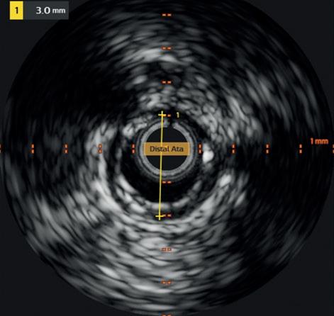
femoropopliteal disease as the precision it brings in intraluminal guidewire positioning, plaque characterisation, lesion length/vessel diameter assessment and post-endovascular therapy quality control appear most likely to move the needle in terms of long-term clinical outcome for this patient group.
References:
1. Allan RB, Puckridge PJ, Spark JI, Delaney CL. The impact of intravascular ultrasound on femoropopliteal artery endovascular interventions: a randomized controlled trial. JACC Cardiovasc Interv. 2022 Mar 14;15(5):536–546. doi: 10.1016/j. jcin.2022.01.001.
Despite lack of RCT data concerning IVUS use in infrapopliteal arteries, there is still compelling evidence supporting its value in treatment of chronic limb-threatening ischaemia (CLTI). Fujihara et al’s singlecentre retrospective comparison of wound
The additional upfront cost of the IVUS catheter is highly likely to be offset by reduced duration of follow-up care due to quicker healing and less CD-TLR.”
Corroboration of these findings outside Japan was recently provided from a US retrospective single-centre comparative analysis of 100 patients, showing significantly lower rates of binary restenosis at one year in cases with IVUS utilisation along with lower TLR.8
A subsequent analysis of 9,845 PAD patients (claudication and CLTI) from a Japanese insurance claims database confirmed that, despite increased initial outlay, IVUS use was significantly associated with reduced total cost from years one to five.9 I expect these savings to be amplified in a CLTI population, and therefore have a very low threshold to use IVUS when performing infrapopliteal endovascular therapy in Rutherford 5/6 patients; the additional upfront cost of the IVUS catheter is highly likely to be offset by reduced duration of follow-up care due to quicker healing and less CD-TLR. The lesion complexity nuance also informs my practice: I am more likely to use IVUS when treating TASC C/D
2. Allan RB, Puckridge PJ, Delaney CL. Three-year outcomes of a randomized controlled trial investigating the use of intravascular ultrasound in femoropopliteal endovascular interventions. J Endovasc Ther. 2025 Dec 18:15266028251398977. doi: 10.1177/15266028251398977.
3. Shi J, Ahn C-M, Lee S-J, et al, IVUS-DCB investigators. Twentyfour-month outcomes of intravascular ultrasound-guided drugcoated balloon angioplasty for femoropopliteal artery disease. J Am Heart Assoc. 2025 Aug 19;14(16):e041564. doi: 10.1161/ JAHA.125.041564.
4. Norgren L, Hiatt WR, Dormandy JA, et al; TASC Working Group. Inter-society consensus for the management of peripheral arterial disease (TASC II). J Vasc Surg. 2007 Jan:45 Suppl S:S5–67. doi: 10.1016/j.jvs.2006.12.037.
5. Lee S-J, Kim T-H, Lee J-H, et al, IVUS-DCB investigators. Intravascular ultrasound-guided vs. angiography-guided drug-coated balloon angioplasty in patients with complex femoropopliteal artery disease. JACC Cardiovasc Interv. 2025 Mar 10;18(5):558–569. doi: 10.1016/j.jcin.2024.10.052.
6. Fujihara M, Yazu Y, Takahara M. Intravascular ultrasoundguided interventions for below-the-knee disease in patients with chronic limb-threatening ischemia. J Endovasc Ther. 2020 Aug;27(4):565–574. doi: 10.1177/1526602820935606.
7. Soga Y, Takahara M, Ito N, et al. Clinical impact of intravascular ultrasound-guided balloon angioplasty in patients with chronic limb-threatening ischemia for isolated infrapopliteal lesion. Catheter Cardiovasc Interv. 2021 Feb 15;97(3):E376–E384. doi: 10.1002/ccd.29347.
8. Snyder DJ, Zilinyi RS, Kido T, et al. The impact of intravascular ultrasound use on one-year outcomes after infrapopliteal endovascular intervention. J Soc Cardiovasc Angiogr Interv. 2025 Mar 18;4(3Part A):102509. doi: 0.1016/j. jscai.2024.102509.
9. Soga Y, Ariyaratne TV, Secemsky E, et al. Intravascular ultrasound guidance during peripheral vascular interventions: long-term clinical outcomes and costs from the Japanese perspective. J Endovasc Ther. 2025 Jun;32(3):698–710. doi: 10.1177/15266028231182382.
Narayanan Thulasidasan is a consultant interventional radiologist at Guy’s and St Thomas’ NHS Foundation Trust in London, UK.
New Delphi consensus offers step-by-step workflow for IVUS in femoropopliteal PAD
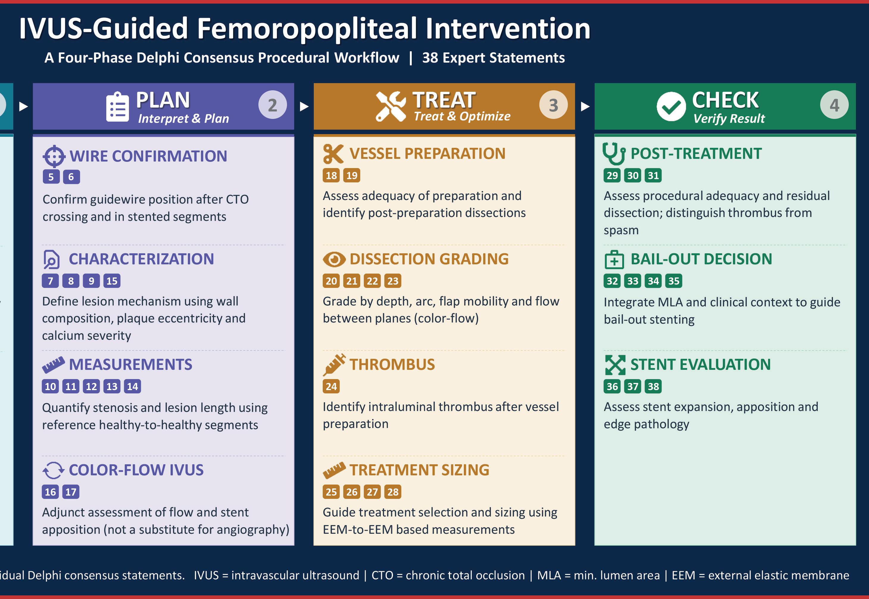
Ashish Patel and Arsalan Wafi (London, UK) speak to Vascular News about their recent Delphi consensus on intravascular ultrasound (IVUS) use in femoropopliteal peripheral arterial disease (PAD). Their message is simple: IVUS is only complicated when the workflow is unclear. The purpose of the Delphi document is to make IVUS use structured, teachable and reproducible in everyday practice.
What first made you feel a Delphi workflow for IVUS was needed?
AP: Anyone who treats femoropopliteal disease will recognise the situation. The angiogram looks acceptable at the end of the case but the patient returns early with restenosis. When those cases are revisited with IVUS, you often find problems that were simply not visible on angiography: the reference vessel was larger than expected so the device was undersized, calcium was underestimated leading to recoil, or dissections were more extensive than appreciated on angiography alone. In our own practice, once IVUS started to be used systematically rather than selectively, those patterns became very obvious.
AW: From a trainee perspective, the difficulty isn’t using the catheter. If you watch experienced operators using IVUS, each one focuses on slightly different features. Early on, what you need is a sequence: where to image, what to measure and which findings should change the procedure.
Why did you choose a Delphi consensus approach?
AP: The evidence base for IVUS in PAD is expanding and there are now multiple studies suggesting improved outcomes with IVUS use. However, what has been missing is practical guidance. The objective was to translate the available evidence and collective experience of experts into a practical workflow that operators can use in daily practice.
AW: Across centres, IVUS use varies considerably. Operators acquire images differently, measure different parameters and sometimes avoid IVUS because they are unsure how to interpret what they are seeing.
You focused on ‘how’ rather than ‘whether’ to use IVUS. Why is that distinction important?
AP: This is a consensus-derived procedural framework. It assumes the operator has already decided that IVUS has a role in their practice and asks: what does good technique
look like? IVUS itself is not complex. The variability comes from how it is used. When different operators assess different parameters at different stages of the case, inconsistency is inevitable. The purpose of the consensus was to bring structure to that process; once technique is standardised it becomes easier to teach, document and evaluate.
How did you design the consensus process?


AW: We began with a scoping review to identify what the literature says about IVUS acquisition and interpretation in PAD and where the evidence remains limited. We also surveyed clinicians who do not routinely use IVUS to understand the common misconceptions and practical barriers. These insights informed the initial consensus statements. An expert roundtable refined the statements before they were circulated internationally through a Delphi process. After the first round most statements had strong agreement but around a third required refinement. After two rounds, consensus was reached.
AP: The panel included experienced IVUS users from multiple high-volume centres internationally. That was important because the goal was to capture practical experience rather than theoretical perspectives.
In practical terms, what does the consensus offer the operator in the room?
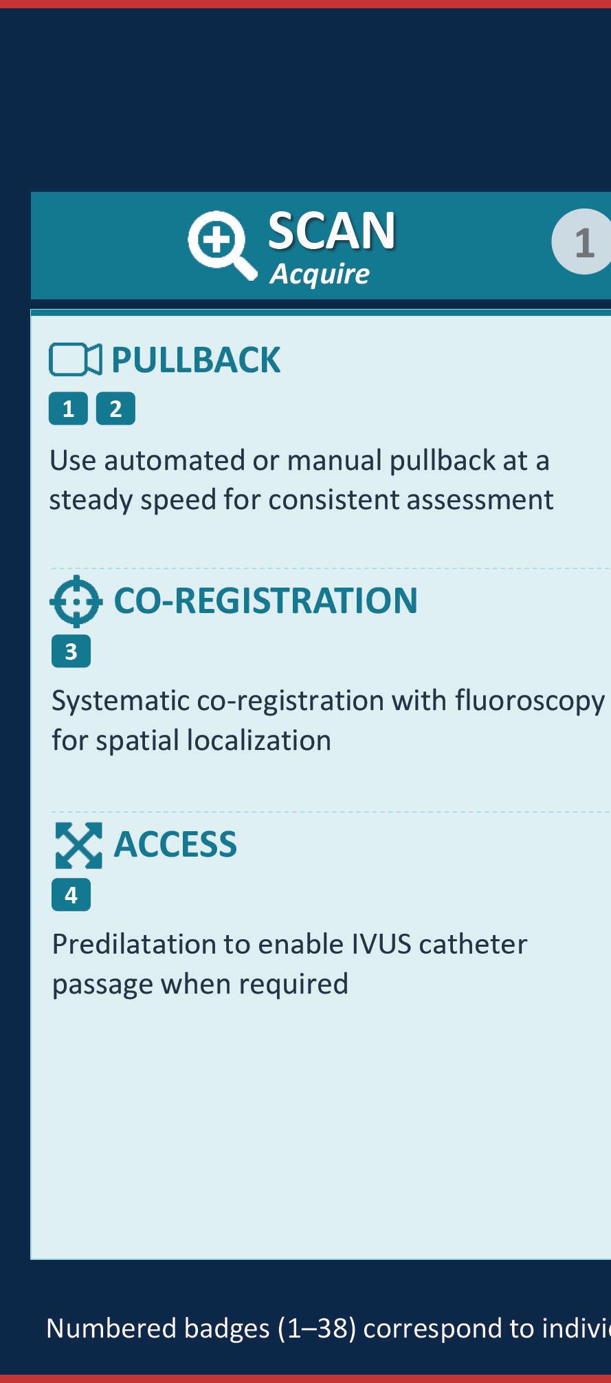
AP: A reproducible procedural framework aligned to the procedure itself: IVUS acquisition, interpretation to guide vessel preparation and definitive therapy, treatment delivery and post-treatment assessment to confirm adequacy and guide optimisation.
The interpretation component is central to the consensus, defining strategy before treatment (through vessel sizing, plaque morphology and lesion assessment) and then reassessing the result after treatment to identify underexpansion, residual disease or dissection requiring optimisation.
Cost is often raised as a concern with IVUS. How do you address that?
AP: The cost of IVUS catheters is real. The only meaningful justification is improved outcomes. If IVUS leads to better sizing, more appropriate vessel preparation and fewer early failures, the additional cost may be offset by reduced reinterventions and complications. What we have observed is that in centres familiar with the workflow, IVUS tends to move from occasional to routine use.
The literature supports improved patency and lower reintervention with IVUS guidance, although certainty varies between studies. In practice, the key is to use IVUS when it changes on-table decision making rather than as reassurance.
AW: A structured workflow also helps from a training perspective. If the operator knows exactly what information they are trying to obtain from IVUS and how it will affect the procedure the cost benefit becomes clearer. I also find that it flattens the learning curve in endovascular decision-making.
Does IVUS use slow procedures down?
AP: Initially it can add time, this is why
Ashish Patel
Arsalan Wafi
the framework matters. When the team understands when IVUS is performed, who is responsible and what needs to be measured, the additional time becomes minimal. Most of the delay reflects the learning curve rather than the technology.
AW: Improvisation slows procedures down. If you are unsure what you are looking for you repeat pullbacks and re-examine images. A clear workflow makes the process faster.
IVUS does not provide haemodynamic information. Is that a limitation?
AP: Yes, and it is important not to frame IVUS as a replacement for angiography. Angiography remains essential for assessing run off and flow. IVUS adds anatomical information: vessel diameter, plaque morphology, calcium distribution, dissections and stent expansion.
In practice, the two modalities are complementary. Where haemodynamics are uncertain, newer imaging technologies may eventually become useful adjuncts but they are not yet established in routine PAD practice.
How did your different perspectives
influence the final workflow?
AP: As someone who spends a fair amount of time teaching endovascular techniques to trainees and early consultants, the challenge is rarely the catheter itself; it is translating the information into a procedural decision. A shared workflow makes IVUS easier to teach and evaluate.
AW: For trainees the early cases benefit from clear structure. Once you follow a consistent sequence the process becomes intuitive and experience adds nuance.
What is the key message for clinicians reading the consensus?
AP: The goal is straightforward. If IVUS is used, it should be used well. A clear workflow makes that possible. The field has reached the point where IVUS is no longer just an adjunct. What has been missing is agreement on how to use it consistently.
What do you hope will happen next?
AW: Ideally the workflow becomes something teams can refer to easily during cases. The learning curve is shorter than many expect and IVUS quickly becomes a natural part of decision making.

In this novel Delphi consensus, we are brought one major step closer to standardising how we use this technology to guide our endovascular procedures. Harnessing the breadth of knowledge of a large, multinational group of experts, this document allows the routine clinician to gain a more complete understanding of how exactly peripheral IVUS should be implemented in clinical practice, which to date has been underexplored. The authors astutely recognise the need for a defined workflow in IVUS practice, one that reduces operational variability and decreases some of the barriers to new users in learning how to employ this technology in their practice. I look forward to seeing more efforts like this as IVUS technology continues to evolve and I expect an uptick in adoption as operators have a readily available playbook to utilise IVUS in PAD.
Scan code to link to corresponding video interview
Eric Secemsky
The use of IVUS in a young patient with multilevel and mixed aetiology lesions presenting with acute-on-chronic CLTI
Mark Portou (London, UK) outlines a case demonstrating the use of intravascular ultrasound (IVUS) in his clinical practice.
A 46-YEAR-OLD MAN PRESENTED to the emergency vascular clinic with sudden onset pain and numbness of the left foot. He previously experienced left-sided buttock, thigh and calf pain on exercise for several years and continued to be a >20/day smoker. For the past five days he had been unable to walk and described severe left foot pain at rest, with pallor of the forefoot.

The arterial duplex scan reported a calcified stenosis with possible dissection in the left common femoral artery (CFA) and damped triphasic flow in the superficial femoral artery (SFA), which occluded in the distal thigh with echolucent disease.
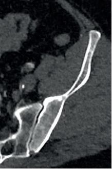
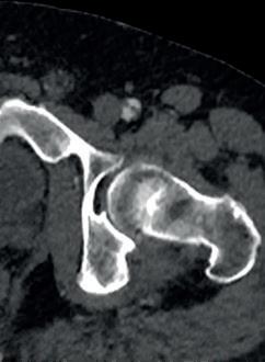
A computed tomography angiogram demonstrated a left occluded internal iliac artery with non-calcified stenosis at the external iliac artery (EIA) origin (Figure 1A). Additionally, a focal dissection of the CFA causing a significant stenosis (Figure 1B) and an occlusion of the popliteal with reconstitution of the anterior tibial was observed.
Open surgical management was considered; however, the aetiology of the acute deterioration was unclear, with several lesions of indeterminate age, including a CFA dissection. An endovascular evaluation with angiography and IVUS was preferred.
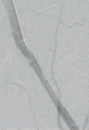
Under local anaesthesia, an ultrasound-guided contralateral retrograde puncture with an 8Fr cross-over sheath was introduced into the left common iliac artery. Angiography demonstrated an irregular EIA lesion (Figure 2A) and a dissected mid-CFA plaque (Figure 3A). A 0.018-inch wire was negotiated into the SFA, and a 6Fr 0.018 IVUS catheter was pulled back through the CFA and EIA lesions (Figures 2B and 3B).
IVUS confirmed luminal position and visualised a significant 66% stenosis of the EIA, underestimated on angiography, with a mixed but predominantly thrombotic appearance and a reference vessel diameter (RVD) of 10mm (Figure 2B). The CFA lesion was near occlusive, with a focal calcified dissection flap (Figure 3B). The RVD of the CFA was 12mm.
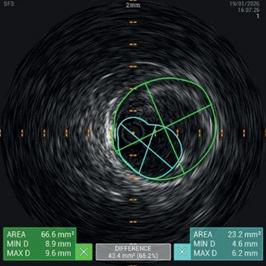
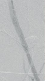
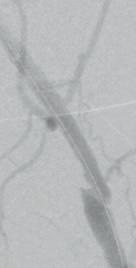
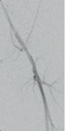
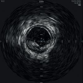
Figure 1A. Left EIA
Figure 1B. Left CFA dissection
Figure 2A: Left EIA stenosis
Figure 2B: IVUS showing 66% thrombotic stenosis
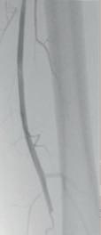
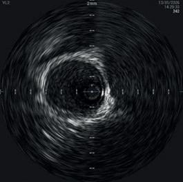
Figure 2C. Covered stent
Figure 3A. Left CFA dissection with wire
Figure 3B: IVUS in true lumen
Figure 3C: Plain balloon angioplasty
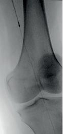
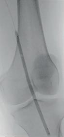
Figure 3D. Repair of dissection flap
A 10mm x 5cm Viabahn (Gore) was placed in the EIA to prevent further embolism (Figure 2C). A prolonged (fiveminute) 12mm angioplasty of the CFA was undertaken (Figure 3C). Significant lumen gain and repair of the dissection was observed on angiography and confirmed on IVUS (Figures 3D and 3E).
The popliteal occlusion was crossed without resistance, and luminal position and the presence of organised thrombus/ soft occlusive material was confirmed on IVUS (Figure 4B). Based on the IVUS sizing and lesion morphology, a 6Fr Rotarex (BD) was chosen to debulk the thrombotic material, followed by a threeminute inflation of a 5x150mm drug-coated balloon (DCB; Figures 4C and 4D). Brisk flow and restoration of three-vessel runoff was confirmed (Figures 4E and 4F). An 8Fr Angioseal (Terumo Interventional Systems) was deployed, and the patient was discharged the next day with palpable pedal pulses.
Mark Portou is a consultant vascular surgeon at the Royal Free Hospital in London, UK.
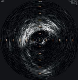
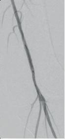
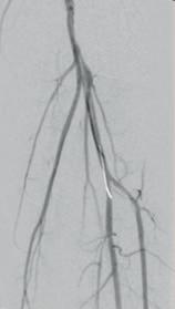
Figure 3E. Repair on IVUS
Figure 4A: Popliteal occlusion
Figure 4B: Luminal position confirmed on IVUS
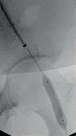
Figure 4C. Mechanical thrombectomy
Figure 4D. 1:1 sized DCB
Figure 4E: Brisk flow
Figure 4F: Three-vessel run-off
Mark Portou
From occlusion to perfusion: IVUS-guided endovascular approach to CLTI
In this case report, María Pilar Lamarca Mendoza (Toledo, Spain) highlights the beneficial impact of intravascular ultrasound (IVUS) on her chronic limb-threatening ischaemia (CLTI) practice.
Clinical case
A 73-year-old woman presented with rest pain in the left lower extremity. Her medical history was significant for obesity, diabetes mellitus, hypertension, dyslipidaemia, chronic kidney disease, and severe two-vessel coronary artery disease. On physical examination, only the femoral pulse was palpable in the affected limb.
Duplex ultrasound demonstrated diffuse fibrocalcific disease of the superficial femoral artery (SFA), along with two critical stenoses in the popliteal artery. Run-off was primarily through the anterior tibial artery (ATA), which showed a proximal occlusion with collateral reconstitution and indirect flow to the dorsalis pedis artery.
diameter measurements (Figures 1–3).
The mid-ATA stenosis was treated with a 3mm non-compliant balloon. The proximal ATA occlusion and adjacent stenosis were treated with a 4mm non-compliant balloon. Completion angiography and IVUS demonstrated an excellent angiographic result, without evidence of residual stenosis or dissection.
Imaging and decision making
Based on clinical assessment and noninvasive imaging findings, diagnostic angiography was performed. This revealed diffuse disease with short-segment subocclusions involving both the proximal and distal popliteal artery. A flush occlusion of the tibioperoneal trunk was identified. The ATA was patent distally, with a proximal occlusion reconstituting at the mid segment and a focal stenosis in the mid-ATA. The ATA provided single-vessel run-off to the foot through a patent dorsalis pedis artery (Figures 1–3).
Given these findings, the treatment strategy aimed to restore direct in-line flow to the ischaemic foot via the ATA.
Endovascular procedure
Antegrade access to the common femoral artery was obtained under ultrasound guidance. The popliteal lesions were successfully crossed; however, the proximal ATA occlusion could not be traversed via an antegrade approach. Therefore, retrograde ATA access was established, and the lesion was crossed using a 0.018-inch guidewire, with re-entry into the true lumen at the proximal ATA.
IVUS (Visions PV 0.018; Philips) was used to confirm true intraluminal recanalisation, characterise lesion morphology, and obtain accurate vessel
Based on IVUS-derived vessel measurements, a 5mm balloon was selected for the distal popliteal artery, while a 6mm non-compliant balloon was used for the proximal popliteal artery and distal SFA. Balloon inflation in the distal popliteal segment produced a characteristic ‘dog-bone’ configuration, suggesting inadequate plaque modification. This finding prompted adjunctive treatment with a 5mm AngioSculpt scoring balloon (Philips) resulting in satisfactory luminal expansion (Figure 4). Drug-coated balloons were subsequently applied to these segments.
IVUS-guided delineation of landing zones led to treatment of longer arterial segments than would have been selected based on angiography alone (Figure 1).
Final angiography demonstrated optimal luminal gain without residual stenosis or dissection, findings that were confirmed by IVUS (Figure 5). A palpable pedal pulse was restored at the end of the procedure. The patient was discharged the following day with complete resolution of symptoms.
Discussion
IVUS-guided treatment of CLTI enables precise characterisation of lesion morphology and extent, accurate vessel sizing, confirmation of intraluminal guidewire position, and detection of angiographically occult complications, including dissections. These advantages facilitate procedural optimisation and may improve technical outcomes.
In selected lesions, the use of a scoring balloon, such as AngioSculpt, can promote optimal lumen expansion while minimising the risk of flow-limiting dissections through controlled plaque modification.
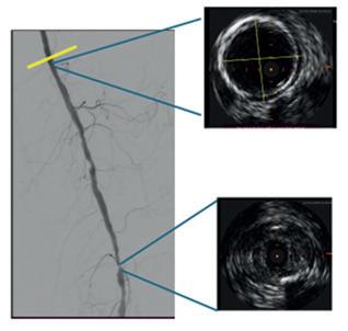
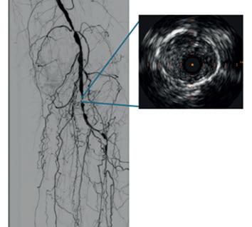
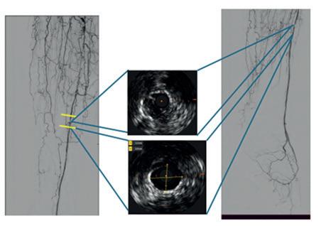
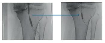
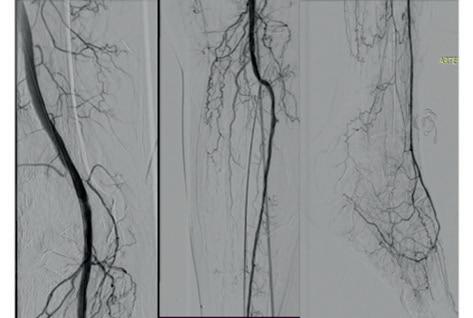
Figure 1. Baseline angiography; IVUS: vessel diameter and landing zones
Figure 2. Baseline angiography; IVUS
Figure 3. Baseline angiography; IVUS: vessel sizing and identification of landing zones
Figure 4. Dog-bone configuration and Angiosculpt treatment
Figure 5. Completion angiogram
María Pilar Lamarca Mendoza is a vascular and endovascular surgeon at the Hospital Universitario de Toledo in Toledo, Spain.
María Pilar Lamarca Mendoza
Mark Portou
Solutions for complex PAD interventions
Digital IVUS

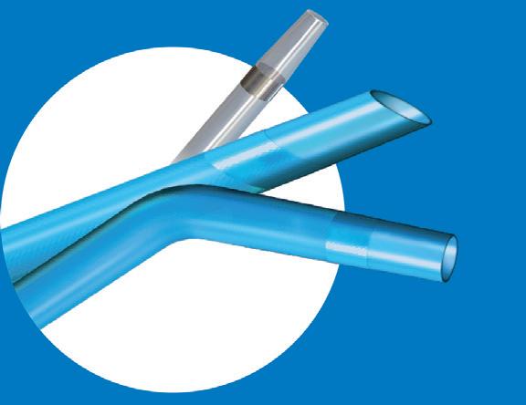
Quick-Cross Support Catheters





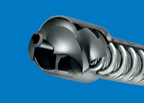






Phoenix Mechanical Atherectomy


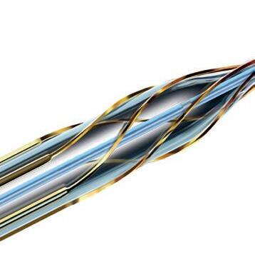
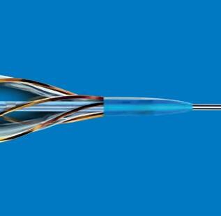
AngioSculpt PTA Scoring Balloon
Turbo-Power Laser Atherectomy