Tenecteplase, blood pressure management, tandem lesions and more
11 Flow diversion
Do the latest unruptured aneurysm guidelines truly reflect clinical practice?
16 Profile
GUILHERME DABUS SNIS president on leadership, research, and the road ahead Debate heats
Stroke thrombectomy beyond 24 hours improves functional outcome but carries increased cerebral haemorrhage and mortality risks
Mechanical thrombectomy can lead to an improvement in 90-day functional outcome compared to standard medical care in anteriorcirculation large vessel occlusion (LVO) stroke patients presenting 24–72 hours after symptom onset, as per the findings of the LATE-MT randomised controlled trial (RCT). However, within the trial, thrombectomy was also associated with increased rates of mortality, symptomatic intracranial haemorrhage (ICH) and other clinically relevant adverse events.
THESE DATA WERE PRESENTED FOR THE FIRST time at the 2026 European Stroke Organisation Conference (ESOC; 6–8 May, Maastricht, Netherlands) by co-principal investigator Jianmin Liu (Naval Medical University, Shanghai, China) alongside several other leading LATE-MT investigators.
interval [CI], 0.39–0.84; p=0.0046) and an adjusted OR of 0.47 (95% CI, 0.32–0.7; p=0.002). Comparisons between 90-day rates of mRS 3–5 versus mRS 0–2, and mRS 3–6 versus mRS 0–2, also indicated that thrombectomy produced better outcome relative to standard medical care. Additionally, significant improvements were seen with thrombectomy versus standard medical care regarding utility-weighted mRS, Barthel Index and health-related quality-of-life scores at 90 days, while follow-up infarct volumes were comparable between the two groups at seven days.
However, primary safety endpoint analyses in LATE-MT found statistically significant increases for both any ICH (28.3% vs 5.5%, respectively) and symptomatic ICH (4.8% vs 0%, respectively) in the intervention group compared to the control group. Severe adverse events also appeared to be more frequent with thrombectomy (22%) versus standard medical care (13.1%), as did rates of adverse events of special interest (15.5% vs 8.9%, respectively), which included procedure-related complications, neurological deterioration, brain oedema, target vessel re-occlusion, pneumonia and recurrent stroke. Lastly, the investigators were “particularly concerned” to see that the mortality rate was greater with thrombectomy (6%) versus standard medical care (1.8%), according to Liu.
Liu began by noting that thrombectomy’s benefits within earlier time windows have been well demonstrated—initially via multiple RCTs that established its clinical efficacy and safety within six hours of symptom onset back in 2015, before results from the DAWN and DEFUSE-3 trials extended this window to 6–24 hours in 2018. It has subsequently been posited that ‘slow progressors’ may also derive benefits from thrombectomy treatments, with observational studies indicating potential improvements in functional outcome in those undergoing the procedure beyond 24 hours.
With this in mind, the prospective, multicentre LATE-MT trial randomised 341 acute ischaemic stroke patients presenting 24–72 hours from time last known well to receive either thrombectomy plus standard medical care (intervention group), or standard medical care alone (control group).
As per the RCT’s primary efficacy endpoint, thrombectomy demonstrated a statistically significant improvement in functional outcome, with an ordinal shift in 90-day modified Rankin scale (mRS) scores favouring the intervention group. Analyses of these data revealed a common odds ratio (OR) of 0.57 (95% confidence
Editor-in-chief: Prof Philip M Meyers
Publisher: Stephen Greenhalgh
Editor: Jamie Bell jamie@bibamedical.com
Editorial contribution: Jocelyn Hudson, Will Date and Éva Malpass
The investigators relayed that further exploratory analyses generally revealed consistent findings across several prespecified patient subgroups—although the benefits of thrombectomy appeared to be greater in patients with internal carotid artery (ICA) occlusions versus other locations.
Based on these results, Liu concluded that “appropriate targeting” of mechanical thrombectomy can be considered effective within this especially late time window, with LATE-MT co-principal investigator Craig Anderson (The George Institute, Sydney, Australia) adding that these data signify a “trade-off” between preventing disability and introducing a “small risk” of further complications in patients presenting at 24–72 hours.
Cover credit: Images by Adobe Stock Creative, concept and photomontage by Terry Hawes
Read it your way
We want to ensure you still receive your copy of NeuroNews the way that works best for you.
To reconfirm your subscription, scan the QR code. You can also let us know if you would like to change the way you read it by opting to receive a digital instead of a printed version or by moving from digital to print
Intra-arterial TNK fails to improve reperfusion after incomplete thrombectomy
Findings from the TECNO trial were presented for the first time at the 2026 European Stroke Organisation Conference (ESOC; 6–8 May, Maastricht, Netherlands), with results indicating that intra-arterial tenecteplase (TNK) did not significantly improve early or late reperfusion rates compared to best medical treatment in stroke patients with incomplete reperfusion following mechanical thrombectomy. However, TNK administration was found to be safe regarding intracranial bleeding.
ACCORDING TO RESEARCHERS, THESE results highlight the need for further refinement of patient selection and treatment protocols in this setting to tackle the “common and clinically important challenge” of incomplete reperfusion after a thrombectomy.
TECNO was a multicentre, randomised, open-label, blinded-endpoint trial conducted across multiple high-volume stroke centres in Europe. It was designed to test whether a 3mg dose of direct intra-arterial TNK in addition to best medical treatment improves reperfusion in patients with incomplete post-thrombectomy reperfusion.
group, confirming an acceptable safety profile with respect to intracranial bleeding.
The trial’s primary efficacy endpoints were early and late reperfusion defined by reperfusion improvement on angiography imaging 25 minutes after randomisation, and complete reperfusion on 24-hour magnetic resonance (MR)/ computed tomography (CT) perfusion imaging, respectively.
On primary analyses, improved early reperfusion was observed in 39% of patients randomised to TNK versus 36% in the control group, representing a difference that was not statistically significant (adjusted risk difference, 3.4%). The between-group difference in late reperfusion was also non-significant, being observed in 51% of TNK patients and 40% of control patients (adjusted risk difference, 15.4%).
Regarding safety, symptomatic intracranial haemorrhage (ICH) occurred in 5.2% of patients in the TNK group compared with 8.6% in the control
Reperfusion-guided BP management improves post-thrombectomy functional outcomes
A SYSTOLIC BLOOD PRESSURE (BP) management strategy tailored to the degree of reperfusion after successful mechanical thrombectomy can improve 90-day functional outcomes in acute ischaemic stroke, according to HOPE trial results presented at ESOC 2026. HOPE was conducted across 11 stroke centres in Spain, enrolling patients within 24 hours of symptom onset who achieved modified thrombolysis
At 90 days, functional independence—as per a modified Rankin scale (mRS) score of 0–2—was achieved by 27% of TNK patients versus 38% of control patients. On this detail, however, the investigators emphasise that the trial was designed and powered to evaluate reperfusion as its primary endpoint, and that definitive conclusions regarding clinical outcomes should not be drawn solely from these data.
“Taken alone, these findings do not support the routine administration of intra-arterial TNK as a pharmacological adjunct in cases of incomplete reperfusion,” commented TECNO principal investigator Johannes Kaesmacher (Bern University Hospital, Bern, Switzerland), who presented these late-breaking data at ESOC 2026.
“Taken alone, these findings do not support the routine administration of intraarterial tenecteplase as a pharmacological adjunct in cases of incomplete reperfusion”
in cerebral infarction (mTICI) ≥2b post-thrombectomy reperfusion. A total of 440 patients were randomised to either individualised BP management—140–160mmHg for those with mTICI 2b reperfusion and 100–140mmHg for mTICI 2c–3— or guideline-recommended BP management.
Some 60% of patients in the intervention group achieved a 90-day modified Rankin scale (mRS) score of 0–2 compared to 46.7% in the control group (odds ratio, 1.71). Haemorrhagic transformation occurred at rates of 22.3% versus 31.6%, respectively, and no mortality difference was observed, with both study arms seeing a rate of 15%. Further analyses revealed no significant heterogeneity in outcomes between different patient subgroups.
Carotid stenting during thrombectomy demonstrates safety in tandem lesions
WHILE CAROTID ARTERY STENTING (CAS) during mechanical thrombectomy appears to be safe, whether or not the approach is non-inferior to deferred management remains to be seen, as per late-breaking results from the CASES trial presented at ESOC 2026.
CASES recruited patients with ischaemic stroke due to a tandem lesion who were suitable thrombectomy candidates at 26 centres across the Netherlands and Belgium. Ultimately, 597 patients were randomised to CAS at the time of thrombectomy, or to deferred treatment, which included carotid endarterectomy, delayed CAS or best medical management. The primary endpoint was modified Rankin scale (mRS) shift, while safety outcomes included symptomatic intracranial haemorrhage (ICH) and mortality— all assessed within 90 days.
Presenting the trial’s full analysis, leading investigator Maarten Uyttenboogaart (University Medical Center Groningen, Groningen, Netherlands) noted that the non-inferiority criterion was not met (adjusted common odds ratio [acOR], 1.06). However, in the per-protocol set, immediate CAS proved non-inferior to deferred treatment (acOR, 1.1). Finally, safety analyses showed rates of symptomatic ICH were 2.1% with CAS versus 3.8% with deferred treatment (OR, 0.55), while respective mortality rates were 21% and 25% (acOR, 0.79).
Flat-detector CT shows high accuracy in detecting intracranial haemorrhage
NEW FINDINGS FROM THE SPINNERS TRIAL demonstrate that—despite failing to achieve non-inferiority versus standard computed tomography (CT), as per its primary endpoint—the Syngo DynaCT Sine Spin (Siemens Healthineers) non-contrast flat-detector CT (FDCT) system provides high diagnostic accuracy for intracranial haemorrhage (ICH) detection in patients with suspected acute stroke.
SPINNERS prospectively enrolled 252 patients across multiple centres in Europe and the USA. Patients with suspected ischaemic or haemorrhagic stroke underwent both FDCT and multidetector CT (MDCT) in a tightly defined time window, with MDCT serving as the reference standard. While non-inferiority versus MDCT was not met in the overall study population, FDCT demonstrated a strong diagnostic performance, achieving 92.6% sensitivity and 93.7% specificity in detecting ICH. Subgroup analyses showed particularly positive results in clinically relevant populations, with sensitivity exceeding 96% in patients with more severe neurological deficits and those with intraparenchymal haemorrhage. Diagnostic accuracy also surpassed 95% in experienced centres.
“Our findings show that FDCT performs particularly well in patients with severe stroke and in experienced centres, supporting its role in streamlined imaging pathways,” said Marios Psychogios (University Hospital Basel, Basel, Switzerland), who shared these data at ESOC 2026.
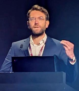
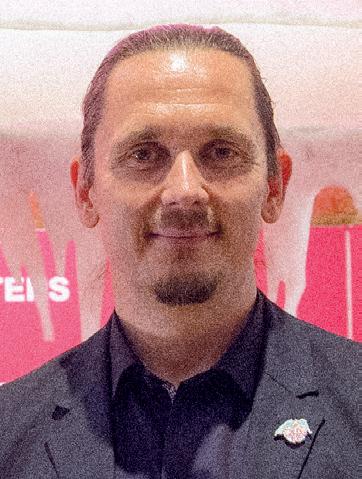
Johannes Kaesmacher
SUCCESS study underscores the safety and value of temporary neck bridging to reduce compromise in aneurysm care
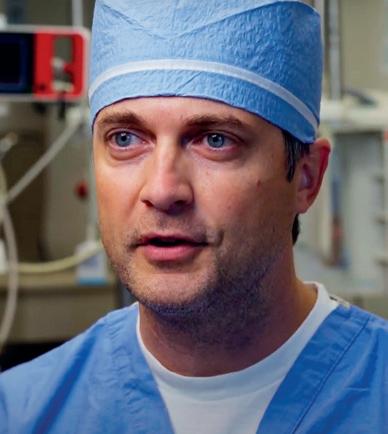
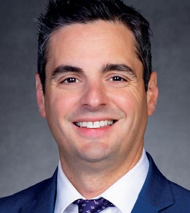
With data from the prospective SUCCESS study having demonstrated the safety and effectiveness of temporary assistance in coil-embolisation procedures aided by the Comaneci device (Rapid Medical), leading investigators Jason Davies (State University of New York, Buffalo, USA) and Ricardo Hanel (Baptist Medical Center, Jacksonville, USA) sit down with NeuroNews to discuss these results, and the wider role for temporary neck bridging in intracranial aneurysm treatments.
THE TREATMENT OF WIDE-NECK ANEURysms requires operators to balance effective coil retention with the limitations and risks of adjunctive techniques—particularly in ruptured cases.
“When you have a wide-neck aneurysm, you need to keep the coils in place, ideally without a permanent implant—especially in ruptured cases, where committing a patient to upfront DAPT [dual antiplatelet therapy], as you would need to with a stent, just complicates everything downstream,” Davies explains. “The other unmet need is […] packing in the coils without flow arrest, as that can cause a number of issues in terms of thromboembolic complications and also compliance in patients who are awake during the procedure.”
As a temporary implant, Comaneci is intended to address these competing demands, providing a scaffold to support coil packing while maintaining blood flow and removing the need to commit to upfront DAPT and its associated risks. Findings from the US Food and Drug Administration (FDA) post-market SUCCESS study—first presented at the 2024 Society of NeuroInterventional Surgery (SNIS) annual meeting (22–26 July, Colorado Springs, USA)—indicate that these benefits extend beyond theory and translate directly into clinical practice.
“Excellent” clinical outcomes
Across 90 consecutive wide-neck aneurysm patients enrolled at 17 US centres, the SUCCESS investigators report immediate occlusion (Raymond-Roy I/II) in 85.6% of cases, increasing to 94.7% at six-month follow-up. Additionally, only one patient (1.1%) required retreatment.
Davies and Hanel agree that these findings speak to the quality of the coiling achieved in the study, with Comaneci-assisted procedural success ultimately reducing the likelihood of recurrence. In Hanel’s view, these “excellent” six-month data are “better than expected”, with the low retreatment rate serving as testimony that coiling “remains a viable option”.
“Even if you just look at Raymond-Roy I at day one [79%] and latest follow-up [89%], those
results should be very reassuring for operators,” he adds. “This also shows how much coiling technology has improved and that a good coiling procedure—especially when you have an assistive device to allow better coil compaction— can achieve very good results.”
The study also demonstrated a minimal thromboembolic complication rate of 5.5%, with no symptomatic events. According to Davies, these figures compare very favourably to the literature on stentand balloon-assisted coiling. He attributes this to the “biological rationale” by which blood flow is maintained during deployment of the Comaneci device.
“What that suggests is that we’re able to pack enough to really establish a condition of stasis within the aneurysm dome,” Davies adds. “You’re getting thrombosis, as opposed to compaction, over time— which is what we always hope for but is difficult to achieve with low packing density.”
The key to success?
In Hanel’s view, a key strength of SUCCESS is its inclusive design. While focused on wide-neck aneurysms (neck diameter, 4–10mm; dome-toneck ratio, <2), he and the other leading investigators kept the inclusion criteria “as broad as possible”, enrolling patients with both sidewall and bifurcation aneurysms as well as ruptured and unruptured cases alike.
And—while its single-arm design limits direct comparison with other techniques—both Davies and Hanel highlight the value held by SUCCESS, with the latter noting that Comaneci-assisted coiling appears to be “as safe as balloon-assisted coiling, and safer than stent-assisted coiling”, based on existing literature.
“These results are robust in that we had independently reviewed core-lab analysis and adjudication from a DSMB [data safety monitoring board], which reassures us that the results are genuine,” Davies adds.
Real-world implications
For Davies, the significance of these findings ultimately lies in their practical impact on one of the most challenging areas in neurointervention.
As a firsthand user of Comaneci, he notes how well the device conforms to the neck of an aneurysm, contributing to the packing density that can be achieved. Another noteworthy trait is that it facilitates having one or multiple backup plans—in any endovascular aneurysm procedure, but, in particular, when faced with more complex cases and tortuous anatomies.
Clinical outcomes from SUCCESS further reinforce Comaneci’s “very strong safety profile”, Davies says. Not only did modified Rankin scale (mRS) scores of 0–2 improve from 73% at baseline to 86% at six months, but ruptured aneurysm patients showed gains in mRS 0–2 from 46.8% at baseline to 73.3% at three months too. Davies calls this a “standout finding” in the setting of acute aneurysm treatment, confirming that upfront heparin administration to reduce thromboembolic risk did not lead to an uptick in adverse events.
“The part of the study that really gave me comfort was that ruptured cohort, because we didn’t see an increase in haemorrhagic complications,” he comments. “Comaneci now plays a more primary role in my ruptured cases.”
“It’s a really nice option to have and, as a result, we’ve ended up doing a lot less stent-assisted coiling because we aren’t relying on it up front; we know it’s there as a bailout strategy, but only in the cases that absolutely need it,” Davies continues.
And, providing a concluding message on what makes these results with Comaneci so pertinent to clinical practice, Davies says: “Wide-neck aneurysms are the hardest thing we’re treating right now, especially in ruptured cases, and having something temporary that obviates the need for DAPT is very useful. We recently published a paper on ‘nuisance bleeding’ following DAPT—something I think is more common than many clinicians outwardly acknowledge. So, having something temporary that doesn’t commit patients to DAPT unless they actually need it represents a tremendous tool in our armamentarium.”
These latest findings also bolster existing evidence on the Comaneci device’s ability to “empower” strong outcomes with the longstanding approach of coil embolisation, according to Hanel.
“For me, SUCCESS shows that Comaneci-assisted coiling is a very safe and effective realworld treatment for wide-neck intracranial aneurysms,” he comments. “It’s very exciting to have this tool available—and I think there’s definitely a role for temporary bridging in wideneck aneurysm treatment.”
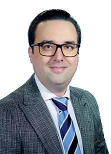
Jason Davies
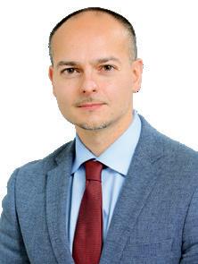
Ricardo Hanel
CX 2026: Carotid trial debate highlights “key
question” of cost-effectiveness
An audience poll asking, ‘What do we believe: ECST-2 or CREST-2?’ following a debate at the 2026 Charing Cross (CX) Symposium (21–23 April, London, UK) revealed a closer-thanexpected result, with attendees voting 60-40 in favour of the latter trial. Subsequent discussion highlighted the pressing need for cost-effectiveness analyses.
While ECST-2 supports treating patients with asymptomatic and low- or intermediate-risk symptomatic carotid stenosis with optimal medical therapy alone, CREST-2 data presented for the first time in 2025 showed that intensive medical therapy (IMT) plus revascularisation with stenting is superior to IMT alone and IMT plus revascularisation with surgery is non-inferior to IMT alone.
“I would suggest to you that we are completely biased, and we need to think about patient preference,” said Alun Davies (Imperial College London, London, UK) in the closing remarks of his argument for ECST-2.
He concluded that it is “probably inappropriate to offer carotid revascularisation to any patient and they should all probably be treated with best medical therapy, other than one or two exceptions”.
William Gray (Main Line Health, Wynnewood, USA), on the other hand, argued that “CREST-2 conclusions are the only believable ones”. ECST2, he asserted, was an “ill conceived, poorly conducted, borderline unethical and incomplete trial”, thus rendering its published conclusions moot.
Richard Bulbulia (University of Oxford, Oxford, UK) then took stock of the debate from the auditorium floor, echoing CREST-2’s win in the audience poll.
“The important question these contemporary carotid trials sought to answer was, what is the
additional benefit of carotid intervention given 21st-century medical therapy? And the answer is clear in CREST-2,” he said.
Bulbulia continued that, considering the field of carotid intervention more widely, “some patients are best treated with medical therapy alone; there’s carotid stenting, there’s carotid surgery, and there’s TCAR [transcarotid artery revascularisation], and really it’s a multidisciplinary team working to decide what’s the best treatment for the right patient.”
“I would suggest to you that we are completely biased, and we need to think about patient preference”
Alun Davies
Davies then raised the “key question” of cost-effectiveness, which he noted “has not been really addressed properly by either study”. On this note, Bulbulia cautioned against seeing intervention as inherently more costly.
“There’s the cost of intervention, but there’s also the cost of strokes that you’re failing to prevent,” he stressed, highlighting the need for a cost-effectiveness analysis of the latest data.
“Incredible” C-GUARDIANS II results point to safety and efficacy of carotid stent
New findings from the C-GUARDIANS II study indicate that the CGuard Prime carotid stent system (InspireMD) achieves high procedural success, durable early luminal restoration, and “excellent” periprocedural safety outcomes, in patients undergoing transcarotid artery revascularisation (TCAR). This is according to data from the first 36 patients enrolled in the study, which were presented for the first time during the carotid and acute stroke session at the 2026 Charing Cross (CX) Symposium (21–23 April, London, UK).
THE STUDY PRESENTER, PATRICK MUCK (Good Samaritan Hospital, Cincinnati, USA), explained that C-GUARDIANS II is a multicentre, single-arm pivotal study designed to evaluate the safety and efficacy of the CGuard Prime (80cm) used in conjunction with the US Food and Drug Administration (FDA)-cleared Enroute transcarotid neuroprotection system (NPS; Boston Scientific) in patients undergoing carotid artery stenting (CAS) via the TCAR approach.
“As a programme director, you talk to your trainees for years about open- and closed-cell stents. Well, this stent is actually both,” Muck said. Muck also put the C-GUARDIANS II trial in the context of other studies from InspireMD, stating: “[Christopher] Metzger already has led the C-GUARDIANS I trial, which led to the FDA approval of the 135cm CGuard Prime stent, and [Patrick] Geraghty and myself are honoured to be able to lead the C-GUARDIANS II trial. A little bit later— probably Q2, perhaps Q3—we’re going to start C-GUARDIANS III, which will include InspireMD’s proprietary SwitchGuard NPS system for TCAR.”
The C-GUARDIANS II study is being conducted across 11 investigational sites in the USA, with target enrolment of 50 patients now complete. Muck commented that the patients enrolled in the study represent a high-surgical-risk group, with ≥25% being symptomatic. In terms of patient demographics, Muck detailed that these were “pretty typical” of the population in question, adding that 30% had prior history of stroke and 11% had prior transient ischaemic attack (TIA).
Sharing podium-first results, Muck relayed a 100% acute device success rate and 97.2% technical success rate in the first 36 patients included in the study. Moving on to secondary clinical endpoints, he reported no death, stroke or myocardial infarction within 30 days of procedure, as well as no stent thrombosis or device-related serious adverse events (SAEs).
In his concluding statements, Muck remarked on the “incredible results” he has seen so far, summarising that primary and secondary procedural endpoints were met, and reiterating the 100% acute device and procedural success as well as >97% technical and treatment success.
The presenter added that all 30-day clinical safety endpoints were achieved, with no deaths, strokes, myocardial infarctions, stent thromboses, or SAEs. Muck also noted that duplex ultrasound confirmed 100% stent patency at 30 days, with no evidence of restenosis or thrombosis, and that the device performed reliably across diverse anatomical and lesion characteristics with significant baseline stenosis.
Richard Bulbulia
OUTSIDE THE OPERATING ROOM
Consensus statement seeks to standardise evaluation of robotic systems in stroke thrombectomy
Researchers have established the first international consensus on how to design, test and evaluate robotic systems for stroke treatment via a new position statement published in the Journal of the American Heart Association (JAHA) that is intended to define standards that put patient safety first.
AS PER A MEDIA RELEASE FROM King’s College London (London, UK)— one of the leading institutions that helped to develop the consensus statement—no such agreed framework existed until now regarding robotic systems for mechanical thrombectomy. And, while robotics hold the potential to expand access to thrombectomy by enabling stroke specialists to perform these procedures remotely, progress has been hampered by a lack of standardisation across studies.
“In 2023, we carried out a systematic review of endovascular robotics and autonomy,” said Harry Robertshaw (King’s College London, London, UK), first author of the consensus statement. “We found that, although several studies existed, they were all testing different tasks, using different models and measuring different outcomes. This made it impossible to compare results or identify the most effective approaches.”
To address this, the team brought together an international group of experts in interventional neuroradiology, robotics, data science, health economics, policy, statistics and patient advocacy to establish consensus frameworks for developing and validating robotic mechanical thrombectomy. The resulting position statement sets out how these systems should be tested, and how their effectiveness should be measured and reported.
“By bringing together experts from clinical practice, academia, industry, and patient representatives, we have defined the first consensus standards for robotics and AI [artificial intelligence] in thrombectomy navigation,” Robertshaw added. “This will help to move the technology forward as its integration with clinical practice moves closer to reality.”
According to the researchers, patient perspectives—including input from patient representatives and organisations like the UK Stroke Association—were key to retaining the centrality of safety, real-world impact and patient benefit.
The researchers hope their position statement will drive more comparable studies, accelerating progress across the field. One requisite patient safety task outlined by the Stakeholder Taskforce for AI-assisted Robotic Thrombectomy (START) in the statement is the present need to correlate in-vitro measurements to in-vivo complications.
“While AI-assisted robotic mechanical thrombectomy is not yet ready for routine clinical use, the speed of innovation in both robotics and AI means transformative advances are likely to be just around the corner,” commented senior author Thomas Booth (King’s College London, London, UK). “Our work lays the foundation for that future by defining clear standards to ensure that global development and validation are well understood.
“By establishing practical, consensus-driven recommendations, we are helping to ensure that—when these technologies do reach patients—they do so safely, responsibly and with meaningful clinical benefit.”
“This will help to move the technology forward as its integration with clinical practice moves closer to reality”
Harry Robertshaw
Medical
societies call for new standard in radiation protection
NINE MEDICAL SOCIETIES ARE calling for updated safety standards in fluoroscopy laboratories, where clinicians face radiation exposure and orthopaedic injuries from heavy protective equipment. A report published simultaneously in multiple journals details the health, financial and workforce impacts of fluoroscopy-guided settings, and proposes an enhanced safety framework dubbed, ‘As Low and As Light as Reasonably Achievable’ (ALARA+), building on the original ALARA paradigm.
The report addresses the dual occupational hazards linked to fluoroscopic procedures—radiation exposure and orthopaedic strain from traditional protective equipment—and aims to ensure that safety is built into the environment, equipment, and standard of care.
This new ALARA+ principle has been endorsed by the Society of NeuroInterventional Surgery (SNIS) and the Society of Vascular and Interventional Neurology (SVIN), as well as several other medical societies spanning radiology, cardiology, electrophysiology and vascular surgery.
US FDA issues warning against suppressing unfavourable trial results
THE US FOOD AND DRUG ADMINistration (FDA) has reminded more than 2,200 medical product companies and researchers of requirements for them to submit certain information on clinical trial results to ClinicalTrials.gov.
of studies subject to mandatory reporting requirements have not submitted results information
As stated in a press release from the US FDA, companies and researchers often fail to disclose negative trial results, resulting in significant gaps in the public record and a publication bias that “obscures the true landscape of drug development outcomes” by “overrepresenting successes and underrepresenting failures”. This gap can also create a “distorted perception” of the safety and efficacy of medical products, the US FDA notes.
According to an internal analysis conducted by the regulator, 29.6% of studies that are “highly likely” to fall under mandatory reporting requirements have no results information submitted to ClinicalTrials.gov. Studies subject to the mandatory reporting requirements include interventional studies with a US nexus and an FDA-regulated product that are past the deadline to report. However, phase-one and device-feasibility studies are excluded.
“Those sponsoring clinical trials have an ethical obligation to make results public, regardless of the data’s influence on the company’s share price,” said US FDA commissioner Marty Makary.
Debate heats up following contrasting trial results on brain cooling during stroke thrombectomy
Two randomised trials presented at the 2026 European Stroke Organisation Conference (ESOC; 6–8 May, Maastricht, Netherlands) have generated new evidence on intra-arterial cooling during mechanical thrombectomy procedures in large vessel occlusion (LVO) acute ischaemic stroke, with one producing data favouring this approach and the other demonstrating safety but suggesting more neutral efficacy findings.
“CHILL-ART AND FOCUS SUGGEST THAT intra-arterial cooling remains a promising neuroprotective adjunct to thrombectomy,” ESO guideline board co-chair Diana Aguiar de Sousa (University of Lisbon, Lisbon, Portugal) told NeuroNews. “While CHILL-ART provides an encouraging efficacy signal, FOCUS is more reassuring on safety than on functional benefit. Taken together, these studies support further investigation—ideally through larger, standardised, preferably international trials designed to clarify protocol, patient selection and the consistency of the treatment effect.”
“The divergent results of the CHILL-ART and FOCUS trials present a classic dilemma: weighing conflicting data from similar protocols before publication,” added World Stroke Organization (WSO) vice president Gustavo Saposnik (University of Toronto, Toronto, Canada), also speaking with NeuroNews in light of these firsttime presentations. “While CHILL-ART suggests a robust functional benefit, FOCUS tempers this optimism, showing no functional gain despite a safety signal in reduced intracranial haemorrhage [ICH]. This inconsistency highlights the uncertainty inherent to neurovascular research, echoing the turbulent history of [mechanical thrombectomy] prior to the 2015 pivotal trials. We must await full publication to parse technical or population differences. Currently, selective intra-arterial hypothermia remains a promise, not a proven standard. These results are hypothesis-generating; more definitive, large-scale data are required before routine integration into thrombectomy workflows.”
Positive efficacy data
CHILL-ART—the first of these multicentre randomised controlled trials (RCTs) to be shared during a late-breaking session at ESOC 2026—demonstrated that delivering targeted brain cooling during thrombectomy significantly improves recovery outcomes, as a meaningful increase in 90-day functional independence was observed in patients receiving adjunctive intra-arterial selective hypothermia versus those receiving standard thrombectomy.
The CHILL-ART RCT enrolled 262 anterior-circulation LVO patients across 26 comprehensive stroke centres in China. Participants aged 18–85 years and treated within 24 hours of stroke onset were randomly assigned to receive either thrombectomy plus intra-arterial infusion of cold saline in the hypothermia group, or thrombectomy with room-temperature saline in the control group.
The trial’s primary endpoint of functional independence (modified Rankin scale [mRS] score 0–2) at 90 days was achieved in 54.7% of patients in the hypothermia group compared to 39.8% in the control group, with an adjusted risk ratio of 1.36 (95% confidence interval [CI], 1.05–1.76; p=0.018). Investigators reported at ESOC 2026 that this equated to a number needed to treat (NNT) of seven. And, “importantly”, in the researchers’ view, safety outcomes were comparable between the hypothermia and control groups, with no significant increase in symptomatic ICH (7% vs 9%, respectively) nor 90-day mortality (13.3% vs 18%, respectively).
Subsequent sensitivity analyses also replicated these findings, indicating the robustness of the trial’s results, and outcomes were shown to be consistent across all study subgroups as well.
“Even when we successfully remove the clot, many patients do not regain independence because of ongoing brain injury after blood flow is restored,” said CHILL-ART principal investigator Zhi-Xin Huang (Southern Medical University, Guangzhou, China), who presented these data alongside Raul Nogueira (University of Pittsburgh Medical Center, Pittsburgh, USA).
According to the investigators, the intervention evaluated in this trial uses standard thrombectomy equipment and refrigerated saline, making it readily scalable in routine clinical practice without requiring specialised devices or additional training.
The researchers also note that, more broadly, CHILL-ART addresses a “critical unmet need” in stroke care: improving outcomes after technically successful clot removal.
By combining reperfusion with targeted neuroprotection, the study introduces a “promising new paradigm” in acute stroke treatment, and its findings may inform future clinical guidelines and support broader adoption of intra-arterial hypothermia as an accessible, cost-effective strategy to reduce disability after stroke, the investigators further posit.
Safe yet neutral outcomes
The FOCUS trial—also investigating selective intra-arterial cooling as an adjunct to thrombectomy for LVO acute ischaemic stroke, and also presented at ESOC 2026—found that, while the cooling technique did not improve functional recovery, it did significantly reduce the risk of any ICH. According to the study’s investigators, these findings therefore provide important insights into the potential role of targeted brain cooling in stroke treatment.
FOCUS was an RCT conducted across 12 hospitals in China that enrolled 258 patients with anterior-circulation LVO stroke who presented within 24 hours of symptom onset. Patients were randomly assigned to receive either selective intra-arterial cooling plus thrombectomy or standard thrombectomy treatment alone.
The trial’s results showed no significant difference in functional outcomes at 90 days between the two groups, with an adjusted common odds ratio of 1.16 (95% CI, 0.75–1.79; p=0.51). However, the cooling technique demonstrated notable safety benefits, reducing the incidence of any ICH at 24 hours compared to standard treatment (adjusted risk difference, -0.174; 95% CI, -0.288 to -0.059; p=0.003). Additionally, there was no difference in the occurrence of symptomatic ICH or mortality between groups.
“This inconsistency highlights the uncertainty inherent to neurovascular research, echoing the turbulent history of [mechanical thrombectomy] prior to the 2015 pivotal trials” Gustavo Saposnik
“Our findings show that targeted cooling delivered directly into the brain at the moment of reperfusion can meaningfully improve recovery without adding risk.”
The FOCUS investigators note that, while hypothermia has long been studied as a potential neuroprotective therapy for stroke based on its ability to reduce brain metabolism and limit secondary injury, its potential benefits require further study at this stage. “These findings validate the feasibility of selective intra-arterial cooling as an adjunctive therapy during endovascular thrombectomy,” commented leading study author Shen Li (Capital Medical University, Beijing, China), who presented these data at ESOC 2026. “The marked decrease in any ICH indicates a potential protective effect on the blood-brain barrier and microvasculature, which may translate into clinical benefits. Although we did not observe a functional improvement in this trial, it paves the way for future studies with larger sample sizes or refined patient selection to fully unlock the neuroprotective potential of hypothermia.”
18th Congress 2026
2 - 4 September 2026
Palais du Pharo, Marseille, France
Emotion, evidence and expertise: European recommendations spark debate over flow diversion’s nuanced role in unruptured aneurysms
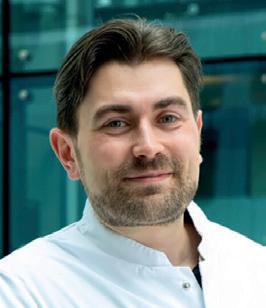
Following the recent publication of European meta-analysis findings on the outcomes and overall body of evidence associated with flow diverters, NeuroNews speaks to multiple physicians from the worlds of interventional neuroradiology and neurosurgery to discuss the much-debated role these devices play in unruptured aneurysm care.
The treatment of unruptured intracranial aneurysms is a contentious topic for several reasons. The very nature of these aneurysms as non-acute— and their interventional treatment therefore being elective—continues to stimulate debate over conservative versus more proactive management strategies. And, even in instances where the latter is favoured, the various merits and pitfalls of the specific techniques available to interventionists are hotly contested too.
Despite open surgical clipping being the longest-standing procedure used to treat brain aneurysms, the turn of the last century saw coil embolisation emerge as a valid, minimally invasive option based on the findings of the ISAT randomised controlled trial—a seminal study that also paved the way for subsequent, alternative endovascular approaches. One such approach involves implanting a flow-diverting stent into a patient’s neurovasculature to channel blood flow away from and ultimately occlude their aneurysm.
The first of these flow diverters to secure major
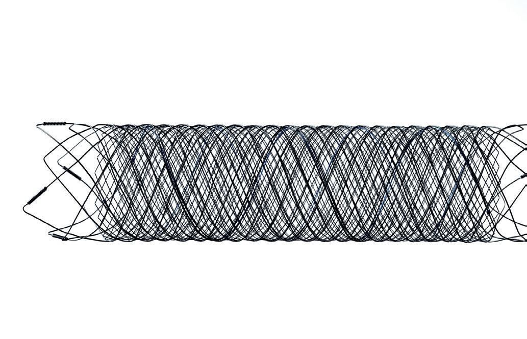
regulatory authorisations and become widely used across the globe was the Pipeline embolisation device (Medtronic), which received a European CE mark in 2008 and US Food and Drug Administration (FDA) approval in 2011. Two other families of flow-diverting stents—Surpass (Stryker) and Fred (Terumo Neuro)—have since been approved in both of these geographies, with Silk (Balt), p64/p48 (WallabyPhenox), Derivo (Acandis) and Tubridge (Microport) also subsequently cleared in Europe.
As such, flow diversion has been a fixture within brain aneurysm care for a number of years, and physicians now have a range of technologies available to them. Despite this, the clinical evidence on the appropriate role for these devices remains a subject of dispute. The most recent example of this came towards the end of 2025 when a research team led by neurosurgeon Nima Etminan (University of Heidelberg, Heidelberg, Germany) published a meta-analysis in the European Stroke Journal assessing existing clinical data on flow diversion in unruptured aneurysms. Their conclusion reads as follows: “The literature
on flow-diverting stents is methodologically weak and potentially biased by financial interests, but still shows relevant proportions of complications and post-treatment morbidity. Currently, there are no good data supporting the use of flow-diverting stents for unruptured intracranial aneurysms where standard treatment options are available.”
This damning verdict is aligned with the European Stroke Organisation’s (ESO) 2022 guidelines on the management of unruptured aneurysms, which recommend that flow diverters should only be considered “when there are no other low-risk options” for aneurysm repair—a notion that was challenged by the neurointerventional community at the time.
The following year saw neuroradiologists Martin Bendszus and Markus Möhlenbruch—also from the University of Heidelberg—respond via an open-access letter published in the same journal. The authors take issue with the aforementioned statement that flow diversion should only be used in the absence of any other valid treatment options, arguing that the sole basis for this was a small, prematurely halted study that “goes back to the early days of the application of flow-diverting stents” and has subsequently been criticised for “severe methodological flaws”. Bendszus and Möhlenbruch also highlight a “plethora” of more recent data—both retrospective and prospective— demonstrating the “overwhelming safety and efficacy” of flow diversion in unruptured intracranial aneurysms.
Etminan and colleagues’ 2025 meta-analysis attempted to address this by providing a more comprehensive and up-to-date overview of the current evidence, but arrived at a similar conclusion to the one outlined by the ESO three years earlier.
Neurosurgeon Victor Volovici (Erasmus University Medical Center, Rotterdam, Netherlands)— who penned an accompanying editorial in the European Stroke Journal that discusses the ongoing challenges faced by flow-diversion treatments—believes “emotion” is among the main reasons why this debate continues to rage on in spite of Etminan et al’s findings.
“I think there’s a lot of emotion, especially in the endovascular world—which, to some extent, is understandable,” he tells NeuroNews. “But we need to free our minds of emotion and look at what the data are telling us in order to provide the best treatments for our patients.”
Volovici also highlights the inherent between-specialty biases underlying many of these discussions: for the most part, interventional neuroradiologists lean towards flow diversion and the other endovascular procedures they typically perform; neurosurgeons lean towards open techniques like clipping; and neurologists tend to hold a preference for avoiding interventional treatment in favour of more conservative medical management strategies.
Nevertheless—and in spite of the available data on flow diverters being “imperfect”—Volovici argues that “we should be able to see exactly what is going on, unshackle ourselves from our respective religions, and look critically at what the data are telling us”.
“For example,” he adds, “I’m a very big proponent of microsurgery—but not for every case, of course, because I’m also a proponent of thinking.”
Evidence versus experience Across 13 studies and more than 800 unrup -
Continued on page 12
tured aneurysms, Etminan et al’s analysis reveals a 3.1% rate of neurological worsening as well as a 12.7% complication rate at three months. Volovici also highlights prior publications showing complication rates as high as 16%, which he describes as “a significant number”, particularly given the context that many of these patients are asymptomatic and are being treated electively.
One of the major points of incongruity within this debate stems from the fact that flow-diverter treatments are extremely nuanced, requiring high degrees of skill, experience and understanding in order to maximise the likelihood of a safe and effective procedure. As such, many of the field’s most renowned neurointerventionists—particularly the flow-diversion pioneers who have been implanting these devices for well over a decade across hundreds of cases—may well be achieving much lower complication rates than those reported in last year’s meta-analysis.
“Most endovascular specialists will say, ‘I do not recognise these numbers—in my practice, we do not have these complication rates’,” Volovici avers. “This is one of the issues that you can see exuding through the results of that meta-analysis. Let’s say your results with flow diverters are better than most—okay, I can live with that. But, if everybody else starts doing what you’re doing based on this, their complication rate could be as high as 16%, because the results of the procedure are so operator-dependent. This creates a problem if you then start claiming that flow diversion is best for patients.”
Perhaps, as Volovici alludes to here—and as others have called for in recent times— the modern proliferation of technologies and treatment approaches creates a need for interventional aneurysm care to be confined to larger, high-volume centres that encounter enough of these cases to develop, and maintain, the requisite capabilities to administer these therapies.
When it comes to firsthand experience, few physicians are better-placed than interventional neuroradiologist István Szikora (Semmelweis University, Budapest, Hungary) to discuss flow diverters and their appropriate usage. Szikora played a leading role in the PUFS study, which helped to establish the benefits of flow diversion, and he has also been implanting these devices since 2009.
While he admits it may not be a particularly “kind or polite” sentiment, Szikora feels that endovascular aneurysm treatments should be restricted to centres like his own that are appropriately equipped in terms of case volume, relevant expertise, availability of devices and overall infrastructure.
“But, for centres that do not have these capacities, I would very strongly recommend that they do not treat aneurysms. It’s that simple,” he continues, speaking with NeuroNews . “There are very few people who can collect sufficient experience in treating aneurysms, either surgically or endovascularly. Even with the
continued growth of endovascular aneurysm treatment, there are not so many cases that people in smaller centres can collect enough experience. And, without sufficient experience, it doesn’t matter which technique you use—your results will be worse.”
Szikora goes on to note that this is especially pertinent regarding flow diversion, as these procedures can appear to be “a piece of cake” when performed by sufficiently trained, experienced operators. While the majority of flow-diverter implantations are relatively straightforward from a technical standpoint and do produce successful results, the minority of cases that go wrong provide a clear demonstration of the importance of understanding coupled with skill. An example Szikora uses is the fact that a simple, successful flow diversion may be comparable to a simple, successful coiling procedure, but
“My problem with the ESO analysis is that the papers they’ve ended up with are quite niche studies that are not very reflective of standard practice”
Philipp Taussky
flow diverters become more problematic when complications arise due to—among other factors—stents being harder to reposition and replace than coils.
Commenting that “centralisation is important” in ensuring optimal outcomes, Volovici is aligned with Szikora as well as a similar assertion made by neurologist Edgar Samaniego (University of Iowa, Iowa City, USA) in NeuroNews last year.
“Guidelines like the ones from the ESO are intended for all centres, including those that only treat 20 aneurysms each year and have nowhere near the experience levels of the leaders in the field,” Volovici adds. “It’s not realistic to expect that flow diversion will produce the same outcomes at these hospitals compared to centres treating a hundred aneurysms every year. Our expectations should be adjusted to the setting where these procedures are taking place.”
Herein lies a fundamental problem associated with these recommendations, however: their attempt to apply universal guidance on such a complex and nuanced therapy appears flawed given the many variables that flow-diverter outcomes are dependent upon. Ultimately, this can be seen to limit the recommendations’ real-world utility—especially for high-volume centres housing experienced proceduralists.
Neurosurgeon Philipp Taussky—an early adopter of flow diversion who, alongside
3.1%
Meta-analysis covering >800 unruptured aneurysms rate of neurological worsening rate of complications
12.7%
Christopher Ogilvy at Beth Israel Deaconess Medical Center (Boston, USA), now places more than 100 of these devices annually—gives a candid take regarding the issue of treatment variability.
In his view, “no one wants to admit to this”, because modern medicine has “rightly” moved towards standardisation and homogenisation, and a multidisciplinary, collaborative approach whereby every patient in the same country—if not across the globe—should get the best treatment possible for their specific condition.
“I do think there will always be differences in brain aneurysms compared to cardiac surgery, for instance, where a bypass is always the same and valve replacements are all very similar,” Taussky tells NeuroNews. “Aneurysms are so heterogenous because of the size, location, morphology and patient anatomy. Every aneurysm is different, so the level of operator skill, experience, knowledge, setup and infrastructure will forever affect the outcomes for these patients.
“There has always been a schism in the flow-diverter world between busy practitioners who have excellent results, and other practitioners who do these procedures less commonly and struggle with higher perioperative complication rates. I do think younger physicians should spend time in busy flow-diverter centres to get a firsthand look at when and how to use these devices.
broader recommendations made by the ESO, at least to the extent that “these technologies should be used carefully”, adding that physicians “need to consider a lot of factors”—including DAPT, which is mandatory alongside flow diversion but not coiling.
“On the other hand,” he continues, “the recommendations—and the 2025 meta-analysis—are clearly coming from people who are not practising [with flow diverters]. There is a difference between very carefully reading what has been published, and actually doing what the subject of those publications is; they’re not the same thing. For instance, in this paper, there are a lot of things that are not considered at all, such as the antiplatelet protocols applied in these cases, and what kind of complications could be related to an appropriate or inappropriate antiplatelet approach. In my mind, these are very important questions—and I acknowledge that it’s very difficult to consider all of those things, but making such strong recommendations is difficult as well.
“So, will I very carefully read the recommendations and use them to decide what I should do in every single case? No, I will not.”
Taussky offers a similar perspective from across the pond. While he acknowledges that it is somewhat “unacademic” to disregard the literature in favour of one’s own experiences—and that, in general, publishing, analysing and comparing clinical data is “extremely important”—he also calls into question the evidence base for Etminan et al’s meta-analysis.
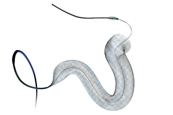
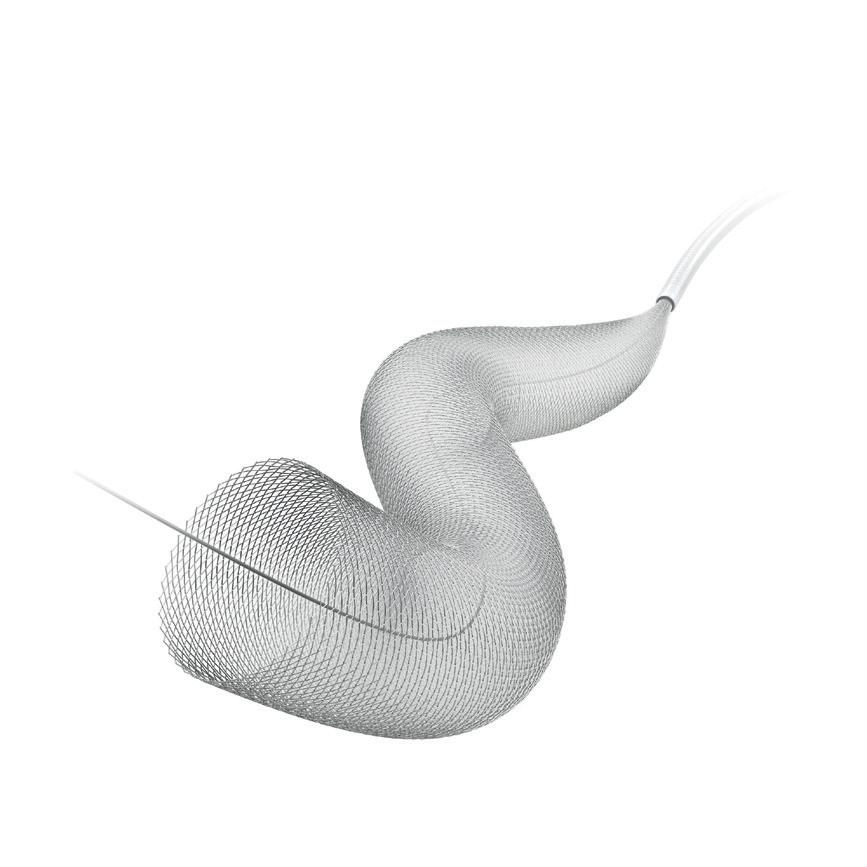
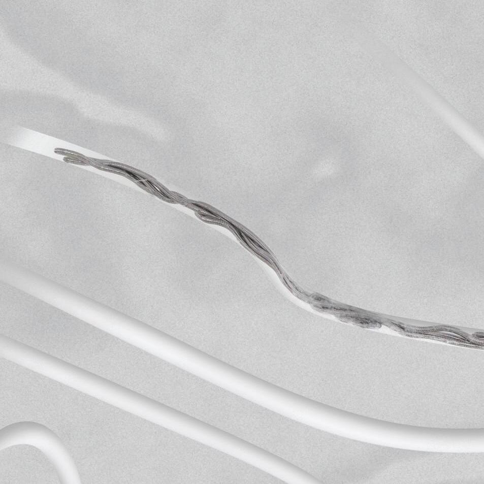
to right: Fred X, Pipeline Vantage and Surpass Evolve devices
“Flow diversion also requires a completely different mindset compared to coiling or clipping, because you need to think about the diseased segment and how to remodel it rather than manipulating the aneurysm itself in order to treat it. As such, you have to think more about proximal and distal landing zones, wall apposition, and DAPT [dual antiplatelet therapy] management.”
Another factor Taussky highlights that must be considered alongside the ESO’s recommendations relates to longstanding geographic variability.
“My initial reaction was that this shows the real difference between practices in Europe and the USA—and it’s been this way since flow diversion entered medical practice,” he says. “When flow diversion entered medical practice in Europe, it was not embraced at all and it was fraught with significant complications, whereas we really adopted and pushed the limits with flow diversion in the USA, and it became the first-line treatment for sidewall carotid aneurysms.”
This discrepancy is apparent within the closest US equivalent to the ESO’s recommendations: the American Heart Association (AHA) guidelines on the management of unruptured intracranial aneurysms. Having been published back in 2015—when the approach was still very much in its infancy—the only reference to flow diversion in these guidelines describes it as “a new treatment strategy that may be considered in carefully selected cases”. The AHA therefore appears to take a more lenient stance on flow diversion as compared to its European counterpart.
“I think those guidelines definitely need to be updated, and I’m sure they’re working on
“The data are poor, and the methodologies they’re based on are not very strong, which leaves more room for interpretation— but my main comment is not that people shouldn’t use flow diversion; it’s that people should be wary and interpret the data with nuance”
Victor Volovici
that,” Taussky comments. “The problem with all of these guidelines is that technical innovation—not just new devices, but management of DAPT and so on—really outpaces any of these recommendations.”
As previously outlined, the initial ESO recommendations published in 2022 were met with umbrage by much of the neurointerventional community—but has Etminan et al’s more recent attempt to synthesise existing data on flow diversion been greeted more positively?
Szikora says he “more or less accepts” the
“My problem with the ESO analysis is that the papers they’ve ended up with are quite niche studies that are not very reflective of standard practice,” he notes. “Many of them include flow diverters that are obsolete and no longer available, and many of them address very niche aneurysm locations like the distal anterior cerebral artery [ACA] or the anterior choroidal artery, meaning the meta-analysis ends up in a rabbit hole that’s not reflective of general clinical practice. That’s the fundamental problem they’re facing. And it’s not clear to me from their methodology how they ended up with these papers— and they are really niche papers, many of which I’d never heard of before—instead of larger, more established series published in better journals.”
The appropriate role for flow diversion
This critique raises multiple key questions beyond the ESO guidance itself, with the most important arguably being the precise role for flow diverters in aneurysm care.
“At this point, the evidence does not suggest that the role for flow diverters is in small or medium-sized aneurysms,” Volovici says. “The evidence only supports their use in the large or giant aneurysms that they were originally made for.”
What Volovici alludes to here is the Pipeline flow diverter’s initial US FDA approval in 2011 limiting its use to patients aged ≥22 years with large or giant wide-necked aneurysms located from the petrous to the superior hypophyseal/ ophthalmic segment of the internal carotid artery (ICA)—with further post-approval data from the PREMIER study later seeing its instructions for use (IFU) expanded to include adult patients with small or medium wide-necked saccular or fusiform aneurysms from the petrous ICA to the ICA terminus.
In his European Stroke Journal editorial, Volo-
Continued on page 14
Left
vici draws attention to the fact that nearly 90% of the 527 aneurysms with available size data included in Etminan et al’s analysis were <10mm in size, meaning “the scientific literature overwhelmingly deals with off-label indications of flow diverters”.
While Volovici is critical of this extrapolation of flow-diverter usage beyond the scope of their original purpose, Taussky points out that such a discrepancy between regulatory indications and real-world clinical practice is not uncommon in the fast-moving neurointerventional space, where innovation continues to outstrip regulation.
“It’s always been the case that flow diversion is being used in off-label situations and, frankly, it’s being used with great success in many instances— for fusiform aneurysms, dissecting aneurysms, and blister-like aneurysms,” he comments.
Meanwhile, Szikora states that, while he does often use flow diversion in larger, wider-necked aneurysms—with coiling being preferred for many smaller aneurysms—this is not the most essential driver of his chosen strategy.
“What really matters is the morphology of the aneurysm and the anatomy of the surrounding vessels—not the size of the aneurysm itself,” he explains. “In my view, flow-diverting stents are designed to treat sidewall aneurysms, and that is very important in my decision-making. Let’s say you have a carotid artery aneurysm arising right at the origin of the ophthalmic artery—a pseudo-bifurcation situation—that is 8–9mm with a 4mm neck; I have no doubt this patient is best treated with a flow diverter. However, if you have a same-sized aneurysm in an MCA [middle cerebral artery] bifurcation, I would definitely not use a flow diverter.”
Szikora goes on to clarify that, while sidewall and ‘pseudo-bifurcation’ aneurysms are a “good indication” for a flow diverter, he typically opts for alternative approaches like coiling or intrasaccular therapy in more symmetrical, ‘true’ bifurcation aneurysms due to challenges created by the second vessel branch regarding device placement and positioning. Perforator-rich areas like the MCA and basilar artery also represent a “potential concern”, he adds.
“Today, there is an increasing tendency of using flow diverters for bifurcation aneurysms, and—in my opinion—that is a questionable practice, and one that I don’t follow,” Szikora continues. “I have a fear that, because flow diverters are promoted so much by some physicians and by industry, it is seen as fashionable. It’s also fashionable—and there’s definitely a strong push from industry here—to go into smaller and smaller vessels to treat more distal, more complicated aneurysms. I believe this is a dangerous path.”
Taussky proffers a similar level of caution; he generally avoids flow diversion in bifurcation aneurysms, and in MCA and ACA locations. However, he is critical of the ESO’s broad recommendation that flow diversion should only be used in cases where no other treatment options are deemed appropriate too.
“There are a few important things that I believe are not considered here,” he explains. “The [greatest asset] of flow diversion is that there is no recurrence—once an aneurysm is occluded, it is not going to recur. It is the only endovascular aneurysm treatment where this is the case. So, if you look at the recurrence and retreatment rates after coiling, and you are thinking about
complications, you should consider complications from potential retreatments as well, and you are then calculating complications from multiple treatments rather than just one. This is not really considered in the meta-analysis, which tries to suggest that treating an unruptured aneurysm with flow diversion leads to significantly more complications, and is associated with poorer outcomes than coiling or clipping. It is a highly oversimplified approach.”
The fact this phenomenon has not been factored into the ESO’s considerations is likely due to a distinct paucity of long-term follow-up data on flow diverters and aneurysm treatments more generally—as opposed to a deliberate omission by the authors. Nonetheless, for Taussky, this is a “very important” detail, as the availability of such data would “definitely” reveal differences between coiling and flow diversion in terms of recurrence rates.
“One of the really unique characteristics of flow diversion is that occlusion rates go up over time, whereas those results get worse over time with both clipping and coiling, and that improved occlusion is not typically associated with higher complication rates,” he continues. “Another is that the treatment is curative; you have a dysplastic, diseased segment, and the aneurysm is just a reflection or a symptom of that disease. Either clipping or coiling really doesn’t address that underlying problem, whereas flow diversion— because it remodels this entire diseased segment— has a chance of curing the patient. This is also reflected in the fact that there are [almost zero] cases of recurrence in the literature.”
Where do we go now?
Even after taking into account the many nuances surrounding flow-diverter therapies, the existing body of evidence on these devices is—to say the least—imperfect.
Volovici highlights the fact that high-quality data on all aneurysm interventions, including flow diversion and other endovascular procedures, but also surgical techniques like clipping, are “really scarce”. Additionally, the lack of evidence regarding the long-term outcomes of these treatments is problematic, with many studies tending to refer to durability at one or two years despite the fact that “we are talking about lifelong bleeding risks here”.
“That remains insufficiently studied at this point, which is a severe issue,” Volovici states. “I hope that, in the future, we can have a wide-scale European registry that allows us to get into the nitty gritty of the data.”
In Volovici’s view, while investigator-led endeavours may yet improve matters, there is no major incentive for flow-diverter companies to fund large studies of products that are already approved and on the market. He also retains a level of scepticism over much of the research conducted on flow diversion to date—particularly those studies that have been driven by the companies developing these devices.
“If you say ‘trial’ in the epidemiological sense, then most people will assume you’re talking about testing a hypothesis and doing randomisation with a control group,” Volovici says. “We do have some one-arm trials, but they’re usually used when randomisation is not feasible or ethical. To me, all of the ‘trials’ of flow diverters are just prospective, multicentre, observational registries, which give you some positive results but are a long way from the evidence generated by trials of thousands of patients. I suspect they are being
“Today, there is an increasing tendency of using flow diverters for bifurcation aneurysms, and—in my opinion—that is a questionable practice, and one that I don’t follow. I have a fear that, because flow diverters are promoted so much by some physicians and by industry, it is seen as fashionable”
István Szikora
referred to as trials in order to create the impression that the level of evidence is higher than it actually is. It’s a very subtle marketing ploy.”
Consistently low numbers of aneurysm cases have always hindered research efforts in the neurointerventional space. Many cardiology trials are able to recruit thousands of patients when evaluating new therapies. The COATING trial—which is comparing surface-modified versus ‘uncoated’ flow diverters, and remains one of the only randomised studies of any endovascular aneurysm therapy in the 25 years since ISAT—enrolled a total of 171. Taussky notes that, as such, the unanswered questions that do exist in the world of flow diversion “are really hard to get to the bottom of”.
Additionally, the industry involvement referred to here by Volovici—and, similarly, the “financial interests” condemned previously by Etminan et al—will forever remain a complex issue, according
to Taussky. In his view, one of the strongest aspects of the 2025 analysis is the fact that it flags up the enduring role played by industry, as this is an especially pertinent topic in the flow-diverter space. When it comes to retrospective case series, for example, Taussky feels physicians should be “very careful” and mindful of possible “underlying agendas”. However, he also warns against being too dismissive of studies like PUFS, PREMIER and SCENT that laid the groundwork for Medtronic and Stryker to gain regulatory approvals for their flow diverters.
“It may lower the bar for scepticism, but the fact a study is industry-sponsored does not automatically mean the data are poor, or biased,” Taussky comments. “And, we have to acknowledge that this innovation is industry-driven and, without industry, we wouldn’t have any of these studies, and we wouldn’t have any of the modern devices we see in [neurointerventional care] as well as cardiology and oncology.
“It’s a complicated discussion; we’re grateful to industry for advancing the management of these disease states, and advancements have been incredible, but we also need to make sure that strong, unbiased data get published.”
While industry input and promotion regarding flow diverters may have an impact “to some extent”, Szikora’s advice to other physicians wishing to mitigate these effects is to have a strong understanding—via clinical evidence and literature—of the technologies at hand, and maintain relationships with sales representatives but, more importantly, with engineers in order to see the “true value” of new devices.
“There are maybe six or seven flow diverters currently being produced by different companies, and each company has some arguments about why its flow
diverter is better than the others—but, if you look at the literature and read the results, there are basically no differences,” he says. “So, if one device is working well in my hands, should I try another device for the same purpose and the same pathology just for the sake of trying it? I don’t do that. In order to switch from one product to another, I need to be convinced that there’s something missing from the one I’m used to. That would be my recommendation: to fully understand what you want to fix and whether it will potentially be improved with a different device.”
Putting the potential challenges created by industry-driven biases to one side, the lack of high-level evidence on flow diversion—particularly longer-term follow-up data—remains a point of contention. The final message delivered by Etminan et al in the conclusion of their meta-analysis is as follows: “Randomised controlled trials are needed to compare safety, efficacy and durability between flow-diverting stents, and coiling or clipping.”
However, Taussky voices reservations regarding how feasible trials of this kind are likely to be, while Szikora harbours doubts over the actual value their results would hold.
“One problem is that I don’t think patients are going to agree to randomisation; another is the question of who is going to pay for these trials,” Taussky comments. “And, by the time many of these studies are finished, the devices will already be outdated, because you need at least 3–5 years to run these trials and have long-term follow-up—and, by that time, you will already have had one or maybe even two new generations of flow diverters. I don’t think randomisation is ever going to happen in this space.”
“In my view, there is no comparison between coiling and flow diversion,” Szikora adds. “Of course, there are aneurysms in which I use a flow diverter plus coils but, outside of that, there are many aneurysms I would not hesitate to treat with coils and many I would not hesitate to treat with flow diversion. So, in my practice, I could not compare them directly, because I use them for different types of aneurysms.”
This concept likely precludes any future randomisation between coiling— the closest thing to a standard of care in contemporary endovascular aneurysm treatment—and flow diversion. The same line of thinking can also be seen when looking back to the early days of flow diversion as, in the PUFS study, it was a prerequisite that enrolled aneurysms had already been deemed untreatable with coils.
“I don’t think the data are sufficient, but when can we say that data are sufficient?” Szikora continues. “The major challenge for clinical research—particularly in our field—is to select comparable patient groups across a large number of centres. A good example of us getting this wrong was the ARUBA trial, which compiled data on any kind of brain AVM [arteriovenous malformation] with the only selection criteria being ‘unruptured’. We know that
there are so many types of AVMs that such a comparison was pretty much useless. “The same problem exists with aneurysms. I would say, if we want to understand the true value of flow diversion, we should look at a large number of flow-diverter cases in infra/supraclinoid carotid artery aneurysms, or in basilar or distal vertebral artery aneurysms, or potentially the sensitive issue of more distal aneurysms in the MCA. These groups should be differentiated, because they are completely different stories that cannot be compared to one another. However, this type of research is something that could only be provided by a very strong international cooperation.”
Recently, the neurointerventional community has attempted to take steps in this direction. A new guideline on flow diverters—endorsed by multiple groups including the European Society of Minimally Invasive Neurological Therapy (ESMINT) and Society of NeuroInterventional Surgery (SNIS)—was also published in 2025, concluding that, “when widely applied, standardisation of methods of measuring and reporting outcomes will help to harmonise the assessment of treatment outcomes in clinical studies, help facilitate communication of results among specialists, and help enable research and development to focus on specific aspects of flow-diversion techniques and technology”.
With randomised trials—for various reasons—appearing very unlikely, Volovici echoes these sentiments, emphasising the pivotal role for multidisciplinary, collaborative decision-making and more comprehensive follow-up protocols in ensuring the highest possible standards of both clinical care and data on unruptured aneurysms. He recommends that, in cases adjudged to be amenable to endovascular and surgical options alike, the patient should be counselled by each of the relevant specialists.
“That’s how it should be,” he explains. “None of us feel like someone else is stealing our cases—there’s enough for all of us, and we don’t have any turf wars. That’s the only way that you can really get this going, aside from the US system where people are dual-trained.
“Another essential point is that people should follow up these patients. Even if they have been treated microsurgically and their aneurysm has completely disappeared on imaging, I still do a six-month MRI [magnetic resonance imaging], neuropsychological assessments, and one- and two-year follow-up. This benchmarking is important. It’s the only way we’ll ever get better data compared to what we have right now, and the only way for us to confirm that the things we assume about post-treatment outcomes are correct as well.
“The data are poor, and the methodologies they’re based on are not very strong, which leaves more room for interpretation—but my main comment is not that people shouldn’t use flow diversion; it’s that people should be wary and interpret the data with nuance.”
The first flowdiverting stent is CE-marked
Flow diversion gains US FDA approval
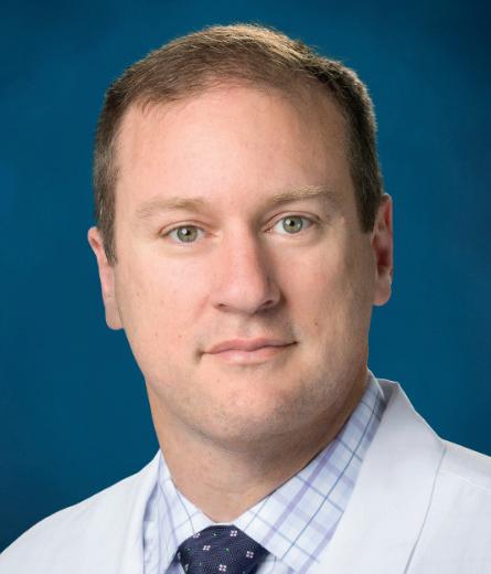
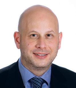
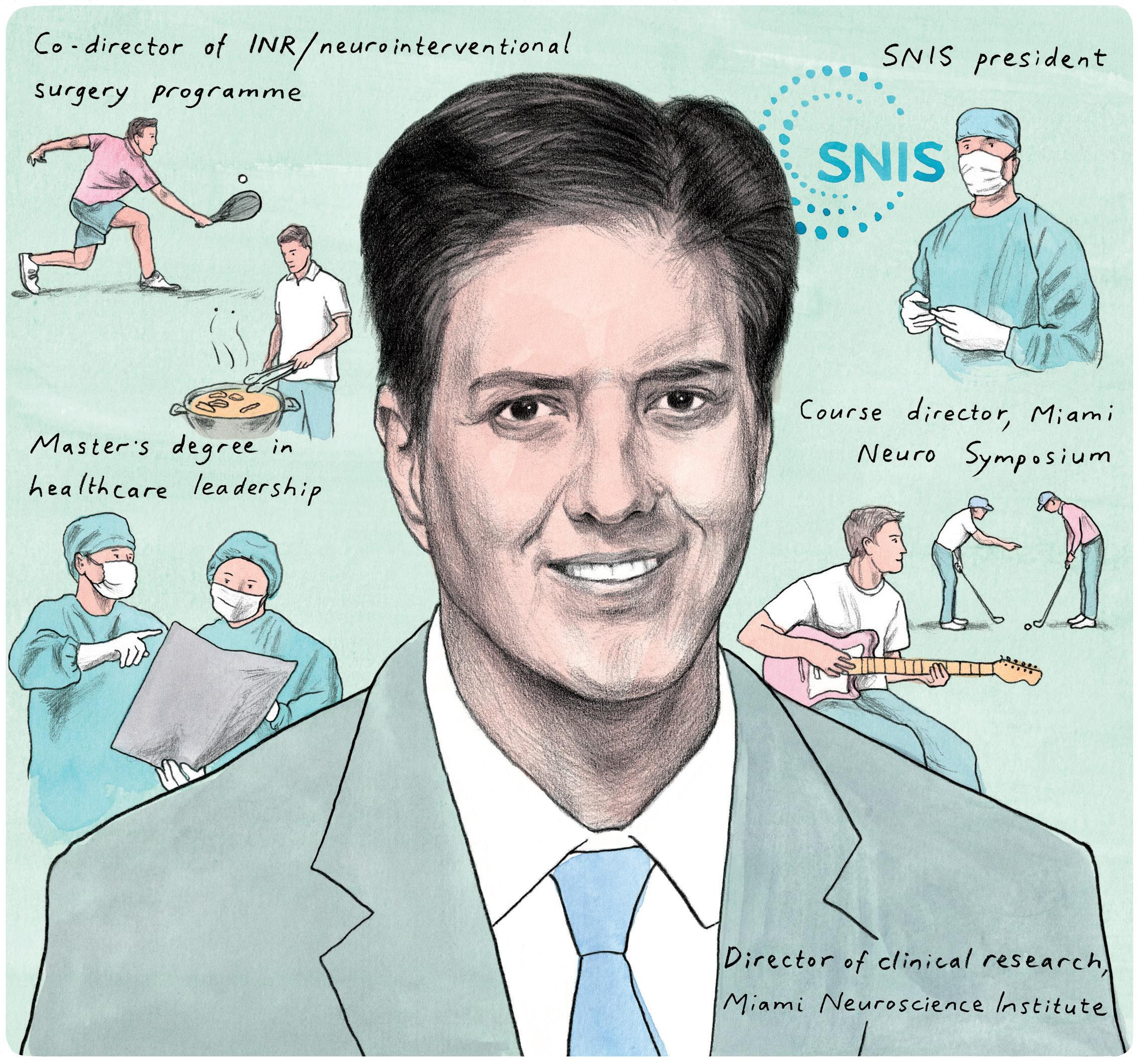
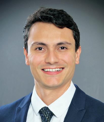
Guilherme Dabus
A rockstar inside and outside of the neurointerventional community, guitar aficionado Guilherme Dabus (Miami, USA) is the current president of the Society of NeuroInterventional Surgery (SNIS), and has been involved in a multitude of impactful clinical studies including CREST-2, HEAT, SCENT, SUMMIT MAX and TIGER. Having finished his medical education and residency training in Santos, Brazil, he moved on to a fellowship in the USA in 2004—and, today, is the director of clinical research and co-director of the interventional neuroradiology/neurointerventional surgery programme at Baptist Health of South Florida’s Miami Neuroscience Institute. Here, Dabus discusses all this and more with NeuroNews.
Why were you drawn to a career in medicine and, more specifically, the neurointerventional space?
Growing up in Brazil, I was surrounded by relatives who were physicians—in particular, my father, who is my greatest influence. I watched how profoundly a single person armed with knowledge and compassion could alter the course of another person’s life. That felt like the most meaningful use of a career I could imagine. During my residency, I was naturally drawn to the procedural aspect of radiology, including biopsies, spinal procedures and, later, angiography and endovascular procedures. The first time I scrubbed in for a neurointerventional surgery during my radiology residency, it felt like standing at the edge of a frontier that nobody had fully mapped. The modern era of neurointerventional surgery was in its infancy and trying to understand where it could take us was really exciting to me. The brain is responsible for everything that makes us human including our capacity to move and breath, our memories, our language, and our personalities—and here was a subspecialty that allowed us to intervene inside that marvellous structure, through a catheter, without opening the skull. The audacity of it, and the responsibility it carries, was something I could not walk away from. Every case still carries that weight for me.
Who have your key mentors been and how have they impacted your career?
I have been extraordinarily fortunate in this regard. From my father who, as I mentioned before, has been my greatest role model and mentor, to everyone else who participated in my formative years from my medical school to my residency in Brazil and, of course, during my training at Northwestern (Chicago, USA), Mallinckrodt Institute of Radiology (St Louis, USA) and Massachusetts General Hospital (Boston, USA), I have been extremely lucky to have had many mentors. I learned that technical mastery is only the foundation—the tool to execute our job. What truly separates a good physician and neurointerventionist from a great one is judgment, humility, compassion, and the willingness to continue to improve. These mentors who shaped me most did not simply teach me procedures but showed me how to think about patients when things are not going in the direction we wanted, how to communicate uncertainty to families, and how to design a question worthy of a clinical trial. I carry their voices into my own teaching of fellows and medical students every day. These teachings and knowledge, I believe, need to be transferred to the younger generation. The obligation to pass it forward is something I feel deeply, especially now in a leadership role. The field’s future lives in the residents and fellows we form today.
How would you describe your experience to date as SNIS president? It has been one of the greatest privileges of my professional life, and also one of the most humbling. When you step into a role like this, you realise quickly that the work of a society like the SNIS is not merely organisational, but also custodial. We are stewards of a specialty that has—in the span of a few decades—transformed how stroke, aneurysm and a host of devastating vascular diseases are treated. My experience as president has been marked by a genuine sense of possibility. Our membership is intellectually curious and bold, our science is accelerating at a pace difficult to follow, and our advocacy for patients continues to grow. But I have also encountered the hard realities of health equity, and the gaps in access to neurointerventional care that still exist across geographies and communities. Carrying both the privilege and the responsibility of this position simultaneously is something I do not take lightly for a single day.
As a reviewer for numerous medical journals, do you have any advice for other neurointerventionists looking to take on similar responsibilities?
Peer review is the immune system of science. It is imperfect but, without it, the literature becomes unreliable, and unreliable literature harms patients. For colleagues considering taking on reviewing responsibilities, my advice is threefold. First, review as you would want your own work reviewed: rigorously, fairly, and with specific rather than vague critique. Second, treat it as a learning exercise, not a burden. Reviewing the work of others forces you to evaluate methodology and results at a level that makes you a better researcher yourself. And, third, be honest about your limits. If a statistical approach or device technology falls outside your expertise, or if a conflict of interest exists, say so. The integrity of the process depends on that honesty. The journals that are shaping our field deserve reviewers who are as committed to rigour as the authors they evaluate.
What is the biggest challenge currently facing the neurointerventional space?
There are certainly several potential challenges, including workforce issues— such as training, burnout, second victim syndrome and moral injury—regulatory and policy issues, and payer down pressure on reimbursement; however, in my opinion, our biggest challenge is access. We have built extraordinary tools and accumulated compelling evidence, but a patient in a rural county or a developing nation, or without adequate insurance, does not benefit from any of this if they cannot reach a centre capable of delivering it. The science is outpacing the systems, and that gap is a moral problem as much as a logistical one. Within the USA, the regionalisation of stroke care has improved outcomes in metropolitan areas, but the map of who receives guideline-based neurointerventional therapy is still drawn in ways that correlate with
Fact file
CURRENT ROLES
Medical director of clinical research; co-director of interventional neuroradiology; co-director of neuroscience operations, Baptist Health Miami Neuroscience Institute
Clinical professor, Florida International University
President, Society of NeuroInterventional Surgery
EDUCATION
2001–2004: Residency, radiology, State University of Campinas
1999–2000: Internship, Santos School of Medical Sciences
1995–2000: Medical degree, Santos School of Medical Sciences
AWARDS (SELECTED)
2022: Fellow of the American College of Radiology
2018: International Visiting Professor of the Radiological Society of North America— Indonesia
2015–2018: Best Doctors Awards
2015: Fellow of the American Heart Association Stroke Council
geography, race, and socioeconomic status. The work of the next generation of leaders in this field will not only be in the angio suite. It will be in policy rooms, in hospital boardrooms, and in communities that have never seen a neurointerventionist. I believe the SNIS has a role in that work and I hope we will continue to push that agenda forward.
Which areas of the neurovascular field do you think will witness the most innovation over the next 5–10 years?
Our field continues to evolve at an exhilarating pace, and we will continue to evolve our treatments of vascular arterial diseases like strokes, aneurysms and carotid disease. However, we will increasingly be treating other types of disease— such as venous disorders, cerebrospinal fluid disorders, migraines, neuro-oncological problems, and degenerative diseases—using the endovascular route. Automation, non-invasive imaging integration, artificial intelligence, brain-com-
“When I imagine the neurointerventional suite of 2035, it looks very different to today— and that is not a source of anxiety. It is a source of profound motivation”
puter interface devices and robotics will be incorporated more and more into our daily practice, assisting our treatments and benefitting our patients. The integration of realtime predictive feedback into procedural decision-making will make our interventions more efficient and effective, and safer. As an example of this transformative future, long-distance, robotic-assisted neurointerventional procedures are no longer hypothetical. The prospect of a skilled interventionist guiding a robotic system from
hundreds of miles away, bringing subspecialty expertise to a patient who would otherwise have none, is one of the most profound equity-levelling possibilities I can envision in medicine. When I imagine the neurointerventional suite of 2035, it looks very different to today—and that is not a source of anxiety. It is a source of profound motivation.
What are your interests outside of medicine?
I play electric guitar. I used to do it a lot, playing all kinds of rock, hard rock and heavy metal as part of a band in my late teens and early 20s, and it remains my main hobby. I also enjoy cooking, in particular grilling, and spending time with family and friends. I enjoy playing any kind of racquet sport—tennis, beach tennis, pickleball—with friends, and playing golf with my father. I also love to watch and cheer for my kids in their sports, including basketball, swimming, and track and field.
Peter James Field / Agency Rush
ADAPT 2.0: A closer look at the latest evolution of aspiration thrombectomy
Thirteen years after the initial publication outlining the potential benefits of ADAPT (a direct aspiration first-pass technique) in stroke thrombectomy, the approach is being adopted by an increasing number of neurointerventionists and continues to evolve. Here, NeuroNews takes a closer look at the core principles, clinical data and firsthand experiences associated with the latest advancement of this technique: ADAPT 2.0.
AS FIRST AUTHOR ON THE 2013 publication that debuted ADAPT, Aquilla Turk (Prisma Health, Greenville, USA) played a central role in introducing this revolutionary concept into the field of interventional stroke care.1 And, today, as Imperative Care’s chief medical officer, he continues to drive progress in aspiration thrombectomy.
Looking back in time, however, Turk recalls that serendipity—as opposed to problem-solving innovation—sparked the initial idea behind direct aspiration.
“We were doing a [thrombectomy] case and, when we pulled the stent retriever through the aspiration catheter, the stent retriever looked brand new,” he says. “But, the aspiration catheter was clogged up, so we pulled it out, and the clot was hanging out of the end of the catheter.”
From that happy accident—and following a handful of cases where Turk and his colleagues saw continued success in advancing an aspiration catheter to the face of the clot and extracting it without a stent retriever—the ADAPT concept snowballed. And, while the technique by no means worked in every case, Turk remembers thinking, “wow, we’ve never done anything this quickly and easily before” in stroke thrombectomy.
This being the neurointerventional space, however, things have progressed markedly over the subsequent decade, with a number of companies developing larger, more trackable catheters tailored specifically to the neurovasculature.
In Turk’s view, a key milestone in this evolution is ADAPT 2.0—a concept that he also describes as a premise upon which Imperative Care was founded. ADAPT 2.0 is the culmination of the company’s attempts to improve “every step along the way” in aspiration thrombectomy, from navigating past the aortic arch to reach the cerebral vessels, to trackability in more distal locations like the M2 segment of the middle cerebral artery (MCA), to engagement and ingestion of the clot itself, and even visualisation of the clot once it has been captured.
ADAPT 2.0 ultimately consists of three key components: 0.088-inch intracranial access, an asymmetric catheter tip, and continuous dual aspiration (CDAT).
Turk explains that the first of these components carries a myriad of potential advantages. By positioning an atraumatic 0.088-inch access catheter above the patient’s neck—meaning it is effectively “anchored” around one or more vessel curvatures at the base of the skull—operators have a stable platform from which to deliver aspiration catheters quickly and effectively to
the target site to remove the clot and restore blood flow rapidly. Furthermore, Turk notes that, in his experience, this closer proximity to the target clot helps improve control during the procedure, and makes performing additional passes quicker and easier while also providing a degree of flow attenuation that may aid clot retrieval.
Next up is the asymmetric tip—a feature that, according to Turk, “really sets these catheters apart”, creating a 15% larger surface area that improves clot engagement and exerts a greater degree of force onto the clot during aspiration.2
“The asymmetric tip also alters the physics of how the clot is engaged,” he explains. “If you look at the vector forces that are influencing the clot, it essentially twists and torques the clot against the vessel wall, which helps to more easily break those forces that are present when a clot is embedded in an artery. In addition, whereas we see clots being pulled straight into the end of a conventional catheter and then ‘mushrooming’, the asymmetric design means the clot is almost being rotated and tumbled when it engages with the catheter tip, and I think that’s why we’re now seeing more frequent ingestion than we did before.”
for itself”. Based upon analyses of a cohort of 211 patients submitted to the US Food and Drug Administration (FDA) in support of clearance of the Zoom System, the study produced a median modified thrombolysis in cerebral infarction (mTICI) 2b–3 recanalisation time of 19 minutes and a symptomatic intracranial haemorrhage (sICH) rate of 0.9%, while 4.7% of cases required rescue therapy using a stent retriever. 3
“These results show meaningful outcomes in all the important areas we’ve talked about, including speed, safety and efficiency,” Turk adds.
ADAPT 2.0 in the real world
While “a lot of important information” was undoubtedly generated by the original Imperative Trial, Maxim Mokin (University of South Florida, Tampa, USA) and his colleagues recently sought to move the needle once again by examining the real-world utility of ADAPT 2.0—doing so via a multicentre, retrospective review of 124 consecutive ischaemic stroke cases treated using the Zoom Stroke System (Imperative Care) and CDAT.
CDAT, meanwhile, perhaps represents the most novel aspect of all within ADAPT 2.0, introducing a concept whereby two catheters— one housed within the other—both simultaneously generate aspiration throughout the procedure via a single vacuum source. This simple approach was designed to maximise clot ingestion without adding extra time and while maintaining procedural efficiency.
“There has been a stepwise evolution in aspiration thrombectomy and, with the combination of these novel innovations that lead to ADAPT 2.0, we’ve seen results that are notably better,” Turk comments, citing a significant reduction in the need for stent retrievers as well as fast procedure times in his own practice.
And, with results from the Imperative Trial now also available—having been published in the Journal of NeuroInterventional Surgery (JNIS) last year—Turk believes this latest data release “speaks
Mokin himself presented findings from this analysis at the 2025 Society of Vascular and Interventional Neurology (SVIN) annual meeting (19–22 November, Orlando, USA), reporting that—across an all-comers patient population with a “very diverse” range of occlusion locations—researchers observed a “remarkable” median procedure time of 17 minutes and a median of one pass per procedure. These analyses also revealed rates of 97% and 80% for final mTICI 2b–3, and mTICI 2c–3, respectively.4
“As an operator, I want to be fast,” Mokin says. “I want to treat a stroke in the shortest amount of time possible because we know very well from prior literature that, the longer the procedure takes, the more damage we then see in terms of clinical recovery. In this series, we achieved a first-pass effect in more than half of the patients, and the procedures were extremely fast—and, in my experience, the two tend to correlate. If you have a procedure that requires an average of one pass and only takes 17 minutes, how can you get faster than that?”
Additionally, while the true clinical ramifications of attaining mTICI 2b–3 versus 2c–3 recanalisation are not fully known, achieving high rates for both remains of paramount importance in every thrombectomy treatment.
“Ideally, we want a procedure that is fast, with the minimal number of passes, and that provides complete or near-complete reperfusion; you really want to nail all three in every case, where possible, rather than settling for one or two,” Mokin explains.
Following on from these positive early indications, the prospective ADAPT 2.0 Study has
Maxim Mokin Aquilla Turk
Josh Abecassis
now begun enrolment, with Mokin serving as co-national principal investigator alongside David Fiorella (Stony Brook Medicine, Stony Brook, USA), Shahram Majidi (Mount Sinai, New York, USA) and Justin Mascitelli (UT Health San Antonio, San Antonio, USA). Investigators expect to enrol a maximum of 750 patients across up to 50 US sites, with rates of mTICI 2c–3 recanalisation and embolisation to new territory (ENT) being among the study’s primary endpoints of interest.
“The prospective study is anticipated to be important in terms of validating our findings, and showing us what is happening in the real world on a daily basis,” Mokin adds. “With independent, core-lab-adjudicated imaging analysis and independently adjudicated safety outcomes, we hope to define where the differences truly are, in which patients, and also with what types of occlusions. Our initial analysis wasn’t that granular, partly because of the sample size, which was decent but not powered for those small details; it was about proving feasibility and looking for that early signal.”
Another finding Mokin is eager to examine further in the prospective study is something that was initially found within the analysis he presented at SVIN 2025: the fact that 27% of cases saw clots ingested into both the inner (smaller) and outer (larger) aspiration catheters during CDAT, while an additional 9% saw clots ingested into the larger, outer catheters alone. For Mokin, this begs the question of what would have happened in these patients if they had only been treated with traditional, single-catheter aspiration. In his view, at best, many would likely require additional passes and may still fail to achieve sufficient recanalisation; at worst, “that clot is heading deeper into the brain”.
“Once you start seeing those clots in the outer [catheter] quite frequently, you realise how many you could’ve missed without it,” Mokin continues. “One could argue, 9% is only a single-digit number, is it really a big deal? My answer would be yes; for the sake of a simple modification that actually makes your life easier, you are capturing clots in 9% of cases where you would otherwise have to do another pass. In this day and age, given how good our thrombectomy devices are now, 9% is huge.”
This early-stage yet intriguing outcome also highlights the potential benefits that may be carried by CDAT—and ADAPT 2.0 more generally—according to Mokin.
“With CDAT, I no longer think of the guide catheter as simply providing access; it becomes a more active component as an aspiration catheter, working in conjunction with the inner catheter to provide dual aspiration,” he explains. “CDAT is intended to make the aspiration you would normally achieve with a single catheter more effective, and more adaptable across different anatomies and clot types, because you have an extra safety basket. And it’s not forcing you to use a new tool—it’s allowing you to leverage a setup that you already have. In my experience, it’s reliable and simple to do, and you can use the same setup for almost every case [with the main exception being tandem occlusions]. That’s why I became a believer.”
An optimised thrombectomy setup
Earlier this year, Imperative Care launched the
Zoom 4S catheter—a new product intended to facilitate thrombectomy treatments in more distal locations, further expanding the capabilities of the company’s Zoom Stroke System alongside its existing Zoom 88 and Zoom 7X catheters. As it pertains to ADAPT 2.0, the larger Zoom 88 catheter is used to provide intracranial access and administer aspiration in proximal locations, with the midsized Zoom 7X and smaller Zoom 4S catheter allowing physicians to advance more distally where required.
Among the key advantages of this setup is the fact the operator has all three catheters at their disposal from the outset of any given case. That is according to Josh Abecassis (University of Louisville, Louisville, USA) who is one of many physicians now employing this triumvirate of catheters—dubbed the ‘Zoom Stack’—in clinical practice.
“I really like the fact that, right out of the gate, I have a single stack with three different catheters available for aspirating a broad range of clot locations,” he reports. “It’s not uncommon that you get some fragmentation when you initially aspirate the clot, and I like having a smaller catheter readily available rather than having to get another device out in order to go up and do another pass.”
Between the increased support offered by Zoom 88, the trackability of Zoom 7X thanks to its self-orienting tip, and the Zoom 4S’s ability to both act as a rail for advancing the larger catheters and extract clots in more distal locations “as needed”, Abecassis feels the Zoom Stack represents a dependable setup that is well-suited to the unpredictability of ischaemic stroke care.
“The main thing that Zoom has revolutionised—in my opinion—is that it sets you up for success in tackling severe tortuosity and really complex anatomies in a way that [has been challenging] with conventional catheters,” he adds. “Zoom has absolutely enhanced our success rates in complex and more basic anatomies, but the results are much more palpable in the complex cases because they’re the ones that have historically made us suffer.”
The ever-growing uptake of tenecteplase—a simpler and potentially more effective alternative to alteplase—for intravenous thrombolysis, while advantageous overall, represents one of the many variables that makes modern interventional stroke care so challenging. Tenecteplase is known to be associated with increased clot migration versus alteplase, with a JNIS paper from March 2026 revealing that this “frequent and time-related” consequence may hinder recanalisation rates associated with thrombectomy.5
“This fragmentation phenomenon is not common, but it’s not necessarily that rare either,” Abecassis notes, citing observations from his own practice. “I think it makes an even stronger case for having multiple tools at your fingertips in every case. I’ve had cases where I thought I would need to use an 0.071-inch or 0.088-inch catheter to treat an M1 but, when I do the run, I see that there are actually multiple M2s, because the clot has fragmented and migrated. In those cases, I have been able to easily adjust my plan and use the Zoom 4S—with or without the Zoom 7X as an intermediate catheter.”
For Abecassis, the take-home message on the Zoom Stack—and, by extension, the ADAPT 2.0 technique he utilises alongside it—relates to its
versatility, and the “impressive” results it has produced across the full gamut of stroke cases. And, echoing Mokin’s advocacy for a standardised setup incorporating CDAT and ADAPT 2.0 due to the fact that “you never know when it might help you out”, Abecassis believes “you have to be prepared for unexpected findings” when treating ischaemic strokes.
“The 0.088-inch intracranial access would be the foundation of ADAPT 2.0 and, to me, it’s probably the most important thing because of how much it changes the procedure for the better,” he adds. “We know about the theoretical benefits of the asymmetric tip due to the increased surface area but, in reality, it’s just a question of how often you’re having to do a second pass with the same catheter or switch to a different aspiration catheter—and I can tell you that the incidence of us having to do this with the Zoom 7X is very low, in our experience.”
And, similarly to Mokin, Abecassis ultimately feels that—with further technical developments and much more clinical evidence on the horizon—Turk and colleagues’ seminal work that helped propel aspiration thrombectomy forward remains a long way from the end of its journey.
“I’m a big believer in ADAPT 2.0,” he concludes. “I think it goes hand in hand with the Zoom technologies, but I also think it creates a framework for future innovation. I don’t think ADAPT had evolved very much since its initial inception and description nearly 15 years ago but, now, there’s nothing stopping us thinking about what ADAPT 3.0 or ADAPT 4.0 might look like. It’s incredibly refreshing to see how much we’re innovating in this space.”
References:
1. Turk A S, Spiotta A, Frei D et al . Initial clinical experience with the ADAPT technique: a direct aspiration first pass technique for stroke thrombectomy. J NeuroInterv Surg 2014; 6(3): 231–7.
2. Data on file at Imperative Care.
3. Zoom System instructions for use (IFU): LBL002069.A.
4. Mokin M. Society of Vascular and Interventional Neurology annual meeting. 2025, 19–22 Nov; Orlando, USA [cited 4 May 2026].
5. Tessier G, Gerschenfeld G, Cortese J et al . When lysis moves the target: thrombus migration after tenecteplase and its impact on endovascular recanalization. J NeuroInterv Surg Published online first: 4 Mar 2026. DOI: 10.1136/jnis-2025024846.
Notes: Individual trials are not directly comparable, and data are presented for observational purposes only. Imperative Care products referenced in this article are approved for use in the USA only, and are not approved or available for sale outside the USA.
Important Safety Information (ISI)
U.S. - Rx only. The Zoom System, when used with the Zoom Aspiration Pump (or equivalent vacuum pump), is indicated for use in the revascularization of patients with acute ischemic stroke secondary to intracranial large vessel occlusive disease (within the internal carotid, middle cerebral - M1 and M2 segments, basilar, and vertebral arteries) within 8 hours of last known well. Patients who are ineligible for intravenous thrombolytic drug therapy or who have not responded to thrombolytic drug therapy are candidates for treatment. There are no known contraindications.
For complete product information including the Zoom System devices, Zoom portfolio, indications, contraindications, warnings, precautions and adverse events, see product IFU included in product packaging, contact Customer Service at +1-408-502-7548, CustomerService@ImperativeCare.com or visit bit.ly/3yWkfEJ.
MAXIM MOKIN and JOSH ABECASSIS are paid consultants for Imperative Care. M003275.A.
Puncture-torecanalisation time shows “unexpected” association with leftsided stroke outcomes
A retrospective analysis of close to 1,000 patients published in World Neurosurgery has revealed that left-sided anterior-circulation large vessel occlusion (LVO) strokes treated via thrombectomy may be associated with worse outcomes compared to their right-sided equivalents—a phenomenon seemingly driven by a multitude of factors including baseline clinical severity and puncture-to-recanalisation (PTR) times. Here, corresponding author Daniel Sahlein (Goodman Campbell Brain and Spine, Indianapolis, USA) discusses the minutiae of these findings with NeuroNews.
What were the most interesting findings within this single-centre study?
We wanted to see whether or not patients with anterior-distribution LVO stroke have different outcomes based on laterality, because there are surprisingly little literature or data on this. As language centres are located in the left side of the brain, it would make sense that patients with left-sided stroke do worse clinically—and we did find that these patients had worse outcomes. That was partly because they had higher National Institutes of Health stroke scale (NIHSS) scores at presentation, which is one of the major determinants of stroke thrombectomy outcomes.
standard Berenstein-tip catheter is going to fail, and then you will eventually switch to a Simmons 2, which essentially means the catheterisation is going to take you an extra 10 minutes.
However, something that really surprised us was the fact that our PTR time was greater on the left than on the right. In fact, when we did a multivariate analysis including PTR time but also things like age and degree of recanalisation, we were able to confirm that discrepancies in time between the two sides seem to make a difference, which was unexpected. There was something even more interesting about the histograms of those times as well: the two sides look very similar up to about 15 minutes, at which point there’s a peak in PTR time. But, while the histogram on the left side falls off pretty quickly after that, it’s actually bimodal on the right side—it goes down, and then there’s another peak 10 minutes later before it starts to decrease again.
What do you think are the underlying reasons for these findings?
When we sat down to try to explain it, we concluded that it’s really to do with aortic arch anatomy. In some cases, the left and right sides look pretty similar, and are both fairly straightforward to catheterise, meaning you end up with a very short PTR time. However, on the left side, you have this fairly common variant anatomy called a bovine arch, where the common carotid artery comes off the subclavian artery and then takes a very tight turn back along the aortic arch. And, ultimately, to catheterise that type of anatomy, you need a catheter with a back bend—called a Simmons 2—in order to make that turn. Using a
We don’t have second-by-second granularity to see exactly what was happening in the left-sided versus right-sided procedures, but this hypothesis seems very plausible and rational. Our analysis also showed that the degree of recanalisation and number of passes were the same on both sides, meaning we were not technically more successful on one side versus the other. In other words, once we got up into the neurovasculature, the procedures went very similarly, so the fact the earlier part of the procedure is not as consistent may account for those differences by laterality. I don’t think that time is the only determinant of the difference in outcomes—there’s also the idea of hemispheric dominance, and so forth— but time was a determinant, and it was more of a determinant than we were expecting.
6, and 12 versus 13, are not the same. That speaks to the overall challenge of coming up with any sort of quantitative scale that accurately captures the true essence of stroke severity—and that’s why it’s important to break these things down into other meaningful and more nuanced categories.
What are the most important messages you would convey based on these data?
“A key learning from this is just to pay really close attention to your stroke outcomes and try to audit every step of the stroke continuum carefully”
Why is there so little published clinical research on stroke laterality?
I don’t know, but I do find it a bit perplexing. I think people feel that a lot of this is covered by NIHSS scores; essentially, you might say that, if you have an NIHSS score of 20, why does it matter which side it’s on? I find that argument to be a little challenging, because NIHSS is both non-linear and non-equivalent. If you’re running a clinical trial, it might work grossly because you need to have some line in the sand in terms of stroke severity, but one NIHSS score of 20 is not necessarily the same as another NIHSS score of 20. They’re composed of a totally different sum of functional deficits, and the likelihood of recovery is not the same even between identical NIHSS scores. The fact it’s non-linear also means the steps between 5 versus
I think one of them is the importance of access and access planning. These data went from 2011–2020, but we have a pretty large amount of more recent data, and that bimodal peak actually disappeared and became unimodal in our more recent dataset. That shows our group has improved over time and become better at predicting these more challenging anatomies based on hundreds of thrombectomy cases. But, for groups that are not doing this many procedures on a per-physician basis, I think there are two things that can help. One of them is getting a better three-dimensional sense of the aortic arch and the anatomic architecture that you’re going to need to traverse, and that’s where I think automated 3D reconstruction technologies offer a really tremendous opportunity because of the confidence they give you in your catheterisation. The second is simply catheter construction and selection, with respect to picking a slip catheter to help you get in position, and a wire that’s stiff enough, and all these kinds of things.
Another key learning from this is just to pay really close attention to your stroke outcomes and try to audit every step of the stroke continuum carefully, with really robust data, and take nothing for granted. We did not expect to see this finding, and we only saw it because the quality of our datasets is extraordinary. So, it starts with the data, and it also starts with being cognisant of your blind spots and having an open mind as to what you might find; that’s how we end up unearthing some of our most interesting discoveries.
Daniel Sahlein
This advertorial is sponsored by Sim&Cure
Further studies and improved integration may see procedural simulations become a routine part of aneurysm care
Following the introduction of Sim&Size v2.1 (Sim&Cure)—the latest advancement in the use of simulation software to support the planning of neurointerventional procedures—early adopter Christian Ferreira (Northwell Health, New York, USA) speaks to NeuroNews to discuss the benefits held by these technologies and their likely role in the future of intracranial aneurysm treatments.
“HEALTHCARE GETS BETTER WHEN we have protocols and standardised treatments,” says cerebrovascular neurosurgeon Ferreira. “The fact that we can help the operator to [model how devices will deform during a procedure] is likely to make life easier for everyone—especially the residents and fellows who don’t have as much experience as a mid-career endovascular surgeon. And, if I was a patient, I would prefer to have my aneurysm treatment simulated before any procedure. Wouldn’t you?”
Given the growing body of evidence evaluating simulation software in endovascular aneurysm care, and the growing number of global centres that have integrated these technologies into their practice, the question posed by Ferreira appears to be a very reasonable one. Last year, he and his colleagues contributed to the aforementioned body of evidence, publishing a meta-analysis intended to synthesise existing data on virtual simulation software in the neurointerventional space.
“Our paper also showed that most of the physicians were choosing a device that was too long, unnecessarily—probably because they feel safer doing so—and we [demonstrated that you can potentially] opt for a shorter device if you go with the simulation,” Ferreira notes, alluding to the mean overestimation of 2.11mm seen with physician-chosen device lengths compared to those generated by the simulation softwares.
Benefits in procedural planning Ferreira’s explanation for this is based on the fact that operators will typically default to selecting excessive device lengths when choosing a flow-diverting stent, believing that the safest approach is to ensure the aneurysm neck is fully covered— even if this means deploying a longer-than-necessary device.
This is where the simulations generated by Sim&Size can, in Ferreira’s view, have a tangible impact on endovascular aneurysm treatments, making these procedures more predictable and ultimately safer by enabling the deployed state of a number of different devices and techniques to be comprehensively modelled beforehand— whether that means brainstorming strategies in the office before the procedure or developing a strategy in the operating room on the day of the procedure.
“You always want to [model] how the device is going to [deform],” he says. “When you plan the treatment of an aneurysm, you have a picture in your mind of exactly how you want the device to be deployed inside the patient. Running a simulation and getting an idea of the device deformation before you start the procedure—even if you don’t have a lot of experience with that device— makes your life much easier, because you can quickly and easily simulate the deployed states of new devices and devices from different companies [without impacting your patient]. The software doesn’t replace your expertise; it enhances it, and helps you transfer what you want to do into the real world.”
Ferreira goes on to state that, in addition to giving him a greater degree of confidence prior to the procedure, these simulations can facilitate more informed discussions between every individual involved in a patient’s treatment as well as serving as an educational tool for less experienced members of the neurointerventional team.
Expanding possibilities
“We wanted to look at what has already been published on these softwares, because we don’t have a major clinical trial attempting to compare cases using these different softwares versus cases without software planning,” Ferreira explains. “Our thinking was to describe what we have so far in terms of data and add that information together to see if the softwares really make a difference.”
The analysis—published in Interventional Neuroradiology—looked specifically at the selection and sizing of flow diverters, pulling together 10 studies comprising a total of 658 intracranial aneurysms. The researchers found that simulation software demonstrated a high level of accuracy, with no statistically significant discrepancy between simulated and postoperative device lengths (mean difference, 1.7mm), while other key results showed no significant difference between physician-selected and software-selected device diameters (mean difference, 0.04mm). Beyond these findings, the meta-analysis demonstrated the clinical efficacy of simulation softwares, reporting a 96% success rate for software-guided deployment alongside a minimal complication rate of 4%.
“When using flow diverters in smaller aneurysms with, for example, a 7mm neck, they might decide to use a device that is 14mm or even 20mm long, so that they have some extra length to play with,” he continues. “The problem is that you may be covering some other [unrelated] branches or areas that you don’t need to cover to treat the aneurysm, which could increase the chances of complications.”
While historical focus has remained on sizing accuracy, there is a growing recognition among operators that precision is merely the founda-
“The software doesn’t replace your expertise; it enhances it, and helps you transfer what you want to do into the real world”
tion, and priorities are shifting towards identifying the optimal treatment strategy rather than simply an effective one. By modelling device-specific mechanics, physicians can better balance effectiveness with safety—improving understanding of how a device may conform to a patient’s unique anatomy to help minimise potential complications.
The simulation capabilities of Sim&Size—a software as a medical device intended solely for healthcare providers—span the full gamut of endovascular aneurysm therapies, from regular stents and embolisation coils to intrasaccular devices and flow diverters. For Ferreira, being able to run simulations involving the latest and most advanced products on the market is critical, as “the field is moving very quickly, and we need to stay updated with all of these innovations”—and the availability of such a wide array of devices means physicians can even simulate deployments of products they do not have in stock at the time.
“That’s another good thing about having the simulation software in the office setting, not just in the operating room. When you’re seeing outpatients, and you’re going to do an elective, unruptured aneurysm case, you can run simulations before you bring the patient in for the procedure. Then, you can call a representative from the company and tell them the specific device you’re going to need,” Ferreira adds.
To account for potential anatomical variations or changes in vessel morphology that can occur between early planning and the actual intervention, a final simulation using Sim&Size should always be performed on the day of the procedure. This ensures the strategy is based on the most recent imaging data and allows the physician to confirm the final device selection against the patient’s current anatomy.
Based on his early experiences with the recently introduced Sim&Size v2.1, Ferreira reports that the software features a more streamlined clinical workflow. Specifically, a reduction
Christian Ferreira
in the number of steps required to execute multiple simulations with different device types in the same case has made the platform’s usage more efficient and straightforward, allowing for faster comparisons during the planning phase.
In addition to simulations providing increasingly sophisticated representations of how a device is likely to deform inside the patient’s vasculature, another key detail Ferreira points to within Sim&Size’s latest update is a greater emphasis on advanced device-specific metrics. The platform provides biomarkers already documented in clinical literature as correlating with occlusion, such as volume embolisation ratio (VER) in coiling, alongside device-aneurysm volume (DAV) ratio and compression in intrasaccular therapies.
Taking this a step further, Sim&Size also incorporates a proprietary metric created by Sim&Cure—specifically, the Spruce Index, which has been retrospectively correlated with aneurysm occlusion status. Finally, for flow diversion, metrics like relative sizing and pore density hold the potential to transform subjective visual assessments into measurable data, allowing the industry to explore their future role as predictive biomarkers.
Ferreira goes on to posit that the objective, data-driven insights provided by the software serve as a “really good starting point”, allowing him to use simulations as a comparator against his initial procedural strategy, quipping that “it’s almost like a second opinion from
a different doctor”.
It is also important to note, however, that the information provided by the software is not intended to in any way eliminate, replace or substitute for—in whole or in part—the healthcare provider’s judgment and analysis of the patient’s condition.
“The performance of the software has improved as well—the time it takes to run a simulation is even faster now, and the simulations are more detailed,” Ferreira adds.
Will simulation become normality?
With the exception of certain urgent cases involving ruptured aneurysms—in which even a small amount of extra time and additional pre-emptive planning is harder to justify— Ferreira is now utilising Sim&Size for the majority of the aneurysms he treats. And, as this technology becomes more seamlessly integrated within current angio systems, and communal experience and familiarity grows, he anticipates that virtual simulations will essentially become another standard part of every elective aneurysm procedure.
“I think there will still be a lot of resistance for a couple of years and we’re going to need more robust data,” he comments. “We will need a trial randomising patients to simulation versus no simulation to really prove its clinical benefit, because most of the studies we have are retrospective.”
To answer this call for higher-level evidence,
initiatives for prospective, randomised evaluations are already beginning to take shape within the neurointerventional community.
“I think that—eventually—everyone will be using these softwares on a daily basis,” Ferreira says, noting that another future goal is for these tools to become native to the angio suite.
Sim&Cure is already leading these efforts through active partnerships with major imaging providers like Siemens and Philips. By enabling seamless simulation overlays today, the company hopes these partnerships will actively evolve towards even deeper angio-suite integration, including future tableside controls.
While there are a variety of reasons why some physicians may remain reluctant to embrace technologies like Sim&Size, history shows that these people are likely to “fall behind” their peers, according to Ferreira.
“It can be really hard to change the way you do something that you are familiar with—especially in elective aneurysm treatments, because the complication rate is so low and people underestimate the fact that you can still make improvements,” he concludes. “It’s the same concept that we saw several years ago with the 3D angiogram. Many people were just relying on 2D angiograms for everything and, when the 3D angiogram came along, everybody said it took too long and it was too complicated—but, now, we all use it every day! I think the simulation software is going to be the same. It is a must-try technology.”
Visit interventionalnews.com and click ‘Subscriptions’ for complimentary print*, digital and e-newsletter subscription**
for US and EU readers only **Available worldwide
This advertorial is sponsored by Penumbra
Science-based approach to aspiration thrombectomy could help physicians achieve more
optimised and effective procedures
The proliferation of new thrombectomy devices coupled with a deepened understanding of these procedures has led to strategies like ADAPT (a direct aspiration first-pass technique) becoming one of the hottest topics in the neurointerventional space. With this in mind, Alex Solich (Karolinska University Hospital, Stockholm, Sweden) sat down with NeuroNews to take a closer look at the concept of Science-Based Aspiration Thrombectomy (S-BAT).
“ASPIRATION HAS BECOME MAINSTREAM over the past couple of years - but it was more marginalised during the dawn of aspiration technology,” Solich comments, highlighting the evolution endovascular stroke treatment has undergone since its safety and effectiveness was fully established in 2015. “Even the best catheters we had were challenging to navigate, especially in anatomically demanding cases. Catheter technologies have taken big leaps forward in recent years, and [newer, more advanced] catheters can be navigated to the occlusion site without any major problems. This has enabled us to use aspiration in an increasing number of patients.”
Through a sprawling portfolio of ischaemic stroke products—including its ENGINE system, RED reperfusion catheters and BMX access catheters—Penumbra has played a leading role in these technological advancements. Now, the company has established a concept within which these newer, more effective tools can be utilised with even greater efficiency—and through which the science underlying aspiration thrombectomy can be better understood.
A scientific basis
clot removal.
“Flow around the outside of the catheter helps push the thrombus inside the catheter, which means you don’t always want to oversize your [device] compared to the vessel,” he adds. “Our experiences and understanding of this have changed how we perform aspiration. We’re looking at the size1 and angulation of the vessel, and trying to anticipate the contact point with the thrombus itself, and then choosing our catheters according to these details.”
The core principles of aspiration physics and vessel sizing also tie into the third pillar of S-BAT. According to Solich, ensuring flow around the catheter helps to maintain the supply of blood to the patient’s collateral vessels, which can have a direct impact on their chances of surviving and recovering post-stroke.
have the best available techniques and tools that you have the most experience with, and aim for a groin-to-groin time of 30–35 minutes.”
Improving outcomes
Over the past 15 years, granular data from the national EVAS registry—which incorporates roughly 99% of stroke thrombectomies in Sweden—have provided the country’s neurointerventionists with a great deal of insight on ischaemic stroke treatments. In Solich’s view, these types of large-scale registries can enable the timing, technique and outcomes of thrombectomies to be evaluated in the real world.
“What we have seen in EVAS is that—in the last 4–5 years—aspiration has started to take over and is generating very good results,” he relays. “When we compare aspiration-first approaches with any other strategy, the procedures are faster and are yielding better outcomes. The next question is, if we compare aspiration strategies—and compare sites or operators that are applying the principles of S-BAT to ones that are not—will there be some dichotomy in the data?”
For Solich, the future of stroke thrombectomy will see S-BAT play a pivotal role alongside greater focus on granular anatomical information gleaned from preprocedural computed tomography angiography (CTA) imaging and key insights on clot radiomics, potentially enabling individualised treatments, enhancing patient selection, and supporting physicians in choosing the most appropriate devices and techniques ahead of a procedure.
“If you want to get aspiration right, first of all, you need to look at physics and how the aspiration works”
S-BAT, Solich explains, is comprised of four key pillars: physics, vessel sizing, flow control, and time.
“If you want to get aspiration right, first of all, you need to look at physics and how the aspiration works,” he says. “We need to understand that we’re not sucking the thrombus inside the catheter—it is actually pushed inside the catheter by the pressure differential we are creating between the catheter and the [vessel] when we aspirate. This is a critical point to understand.”
Solich also emphasises the importance of appropriate sizing, warning his peers of the potential pitfalls in selecting a larger-diameter or even ‘super-bore’ aspiration catheters that will occupy the entire lumen of the target vessel, as this reduces the pressure differential between thrombus and vacuum source—a pressure differential that is believed to support effective
“We need to think about how important the collaterals [and protective flow around the catheter] are in keeping the parenchyma alive,” he says. “Patients who are arriving for thrombectomy in the late window, for example, often have very good collaterals; their brain has potentially survived for more than six hours. By putting these patients on the table, and inserting larger-bore catheters or balloon guide catheters, you are creating an environment with diminished flow—the collateral system may then collapse and, if recanalisation cannot be achieved quickly enough, we are likely to see worse outcomes.”
Lastly, the ‘Time is brain’ adage continues to be bolstered by evidence indicating that reducing the duration of a thrombectomy procedure can have a tangible impact on patient outcomes. Solich sites recent data suggesting that completing a thrombectomy within 30 minutes appears to be just as vital as achieving the often-lauded first-pass effect in terms of optimising clinical results, with patients undergoing multiple passes to reach successful recanalisation inside 30 minutes demonstrating comparable outcomes to those only requiring a single pass.2
“In order to achieve this,” Solich adds, “you need to prepare yourself for the thrombectomy so you know what you’ll be facing. Make sure you
“Thrombectomy is a technical sport and should be treated as such; you need to review your cases and learn from your mistakes,” he concludes. “Also, all good masters of the trade need the right tools, and treating the whole portfolio of devices as your toolbox is the way to go. It’s important to size your catheter to the vessel— one size doesn’t fit all. 3 We are also talking more and more about distal aspiration, and you really need the whole toolbox to reach those more distally located clots.”
References:
1. Saber H, Froehler M, Zaidat O et al. Variation in Vessel Size and Angiographic Outcomes Following Stent-Retriever Thrombectomy in Acute Ischemic Stroke: STRATIS Registry. S:VIN. 2024; 4(3): e000978.
2. Koo A, Reeves B, Renedo D et al. Impact of Procedure Time on First Pass Effect in Mechanical Thrombectomy for Anterior Circulation Acute Ischemic Stroke. Neurosurgery. 2024; 95(1): 128–36.
3. Charbonnier G, Primikiris P, Desmarets M et al. Defining the optimal size of an aspiration catheter in relation to the arterial diameter during mechanical thrombectomy for stroke. J Neuroradiol. 2024; 51(1): 47–51.
DISCLAIMER:
Procedural techniques and considerations are illustrative examples from representative experience. Physicians’ treatment and techniques will vary on their medical judgement.
Alex Solich
POINT OF VIEW
Five questions every neurosurgeon should ask before implanting an AI-enabled neural device
As brain-computer interfaces (BCIs) and other artificial intelligence (AI)-enabled neural devices become increasingly commonplace, Amin Tavallaii (Mashhad, Iran), Antonio Di Ieva (Sydney, Australia) and Aimee DeGaetano (Buffalo, USA) provide a snapshot of the ethical dilemmas these implants create for neurosurgery.
A PATIENT WALKS INTO THE CLINIC. Eighteen months ago, before the implant, they could not. Their surgery went well; bilateral leads in the subthalamic nuclei. The algorithm reading their brain right now listens to local field potentials, reads the beta-band signature that tracks their motor state, and acts. It has been updated twice since their operation. The version running today is not the version named on the consent form they signed almost two years ago—before device placement.
The patient is doing well. Their tremor is gone, their gait is improved, and their partner says they are more like themself than they have been in a decade. On the way out, the patient asks what happens to all of this if the company is sold. You do not have an answer. We are operating on patients affected by diseases we do not fully understand, with devices running algorithms that will not stand still. None of that is new in medicine. What is new is that the algorithm is now part of the implant. For most of the history of neurosurgery, the device we put in was the device we put in. A shunt, a clip, a stimulator with fixed parameters; once the operation was done, the system was stable, and what changed afterwards was the patient—or the disease— but not the hardware. Adaptive and AI-enabled neural devices break that assumption. The system continues to make decisions long after we have closed. It updates. It learns. It is configured by people we have never met, on data we will never see, against benchmarks we are rarely shown.
This is not a reason to refuse the technology. The clinical case for adaptive deep brain stimulation (DBS), responsive neurostimulation, and emerging BCIs, is strong and growing. It is a reason to operate on these patients with a different kind of forethought than the field has yet built into training, consent, or follow-up.
The Declaration of Sydney, ratified at
Amin Tavallaii
Antonio Di Ieva
Aimee DeGaetano
the first World Conference of Computational Neurosurgery (WCCNS; 13–15 February 2026, Sydney, Australia), sets out an ethical framework for that forethought. Two of its articles speak directly to BCIs—article 14 concerns their governance, while article 15 centres on the line between therapy and enhancement. Here are five questions that this framework asks us to answer in our own voice before we next pick up the drill.
1. Who designed the algorithm running on this device, and who can change it?
The hardware is one decision. The decoder, the closed-loop controller, the model that decides when and how to stimulate, is another, and it is rarely scrutinised with the same rigour. Was it validated on a population that resembles your patient? Has it been independently audited, or only by the manufacturer? Will it update after implantation and, if so, who decides what triggers an update and what it does? Most consent forms do not distinguish between the device and the algorithm running on it, but they should. The patient is consenting to both despite the fact they are distinct.
2. What did the patient in question actually consent to?
Surgical consent is point-in-time: this device, this indication, these risks. Adaptive systems do not respect that frame.
The patient consents to surgery; the algorithm then makes thousands of decisions about how to modulate their basal ganglia, cortex, or seizure focus. None of those decisions were on the form. There is also a quieter question we rarely raise: the neural data the manufacturer retains may be re-analysed years later by models more powerful than anything available today. What can be inferred from a brain signal in 2030 is not what could be inferred when the patient signed. Consent at induction cannot reach forward to inferences that do not yet exist.
3. What happens to this patient when the trial ends, the company pivots, or the manufacturer is acquired?
Argus II (Second Sight) retinal-implant recipients lost support after the programme ended. Nuvectra’s 2019 collapse left spinal cord stimulator patients in a similar position. The pattern is not exotic—it is what venture-funded
neurotechnology looks like when the funding logic and the patient’s lifespan diverge. Recommending a patient for implantation commits them to a relationship that may outlast the trial or the company. Whose name is on the maintenance contract in 2035 is not a question we have generally asked, but it is one we should be asking before the patient signs.
4. Where is the therapy-enhancement line in this case, and who is drawing it?
The framework treats enhancement as a category demanding caution. In practice, the line between restoring function and exceeding it is drawn, case by case, by us in the clinic—often without acknowledging that we are doing so. A device tuned to bring a patient’s motor control to a population baseline and one tuned to push past it are not separated by anything in current regulation. Only by a parameter and a conversation. As the technology matures, more patients will sit in the ambiguous middle: not enhancement in the science-fiction sense, but no longer plain restoration either. The decision about where on that spectrum to operate is, increasingly, a clinical one, and we should be making it deliberately.
5. Can I justify this implant if the algorithm fails silently tomorrow?
“An [adaptive
or AI-enabled neural
device] continues to make decisions
long after we have closed. It updates. It learns. It is configured by people we have never met, on data we will never see, against benchmarks we are rarely shown”
The most dangerous failure mode in computational neurosurgery is not a system that crashes. It is one that quietly degrades; a decoder whose accuracy drifts, a stimulation parameter that desyncs from the patient’s clinical state, a model whose confidence remains high while its outputs become unreliable, or a signal whose quality degrades over time. If the device stopped working in an obvious way, the patient would tell you. If it works but worsens, neither of you may notice for months. Before implantation, it’s time to ask: what monitoring is in place to catch that, who is responsible, and what is the plan if it happens? If you cannot answer those three questions, the patient is bearing a risk you have not yet identified.
These questions do not have settled answers yet—and that is the point. The Declaration of Sydney sets the framework within which the answers can be worked out, patient by patient, and institution by institution, but the work itself sits with us.
The patient who walked into your clinic will be back in three months. By then, they will know whether the company has been acquired, whether their next firmware update changes anything they can feel, and whether the algorithm running in their head this winter is the same one that was running in spring. They will ask you again what happens to all of this if the company is sold. You will need a better answer than the one you have today. Building it is work for the field, and the field has not yet done it. The questions above are where that work begins.
The authors’ biographies and relevant conflicts of interest they disclosed can be found online at neuronewsinternational.com/aienabled-neural-devices-declarationof-sydney.
This
advertorial is sponsored by B Braun
Robotic-assisted digital systems herald ‘new era’ in neurosurgical microscopy
Enhanced digital capabilities, robotic assistance and the use of an exoscope—as opposed to the more traditional microscope—are among the most prominent recent developments to the neurosurgical systems used in daily practice. According to Jean Soustiel (Galilee Medical Center, Nahariya, Israel), all of these characteristics have been integrated into the Aesculap Aeos system (B Braun) to expand situational awareness, improve operator comfort, and ultimately harmonise surgical teams more effectively within the operating room (OR), leading him and his colleagues to deploy the system for the vast majority of all relevant neurosurgery procedures today. Here, Soustiel discusses his experience with Aeos to date and the ways in which other neurosurgeons may benefit from adopting it at their centres.
“I DO THINK THAT, AS A NEUROSURGICAL team, if you have better awareness of everything that’s going on, and you are more involved and synchronised with what you’re doing, you are able to improve things for the patient in terms of safety and adverse procedural events by having a more accurate performance within the surgical theatre,” Soustiel comments, outlining his own personal perspective.
In his view, the Aeos system carries appreciable benefits for the operators themselves too, providing a more comfortable, ergonomic surgical setting as well as potentially enabling shorter procedure times.
“This is important,” he adds. “Cervical spine degenerative disease is extremely common in endoscopy users and other aspects of medicine where your head and neck are frequently in awkward positions.”
Initial experiences
Soustiel recalls being invited to a live surgical procedure in Prague, Czechia as the first time he witnessed the potential held by exoscopes, and a driving factor in his decision to switch away from traditional microscopy.
“Immediately, I saw the truth,” he notes. “I saw what this kind of technology can do in terms of gathering people together and involving them in the surgical theatre. Everyone is aware of what is going on, which is quite important. I spoke with the head of the department at that hospital, and he said he was limiting use of the technology to simple, superficial cranial surgeries, but it was very clear—in my mind—that it could do a lot beyond that.”
According to Soustiel, while he knew there were “clear limitations” associated with traditional microscopes—including a ‘trade-off’ between magnification and field of view, and the fact that OR staff can only observe the procedure on 2D imaging—it was only retrospectively that he came to realise just how much of an improvement the Aeos system had brought to his clinical practice.
“With Aeos, you get a much larger field of view when using the same magnification, which expands your situational awareness,” he continues. “And, as a surgeon, when you’re working for hours in positions that are not very ergonomic, it makes a huge difference to be able to work comfortably—for example, while sitting—using the exoscope. It’s a different world.”
Between the microscopy equipment, surgical aspirators, navigation systems and imaging platforms, as well as additional machines electrophysiologists, anaesthesiologists and nurses may bring with them, the operating theatre is already a crowded environment. As such, accommodating a new technology is often a challenge in itself. Soustiel reports, however, that the Aeos system takes up a relatively small amount of space in the OR. In his view, this—coupled with a short learn-
“Certainly, when we have a digital platform that can harness [augmented reality] alongside the convenience of a robotic arm, it is very clear to me that that will be the end of the microscope”
ing curve and it being a “user-friendly” piece of equipment featuring a straightforward 3D imaging display—means the system can be “very easily implemented”.
“I don’t believe that your level of experience is a factor in getting along with this system,” he adds. “I can tell you, for instance, that my residents adopted it immediately, and they only want to operate with Aeos now. Getting along with the system was extremely smooth, because it’s a user-friendly technology—and, from the very beginning, we saw important advantages in our daily practice including situational awareness and involvement of the whole team. The feeling of being one single, [harmonised] team is certainly a reality when using Aeos.”
Robotic and digital features
While the ‘robotic’ aspect of the Aeos system was not a pivotal factor in his decision to implement it within clinical practice, Soustiel notes that— again, retrospectively—he discovered that the robotic arm carries “serious advantages”.
“This is something you come to fully understand upon using the system,” he says. “For instance, it is a very simple and very effective tool in anterior cervical spine surgery. When you’re looking at the intervertebral space, you need to see the nerve root exiting the spine on both sides, and you need to move from one side to the other to create the right angle. With Aeos, you can define a reference point from which the robotic arm will go and then come back to along the same axis. This feature makes our lives much easier.”
In Soustiel’s view, the digital features of the Aeos system are also “extremely important”, and
Aesculap Aeos system
provide operators with many advantages—for example, in primary brain tumour surgery, by combining fluorescence imaging and 3D visualisation of the tumour within a single picture, removing the need to alternate between the two. He describes this as a me aningful development that can optimise and reduce the procedural duration of tumour resections.
“And, [in my experience], unlike with some endoscopic systems,” he continues, “there is [minimal] lag time between what you are doing and what you see on imaging, which is very important in delicate microsurgical procedures.”
In addition to the improved comfort and security the system provides to him as a practising surgeon, Soustiel is keen to highlight the educational benefits he sees as head of his centre’s neurosurgery department.
“The residents love it because they are totally involved and they can easily see everything I’m doing,” he avers, also claiming that Aeos has had a transformational impact on surgical teaching within his hospital’s OR.
Future considerations
According to Soustiel, there are two notable limitations he has observed while using the Aeos system in his practice—neither of which, he says, are directly associated with the technology itself.
The first is its present inability to synchronise fully with third-party navigation systems to
create an augmented reality overlay on imaging; something he feels is “greatly anticipated” and “clearly needed” when using a digital platform. The other limitation he highlights is a slight reduction in ‘sharpness’ and visualisation of smaller details on the exoscope’s digital images relative to those generated by a microscope. He advises that employing increased magnification can mitigate the impact of this, adding that the problem is linked to the third-party imaging screens displaying these pictures as opposed to the image quality produced by the exoscope itself. B Braun is currently evaluating ways to alleviate these two limitations within future updates to the Aeos system.
“Certainly, when we have a digital platform that can harness AR alongside the convenience of a robotic arm, it is very clear to me that that will be the end of the microscope,” Soustiel adds. “We are talking about the end of the era of optic imaging in neurosurgery.”
Finally, regarding the need for experienced surgeons to embrace rather than resist newer technologies like the Aeos system, he comments that “those who do not renew themselves from time to time will inevitably go backwards”.
“We have to do it, because it will make us better and more aware of things that should be improved,” Soustiel concludes. “It’s a necessity to look at new technologies, and think about
“I do think that, as a neurosurgical team, if you have better awareness of everything that’s going on, and you are more involved and synchronised with what you’re doing, you are able to improve things for the patient in terms of safety and adverse procedural events”
how they can help you to circumvent the frustrations and challenges you may have in your daily practice.”
DISCLAIMER:
The content and insights included in this article are completely independent, and express only the clinical perspectives of the interviewee(s).
Jean Soustiel
POINT OF VIEW
Rethinking interventions for asymptomatic carotid stenosis
Caitlin Hicks
Caitlin Hicks (Baltimore, USA) reflects on CREST-2 following publication of the trial late last year.
AFTER NEARLY A DECADE OF ENROL ment and follow-up, CREST-2 provides the most contemporary randomised evidence comparing carotid revascularisation with intensive medical therapy for asymptomatic disease. The investigators deserve considerable credit. Randomised trials in asymptomatic carotid disease have become increasingly difficult as medical therapy improves and clinical practice evolves. CREST-2 required sustained collaboration across hundreds of sites, rigorous credentialing of operators, and a highly structured protocol for risk-factor management.
The most important lesson from CREST-2 is not about which procedure performs best. Instead, the trial highlights just how dramatically the natural history of asymptomatic carotid disease has changed. Put simply: modern medical therapy works.
The most striking observation in CREST-2 is the low stroke rate observed among patients treated with intensive medical therapy. The annual stroke risk in the medically managed cohorts was approximately 1.5% per year.1 That number matters because it fundamentally reshapes the risk–benefit equation that has historically driven decisions about intervention.
Earlier trials that established the benefit of carotid revascularisation were conducted in an era when medical therapy was far less effective. Statins were not widely used, blood pressure control was less aggressive, and preventive cardiovascular care was inconsistent. CREST-2 reflects a very different treatment landscape. With contemporary medical therapy, including aggressive lipid control, antiplatelet therapy, and structured follow-up, many patients with asymptomatic carotid stenosis now face a relatively low short-term risk of stroke. For some patients, particularly older individuals with competing comorbidities, this means the immediate procedural risk of intervention may outweigh potential longterm benefits.
That does not mean carotid revascu -
larisation for patients with asymptomatic disease is obsolete. But it does mean the threshold for intervention deserves careful reconsideration.
It is important to recognise that the results of CREST-2 were achieved under highly controlled conditions. 2 Patients enrolled in the trial received structured follow-up and intensive risk-factor management that may be difficult to reproduce in routine clinical practice. Medication adherence was closely monitored, and risk factors were aggressively addressed.
Operator credentialing was also rigorous. Interventionists performing transfemoral carotid artery stenting were required to demonstrate extensive experience before being allowed to participate in the trial, and a significant proportion of applicants were not approved. 3
In addition, patients with unfavourable anatomy, including complex arch anatomy or calcified lesions, were excluded from stenting randomisation. In contrast, anatomic limitations for carotid endarterectomy were minimal.
These safeguards undoubtedly contributed to the excellent outcomes reported in CREST-2. The question now facing clinicians is not whether these outcomes are achievable, but whether they are reproducible in everyday practice.
One of the most notable aspects of CREST-2 is what the trial did not include.
Transcarotid artery revascularisation (TCAR) has become a major component of carotid intervention in the USA. However, TCAR was not incorporated into the CREST-2 trial design because the study began before the technology achieved widespread adoption. As a result, CREST-2 compares intensive medical therapy with carotid endarterectomy and transfemoral carotid stenting, but does not address the approach that many vascular surgeons use with increasing frequency in contemporary practice.
Registry data suggest favourable outcomes with TCAR, particularly when compared with transfemoral stenting.4 Whether TCAR would have influenced the conclusions of CREST-2 remains unknown, but its absence is an important consideration when applying the trial’s findings to contemporary practice.
So, what should clinicians take away from CREST-2?
First, the trial reinforces the importance of aggressive medical therapy for all patients with carotid disease. Risk-factor control is no longer simply an adjunct to intervention; it is a central component of stroke prevention.
Second, the CREST-2 results highlight the importance of patient selection. Not every patient with asymptomatic carotid stenosis requires intervention. For some individuals, such as older patients with well-controlled risk factors, medical therapy alone may be a reasonable strategy. Other patients, such as those with long life expectancy and favourable anatomy, may still derive meaningful benefit from revascularisation. Even modest annual stroke risk can accumulate over time, making long-term risk reduction relevant for carefully selected individuals.
Finally, shared decision-making will become increasingly important. Patients should understand both the relatively low stroke risk associated with modern medical therapy and the potential long-term benefits—balanced against the short-term risks—of intervention.
“CREST-2 does not end the debate over asymptomatic carotid stenosis, but it reframes it”
The annual stroke risk in the medically managed cohorts was approximately per year 1.5%
CREST-2 does not end the debate over asymptomatic carotid stenosis, but it reframes it. The future of carotid disease management will depend on better tools to identify which plaques are truly high risk and which patients stand to benefit most from intervention. Advances in imaging, plaque characterisation, and risk stratification may ultimately help guide these decisions.
In the meantime, the central message of CREST-2 is clear: optimise medical therapy, intervene selectively, and individualise treatment decisions.
Carotid revascularisation remains an important tool for preventing stroke. But CREST-2 reminds us that the goal is not simply to perform procedures—it is to ensure that the right patients receive the right treatment at the right time. This shift from routine intervention toward precision patient selection may ultimately be CREST-2’s most lasting contribution.
References:
1. Brott T G, Howard G, Lal B K et al ; CREST-2 investigators. Medical management and revascularization for asymptomatic carotid stenosis. N Engl J Med . 2026; 394(3): 219–31.
2. Turan T N, Voeks J H, Chimowitz M I et al Rationale, design, and implementation of intensive risk factor treatment in the CREST-2 trial. Stroke 2020; 51(10): 2960–71.
3. Lal B K, Meschia J F, Roubin G S et al ; CREST-2 investigators. Factors influencing credentialing of interventionists in the CREST-2 trial. J Vasc Surg 2020; 71(3): 854–61.
4. Columbo J A, Martinez-Camblor P, Stone D H et al . Effectiveness of transcarotid vs transfemoral carotid stenting for stroke prevention. JAMA Netw Open . 2025; 8(4): e259143.
CAITLIN HICKS is vice chair of research in the Department of Surgery and associate professor of surgery at The Johns Hopkins University School of Medicine in Baltimore, USA.
The author declared no relevant disclosures.
Brain Interchange BCI system enables stroke patient to control computer via thought CorTec has announced that the first participant in the University of Washington’s (Seattle, USA) National Institutes of Health (NIH)-funded clinical trial of the Brain Interchange brain-computer interface (BCI) system has successfully controlled a computer through thought alone using the implanted device and cortical electrodes placed to support his motor recovery after stroke.
In a press release, CorTec says this is the first reported instance worldwide in which a single, fully implanted, wireless BCI system has demonstrated both brain stimulation for therapeutic stroke rehabilitation enhancement and thought-based computer control in the same patient using identical hardware.
“For the first time, we are decoding real-time intent from a fully implanted BCI in an individual with stroke, and the signals are clear and consistent,” said Jeffrey Herron (University of Washington School of Medicine, Seattle, USA). “The same capability could ultimately serve patients with a wide range of neurological conditions.”
A “remarkable” aspect of this study, according to CorTec, is that no modification to the implanted system was required. The Brain Interchange— the same fully implanted, wireless device delivering therapeutic cortical stimulation for stroke rehabilitation— performed the neural decoding using identical hardware and the same surgical placement.
Sensome achieves accurate clot characterisation within CLOT OUT study
Sensome has announced the publication of first-in-human results from its CLOT OUT study demonstrating, “for the first time”, the ability to accurately characterise clot from inside a fully occluded brain vessel. Now published in the Journal of NeuroInterventional Surgery, the study shows that Sensome’s Clotild smart guidewire system can differentiate clot from surrounding tissue and reveal true clot boundaries during a mechanical thrombectomy. The system also met all primary endpoints in the study.
By integrating the “world’s smallest” electrical impedance sensor with predictive algorithms into a standard 0.014-inch guidewire, the Clotild clot-sensing guidewire is designed to provide physicians with real-time, in-situ biological intelligence about the occlusion. This new source of information aims to reduce guesswork during thrombectomy and support more informed treatment decisions, as per a Sensome press release.
CLOT OUT is described by Sensome as a first-in-human, international, multicentre, single-arm study of the
Clotild smart guidewire system used in 41 acute ischaemic stroke patients at three centres in Australia and France. In the study, the Clotild guidewire successfully differentiated between blood, clot and arterial wall; identified the distal end of clots and thereby characterised clot length, with no use of contrast agent; and showed no vessel perforation, dissection or serious adverse events.
The authors conclude that, “to our knowledge, this is the first medical device able to provide in-situ clot characterisation with high precision”, adding that, “in the future, it may be considered to complement angiography, providing periprocedural insights to guide the treatment strategy in neurovascular interventions”.
Viz.ai highlights study demonstrating 44% reduction in interfacility stroke transfer times Viz.ai has highlighted the presentation of new clinical data demonstrating a 44% reduction in door-in-door-out (DIDO) times—the time required to evaluate, coordinate and transfer a patient to a comprehensive stroke centre (CSC)—for large vessel occlusion (LVO) stroke cases in regional care settings.
The study, led by Caezar Jara at Adventist Health and Rideout (Marysville, USA), evaluated the impact of the Viz.ai platform on DIDO times at a regional primary stroke centre within a hub-and-spoke network.
Following implementation of a comprehensive quality improvement initiative—including deployment of the Viz.ai platform, partnership with a CSC, and standardised transfer protocols— average DIDO times decreased from 202 minutes to 113 minutes, exceeding the US Joint Commission’s 120-minute national benchmark by nearly 6%.
According to Viz.ai, performance gains were driven by faster LVO identification and team activation, including an 84% reduction in time from computed tomography angiography (CTA) completion to detection, and careteam notification times dropping from 45 to seven minutes. These workflow changes occurred in the context of Viz.ai’s real-time imaging analysis and automated care coordination capabilities being implemented as part of the broader quality improvement effort, the company also notes in a recent press release.
“Our initiative was driven by the need to eliminate manual bottlenecks that delay stroke care in regional settings,” said Jara. “By integrating Viz.ai, we replaced a complex transfer process with an automated, real-time workflow, helping patients reach life-saving intervention sooner.”
First US patients treated in WallabyPhenox’s PIANO trial WallabyPhenox has announced that the first US patients have successfully been treated in the PIANO investigational device exemption (IDE) trial evaluating the company’s p48 and p64 MW Hydrophilic Polymer Coating (HPC) flow-modulation devices for intracranial aneurysms. The first two procedures were performed by Adel Malek at Tufts Medical Center (Boston, USA).
“We always welcome the opportunity to offer new innovations and technologies to our patients through cutting-edge clinical trials,” said Malek. “A device that proves to be safe, effective and efficient will only enhance our ability to treat intracranial aneurysms and ensure the best possible outcomes for our patients.”
A WallabyPhenox press release notes that the p48 and p64 MW HPC devices bring with them “multiple novel features”, including the first ‘Movable Wire’ technology designed for stable and controlled positioning as well as the HPC surface modification intended to reduce the thrombogenicity of the implant. Smaller microcatheter delivery simplifies procedural access, while the nitinol wires with a platinum core provide full radiographic visibility, WallabyPhenox also claims.
“We are pleased to usher in this unique flow diverter that the US physician community has been eagerly awaiting,” commented Demetrius Lopes (Advocate Health Care, Chicago, USA), who serves as the national principal investigator for PIANO along with Jared Knopman (Weill Cornell Medicine, New York, USA) and Eytan Raz (New York University [NYU] Langone Health, New York, USA).
Panama’s president hails “historic achievement” following telerobotic thrombectomy procedure XCath has announced the successful completion of the “world’s first” telerobotic stroke procedure as part of a clinical investigation dubbed ‘Operation Robo Angel’. This landmark remote mechanical thrombectomy was performed by Vitor Pereira (University of Toronto, Toronto, Canada) using the company’s Iris surgical robotic system. Pereira led the procedure from Clinica Norte in Santiago, Panama—more than 200km away from the patient, who was located in Panama City.
In a press release, XCath details that, during the procedure, the Iris system
demonstrated precision in navigating and delivering therapeutic devices over a long-distance connection with “no perceptible latency”.
“Panama is honoured to be the birthplace of a medical breakthrough that will save countless lives around the world,” said Panama’s president, José Raúl Mulino. “XCath’s Operation Robo Angel demonstrates the extraordinary potential that emerges when visionary leadership meets the dedication of our physicians and institutions. This historic achievement reflects the spirit of Panamanian collaboration and our nation’s commitment to advancing healthcare for humanity. On behalf of the people of Panama, we proudly celebrate this milestone, and the global hope it represents for patients and families affected by stroke.”
“Mechanical thrombectomy is proven to reduce mortality and disability in patients with ischaemic stroke but, unfortunately, access to the procedure is extremely low,” Pereira stated. “The work XCath has done with the Iris system is a giant step toward broadening access to mechanical thrombectomy for patients around the world. The experience of performing the first-in-human procedure matched the successful simulated procedures we previously performed with the team, with imperceptibly low latency and no disruptions to the surgical workflow. It is the highlight of my career to be involved in this historic achievement.”
XRlabs achieves first-inhuman neurosurgical use of physical AI with Orbeye XRlabs announced recently that it has achieved the first-in-human use of real-time physical artificial intelligence (AI) integrated with the Orbeye 4K 3D exoscope during a neurosurgical procedure performed by David Langer and Randy D’Amico at Lenox Hill Hospital (New York, USA).
As per an XRlabs press release, this marks the first clinical deployment of Jetson Thor, Nvidia’s next-generation edge AI computing platform, and is also the first documented use of a physical AI-enabled exoscope in neurosurgery.
“We always believed this would be the next way of operating, and now it is here,” said Langer. “This was not incremental progress—this was a meaningful leap forward.”
Clotild smart guidewire system
Vitor Pereira (left) and XCath chairman Fred Moll
Galaxy submits final PMA module for Seal device in wide-neck bifurcation aneurysms
Galaxy Therapeutics has announced the submission of the final module of its premarket approval (PMA) application to the US Food and Drug Administration (FDA) for the Seal device for the treatment of wide-neck bifurcation aneurysms (WNBAs).
This modular PMA submission is supported by data from the WNBA cohort of the SEAL IT investigational device exemption (IDE) trial, which enrolled 163 patients across leading US neurointerventional centres. The submission includes the WNBA cohort only, Galaxy notes in a press release.
SEAL IT was designed to evaluate the Seal device in a real-world patient population, including a broad range of aneurysm sizes and anatomical complexities, the company adds, also stating that the study represents “one of the most comprehensive prospective US studies in intracranial aneurysms”, with dedicated cohorts evaluating wide-neck bifurcation, sidewall and ruptured aneurysms.
“Submitting the final PMA module for Seal marks a major milestone in our journey toward bringing this technology to patients,” said Osama Zaidat (Mercy Hospital, Toledo, USA), president and chief executive officer (CEO) of Galaxy. “This achievement reflects the strength of our clinical programme and our commitment to advancing the treatment of complex brain aneurysms.”
Elucid launches lesion inspection tool for coronary and carotid plaque analysis
Elucid has announced the commercial availability of a new lesion inspection tool for coronary and carotid plaque analysis in its novel Plaque-IQ software suite.
The tool provides physicians the ability to interrogate lesion-specific composition and plaque burden across any vessel, which can help the physician better understand the patient risk profile to enhance clinical decisionmaking, Elucid claims.
“The lesion inspection tool provides a detailed view of plaque composition at the lesion level, beyond giving just overall plaque metrics,” said Jimmy Kerrigan (Ascension Saint Thomas Heart, Nashville, USA). “Because cardiovascular events like heart attacks occur at the lesion level, having the ability to characterise individual lesions enhances my understanding of a patient’s disease process and supports more informed clinical decision-making.”
Plaque-IQ’s lesion inspection tool allows users to interrogate any lesion or region, and access quantitative results displayed for the specific segment. By providing precise quantification of plaque types at the lesion level, the tool
enables physicians to quantify high-risk features like lipid-rich necrotic core (LRNC). These objective and actionable data support proactive, individualised treatment strategies, as noted in an Elucid press release.
Hyperfine secures CE and UKCA marks for latest versions of Swoop system and Optive AI software
Hyperfine has announced receipt of CE marking and UK Conformity Assessment (UKCA) approval for both the next-generation Swoop system and the latest advancement in its Optive AI software. The company notes that, together, these regulatory approvals enable commercialisation of Hyperfine’s most advanced portable magnetic resonance imaging (MRI) technology across Europe and the UK, expanding access to high-quality brain imaging at the point of care and representing “a pivotal milestone” in its international commercial growth strategy.
procedural efficiency, requiring only one minute of ‘shake time’ compared to the 20-minute preparation demand of existing tantalum-based products. Medtronic says that early-case physicians have noted “particularly strong impressions” in two key areas: reflux control and distal penetration. The ability to precisely govern embolic delivery—limiting unintended reflux while achieving targeted distal reach—has been highlighted as a standout characteristic based on early clinical experiences. Onyx 12’s radiopacity ensures clear fluoroscopic visualisation, giving physicians the confidence to monitor embolisation in real time, the company also claims.
the fact that Apollo Dream remains an investigational device that is yet to receive full US FDA approval or clearance for commercial distribution in the USA.
Peijia Medical receives US FDA 510(k) clearance for DCwire micro guidewire
Peijia Medical has announced that its DCwire micro guidewire has received 510(k) clearance from the US Food and Drug Administration (FDA). The product was independently designed and developed by Achieva Medical, a subsidiary of Peijia.
The next-generation Swoop scanner—which was cleared by the US Food and Drug Administration (FDA) in 2025—represents a “significant advancement” in portable brain MRI, delivering “substantial improvements” in image quality, workflow, and overall user experience.
In addition, Hyperfine has received European approvals for the latest advancement in its Optive AI software, which was also cleared by the US FDA last year. The updated software includes a new multi-direction diffusion-weighted imaging (DWI) sequence that acquires and averages images from multiple diffusion directions—similarly to the method used in high-field MRI scanners.
Hyperfine plans to initiate commercialisation of the nextgeneration Swoop system and the latest Optive AI software, both in Europe and the UK, early in the third quarter of 2026.
Medtronic provides European and US updates on Onyx liquid embolic system
Medtronic has announced that, following CE-mark approval in February this year, its Onyx 12 liquid embolic system (LES) has entered its first patient cases—with promising early feedback being reported by physicians. A full market release of the product in western Europe is targeted for June 2026.
Onyx 12 is CE-mark approved for the embolisation of lesions in the neurovasculature, including arteriovenous malformations and hypervascular tumours. According to Medtronic, the product delivers a “meaningful step forward” in
Medtronic also recently announced that Onyx 18 and 34 have received US Food and Drug Administration (FDA) approval covering the aforementioned one-minute shake time alongside the same product performance. The company adds that, through this approval—which pertains to the US market only—Onyx 18 and 34 will have updated packaging with reduced plastic and improved ease of use.
MicroPort NeuroScientific granted US FDA breakthrough designation for Apollo Dream stent system
MicroPort NeuroScientific Corporation has announced that its Apollo Dream sirolimus target-eluting stent system has been granted Breakthrough Device designation by the US Food and Drug Administration (FDA), providing opportunities for early and frequent interaction with the US regulator as development of the system progresses.
Apollo Dream—MicroPort NeuroScientific’s investigational, balloon-expandable, rapid-exchange drug-eluting stent system—has been designed to provide vascular scaffolding and restore cerebral blood flow. The company says it is not aware of any other devices with this specific combination of features that have received a US FDA Breakthrough Device designation covering this indication.
The system incorporates a proprietary ‘microgroove’ drug-delivery architecture, whereby sirolimus and a bioabsorbable polymer are precisely placed into micron-sized grooves on the abluminal surface of the stent, while the luminal surface remains in a baremetal state. As the polymer degrades, sirolimus is delivered to the vessel wall.
MicroPort NeuroScientific notes that Apollo Dream’s drug load is lower as compared to traditional drug-eluting stents. Long-term safety outcomes with this stent system are currently under clinical investigation, the company adds, also highlighting
This milestone represents the first US FDA-cleared product in the company’s portfolio and underscores Peijia’s continued commitment to innovation in the field of neurointerventional medical devices, as stated in a press release. The clearance is also said to mark a significant step forward in Peijia’s global expansion strategy, enabling its entry into the US market.
Peijia’s goal is to develop comprehensive treatment solutions for structural heart and neurovascular diseases. The company now has four transcatheter aortic valve implantation (TAVI) systems and close to 20 neurointerventional devices commercialised in China as well as various innovative product candidates at different stages of development.
Perfuze receives US FDA clearance for Millipede88 superbore aspiration catheter
Perfuze has announced the receipt of US Food and Drug Administration (FDA) 510(k) clearance for the Millipede88 aspiration catheter. This clearance follows the successful completion of the MARRS clinical study—which met all of its primary objectives—and, according to the company, positions Perfuze as a leader in the super-bore aspiration segment of the mechanical thrombectomy market.
“The MARRS study results speak for themselves. A 96% deliverability rate for a super-bore catheter is among the best reported in its class, and a 77% first-pass effect in M1 occlusions is something the stroke community has not previously seen in an independently adjudicated thrombectomy trial,” said MARRS principal investigator Raul Nogueira (University of Pittsburgh Medical Center, Pittsburgh, USA).
“Millipede88’s corrugated design helps preserve lumen integrity in tortuous anatomy, where conventional catheters can ovalise, enabling more consistent
Onyx 12
Millipede88
clot removal. In stroke—where every minute of delay costs brain tissue— technology that improves the likelihood of achieving reperfusion on the first pass represents a meaningful advance.”
A standalone direct aspiration indication means physicians can achieve reperfusion with the Millipede88 device alone, potentially reducing procedural complexity, lowering per-case device costs, and saving critical minutes for patients, Perfuze further claims in a press release. With this clearance, Millipede88 marks Perfuze’s entry into the super-bore aspiration category as the first standalone 0.088-inch catheter indicated for direct aspiration, bringing its distinctive, patented corrugated technology to “a segment where no comparable solution currently exists”, the release adds.
Prolong’s investigational therapy for acute ischaemic stroke receives US FDA breakthrough designation Prolong Pharmaceuticals has announced that its investigational therapy, pegylated carboxyhaemoglobin bovine (PP-007), has received a breakthrough therapy designation (BTD) from the US Food and Drug Administration (FDA) for the treatment of acute ischaemic stroke with anterior-circulation large vessel occlusion (LVO).
In a press release, the company notes that BTD is intended to expedite the development and review of investigational therapies for serious or life-threatening conditions where preliminary clinical evidence indicates the potential to demonstrate substantial improvement over existing therapies on clinically meaningful endpoints.
“This designation from the US FDA supports the significant potential of PP007, a novel therapeutic that improves oxygen delivery and cerebral blood flow, to be a first-in-class therapy to improve outcomes in acute ischaemic stroke patients with anterior-circulation LVOs,” said Kirsten Gruis, chief medical officer at Prolong.
PP-007 is an investigational therapy designed to improve oxygen delivery in ischaemic tissue.
The therapy is currently being evaluated under an active investigational new drug (IND) application, according to Prolong.
Conference calendar
1–3 June
LINNC Paris 2026 Paris, France
W: linnc.com/Courseinformation/LINNC-Paris-2026
22–26 June
European Course in Minimally Invasive Neurological Therapy (ECMINT) 6.3 - Ischaemic stroke Oxford, UK
W: esmint.eu/education/ecmint
Radical gains US FDA 510(k) clearance for 6F neurovascular access catheter
Radical Catheter Technologies—a NeuroTechnology Investors (NTI) portfolio company—has announced that the US Food and Drug Administration (FDA) has granted 510(k) clearance to its 6F neurovascular access catheter.
The company states in a press release that this latest milestone marks another important step as it accelerates the evolution of its catheter platform, with the 6F joining Radical’s first two US FDA-cleared catheters, and broadening therapeutic options— particularly for middle meningeal artery (MMA) embolisation—and further expanding availability in radial access procedures.
Designed specifically for large clot burden in proximal vessels, Tigertriever 25 combines a 53mm capture length with proprietary real-time force control. Rapid states in a press release that this capability, which is unique to the Tigertriever platform, enables physicians to actively control clot interaction during procedures, driving consistent performance across a wide range of clinical scenarios—including long thrombi and stenotic vessels— where conventional devices are “limited”.
“The FDA clearance of the 6F Radical catheter is a meaningful advancement for our patients,” commented Johanna Fifi (Mount Sinai Hospital, New York, USA). “The 6F’s lower profile delivers best-in-class performance across both radial and femoral access. Built on the foundation Radical Catheter Technologies has already established, the 6F becomes an immediate top choice, particularly in challenging cases.”
Rapid Medical announces US FDA clearance of Tigertriever 25 alongside first clinical cases
Rapid Medical has announced the US Food and Drug Administration (FDA) clearance of its Tigertriever 25 device— the latest addition to its Tigertriever platform—along with the first clinical cases demonstrating its performance in treating large vessel occlusion (LVO) stroke.
27–30 June
Congress of the European Academy of Neurology (EAN)
Geneva, Switzerland
W: ean.org/congress2026
29 June–2 July
Cerebrovascular Complications Conference (3C)
Teton Village, USA W: jimeetings.org
20–24 July
“With every thrombectomy, we aim at our first pass being the last,” said Edgar Samaniego (University of Iowa, Iowa City, USA), who performed the first US case. “The Tigertriever 25 demonstrated excellent conformability to the vessel anatomy, accommodating the MCA [middle cerebral artery] diameter with a smooth transition into the terminal ICA [internal carotid artery].
This adaptability allows for effective engagement across long segments and makes it particularly valuable in LVOs with high clot burden—even in challenging, angulated anatomy.”
Route 92 Medical announces US$50 million in growth financing Route 92 Medical has announced that it has raised US$50 million in growth financing.
New investor Sectoral Asset Management joined lead investor Novo Holdings and returning investors US Venture Partners, Norwest Venture Partners, InnovaHealth Partners, and The Vertical Group, in this financing round.
Proceeds from the financing will support the next phase of Route 92’s growth strategy by helping to accelerate global commercial expansion, advance clinical evidence to support broader adoption of the company’s neurovascular procedural approach, and invest in continued product innovation across its platform, as per a press release.
Sonorous receives US FDA 510(k) clearance for BosCATH neurovascular catheter
Sonorous Neurovascular has announced that it has received 510(k)
Society of NeuroInterventional Surgery (SNIS) 23rd Annual Meeting & Fellows Course
Seattle, USA W: snisonline.org/meetings/ snis-23rd-annual-meeting-andfellows-course
6–8 August
iCure Stroke 2026
Istanbul, Türkiye W: icurestroke.org/en
clearance from the US Food and Drug Administration (FDA) for BosCATH, a next-generation neurovascular catheter designed to enhance access, delivery and procedural efficiency in complex cerebral venous and arterial anatomies. This clearance allows Sonorous to commercialise BosCATH in the USA, expanding treatment options for interventional neuroradiologists and neurosurgeons, the company states in a press release.
BosCATH is engineered to provide physicians with superior trackability, support and navigability in challenging neurovascular procedures, according to Sonorous, with the company noting in its recent release that the device complements its innovative portfolio and has been specifically designed to support delivery of advanced therapies like the BosSTENT in tortuous venous anatomy.
Sonorous has also initiated the international B-SILENT study in France and Canada to evaluate the investigational use of its BosSTENT device for the treatment of debilitating pulsatile tinnitus caused by symptomatic cerebral venous sinus stenosis. This clinical study is designed to generate “essential” safety and performance data to support a future CE-mark submission, the company says.
Stereotaxis to acquire Robocath Stereotaxis has entered into a definitive agreement to acquire Robocath, a venture-backed innovator of robotic technologies for interventional cardiology and neurointerventions.
Robocath is developing a nextgeneration system designed to enable simultaneous manipulation of up to five interventional devices. Initial first-inhuman procedures with the company’s flagship R-One+ system were recently completed successfully in France.
Stereotaxis says it plans to accelerate development of this system, and pursue regulatory submissions in the USA and Europe within the next two years.
The present transaction includes an upfront payment of US$20 million, and additional contingent payments of up to US$25 million tied to regulatory and commercial milestones. Upon closing— which is expected in mid-2026— Robocath will operate as a wholly owned subsidiary of Stereotaxis.
2–4 September
European Society of Minimally Invasive Neurological Therapy (ESMINT) Congress
Marseille, France W: esmint.eu/congress
29 September–2 October
Pan Arab Interventional Radiology Society (PAIRS) Neuro Dubai, UAE W: pairscongress.com/pairs-neuro
30 September–4 October
European Society of Neuroradiology (ESNR) Annual Meeting London, UK W: esnr.org/event-details/6941
30 September–4 October
European Association of Neurosurgical Societies (EANS) Congress