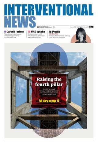
New editorial argues carotid artery stenting should be considered first-line
16 UAE uptake Miscommunication and multidisciplinary dysfunction mar global UAE access
18 Profile TZE MIN WAH Professor of interventional radiology tracks key career highlights
EDITOR’S LETTER
What’s in a name?
It’s been going on since I applied for residency in the 80s and I got to thinking about it for the umpteenth time after reading a Society of Interventional Radiology (SIR) blog post lamenting our sad situation. I stewed over it and concluded: Bloggers, you are wrong. This perennial is dead.
I REFER, OF COURSE, TO OUR PROFESSIONAL identity crisis, our toxic mix of pride and self-loathing. The conversation usually begins with recounted conversations with family or friends who ask: “What do you do?” Which is followed by contradictory pronouncements based on professional genealogy, taxonomy, or genetic expression (or bias). The conversation has not matured as rapidly as the field, and while I’d venture to guess most of us know what we are, we are challenged to find the right words, or we trip ourselves up with too much detail in a self-defeating push for irrelevant accuracy.
So let me tell you what you are:
1. You are a surgeon—a medical doctor who performs invasive procedures to diagnose and treat disease.
(If you agree, skip this paragraph): For those still stubbornly holding on to the life raft, remember that all doctors diagnose and treat disease. Surgeons are the subset that do some of this through procedural care. But remember, that most surgeons—us included—spend as much time in the non-invasive realm seeing patients, talking, diagnosing and prescribing, as they do operating. Gastroenterologists, cardiologists, anaesthesiologists, dermatologists, even radiation oncologists, are all surgeons. It’s defined by what you do, the highest form of your art.
2. You are also an interventional radiologist or ‘IR’. (If you agree, skip this paragraph): You are probably troubled by the ‘R’. I get it. But the marketplace has you labelled. Admit it—when you tell people that you’re an ‘IR’ you get the most—generally positive— name recognition. It could be worse. You could be called an ‘ENT’ (ear, nose, throat). An oto...what?
Embrace our acronyms/initialism. Stop the fussing and promote our ‘IR’ brand, it’s baked in exclusivity.
So, you’re an IR? What’s that?
The answer depends on the audience. Most doctors trained in the last 20 years know what
Editors-in-chief:
Publisher: Stephen Greenhalgh
Editor: Éva Malpass
Design: Terry Hawes
Advertising:
IRs are. So, I’ll focus on the public: IR is virtual reality surgery. We travel throughout the body in an environment created by imaging machines and solve problems by deploying highly engineered devices and drugs. It’s as cool as it sounds.
So, what’s in a name? I submit that we are the fortunate inheritors of a name that is at once accurate and trendy. I can’t think of a better place to be or a better time. Our adolescent identity crisis has passed. We are IRs!
BRIAN STAINKEN is a professor of interventional radiology and Interventional News’ co-editor-in-chief.
If you have comments on this issue or suggestions for upcoming editions write to eva@ bibamedical.com
Opening New TACE Horizons
CONFERENCE CIRCUIT
C-TRACT trial results
hail “new era” for postthrombotic syndrome following DVT
“These patients have suffered in silence because few physicians are aware of this complication or know how to effectively treat it,” said Suresh Vedantham, an interventional radiology physician and professor of radiology at WashU Medicine Mallinckrodt Institute of Radiology in St Louis, USA, and principal investigator of the C-TRACT trial. The results of the National Institutes of Health (NIH)-sponsored trial were presented at the Society of Interventional Radiology (SIR) 2026 annual scientific meeting (11–15 April, Toronto, USA) and were simultaneously published in the New England Journal of Medicine.
RESULTS FROM THE C-TRACT TRIAL showed that endovascular therapy, including iliac vein stenting and enhanced antithrombotic treatment, significantly reduced post-thrombotic syndrome (PTS) severity and improved quality of life compared with standard care alone in patients with moderate-to-severe PTS following deep vein thrombosis. Among 225 patients, severe disease rates fell substantially in the stent group at six months. Investigators said the findings provide strong evidence that severe PTS is treatable, though bleeding rates were higher with endovascular therapy.
During a roundtable hosted at SIR 2026, Vedantham spoke to the significance of the C-TRACT results: “This study suggests that even severe PTS is, in fact, a treatable condition in many patients,” he said. “We think that good patient selection
Prospective registry analysis supports radial access for uterine fibroid embolization
A SUB-ANALYSIS OF THE PROSPECTIVE RAVI (Radial Access and Visceral Interventions) registry found that uterine fibroid embolization (UFE) via radial artery access achieved high
and post-stenting management are going to optimise patient benefit, enable us to select the right patients and minimise bleeding from the anti-clotting medications,” Vedantham continued. “Standard treatments such as anticoagulation and compression are important, but for many patients, such treatments are not enough to improve leg symptoms and enable patients to function normally in their daily lives,” said Vedantham. “C-TRACT shows that patients with moderate-to-severe PTS and large vein blockage could benefit greatly from endovascular stent treatment, so patients should consult with an interventional radiologist for proper evaluation and management.” Vedantham stated that these results “usher in a new era” in which patient symptoms and quality of life reports are “no longer ignored”.
Catching up with Interventional News, Vedantham shared: “I am so pleased that the determined efforts of the C-TRACT investigators ultimately came to fruition, with demonstration of compelling clinical benefit for venous stenting in patients with PTS through six months. We have already started the work of further exploring this one-of-a-kind dataset to evaluate longer-term quality of life, cost-effectiveness, and important clinical-imaging correlations that pertain to venous stenting in this population.”
“This study suggests that even severe PTS is, in fact, a treatable condition in many patients”
procedural and technical success, with no major access-site complications.
Presented at the Global Embolization Oncology Symposium Technologies (GEST) annual meeting (14–17 May, New York, USA), the multicentre study of 70 women reported significant improvements in bleeding, bulk-related symptoms and quality of life through 12 months. Most patients were discharged the same day, while imaging data showed a 90–100% rate of fibroid infarction among those assessed.
MOTIV scaffold found to be “clearly superior at 12 months” to standard balloon angioplasty on efficacy endpoint
PRIMARY RESULTS FROM THE MOTIV-BTK trial showed that the Motiv sirolimus-eluting bioresorbable scaffold was superior to standard balloon angioplasty for treating infrapopliteal lesions in patients with chronic limb-threatening ischaemia. Presented at the 2026 Charing Cross (CX) Symposium (21–23 April, London, UK), the study reported a 22% absolute improvement in the composite endpoint of limb salvage and primary patency at 12 months, with benefits evident from six months. Safety was maintained, with low rates of adverse limb events and no perioperative deaths or major amputations.
Y-90 SIRT associated with complete local tumour control for unresectable HCC, study
finds
“THESE 12-MONTH RESULTS demonstrate the consistency of response achievable with personalised dosimetry,” said Armeen Mahvash (MD Anderson Cancer Center, Houston, USA) during the Society of Interventional Radiology (SIR) annual scientific meeting (11–15 April, Toronto, Canada). Results from the DOORwaY90 trial showed that yttrium-90 (Y-90) selective internal radiation therapy (SIRT) using personalised partition dosimetry achieved 100% local tumour control in patients with unresectable hepatocellular carcinoma—one of the “highest” reported response outcomes in Y-90 therapy, stated Mahvash. The study reported a 90% complete response rate and a 99% overall response rate, with preserved liver function in more than 95% of patients.
Randomised trial demonstrates MRIguided TULSA benefit for intermediate-risk prostate cancer
A RANDOMISED CONTROLLED TRIAL OF 212 patients with localised, intermediate-risk prostate cancer found that magnetic resonance imaging (MRI)- guided transurethral ultrasound ablation (TULSA) enabled faster recovery and less short-term impact on daily life than robotic prostatectomy. Presented at the Society of Interventional Radiology (SIR) annual scientific meeting (11–15 April, Toronto, Canada), the CAPTAIN trial showed TULSA patients experienced less blood loss, shorter hospital stays, less pain and quicker return to normal activities. Researchers will continue follow-up for 10 years to assess longterm cancer control and functional outcomes.
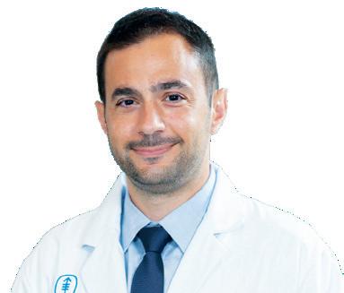
Suresh Vedantham
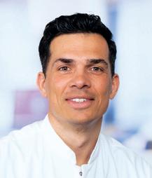
Armeen Mahvash
UK survey reveals need for national and international guideline to optimise radiation exposure during UAE
The development of diagnostic reference levels (DRLs) is pertinent to uterine artery embolization (UAE) as the “first step” to optimise the radiation risks to patients and staff while maintaining the highest level of patient care, results from a UK-wide survey have shown.
Omar Marei (Imperial College Healthcare NHS Trust, London, UK) presented the findings of RADSAFE, a UK survey of radiation doses during uterine fibroid embolization (UFE), at the European Conference of Embolotherapy (ET; 3–5 June, Valencia, Spain).
According to Marei, UAE remains a procedure with substantial radiation exposure which could benefit greatly from the implementation of national DRLs to improve patient and clinician safety. He stated that, “due to the radiosensitive nature of the ovaries, radiation exposure is inevitable during UAE”. Currently, no national or international guidelines exist on radiation doses during UFE, with some guidance suggesting a target dose area product (DAP) threshold of <50Gycm2 (gray square centimetre) in the literature.
Marei et al retrospectively collected radiation and demographic data from 11 vascular and interventional radiology (IR) centres in the UK for all patients undergoing UAE between 2020 and 2025. Data collected included uterine volume, number and size of fibroids, fluoroscopy time, procedure time, DAP and cumulative air kerma (CAK).
Data from 1,039 UAE procedures were included. Patients had an average age of 46 years, a median uterine volume of 733.6 and patients had an average of five fibroids. Median fluoroscopy time was 18.7 minutes, and median proce -
dure time was 90 minutes. The authors noted that uterine volume demonstrated a “weak” but statistically significant positive correlation with DAP. Centre experience demonstrated a strong but statistically insignificant negative correlation with median DAP, Marei stated. He continued that, based on current data, the proposed DRL for DAP is 85.5Gycm2 for standard UAE.
The investigators compared machine type and DAP; they included that 733, 300 and 5 UFE procedures were carried out using Siemens, Philips and GE HealthCare machines, respectively. Marei reported that, in a post-hoc analysis, the research team found that Siemens technology was associated with significantly longer fluoroscopy times compared to that of both Philips and GE HealthCare.
When questioned of the findings, Marei stated that this difference may be due to Philips models being “older”. He added that there are many factors involved in this finding, however, including operator experience and training, and the impact these may have on fluoroscopy duration.
Marei stated that age, uterine volume, size of largest fibroid, machine manufacturer and centre experience are key areas of consideration when planning a UFE procedure. Finally, the speaker stated that data collection will continue with the aim of strengthening and later publishing their findings to implement a national DRL.
“Radiation exposure is inevitable during UAE”
fluoroscopy (mins)
18.7
procedure time (mins)
FIRST SAFETY AND EFFICACY DATA for the Liverty (BD) transjugular intrahepatic portosystemic shunt (TIPS) graft have demonstrated durable control of variceal bleeding, refractory ascites and hydrothorax related to portal hypertension.
Presenting at the Society of Interventional Radiology (SIR) annual scientific meeting (11–15 April, Toronto, USA), Ziv Haskal (University of Virginia School of Medicine, Charlottesville, USA) reported that technical success was achieved in 100% of the 177 patients enrolled across 26 centres in the USA, France, and Germany.
Freedom from major complications through 30-days was 85.8%, Haskal stated. The speaker noted that novel smaller 6–7mm-diameter TIPS resulted in lower incidence and severity of hepatic encephalopathy (HE) at all timepoints compared to “traditional” shunt diameters of 8–10mm.
First-look Liverty TIPS trial data suggest new small-shunt era HEMBO-1 data suggest haemorrhoidal embolization with large particles and coils may improve bleeding symptoms
INITIAL RESULTS FROM THE PROSpective HEMBO-1 trial—presented at the Global Embolization Oncology Symposium Technologies (GEST) annual meeting (14–17 May, New York, USA) by Scott Thompson (Mayo Clinic, Rochester, USA)—indicate that haemorrhoidal artery embolization (HAE) using large particles combined with hydrogel coils may provide “significant” early symptom improvement.
The prospective trial aims to enrol 20 patients with bleeding-predominant internal haemorrhoids. At the time of presentation, 19 patients had been enrolled and 18 treated.
Thompson outlined that four patients (57%) had grade II haemorrhoids, and three patients (43%) had grade III disease. Median duration of bleeding symptoms was five years, with a range of one to 20 years.
At three-months, median bleeding and haemorrhoid bleeding scores each improved by two points, while quality-of-life scores improved by one point. When asked whether they were satisfied with their symptom improvement, six of the seven patients (83%) reported satisfaction.
Thompson stated that ongoing enrolment and longer-term follow-up are expected to further define the role of this embolization strategy in this patient population.
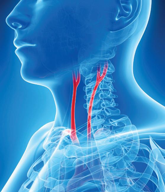
Editorial argues carotid stenting has reached “prime time” following CREST-2
An editorial published in CVIR Endovascular argues that carotid artery stenting (CAS) should be considered a first-line treatment option for appropriately selected patients with both symptomatic and asymptomatic carotid artery stenosis, contending that recent evidence, led by the CREST-2 trial results, marks a turning point for the procedure..
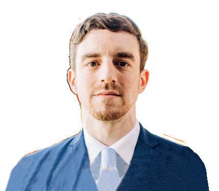
THE ARTICLE, LED BY STEFAN MÜLLERHülsbeck (Malteser Fördeklinikum St Katharina, Flensburg, Germany) and colleagues, examines how evolving evidence and contemporary clinical practice could reshape the role of CAS. The authors argue that the field has moved beyond questions of whether carotid stenting is effective and should instead focus on identifying which patients are most likely to benefit from intervention.
The publication follows the release of results from CREST-2, widely regarded as one of the most anticipated carotid trials in recent years. While debate continues over how the findings should influence practice, Müller-Hülsbeck et al suggest the study has strengthened the case for carotid stenting in carefully selected patients—particularly when performed in experienced centres using state-ofthe-art devices.
The authors ask: “Has carotid artery stenting reached prime time?” and their answer is unequivocally “yes”.
The editorial acknowledges that current guideline recommendations remain cautious, particularly in asymptomatic carotid disease. While carotid endarterectomy continues to be regarded as the standard treatment for many patients requiring intervention, the authors argue that the evidence base underpinning CAS has evolved considerably since many of those recommendations were written.
transcarotid artery revascularisation (TCAR) in appropriately selected patients. Müller-Hülsbeck and colleagues argue that these differing interpretations reflect a field in transition rather than one in disagreement.
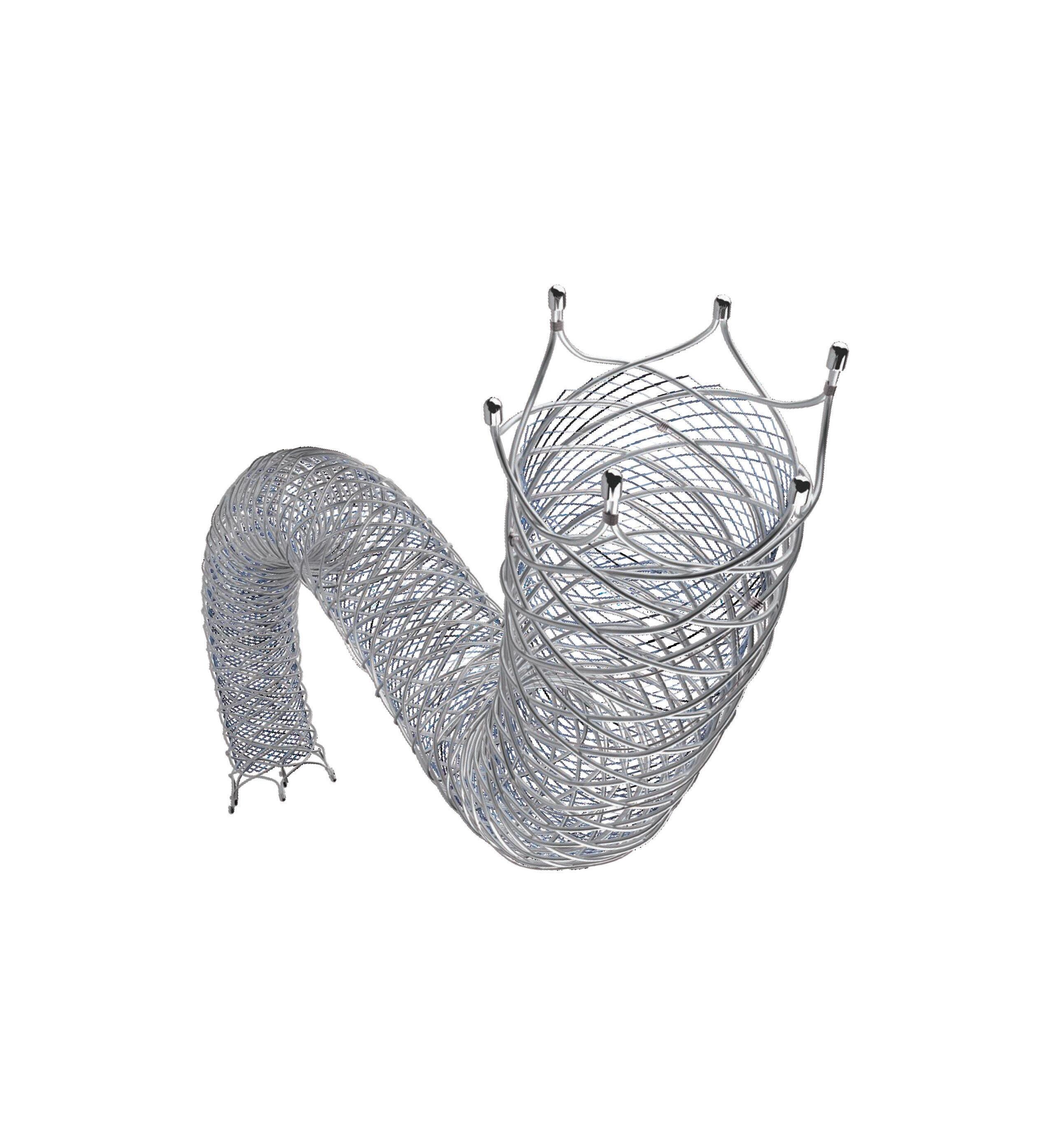
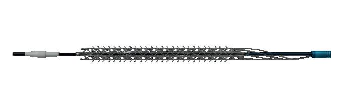
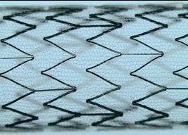
The editorial also places focus on significant changes in carotid stenting practice since the early randomised trials. The authors note that most patients enrolled in CREST-2 were treated with first-generation single-layer carotid stents. Since then, mesh-covered and dual-layer devices have increasingly become available, designed to reduce plaque prolapse and distal embolization by providing improved plaque coverage. Combined with advances in embolic protection systems, imaging, procedural technique and operator experience, they argue these developments have contributed to improved procedural safety. Consequently, the authors suggest that outcomes reported in contemporary practice may underestimate what can now be achieved using current-generation technology.
Müller-Hülsbeck et al emphasise that patient selection remains central to the discussion and advocate for a tailored approach based on plaque characteristics, anatomical suitability, life expectancy and procedural expertise. The authors also reiterate the importance of multidisciplinary decision-making and adherence to established standards of practice.
MELD-based mortality score performance differs by TIP indication, metaanalysis finds
MODEL FOR END-STAGE LIVER DISEASE (MELD)-based score performance differs by transjugular intrahepatic portosystemic shunt (TIPS) indication, reports a new meta-analysis comparing the prognostic accuracy of MELD and the Freiburg index of post-transjugular intrahepatic portosystemic shunt survival (FIPS) for predicting patient mortality.
Led by interventional radiologist Maria del Pilar Bayona Molano from the Miller School of Medicine in Miami, USA, the analysis included 11 studies accounting for 5,180 patients. Those included were undergoing TIPS for ascites or gastroesophageal bleeding (GEB).
The authors report that MELD, MELD-Na, and MELD 3 provide “good to excellent” discrimination for short-term mortality in patients undergoing a TIPS procedure for GEB, whereas their performance is “fair” in patients treated for ascites. This difference is particularly evident in the early postprocedural period (30–90 days), where MELD-based models consistently achieve higher area under the curve (AUC) values in bleeding cohorts than in ascites cohorts.
SWEDEPAD 2 prompts guideline update for asymptomatic PAD and intermittent claudication
THE EUROPEAN SOCIETY FOR VASCULAR Surgery (ESVS) has incorporated new data from SWEDEPAD 2 and other randomised trials into updated recommendations on the management of asymptomatic peripheral arterial disease (PAD) and intermittent claudication.
Writing in the European Journal of Vascular and Endovascular Surgery (EJVES), SWEDEPAD investigator Joakim Nordanstig (University of Gothenburg, Gothenburg, Sweden) and colleagues recommend clinicians “should carefully balance the limited short-term procedural benefits of paclitaxel-coated devices against the lack of proven quality-of-life improvement and the persisting uncertainty regarding long-term mortality risk”.
Rather than viewing CREST-2 in isolation, they place it alongside findings from studies including SPACE-2 and ECST-2, as well as contemporary registry data, arguing that together they demonstrate the safety and effectiveness of carotid stenting in appropriately selected patients.
However, not all commentators have interpreted the evidence in the same way, as the authors highlight differing reactions following publication of CREST-2. Some have suggested that the trial reinforces the value of intensive medical therapy, while others have argued that it supports broader use of carotid revascularisation. The Society for Vascular Surgery (SVS), for example, concluded that the study “does not change standards of care”, while continuing to support carotid endarterectomy, transfemoral CAS and
Looking ahead, Müller-Hülsbeck and colleagues call for future guideline updates to reflect the growing body of evidence supporting CAS. They also acknowledge that while further randomised trials evaluating newer-generation stents would be valuable, such studies may prove difficult to undertake because of cost and logistical challenges.
Concluding their editorial, the authors underscore the “bright future” ahead for stenting and minimally invasive treatment in “experienced hands”. They determine that the combination of contemporary evidence, technological advancements and developing operator expertise will enable carotid stenting to be placed alongside surgery as a routine treatment option for selected patients, rather than as an alternative reserved for highly specific clinical scenarios.
The authors note that ESVS focused guideline updates are issued to convey important data that emerge between the publication of full guidelines. They state that, in this case, updated evidence that has emerged since the society’s 2024 publication on asymptomatic PAD and intermittent claudication management “suggested limited clinical benefit and possible long-term harm from paclitaxel-coated devices” in this patient population.
“The update consistently emphasises supervised exercise therapy and best medical therapies as first-line treatment,” the authors write, summarising a key takeaway from the EJVES paper. They add that endovascular revascularisation is recommended only for “persistent lifestyle-limiting symptoms despite conservative therapy”. Furthermore, the authors advise that paclitaxel-coated devices “may be considered only for selected cases with restenosis following transparent discussion of uncertain long-term safety and, at best, modest symptomatic benefit”.
Read it your way
We want to ensure you still receive your copy of Interventional News the way that works best for you.
To reconfirm your subscription, scan the QR code. You can also let us know if you would like to change the way you read it by opting to receive a digital instead of a printed version or by moving from digital to print
FROM THE JOURNALS
Long-term survey maintains “lifechanging” benefit of UFE, despite symptom recurrence in younger patients
Women under 40 years old who undergo uterine fibroid embolization (UFE) are highly likely to experience symptom recurrence before menopause and may require additional procedures, yet report high levels of treatment satisfaction.
THESE WERE THE KEY TAKEAWAYS OF A new long-term retrospective survey across two institutions led by Elisabeth R Seyferth (Perelman School of Medicine, Philadelphia, USA) et al. Published in the journal CardioVascular and Interventional Radiology (CVIR), the authors identified 976 women who underwent UFE between 2001–2021, 173 of whom were younger than 40 at the time of treatment; 80 of 173 participated in the survey.
Median age at embolization was 37.2 years and when surveyed was 52.7 years. Among respondents, 70% (56/80) and 18.8% (15/80) of patients reported “a lot” or “a little” symptom improvement, respectively, in the first six months after UFE; 11.2% (9/80) reported no change. Of those with improvement, symptoms returned in 57.7% (41/71) of patients, with median time to recurrence of 11 years. Freedom from recurrence of symptoms was 95.8% at one year, 67.6% at five years, and 50.7% at 10 years. Freedom from hysterectomy for recurrent symptoms was 98.6% at one year, 84.5% at five years, and 71.8% at 10 years. Patient satisfaction having received UFE was a median of 5 on a scale of 1–5 (5=very satisfied).
Seyferth and colleagues report that 12.5% of women attempted to become pregnant after UFE. Of these, 40% (4/10) had a successful live birth. In their discussion, the authors note that, although
fertility rates for UFE remain limited, this finding is comparable to the 2008 trial by Michal Mara (Charles University, Prague, Czech Republic) et al which reported a labour rate of 48% among patients who attempted to become pregnant after myomectomy.
The authors note that most patients endorsed high satisfaction with UFE despite high rates of symptom recurrence, with multiple patients choosing “very satisfied”, stating that UFE had “changed their life”. Notably, while some patients mentioned that they were counselled that symptoms may recur, others felt that they were not told of that possibility and therefore chose a lower satisfaction score. Although this finding emphasises the importance of appropriate pre-procedure counselling across centres, Seyferth and colleagues maintain that “satisfaction following UFE remained high, with many experiencing years of symptom relief”.
“Satisfaction following UFE remained high, with many experiencing years of symptom relief”
ENDOLOGIX HAS ANNOUNCED PUBLICAtion of the 36-month results from the DETOUR2 clinical trial demonstrated sustained safety and durable outcomes for percutaneous transmural arterial bypass (PTAB) using the Detour system in patients with complex femoropopliteal disease. The prospective, single-arm, international, multicentre study enrolled 202 patients across 32 sites with long, complex superficial femoral artery (SFA) lesions. The cohort had a mean lesion length of 32.7cm, with 96% chronic total occlusions and 70% severely calcified lesions.
At 36 months, primary patency was 58.2%, while patency defined as freedom from occlusion was 84.0%. Freedom from clinically driven target lesion revascularisation was 66.8%, and freedom from major adverse limb events was 64.5%. Freedom from major amputation remained 97.9%, unchanged from 12 months, while symptomatic deep vein thrombosis occurred in 4.1% of patients, with no pulmonary emboli reported. Clinical success, defined by improvement in Rutherford classification, was achieved in 96.7% of patients.
TARE standardisation needed between North America and Europe, finds CIRSE survey
A NEWLY PUBLISHED INTERNATIONAL survey by the Cardiovascular and Interventional Radiological Society of Europe (CIRSE) offers a real-world evaluation of intercontinental disparities in transarterial radioembolization (TARE) practices between North America and Europe. Among key variations, the survey, published in the journal Current Oncology, reveals differences in hospitalisation, product utilisation, and procedural techniques.
Led by Grace Keane from the University Medical Centre Utrecht (Utrecht, Netherlands) et al, a survey of 32 multiple-choice questions was distributed to CIRSE members between November and December 2022. The questions addressed steps of the TARE workflow, including treatment work-up, planning and dosimetry, intervention, follow-up and innovations.
Responses were obtained from 30 countries and 133 hospitals, including 87 European and 21 North American centres. North America predominantly used yttrium-90 (Y-90) glass microspheres, whereas Europe used Y-90 resin. Procedural differences included the adoption of intra-procedural computed tomography (CT) imaging, employed by all North American sites, versus 89% of European sites. Outpatient treatments were favoured in North America (85%), while in Europe, most patients remained hospitalised for one night (51%). Both regions increasingly emphasised dosimetry-guided treatments, with personalised dosimetry planning in 71% and 84% of North American and European sites, respectively.
Keane et al note that this analysis illustrates variability in TARE practices, underscoring the need for standardisation efforts, asserting that organisations such as CIRSE must play a “crucial role” in achieving harmonised global practices.
Iceball margin of less than 5mm associated with local treatment failure following cryoablation for RCC
INSUFFICIENT ICEBALL MINIMAL treatment margins (MTMs)—equalling <5mm— when delivering computed tomography (CT)-guided percutaneous cryoablation of cT1a renal cell carcinoma (RCC) are associated with local treatment failure, finds a recent study led by Jens Borgbjerg (Akershus University Hospital, Nordbyhagen, Norway) et al Between 2015–2022, 575 RCCs were identified with ≥12 months of imaging follow-up. Of these, 17 demonstrated local treatment failure with a median follow-up of 1,173 days. Tumours with local treatment failure were larger than tumours with local control (29.2mm vs. 23.7mm), and MTM was smaller in the local treatment failure group (0.4mm vs. 7.1mm). The authors state that all in-zone failures occurred in tumours with margins of less than 5mm, supporting their conclusion that inadequate margins cause failure.
BEYOND THE VESSEL
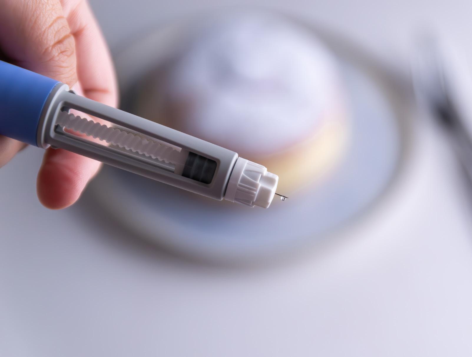
IR-specific guidance needed for management of patients taking GLP-1 agonists
Recognising an increase in patients taking glucagon-like peptide 1 (GLP-1) agonists, a recent review article has underscored the importance of understanding its effects and implications for quality of care and daily practice in interventional radiology (IR). Led by Warren Clements (Alfred Health, Melbourne, Australia), the research team highlight the “urgent” need for IR-specific evidence regarding pharmacological effects and periprocedural implications in patients who take GLP-1 agonists.
Published in the Journal of Medical Imaging and Radiation Oncology, Clements and colleagues first highlight the significant growth in the use of GLP-1 agonists, referencing a report which states that overall GLP-1 agonist use has increased by over 700% among individuals without type 2 diabetes.
The authors note that GLP-1 agonists may have a positive influence on IR practice, stating that higher body mass index (BMI) is a common risk factor for increasing the rate of post-procedure complications, including impaired wound healing and thromboembolism. In reducing BMI, they note that additional practical difficulties can also be avoided such as during fluoroscopic procedures, as increased tissue thickness reduces image quality.
The authors equally present the potential downsides interventional radiologists must consider in these patients, underscoring the impact of GLP-1 agonists on fasting glucose levels. They discuss the risks of hypoglycaemia but note that, despite GLP-1 agonists’ enhancement of insulin secretion, they carry a “relatively low risk” of causing the patients to develop the condition due to their
glucose-dependent mechanism.
However, the research team highlight that, in diabetic patients, the use of multidrug anti-hyperglycaemic regimens could have implications in the periprocedural setting, with some studies suggesting increased incidence of hypoglycaemia in these patients. In their recommendation, the authors state that it may be beneficial to treat all patients taking GLP-1 agonists like regular diabetic patients, “placing them first on a morning list, when possible, to reduce prolonged and potentially unnecessary fasting”.
Continuing, the authors underscore aspiration as a potential concern when performing IR procedures for patients taking GLP-1 agonists. This relates to delayed gastric emptying when providing IR-initiated sedation, they state.
“Minimal to moderate IR-initiated sedation is often preferred and sufficient as a balance between achieving ideal analgesia and anxiolysis, while minimising the need for specialist anaesthetic support, which is not always readily available and is not always cost-effective. This approach raises concerns about the theoretical suitability of continuing GLP-1 agonists in the
perioperative setting, particularly with the use of moderate sedation in an unsecured airway,” write the authors.
Further, they highlight that interventional radiologists must consider the effect on gastric emptying alongside gastric-specific procedures such as radiologically inserted gastrostomy, noting that a prolonged period of fasting may be necessary to ensure adequate gastric emptying.
They explain that, in settings where evidence is “not clear”, it is relevant to turn to society commentary to provide guidance via pooled expert consensus, yet there remains a “relative disconnect” between the service interventional radiologists provide and existing recommendations for other specialties.
“Looking forward, it is now time for the provision of IR-led evidence on the safety of using GLP-1 agonists specifically for IR procedures, so that IR societies can provide their own guidelines that are based not just on expert consensus but also relevant evidence,” the authors write. “This may require leadership from societies and eminent interventional radiologists in each region to coordinate. The importance of IR-specific guidelines cannot be overstated, as the recovery and risk profile is different from endoscopy and open surgery.”
For now, Clements et al offer three options that interventional radiologists can follow in these circumstances. Option 1—modelling care on surgical protocol that suggests ceasing subcutaneous GLP-1 agonists for seven days prior to a procedure, which the authors assign as a “conservative approach”.
Option 2 is to continue GLP-1 agonists unless performing a procedure in the gastric lumen, which is “likely to provide a more real-world approach for interventional radiologists where minimal-moderate sedation is associated with a very low background aspiration risk”, the authors write.
“Until a major IR society provides a consensus recommendation, interventional radiologists will have to make these decisions based on the evidence provided in this review, their own experiences, and local hospital influences”
Finally, Option 3 is to consider a hybrid model, which is triaged based on procedure type and patient risk.
“There is no clear evidence-based answer, with studies showing positives for each option and different societies making recommendations for each. Additional universal diabetic precautions may be useful such as observation of fasting blood sugar levels in patients where medication was not ceased and performing procedures first on a morning list.
“Until a major IR society provides a consensus recommendation, interventional radiologists will have to make these decisions based on the evidence provided in this review, their own experiences, and local hospital influences.”
The authors conclude that IR-specific evidence is urgently needed regarding GLP-1 agonists and recommend that major societies take “a leadership role in promoting research” to provide clinical practice guidelines that local interventional radiologists can follow moving forward.
The number of centres enrolled in the International Accreditation System for Interventional Oncology Services (IASIOS) has surpassed 100, marking five years since the accreditation framework was launched with an initial cohort of 12 pilot institutions.
ACCORDING TO IASIOS, PARTICIPATING centres now span university hospitals, dedicated cancer centres and regional hospital networks across Europe, Asia, Australia, the Americas and the Middle East. The programme is based on the Cardiovascular and Interventional Radiological Society of Europe (CIRSE) Standards of Quality Assurance in Interventional Oncology, which are endorsed by more than 40 oncology and radiology societies.
The standards are intended to provide a framework for assessing institutional organisation and quality assurance in interventional oncology (IO), focusing on areas including multidisciplinary collaboration, patient selection, peri-procedural care, complication monitoring and follow-up, rather than specific clinical techniques.
IASIOS seeks to operationalise those standards through a three-tiered structure. Institutions first enrol and undertake a self-assessment of their services before progressing to accredited centre status following external peer review. A centre of excellence designation is awarded to institutions meeting both core and extended criteria, including academic activity and longitudinal outcomes monitoring.
process has allowed us to thoroughly audit our practices and improve them. Much more than an accreditation process, IASIOS represents an affirmation of our unwavering commitment to excellence; it’s a promise to our patients and the desire to remain at the forefront of oncology treatments.”
Similarly, Uli Fehrenbach and colleagues at Charité University Hospital Berlin (Berlin, Germany) said the process enabled their department to “refine our workflows and align our services with international gold standards”, adding: “This achievement not only validates our high quality of patient care but also motivates us to continue advancing our clinical practice. We are proud to be part of the global IASIOS community and look forward to further improving oncological outcomes through innovation and standardised excellence.”
“It’s a promise to our patients and the desire to remain at the forefront of oncology treatments”
also achieved accredited centre status, while St Jude Children’s Research Hospital in Memphis, USA has become the first paediatric institution to enrol in the programme.
In Portugal, Paulo Donato, Alfredo Agostinho and colleagues at Hospital da Universidade de Coimbra described the accreditation process as “an excellent opportunity to evaluate how cancer patients are treated by IR at our hospital”, adding that it “allowed us to thoroughly assess our procedures, refine our methodologies, and strengthen our commitment to quality and patient-centred care.”
According to IASIOS, one objective of the framework is to encourage greater integration of IO services within multidisciplinary cancer care by requiring participating centres to demonstrate formal involvement in tumour boards, defined referral pathways, structured communication with referring clinicians and longitudinal patient follow-up.
The growth of structured quality assurance frameworks comes as IO continues to seek broader integration within multidisciplinary cancer care. While the role of image-guided cancer therapies has expanded, the degree to which interventional radiologists participate in treatment planning varies between institutions and healthcare systems.
Referral pathways, formal involvement in multidisciplinary tumour boards and long-term patient follow-up are increasingly recognised as important components of an established IO service. The CIRSE Standards of Quality Assurance in Interventional Oncology include these organisational measures among the criteria assessed during accreditation.
IASIOS states that requiring centres to demonstrate consistent participation in multidisciplinary cancer pathways aims to strengthen the visibility and accountability of IO within wider oncology teams.
Andreas Adam, chairperson of the IASIOS steering board said: “Patient care matters in all of interventional radiology [IR], but it is particularly important in the treatment of cancer. By emphasising the clinical aspects of IR, IASIOS ensures that patients treated in accredited centres can be confident of receiving the best possible care, not only in the procedure room but also in the ward and in the outpatient clinic.”
Lucas Smagge, Behnam Mohseny and colleagues at Erasmus MC (Rotterdam, the Netherlands) reported that participation prompted a review of pre- and post-procedural care pathways and patient safety protocols. “This meticulous approach has allowed us to optimise our practices where necessary, reinforcing our commitment to delivering consistent, high-quality care. As a result, we have built greater trust and confidence among our patients and colleagues, ensuring superior outcomes now and in the future.”
Afshin
Gangi and Roberto Luigi Cazzato
IASIOS states that the programme is designed to support departments at different stages of development while promoting greater consistency in the delivery of IO services.
Several participating centres described the accreditation process as an opportunity to review existing practices and identify areas for improvement.
Afshin Gangi and Roberto Luigi Cazzato, from the University Hospital of Strasbourg in Strasbourg, France—the first institution to achieve IASIOS centre of excellence designation—said: “This journey has allowed us to surpass ourselves by constantly questioning our practices in a quest for excellence that inspires respect and trust. The
The programme has continued to expand internationally since its launch in 2021.
Mohammad Arabi, Shaker Alshehri and colleagues at King Abdulaziz Medical City (Riyadh, Saudi Arabia) said “the accreditation process challenged us to reflect, refine, and grow, not only in clinical delivery but also in teamwork and collaboration. We took a deeper look into our workflows, identified areas for improvement, and strengthened our commitment to excellence.”
In North America, the Mallinckrodt Institute of Radiology at Washington University in St Louis became the first accredited IASIOS centre in the USA. “We are the first institution in the USA to become associated with this accreditation and hope to lead the way for other IO practices in the USA,” said Jack Jennings and colleagues. Since then, the National Institutes of Health Clinical Center’s Center for Interventional Oncology has
To date, six institutions have been designated IASIOS centres of excellence: the University Hospital of Strasbourg (France), Singapore General Hospital (Singapore), Institut Bergonié (Bordeaux, France), Leiden University Medical Centre (Leiden, the Netherlands), SLK-Kliniken Heilbronn (Heilbronn, Germany) and Cantonal Hospital Winterthur (Brauerstrauss, Switzerland).
Representatives from Singapore General Hospital (Singapore, Singapore) said: “The award reinforces our responsibility to lead, educate and collaborate globally to promote high-quality IO services so that patients everywhere can benefit from this emerging field of cancer care.”
Despite growth in the number of participating centres, access to IO services continues to vary considerably between countries and institutions. Differences in infrastructure, workforce, referral pathways and resources mean that the availability of specialist IO services remains inconsistent.
IASIOS says one objective of expanding its international network is to encourage greater standardisation of organisational practices across diverse healthcare settings. The organisation argues that wider adoption of quality assurance frameworks could help support more consistent service delivery and facilitate the integration of IO into established cancer pathways.
The programme has now enrolled more than 100 centres worldwide, although IASIOS acknowledges that disparities in infrastructure and access remain, and that broader implementation of quality standards will require continued international engagement.
Predictability above all: Key characteristics of a successful drainage catheter
For interventional radiologists managing patients with complex oncological disease, percutaneous drainage is rarely a standalone procedure. Instead, it represents one component of a broader multidisciplinary treatment strategy, with the overall aim to improve symptoms or allow the patients to proceed with further oncologic interventions.
SHARING HIS CLINICAL EXPERIENCE, Franco Orsi, director of the Interventional Radiology department at the European Institute of Oncology (Milan, Italy), says that drainage procedures are frequently required in patients with malignant biliary obstruction, postoperative or infected fluid collections, liver abscesses and in selected patients with the need for gallbladder drainage when surgery or endoscopic treatment is not appropriate or has not been successful.
Working within a comprehensive cancer centre means that many patients present with anatomically complex disease and significant treatment histories, factors that place particular emphasis on the performance and reliability of drainage devices to allow patients to advance to further treatment, Orsi explains.
“In oncological patients, drainage is usually part of a multimodal treatment pathway. For example, patients may need systemic therapy but cannot receive it because their bilirubin level is too high, so we first need to palliate the obstruction before treatment can begin,” he describes.
Procedural challenges beyond initial placement
cal patients often require long-term catheter management.
Defining device characteristics
To Orsi, predictability is one of the key characteristics sought after in any interventional radiology (IR) device, “and drainage catheters are no exception”, he adds. “The catheter should advance smoothly while providing adequate pushability and torque control—these qualities are extremely important in daily practice.” Under fluoroscopy, the relationship between the marker, the drainage holes and the pigtail should be “unambiguous” he adds, while the locking mechanism should be “simple and reliable”.
In patients who have complex anatomy due to tumour-related distortion of the organs, previous surgery, or earlier interventional procedures, achieving access can be challenging, Orsi explains.
“In biliary procedures, one of the most difficult scenarios is when the ducts are completely disconnected after surgery, such as following an anastomotic leak. The ducts may also be minimally dilated, making access difficult. In benign obstructions, crossing the stricture itself can be particularly challenging, and the risks of infection and bleeding must always be considered.”
Yet, he states that technical placement is only the first step of the procedure that interventionists must consider, and the catheter must continue to function reliably after the patient leaves the interventional suite. “Dislodgement, occlusion, kinking, leakage, skin discomfort and the burden of catheter care may become clinically relevant during the following days or weeks,” Orsi says, acknowledging that oncologi-
an angled tip—it can sometimes exit through one of the side holes. “If the holes are clearly visible under fluoroscopy you can use torque control to redirect the wire and continue straight through to the catheter tip.”
An added benefit of reducing catheter exchange time is reduced radiation exposure to the operator. “Lower radiation exposure is important for operator safety,” Orsi says, “I started doing this work 30 years ago, and, at that time, devices and materials were far less advanced, and we often spent a long time positioning catheters correctly. Radiation exposure should, of course, be minimised for every patient. For operators, there is the additional concern of cumulative exposure across multiple procedures performed every day.”
Improving everyday practice
Overall, Orsi describes the ReSolve catheter as a technically well-developed device whose advantages lie in practical refinements rather than changing the underlying principles of drainage procedures.
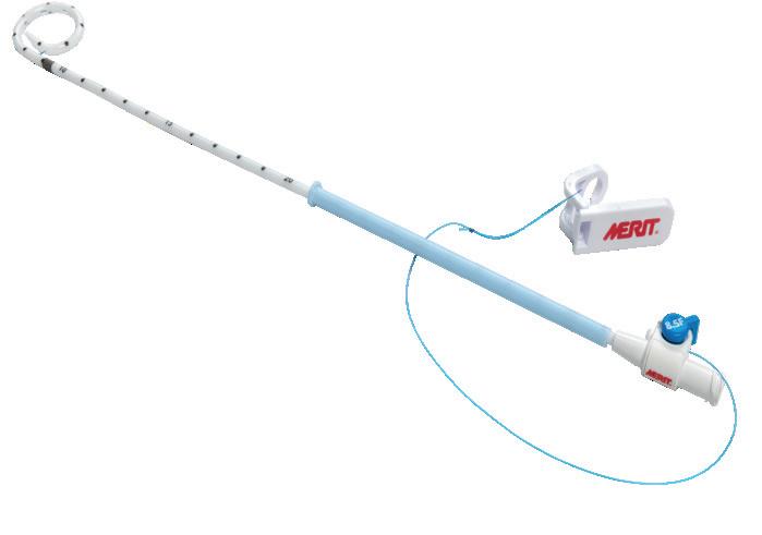
Day-to-day, Orsi uses the ReSolve® catheter (Merit Medical) for his drainage procedures. He states that the characteristics of the device contribute to procedural efficiency by allowing him and his team to concentrate on patient anatomy rather than device behaviour.
Returning to the concept of predictability, he details that the formation of the pigtail before deployment is critical: “The pigtail should form exactly as expected upon deployment, ensuring it sits correctly within the cavity for fluid collection.” This factor becomes increasingly important when fluid has been completely drained and the cavity walls collapse. If the catheter tip remains in the same plane as the pigtail, it is less likely to become embedded in the wall.
“This may not be the first feature interventionists would think about because they’re so used to benefitting from the design, but it’s a very important one,” Orsi says.
Other important features include the device’s locking mechanism and the configuration and size of the drainage holes. Regarding the former, Orsi notes that the locking suture should pass separately, rather than through the catheter lumen. This is a particularly valuable feature, as, in his experience with other devices, he has seen air leakage through the locking system. “When you’re dealing with pleural effusions or pneumothorax, air escaping through the locking mechanism can become a significant clinical problem,” he states.
Ensuring that drainage holes are distributed evenly along the catheter also helps during catheter exchange. Orsi illustrates that, when advancing the guidewire—especially one with
“Its value is not that it fundamentally changes the drainage procedure, but one that reduces unnecessary device-related difficulties during an already complex intervention,” he states. “A well-designed catheter can support patient safety and comfort by facilitating controlled placement, limiting unnecessary manipulation and providing stable and effective drainage. A low-profile and manageable external component may also make catheter care easier for both the patient and the clinical team.”
However, he underscores an important principle: “The catheter alone does not determine the clinical outcome”, instead, outcomes depend primarily on correct patient selection, the timing of drainage, imaging guidance, operator technique, infection control, management of coagulation and appropriate follow-up.”
Orsi therefore defines the role of a high-quality catheter as a device which supports the procedure and reduces avoidable device-related problems: “Its an important component of treatment, but only one component. A good drainage catheter does not replace clinical judgement or technical expertise. Its value is that it is predictable, allowing the operator to concentrate on the anatomy and on the patient rather than on the device.”
Franco Orsi
ReSolve drainage catheter
International societyendorsed consensus documents on liver tumour thermal ablation published
An international panel of experts has released two Delphi consensus statements for liver tumour thermal ablation, outlining procedural and practice standards, as well as margin assessment. The results were published earlier this year in The Lancet Oncology.
CONCERNING THE FORMER, KEY takeaways include prioritisation of the least invasive approach—typically percutaneous, while margin adequacy is reaffirmed as the principal technical goal. Insights from the latter document outline that margins should be assessed quantitatively in three dimensions with contrast-enhanced computed tomography (CT) or magnetic resonance imaging (MRI), preferably intraprocedurally with ablation confirmation software.
The consensus was initiated by co-principal investigators Reto Bale (Medizinische Universität Innsbruck, Innsbruck, Austria) and Bruno Odisio (MD Anderson Cancer Center, Houston, USA) who appointed a steering committee which also included Iwan Paolucci (MD Anderson Cancer Center, Houston, USA), Grego Laimer (Medizinische Universität Innsbruck, Innsbruck, Austria), Edward Johnston (Royal Marsden Hospital, London, UK), and Christiaan Overduin (Radboud University Medical Centre, Nijmegen, Netherlands).
In the first paper, consensus was achieved for 94 (70%) of 135 statements, while the second achieved consensus for 150 (75%) of 199 statements. The authors add that strong consensus was observed between interventional radiologists and surgical oncologists—who were actively involved with the initiative—with only 12 (6%) of the199 statements showing significantly different ratings.
To develop the consensus, 72 global experts were engaged during three iterative rounds of Delphi scoring across 135 statements relating to procedural and practice standards, and 199 statements relating to margin assessment between June and December 2024.
The initiative was held both in-person in Innsbruck and online, to support the attendance of specialists “from every continent”, said Odisio speaking to Interventional News alongside Bale ahead of the release of the consensus papers. “It was touching to see everyone come together,” Odisio continued. “There’s motivation— there is desire to improve local tumour control and success rates with ablation, but there isn’t a roadmap for how we can implement that in clinical practice, and that was the main incentive.” As Bale added: “[The initiative] is all about standardisation and reliability.”
Among key points of consensus in practice standards were organ displacement techniques, which were endorsed to maintain safety and expand treatable indications when performing liver tumour thermal ablation, and the assertion that complex ablations should only be performed by experienced operators (more than 100 previous cases). For margin assessment, participants agreed that margins should be assessed and documented for every treated tumour and categorised as A0 (tumour completely covered with sufficient margin), A1 (tumour completely covered but insufficient margin), or A2 (portion of tumour remains unablated).
Concerning practice standards, consensus was not reached on statements including: the minimum number of cases required before independently performing liver tumour thermal ablation; the appropriateness of offering thermal ablation if the tumour cannot be completely covered by the ablation zone; the use of coaxial or non-coaxial needles affects case complexity; and, if the operator experience should directly influence selection of ablation settings, among others.
In the second paper, consensus was reached on the need for standardised methodology and outcome reporting in scientific studies focusing on ablation margin assessment, while statements such as the maximum time between preprocedural imaging and ablation did not. Elsewhere, consensus was not reached
on whether sufficient evidence exists to define a minimum ablation margin threshold for non-colorectal liver metastases or the acceptable increase in procedural time to perform ablation margin assessment.
Bale, on why 100% consensus was not reached, stated: “I sense that insufficient evidence [at the time], or the now emergent evidence base following the publication of the COVER-ALL and COLLISION trials, is what left experts relying on opinion rather than data across several statements,” he said. “This, as well as conceptual disagreement on how to define some of the core terms, such as case difficulty and ablatability, which made clear that future research should focus on the definition of such terms.”
In Odisio’s view, the consensus papers sit alongside recent evidence, bolstering the position of thermal ablation for liver tumours and provides a guide to implementing the technique within practice. “It’s a document that can demonstrate the necessary steps to achieve the same results that are being published with very high rates of local tumour control,” said Odisio.
“It’s also a document that will guide us in the next steps in terms of research, to better understand the role of locoregional therapy for patients regarding how local therapy with curative intent could potentially have systemic effects,” Odisio continued.
“It’s a document that can demonstrate the necessary steps to achieve the same results that are being published with very high rates of local tumour control”
Bruno Odisio
Both Bale and Odisio commented that the consensus papers seek to “democratise” ablation across centres internationally. However, they acknowledged heterogeneous access to the latest technology across hospitals due to funding can affect outcomes.
“This is very important. As a pragmatic interim approach, the use of image fusion with manual quantitative minimum ablation margin assessment, which was not explicitly included in the questions, was endorsed,” stated Bale. “Image fusion software is available on many standard scanners, and these have been shown to improve local tumour control when compared with side-by-side comparisons.”
Odisio explained that, during the consensus meetings, this point was carefully considered. “It’s a difference between enforcing or advising the use of a technology. For instance, we had a consensus recommendation on the use of ablation confirmation software given the data we have right now, but we understand that the adoption of that software might take time and money,” he said. “Finance is a pervasive element here, so we also provide alternatives in our discussion to show that, even if you don’t have ablation confirmation software, there are still tools available that can help you progress. The statements create a roadmap, but at the same time, balance constraints.”
The guidelines are endorsed by the European Society of Surgical Oncology (ESSO) , the Cardiovascular and Interventional Radiological Society of Europe (CIRSE), and the Society of Interventional Oncology (SIO).
Reto Bale
Bruno Odisio
New GAE position statement bridges ‘huge gap’ for treatment of knee osteoarthritis
A new Society of Interventional Radiology (SIR) position statement has sought to provide evidence-based support for the use of genicular artery embolization (GAE) as a minimally invasive treatment option for patients with symptomatic knee osteoarthritis (KOA). Following its publication in the Journal of Vascular and Interventional Radiology (JVIR), co-authors Osman Ahmed (Joint and Vascular Institute, Libertyville, USA) and Jafar Golzarian (North Star Vascular and Interventional, Minneapolis, USA) unpick central tenets of the statement and their significance in today’s clinical landscape.
Before GAE, what treatments were available for KOA? Did GAE fill a gap for this condition?
JG: [Before GAE] interventional radiology (IR) had little to offer. Centres would sometimes offer aspiration or perform corticosteroid injections and perhaps nerve ablation, but before embolization, our only access to knee intervention was post-surgical total knee replacement bleeding and haemarthrosis.
OA: Prior to now, most interventional radiologists were not treating KOA. While we have the ability and expertise to do so, only a small percentage did. Most clinicians treating KOA were performing nerve ablation or cryoneurolysis to manage pain, which allowed us to breach the threshold into embolization for a wider set of indications such as musculoskeletal intervention.
How does the position statement define the ‘optimal patient’ for GAE?
JG: The statement suggests that the ideal candidates are patients who have failed conservative management and are either poor surgical candidates or would prefer not to undergo surgery. However, I would push the envelope on this concept a bit further.
One of our recommendations was based on data which demonstrated that GAE provides over two years of stability and improvement of symptoms. And so, in patients who are good candidates for surgery but would like to postpone, GAE is an option.
We know that surgery is associated with continuous pain in up to 20% of the patients after the procedure and the lifespan of a knee replacement is about 15 years. Can we postpone surgery with
GAE so the patient will undergo only one surgical procedure in their lifetime? That was a key discussion point when putting together the statement. OA: There’s a huge treatment gap and, right now, for better or worse, we have a relatively broad indication for GAE which really tries to bridge the gap between conservative management and surgery.
We have a lot to offer patients, and I think the statement does acknowledge that there’s a tremendous amount of work that needs to be done to further refine patient characteristics that indicate whether they will be a good candidate for GAE or not.
How does the statement handle cost-effectiveness debates regarding durability of benefit and repeatability of treatment?
symptom improvement, as Golzarian previously mentioned. That cost is very low as opposed to steroid injections, which are cheap but must be administered at the hospital every three months, incurring several additional costs. Ultimately, we did find that GAE and radiofrequency ablation (RFA) was more cost effective than steroid injections.
What is the learning curve for GAE in KOA? What experience must an interventionist have to adequately perform this treatment?
JG: Do you want the correct answer or the politically correct answer?
Either way, it’s important for the SIR to release this statement as the skills required to perform GAE are intrinsic to IR training. For any new procedure there is always a learning curve, but the learning curve for an experienced interventional radiologist is different to that of someone who is doing knee arthroscopy and wants to perform embolization.
In general, I would say it’s a technique that is totally accessible to interventional radiologists. An understanding of catheterisation of small vessels is essential, however. The genicular arteries can be challenging which may require additional time during the initial stages of the procedure.
If you are interested in performing GAE, I recommend watching it being performed—try to learn about anatomy, read publications, go to meetings and talk about it.
OA: The godfather of embolization [Golzarian] said it perfectly, but I would emphasise that every procedure is a risk. This is an elective procedure for benign disease where there are multiple options, and so our risk is that the tolerance threshold for complications is very, very low. Risk to the patient should be close to zero. So, those performing GAE should be very comfortable with embolization.
What prior methodological limitations does the position statement address?
OA: Methodologically, for new procedures or innovations, studies always start in a similar fashion. You’re always going to start with an early feasibility study to say, ‘Hey is what we’re doing safe?’ And once you know it’s safe, you ask, ‘Well then, how effective is it?’
“Can we postpone surgery with GAE so the patient will undergo only one surgical procedure in their lifetime?”
Jafar Golzarian
OA: We recently published on cost-effectiveness, although not directly comparing knee replacement to GAE, but to other ‘competing’ (and I’ll use air quotes here as they’re not really competing) treatments.
Patients with arthritis will largely end up receiving every available intervention at some point because it’s an incurable disease. I think criticism of IR in general, not just with GAE specifically, is, ‘Hey, your procedures are expensive because they require a lot of fancy equipment and a lot of technical skill’, but that’s a one-time upfront cost.
So, in our analysis, we demonstrated that, while you may be paying more initially, this amount is spread across the two to four years of
In our specialty—for better or worse—we conduct a lot of retrospective studies and then that evolves into prospective, single-centre trials, which develop into levelone evidence in randomised controlled trials. The aim then is to get into guidelines.
GAE has generated a lot of data and evidence which is why we have issued this position statement.
We’re now entering a new phase in pursuit of bigger studies, but the difficulty that we encounter now is due to many of our device uses being off label. So, if we wanted to conduct a retrospective paper prospectively, we would need to receive US Food and Drug Administration (FDA) investigational device exemption status, and that takes a significant amount of time and money.
JG: This is a prevalent limitation in our field, as legal approval of devices always lags behind the speed of innovation.
Osman Ahmed Jafar Golzarian
INTERVIEW
Interventional societies press for “immediate” action to mitigate avoidable radiation exposure
Following the first evaluation of exposure to X-rays in interventional neuroradiology and a new multisociety consensus statement on the immediate implementation of enhanced radiation protection for fluoroscopic laboratories, calls for urgent action continue to build across interventional specialties.
EARLIER THIS YEAR, CATHPAX—A SPINOFF of the Lemer Pax group that designs and develops full-body radiation protection systems for interventional medicine practitioners—announced positive results from the NOVARAD clinical study. The study was described as the “first ever” to evaluate exposure to X-rays in interventional neuroradiology, with the aim of assessing the effectiveness of the Nova-X radiation protection system in shielding the entire staff team in cath labs “from head to toe” during interventional procedures.
The multicentre, two-arm prospective NOVARAD study analysed the X-ray doses received by three operators in seven distinct anatomical areas with operational dosimeters measurements as per a minimum detection threshold of 0.1µSv. In total, 178 procedures at four French centres— the university hospitals of Besançon, Bicêtre, Nantes and Toulouse—were included. Some 88 of these procedures were performed with Nova-X compared to 90 without.
The dose received with Nova-X was found to be 11 times lower for the head, 11 times lower for the arms, and 38 times lower for the feet, compared to the conventional approach, according to Cathpax.
In the NOVARAD study, ‘Operator 1’ presented an average dose reduction of 91% across their entire body, with an average value of 1µSv per Nova-X procedure compared to 11.1µSv without Nova-X. The dose for ‘Operator 2’ went from 4.8 µ Sv on average per procedure to 0.8 µ Sv through the use of Nova-X, while ‘Operator 3’ received an average overall dose of 0.4µSv with Nova-X versus 1.1µSv without.
As per attempts to assess the stochastic risk within these data, effective dose was found to be 1.33 without Nova-X and 1.24 with Nova-X in staff members who were not wearing a lead apron. According to Cathpax, obtaining these results alongside deterministic risk findings demonstrat-
ing exposure reduction factors greater than 90% over the unprotected anatomical areas validates the possibility of working without a lead apron. Since the release of the results, Cathpax has announced the commercial launch of its radiation protection system and aims to have a total of 50 systems installed in the Europe, Middle East and Africa (EMEA) region by the end of 2027.
Although focused on a single device in the neurointerventional space, the NOVARAD results signify increasing interest in a new way of working for many interventionists, with the potential to
“Protecting access to that care means protecting the teams who deliver it”
Saher S Sabri
dispense of lead aprons allowing them to essentially ‘go naked’. Acknowledging this shift, a new multisociety consensus statement has called for hospitals, manufacturers, regulators, and professional societies to immediately adopt enhanced radiation protection technologies, modernise radiation safety standards, and strengthen monitoring and reporting practices to better protect the healthcare teams working in fluoroscopy laboratories. The consensus was jointly published in the Journal of the Society for Cardiovascular Angiography & Interventions (JSCAI), the Journal of Vascular and Interventional Radiology (JVIR), and JACC: Cardiovascular Interventions
David G Rizik (Banner Health, Scottsdale, USA), chair of the writing group, states that interventionists working in cath labs should “no longer have to accept preventable radiation exposure and orthopaedic injuries at part of their jobs”.
Given the availability of safer technologies, the authors say broader implementation of enhanced radiation protection devices (ERPDs) is both an ethical responsibility and a necessary evolution of ALARA (As Low As Reasonably Achievable)—the longstanding radiation safety standard. ERPDs can reduce radiation exposure through engineering controls rather than relying primarily on personal protective equipment. The consensus statement cites evidence that ERPDs can reduce operator and staff radiation exposure by up to 99%.
The statement proposes several actions to be taken by healthcare organisations and policymakers to improve occupational safety in fluoroscopy laboratories, including: the adoption of ERPDs to reduce radiation exposure; modernisation of safety regulations to reflect today’s technological advancements and evidence; the measurement and reporting of radiation safety performance using real-time dosimetry and quality metrics; the integration of enhanced radiation protection into imaging systems rather than relying on wearable lead; and, the expansion of education, training and research to promote further improvement.
The consensus statement is endorsed by the Society for Cardiovascular Angiography and Interventions (SCAI), the American College of Cardiology (ACC), the American Society of Echocardiography (ASE), the Heart Rhythm Society (HRS), the Society of Interventional Radiology (SIR), and the Society for Vascular Surgery (SVS).
“For decades, we accepted occupational radiation exposure and the physical burden of heavy protective equipment as unavoidable realities of working in a fluoroscopy laboratory,” says Rizik. “That is no longer acceptable. Technologies available today can greatly reduce radiation exposure while also addressing the orthopaedic injuries associated with traditional lead protection. The question is no longer whether these solutions exist. It is whether we are willing to make protecting healthcare workers the priority it deserves. This consensus makes clear that the time for implementation is now.”
Weighing in, interventional radiologist and SIR president Saher S Sabri (MedStar Health, Washington DC, USA), emphasises that patients depend on them and their teams “for timely access to image-guided treatments that can relieve pain, restore function and save lives”.
“Protecting access to that care means protecting the teams who deliver it,” Sabri continues. “Enhanced radiation protection in the angio suite is not an optional workplace upgrade, but a key component of ensuring patient access to interventional radiology teams who can safely deliver these critical minimally invasive procedures. These guidelines set a clear expectation: safer technology exists, and it should be adopted now.”
Crossing specialty borders, the consensus statement underlines the universal benefit of improving education, adherence to established safety principles, and the implementation of effective enhanced protection technologies for all interventionists. The authors of the consensus affirm that the statement should act as a mandate “grounded in scientific evidence and moral responsibility to safeguard the health of those who have dedicated their professional lives to caring for others in this high-risk environment”, pledging commitment to the “immediate implementation of regulation-mandated enhanced radiation protection”.
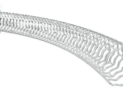
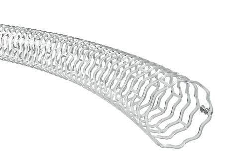
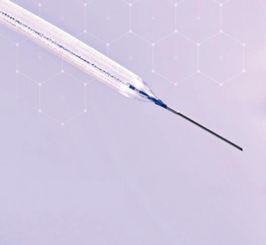
A self-expanding nitinol stent on a rapid exchange delivery catheter, designed for flexibility, conformability and accurate placement1
“Gatekeeping”, “fear” and “misinformation”
continue to mar global UAE uptake, say experts
Following a recent review which surveyed global access to uterine artery embolization (UAE) across each continent, Interventional News speaks to multiple clinicians who trace misunderstandings, miscommunication and multidisciplinary dysfunction at the core of symptomatic fibroid care.
Uterine fibroids are a common benign tumour affecting between 40–60% of women of reproductive age. Although many fibroids remain asymptomatic, a significant portion of women experience heavy bleeding, pain and/or infertility. Despite hysterectomy being the long-standing definitive treatment for symptomatic fibroids—or myomectomy for women wishing to preserve fertility—over the last two decades UAE has established its role as a minimally invasive alternative based on findings from the EMMY and FEMME randomised controlled trials, among others.
Despite level-one evidence, access to and awareness of UAE remains limited with varying severity depending on geographic, socioeconomic and healthcare system factors. This was the broad finding of the global UAE review published in CardioVascular and Interventional Radiology (CVIR) Endovascular for which interventional radiologist Sara Lojo-Lendoiro (Hospital Arquitecto Marcide, Ferrol, Spain) was a study investigator.
“The challenge today is less scientific than cultural and organisational,” Lojo-Lendoiro tells Interventional News “The question is no longer whether UAE works, we answered that years ago. The question today is whether every woman who could benefit from UAE is actually given the opportunity to consider it.”
Who benefits from UAE?
Defining which patients would benefit from UAE relies heavily on shared decision making. Lojo-Lendoiro describes that these are often patients “who are looking for a treatment that is effective but also minimally disruptive to their lives”. She states that these are women with demanding jobs, caregiving responsibilities, multiple fibroids, previous surgeries, or significant comorbidities who may particularly appreciate avoiding a major operation and prolonged recovery.
Recovery time following UAE is approximately four to five days, but most patients return home the same day or the following morning. “From a patient experience point of view, that’s a huge difference,”
highlights Dania Daye, associate professor of radiology and physician-scientist at University of Wisconsin School of Medicine and Public Health (Madison, USA), sharing her clinical experience treating fibroids with UAE.
Although procedurally UAE appears favourable, treatment effect and symptom relief are factors which significantly influence patient preference. In women who experience heavy menstrual bleeding (HMB) as a main symptom of fibroids, Daye explains that the effects of UAE can be relatively quick, although hysterectomy instates an immediate effect.
Fertility fears
Aside from shrinking fibroids, UAE offers women the possibility of symptom relief without resorting to irreversible treatment—a consideration which may matter greatly to women seeking fertility preservation. Lojo-Lendoiro describes that, historically, many physicians adopted a cautious approach because long-term reproductive outcomes after UAE were not as well understood as they are today. Although caution is “justified”, she warns that “caution can sometimes evolve into paternalism; for clinicians, fertility concerns create uncertainty and, in patients, they often create fear”.
“There is a difference between saying: ‘We don’t know everything’ and ‘We don’t know everything, therefore you shouldn’t have access to this treatment’. Younger women deserve honest conversations about uncertainty, not automatic exclusion from treatment options,” Lojo-Lendoiro notes. “The recent data—including long-term follow-up studies—increasingly suggest that many younger women can achieve excellent fertility outcomes after UAE. The goal should not be to convince every young woman to choose UAE. The goal should be to trust women with the information necessary to decide for themselves.”
The misinterpretation that UAE is contra-indicated in women wishing to become pregnant is compounded by the fact that chronic disease in women has a natural recurrence rate, explains Warren Clements (The Alfred Hospital, Melbourne, Australia), principal inves-
tigator of the CVIR Endovascular global UAE review.
“There is a 20% chance of a woman needing a repeat treatment on their uterus after UAE, and this recurrence rate has been coopted as a reason to suggest the treatment’s ‘failure’,” Clements says. “In reality, UAE is a uterus-preserving treatment that exists on a continuum between conservative management and surgery.” Yet, women are still not being made aware of their options, adds Clements, pointing to the median age of his patients being 45 years as an indicator of continuing reluctance to refer younger women for UAE.
‘Outdated’ clinical understanding
Results from the CVIR Endovascular review demonstrated that, despite broad availability of UAE in several geographies, the procedure is rarely employed. Alongside misapprehensions of UAE and fertility, Clements believes that the treatment’s intrinsic association with IR—and the lack of IR’s recognition as an independent specialty—has meant access to mandatory clinical services is lacking, such as outpatient clinics, admitting rights, and a team of junior staff.
“Without these, it is very difficult to offer a holistic and rounded clinical service, and we know that simply offering a technical service is not enough,” says Clements. “Patients and other doctors are then left not knowing what services are available to them.”
“Non-IR doctors don’t keep abreast of the most recent evidence and clinical practice guidelines that govern UAE,”
Sara Lojo-Lendoiro
Warren Clements
Dania Daye
Clements states, underscoring that only interventional radiologists know the indications and suitability of UAE and can council a patient as to treatment appropriateness.
Clements continues: “It is not feasible for a doctor of another specialty to fully understand UAE, and as such it is rarely offered, or glossed over as a non-viable treatment approach. Many non-IR doctors haven’t read IR literature and make decisions based on data from over 20 years ago.
“Doctors aren’t super-human, they can’t know everything. If a woman is being considered for surgery, then she should equally be given an opportunity to consider UAE, and the only way this can be achieved is through consultation with an interventional radiologist. This would then allow for access to unbiased information and true informed consent regardless of whether she chooses UAE or surgery.”
Gynaecological “gatekeeping” and treatment pathway issues
Among other barriers to access, speakers shared that women are often not able to be directly connected with an interventional radiologist, which they posit is an issue that starts at the primary care level.
Clements explains that, in the case of HMB, there is a “misconception” that all women must first see a gynaecologist to access second line care. He acknowledges that general practitioners (GPs) play an important role in the investigation of the early causes of HMB, however notes that when a diagnosis is made and the symptoms are refractory to simple treatments, women should be offered the opportunity to discuss UAE with an interventional radiologist and concurrently see a gynaecologist to discuss surgery.
Using data from their global review, Clements highlights a trend which shows women are “almost exclusively” sent to a gynaecologist first. “Unfortunately, this creates a gatekeeping role and only very select patients make it to an interventional radiologist,” he adds. “This means many women miss out on a specialist opinion, advice, and an opportunity to consider a less invasive treatment approach.”
To this Lojo-Lendoiro agrees, stating that often it is the first specialist a woman sees that largely determines the treatment she is offered. “That fact should make us uncomfortable,” she says, adding that UAE sits at the intersection between
“Whenever a treatment challenges traditional ownership of a disease pathway, controversy tends to persist longer than the evidence would justify”
Sara Lojo-Lendoiro
specialties which causes referral difficulties.
“Whenever a treatment challenges traditional ownership of a disease pathway, controversy tends to persist longer than the evidence would justify,” states Lojo-Lendoiro. “Ultimately, I don’t think the debate is really about UAE anymore. I think it is about whether we are willing to move towards genuinely multidisciplinary fibroid care.”
UAE ‘reflective’ of women’s health inequities In 2025, a cross-sectional study published in
the journal JAMA Network found that UAE was underutilised with significant disparities across socioeconomic factors. The study, led by Tarig S Elhakim (Perelman School of Medicine, Philadelphia, USA), utilised the 2016 to 2022 National Inpatient Sample obtained from the US Healthcare Cost and Utilization Project (HCUP).
As corresponding author for this study, Daye details that results showed African American women were less likely to receive UAE compared to other ethnicities. Similarly, Hispanic patients were also less likely to receive a UAE and more likely to receive a hysterectomy or myomectomy. When comparing metropolitan versus non-metropolitan areas, Daye and colleagues found that UAE was more likely to be performed in metropolitan areas, while in rural areas, there are still more hysterectomies and myomectomies—a result that was expected, Daye adds.
“We also saw some interesting trends when we looked at the payers. We are seeing more patients on Medicaid, for example, receive UAE compared hysterectomy, which was something that was very surprising to us,” says Daye. “Overall, that paper taught us that access to UAE is not uniform across the country or different patient groups. We need to do better, and we need to understand why certain populations have more access to procedures and not others.”
Although Lojo-Lendoiro promotes caution when interpreting treatment utilisation patterns as evidence of equity, she believes that “fibroid care often reflects broader social inequities affecting women’s health”, which can “rarely” be assigned a single explanation.
“Race, income, insurance status and geography continue to influence what treatments women receive and when they receive them. The fact that these disparities persist in 2026 should remind us that improving technology is not enough,” Lojo-Lendoiro tells Interventional News
Guideline inclusion
Currently, UAE is not officially included in many practice guidelines, although grants such as the Society of Interventional Radiology (SIR) Foundation’s Scott C Goodwin Grant for adenomyosis seek to expand clinical knowledge of the procedure.
“The grant reflects national recognition that we simply don’t have enough data to support the evidence base,” says Daye. “Frankly, we need randomised trials. We need larger, gold-standard clinical trials to support inclusion in clinical guidelines. We need to generate the evidence that drives medical decision-making, but right now, we don’t have enough high-quality randomised clinical trial evidence yet.”
To Clements, access and awareness of UAE is multifactorial, but interventional radiologists play an important role in ensuring that current governance supports evidence-based practice. “This means a top-down approach to ensure that UAE is built into all relevant guidelines—government, college, societal, and at a local hospital level,” he states.
Once established, Clements hopes that the guidelines—written by interventional radiologists and supported by major IR societies—will be readily accessible to GPs and other primary care providers who can easily review for advice on a daily basis. “Ideally, interventional radiologists should be available to consult via direct referral and IR outpatient clinics made available for patients,” he adds, to provide a rounded, evidence-based approach to fibroid care.
Tze Min Wah
Driven by a passion for both science and art, professor of interventional radiology (IR) Tze Min Wah (Leeds, UK) has built a career that combines innovation, technical expertise, and creativity. After initially pursuing surgical training, she discovered IR—a specialty that blends clinical practice with procedural innovation. Today, as a consultant interventional radiologist at Leeds Teaching Hospitals NHS Trust, she combines patient care with pioneering research, advancing new technologies and pushing the boundaries of IR in research and education. Here, Wah tracks key career highlights with Interventional News
Why did you initially choose to become a doctor and what was it that made you decide to specialise in IR?
Growing up on the tropical island of Penang in Malaysia, I was equally captivated by art and science. As a child, I dreamed of becoming a fashion designer, drawn by the creativity and innovation of the design world. However, my aptitude for science gradually steered me towards medicine—a direction gently encouraged by those around me.
Throughout medical school and my early years as a house officer, I enjoyed clinical practice but felt that something was missing. That changed during my surgical rotation in the professorial surgical unit, where I first encountered interventional radiologists. I was fascinated by their ability to solve complex clinical problems, often providing lifesaving minimally invasive treatments when conventional approaches had reached their limits. They were the team everyone relied upon in difficult situations, combining technical expertise with clinical judgement to make an immediate difference to patient care. It was then that I realised that IR represented the missing piece of the puzzle for me.
Towards the end of my house officer training, I was offered a senior surgical house officer post in the professorial surgical unit. I remember joking with the two professors that perhaps I would be more useful as an interventional radiologist, so that I could always help them whenever they needed IR support. They both laughed and remarked that, if they had the opportunity to choose their careers again, they too would choose IR. That conversation marked the beginning of my IR jour-
ney in 1996.
After securing a radiology training post in Leeds, I quickly discovered that pursuing IR as a woman was not universally encouraged. At that time, misconceptions surrounding radiation exposure during pregnancy, together with the perception that IR was a male-dominated specialty with poor work–life balance, created significant barriers. During my second year of training, I came close to leaving the specialty altogether.
Who were the biggest influences on your early career?
Everything changed when I met Dr Sam Chakraverty during a rotation at a district general hospital. He welcomed me into his interventional practice, entrusted me with increasingly complex procedures and, most importantly, believed in my potential. Those three months transformed my career trajectory. They rekindled my passion for IR and gave me the confidence to persevere despite the challenges. I remain profoundly grateful for his mentorship, which fundamentally changed the course of my professional life.
Could you describe one of your most memorable cases?
Among the many memorable patients I have had the privilege to treat, one experience stands out above all others. In 2021, we performed one of the first liver treatments in the global #Hope4Liver trial, evaluating image-guided histotripsy for liver cancer. Histotripsy represents a completely different treatment paradigm, using focused cavitating ultrasound to mechanically destroy tumour tissue without needles or ionising radiation.
The following morning, I visited my patient, Sheila, expecting to assess her recovery. She told me she had experienced no pain. When I asked if I could examine her wounds, she smiled and replied: “There are no wounds.” At that moment, the significance of what we had achieved truly resonated with me. We had reached a point where meaningful cancer treatment could be delivered entirely non-invasively. It was one of those rare moments that reminds you why innovation in medicine matters.
You were the first UK professor of interventional oncology (IO). What drove you to pursue this qualification in a relatively underdeveloped field?
Research has always been central to my vision of practising evidence-based medicine. During my radiology training, I aspired to undertake a PhD, believing it would provide the academic foundation necessary to advance IR. At the time, however, there was little encouragement to pursue research because of workforce demands. The priority was understandably to train more practising interventional radiologists.
Rather than abandoning that ambition, I sought permission to undertake a PhD after becoming a full-time consultant. My organisation generously supported the opportunity, although the research itself had to be completed almost entirely in my own time—during annual leave, evenings and weekends. Importantly, I was working in an institution without an academic radiology department, making the journey even more challenging.
I was exceptionally fortunate that Prof Peter Selby agreed to supervise my doctoral studies. Looking back, I embarked on the PhD with little appreciation of how demanding the journey would become. Balancing a full-time consultant practice while raising two young daughters required considerable perseverance and, above all, the unwavering support of my husband, family and colleagues.
When I successfully defended my PhD, Selby remarked that he was particularly proud because, statistically, the likelihood of completing such a journey under those circumstances was remarkably low. His words have stayed with me ever since. There were undoubtedly moments when others believed in me more than I believed in myself, and I remain deeply grateful for their encouragement.
My PhD marked the beginning rather than the culmination of my academic career. For many years, research remained something I pursued alongside a full-time clinical practice. It was only after my appointment as the UK’s first professor of IO in 2022, followed by the award of the National Institute for Health and Care Research (NIHR) Senior Clinical and Practitioner Research Award (SCPRA) in 2024, that I was able to dedicate protected time to developing a truly integrated clinical academic programme.
Everything I do in research is underpinned by a simple principle: patients deserve the highest quality, evidence-
Fact file
CURRENT APPOINTMENTS
2022–present: Honorary professor of interventional radiology, University of Leeds (Leeds, UK)
2017–present: Honorary professor, Universiti Tunku Abula Rahman (Kuala Lumpur, Malaysia)
1995: MBBS, PhD, University of Leeds (Leeds, UK) AWARDS (SELECTED)
2024: Senior Clinical Practitioner Research Award (SCPRA), National Institute of Health Research (NIHR)
2022: N1 Level Award, National Clinical Impact Award (NCIA)
2018: Bronze National Clinical Excellence Award, Advisory Committee on Clinical Excellence Awards (ACCEA)
based care. Innovation alone is never enough; it must be supported by robust scientific evaluation before it can become routine clinical practice. That philosophy continues to drive my commitment to research in IR.
How did you become involved in research for histotripsy and what role will the modality play in the future treatment of liver tumours?
In 2020, at the height of the COVID19 pandemic, I was approached by the research and innovation team at HistoSonics regarding the possibility of introducing histotripsy at my centre in Leeds. My initial reaction was that the timing seemed almost impossible. However, following discussions with our multidisciplinary team, we recognised the potential importance of the technology.
My instituion subsequently underwent a competitive international selection process and was chosen as the chief investigator site for the #Hope4Liver trial in the UK. In 2023, we were also privileged to
“At that moment, the significance of what we had achieved truly resonated with me”
perform the world’s first image-guided renal histotripsy treatment. This technology is transformative and we will have many exciting opportunities to translate this into various organs and diseases in years to come.
What is the biggest challenge facing IO (or IR) today?
Looking ahead, I believe the greatest challenges facing IO are workforce sustainability and the pace of technological innovation. Novel technologies are evolving more rapidly than our traditional evidence-generation pathways.
We need more agile and proportionate approaches to evaluating medical technologies, enabling promising innovations to be
translated safely and efficiently into definitive clinical trials and, ultimately, routine patient care.
Were there any career risks you took that, in hindsight, proved pivotal?
Reflecting on my career, two decisions have shaped my professional life more than any others: persevering in my pursuit of IR despite significant obstacles and insisting on pursuing a PhD when the conventional path would have been to focus solely on clinical service. Those decisions taught me that resilience, supported by good mentors and colleagues, can transform challenges into opportunities.
What does your life outside of medicine look like?
Outside medicine, I enjoy travelling, particularly when it allows me to spend time with my parents and siblings in Malaysia. I also enjoy water sports, cooking and, perhaps unsurprisingly, I still indulge my lifelong interest in fashion design whenever time permits.
Andrew Holden
People, place and purpose propel IR, says Andrew Holden
“He aha te mea nui o te ao? He tāngata, He tāngata, He tāngata,” said Andrew Holden (Auckland City Hospital, Auckland, New Zealand), quoting a Māori phrase to open his Charles Dotter Lecture at the 2026 Society of Interventional Radiology (SIR) annual scientific meeting (11–15 April, Toronto, Canada). The phrase asks: What is the most important thing in the world? It is people, it is people, it is people—and embodies the most important aspect of Holden’s path in interventional radiology (IR). In this interview, Holden reflects on what his lecture, titled ‘Interventional radiology: A journey of creativity’, sought to convey.
What is meant by ‘a journey of creativity’?
Through IR, I fell into a more academic role and that’s really what my Dotter Lecture was about: a journey of creativity. People often associate creativity with the arts, but scientists and doctors can be just as creative if they’re able to see solutions to problems that other people haven’t recognised. For me, that became the challenge. I identified areas where I didn’t think we were performing particularly well in IR, attended meetings, saw people trying new approaches, and came away with a few ideas of my own.
It was a time when it was somewhat easier to innovate before all of the regulatory requirements associated with formal clinical trials had developed to the extent they have today. That gave me a real taste of making a difference through innovation and being creative.
From there, things gradually gathered momentum. Quite early on, I realised that Auckland was never going to compete with the world’s largest academic centres on volume alone—we simply didn’t have the same number of patients. Instead, we made a conscious decision that our strength would be in being among the first to evaluate new technologies. That became our niche, assessing new devices early, providing valuable feedback and helping to develop technologies that ultimately benefit patients around the world.
Multidisciplinary working was one of the central themes of your lecture. What does that look like in your day-to-day practice?
Where I trained, multidisciplinary working was the norm, and I could immediately see the benefit, particularly for patients. Whenever I’ve worked in environments where patients weren’t placed first and specialties operated more independently, I’ve generally seen poorer practice and poorer outcomes.
Today, multidisciplinary meetings are fundamental to everything we do. Every patient’s imaging, clinical presentation and pathology are reviewed collectively, and management decisions are reached by consensus. Beyond that, we recognise there is considerable overlap between IR and other specialties. Particularly in complex vascular disease, we recognise that there is a crossover in expertise and training between IR and vascular surgery we need to work hand-in-hand. Interventional radiologists are strong on procedural aspects, but during training, we need a greater emphasis on clinical practice and managing patients. Conversely, almost the opposite is true of vascular surgeons who have strong clinical skills, but may lack endovascular expertise.
Vascular specialists of a similar vintage to me will have definitely seen a striking change in the patient population and the Dotter Lecture was an opportunity to ask exactly that question: Is this true and why is it happening?
We compared our practice over two years separated by two decades (2005 and 2025), and the differences were dramatic. Not only were we performing significantly more procedures for a similar population, but the patients themselves had changed. Twenty years ago, many patients were referred with intermittent claudication or pain on walking. Today, while we still see those patients, virtually all of the growth has been in people presenting with chronic limb-threatening ischaemia (CLTI).
The pattern of disease has also evolved. Twenty years ago disease was predominantly affecting the iliac and femoral arteries. Those vessels are still commonly involved, but we now see far more disease extending into the arteries below the knee and into the foot.
What’s driving that? We’re increasingly able to answer this question, even down to the cellular level. Although there are genetic factors, the biggest drivers are environmental and metabolic. Smoking still plays an important role, but obesity, diabetes and renal disease have become much more significant contributors. These conditions produce changes within the endothelium, platelets and smooth muscle cells that ultimately alter the biology of the arterial wall; one of the most important consequences is vascular calcification. Twenty years ago, heavily calcified arteries were relatively uncommon, but today, they’re present in the vast majority of patients we treat.
That’s one reason why technologies specifically designed to modify or treat calcium have become so important. They’re addressing an unmet need because our older technologies do not manage calcific arterial disease well. It’s encouraging that we are no longer just observing the change in distribution and severity of vascular disease, we’re understanding the biological mechanisms behind it.
“People often associate creativity with the arts, but scientists and doctors can be just as creative”
You’ve spoken about the importance of being a clinician rather than a proceduralist. How does that philosophy shape your own practice? Essentially, it means building a practice around patients and clinics rather than procedures. That clinical involvement is critical. We see patients before intervention, manage them during their hospital admission and continue to follow them afterwards. That’s what being a vascular specialist means, and of course, IR extends well beyond vascular disease.
Increasingly, we’re seeing some overlap between specialties, for example a vascular specialist might be a vascular surgeon with excellent endovascular skills, or an interventional radiologist with strong clinical expertise. Likewise, an interventional oncologist may be an interventional radiologist with specialist oncology knowledge, but in some centres oncologists themselves are developing interventional skills.
During your talk you presented an audit showing an increase in lower limb interventions in the last 20 years. What is driving that change?
For me, the future is about combining technical skills with clinical care. For our trainees, they are required to do a significant amount of clinic work in their training, so that they are comfortable with the clinical management of the patients they treat.
POINT OF VIEW
Endovascular therapy for acute pulmonary embolism: A field moving from promise to proof
The management of acute pulmonary embolism (PE) is undergoing a meaningful transformation. Following the release of several pivotal datasets, co-global principal investigator and professor of radiology and surgery at the Icahn School of Medicine at Mount Sinai (New York, USA) Robert Lookstein plots an evidence-based trajectory for endovascular PE therapy.
FOR YEARS, ENDOVASCULAR THERapy occupied an uneasy space between physiologic logic and incomplete clinical evidence. Interventionalists could see, often immediately, that catheter-based therapy improved pulmonary artery obstruction, right ventricular strain, oxygenation, and patient haemodynamics. Yet, the broader clinical community reasonably asked whether these improvements translated into patient-centred outcomes.
Recent randomised trials now provide an increasingly persuasive answer: in carefully selected patients with intermediate-high-risk PE, endovascular therapy can meaningfully improve early clinical trajectory and may prevent deterioration.
The field should therefore move beyond the outdated binary debate of “anticoagulation versus intervention” and toward a more sophisticated framework: which PE patients are most likely to benefit from timely, targeted reperfusion? Low-risk PE remains appropriately managed with anticoagulation, risk-adjusted disposition, and follow-up. Massive or high-risk PE with shock remains a reperfusion emergency. The most important evolving population is intermediate-high-risk PE: patients who are normotensive but demonstrate right ventricular dysfunction, biomarker elevation, hypoxaemia, tachycardia, syncope, or limited cardiopulmonary reserve.
Earlier trials established proof of biologic effect. ULTIMA showed that ultrasound-assisted catheter-directed thrombolysis improved right ventricle/ left ventricle (RV/LV) ratio compared with anticoagulation alone. CANARY, although prematurely terminated,
Robert Lookstein
similarly suggested improved right ventricular recovery with conventional catheter-directed thrombolysis and a low major bleeding signal. These studies were important, but they largely centred on imaging and physiologic endpoints. The newer trials are more consequential because they move the field closer to clinical relevance. STORM-PE randomised patients with intermediate-high-risk PE to computer-assisted vacuum thrombectomy plus anticoagulation versus anticoagulation alone. The thrombectomy strategy produced greater early reduction in RV/LV ratio, greater reduction in pulmonary obstruction, and more rapid normalisation of vital signs. This is not trivial. In acute PE, early right ventricular recovery and stabilisation of vital signs are clinically meaningful because the central threat is RV failure. Although STORM-PE was not powered to demonstrate a mortality advantage, it provides randomised evidence that mechanical thrombectomy can rapidly improve the pathophysiology that drives PE decompensation, without an obvious major safety penalty in the studied population.
HI-PEITHO is another very important clinical trial. In patients with acute intermediate-risk PE and additional severity features, ultrasound-facilitated catheter-directed fibrinolysis plus anticoagulation reduced the sevenday composite of PE-related death, cardiorespiratory decompensation or collapse, or symptomatic recurrent PE compared with anticoagulation alone. The benefit was driven primarily by reduction in clinical decompensation or collapse. That finding matters enormously. Mortality in intermediate-risk PE trials is often low, making death alone an insensitive endpoint. Preventing deterioration, escalation, shock, rescue therapy, and collapse is a clinically meaningful therapeutic goal. HI-PEITHO supports the idea that early catheter-directed therapy can alter the natural history of selected intermediate-risk PE before patients cross the threshold into overt haemodynamic failure. Taken together, these trials justify a more confident posture toward endovascular PE therapy. The evidence no
longer supports viewing catheter-based intervention as merely experimental, anecdotal, or reserved only for last-ditch rescue. In the appropriate patient, endovascular therapy is becoming a legitimate early treatment strategy designed to unload the RV, stabilise physiology, reduce decompensation, and accelerate recovery. This does not mean every intermediate-risk PE requires intervention. It does mean that patients with clear intermediate-high-risk features deserve timely multidisciplinary evaluation and that catheter therapy should be considered proactively rather than only after clinical collapse. The next challenge is implementation. Endovascular PE therapy should include rapid diagnosis, standardised RV assessment, biomarker interpretation, bleeding risk assessment, pulmonary embolism response team (PERT) activation, and follow-up. Intervention should be selected based on clinical phenotype, not clot burden alone. A saddle embolus in a stable patient is not the same as segmental-level clot burden in a patient with severe hypoxemia, rising biomarkers, RV dysfunction, syncope, and poor reserve. The decision should be physiologic, not purely anatomic.
“The evidence no longer supports viewing catheterbased intervention as merely experimental, anecdotal, or reserved only for last-ditch rescue”
The field also needs continued investigation. Future trials such as PE-TRACT will evaluate functional recovery, quality of life, and chronic thromboembolic disease, among other factors. Mortality will remain important, but it should not be the only measure of success in a disease where the central clinical objective is often prevention of deterioration.
Endovascular therapy for acute PE has reached an inflection point. STORM-PE strengthens the case for mechanical thrombectomy as a rapid, non-lytic strategy for improving right ventricular strain and physiologic instability. HI-PEITHO provides randomised clinical endpoint evidence that catheter-directed fibrinolysis can reduce early decompensation in selected intermediate-risk patients. These data do not eliminate the need for judgment; they elevate the importance of judgment. The most effective PE programmes will be those that identify the right patient early, match the intervention to the clinical problem, and act before RV failure becomes irreversible. The message is no longer simply that endovascular PE therapy is promising. The emerging message is that, when applied selectively and systematically, it is becoming an evidence-based tool for preventing deterioration and improving early recovery in a highrisk subset of patients who historically had few options between anticoagulation and crisis-driven rescue.
ROBERT LOOKSTEIN is a professor of radiology and surgery at the Icahn School of Medicine at Mount Sinai in New York, USA.
The author declared that he is principal investigator for the Penumbrasponsored STORM-PE trial.
POINT OF VIEW
The transition to a more sustainable IR service: From awareness to action
Simon Lambracos
As the climate crisis continues to affect population health, healthcare systems face the dual challenge of mitigating its effects while addressing their own contribution to it. Interventional radiology (IR), with its reliance on advanced imaging technology and extensive use of medical devices, therefore, sits at an intersection between innovation and sustainability. Here, Simon Lambracos (London, UK) puts forward key actions to mitigate emissions in IR, addressing energy consumption, waste management, supply chain and departmental infrastructure.
THE HEALTHCARE SECTOR CONTRIbutes approximately 5% to global greenhouse gas emissions, and medical imaging represents 10% of that figure.1 As a result, in 2020, the UK National Health Service (NHS) became the first healthcare system worldwide to commit to achieving net zero emissions, and in doing so embedded this within UK law. Yet awareness of this alone is insufficient. Meaningful change requires clinicians and organisations to recognise where emissions arise within everyday workflows and to translate the evidence into tangible interventions. But what is meant by sustainability in this context?
1. Steps to minimise energy consumption Energy consumption is one of the most visible challenges within the IR environment. Imaging and ventilation systems are energy intensive and alarmingly often consume the most power during periods of inactivity. There is little excuse for this; we would not leave the heating and electrical appliances running when away from home, so should we not afford the same courtesy when leaving the hospital environment? Simple changes such as switching systems off when not required, optimising equipment standby settings and reducing system idling through optimised patient scheduling, represent some of the lowest hanging fruit to reduce unnecessary energy use. These measures may appear small at an individual departmental level, but would represent a substantial cumulative effect across multiple sites.
The environmental burden associated with digital data is also becoming increasingly relevant. As demand for imaging grows and artificial intelligence (AI) applications become more integrated into clinical practice, the volume of stored data is increasing exponentially. With data centre emissions currently being compared to those of the global aviation industry and
projected to at least double in terms of energy consumption within the European Union in the next decade, this is naturally a major concern.2 That said, the beneficial role for AI in healthcare and IR cannot be understated. AI has the potential to counteract environmental impact through improved workflow planning, reduction of repeat procedures and more efficient use of existing resources. 3
2.Reducing waste and rationalising single-use products
In the NHS, single-use products make up 90% of medical device waste with their disposal amounting to an estimated annual cost of £28 million.4,5 Whilst this may seem like an insurmountable challenge, simple actions such as improving stock management to prevent avoidable product expiry and opening products “just in time” as opposed to “just in case” have been shown to significantly reduce both carbon emissions and financial cost in some centres.5,6 Similarly, appropriate waste segregation can also play its part through avoidance of erroneous incineration of recyclable or non-hazardous materials, which can sometimes also incur financial penalties. Simply improving waste bin placement, signage and staff education can help to address this issue.
3. Collaboration with industry is key
A significant proportion of the environmental footprint of IR lies within the manufacturing, packaging and transportation of medical devices. Clinicians therefore have an important role in influencing purchasing decisions and engaging with industry partners. Moving towards a circular economy—where products are designed for reuse, reprocessing or refurbishment—will require collaboration between healthcare providers, manufacturers and regulators. Imaging equipment itself provides
another example of where sustainability and innovation can align. Longer equipment lifecycles, energy-efficient technologies and responsible replacement strategies may reduce the environmental impact associated with manufacturing and disposal.7
4. Affecting change at the departmental level
Sustainability also extends beyond the walls of the IR suite. Consideration of the whole patient pathway can uncover significant pre- and post-procedural contributors to the overall IR carbon footprint. Reducing unnecessary hospital visits through virtual consultations, remote digital consent processes and reducing length of stay with ringfenced day-case units can optimise patient experience, environmental outcomes, departmental productivity and financial savings.8
“These measures may appear small at an individual departmental level but would represent a substantial cumulative effect across multiple sites”
Despite these opportunities, barriers remain. Cultural inertia, financial pressures and regulatory constraints can significantly delay progress.9 It is for this reason that achieving a sustainable IR service requires leadership and collaboration, at an individual level, organisational level and cross-sector. Embedding these principles into the infrastructure of organisations and societies is a key mechanism in their consequent adoption into business-as-usual at ground level. By committing to sustainable practice, IR can again innovate by delivering high-quality care whilst simultaneously contributing to a healthier future for both patients and the planet.
References
1. Picano E, Mangia C, D’Andrea A. Climate change, carbon dioxide emissions, and medical imaging contribution. J Clin Med . 2023;12(1):215. doi:10.3390/ jcm12010215.
2. Buckley BW, MacMahon PJ. Radiology and the climate crisis: Opportunities and challenges—Radiology in training. Radiology . 2021;300(3):E339-E341. doi:10.1148/ radiol.2021210851.
3. Doo FX, et al . Environmental sustainability and AI in radiology: A double-edged sword. Radiology 2024;310(2). doi:10.1148/radiol.232030.
4. UK Government. Design for Life roadmap [Internet]. 2024 [cited 2026 Jul 28]. Available from: https:// www.gov.uk/government/publications/design-forlife-roadmap/design-for-life-roadmap--4
patient and population outcomes
5. Demmert A, Hong K. 03:54 PM Abstract No. 147: Using human psychology to reduce equipment waste and decrease inventory costs. J Vasc Interv Radiol . 2019;30(3 Suppl):S68. doi:10.1016/j. jvir.2018.12.197.
6. Chasseigne V, et al . Assessing the costs of disposable and reusable supplies wasted during surgeries. Int J Surg . 2018;53:18-23. doi:10.1016/j. ijsu.2018.02.004.
7. Green Surgery. Reducing the environmental impact of surgical care [Internet]. UK Health Alliance on Climate Change; n.d. [cited 2026 Jul 28]. Available from: https://ukhealthalliance.org/sustainablehealthcare/green-surgery-report/
8. Lambracos S, Kurek N, Gray WK, Briggs TWR, Sheard S, Barnacle A. Call to action: A sustainable interventional radiology service. Cardiovasc Intervent Radiol . 2026. doi:10.1007/s00270-02604392-9.
9. Lojo-Lendoiro S, Abadal Villayandre JM, Lonjedo Vincent E, Morales Santos Á, Rovira À. A greener path for interventional radiology. J Med Imaging Radiat Oncol . 2025;69(4):498-508. doi:10.1111/17549485.13867.
SIMON LAMBRACOS is a radiology resident at Guy’s and St Thomas’ NHS Foundation Trust in London, UK.
POINT OF VIEW
Histotripsy: Less invasive does not equal less risk
Vlasios S Sotirchos
For histotripsy, as with every disruptive medical technology, the transition from carefully controlled clinical trials to clinical practice has inevitably revealed adverse events that were not apparent during premarket evaluation, says Vlasios S Sotirchos (New York, USA).1-3 Here, he discusses how this pattern is neither unique to histotripsy nor unexpected; rather, it reflects the natural evolution of innovation as new technologies are applied to broader patient populations and increasingly complex clinical scenarios.
HISTOTRIPSY IS A NEW TECHNOLOGY with potentially important applications in tumour ablation. By using focused ultrasound to generate acoustic cavitation and mechanically
This advertorial is sponsored by Reflow Medical
destroy tissue without needles or thermal injury, histotripsy has generated considerable enthusiasm among clinicians and patients alike.
Recent reports describing portal vein thrombosis, vascular injury, and acute kidney injury should therefore be viewed within this context.4-6 Overall, these adverse events appear relatively uncommon, and early clinical experience has already suggested potential mitigation strategies, including tailored anticoagulation protocols, careful treatment planning for centrally located tumours, and greater awareness of treatment-volume thresholds. These observations also emphasise that histotripsy should be performed within an appropriately equipped multidisciplinary environment, where intraprocedural computed tomography (CT) and interventional expertise are available to facilitate the prompt recognition and management of procedure-related complications.
appropriate role of histotripsy within interventional oncology.
“Scientific rigour must keep pace with technological innovation”
As enthusiasm for histotripsy grows, scientific rigour must keep pace with technological innovation. The next phase of clinical development should prioritise prospective studies with standardised adverse event reporting and established oncologic outcome measures. By applying the same standards that have guided the evolution of other technologies, the interventional oncology (IO) community can ensure that histotripsy realises its considerable promise while building a robust evidence base that supports its long-term integration into clinical practice.
References:
1. Mendiratta-Lala M, Wiggermann P, Pech M, et al. The #HOPE4LIVER single-arm pivotal trial for histotripsy of primary and metastatic liver tumors. Radiology. 2024;312(3):e233051.
The more important question is not whether adverse events occur, but how systematically the field responds to them. Large prospective registries and multicentre studies will be instrumental in quantifying adverse event rates, identifying patient- and procedure-specific risk factors, and refining best practices. Equally important, future studies should move beyond procedural safety alone. Histotripsy should be evaluated using standardised oncologic endpoints that have been established for other image-guided ablative therapies, including local tumour progression, progression-free survival, overall survival, and quality-of-life measures. Adoption of these established endpoints will facilitate meaningful comparisons with existing ablative modalities and provide the evidence needed to define the
A novel temporary scaffold approach to “keep options open” in complex BTK CLTI
Detailing his evolving treatment algorithm for complex below-the-knee (BTK) disease in patients with chronic limb-threatening ischaemia (CLTI), interventional radiologist Daniel van den Heuvel (St Antonius Hospital, Nieuwegein, Netherlands) discusses patency rates, lesion preparation strategies and what’s on the horizon for the care continuum.
WHEN USING A CONVENTIONAL ENDOVAScular approach to treat complex BTK lesions in patients with CLTI, small, diffusely diseased vessels and vessel wall calcification are among the “most determining factors” with respect to treatment patency, states van den Heuvel. He details that it can be difficult to obtain an optimal result in non-calcified arteries with conventional treatment (often meaning balloon angioplasty), let alone in those that are calcified.
“A stiff, calcified small vessel doesn’t dilate evenly under balloon pressure; it can crack unpredictably, dissect, or simply recoil once the balloon deflates, because there’s less elastic ‘give’ and less room to absorb any injury,” notes van den Heuvel.
Describing his practice, van den Heuvel states that if a patient requires long-term patency—often those with a high Wound, Ischemia, foot Infection (WIfI) classification for whom early failure is a risk—he opts to deploy a lesion preparation
Spur peripheral retrievable scaffold system
“Spur optimises BTK lesion preparation and drug uptake without leaving a permanent implant”
device. Using the Spur peripheral retrievable scaffold system (Reflow Medical) alongside a drug-coated balloon in cases with limited to no calcium, van den Heuvel can prevent flow-limiting dissections and recoil by avoiding the rota-
2. Wehrle CJ, Burns K, Ong E, et al. The first international experience with histotripsy: A safety analysis of 230 cases. J Gastrointest Surg. 2025;29(4):102000.
3. Worlikar T, Caoili E, Navarro A, et al. Management and prevention of histotripsy-induced acute bland portal vein thrombosis in histotripsy treated liver tumors. J Vasc Interv Radiol. 2026. Article 108883. Epub ahead of print.
4. Poe LL, Kisting AL, Mendiratta-Lala M, et al. Histotripsy-induced acute kidney injury following liver tumor treatment: A preliminary report of 9 cases. J Vasc Interv Radiol. 2026. Article 108882. Epub ahead of print.
5. Breuer JA, Mendiratta-Lala M, Lee FT Jr, et al. Vascular injury after histotripsy: A case series of hemorrhage and pseudoaneurysm complications in human patients. J Vasc Interv Radiol 2026;37(6):108576.
6. Cornelis FH, Bokhari R, Solomon SB. Public interest and academic trends in percutaneous ablations: Histotripsy’s reversed innovation pathway. CVIR Oncol. 2025;1(1):14.
VLASIOS S SOTIRCHOS is an interventional radiologist at Memorial Sloan Kettering Cancer Center in New York, USA.
tional forces the balloon would otherwise exert on the vessel wall.
He adds that Spur also allows for better drug uptake, noting, “with a complete and intact ring of calcium it is hard to imagine that drugs from drug-eluting balloons would penetrate the deeper wall layers. Yet, the channels created by the spikes facilitate improved drug uptake and potentially benefit long-term patency”.
Casting an eye ahead, van den Heuvel hopes that technologies such as Spur will improve patient quality of life by reducing reintervention and major amputation rates.
“For the time being,” he continues, “we must accept that our patients will have multiple interventions during their lifetime.” He emphasises that preventing permanent implants while being able to treat long lesions is an “absolute advantage” of Spur, allowing for repeat treatment while “keeping options broadly open”. Van den Heuvel says that “this matters enormously in a population where repeat intervention is the norm, not the exception”.
Scan for more information on the Spur system and the CE indication for use.
Daniel van den Heuvel
• BeBack with 2.9 F or 4.0 F for retrograde, antegrade and crossover approach
• Needle out of the tip in adjustable length and direction
• The e ective low profile catheter for support, crossing and re-entry
Avoiding the cost of crossing failure: The benefits of early escalation in chronic complex iliac occlusions
Over the last two decades, endovascular treatment has developed as the preferred strategy for chronic complex iliac occlusions due to lower procedural time and risk, and proven long-term patency in comorbid patient groups. While contemporary trials seek to identify whether the clinical and cost-effectiveness of open surgery for severe occlusive aortoiliac disease is superior to endovascular treatment, the latter is typically selected even for complex TASC C/D iliac artery chronic total occlusions (CTOs). Here, one expert describes her clinical experience with the BeBack crossing and re-entry catheter (Bentley), underscoring the importance of early escalation and preserving treatment options in the context of complex disease.
KATHARINE LEWIS, INTERVENTIONAL radiologist at Somerset NHS Foundation Trust in Taunton, UK describes that endovascular treatment of iliac CTOs typically begins with an attempt to cross the occlusion using a variety of guidewires and catheters. This step remains the most technically demanding part of the procedure.
“CTOs often exhibit dense fibrosis, circumferential calcification, and complete loss of luminal architecture, making true lumen passage difficult. Using larger-bore catheters and hydrophilic wires can often result in formation of a wide subintimal plane which extends beyond the bifurcation into the distal aorta, causing dissection and extending the landing zone of any ‘kissing’ iliac stents”, Lewis explains.
Once an established subintimal tract is formed, thrombus precipitates in the dissection flap and it can be challenging to convert and re-enter the intraluminal passage. Therefore, Lewis emphasises, early use of a targeted crossing and re-entry device can avoid potential complications.
Lewis explains that additional complexity is a given due to the diverse plaque morphology within iliac CTOs, which generally consists of heavily calcified proximal and distal plaques with areas of mixed-morphology or fibrous fattyplaque within the mid occlusion. This results in catheter-wire combinations, traversing the occlusions with mixed subintimal and intraluminal passages. “The ability to interchange between these strategies with a single device, minimising the need for catheter exchange, is beneficial,” she notes. “This reduces procedure time, exposure to radiation and risk of losing wire access.”
Rather than simply achieving passage through an occlusion, controlling where and how the lesion is crossed is essential. Even in iliac CTOs, Lewis suggests initiating the crossing intraluminally either with a specialised CTO crossing catheter or a weighted wire to pierce any calcified cap.
“It is then possible to cross largely intraluminally, avoiding the creation of a wide dissection flap, which can cause complications,” she describes. “The BeBack catheter can also be used for this purpose with the steerable, length-adjustable nitinol needle used on the lowest—3mm— setting to initiate tracking so that the catheter can be traced over a wire.”
Today, many operators begin with standard wire-and-catheter techniques prior to escalating
if they encounter difficulties. And to Lewis, this is not “unreasonable”. However, she believes that “early escalation to a crossing catheter and a 0.018-inch CTO wire is preferable, and consideration of a re-entry device, as opposed to persisting with multiple catheter-wire combinations, makes the likelihood of technical success in a single session higher and avoids the patient reattending for a further procedure”.
ration technologies, such as intravascular lithotripsy (IVL), are used before stenting. In these cases, staying intraluminal is “beneficial although not essential to ensure close contact between the transmitted ultrasound wave and plaque,” Lewis notes. “If the wire takes an uncontrolled subintimal route, vessel preparation and final stent expansion may become less predictable and can extend further into the aorta,” she describes. In Lewis’s experience, BeBack can help preserve a controlled route through the lesion or support targeted re-entry when needed.
Imparting advice to operators seeking to improve procedural predictability when managing complex iliac CTOs, Lewis advocates for controlled intraluminal passage “wherever possible”. She acknowledges that this can sometimes be achieved with a standard catheter and hydrophilic wire combination, but if an initial attempt
CASE REPORT
A male in his 50s had previously experienced life-limiting claudication at 20 metres walking distance, which had been affecting his ability to work as a chef. Following computed tomography (CT) imaging, a 5cm right iliac occlusion from the aortic bifurcation was identified with significant claudication in the distal aorta. Previous retrograde crossing had failed with precipitation of a subintimal tract into the aorta.
Following a three-month window to allow remodelling, the patient was rebooked with a plan to use the 4Fr BeBack crossing and re-entry catheter as first-line treatment, followed by deployment of a balloon expandable covered stent graft. As an adjunct, the Cydar EV (Cydar Medical) image fusion platform was employed to overlay the occlusion and plan targeted re-entry (Figure 1).
The 4Fr BeBack catheter of 120cm in length was used as a crossing catheter with the 0.018inch Asahi Gladius and Halberd (Asahi Intecc) to cross the distal occlusion intraluminally. However, conversion to subintimal passage at the proximal aspect was required with targeted re-entry at bifurcation using the BeBack device. Figure 2 shows orientation of the device using C-shaped radiopaque markers with directional control of the needle tip to re-enter the lumen medially at the aortic bifurcation. Once intraluminal access was established, a 10x57mm balloon expandable covered BeGraft stent was deployed in the common iliac artery due to calcific burden (Figure 3), with excellent angiographic and haemodynamic results restoring patency and flow (Figure 4).
As for aortoiliac occlusive disease cases, Lewis emphasises the importance of the crossing route to preserve the intended reconstruction. “It’s not only about crossing the occlusion, but early re-entry into the lumen at the bifurcation.” She adds that the BeBack catheter—with a 360-degree rotational nitinol needle and a clear directional C-shaped marker—can enable precise luminal re-entry at the bifurcation, which allows for intended geometry, improving flow dynamics. In heavily calcified iliac lesions, vessel prepa-
using this approach is unsuccessful, then early escalation to the BeBack catheter is preferred.
“Adopting this strategy—using a dual-purpose crossing catheter and controlled re-entry device— has proven highly successful,” Lewis shares. “It can be used for retrograde and antegrade approaches. The length-adjustable needle and directional rotational markers allow controlled passage and precise re-entry into the true lumen, minimising complications and extensive dissection, facilitating treatment in one sitting.”
This advertorial is sponsored by Bentley
Context matters: Adverse event severity in weekend and after-hours IR POINT OF VIEW
Gavin Wu
After-hours and weekend interventional radiology (IR) procedures may be associated with more severe adverse events, but severity alone does not tell the whole story. Gavin Wu (Columbus, USA), Jeffrey Forris Beecham Chick (Los Angeles, USA), David S Shin (Los Angeles, USA), and Mina S Makary (Columbus, USA) discuss why higher adverse event severity during afterhours and weekend procedures should be interpreted in the context of patient acuity, procedural urgency, and opportunities for system-level quality improvement.
IR HAS BECOME AN ESSENTIAL COMPOnent of acute hospital care. Presentation of emergent conditions, such as traumatic haemorrhage
A hub for interventional radiologists in training and early practice
NEW HORIZONS
APSCVIR president shares why congresses are essential for interventional radiologists
in training
Following this year’s instalment of the Asia-Pacific Society of Cardiovascular and Interventional Radiology (APSCVIR) annual scientific meeting, the society’s president and interventional radiologist Tay Kiang Hiong (Singapore General Hospital, Singapore, Singapore) discusses the core themes that emerged, how the society can support young interventional radiologists and why attending congresses is invaluable for interventionists in training.
What were the key themes and talking points
and gastrointestinal bleeding, does not adhere to routine weekday schedules. Despite the critical role of IR in after-hours care, limited data exist regarding whether procedure timing is associated with differences in adverse event severity.
To explore this question, a multi-institutional review of 547 adverse events reviewed at IR morbidity and mortality conferences was conducted. Adverse events occurring after hours and on weekends were more likely to be associated with higher-severity outcomes, including death, than those occurring during routine weekday daytime hours.
At first glance, these findings appear consistent with the broader “weekend effect” described across other medical specialties. However, the more important observation may be what did not differ: quality-of-care assessment scores were not significantly different among weekday daytime, weekday after-hours, and weekend procedures. In other words, although adverse events occurring after hours and on weekends were associated with greater severity, including higher mortality, the case review results did not demonstrate a corresponding decline in the quality of care delivered.
patient in septic shock should not be interpreted in the same context as an elective daytime procedure. Accordingly, the greater severity of adverse events observed after hours and on weekends may reflect higher patient acuity and clinical urgency rather than a decline in the quality of procedural care.
The next step is not to avoid after-hours IR, but to study it more rigorously. Future quality improvement initiatives should move beyond adverse event review alone by incorporating overall procedural volume, standardised measures of adverse event severity, objective characterisation of patient acuity, and systems-level assessment of factors such as staffing, provider fatigue, and anaesthesia support. Indeed, as the practice and scope of IR continues to expand, so does the need to better understand how outcomes differ when procedures are performed outside of regular working hours.
“The greater severity of adverse events observed after hours and on weekends may reflect higher patient acuity and clinical urgency rather than a decline in the quality of procedural care”
This distinction is crucial. In IR, procedures performed outside routine working hours are rarely elective; instead, they are typically undertaken for urgent or emergent indications in critically ill patients. For example, a weekend embolization for a patient in haemorrhagic shock or an after-hours drainage for a
from this year’s APSCVIR meeting?
This year’s APSCVIR meeting (11–14 June, Suzhou, China) focused on medtech innovation and the future of intelligent interventional radiology (IR). Several medtech innovations were featured, in particular those developed in mainland China. These included an ablation probe capable of delivering both cryoablation and thermal energy for tumour ablation, yttrium-90 (Y-90) loaded on carbon microspheres, ‘noiseless’ digital subtraction angiography, robotics and artificial intelligence (AI) applications in IR, among others. This edition of the APSCVIR meeting was also jointly organised with the International Society of Multidisciplinary Interventional Oncology (ISMIO), which meant interventional oncology (IO) was prominently featured with multidisciplinary specialists involved in cancer care and treatments.
GAVIN WU is a a graduate of the Ohio State University College of Medicine in Columbus, USA and an incoming IR resident at the University of Pennsylvania in Philadelphia, USA; JEFFREY FORRIS BEECHAM CHICK and DAVID S SHIN are interventional radiologists at the University of Southern California in Los Angeles, USA; and MINA S MAKARY is an interventional radiologist at The Ohio State University Wexner Medical Center in Columbus, USA.
virtual webinars.
How important is international collaboration in growing a standardised IR service? International collaboration is crucial in this respect. Collaborative agreements such as the updated global statement defining IR led by Robert Morgan (St George’s Healthcare NHS Trust, London, UK) and colleagues was supported by dozens of global IR societies to define the foundational principles of IR, and this standardisation works to ensure patients receive equitable care regardless of geographic location and helps to drive a unified IR identity. International partnerships facilitate knowledge and skill transfer to less developed regions and allow centres in developing regions to learn best practices from established centres.
How does the APSCVIR support young interventional radiologists?
APSCVIR supports young and early career interventional radiologists in the Asia Pacific region by offering dedicated travel grants to attend our annual meeting, namely the Young IR Grant. The meeting includes specialised sessions for juniors on basic concepts and technical pearls as well as networking opportunities during the annual meeting to connect with peers, world experts and experienced mentors. Our society also has an outreach programme that helps improve IR training in less developed countries in our region. Young interventional radiologists benefit most from the programme where it focuses on hands-on proctorships, localised workshops and
How essential is it for aspiring interventional radiologists to attend conferences? Attending conferences is highly essential for aspiring interventional radiologists. As IR is a highly procedural and rapidly evolving field, conferences provide indispensable training, research exposure and knowledge updates, especially of new treatments and technologies. Major conferences often go beyond mainstream IR techniques/procedures and cover topics such as practice development, business management, patient care, and multidisciplinary collaboration. It is also a great opportunity to network with peers, world experts, IR leaders, mentors and industry partners which could help with research collaboration, job opportunities and career advancement.
Tay Kiang Hiong
BROWSE FULL PROGRAMME HERE
REGISTER TODAY BSIR 2026
4TH – 6 TH NOVEMBER | BIRMINGHAM, UK
IR Innovation - improving patient care
Join us at the BSIR Annual Scientific Meeting 2026 to explore novel interventional treatments from developments in venous and arterial IR, continued advances in thyroid pathology and musculoskeletal embolotherapy, and the evolving landscape of interventional oncology.
Applying knowledge
With a strong focus on how treatments are implemented in real-world practice, the programme has been designed to facilitate discussion - from panels exploring thoracic venous occlusions to live acute limb ischaemia MDT discussions.
Building the future of IR
Don’t miss debates on the role of IR within the RCR, the practical requirements of a modern IR service and the Wattie Fletcher Lecture focussing on the importance of IR education. Make the most of the dedicated Trainee and Nurses & Radiographers programmes, designed to inspire the next generation of IRs and the whole IR team.
Hands-on learning
The Active Learning Zone returns bigger and better than ever, allowing attendees to explore devices and phantoms in their own time. In-depth hands-on workshops and masterclasses will also offer small-group learning under expert guidance.
Register by Thursday 17th September to access early bird fees
Virtual registration available!
SIR BUSINESS of IR
Jan. 29–30, 2027
Atlanta, Georgia, United States Where interventional radiology meets business strategy.
2-FOR-1 REGISTRATIONS
sirweb.org
Track 1: Revenue and performance optimization
Track 2: Practice growth and innovation
In today’s rapidly evolving healthcare landscape, success in IR requires more than clinical expertise—it requires strong business strategy. SIR Business of IR is the only conference designed to help interventional radiologists build, grow and lead successful practices. Register today with the lowest price possible to master the business of IR!
sirweb.org
CGUARDIANS II shows positive 30-day outcomes for carotid stent use in TCAR procedures
InspireMD, developer of the CGuard Prime carotid stent system, has announced positive 30-day outcomes from the CGUARDIANS II clinical trial of the CGuard Prime 80cm device for use in transcarotid artery revascularisation (TCAR) procedures.
Key 30-day outcomes observed in the CGUARDIANS II trial include acute device success in 100% (50/50) of patients; no deaths, strokes or myocardial infarctions reported within 30 days; no stent thrombosis within 30 days; and complete stent patency at 30 days in evaluable subjects.
Patrick J Geraghty (Washington University School of Medicine, St Louis, USA), co-lead investigator of the CGUARDIANS II study, commented:
“The 30-day results observed in the CGUARDIANS II trial suggest that the CGuard Prime 80cm stent may deliver exceptional safety and efficacy when
used with a TCAR approach. Notably, the 100% acute device success rate with zero major adverse events has the potential to make CGuard Prime the gold standard implant for high-risk patients undergoing TCAR procedures.”
Trial of restorative vascular access conduit reaches 50% enrolment
Xeltis has revealed that it has exceeded 50% enrolment in its US pivotal trial for aXess, its restorative vascular access conduit for haemodialysis treatment.
The US pivotal trial is a prospective, multicentre study evaluating the patency, safety, and performance of aXess in adult patients with end-stage renal disease (ESRD) requiring vascular access for haemodialysis.
To date, over 50% of the target patient enrolment has been achieved across 20 sites in the USA, marking a significant milestone towards the planned enrolment of 140 patients in total. The study is supported by US Food and Drug Administration (FDA) breakthrough device designation, which was granted in November 2024.
“Reaching this milestone reflects the strong momentum behind the aXess programme as we advance towards FDA approval in the USA,” Shawn Gage, vice president of US clinical affairs, Xeltis, said. “This achievement would not have been possible without the trust and commitment of
the patients participating in the study, and we extend our sincere thanks to them and to the clinical sites for their continued support in executing this trial with excellence.”
SYMPHONY-PE analysis finds early thrombectomy improves recovery in intermediaterisk PE
Imperative Care has released findings from a post-hoc analysis of the SYMPHONY-PE trial evaluating the effect of early mechanical thrombectomy in patients with intermediate-risk pulmonary embolism (PE). The analysis was published in Circulation: Cardiovascular Interventions. Imperative Care also announced that more than 500 pulmonary embolism (PE) patients in the US have been treated with the Symphony thrombectomy system to date.
“We set out to establish a new standard in PE treatment by developing a large-bore system that removes the guesswork from thrombectomy and is designed to enable more efficient procedures and real-time decisionmaking so physicians can focus on what matters most: the patient,” said Fred Khosravi, Imperative Care’s chairman and chief executive officer. “Now, with more than 500 PE patients treated since commercial launch, we are encouraged by the growing physician confidence in Symphony and the continued clinical evidence supporting its procedural efficiency and favourable safety profile.”
“Behind this milestone are hundreds of patients and families affected by PE, and we are grateful to the patients and physicians who have placed their trust in Symphony as they work to deliver timely, life-saving care,” Khosravi added.
VentiV Scientific completes first-in-human use of V-Pulse mechanical thrombectomy system
VentiV Scientific has reported the successful completion of its first-inhuman (FIH) clinical cases utilising the V-Pulse mechanical thrombectomy system, a novel thrombectomy platform developed to remove blood clots from the peripheral vasculature.
“The V-Pulse system removed significant clot burden and provided immediate visual confirmation of thrombus capture through its integrated filtration technology,” said Mahmood Razavi (St Joseph Heart & Vascular Center, Orange, USA). “In our initial experience, the device demonstrated excellent control and procedural efficiency. Effective clot removal is critical for patients suffering from venous and arterial thrombotic disease, where delayed treatment can lead to serious complications.”
US FDA approves Abbott’s longer-length Esprit for severe PAD
The US Food and Drug Administration (FDA) has approved Abbott’s new longer-length Esprit BTK system for the treatment of chronic limb-threatening ischaemia (CLTI).
The longer-length (76mm) Esprit is designed to improve procedural efficiency by helping physicians treat lesions using fewer devices and supporting a more streamlined workflow.
Abbott notes that the device addresses a realworld clinical need, as most below-the-knee (BTK) lesions in CLTI patients exceed 100mm in length. Existing lengths can require multiple Esprit BTK scaffolds, increasing procedural time and complexity.
Volante features a hydrophobiccoated sheath available in five different lengths and six different French sizes to suit a range of procedural approaches. The company has stated that the radiopaque tip enhances visibility under fluoroscopy, and the ergonomic, self-locking knob lets operators lock in the desired angle, so focus can remain on therapy delivery instead of sheath manipulation. Another feature is the braided Flexsteer shaft technology, which offers precision and control during procedures.
Along with products like the Coda balloon catheter and the Indy OTW vascular retriever, Volante is a part of Cook’s Aortic Essentials portfolio.
Cook Medical notes that it also provides comprehensive clinical education, procedure support and customer service to ensure successful adoption of Volante.
Esprit Long Length builds on Abbott’s proven Esprit BTK scaffold design and over-the-wire (OTW) platform, which the company highlights is the only dissolving resorbable scaffold (DRS) on the market with robust long-term clinical evidence. Abbott adds that the device is designed for consistent coverage throughout the target lesion with marker systems to support accurate deployment.
Sentante lands CE-mark approval for endovascular robotic platform
Sentante has secured CE-mark approval for its flagship device—a multi-domain robotic platform technology—marking a “major transition” from research and development to full commercial readiness. That is according to a press release from the company.
This certification enables Sentante to begin marketing and selling its robotic technology across European markets, following a period of “intensive” development and regulatory validation.
“This is a defining moment for Sentante,” said Edvardas Satkauskas, chief executive officer (CEO) and co-founder of Sentante. “Achieving CE mark is not only a validation of our technology, but also of the rigorous work undertaken by our team to meet the highest regulatory standards. We are now positioned to move decisively into commercialisation and begin delivering real impact for clinicians and patients.”
Cook Medical announces Volante steerable guiding sheath is now available in the USA
Cook Medical reports that the Volante steerable guiding sheath is now commercially available to physicians in the USA and select global markets.
“With Volante, we set out to give physicians fingertip control during fenestrated and branched endovascular procedures,” said Johnny LeBlanc, director of Cook Medical’s Aortic specialty.
FastWave Medical secures four new international patents
FastWave Medical has added four international patents to its intellectual property (IP) portfolio, the company revealed recently. This brings FastWave’s total to 13 patents worldwide, including nine in the USA. The new patents cover both of FastWave’s intravascular lithotripsy (IVL) platforms— Artero, the company’s electric IVL system for peripheral arterial disease (PAD), and Sola, its laser-based IVL (L-IVL) system for coronary artery disease (CAD)—with two issued in Australia and two in Japan. FastWave states that it is advancing the next generation of IVL technology to address certain limitations associated with firstgeneration devices across both peripheral and coronary applications.
“Calcified arterial disease remains one of the most difficult challenges in interventional medicine, particularly when calcium limits device delivery, lesion expansion, or procedural efficiency,” said Miguel Montero-Baker (HOPE Amputation Prevention Center, Houston, USA). “As IVL becomes more widely adopted, physicians will continue to look for technologies that can make calcium modification predictable.”
Last year, FastWave completed a 30-patient first-in-human study of its L-IVL coronary platform, following an earlier first-in-human study peripheral IVL platform.
CGuard Prime
Esprit BTK system
Copenhagen, Denmark September 5-9
CIRSE 2026
A programme you cannot miss
Join us in Copenhagen for the must-see IR congress of the year! CIRSE 2026 will feature 750 presentations given by an international faculty of more than 500 specialists throughout 200+ sessions. Scan the QR code on the right to browse all sessions and put together your personal itinerary!
Hands down the best hands-on
Improve your skills and gain practical knowledge on the latest tools and devices through the 80 hands-on training sessions covering a gamut of topics ranging from stroke thrombectomy to vertebral augmentation all the way to safe sedation!
Spotlight on AI
SPHAIRE, the CIRSE 2026 programme on AI and other emerging technologies, will feature a variety of lectures, industry presentations, and a dedicated exhibition, letting you tap into the future of IR! Scan the QR code on the right to find out more.
Vesalio appoints Greg Cook as chief commercial officer
Vesalio has appointmented Greg Cook as chief commercial officer (CCO).
The company has stated that Cook will be responsible for leading Vesalio’s commercial strategy, market development, sales, marketing, and commercial operations. A recent press release made by the company describes how Cook will play a key role in supporting Vesalio’s commercial expansion as the company continues advancing its thrombectomy platform across neurovascular, coronary, and peripheral vascular markets.
With more than 20 years of medical device experience, Cook most recently served as senior vice president of sales at Philips Image Guided Therapy Devices, where he led US commercialisation of the VeriSight intracardiac echocardiography platform. He previously led Philips’ cardiac lead management sales organisation.
Access Vascular and Medline enter multi-year agreement to launch cobranded nextgeneration vascular access catheters
Access Vascular Inc (AVI) and Medline have signed a multi-year agreement to accelerate the rollout of AVI’s nextgeneration catheters, HydroMID and HydroPICC, which recently received an anti-thrombogenic indication from the US Food and Drug Administration (FDA).
Millions of peripherally inserted central catheters (PICC) and midline catheters are placed annually in acute care settings, the press release notes, supporting a market of over US$600 million. However, the category continues to be anchored in established polyurethane-based catheter technologies, with limited recent innovation.
The companies share that AVI’s HydroMID and HydroPICC catheters feature a proprietary hydrogel material with anti-thrombogenic properties to help address catheter-related complications such as thrombosis and occlusion—potential sources of morbidity, readmissions, and cost for health systems. Addressing these challenges positions AVI and Medline to meet a currently underserved need for providers.
5–9 September
Cardiovascular and Interventional Radiological Society of Europe (CIRSE) Copenhagen, Denmark cirsecongress.cirse.org
3–4 October
The VEINS (Venous Endovascular INterventional Strategies) Las Vegas, USA viva-foundation.org/veinsprogramming
Spectrum Vascular announces agreement to acquire Piccolo Medical Augustus Shanahan, chief executive officer (CEO) of Piccolo, will join Spectrum alongside the legacy Piccolo team. A press release issued by the companies states that the group will advance development and commercialisation of its 510(k)cleared catheter guidance and location products.
Piccolo’s real-time catheter guidance utilises proprietary blood flow sensing technology to precisely guide catheter tip placement, eliminating the need for confirmatory chest X-rays. Piccolo completed the first successful patient placements of its next-generation Nav+ Stylet in May 2026.
“Joining the Spectrum team marks an exciting new chapter for Piccolo and for vascular access as a whole,” said Shanahan. “I am incredibly proud of what the Piccolo team has built over the past decade, and together with Spectrum, we now have the platform, the portfolio, and the reach to set a new standard, delivering better outcomes for patients and real economic value for the hospitals that serve them.”
Advanced NanoTherapies secures over US$31 million Series B to deliver dualdrug nanoparticle-coated balloon platform
Advanced NanoTherapies (ANT) has announced the closing of an oversubscribed Series B financing totalling more than US$31 million. The round was co-led by an undisclosed strategic investor and S3 Ventures, with participation from the T45 Fund and new and existing investors.
ANT states in a press release that it is “redefining the vascular intervention paradigm” with a next-generation dual-drug (paclitaxel and sirolimus) nanoparticle on a percutaneous balloon platform. The company notes that this approach aims to maximise therapeutic effects while promoting a more predictable, sustained vascular response compared with firstgeneration drug-coated balloons (DCBs) with a crystalline single drug.
“Restenosis after PCI [percutaneous coronary intervention] continues to be a meaningful clinical challenge,
4–7 October VIVA (Vascular InterVentional Advances) Las Vegas, USA viva-foundation.org/vivaprogramming
4–6 November
British Society of Interventional Radiology (BSIR) Birmingham, UK bsir.org/events/bsir-asm/
largely driven by cell proliferation at the treatment site. This novel platform aims to deliver two drugs simultaneously to the lesion site using functionalised nanoparticles, enabling sustained local drug retention at lower doses,” said Rishi Puri (Cleveland Clinic, Cleveland, USA).
Researchers secure funding to advance Y-90 radioembolization planning for liver cancer
Jason Chiang and Kyung Sung of the department of Radiological Sciences at the University of California (UCLA)’s David Geffen School of Medicine and the UCLA Health Jonsson Comprehensive Cancer Center have received a US$3.2 million, five-year grant from the National Cancer Institute (NCI) to develop an artificial intelligence (AI)-enhanced imaging platform designed to improve yttrium-90 (Y-90) radioembolization planning for patients with liver cancer.
A recent press release details that, before treatment with Y-90, clinicians often use imaging to estimate where the Y-90 microspheres will travel inside the liver and tumour. However, current methods cannot fully capture the complex and often unpredictable blood flow within tumours, making it difficult to accurately predict how the beads will distribute during treatment.
To address this challenge, Chiang, a physician-scientist who is also a member of the UCLA Broad Stem Cell Research Center, and Sung, an expert in AI and magnetic resonance imaging (MRI), are leading the research team to develop an AI-enhanced imaging approach that uses special dynamic contrast-enhanced (DCE)-MRI scans to better characterise tumour blood flow and predict how Y-90 microspheres distribute within liver tumours.
14–15 November Symposium on Clinical Interventional Oncology (CIO) Fort Lauderdale, USA hmpglobalevents.com/cio/staytuned-2026
17–21 November VEITHsymposium New York, USA veithsymposium.org
The new NCI funding will support the development and validation of the AI platform to optimise Y-90 radioembolization treatment planning. Using specialised hepatic vascular phantoms, the team will first evaluate how Y-90 microsphere distribution is affected by arterial flow pattern, catheter position and tumour vascularity. These phantom models will allow investigators to train and validate computational tools linking perfusion patterns derived from DCE-MRI scans to the Y-90 microsphere density. The validation process will then be extended to large animal liver tumour models using clinically relevant MRI scanners and imaging protocols.
The new NCI funding will support the development and validation of the AI platform to optimise Y-90 radioembolization treatment planning. “By combining advanced MRI techniques with artificial intelligence, we hope to better predict how Y-90 microspheres distribute within liver tumours and improve the precision of radioembolization therapy,” said Chiang.
Stryker completes acquisition of Amplitude Vascular Systems
Stryker has revealed that it has completed the acquisition of Amplitude Vascular Systems (AVS), developer of a next-generation intravascular lithotripsy (IVL) platform designed to treat complex peripheral arterial disease (PAD).
“This acquisition is a significant milestone in expanding our peripheral vascular portfolio and enhancing our ability to address challenging arterial disease,” said Kevin Lobo, chair and chief executive officer (CEO), Stryker.
“By integrating AVS’s innovative CO2generated pressure wave technology with Stryker’s scale and resources, we are well-positioned to deliver solutions that support physicians and patient outcomes.”
The addition of an IVL platform will strengthen Stryker’s impact in peripheral vascular solutions, the company states in a press release.
3–5 December Paris Vascular Insights (PVI) Course Paris, France paris-vascular-insights.com
15–17 January Spectrum Miami, USA thespectrumconference.org
18–21 January International Symposium on Endovascular Therapies (ISET) Miami, USA hmpglobalevents.com/iset
24–28 February Society of Interventional Oncology (SIO) Toronto, Canada sio-central.org/SIOToronto
SirPlux Duo
Jason Chiang and Kyung Sung
Interventional specialists begin each day knowing that every decision matters. Years of learning and preparation give them the confidence to act when the moment allows no hesitation.
Terumo Interventional Systems exists for those moments. Because when every decision matters, there can be no compromise.