www.interventionalnews.com
5 SIO 2026:
March 2026 | Issue 101
EuRECA registry analysis yields positive result
14 Profile:
Gerry O’Sullivan
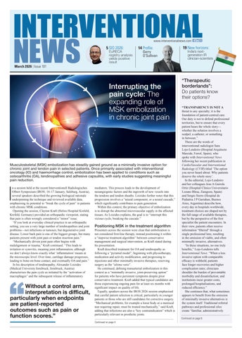
Interrupting the pain cycle: The expanding role of MSK embolization in chronic joint pain
Musculoskeletal (MSK) embolization has steadily gained ground as a minimally invasive option for chronic joint and tendon pain in selected patients. Once primarily associated with interventional oncology (IO) and haemorrhage control, embolization has been applied to conditions such as osteoarthritis (OA), tendinopathies and adhesive capsulitis, with early studies suggesting meaningful pain reduction.
I
n a session held at the recent Interventionell Radiologisches Olbert Symposium (IROS; 14–17 January, Salzburg, Austria), several speakers described the growing biological rationale underpinning the technique and reviewed available data, emphasising its potential to “break the cycle of pain” in patients with chronic MSK conditions. Opening the session, Clayton Kraft (Helios Hospital Krefeld, Krefeld, Germany) provided an orthopaedic viewpoint, stating that pain is often wrongly considered a “minor” issue. “If you look at everyday clinical practice in an orthopaedic setting, you see a very large number of tendinopathies and joint problems—not infections or tumours, but degenerative joint disease. Lower back pain is one of the biggest groups, but many patients present with joint pain or tendon insertion pain.” “Mechanically driven joint pain often begins with malalignment or trauma,” Kraft continued. “This leads to irritation or synovitis, meaning joint inflammation, although we don’t always know exactly what ‘inflammation’ means at the microscopic level. Over time, cartilage damage progresses, leading to bone-on-bone contact, and eventually OA and pain.” In his description of tendinopathy, Alexander Loizides (Medical University Innsbruck, Innsbruck, Austria) characterises the pain cycle as initiated by the “activation of macrophages” and the subsequent release of inflammatory
Without a control arm, interpretation is difficult, particularly when endpoints are patient-reported outcomes such as pain or function scores.”
mediators. This process leads to the development of neoangiogenic factors and the ingrowth of new vessels into the tendons and tendon sheaths. Loizides further notes that this progression involves a “neural component, or a neural cascade,” which significantly contributes to pain generation. Within this context, the primary objective of embolization is to disrupt the abnormal microvascular supply in the affected tissues. As Loizides explains, the goal is to “interrupt this vicious cycle, breaking the cascade.”
Positioning MSK in the treatment algorithm
Presenters across the session were clear that embolization is not considered first-line therapy, instead positioning it within a “stepwise treatment algorithm” between conservative management and surgical intervention, as Kraft stated during his presentation. Kraft described treatment for OA and tendinopathy as following a “pyramid model”, beginning with physiotherapy, medication and activity modification, and progressing to injections and other minimally invasive therapies, reserving surgery as the “ultima ratio”. He continued, defining transarterial embolization in this context as a “minimally invasive, joint-preserving option” for patients who have persistent symptoms despite prior conservative treatment. Kraft added that typical candidates are those experiencing ongoing pain for at least six months with significant impact on quality of life. Equally, speakers across the IROS 2026 session emphasised that careful patient selection is critical, particularly in younger patients or those who are still candidates for corrective surgery. “Mechanical problems, for example a loose body or a meniscal tear requiring repair, must be treated mechanically,” said Kraft, adding that infections are also a “key contraindication” which is particularly relevant in prosthetic joints.
19 New horizons:
India’s nextgeneration IR clinician-scientists
“Therapeutic borderlands”: Do patients know their options? “TRANSPARENCY IS NOT A threat to any specialty; it is the foundation of patient-centred care. Our duty is not to defend professional territories, but to ensure that every patient hears the whole story— whether the solution involves a scalpel, a catheter, or something in between.” These are the words of interventional radiologist Sara Lojo-Lendoiro (Hospital Arquitecto Marcide, Ferrol, Spain), who spoke with Interventional News following her recent publication in CardioVascular and Interventional Radiology (CVIR) titled ‘The option you never heard about: Why patients deserve the whole story’. In the editorial, Lojo-Lendoiro and her colleagues Jose A Guirola Ortiz (Hospital Clinico Universitario Lozano Blesa, Zaragoza, Spain) and Sergio Sierre (Hospital de Pediatria J P Garrahan, Buenos Aires, Argentina) describe how, every day in hospitals worldwide, treatment choices are shaped not by the full range of available therapies, but by the perspective of the first specialist the patient encounters. In their view, patients often receive information “filtered” through a single professional lens, resulting in the omission of viable, and often minimally invasive, alternatives. “In these situations, no one truly benefits,” Lojo-Lendoiro tells Interventional News. “When a less invasive option with comparable efficacy is withheld, patients face longer recoveries and higher complication rates; clinicians shoulder the burden of preventable morbidity and dissatisfaction; and institutions incur greater costs, prolonged hospitalisations, and reduced efficiency.” She continues that, what sometimes appears to benefit from the omission of minimally invasive alternatives is the system itself. Traditional referral pathways and professional silos create “familiar, administratively Continued on page 6
Continued on page 4