Screening and development of a base stable, mild elution condition affinity ligand for AAV9 C. Arai, C. Burdett, C. Daye, S. Yang, S. Arroyo, C. Whitehouse, I. Scanlon Astrea Bioseparations, Horizon Park, Barton Road, Comberton, Cambridge, CB23 7AJ, UK
Introduction
AAV9
Adeno-associated virus (AAV) vectors are widely adopted as delivery vehicles for the treatment of genetic disorders. Of the multiple AAV serotypes, AAV9 has demonstrated tropism towards the central nervous system, retina, inner ear, lung, liver, cardiovascular, and multiple other targets and has been reported as being used in 25% of the programs in clinical development (Advancing AAV production with high throughput screening and transcriptomics Cell & Gene Therapy Insights 2024; 10(6), 821–840).
4
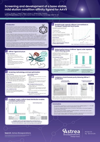
Breakthrough capacity Affimer®s immobilized on PuraBead® loaded with AAV9 lysate
Two candidates screened with AAV9 lysate (expressed in suspension HEK 293 cells).
0.09 0.08
1 mL packed column 1-minute residence time Load: ≈5 × 10¹³ capsids/mL adsorbent
0.07
10% breakthrough not reached on either prototype by detection of AAV9 particles by ELISA, indicating a high dynamic binding capacity.
We present a screening process for an Affimer® protein ligand to selectively bind AAV9. Affimer®’s are cystatin-derived single-domain proteins (10.7 kDa), affinity is tested as a function of the inclusion of randomized sequences in loops 2 and 4. The highest performing candidates were immobilized on an agarose bead matrix (PuraBead®) and the resulting adsorbent demonstrated binding at pH 7.5 and elution at pH 4.5 of clarified AAV9 from transiently transfected HEK 293 cells.
Affimer® 1 selected for further testing due to milder elution conditions
Binding loops
0.01 0 0
Small, single‑domain proteins
•
95 amino acids; ~10.7 kDa without inserts
•
ngineered scaffolds derived from the cystatin E protein family
Chromatography method
•
aturally lack cysteines and post‑translational N modifications
• Equilibration: PBS, 0.2 M NaCl, 0.001% Pluronic F‑68, pH 7.5
•
inding specificity generated by introducing B randomized peptide sequences into loops 2 and 4
• Load: >1 × 10¹⁴ capsids/mL adsorbent (AAV9)
• •
Phage display was used to generate a library of His‑tagged proteins. Screening was performed on a flow cytometry platform using a sandwich assay format. Anti‑His antibody–conjugated beads were used to capture Affimer® candidates, which were then incubated with clarified lysate containing recombinant AAV9. Binding was detected using an AlexaFluor‑488–labelled anti‑AAV9 antibody.
Hit validation Hits from the initial screen were verified using a reverse‑sandwich flow cytometry assay. In this format, anti‑AAV9 antibody was immobilized, recombinant AAV9 lysate was applied, and binding of Affimer® hit candidates was detected using an AlexaFluor‑488 anti‑His antibody.
Cross-validation by ELISA Both assay formats were subsequently repeated using ELISA to cross‑validate and strengthen the hit identification dataset.
lean‑in‑place (CIP): 0.1 M NaOH, 15 column C volumes at 1 mL/min flow rate (15‑minute exposure per cycle)
Lead candidate evaluation Initial screening • Fourteen compounds were identified in the primary screen
6
• Seven sequences were selected for scale‑up to 1 L expression in E. coli
2
3
4
5
6
kDa 191
• Binding performance was evaluated for total AAV9 particle capture in a high‑throughput plate‑based format • Recovery was quantified using an AAV9‑specific ELISA
97
VP1 VP2 VP3
64 51
Column‑scale evaluation
39
• Lead candidates from the plate assay were further assessed in a 1 mL column format
28
30
35
Figure 1. Breakthrough of AAV9 particles from clarified HEK 293 lysate
AAV9 recovery by elution at pH 4.5 1.2E+14 1E+14 8E+13 6E+13
1.1E+14 9.7E+13
9.7E+13
11
21
4E+13 2E+13 0
1
Cycling number
• Affimer® 1 consistently loaded to >1 × 10¹⁴ capsids/mL adsorbent across all AAV9 load cycles
• No reduction in performance was observed following repeated exposure to 0.1 M NaOH, indicating strong caustic stability
AAV9 elution profile following repeated CIP 1
• Purified candidates were immobilized on the PuraBead® Edge matrix
25
Affimer® 4
Validation of AAV9 eluate purity following Affimer® 1 purification
• Proteins were purified using IMAC followed by SEC, achieving >95% purity as confirmed by capillary electrophoresis
High‑throughput binding assessment
20
Figure 2. Recovery of AAV9 over 20 exposures to CIP
• Elution recovery corresponding to ~75% recovery in the pH 4.5 elution pool
Screening methodology and lead optimization
Primary screening
15
Affimer® 1
Analysis: ELISA and SDS‑PAGE
N-terminmus
Image by Avacta Life Sciences via Wikipedia (Public Domain)
2
Elution 1: 50 mM citrate, 0.3 M NaCl, pH 4.5
• Elution 2: 50 mM citrate, 0.3 M NaCl, pH 3.0
• Allows for large display libraries with >1010 sequence diversity
10
Robust performance of Affimer® ligand under repeated NaOH cleaning cycles
•
β-turn 3
5
CV
21 sequential column runs were performed. AAV9 lysate was loaded on cycles 1, 11, and 21, corresponding to: post‑0, post‑10, and post‑20 exposures to 0.1 M NaOH.
Affimer®: Key features
0.04
0.02
Gradient elution from pH 6 → pH 3 Affimer® 1: elution at ~pH 5 Affimer® 4: elution only at pH 3 hold
Total AAV9 recovery (capsids/mL adsorbent)
Affimer® ligand structure
0.05
0.03
Elution behavior
5
1
C/C0
0.06
Serotype independent ligands demonstrate poor binding of AAV9 (Efficient AAV9 Purification Using a Single-Step AAV9 Magnetic Affinity Beads Isolation Int. J. Mol. Sci. 2024, 25(15), 8342;). The potency of AAV9 is dramatically affected by pH and salt conditions, due to aggregation, depurination, and other structural factors. (Lengler, J., Gavrila, M., Brandis, J. et al. Crucial aspects for maintaining rAAV stability. Sci Rep 14, 27685 (2024). ). Many current affinity ligands require pH of 3 or below for full elution, whereas at pH 4 AAV9 has demonstrated maintenance of physical structure and activity. We determined that a serotype specific AAV9 ligand with a milder elution pH would be beneficial for efficient clinical production of AAV9.
19
14
7
8
Lane
Comparison to benchmark resin for AAV9 elution SDS-PAGE load (µL)
Sample
Dilution
M
Marker
Neat
5
1
C1 (EL1)
Neat
25
2
C11 (EL1)
Neat
25
3
25
C21 (EL1)
Neat
4
Sample buffer
Neat
5
5
C1 (CIP)
Neat
25
6
C11 (CIP)
Neat
25
7
C21 (CIP)
Neat
25
8
Sample buffer
Neat
5
1 kDa 191 97
2
3
4
Lane
Marker
2
Product A* Run 1
3
Product A* Run 2
4
Affimer® 1 resin Run 1 + 2 pooled
64 51
Sample
1
39 28 19 14
Affimer 1 Elution 1 fractions demonstrated high AAV9 purity. Consistent elution profiles observed across all runs. No AAV capsid proteins were detected in the CIP strip fractions. Indicating effective cleaning and no product carryover. The elution pattern was comparable to that of a commercial AAV9 affinity adsorbent*. ®
3
PuraBead® matrix: Uniform bead distribution enables consistent performance Market leading product Market leading product
1000
1000
800
800
Frequency
1200
600 400
200 0
(11, 12] (14, 15] (17, 18] (20, 21] (23, 23] (26, 26] (28, 29] (31, 32] (34, 35] (37, 38] (40, 40] (43, 43] (45, 46] (48, 49] (51, 52] (54, 55] (57, 58] (60, 60] (62, 63] (65, 66] (68, 69] (71, 72] (74, 75] (77, 77] (80, 80] (82, 83] (85, 86] (88, 89] (91, 92] (94, 94] (97, 97] (99, 100] (102, 103] (105, 106] (108, 109] (111, 111] (114, 114] (116, 117] (119, 120] (122, 123] (125, 126] (128, 129] (131, 131] (133, 134] (136, 137] (139, 140] (142, 143] (145, 146] (148, 148]
• The lead Affimer® immobilized on an agarose matrix demonstrated a binding capacity of ~1 × 10¹⁴ AAV9 particles/mL adsorbent, despite using a low ligand density (~2 mg/mL). • The adsorbent enabled mild elution at pH 4.5, achieving ~75% recovery, with no loss of capacity or recovery across 20 CIP cycles.
400
0
• These results highlight excellent suitability for AAV9 purification, combining high capacity, gentle elution conditions, and excellent caustic stability. 50
100
150
Diameter (ESD)
Diameter (ESD) Porosity (Kav)
Conclusions
600
200 [0, 1] (3, 4] (6, 6] (9, 9]
Frequency
® ® PuraBead Edge average particle distribution PuraBead EDGE average particle distribution
1200
0.43
Porosity (Kav)
Flow (cm/hr at 15 psi)
364
Flow (cm/hr at 15 psi)
525
Mean diameter (µm)
69.10
Mean diameter (µm)
96.36
Distribution (% beads in range 44–96 μm)
95.8
Distribution (% beads in range 76–141 μm)
66%
PuraBead® matrix is a 6% agarose base matrix, manufactured via a sustainable aqueous process, and is available in 60 µm or 90 µm bead sizes. The narrow size distribution enables robust packing and flow properties.
Search: Astrea Bioseparations Any data or results provided are only examples and do not provide any guarantee of similar results in future. No conflicts of interest to disclose. The products of Astrea Bioseparations may be covered by or for use under one or more patents: astreabioseparations.com/patents All trademarks, trade names, trade dress, product names and logos are the property of Astrea UK Services Ltd. © 2026 Astrea Bioseparations Ltd. All rights reserved
0.35
• With further optimization of ligand immobilization, this Affimer®‑based adsorbent has the potential to become a robust, scalable platform for clinical‑grade AAV9 manufacturing.