Viral clearance from mAbs with HCPure™ and Q PuraBead® adsorbents
Abstract
Biopharmaceutical products such as monoclonal antibodies (mAbs) which are derived from mammalian systems require evidence of viral clearance in the manufacturing process. Viral spiking studies are utilized to determine the efficacy of viral clearance in downstream purification methods (1).
Minute Virus of Mice (MVM) is a small non-enveloped parvovirus which has been found as an adventitious contaminant of CHO derived drug products and which is commonly used for viral clearance studies. Cygnus Technologies has developed a BSL-1 compatible surrogate of MVM which has been shown to be physiochemically similar to live MVM and can be used as an alternative to MVM for viral clearance studies (2).
Cygnus Technologies’ Type C Retrovirus Like Particles (RVLP) are endogenously produced during CHO cell expression. Endogenous viral clearance is generally demonstrated with a model mammalian virus, XMuLV, however, the establishment of RVLP quantification methods have made it possible to track the removal of actual endogenous RVLP throughout the downstream process.
Astrea Bioseparations Ltd partnered with Cygnus Technologies to demonstrate viral clearance using HCPure mixed-mode and Q PuraBead® Anion Exchange Chromatography resins with MVM-MVP and RVLP spiking studies.
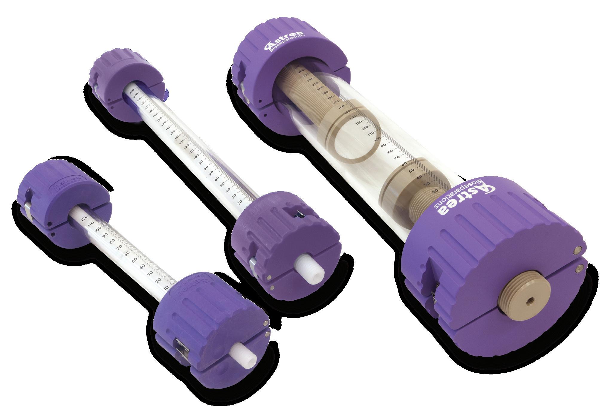
A CHO derived IgG was partially purified by Protein A capture, viral inactivation with low pH hold and intermediate purification with CEX chromatography (SP PuraBead® HF) in bind and elute mode. The SP eluate was buffer exchanged by TFF into 50 mM Tris pH 8 and supplied to Cygnus Technologies with HCPure and Q PuraBead® HF columns packed in to Astrea Bioseparations SNAP® Laboratory Glass Columns at 10 cm bed height.
Cygnus Technologies spiked MVM-MVP or RVLP into the feedstock before triplicate purification runs with each adsorbent in flow through mode. Viral clearance was assessed by qPCR methods provided by Cygnus Technologies.
Carry over experiments with no viral load were performed to confirm the strip and CIP were sufficient between replicate runs on the same column.
Multiple batches of CHO IgG harvest were prepared in shake flasks (~ 28 L) and processed on a 9 mL GORE® Protein Capture Device with Protein A over 20 cycles. Typical buffer conditions for the Protein A purification are shown.
Pressure increases from multiple cycles was recovered using the extended wash regime recommended by the supplier which utilizes an acidic wash (100 mM arginine pH 2.3) and a reducing agent wash (50 mM tris, 100 mM NaCl, 1% SDS, 10 mM DTT pH 10.4).
The Protein A elution was held at pH 3.4 for 1 hour for viral inactivation before neutralization with 1 M Tris to pH 6.
An example purification run with the same buffer conditions to be used for the viral clearance study was performed on 1 mL pre-packed HCPure and Q PuraBead® HF columns with CHO IgG after Protein A purification and buffer exchange in to 50 mM Tris pH 8 (no SP step was used for this feedstock).



The columns were equilibrated with 50 mM Tris
Triplicate runs on the 10 cm bed height HCPure and Q PuraBead® HF columns were performed with CHO IgG spiked with RVLP or MVM-MVP in 50 mM Tris pH 8.
The columns were equilibrated with 50 mM Tris pH 8 before loading the spiked feedstock at 20 mg IgG/mL adsorbent and collecting the UV peak in the flow through.
The columns were regenerated with 50 mM sodium citrate, 1 M NaCl, pH 3 followed by cleaning with 0.5 M NaOH to ensure any bound materials were removed between runs.
Carry over experiments confirmed the success of the strip and CIP The virus concentration in the load, flow through and strip were measured by qPCR and the log clearance calculated.
Excellent recovery of IgG was seen for all runs (94 – 97%). Viral clearance to below the limit of detection was seen for both model viruses with the Q PuraBead® adsorbent and for the RVLP for the HCPure™ adsorbent, corresponding to ≥3.3 log for RVLP and ≥4.2 log for MVM-MVP. No clearance of MVM-MVP was seen for HCPure under these conditions.
Therefore, for the CHO IgG used in this example, a
capture step followed by HCPure for
clearance.