Bacterial Diversity in Invertebrates: A 16S rRNA Nanopore Sequencing Project LaShawn Bowe, Felicia Silva, Dr. Barbara Murdoch Eastern Connecticut State University, Willimantic CT USA Figure 3. PCR Process.
Introduction • • • •
•
Invertebrates are animals that lack a backbone. They are the largest group of animal species. The invertebrate microbiome refers to the collection of all microorganisms living in and on the host. Microbiomes are important for immune function, mental health, digestion, and more. 16S rRNA gene sequencing identifies bacteria by analyzing the sequences of the 16S ribosomal gene, which is a reliable marker for bacterial identification and phylogeny. Nanopore sequencing is a DNA sequencing method that utilizes tiny protein pores (nanopores) to identify the sequence of DNA or RNA Hypothesis Giant Mesquite Bug (Thasus gigas) will show higher bacterial diversity than the Whip Spider (Amblypygid).
Figure 4. AMPure bead DNA cleanup.
1. Giant Mesquite Bug & Whip spider
A
3. Sequenced Bacteria from Invertebrates at Genus Level
Figure 8. Abundance of genera found in microbiome of invertebrates. Analysis done through EPI2ME bioinformatics.
Results
B Figure 5. Amplified invertebrate 16S rRNA gene from PCR.
Figure 9. Rarefaction Graph. Amount of different bacteria found over the course of the sequencing run shown using EPI2ME bioinformatics.
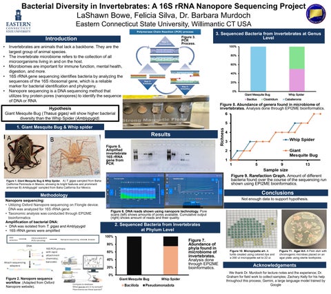
Figure 1. Giant Mesquite Bug & Whip Spider. A) T. gigas sampled from Baha
California Peninsula in Mexico, showing its bright features and prominent antennae B) Amblypygid sampled from Baha California Sur Mexico.
Conclusions
Methodology Nanopore sequencing: • Utilizing Oxford Nanopore sequencing on Flongle device. DNA was analyzed for 16S rRNA gene • Taxonomic analysis was conducted through EPI2ME bioinformatics Amplification of bacterial DNA: • DNA was isolated from T. gigas and Amblypygid • 16S rRNA genes were amplified
Not enough data to support hypothesis. Figure 6. DNA reads shown using nanopore technology. Pore scans (left) shows amounts of pores available. Cumulative output (right) shows amount of reads and their quality.
2. Sequenced Bacteria from Invertebrates at Phylum Level Figure 7. Abundance of phyla found in microbiome of invertebrates. Analysis done through EPI2ME bioinformatics.
Figure 2. Nanopore sequence workflow. (Adapted from Oxford Nanopore website).
Figure 10. Micropipette art. A turtle created using colored dye and a 200 ul micropipette set to 22 ul.
Figure 11. Agar Art. A Petri dish with chromogenic microbes placed on an agar plate using sterile toothpicks.
Acknowledgements We thank Dr. Murdoch for lecture notes and the experience; Dr. Graham for field work to collect samples; Zachary Kelly for his help throughout this process; Gemini, a large language model trained by Google