ARI Activity Report 2025
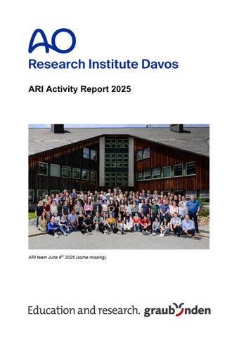
ARI team June 6th 2025 (some missing).
Contents 1
Introduction .................................................................................................................... 4
2
ARI Purpose / Goals / Outlook........................................................................................ 6
3
Funding Summary .......................................................................................................... 8
4
Research Structure & Advisory Committees ................................................................... 9 4.1
AO Research Institute Davos (ARI) Organigram ................................................ 9
4.2
AO Foundation Executive Committee (AOEC) ................................................... 9
4.3
AO Foundation R&D Platform and Innovation Platform ...................................10
4.4
AO Research Institute Davos Advisory Committee (ARI AC) ..........................11
4.5
AO CMF Research Commission (AO CMF RC) .................................................12
a.
Scientific Knowledge Development:...............................................................................12
b.
Individual Research Career Development: ....................................................................12
5
4.6
AO Spine Research Commission (AO SRC)......................................................13
4.7
AO Trauma Research Commission (AO TRC)...................................................14
4.8
AO Vet Research Commission (AO VET RC) ....................................................15
4.9
AO Research Review Task Force (AO RRTF)....................................................15
4.10
AO Network Preclinical Research (AO NPR) .....................................................16
ARI Teams / Personnel .................................................................................................16 5.1
Biomedical Development ...................................................................................16
5.2
Preclinical Services ............................................................................................17
5.3
Regenerative Orthopaedics................................................................................18
5.4
ARI Administrative Services ..............................................................................20
5.5
Operations standards and safety.......................................................................21
6
Gender Equality Initiative...............................................................................................22
7
ARI Abstracts periodical / ARI conferences ...................................................................23 7.1
ARI Abstracts periodical.....................................................................................23
7.2
AO Orthopaedic Research Summit 2025 ...........................................................23
8
Institutional and Professional Relations .........................................................................24
9
Good News ...................................................................................................................29 9.1
New noncommercial extramural funding ..........................................................29
9.2 AO intramural funding (grants beyond ARI retainer & Clinical Division Research Commission grants) ......................................................................................29 9.3
Professorship / Habilitation................................................................................29
9.4
ECLAM diplomate ...............................................................................................30
9.5
Awards .................................................................................................................30
9.6
New Board Positions ..........................................................................................31
9.7
ARI MOU's (Memorandums of Understanding) .................................................31 2
9.8
Collaborations .....................................................................................................32
9.9
Visits ....................................................................................................................33
10 ARI Medical Research Fellows ......................................................................................35 11 Project Abstracts by Sponsors.......................................................................................45 11.1
AO CMF................................................................................................................45
11.2
AO Spine..............................................................................................................46
11.3
AO Trauma ..........................................................................................................48
11.4
AO VET ................................................................................................................62
11.5
AOTC System ......................................................................................................64
11.6
ARI AC (AO Direct Funds) ..................................................................................66
11.7
AO Development Incubator ................................................................................76
11.8
AO Education Institute Funding.........................................................................80
11.9
Extramural Projects ............................................................................................83
12 Team Members ...........................................................................................................100 13 ARI Patents .................................................................................................................105 14 Publications & Presentations .......................................................................................108 14.1
2021-2025 Five-year ARI Key Performance Indicators ...................................108
14.2
2025 Published peer reviewed papers (epub & in print).................................109
14.3
2024 epub, 2025 in print ...................................................................................118
14.4
Theses ...............................................................................................................119
14.5
Abstracts published in journals .......................................................................120
14.6
Abstracts (conference presentations) .............................................................123
14.7
Presentations (not in conference proceedings) .............................................131
3
1 Introduction 2025 was a year of momentum, achievement, and looking confidently toward the future. Across the AO Research Institute Davos (ARI), our scientists, surgeons, fellows, support teams, and collaborators continued to advance innovation in orthopaedics through high-quality translational research and development, always guided by the AO mission of improving patient care. One of the defining moments of the year was the successful organization of the AO Orthopaedic Research Summit 2025 in Davos. By bringing together the annual meetings of ARI Orthopaedics, the European Orthopaedic Research Society (EORS), and the Computer Assisted Orthopaedic Surgery (CAOS) Society, the summit created a unique global platform for scientific exchange. More than 860 participants from 53 countries joined us to share new discoveries, discuss emerging technologies, and strengthen collaborations across disciplines. The event demonstrated not only the scientific reputation of ARI but also the ability of our teams to convene and inspire the international orthopaedic research community. Equally significant was the beginning of a new chapter for the AO in Davos. During 2025, construction commenced on the Science Circle, following the groundbreaking ceremony that formally launched this major AO Campus development. Science Circle represents far more than a new building. It is an investment in the future of research, education, and innovation, providing modern laboratories, collaborative workspaces, and infrastructure designed to foster interdisciplinary collaboration and scientific excellence. As the foundations were laid during 2025, so too were the foundations for decades of future discovery. The new facility will ultimately house the Regenerative Orthopaedics laboratories and the majority of ARI staff, bringing together researchers, clinicians, educators, and innovation partners within a vibrant AO Campus environment. On a personal note, I have been privileged to spend 34 years at the AO and to have worked across each phase of ARI’s development in Davos – from the early days of the Laboratory for Experimental Surgery (LECD) at Villa Fontana in the town, through the establishment and growth of the ARI on our own land, where the AO campus is now and soon into the next era represented by Science Circle. It is both professionally rewarding and deeply meaningful to witness this continued evolution of the AO’s research infrastructure, and I look forward to contributing to this exciting new chapter as Science Circle becomes the future home of much of ARI’s scientific community. The year also brought continued scientific success. ARI researchers remained active across musculoskeletal biology, biomaterials, biomechanics, infection research, regenerative medicine, computational modelling, and translational technologies. Our teams secured new grants, strengthened international collaborations, contributed to global scientific societies, and continued to publish and present research that advances understanding and improves clinical practice. Several ARI innovations continued their path toward translation, while others further matured within the research pipeline, reinforcing our commitment to transforming scientific discoveries into solutions that ultimately benefit patients. Beyond the many scientific achievements recorded throughout this report, I am particularly proud that ARI continues to combine world-class science with tangible impact. Our teams continued to publish, innovate, secure competitive funding, develop intellectual property, educate future leaders, and strengthen collaborations across the globe. Numerous colleagues received recognition through awards, invited lectureships, leadership positions, and international honours, while ARI researchers continued to feature amongst the world's most highly cited scientists. At the same time, our translational technologies progressed further along the path toward clinical application, reflecting our commitment to converting scientific discoveries into practical solutions for patients. These achievements are only possible because 4
of the dedication, professionalism, and collaborative spirit of our staff, fellows, AO surgeons, partners, and supporters, all of whom contribute to the unique culture that makes ARI such a special place to work and innovate. Beyond scientific output, 2025 highlighted the importance of investing in people and culture. The implementation of the updated ARI career path continued to support professional development and internal progression, while ongoing initiatives in inclusive excellence, mentorship, fellowships, and international collaboration strengthened our position as a welcoming and supportive research environment. ARI's greatest asset remains its people, and their commitment, creativity, and collaborative spirit continue to drive our success. As we reflect on 2025, we can take pride not only in our scientific achievements but also in the strong foundations we are building for the future. From advances in translational research and innovation to the construction of Science Circle and the success of the AO Orthopaedic Research Summit, the year demonstrated that ARI continues to evolve while remaining true to its purpose: advancing innovation in orthopaedics through research and development to improve patient care. On behalf of the leadership team, I would like to sincerely thank all ARI and AO Network Preclinical Research staff, our AO clinical specialties – particularly their Research Commissions – our Advisory Committee, external partners, grant agencies, project collaborators, fellows, and the global AO surgeon network for their dedication, expertise, and support. I would also like to acknowledge the strong collaboration and partnership of our colleagues within the AO Innovation Translation Center (AO ITC), the AO Education Institute (AO EI), AO Community Development, and AO Governance, whose contributions help ensure that our research remains relevant, impactful, and aligned with the AO mission. Furthermore, I would like to express my appreciation to the AO support units, including Finance, Human Resources, Communications & Engagement, Marketing, Legal, and many others, who work closely with us every day and whose professionalism and service are essential to the success of our institute and projects. Together, we continue to create knowledge, develop innovative solutions, educate future leaders, and make a meaningful difference to patient care worldwide. It is this exceptional AO community, united by a shared commitment to improving outcomes for patients, that continues to inspire and motivate me every day and remains a primary reason why, after more than three decades with the AO, I remain deeply enthusiastic and privileged to serve in this role. Sincerely
Prof Dr R Geoff Richards FLSW, FBSE, FIOR, FORS, FTERM Executive Director AO Research & Development, Director AO Research Institute Davos (ARI)
5
2 ARI Purpose / Goals / Outlook Purpose To further the AO Foundations’ mission, ARI advances innovation in orthopedics through translational Research and Development (R&D). Orthopedics concerns musculoskeletal, spine, and craniomaxillofacial trauma, degenerative musculoskeletal diseases, infections, and congenital disorders.
Overall goals • Create evidence and knowledge through our expertise and high quality translational preclinical R&D focused on clinical problems within the AO mission. • Disseminate our created knowledge globally to advance our world class reputation. • Foster a close relationship with the AO health care professional network, academic societies, universities and collaborative hospitals for research. • Develop and valorize ARI intellectual property (IP) to improve patient care. • Strengthen education through Medical Research Fellowships, nurture future AO key opinion leaders and also support AO’s education developing advanced technologies. • Provide a supportive & inclusive environment & mentorship for all our employees & guests. • Maintain high level ARI Contract Research and Development.
2023-2025 Goals • Valorize AO Fracture Monitor with AO Innovation Translation Center (AO ITC). The AO Fracture Monitor has progressed to clinical handling testing in Germany and continues to advance toward clinical translation. • Implement the specific-pathogen-free sheep flock in studies. Implementation is ongoing, with the flock being progressively integrated into ARI preclinical research activities. • Valorize the biphasic plate together with AO ITC. The Biphasic Plate has successfully completed development milestones and requests for proposals have been issued to multiple companies to support future commercialization and translation. • Strengthen and advance research activities in patient diagnostics and personalized medicine. Research activities have expanded substantially, including continued development of diagnostic biomarkers, fracture-healing monitoring technologies, computational simulations, and personalized medicine approaches. • Develop training technologies to support education within the AO network. Digitally Enhanced Hands-on Surgical Training (DEHST) has been developed and implemented in multiple AO educational courses, establishing a distinctive capability within AO’s educational portfolio. • Continue developing 3D (bio)printing and SIM technologies. Development activities continue, supported by successful acquisition of competitive research funding and external grants that are advancing both technology platforms.
ARI principles • Maintain and advance our world-class research standing with our expert team. • Nurture in-house employee development through educational training and mentoring for long-term knowledge creation and innovation. • Support the AO health care professional network with cutting-edge R&D for musculoskeletal clinical problems. • Continue developing ARI technology portfolio. Translate and valorize ARI innovations together with the AO ITC's Technology Transfer team and the AO EI. • Maintain our world-class certifications (ISO, AAALAC, GLP). • Engage with scientific networks, societies, consortia, universities, institutions and hospitals at regional and global level.
6
ARI projects are translational preclinical research and development projects focused towards specific clinical and educational applications. Definitions (within the translational research): 1. Preclinical Research is fundamental research to solve major clinical problems over an extended time frame (over 10 years). 2. Translational Preclinical research aims at developing a clinically applicable result in around 5 years and builds upon the fundamental preclinical research. This research is usually not possible without the previous fundamental preclinical research. 3. Translational Development / Innovation takes the next step to valorize the translational research, in ARI’s case hand in hand with AO ITC and AO EI.
Outlook The AO Foundation's strategic collaboration with Synthes GmbH (Johnson & Johnson MedTech), which started new in January 2016 was signed, extending AO’s Cooperation Agreement through December 31, 2030. This renewal reinforces the shared commitment to advancing surgical education and innovation worldwide. The AO retains full independence, supported by JJMT through an unrestricted grant. Though research is not part of the Cooperation Agreement, ARI does conduct research with JJMT through Project‑specific contracts involving JJMT and ARI, with signed Research Funding Agreements between Synthes GmbH / Johnson & Johnson entities and ARI. The ARI budget received from the AO Foundation is taken from the AO Foundation's endowment funding stream, giving the ARI freedom to operate without direct obligations to the AO Foundation's industrial partners. With the signing of the EU Programs Agreement on 10 November 2025, Switzerland reaffirmed its commitment to international collaboration in research and innovation. This retroactive association to Horizon Europe, the Digital Europe Program, and the Euratom Program from 1 January 2025 strengthens the foundation for dynamic partnerships, pioneering innovation, and a future shaped by shared scientific ambition. The EU Programs Agreement represents a significant milestone in Switzerland's long-standing and forward-looking cooperation with the EU in education, research, and innovation. Being actively engaged in these fields in EU programs is strategically vital. It provides access to leading international networks, promotes scientific excellence, and enables joint responses to global challenges. As a result of the Agreement, researchers and innovators in Switzerland will once again be funded directly by the European Commission from 2025 onward. To this end, Switzerland will contribute financially to the EU, thereby reaffirming its commitment to international cooperation in research and innovation. This means that ARI can submit project proposals in the role of coordinator again. It also means that ARI budget of these applications needs to be included in the total amount of project funding requested by the consortium from the EU. Applying in the role of Beneficiary is compulsory in order to have funding guaranteed. There are no direct implications on running applications or projects.
7
,000 3CHF inFunding Summary 2024 Actual
Management & Overhead ARI Regenerative Orthopaedics Biomedical Development Preclinical Services Fellowships Network Preclinical Research Total Income Management & Overhead ARI Regenerative Orthopaedics Biomedical Development Preclinical Services Fellowships Network Preclinical Research Total Expenses Total Net Result
2025 Actual
2025 Budget
abs
%
abs
%
abs
%
751 1,918 1,803 918 0 32 5,422 -2,351 -6,373 -3,074 -3,041 -546 -1,984 -17,370 -11,948
14% 35% 33% 17% 0% 1% 100% 14% 37% 18% 18% 3% 11% 100%
1,295 1,689 1,579 1,013 0 8 5,585 -2,458 -6,362 -2,929 -2,761 -454 -3,035 -17,998 -12,413
23% 30% 28% 18% 0% 0% 100% 14% 35% 16% 15% 3% 17% 100%
1,105 1,661 1,608 835 5,208 -2,210 -6,271 -2,932 -2,893 -640 -2,900 -17,847 -12,638
21% 32% 31% 16% 0% 0% 100% 12% 35% 16% 16% 4% 16% 100%
Variance A25/B25 abs 190 28 -28 178 0 8 377 -248 -90 3 132 186 -135 -152 225
% 17% 2% -2% 21% 7% 11% 1% 0% -5% -29% 5% 1% -2%
Comments: Overall, the ARI closed the year with a net result of CHF -12,413 K, CHF 225 K below budget. For the unit ‘Network Preclinical Research’ (AO NPR), an underspending was expected (in Autumn) and a rollover of CHF 316 K from 2025 to 2026 was foreseen in the 2026 budget. Fortunately, this scenario did not occur, and AO NPR was able to spend the funds as planned. The final ARI costs even show an expenditure surplus of CHF -152 K. The good result, with a final underspending of CHF 225 K, corresponding to a deviation of less than 2% from the budget, was achieved mainly due to higher-than-planned income from public grants and commercial studies. Income: The ‘Management & Overhead ARI’ (M&O) division generated higher revenues of CHF 190 K thanks to the successful staging of the ‘AO Orthopaedic Research Summit’ (three conferences in one) in Davos in June 2025 and higher subsidies following successful contract negotiations. Contrary to expectations, the ‘Regenerative Orthopaedics’ division was able to increase its income from public grants. The reported income surplus of only CHF 28 K is owed to the cancellation of a development incubator project, which reduced intercompany income accordingly. ‘Biomedical Development’ also increased its revenue from public grants. The reported loss of CHF -28 K is attributable to various projects in collaboration with the Technical Commission (TC) / DePuy Synthes (DPS) being put on hold by DPS, which prevented the achievement of intercompany income. Preclinical Services acquired various additional commercial studies, resulting in a revenue surplus of CHF 178 K. Expenses: The higher expenses shown in M&O are mainly driven by increased costs for the organization of the 2025 ‘AO Orthopaedic Research Summit’ (personnel, expenses non-employees), but also by price increases for electricity and higher costs for input tax deduction. The overspending in ‘Regenerative Orthopaedics’ is due to higher housing costs (shown under rental) for the many scientific guests and interns, and higher maintenance costs for aging machinery. The underspending in ‘Preclinical Services’ is due to personnel not being replaced. In ‘Fellowships,’ fewer interns could be hired, and salary costs for fellows were partly granted from other sources. In the AO NPR area, project delays due to ongoing contract negotiations and other reasons led to various payments being postponed for years. Most of these problems have now been resolved, and the corresponding amounts have been transferred, resulting in overspending in 2025. Cost category: The main cost categories were ‘Personnel Expenses’ with 58% of the total, followed by ‘Scientific & Regional Expenses’ with 11%, and ‘Material Expenses’ with 10%. 8
4 Research Structure & Advisory Committees 4.1 AO Research Institute Davos (ARI) Organigram
(September 2025)
4.2 AO Foundation Executive Committee (AOEC)
The AO executive committee. Claas Albers officially became CEO and Vice-Chairman of the AO Foundation on 1 January 2025. His position as head of Innovation Translation was taken by Roland Herzog.
9
4.3 AO Foundation R&D Platform and Innovation Platform The AO R&D Platform and Innovation Platform supports the active exchange and mutual discussion about strategies of the AO units with respect to their related goals in R&D and innovation. It supports the AO Foundation Board (AOFB) in defining general strategic areas and their implementation in an advisory function. It ensures that relevant activities are in line with the AO Mission and strategies as defined by the AOFB. All research and innovation stakeholders are finally accountable to the AOFB. The AO R&D Platform and Innovation Platform further develop the strategies and their implementation on behalf of the AOFB in an advisory capacity. They have no funding and decision authority. The R&D Platform is represented on the AO EC by the AO Executive Director of Research and Development and the Innovation Platform by the AO Executive Director of Innovation Translation. The R&D expert of the AOFB is the Chair of the R&D Platform (and currently the Innovation Platform), currently Prof Joost De Brujin, President, Innovation & Strategy at Kuros Biosciences, Utrecht, Netherlands.
10
4.4 AO Research Institute Davos Advisory Committee (ARI AC) The ARI Advisory Committee (ARI AC) provides operational and strategic scientific advice to the ARI on behalf of the AO Foundation Board (AOFB). ARI AC acts as both a sounding board and sparring partner for the Director and scientists of the ARI. The ARI AC's tasks and responsibilities include advising ARI on: - Portfolio of competencies (skills of personnel and type of equipment) - Strategy and priority setting for direct funds of ARI - Business development and initial advice on technology transfer - Regulatory issues - Use of ARI funds - Advancement of the ARI capabilities, to ensure the efficient use of the infrastructure The ARI AC comprises the following members in 2024: - Prof Brian Johnstone, (Chair, represents the ARI AC and is member of the AO R&D Platform and the AO Innovation Platform), Oregon Health and Science University, USA - Prof Chris Evans, Mayo Clinic, USA - Prof Jürg Gasser, Basel, CH - Prof Hamish Simpson, Professor of Orthopaedics & Trauma, University of Edinburgh, Scotland, UK - Prof Gerjo Van Osch, Vice dean of Research Erasmus, Rotterdam, the Netherlands Two ARI AC f2f meetings were held in 2025 (June and December).
11
4.5 AO CMF Research Commission (AO CMF RC) The AO CMF RC is the international coordination body for all activities of the AO CMF clinical specialty for research and development of the AO Foundation (AO). Its mission is promoting excellence in patient care and treatment outcomes in trauma and musculoskeletal disorders. AO CMF RC’s main activities. Include coordinating international research across specialties as a central body for CMF research within the AO. The commission collaborates with external partners (consortia) and runs large-scale projects such as clinical priority programs (CPP), Seed Grants, and other global initiatives. Commission members as regional representatives are also members of regional boards and ensure that the flow of information is bidirectional. Key Research Areas include a. Scientific Knowledge Development: - Enhances academic credibility and leadership, focusing on the clinical quality program. - Projects involve external and internal bodies (AO ITC and ARI), with ongoing projects like the AO CMF CPP. - Long-term projects, typically 5-year terms, may be renewed. Additional activities include studies and projects by AO ITC and ARI. b. Individual Research Career Development: - Provides individual support through small grants (seed grants). - Supports research fellowships (AO ITC, ARI, research symposia, AO PEER offerings (f2f, online courses), etc. The AO CMF Research Commission comprises the following members, permanent guests and AO representatives: Dr Thomas B. Dodson, AO CMF Research Commission (RC) chair, Seattle, WA, USA Dr Rodrigo Pereira, AO CMF RC member (representative AO CMF LAT), Rio de Janeiro, Brazil Dr Brad Strong, AO CMF RC member (representative AO CMF NA), Davis, CA, USA Dr Patricia Stoor, AO CMF RC member (representative AO CMF ESA), Helsinki, Finnland Dr Khalid Abdelgalil, AO CMF RC member (representative AO CMF MENA), Abu Dhabi, UAE Dr Takahiro Kanno, AO CMF RC member (representative AO CMF AP), Izumo, Japan Philipp Buescher, ARI Head AO Network Preclinical Research (NPR) Prof Martin Stoddart, ARI Program Leader Regenerative Orthopaedics, Davos, Switzerland
12
4.6 AO Spine Research Commission (AO SRC) AO Spine's preclinical research activities, led by Principal Scientist, Dr Sibylle Grad at the ARI focus on intervertebral disc (IVD) degeneration and postoperative spine infection, with expertise in organ models and biomarker discovery. Preclinical findings are systematically translated to the AO Spine Knowledge Forums (KF), expert-driven global clinical study groups, for clinical evaluation. In 2025, key outcomes included: The “Immunospine” study identified a distinct serum protein signature that differentiates early surgical site infection (SSI) from normal postoperative recovery, highlighting biomarkers as a potential tool for early SSI detection, enabling differentiation between infection and sterile postsurgical inflammation. A bioreactor-based IVD degeneration model demonstrated that a single injurious event provokes both biological and mechanical deterioration. When the discs are then dynamically unloaded for a few days, some signs of recovery appear, such as reduced inflammation, less matrix loss, and partial height restoration suggesting that unloading or therapeutic movement may help support disc regeneration. Using the ARI’s whole-spine bioreactor system which simulates 6DOF loading, Sibylle and her team demonstrated that dynamic multiaxial loading of IVDs induces region-specific inflammatory and catabolic responses, particularly in the annulus fibrosus; dorsal root ganglion studies revealed early discogenic sensitization, while IVDs structure remained preserved after one week of mechanical loading. This work received the 2026 International Society for the Study of Lumbar Spine (ISSLS) Prize in Basic Science. The AO SRC consists of the following members: Dr Klaus Schnake, Chairperson, Erlangen, Germany Dr Shekar Kurpad, AO Spine KF Spinal Cord Injury Representative, Milwaukee, WI, USA Dr Michael Kelly, AO Spine KF Deformity Representative, San Diego, CA, USA Dr Samuel Cho, AO Spine KF Degenerative Representative, New York, NY, USA Dr Ilya Laufer, AO Spine KF Tumor Representative, New York, NY, USA Dr Gregory Schroeder, AO Spine KF Trauma & Infection Representative, Pittsburgh, PA, USA Dr Alfredo Guiroy, AO Spine Latin America Regional Research Officer, Mendoza, Argentina Dr Jefferson Wilson, AO Spine North America Regional Research Officer, Toronto, Canada Dr Kenny Kwan, AO Spine Asia Pacific Regional Research Officer, Hong Kong, China Dr Kabir Abubakar, AO Spine Middle East & Northern Africa Regional Research Officer, Kano, Nigeria Dr Marcin Czyz, AO Spine Europe and Southern Africa Regional Research Officer, Birmingham, England Dr Sibylle Grad, ARI Representative, Davos, Switzerland
13
4.7 AO Trauma Research Commission (AO TRC) The AO TRC is the international coordination body for all activities of the AO Trauma sprecialty for research and development of the AO. The AO TRC funds research projects and clinical studies in collaboration with external institutes as part of consortia within clinical priority programs (CPP). AO Trauma Research strategy focuses on two fields: 1) To be a knowledge leader, performing large research projects such as Clinical Priority Programs (CPPs) as a consortia with external key opinion leaders, experienced clinicians and researchers in collaboration with ARI and AO ITC that help AO Trauma gain scientific knowledge and enhance academic recognition and credibility. Gaining state-of-the-art knowledge serves to promote AO Trauma to maintain its leadership position. To this aim, AO Trauma conducts two CPPs that focus on clinically highly relevant topics. 1) AO Trauma CPP Patient Outcome lead by Dr Marylin Heng (Miami, USA). 2) AO Trauma CPP focus on bone non-union (led by Prof Elizabeth Balmayor, Aachen University Hospital, Germany). 2) AO TRC provides individual support to young clinicians to increase awareness of research and provide training in the fundamentals of research processes. Within this framework, the AO TRC offers funding programs for smaller projects. These grants follow the AOFB guidelines in terms of target group (young clinicians < 40 years), access (open to all Clinical specialties). Out of this pool of young clinicians, new talents are identified. AO TRC also coordinates research symposiums and offers research fellowship programs. AOTRC comprises the following members and AO representatives: Prof Peter Giannoudis, AO TRC chairperson, Leeds, UK Prof Dhaval Desai, AO TRC member (representative AO TAP), Surat, India Dr Leah Gitajn, AO TRC member (representative AO TNA), Lebanon, USA Dr An Sermon, AO TRC member (representative AO TESA), Leuven, Belgium Dr Carlos Valderrama, AO TRC member (representative AO TLAT), Medellín, Colombia Dr Ayesha Saeed, AO TRC member (representative AO TMENA), Islamabad, Pakistan Philipp Buescher, ARI Head AO Network Preclinical Research (NPR), Davos, Switzerland Dr Alex Joeris, AO ITC Head of Clinical Science, Dübendorf, Switzerland Prof Geoff Richards, AO Executive Director Research & Development, Davos, Switzerland
14
4.8 AO Vet Research Commission (AO VET RC) AO VET RC pursues two main goals with its research activities. First one is to perform research activities that help to gain scientific knowledge and enhance academic recognition and credibility. Gaining state-of-the-art knowledge serves to promote the AO to maintain its leadership position. AO VET RC also provides individual support to young clinicians to increase awareness of research and provide training in the fundamentals of research processes as well as identifying new talents. The preclinical research activities of AO VET are coordinated at ARI by Dr med vet Stephan Zeiter, Program manager Preclinical Services. AO VET RC also supports the other AO Clinical specialties as an advisory body (Animal Welfare Advisory Committee (AWAC) and AAALAC. The AO VET RC comprises the following members and AO representatives: Ass Prof Kyla Ortved, AO VET RC chair, Pennsylvania, MI, USA Dr Yukihiro Fujita, AO VET RC member (representative AP), Tokyo, Japan Dr Sushmitha Durgam, AO VET RC member (representative NA), Columbus, OH, USA Dr Kevin Parsons, AO VET RC member (representative ESA), Bristol, UK Dr Anderson Souza, AO VET RC member (representative LAT), São Paulo, Brazil. Philipp Buescher, Head AO Network Preclinical Research (NPR), Davos, Switzerland Dr Stephan Zeiter, ARI Program Leader Preclinical Services, Davos, Switzerland Dr Ivan Zderic, ARI Focus Area Leader Biomechanics and Modeling, Biomedical Development, Davos, Switzerland
4.9 AO Research Review Task Force (AO RRTF) The AO RR TF is an independent peer review body valid for all AO decision-making bodies for grants to all external applicants for AO research funding. The AO RRTF is assigned jurisdiction over many external AO peer review process, while other internal AO Peer Review Policies and expectations govern specific AO Institute research programs, partnering, internal research contracting, and some limited external research funding processes. Decision-making bodies are defined as bodies that have funding allocation roles within the AO, including AO Trauma, AO Spine, AO CMF, AO VET, and their respective Research Commissions (RCs). For ARI Collaborative Research Programs, the decision-making body is ARI together with the ARI Advisory Committee (ARI AC). For each Clinical Specialty research grant, the decision-making body is that respective CD RC. The chairperson of the AO RR TF is Jaimo Ahn, Ann Arbor, USA.
15
4.10 AO Network Preclinical Research (AO NPR) The goal of the AO Network Preclinical Research (AO NPR) is to gain in efficiency and effectiveness with one central team for all AO funded external preclinical research. AO NPR is the international coordination group for all external preclinical research activities of the AO. AO NPR manages and supports the global research commissions of the AO Trauma, AO CMF, and AO VET to establish a cohesive global research vision and strategy for AO worldwide. AO NPR supports coordination between external partner institutes and AO Institutes. AO NPR is the entry point for all external research partners for preclinical research. AO NPR promotes excellent research of all AO partners, which are directly or indirectly related with clinical needs in patient care. It helps to strengthen networking among AO clinicians and researchers worldwide, making clinically relevant research attractive for the young generation of AO surgeons. AO NPR Manages the Clinical Priority Programs (CPP’s) of Clinical Specialties and the Research activities of Clinical Divisions AO Trauma, AO CMF, and AO VET. AO NPR manages the research governance of the Research Commissions of the Clinical Specialties AO Trauma, AO CMF and AO VET, the AO R&D Platform, the AO Innovation Platform and the AO Research Review Commission (AO RRC). AO NPR is headed by Philipp Buescher. Team members are Tania Bosque, Anna Dönz, Larissa Welti.
5 ARI Teams / Personnel 5.1 Biomedical Development Program Leader: Peter Varga, Deputy: Boyko Gueorguiev Team Members: Gordian Banzer, Jan Barcik, Zoé Beer, Jan Buschbaum, Jan Caspar, Daniel Ciric, Ivan Dimitrov, Manuela Ernst, Alicia Feist, Dominic Gehweiler, Alisa Hangartner, Flurin Heimo, Carla Hetreau, Maximilian Heumann, Fabian Kellner, Angelov Lyubomir, Dominic Mischler, Alia Pfiffner, Helcio Pinheiro, Miroslav Raykov, Sidney Schmuki, Peter Schwarzenberg, Flurin Spiller, Richie Strain, Antoine Vautrin, Teng Ye, Ivan Zderic, Calvin Zeller, Franziska Ziegenhain, Erich Zweifel The Biomedical Development (BD) Program aims to provide innovative, translation‑oriented solutions that address key clinical challenges in traumatology and orthopedics towards improved patient care. This is achieved in close collaboration with clinical, scientific, and industrial partners, as well as with the AO Clinical Specialties, AOITC, and the AO Education Institute (AO EI). The R&D team offers extensive know-how, expertise and experience in biomechanical testing, computational analysis, and the development and design of medical devices in accordance with EN ISO 13485. New concepts, tools, implant systems, and sensors for surgical applications and research are created, alongside digital and hands‑on technologies for surgical training and education. The development of optimal solutions to clinical questions is supported by utilizing capabilities ranging from in silico approaches to anatomical laboratories, enabling rapid and effective evaluations in realistic environments. Tailored biomechanical test procedures incorporating radiography, video and motion tracking analysis are applied in experiments focusing on fracture fixation and joint reconstruction. State‑of‑the‑art technologies, powerful numerical methods, AI-supported analyses and comprehensive tools for virtual simulation support investigations into the biomechanical performance of bone–implant constructs and fracture healing. Medical imaging, processing, and analysis capabilities—featuring CT scanners with a wide range of resolutions and scan volumes—enable detailed morphological assessments, extraction of statistical and patient‑specific information, and deeper insights into variations in bone characteristics and fracture patterns. The Program’s capabilities are completed by the Prototype Workshop that provides rapid and high‑quality manufacturing of devices, tools, and implants. 16
5.2 Preclinical Services Program Leader: Stephan Zeiter, Deputy: Nora Goudsouzian Team Members: Daniel Arens, Milena Benz, Barbara Brändle, Carmen Brazerol, Klemens Driver, Lorena Faoro, Loris Faoro, Pierina Faoro, Andrea Furter, Lena Gens, Martin Hächler, Meredith Halbeisen, Genevieve Hall, Maria Hildebrand, Lean Jabali, Urban Lanker, Salome Leuthold, Leonie Mollet, Reto Müller, Dirk Nehrbass, Ann-Kathrin Ostertag, Dominic Perren, Arina Polujanenkova, Irene Sanchez, Monika Schneider, Zdenka Slavikova, Beni Szulyovszky, James Tapia-Dean, Claudia Zindl The Preclinical Services (PS) Team at ARI, encompassing Preclinical Surgery and Histology, continues to strengthen its commitment and dedication to both animal welfare and performing in vivo preclinical research at the highest standards. Our team of dedicated veterinarians, biologists, animal care takers, histopathologist and technicians delivers end-to-end support, accompanying projects from early planning and surgical procedures to data analysis and publication. Together with the Medical Imaging, Infection Biology and Bone Biology teams of other programs we continue to foster fruitful collaborations with ARI internal partners. In 2025, we successfully conducted over 15 studies and carried out more than 400 surgical procedures, utilizing various animal models, including mice, rats, rabbits, and sheep. Our in-house flock of SPF (Specific Pathogen Free) sheep continues to grow healthily and expanded by 59 newborn lambs (21 pairs of twins, and one pair of triplets). Underlining our commitment to animal health and research advancements. To stay at the forefront of developments surrounding animal welfare and preclinical testing our team has gained further expertise. As a result, we gained a further ECLAM Diplomate (European College of Laboratory Animal Medicine) strengthening a program dedicated to the promotion of scientific progress in laboratory animal medicine. Additionally, further achievements of our animal caretaker staff with successful completion of an apprenticeship in Animal Care (EFZ Tierpflege) and Operations Manager in Agriculture (Betriebsleiterin Landwirtschaft) bringing further expertise to our team. By contributing to the chain of knowledge and passing on our expertise, we continually promote the veterinary student scholarship program in partnership with Aberystwyth School of Veterinary Science, Aberystwyth University, Wales. Through this and other global university collaborations, we provided practical training and a solid foundation in preclinical research to veterinary students from different countries around the world. We continue to uphold the highest standards through robust quality management systems, including GLP and AAALAC accreditations. Our dedication to transparency, ethical research practices, and increasing public understanding of the importance of our work remain as key values. Looking ahead, we remain committed to advancing preclinical science while preserving the integrity and values that have always guided our approach. Figure: Surgical intervention in sheep knee joint with Osteochondral defect in the lateral condyle, metal cube for size reference (1cm), study aimed to test the safety and efficacy of a treatment method for cartilage lesions.
17
5.3 Regenerative Orthopaedics Program Leader: Martin Stoddart, Deputy: Sibylle Grad Team Members: Saif Ali, Mauro Alini, Nada Yasmine Amjid, Sebastian Amport, , Ezgi Irem Bektas Tas, Xander Bemelmann, Luca Buonarriva, Simona Casutt, Claire Chabot, Wen Chen, Marco Chittò, Alessandro Cianciosi, Eda Ciftci-Dede, Carolina Maria Cordeiro, Darine D’Adam, Elena Della Bella, Matteo D’Este, Nicolas Devantay, Jun Dong, Pia Fehrenbach, Nicolas Fischlin, Severine Flück, Pamela Furlong-Jäggi, Nico Giger, Melanie Grimm, Anita Jose, Neira Junuzovic, Iris Keller-Stoddart, Nada Kholeif, Livia Kiener, Thomas Krüger, Barbora Kubincova, Eliane Kuhn, Charlotta Kulik, Marina Kurz, Puk Kwant, Zhen Li, Wentao Liu, Yugi Liu, Junxuan Ma, Nader Maai, Laura Mecchi, Huan Meng, Danilo Menghini, Ursula Menzel, Mariangela Miccoli, Fintan Moriarty, Jeannine Müller, Marcia Mürner, Pamela Nylund, Federica Orellana, Virginia Post, John Premnath, Clara Presciutti, Jill Raimann, Noémie Reinert, Fiona Rojo Acero, Fatemeh Safari, Daiana Salguero, Marta Santolini, Jonathan Save, Maja Schlittler, Maria Schröder, Tiziano Serra, Emily Sharp, Claudia Siverino, Christoph Sprecher, Léa Stahlberg, Jorge Ubeda Garrido, Desiré Venegas, Sophie Verrier, Svenja Wacker, Esther Wehrle, Liru Wen, Katrin Wendrich, Jacek Wychowaniec, Jiangyao Xu, Jian Zhang, Daniele Zuncheddu Biomedical Materials Focus Area The Biomedical Materials Focus Area is committed to the design of advanced biomaterials and the development of (bio)manufacturing technologies to achieve improved patient care and outcomes in musculoskeletal disorders. Using a variety of chemical approaches, we create responsive biomaterials that react to environmental stimuli and actively interact with cells and tissues. We design biomaterial surfaces and antibacterial delivery systems for prevention and treatment of infections, and we are investigating how materials "talk" to the body at the cellular level by harnessing the inflammatory processes to trigger a healing response and prevent chronic inflammation. We also develop bio-processing technologies for translating tissue engineering approaches to regenerative, patient-tailored precision medicine. By deepening our understanding on how materials dynamically interact with/in the body, and how additive manufacturing and bioprocessing modulate these interactions, we aim to advance orthopaedic patient care. Bone Biology Focus Area Bone healing depends on biological factors and the mechanical conditions in the defect region. Despite the advances in fracture fixation, there remains a subset of patients that suffer from healing complications, resulting in delayed healing and non-unions. Currently it is not possible to reliably identify healing complications at an early stage when treatments may be more effective. We study biological factors involved in the different phases of bone healing with a major focus on early immunological, angiogenic and mechano-molecular components. The immune system is involved in guiding and directing the healing response. We are investigating how modulation of inflammation may be used to enhance the bone healing process, as well as assessing the potential of immune cell characterization to be used as a predictive biomarker of the individual healing potential. Mechano-molecular mechanisms are important for successful bone healing. Via our novel technology we aim to precisely study how mechanics influence molecular mechanisms during bone healing in vivo (femur defect loading model in mice) and in vitro (bone bioreactor). In combination with emerging molecular omics techniques, we want to comprehensively characterize the local and systemic mechano-molecular regulation of bone healing. Via this combined in vivo and in vitro approach, we aim for identification of novel therapeutic targets, systemic biomarkers, and mechanical intervention therapies relevant towards translation of personalized medicine approaches for impaired healing conditions.
18
Disc and Cartilage Biology Focus Area Traumatic and degenerative damage to the articular joint and intervertebral disc (IVD) are major causes of pain and functional impairment. Nonetheless, the factors that contribute to the loss of function and underlying pathophysiology are still poorly understood. In addition, current clinical approaches barely address the underlying pathology and are often unsatisfactory. We investigate mechanical and molecular mechanisms leading to cartilage and IVD damage and identify tissue and systemic biomarkers, which may serve as diagnostic and therapeutic targets. Collaboration with clinical partners provides access to patients’ samples, data, and clinical context. We have established whole IVD organ culture systems with the ability to maintain entire IVDs for several weeks under controlled nutrition and mechanical loading. Our unique multiaxial bioreactors enable us to apply load and motion in six degrees of freedom onto the spinal segment, reproducing conditions of human spine movement. Ex-vivo defect and degeneration models allow us to design and evaluate new biological treatment strategies, including the delivery of therapeutic cell populations, anabolic, anti-catabolic or anti-inflammatory molecules, biomaterials or combinations thereof. The goal is to develop functional therapies which will restore the mechanical properties, enhance endogenous regenerative processes, or inhibit paradox nerve or vessel growth and activation. To study the potential of new therapies for articular cartilage repair, we have implemented jointspecific bioreactor systems applying multiaxial load to tissue-engineered constructs or osteochondral explants. Co-culture models, osteoarthritis-mimicking proinflammatory conditions, and a physiological oxygen environment are employed to investigate disease mechanisms and test tailored treatments. Field-Assisted Biofabrication The Field-Assisted Biofabrication (FAB) team focuses on advancing the engineering of living systems using extrinsic field-based biofabrication technologies, including hydrodynamic waves, magnetic fields, light, and stimuli-responsive materials for biological modelling and regeneration of musculoskeletal tissues. Our research leverages these approaches to spatially pattern and assemble cells, aggregates, organoids, and extracellular matrices in a controlled and programmable manner. By orchestrating multicellular organization, we aim to generate advanced human in vitro multicellular systems that more faithfully reproduce the structural and functional features of native tissues, particularly in the musculoskeletal field. These models are designed to support biomedical research and drug development while contributing to the 3Rs principles (Replace, Reduce, Refine) by reducing reliance on animal experimentation. A key innovation developed by the team is Sound Induced Morphogenesis (SIM), a technology that enables contactless bioassembly through hydrodynamic waves under fast and mild culture conditions. SIM allows the rapid organization of biological building blocks into morphologically relevant structures, opening new opportunities for the generation of functional multicellular systems. In 2020, the ARI SIM technology was licensed to mimiX Biotherapeutics, a Swiss startup advancing the clinical translation of field-assisted biofabrication technologies. The innovation was also selected for the “Technology Outlook 2023” published by the Swiss Academy of Engineering Sciences (SATW), highlighting its potential impact on the future of biomedical engineering. Infection Biology Focus Area Fracture-related infection (FRI) remains one of the most challenging complications in orthopaedic and musculoskeletal trauma surgery. FRI has been convincingly shown to delay fracture healing, worsen functional outcomes, and incur substantial socio-economic costs. While antibiotic prophylaxis, wound debridement, and post-surgical care can reduce the incidence of FRI, they cannot fully prevent it, highlighting the need for novel interventional strategies. The Infection Biology team conducts in vitro, ex vivo, and in vivo studies to improve the understanding, prevention, and treatment of FRI. A significant proportion of this work is 19
performed in close collaboration with the Preclinical Services team at ARI, enabling the modelling of FRI in complex living systems and the robust evaluation of new interventional technologies under development, such as antibiotic-loaded hydrogels. This expertise also supports extramural studies with industrial partners, allowing the assessment of external innovations for the prevention and treatment of FRI prior to clinical implementation. In parallel with preclinical in vivo evaluations, increasing emphasis has been placed on research involving human materials. This includes in vitro studies using basic cell culture models as well as clinical studies involving patients with FRI. Through partnerships with clinician-scientists within the AO network, the team has gained access to biological materials from patients with FRI, enabling more physiologically relevant investigations of host–pathogen interactions. Laboratory Facilities High quality research requires high quality facilities. The Laboratory Facilities focus area is responsible to ensure the smooth operation of the laboratories to maximize the efficiency and quality of the research done at the ARI and ensure ISO 9001 compliance. Researchers, students and other personnel are trained in routine methods to ensure compliance with safety regulations and protocols. Resources such as laboratory equipment and consumables are managed to ensure long service life and reliable results. Effective laboratory management significantly contributes to the efficient and successful execution of research projects and creates a structured and safe working environment that fosters the productivity and innovation of the laboratory. Progenitor Cell Biology Focus Area Work is dedicated to advancing stem cell therapies for bone and cartilage, with the goal of clinical application. We have identified predictive markers of donor variation to assess the potency of cells from individual donors. In our search for biomarkers that can determine patient specific healing potential, extracellular vesicles and non-coding RNA sequences, such as miRNA, are increasingly being utilized as diagnostic and therapeutic tools. Developing a serum-based biomarker approach would significantly enhance patient-specific clinical decision making. Additionally, we aim to investigate the role of mechanical and soluble factors in the activation of mesenchymal stem cells, as well as in promoting differentiation and tissue repair. Mechanical forces, applied through rehabilitation protocols, can modify the function of both stem cells and immune cells. These studies are contributing to the emerging field of regenerative rehabilitation. Beyond direct differentiation, it is known that biomechanical stimulation can also influence the cell secretome. Investigating these changes may uncover new targets present during articulation, opening up potential avenues for clinical therapies.
5.4 ARI Administrative Services Manager Admin Services: Manager Purchasing:
Claudia Barblan Ulrich Bentz
Team Members: Isabella Badrutt, Nunzia Di Luise, Carla Escher, Scarlett Kollipka, Shannon Smit, Marisa Vivalda, Sonia Wahl The ARI Administrative Services team plays a central role in ensuring the seamless and efficient operation of the institute. By providing high‑quality administrative and organizational support across all ARI units and to numerous AO partners, the team contributes significantly to the reliability and professionalism of ARI’s daily activities. A significant achievement during the reporting period was the successful organization and execution of the AO Orthopaedic Research Summit 2025, carried out in close cooperation with ARI scientists.
20
5.5 Operations standards and safety Quality Manager: Ulrich Bentz Successful 2025 routine audit of AO Research Institute Davos: From April 1 to 2, 2025, an external auditor from the SQS (Swiss Association for Quality and Management Systems) inspected ARI two days for the routine audit of the institute. ARI has passed the routine audit with three minor conconformities. The entire ARI is certified according to the international standard ISO 9001:2015. Parts of the Biomedical Development Program are additionally certified to develop medical devices according to EN ISO 13485:2016. ARI is one of the very few academic research organizations to have achieved this certification. ARI is a GLP (Good Laboratory Practice) compliant test facility since February 2016. The fourth inspection by Swissmedic took place in April / October 2024 and ARI has received the renewed statement of GLP compliance on January 30th, 2025, from the Swiss Federal Office of Public Health for the next 3 years. We can offer contract research services to all interested customers under GLP, especially if they want to get their medical devices approved by the FDA. Since the achievement of the GLP certification all major commercial studies have been conducted under GLP (without pilot studies). AAALAC international accreditation of Preclinical facility: The Preclinical Facility was first accredited by AAALAC International in early 2013. The Association for Assessment and Accreditation of Laboratory Animal Care International (AAALAC), is a private, nonprofit organization that promotes the humane treatment of animals in science through voluntary accreditation and assessment programs. ARI is one of only 4 21
accredited institutions in Switzerland, and the only accredited academic Research Institute in Switzerland. In November 2024 we had the fifth AAALAC international site visit and got some great comments on our facility and team. The final confirmation for the renewal of the full accreditation was received March 24th, 2025, after the AAALAC international council meeting. The next reaccreditation site visit is due in 2027.
6 Gender Equality Initiative
Gender equality, diversity, equity, inclusion, and accessibility (DEIA) remain central to the ARI’ organizational culture, research excellence, and long-term strategy. The third and final year of the Gender Equality Plan (GEP) 2023–2025 marked a period of consolidation and continued progress, reinforcing ARI’s commitment to fostering an equitable, inclusive, and supportive working environment. ARI’s Gender Equality Working Group (GEWG) continued to guide, monitor, and evaluate initiatives. Following organizational adjustments, the group was reorganized to ensure continuity and multidisciplinary representation. Sex-disaggregated workforce data continued to demonstrate balanced representation. Headcount and full-time equivalent distributions remained close to parity, indicating equitable participation and comparable working-time patterns across sexes. Career development remained a strong area of progress. The revised ARI career path, introduced in 2024, showed continued positive impact during its second year of implementation. The sustained number of career requests, primarily promotions, highlights engagement with transparent advancement mechanisms and ARI’s commitment to equitable professional growth. Awareness-raising and capacity-building activities represented a visible achievement of 2025. ARI fostered dialogue on DEIA topics through internal and external initiatives, including the Diversity-gr event “Diversity in Research and Beyond” and a dedicated workshop at the AO Orthopaedic Research Summit on allyship, mentorship, and inclusive workplace culture. These initiatives strengthened institutional awareness and encouraged shared responsibility for inclusive practices. The integration of sex and gender considerations into research continued to gain momentum. Scientific outputs and funded projects increasingly reflect awareness of biological sex as a relevant analytical variable, reinforcing the link between inclusive research design and scientific quality. Despite these advances, ARI recognizes persistent challenges. Women remain underrepresented in leadership and certain high-visibility scientific activities. Addressing these structural imbalances will remain a priority in future planning, with emphasis on visibility, leadership development, and equitable participation. The conclusion of the GEP 2023–2025 cycle confirms that sustained, structured initiatives can generate meaningful institutional progress. Overall, 2025 marked a year of steady advancement, strengthening awareness, supporting career development, enhancing flexibility, and advancing inclusive research practices.
22
7 ARI Abstracts periodical / ARI conferences 7.1 ARI Abstracts periodical ARI Abstracts Periodical is a non-profit online platform dedicated to publishing supplements from the ARI Orthopaedics Conference and various third-party events. It operates as an openaccess resource, featuring collections of congress abstracts in PDF format. These abstracts have been peer-reviewed by the respective conference organizers. The platform is managed by ARI, a non-profit foundation based in Switzerland and is designed by scientists for scientists. While the abstract collections do not have a DOI and are not searchable on PubMed, they may be cited, depending on the policies of the relevant journal. ARI Abstracts also includes all eCM official society meeting abstracts up until July 2023, when the eCM platform was rebranded as the ARI Orthopaedics Conference, in addition to abstracts from other congresses. All content is permanently recorded in the ISSN Register, ISSN: 2522-235X, by the ISSN International Centre. For more information, visit ARI Abstracts.
7.2 AO Orthopaedic Research Summit 2025 The AO Orthopaedic Research Summit 2025, organized by the ARI and held at the Davos Congress Centre from June 16 to 19, 2025, was a landmark interdisciplinary event. The summit combined three major international meetings: • • •
ARI Orthopaedics, focusing this year on biofabrication European Orthopaedic Research Society (EORS) Annual Meeting, and International Society for Computer Assisted Orthopaedic Surgery (CAOS)
Together, these events attracted over 860 attendees from 53 countries, creating a vibrant forum for translational research, digital innovation, and clinical practice in orthopaedics. The guest nation was China, representing approximately a remarkable 13% of all participants. The summit featured: • • • •
92 keynote and plenary presentations, 447 oral presentations, 310 posters, and 112 abstract reviewers from 26 countries
Daily plenaries were delivered by distinguished experts (2 men and 2 women): Dr med Florian Gebhard, Ulm, Germany as CAOS president and AO Past President, Prof Rocky Tu-an, past Vice-Chancellor and President of the Chinese University of Hong Kong, Prof Britt Wildemann from Jena University Hospital, Germany and Prof Jinah Jang from Pohang University of Science and Technology in South Korea united participants from all three societies before they engaged in parallel sessions, workshops, and panel discussions. A highlight of the program was the Berton Rahn Research Award, named after ARI’s former vice director for many years, who sadly passed away in 2008. The award was presented to the spine surgeon Marcel Dvorak (who unfortunately passed away after he had won the award, before the congress), for his groundbreaking AO-funded work on thoracolumbar burst fractures, recognizing his lifelong contributions to spine research and education. His AO career, scientific contributions, and personal reflections were beautifully highlighted by his mentee, Dr Charlotte Dandurand, spinal neurosurgeon at Vancouver General Hospital, during the award ceremony. The CAOS section celebrated its 25th anniversary, marking its return to Davos, where the first congress was hosted. Historical reflections were provided by Lutz-Peter Nolte, an honorary member of the society, with new leadership passing to Masaki Takao, who will host the 2026 CAOS meeting in Japan. Beyond academic sessions, the conference dinner, the run and walk around Davos Lake and the new investigators event offered social highlights, promoting wellness and community engagement among participants. 23
8 Institutional and Professional Relations Director, Program Leaders & Managers and Focus Area Leaders R. Geoff Richards has been Director of the ARI since 2009 (having been at ARI since 1991). He is a full Professor at the Medical Faculty of Albert-Ludwigs University, Freiburg, Germany (since 2015). He currently holds honorary Professorships at Cardiff University, Wales, GB (since 2007) Aberystwyth School of Veterinary Science, Aberystwyth University, Wales, UK since 2022. In 2025 Richards was appointed Visiting Professor in Locomotor and Neurological Disorders, at the Department of Development and Regeneration, Faculty of Medicine, KU Leuven in Belgium. He was also appointed an Adjunct Professorship, Department of Orthopaedics and Traumatology, Faculty of Medicine, The Chinese University of Hong Kong (CUHK). He has Doctor Honoris Causa from the Technical University of Varna, Bulgaria. He is an elected Fellow of the Learned Society of Wales (FLSW) since 2020 (the national academy for arts and sciences of Wales). He is also a Fellow of: Biomaterials Science and Engineering (FBSE) since 2012, International Orthopaedic Research Societies (FIOR) since 2016, Orthopaedic Research Society (FORS) since 2021, Tissue Engineering and Regenerative Medicine International (FTERM) since 2021. He was also awarded honorary Fellow in 2019 of his alma mater at Aberystwyth University in Wales. In 2024, but presented in September 2025, he won the Klaas de Groot Award (European Society for Biomaterials) – The Klaas de Groot Award was established as a prestigious recognition of scientists who have shown a distinct ability to provide excellent mentorship and guidance to young researchers, helping them to establish their own independent careers. In 2022 he was awarded the International Combined Orthopaedic Research Societies (ICORS) Transformative Contribution Award. (ICORS Transformative Contribution Award recognizes and acknowledges individuals who have had a major impact on the global orthopaedic community via sustained strategic and visionary activities). Specifically for the outstanding contribution to the establishment and governance of the International College of Fellows for Orthopaedic Research, and governance in ICORS collaboratively leading to a substantial ICORS transformation evident by ICORS globalization, progressive development and expanded networking. In 2017 Geoff co-founded the International College of Fellows for Orthopaedic Research at the ICORS, where he represents AO as a steering committee member. Geoff is a cofounder of arguably the first ever open access journal worldwide, the Not-for-Profit open access eCM Journal. He is a past president of TERMIS Global (Tissue Engineering & Regenerative Medicine International Society) and past Chair of International Fellows of Tissue Engineering and Regenerative Medicine (2022-2024). He is also Past Chair of the International College of Fellows for Orthopaedic Research (2022-2025) and is a member of the ICORS steering committee. He is the ARI representative to the AO Trauma R&D Commission. Locally, Geoff is Past President of Science City Davos (since 2021, member since 2013). He was elected to the "Stiftungsrat" (Board of Trustees), Stiftung Sport Gymnasium Davos (Sport Foundation, Gymnasium high School Davos), Swiss Olympic Sport School, Davos in 2022. He was a member of numerous Davos and Graubünden committees.
24
Martin Stoddart is the Vice Director of the ARI since January 2025, Program Leader of Regenerative Orthopaedics at the ARI since 2020 and a Principal Scientist (having been at ARI since 2005). He is a full Professor at the Medical Faculty of Albert-Ludwigs University of Freiburg, Germany (since 2015). He is honorary Professor at the Institute for Science and Technology in Medicine, University of Keele, UK (since 2016) and Adjunct Associate Professor, Department of Orthopaedics and Traumatology, The Chinese University of Hong Kong (Since 2025). In 2016 he was elected Fellow of the Royal Society of Biology (FRSB) and an ICRS Fellow member. Since 2022 he is a Fellow of the International Combined Orthopaedic Research Societies (FIOR). He lectures on the Skeletal Repair MSc module at the Department of Health Sciences and Technology (D-HEST) of ETH Zurich. He is a member of the ORS Board of Directors in his role as Education Council Chair. He is a member of the ICORS Executive Committee in his new role as Chair of the FIORS College of Fellows and a member of the ICORS steering Committee. He is Member at large on the TERMIS EU Council, Global Membership Committee and Global Governing Board. He is a member of the International Consortium for Regenerative Rehabilitation Leadership Council. He is Editor-in-Chief of eCM Journal. He is an editor of BioMed Research International Orthopedics, an editor of Journal of Functional Morphology and Kinesiology, an Associate editor for Frontiers in Bioengineering and Biotechnology, and a member of the Review Editorial Board of Frontiers in Craniofacial Biology. He is the Co-coordinator and organizer of the yearly ARI Orthopaedics Conferences (formerly eCM) and a web editor of ARI Abstracts periodical (formerly eCM periodical). He is the ARI representative to the AO CMF Research Commission (AO CRC). Peter Varga is the Program Leader of Biomedical Development at the ARI (since 2025). He has habilitation in Biomedical Engineering at the Medical Faculty of the University of Bern and is the lecturer of the virtual Tissue Biomechanics Laboratory course. He is a guest lecturer in the MSc Course Skeletal Repair at the Department of Health Sciences and Technology (D-HEST) of ETH Zurich. Peter is a member of the European Society of Biomechanics council.
Stephan Zeiter is the program manager of the Preclinical Services at the ARI since 2014 (having been at ARI since 2003). He is the past president of the European College of Laboratory Animal Medicine (ECLAM). In Davos, he is a member of the board of the Society for Natural Sciences (NGD). Stephan is a guest lecturer in the MSc Course Skeletal Repair at the Department of Health Sciences and Technology (D-HEST) of ETH Zurich. He is ARI`s radiation safety and ARI`s representative to the AO Vet Research Commission. He has been co-founder of the Preclinical Model Section of Orthopaedic Research Society (ORS) and the European Academy of Laboratory Animal Surgery (EALAS).
25
Mauro Alini was the Vice Director of the ARI since 2009 (having been at ARI since 1998) until his partial retirement in October 2023. He remains in 2025 as an Emeritus Research Advisor for ARI. He was an adjunct Professor at the Division of Orthopaedic Surgery of the McGill University, Montreal, Canada. He is a Fellow of: International Orthopaedic Research (FIOR) since 2016, Orthopaedic Research Society (FORS) since 2021, Tissue Engineering and Regenerative Medicine International (FTERM) since 2018. He is co-Editor-in-Chief of Advanced Orthopaedics (KeAi Publishing) together with Zhiyu Zhou (Shenzhen) and Xuenong Zou (Guangzhou). He is a member of the Scientific Editorial Board of the eCM Journal. He is also on the international Editorial Board of the Journal of Orthopaedic Translation and Journal Orthopaedic Research and International Editorial Board Member, Journal of Orthopaedic Research (JOR). Boyko Gueorguiev-Rüegg was the Vice Director of the ARI since September 2023 until the end of 2024. He was program leader of Biomedical Development at the ARI since 2010 (having been at ARI originally in 2003). He currently is employed as an Emeritus R&D Advisor for ARI. He is an Honorary Professor at the Technical University of Varna, Bulgaria in the fields of biomedical engineering and biotechnology (since 2016). He is current President of the European Orthopaedic Research Society (EORS), until Sept. 2026 and in the board since 2018. He is Honorary Member of the Bulgarian Orthopedic and Traumatology Association and of the Serbian Trauma Association (2019). He is a Member of the Academic Council at the University Multiprofile Hospital for Active Treatment and Emergency Medicine 'N I Pirogov', Bulgaria (2017). He is Honorable Research Fellow of the Institute of Metal Science, Equipment and Technologies with Hydro- and Aerodynamics Centre "Acad A Balevski" at the Bulgarian Academy of Sciences (2022). He is appointed as Associate Editor and Editorial Board Member of the Journal of Orthopaedic Trauma, BMC Musculoskeletal Disorders, Bone & Joint Research, and Medicina, Section Editor for Orthopaedic Biomechanics at the Indian Journal of Orthopaedics, Academic Editor at the Editorial Board of Medicine, and Editorial Board Member of International Journal of Orthopaedics. He is the ARI representative of the AO TC System. Sibylle Grad is a Principal Scientist and Focus Area Leader for Disc and Cartilage Biology at the ARI. She is deputy Program Leader of Regenerative Orthopaedics. She is Adjunct Professor at the Institute for Biomechanics within the Department Health Sciences and Technology (D-HEST) of the ETH Zurich, organizer and lecturer of the ETHZ courses Skeletal Repair and Functional Anatomy, and coorganizer of the course Practical Methods in Tissue Engineering. She has been a fellow of Orthopaedic Research Society (FORS) since 2024. She is a scientific editor for the eCM Journal and a coorganizer of the annual ARI Orthopaedics Conferences (formerly eCM) on the topics disc and cartilage. She is a member of the International Review Board of JOR Spine and associate editor for Frontiers in Bioengineering and Biotechnology. She is an executive Committee Member of the International Society for the Study of the Lumber Spine (ISSLS), and ARI representative to the AO Spine Research Commission (AO SRC). Locally she is a Board member of Academia Raetica.
26
Matteo D'Este is Principal Scientist and Focus Area Leader for Biomedical Materials at the ARI. He is Adjunct Professor at the Département de génie des mines, de la métallurgie et des matériaux of the Laval University, Québec City, Canada. He is a member of the European Society for Biomaterials Council, and he is Vice President of the Swiss Society for Biomaterials and Regenerative Medicine (SSB+RM). He is a lecturer at the Department of Health Sciences and Technology (D-HEST) of ETH Zurich, teaching Biomaterials for the Skeletal Repair and Advanced Hydrogels for the Practical methods in tissue engineering course. Matteo is a Scientific Editor of the eCM Journal and co-organizer of the annual ARI Orthopaedics Conferences (formerly eCM conferences) on the topics of biomaterials and biofabrication. Fintan Moriarty is a Principal Scientist and Focus Area Leader for Infection Biology at the ARI. He is lecturer in infection biology at the Center for Muskuloskeletal Infections (ZMSI), University Hospital Basel, Basel, Switzerland (since 2022). Fintan Moriarty is a lecturer in the MSc Course Skeletal Repair at the Department of Health Sciences and Technology (D-HEST) of ETH Zurich. He is a scientific editor for the eCM Journal and a co-organizer of the annual ARI Orthopaedics Conferences (formerly eCM) on the topic infection. He is also a member of the Editorial Board of Journal of Orthopaedic Trauma (JOT). He has a visiting professorship at the Beijing University of Chemical Technology in Beijing China and is co-Editor in Chief of the Journal of Bone and joint Infection. Tiziano Serra is a Focus Area Leader of Field-assisted Biofabrication at ARI. He is Assistant Professor at the Complex Tissue Regeneration Department, MERLN Institute for Technology-Inspired Regenerative Medicine (Maastricht University, NL) and Adjunct Professor at the University of Eastern Piedmont "Amedeo Avogadro", UPO (Novara, Italy) where he held a course of Bioengineering within the Master Degree in Medical Biotechnology. He is Visiting Professor at the School of Medicine (University of Sydney, AU). He is co-organizer of the annual ARI Orthopaedics Conferences (formerly eCM) on the topics of biomaterials and biofabrication. He is the inventor of SIM, a sound-driven biofabrication technology licensed to mimiX Biotherapeutics, a startup currently advancing its clinical translation with a product targeting complex wound healing. Esther Wehrle is the Focus Area Leader for Bone Biology at the ARI. She is a lecturer at the Department of Health Sciences and Technology (D-HEST) of ETH Zurich where she lectures on the MSc Courses “Skeletal Repair” and “Bone Biology: Basics, Research and Clinics”. She is a member of the ICORS Steering Committee, an International Editorial Board Member of the Journal of Orthopaedic Translation, senior advisor of the International Society for Bone Morphometry (ISBM) working group on "Spatial transcriptomics in skeletal tissues" (2024-2026) and a co-organizer of the annual ARI Orthopaedics Conferences (formerly eCM) on the topic Bone and Fracture Repair.
27
Other Professional Relations of ARI Team (alphabetical order) Daniel Arens is a member of the credential committee of Specialized Veterinarians in Laboratory Animal Science (SVLAS). Elena Della Bella is Deputy Focus Area Leader Progenitor Cell Biology at ARI. Elena achieved the national scientific qualification as Associate Professor in the Italian higher education system for the disciplinary field of 05/F1 - Experimental biology (valid until 2034). She is cochair of the ORS International Section of Fracture Repair (ISFR) Membership Committee. Elena is associate editor for the Craniomaxillofacial Trauma & Reconstruction Journal (AO CMF journal) and member of the eCM Journal International Review Panel. Dominic Gehweiler is the Focus Area Leader for Imaging & Prototyping at the ARI and has habilitation at the Faculty of Medicine of the University of Münster, Germany. Zhen Li is a Principal Scientist and Deputy Focus Area Leader for Disc and Cartilage Biology at the ARI (having been at ARI since 2004). Zhen Li is a Visiting Professor at the Fuzhou Second General Hospital, Fuzhou, China. She has been the Co-Chair of the European Orthopaedics Society (EORS) Equal Representation Committee since 2024, and Osteoarthritis Research Society International (OARSI) Finance Committee Member for the term of 20242027. She is the European Development Committee Member of International Chinese Musculoskeletal Research Society (ICMRS). Zhen Li is the Executive Editor-in-chief of Advanced Orthopaedics journal, the International Editorial Board Member of Journal of Orthopaedic Translation, a member of the JOR Spine Advisory Review Board and eCM Journal International Review Panel. She is also a co-organizer of the annual ARI Orthopaedics conferences (formerly eCM conferences) on the topic of Cartilage and Disc Biology. Peter Schwarzenberg is Member of the Orthopaedic Research Society International Section of Fracture Repair (ORS ISFR) Membership Committee (2-year term). The aim of the Committee is to promote the section worldwide. Claudia Siverino is a Research Scientist and Deputy Focus Area Leader for Infection Biology at the ARI. Claudia is a Communications Committee Member of the Preclinical model section at the ORS since 2023 and Section Communications Chair of the Preclinical model section since 2026. She is also part of the ORS Basic Science Tip team. She is the Chair of the social media at EORS and a member of the Webinars Committee since 2024. She is also a coorganizer of the ARI Orthopaedics conference on the topic of Infection Biology. Christoph Sprecher is lecturer at the block course for ETHZ/ZHAW students at ARI; additionally, he contributed to teaching activities for high school students from the Schweizerische Alpine Mittelschule Davos and for the Zukunftstag (Future Day) of school children in the age of 11-13 years. Sophie Verrier is Principal Investigator in the Regenerative Orthopaedics Program, Bone Biology Focus Area. She is a board member of the Swiss Bone and Mineral Society (SBMS) and a member of the ISFR Education Committee. She is an Associate Editor of the journal Frontiers in Bioengineering and Biotechnology, a member of the eCM International Review Panel (eCM Journal) and co-organizer of the annual ARI Orthopaedics Conferences (formerly eCM) on the bone topic.
28
9 Good News 9.1 New noncommercial extramural funding Innosuisse: Osteotrack - feasibility phase. Overall Budget CHF 224K, 2025-2026. ARI personnel: Manuela Ernst, Max Heumann. NFS(USA)-SNFS Lead Agency MINT: “Advancing Predictive In Silico Models of Bone Healing”. Swiss National Science Foundation grant number 238018. Overall Budget CHF 600K, 2026-2030. ARI Personnel: Peter Schwarzenberg, Peter Varga. Collaboration partner (NFS): Hannah Dailey, Lehigh University, USA. Fintan Moriarty received a grant from the European Union’s HORIZON- MSCA-2024-DN-01, to fund a PhD student working on infection associated with medical devices. Project duration: 09.01.2025 – 08.31.2029. Fintan Moriarty received a joint Eureka and European Commission funding program (Eurostars) grant to perform preclinical evaluation of a novel antibacterial strategy in a large animal model. Project Duration: 01.10.2025 – 30.09.2028. Fatemeh Safari obtained funding from SNSF on her SPARK project titled Deciphering osteoclast-chondrocyte interactions in a physioxic organoid model: implications for inflammatory responses and sexual dimorphism – MiniJoint. The project period is 01.04.202531.03.2026, with funding amount of CHF100K.
9.2 AO intramural funding (grants beyond ARI retainer & Clinical Division Research Commission grants) AO Development Incubator (AODI): AO Fracture Monitor – development phase. Overall Budget CHF 5.3 Mio, 2019-2025. ARI personnel: Manuela Ernst. AO Development Incubator (AODI): Growth modulation implant. Overall budget CHF 1.6 Mio, 2021-2025. ARI personnel: Jan Buschbaum, Manuela Ernst, Max Heumann. AO Education Institute: Digitally enhanced hands-on surgical training – development of DEHST extensions and field testing. Budget 2025 CHF 320K. ARI personnel: Jan Buschbaum, Daniel Ciric, Carla Hetreau. AO Education Institute: OSapp integration into AO Surgery Reference. Budget 2025 CHF 60K. ARI personnel: Alicia Feist, Peter Varga.
9.3 Professorship / Habilitation 2025 R. Geoff Richards became Visiting Professor in Locomotor and Neurological Disorders, Dept. of Development and Regeneration, Faculty of Medicine (from 1 July 2025 to 30 June 2030), KU Leuven. 2025 R. Geoff Richards became Adjunct Professor, Department of Orthopaedics and Traumatology, Faculty of Medicine, The Chinese University of Hong Kong (CUHK). 2025 Prof Martin Stoddart became Adjunct Associate Professor, Department of Orthopaedics and Traumatology, The Chinese University of Hong Kong.
29
9.4 ECLAM diplomate Following a three-year residency, Lena Gens successfully passed the examination of the European College of Laboratory Animal Medicine (ECLAM). Her achievement underscores the ARI’s commitment to advanced specialist training as part of the Swiss ECLAM residency program.
9.5 Awards Boyko Gueorguiev – Fellow of International Orthopaedic Research (FIOR) At the 2025 World Congress of Orthopaedic Research, organized by the International Combined Orthopaedic Research Societies (ICORS), Boyko Gueorguiev — current President of the European Orthopaedic Research Society (EORS), an ICORS member organization — was awarded the honorary status Fellow of International Orthopaedic Research (FIOR) by the ICORS International College of Fellows. This distinction highlights the honoree’s outstanding scientific achievements in orthopaedic research and their role as a model within the international research community. The recognition further emphasizes their contributions to fostering professional exchange among colleagues and ICORS member societies, enhancing public awareness of the field of orthopaedic research, supporting professional and continuing education, and promoting the advancement and dissemination of excellence in orthopaedic science and scholarship. Brian Johnstone, Member of the ICORS Executive Committee and Chair of the ICORS International College of Fellows (left), presents the Fellow of International Orthopaedic Research (FIOR) certificate to Boyko Gueorguiev (right).
Comenius Award for OSapp OSapp was honored with the Comenius EduMedia Award (Seal), an internationally recognized certification that distinguishes high‑quality digital educational media and is one of the most prestigious awards in the field of digital educational media in Europe. The Seal is granted based on scientifically developed evaluation criteria for pedagogical value, content quality, and digital design. Receiving this award highlights OSapp’s excellence in fostering a deeper understanding of biomechanical principles in fracture fixation through its interactive, simulation‑based learning environment. This recognition underscores OSapp’s contribution to advancing digital education in the field of orthopedics.
Alicia Feist (left) receives the Comenius EduMedia Award on behalf of the OSapp team at the Award Ceremony in Paderborn, Germany.
30
Best Visual Abstract Award of the Swiss Trauma Society Lausanne 22.05.2025 Moritz Kraus, Luke van Rossenberg, Ivan Zderic, Boyko Georgiuev, R. Geoff Richards, HansChristoph Pape, Tatjana Pastor, Klaus Burkhart, Torsten Pastor. Comparative Biomechanical Analysis of Radial Neck Plate Versus Tripod Fixation in Complex Mason Type III Radial Head Fractures. Zhen Li received the 2025 EORS-Aesculap Award on Translational Research Towards Clinical Applications.
Zhen Li (fifth from the left).
9.6 New Board Positions Prof Martin Stoddart was elected to the ORS Board of Directors as Education Council Chair for a period of two years. Prof Martin Stoddart was elected as the ICORS Chair of the FIORS Collage of Fellows for a period of three years.
9.7 ARI MOU's (Memorandums of Understanding) Running • Aberystwyth University, UK • Balgrist University Clinic, Zurich, Switzerland • Beijing University of Beijing, Chfina • Beijing University of Chemical Technology, Beijing, China • Bio-Genetix Institute and Klinik Gut, St. Moritz, Switzerland • China Association for International Science and Technology cooperation and Beijing Visual MedTech Co. • Chinese University of Hong Kong SAR, China • Kyoto University of Advanced Study, Kyoto, Japan • Medical University, Sofia, Bulgaria • Shenzhen University, China • Trakia University, Stara Zagora, Bulgaria • University Hospital, Trauma Surgery, Zurich, Switzerland Expired • Swiss Society for Biomaterials and Regenerative Medicine, Switzerland • University Hospital Tübingen, Dept of Oral and Maxillofacial Surgery, Tübingen, Germany 31
9.8 Collaborations DEHST project for operational commercialization The Digitally Enhanced Hands-On Surgical Training (DEHST) project continued its strategic evolution in 2025 with the development and validation of DEHST Essentials, a new streamlined and cost‑efficient version designed for scalable integration across AO’s global educational portfolio. Developed through close collaboration between ARI and the AO Education Institute (AOEI), Educational Delivery, AO Finance, and AO North America (AO NA), DEHST Essentials represents a major step toward operational valorization and sustainable deployment.
OSapp integration into AO Surgery Reference in collaboration with the AO EI Following the handover of the strategic ownership of OSapp (https://osapp.AO Foundation.org) to AOEI at the end of 2024, 2025 marked the first year with AOEI holding project leadership, maintaining the tight collaboration with ARI. Our team has stayed closely involved by developing new biomechanical models and providing scientific and technical support for their integration into AO Surgery Reference (AOSR), including content augmentation across multiple divisions under the guidance of our Medical Advisory Board. This collaboration ensured the continued development, quality assurance, and systematic expansion of OSapp content within AOSR and the wider AO digital education landscape.
32
9.9 Visits Clinical fellows’ visit, 4 March 2025 • Radu Filote (Romania, Spine) • Raghav Suthar (India, Trauma) • Bo Hu (China, Spine) • Seneeth Peramunugamage (Sri Lanka, Trauma) • Atta Bhatti (Pakistan, Neuro) • Oleksndr Ptitsin (Ukraine, Trauma) • Bogdan Trandabat (Romania, Trauma)
Perren fellows in ARI, 26 May – 6 June 2025 • Esmee Engelmann (Netherlands) • Gálvez Márquez Gonzalo (Spain) • Mads Emil Jacobsen (Denmark) • Mahmoud Mohammad Saad Thabet (Egypt) • Shareef Traigy (Egypt) • Salam Ismael (UK)
33
Clinical fellows’ visit, 5 September 2025 Sahil Arora (India, Trauma); Jen-Chen Huang (New Zealand, Trauma); Muhammed Fatih Serttas (Turkey, Spine); Elancheral Ayanambakkam Nambi (UK, Trauma); Neha Umakant Chodankar (India, CMF)
Insights into Research Around 30 high school students from the Schweizerische Alpine Mittelschule Davos (SAMD) visited the AO Center on Monday, November 10, 2025. They learned about the surgical treatment of bone fractures and gained insight into the wide range of career opportunities in research. The SAMD, Davos’ hospital (Spital Davos), and the AO have a long history of collaboration. Originally established by Peter Matter, AO Past President and former chief surgeon at the hospital, it provides SAMD students with the opportunity to learn more about the work of the AO and the surgeons at the hospital. In addition, it also aims to offer those who have not yet decided on a course of study or vocational training an insight into the diverse career opportunities in medical research. Christoph Sprecher, Project Leader Biomedical Materials at the ARI, presented one of the newer research projects at the ARI to the students: the AO Fracture Monitor, an implanted sensor that continuously monitors bone healing. The project shows how many different professions and specializations work together in research: “Surgical fracture healing is not a biological or technical problem, but a joint task.” The presentation was followed by a tour of the AO Center, during which the students were able to take a look at the various work areas and laboratories. Together with Marcia Mürner, a PhD student at ARI, those who felt confident enough were able to try their hand at dissecting intervertebral discs from cow tails to get an idea of their different components. The cow tails are slaughterhouse leftovers and are used at the ARI to research minimally invasive treatment options for disc degeneration. Mürner also willingly shared insights into her career path to date and encouraged those interested in a career in research to contact her and attend a taster day. In part two of the program, students visited Spital Davos, where they had the opportunity to practice their fine motor surgical skills in a “skills lab.” Those specializing in science also had the opportunity to experience the AO Davos Courses at the Congress Centre in early December, where they performed the same exercises as aspiring surgeons. 34
10 ARI Medical Research Fellows The ARI's Research Fellowship program again attracted residents and senior surgeons from around the world. Some of the many benefits to a surgeon are: • • • • • • •
Creation of tangible research results. Possibility of a research publication as a co-author. Knowledge about how to approach research challenges. Inspiration from being part of a world-renowned multidisciplinary R&D team. Inside knowledge of the AO. Enlargement of personal network for future R&D and AO Foundation activities. Chance to have a research friend/mentor that is always easy to contact.
Research Fellows Ivan Dimitrov: Clinic of Orthopedics and Traumatology, University Hospital Stara Zagora, Bulgaria ARI Project: Stabilization Techniques for Acromion Fractures Including Minimally Invasive Approaches. I joined the ARI in September 2025 and spent three months there. During this time, I worked on exploring new methods to stabilize acromion fractures, with a particular focus on minimally invasive techniques. The aim was to evaluate alternative fixation strategies and contribute to improving surgical options for these challenging injuries. Even before coming to Davos, I had a strong interest in developing practical and clinically usable 3D-printed guides for the treatment of bone defects. At ARI, I was able to further build on this interest and apply it in different research settings. In addition to my main project, I supported fellow researchers by designing and producing individualized 3D-printed resection guides for various orthopedic and trauma-related projects. These included work on multifragmentary intraarticular distal humerus fractures as well as unstable intertrochanteric fractures treated with lateral plating. Being involved in these different topics allowed me to deepen my experience in 3D planning and printing and to contribute to collaborative research across teams. Working at ARI had a very special atmosphere. The somewhat secluded alpine setting created a unique environment to fully focus on scientific work, allowing for deep concentration and many productive discussions. At the same time, there was a vibrant and welcoming social life. Regular pub quizzes, shared dinners, and spontaneous activities brought everyone together beyond the lab and made it easy to feel part of the community. Outside of work, I enjoyed spending time in the mountains around Davos. I went on several hikes and was fortunate to catch the beginning of the ski season. The combination of focused scientific work, strong team spirit, and the exceptional alpine surroundings made my stay both professionally enriching and personally unforgettable. Barbora Kubincova: Orthopeadic Surgery Hospital Interlaken ARI Project: Investigation of early biological, biomechanical, and pain-related markers of intervertebral disc (IVD) degeneration using a multiaxial bioreactor model. During my eight-month fellowship at ARI, I worked within the IVD research group investigating early markers of disc degeneration under multiaxial loading. The team’s expertise and mentorship were exceptional. They challenged me to think critically, refine experimental designs, and interpret data with scientific rigor, while always being approachable and supportive. From collecting bovine tails at the slaughterhouse, a truly memorable experience, to carefully isolating IVDs almost feeling like
35
microsurgery, loading them into the multiaxial bioreactor, and performing downstream analyses including cell viability and structural assays, neurosensitization experiments, gene expression studies, and biomechanical testing, I was involved in every step of the project. Conducting a study from start to finish allowed me to grow substantially both technically and scientifically. Beyond the lab, skiing and trail running in the mountains around Davos made this time both professionally and personally enriching. As I return to continue my residency, I leave with new skills, meaningful connections, and sincere gratitude for a team that significantly shaped my scientific development. Miroslav Raykov: Reconstruction of the lateral wall in unstable trochanteric fractures ARI Project: Reconstruction of the lateral wall in unstable trochanteric fractures. I joined the ARI as a Medical Research Fellow from September to December 2025, working within the Biomedical Development program. My primary research focused on the reconstruction of the lateral wall in unstable trochanteric fractures. In addition to my main study, I contributed to projects investigating knee instability and various fixation techniques for distal humeral fractures. This fellowship provided a unique platform to exchange knowledge with international colleagues and establish lasting professional friendships. I thoroughly enjoyed working in a field I am passionate about, while also exploring the natural beauty of the Davos region through biking, running, and skiing. Most importantly, the incredible team at ARI made my stay truly memorable. This has certainly been a once-in-a-lifetime experience. Irene Sanchez: Complutense University of Madrid, Faculty of Veterinary Science, Madrid, Spain I joined the ARI as a Veterinary Research Fellow in winter 2025 and worked with the team of Preclinical Services. During my clinical work back in Madrid, I already had some experience on animal experimental models and translational medicine. In the ARI, I was given the opportunity to continue my training with a highly experienced team. I contributed to multiple ongoing projects within the focus area. I worked in projects aiming to investigate topics like fracture-related infection, effect of diabetes on bone healing or the effect of stability on the bridging of bone defects, among many. Additionally, we applied and were funded a project aiming to improve laboratory rat welfare. Working with people from my team and other areas was incredible, as I was able to experience a friendly and supportive working environment. Additionally, life in Davos was wonderful, I was able to enjoy activities in the snow, like crosscountry skiing or sledging, as well as during summer, like swimming in the lake or going hiking. Overall, the ARI team and the beautiful nature around Davos made my stay an unforgettable and amazing experience. Richie Strain: Department of Trauma and Orthopaedics, Leeds General Infirmary, United Kingdom ARI Projects: - The study of anti- and pro-coagulants on the behaviour of staphylococcus Aureus micro aggregates. Bacterial colonisation dynamics on absorbable magnesium implants: an in-vitro study. The effect of various malreduction on intramedullary implant behaviour in subtrochanteric femur fractures: a biomechanics study I had the honour of joining the ARI for 5 months as a clinical research fellow. I worked closely with the infection biology and biomedical development group. I studied the effect of clinical anticoagulants on staphylococcus abscess communities, the bacterial dynamics of a degradable magnesium alloy as well as some preliminary work on 36
investigating failure mechanisms of intramedullary implants. During my tenure I found myself immersed in the cutting edge pre-clinical research and received support and knowledge from some of the leaders in their respective field. The institute fosters a friendly and supportive environment with genuine scientific curiosity and collaboration. Beyond research I had the pleasure of witnessing the beauty of Davos from the summer skies, through the colours of autumn and the exhilarating ski-season. I have made lifelong connections and broadened my knowledge and cultural experience. I will always revere my time in ARI and remember it with pride and fondness. Franziska Ziegenhain: Department of Trauma Surgery, University Hospital Zurich, Switzerland ARI Project: The effect of auxillary plating in highly unstable subtrochanteric fractures treated with intramedullary nailing; Minimally invasive treatment strategies for fracture of the clavicle and ankle. I had the pleasure of being a fellow at ARI for six months in 2025, where I worked in the Biomedical Development Program. My main project was the PeriNail study, which investigated the biomechanical advantages of adding an auxiliary plate in highly unstable subtrochanteric fractures. In addition, I was able to complete several biomechanical projects focusing on minimally invasive treatment strategies for clavicle and ankle fractures. Beyond my own research, I had the opportunity to contribute to different studies within the BMD. The exchange with other fellows allowed me to gain insights into a wide range of research fields within the ARI. From the very beginning, the team at the BMD made me feel welcome and truly appreciated, which played a major role in my very positive experience in Davos. Working in an international team of accomplished researchers from diverse backgrounds was both inspiring and enriching. Outside of work, I greatly enjoyed my time in the mountains - alpine and cross-country skiing in winter and hiking the many trails around Davos in summer. Overall, my fellowship at ARI was an exceptionally positive and enriching experience, and I am very grateful for the opportunity to work in such a supportive, collaborative, and inspiring research environment. Guest Students Sebastian Amport: Swiss Federal Institute of Technology (ETH), Zurich, and Università della Svizzera italiana (USI), Lugano, Switzerland ARI Project: Assessment of bacterial attachment on differently processed 3D-printed implant surfaces. I had the opportunity to spend three months at the Infection Biology Focus Area in ARI, completing a research internship followed by my Master’s thesis, where I investigated bacterial adhesion on 3D-printed implants with different surface treatments. Prior to coming to the ARI, I had little lab experience, but I was able to gain hands-on experience with new techniques, supported by competent and friendly supervision. The group fostered a supportive environment where I could quickly learn new methods and apply them independently. The team atmosphere was open and welcoming, and I felt integrated from the beginning. Whenever I had questions or needed guidance, support was readily available, which made it easy to contribute meaningfully. Outside the lab, I also built great personal connections with colleagues, some of whom I now consider friends.
37
Xander Bemelman: University of Utrecht, Utrecht, Netherlands ARI Project: siNPain project in developing an in vitro 3D osteochondral construct model. During my three months at the ARI, I had the opportunity to serve as a research intern in the Disc and Cartilage Biology Focus Area, where I contributed to the development of an in vitro 3D osteoarthritis model in a bioreactor. The tasks I performed were part of the larger siNPain project, which aims to develop a nano-therapy based on siRNA treatment for multiple stages of knee OA. During my work at ARI, I got in contact with a lot of other students and researchers, which gave me interesting insights into the research performed at ARI. In addition to the supportive and fun working environment, I made a lot of friends with whom I went on a lot of side quests with. I enjoyed my time at the ARI, and I am very grateful for the opportunity given to develop myself in the research field. The beautiful surrounding Swiss mountains gave me enough distraction of the lab work and made my stay in Davos very inspiring and memorable. Nicolas Fischlin: Swiss Federal Institute of Technology (ETH), Zurich, Switzerland ARI Project: Bridging Biochemical and Biomechanical Profiles in whole-organ IVD Degeneration Models. I had the opportunity to write my Master's thesis as a joint project between the BMD and disc Focus Area. I investigated the complex interplay between biochemistry and biomechanics in whole-organ IVD models. As the final project of my Biomedical Engineering study program at ETH Zurich, I appreciated the opportunity to complete my degree in such an inspiring and supportive working environment. In addition to the enriching work experience, I enjoyed the advantages of the life in a mountain town, such as hiking, skiing and cycling. Fabian Kellner: Swiss Federal Institute of Technology (ETH), Zurich, Switzerland ARI Project: Validated simulations of bone fracture healing (SimBo). During my six-month internship at the ARI, I was part of the Biomedical Development Focus Area and contributed to the project Validated simulations of bone fracture healing (SimBo). The project aims to develop and validate a mechanoregulatory modelling platform capable of predicting the structural time course of bone healing based on mechanical stimuli. My work focused on the evaluation of in vivo sensor data and the development of bone models used for simulation and validation purposes. By analysing experimental datasets and contributing to model construction, I gained deeper insight into the interaction between mechanical stability and fracture healing. This allowed me to strengthen my analytical skills and broaden my understanding of computational modelling in orthopaedic research. I particularly appreciated the dynamic and collaborative environment at ARI, characterised by many Master’s students, interns, and young scientists. The open exchange of ideas across projects created a motivating and supportive atmosphere. Outside of work, I joined a local volleyball club and competed in the 3rd league, which enabled me to connect with people beyond the AO community and integrate into local life in Davos. The combination of engaging research and an active social life made my stay at ARI a very enriching experience.
38
Charlotta Kulik: University of Freiburg, Germayn ARI Project: The effect of Triamcinolone Acetonid on in vitro cultured chondrocytes under simulated inflammatory surroundings. As a fifth-year medical student from the University of Freiburg, I had the opportunity to join the ARI to work on my doctoral thesis. My project focused on investigating the effects of a synthetic glucocorticoid on in-vitro cultured chondrocytes under inflammatory conditions. During my eleven-month stay, I gained valuable insight into scientific research, expanded my laboratory skills, and connected with scientists from around the world. This experience allowed me to deepen my academic interests in orthopaedics and sports medicine while further developing practical competencies. Beyond the scientific environment, my time in Davos also allowed me to build valuable friendships and enjoy the mountains, as activities such as ski touring, climbing and hiking with colleagues were a regular way to recharge on weekends. I am grateful for this experience and for being part of such an inspiring and supportive environment. Jakob Schneider: Swiss Federal Institute of Technology (ETH), Zurich, Switzerland ARI Project: Automated Image Analysis of Cellular Calcium Dynamics in the Bovine Dorsal Root Ganglion Explant. This project, which I am completing to culminate my master’s degree at ETH with a written thesis, focusses on computational techniques to analyze neuronal activity in the bovine Dorsal Root Ganglion tissue. This structure plays a critical role in pain perception and generation for patients suffering from degenerative vertebral disc disease and therefore presents an interesting analytical target to understand the pathology better. Manual activity modeling by examination of the retrieved microscopy data is however time-intensive and costly. The project aims at automating this workflow, streamlining work efforts and unlocking interesting research opportunities. To do so, I am implementing an automated, unsupervised machine learning pipeline to automatically isolate cells from the tissue. Once isolated, I can retrieve their unique activity trace and use it to model correlated activity in the tissue. This correlation analysis can then be used to investigate and quantify the impact degenerative discs have on pain related signaling changes in the next step. Having completed several software projects in the field of AI models with biomedical applications, I am excited to tackle this specific image analysis challenge. I am thrilled to refine my machine learning expertise here, while collaborating with and learning from a team of world-class biological and clinical experts. Emily Sharp: PhD Candidate at the University of Pennsylvania, USA ARI Project: Durability and Impact of Annulus Fibrosus Repair with Functionalized Hydrogels Under Loading. Through the support of the Dr Peter Roughley Award from the Orthopaedic Research Society (ORS), I was able to travel to ARI and collaborate with Sibylle Grad. As part of my dissertation research, I was involved in developing a hydrogel system for annulus fibrosus repair following intervertebral disc herniation. This collaboration allowed me to explore the therapeutic efficacy of this repair strategy in Sibylle Grad's uni- and multi-axial bioreactor organ culture systems. The results of this study provided valuable information as I continue to refine this system and translate it into large animal models for further evaluation. In addition to the many scientific benefits of this collaboration, I had the privilege to learn from, work alongside, and get to know incredible researchers from all across the globe. This was truly an unforgettable experience, and I am immensely grateful to ARI and the ORS for their support. 39
Léa Stahlberg: Swiss Federal Institute of Technology (ETH), Zurich, Switzerland ARI Project: Sustained local ionic homeostatic imbalance to trigger ectopic bone formation and boost orthotopic bone formation. During my stay at the ARI, I worked as a guest student in an internship on the project SLIHI4BONE. I gained hands-on experience in musculoskeletal research, including data analysis and experimental approaches related to bone formation and regeneration, while working in an interdisciplinary and international research environment. Alongside the rewarding work experience, I enjoyed my time in Davos, taking advantage of the alpine surroundings through hiking and biking. Calvin Zeller: Swiss Federal Institute of Technology (ETH), Zurich, Switzerland ARI Project: Global Sensitivity Analysis and Parameter Reduction for Bone Fracture Healing Simulations. I conducted my Master’s thesis over a seven-month period in the Biomechanics and Modeling Focus Area at the ARI. During this time, I performed global sensitivity analyses on a computational framework for bone fracture healing, with the aim of identifying the most influential model parameters. The project was part of a larger research project at ARI, which allowed me to work closely with my supervisor and the research team. This provided an excellent opportunity to apply and deepen the theoretical and computational knowledge I had gained during my Bachelor’s and Master’s studies at ETH Zurich. I will continue this line of research as a PhD student over the next four years within the same group at ARI. I particularly appreciated the open, collaborative work environment and the strong scientific exchange within the team. Being part of the ARI is both a privilege and a valuable opportunity for further academic and professional development. In addition to the research, living in Davos was a very positive experience. The close connection to the mountains and the wide range of outdoor activities throughout the year made everyday life especially enjoyable. Spending time running, cycling, or snowboarding with colleagues from the lab helped build strong personal connections, and I look forward to continuing this balance between research and outdoor life in the future. Internships Nada Yasmine Amjid: Université de Paris Cité, Paris, France ARI Project: An in vitro platform to decipher the correlation between macrophage polarisation and bone remodelling. I had the pleasure to conduct my 6-month Master’s thesis in Biomedical Engineering (Molecular and Cellular Biotherapies) in the Regenerative Orthopaedics program. I worked on understanding the effect of macrophage secretome on osteoclast activity as part of a project in collaboration with the University of Basel, thus mastering human cell culture, conducting qPCR and cytochemical assays as well as analyzing data, under the supervision of Dr Elena Della Bella, who provided exceptional mentorship and support throughout my time in Davos. I am honoured to have had the opportunity to learn and grow in such a great working environment surrounded by the beautiful mountains of Davos.
40
Zoé Beer: Polytechnique Montréal, Montréal, Canada During my six-month internship at the ARI, within the Biomechanics & Modeling Focus Area of the Biomedical Development Program, I gained hands-on experience in applied biomechanical research in an orthopedic context. As a master’s student in Biomedical Engineering at Polytechnique Montréal, my goal was to strengthen the link between theoretical knowledge and experimental practice. My work primarily involved conducting biomechanical tests on synthetic and anatomical specimens, including the setup and calibration of mechanical testing systems and the execution of non-destructive loading protocols. I regularly contributed to specimen preparation prior to testing and collaborated with engineers and clinicians to ensure proper experimental setup and protocol consistency. In parallel, I analyzed experimental data using optical strain measurement techniques and post-processed results using custom data analysis scripts. Through this internship, I developed strong technical autonomy in mechanical testing, data handling, and experimental troubleshooting, while gaining insight into the workflow of preclinical orthopedic research within a multidisciplinary team. Beyond the laboratory, my stay in Davos provided a unique opportunity to balance intensive research work with outdoor activities and an international working environment, making the experience both professionally and personally rewarding. Luca Buonarrivo: University of Eastern Piedmont, Italy ARI Project: AO CMF Bone Consortium. During my internship at the ARI, I contributed to the AO CMF Bone Consortium. The project’s main aim was to identify new strategies for bone regeneration in the craniomaxillofacial area, particularly for applications following trauma or tumor resection. My work involved developing bioactive membranes using fibrin hydrogels supplemented with self-assembling peptide amphiphiles and calcined bone particles. A significant part of my research focused on Sound-Guided Bioassembly, utilizing acoustic fields to spatially organize bone particles within composite gels to create reinforced, anisotropic structures. To evaluate the effectiveness of these materials, I performed various biological assays, including immunostaining and gene expression analysis, to monitor osteogenic differentiation over different time points. Outside the lab, my time in Davos was a truly rewarding experience that went well beyond the research! I made the most of the Alpine setting by spending my free time snowboarding and playing frisbee with colleagues. The incredible mountain landscapes also provided the perfect subjects for my passion for photography. Overall, this stay at ARI provided a fantastic balance, allowing me to grow professionally in a world-class environment while enjoying the unique outdoor lifestyle of the Swiss Alps. Rachel Silva Cordeiro: Centre for Rapid and Sustainable Product Development (CDRSP-PLeiria), Marinha Grande, Portugal ARI Project: Chondrogenic factors and cell architecture in multiaxial stimulated hybrid scaffolds. I had the opportunity to spend three months at ARI, testing columnar scaffolds made of thermoplastic polyurethane (TPU), with different distances between columns, for mimicking the deep zone of the articular cartilage. The columnar scaffolds were subjected to mechanical stimulation and analysed biochemically after 21 days. This project was funded by the EMBO Scientific Exchange Grant (10990), but I must thank the entire team of Professor Martin Stoddart, who welcomed me and made me feel at home during my stay. It was an incredible experience to have been at the ARI and to have learned from its team. The knowledge I acquired will be essential for completing my PhD. Davos is a wonderful city, where it felt like living in a postcard. I was able to share moments outside of the laboratory with other colleagues, go for walks, and visit neighboring cities. It was a memorable experience. 41
Jeannine Müller: Swiss Federal Institute of Technology (ETH), Zurich, Switzerland ARI Project: A neurovascular IVD system to investigate the role of endothelial cells on CGRP+ neurite outgrowth. During my four-month internship at ARI, I investigated neurovascular growth into the annulus fibrosus as a potential driver of discogenic pain. My results indicate a synergistic effect between endothelial cells and cytokine-primed annulus fibrosus tissue, leading to a significant increase in both the frequency and length of CGRP-positive axonal outgrowth compared to conditions with non-inflamed annulus fibrosus explants and without endothelial cells. As a Master’s student in Biomedical Engineering at ETH Zurich, I used the opportunity at ARI to further develop my expertise in cell culture techniques and strengthen my skills in experimental design and planning. This work provided valuable insight into regenerative orthopaedics research and supported my professional and personal growth within a highly collaborative and supportive environment. Beyond the laboratory, I enjoyed the natural beauty of Davos, which further enriched my time at ARI. Alia Pfiffner: Swiss Federal Institute of Technology (ETH), Zurich, Switzerland ARI Project: Feasibility of Implant Load Monitoring to Assess Femoral and Tibial Fractures. I completed a three-month internship followed by a seven-month master’s thesis at the ARI. During my master’s thesis, I investigated the feasibility of implant load monitoring to assess fracture healing in femoral and tibial fracture models, primarily using the AO Fracture Monitor. My work focused on biomechanical testing of different osteosynthesis techniques to evaluate changes in implant load during simulated healing stages. This involved preparing synthetic bone models with implants and sensors, followed by mechanical testing and data analysis. Growing up in Davos made my time at ARI especially meaningful. I gained hands-on experience and valuable first insights into research. Working in a collaborative international environment allowed me to deepen my scientific knowledge, develop new skills, and form new friendships. The familiar alpine surroundings made this experience even more enjoyable, and I am truly grateful for the opportunity. Arina Polujanenkova: Estonian University of Life Sciences, Tartu, Estonia I am a veterinary medicine student from Estonian University of Life Sciences. I spent 8 weeks in the Preclinical Facility of ARI Davos after my 4th year of studies due to personal interest in getting experience in the field of research. This opportunity gave an excellent overview on the impressive work conducted in PCF on a daily basis as well as precious experience in handling animal species used for research here, including species-specific features of anesthesia, welfare and post-operative care. Working in this team was an absolute pleasure and an important point in my personal growth.
42
Jill Raimann: Zurich University of Applied Sciences (ZHAW), Zurich, Switzerland ARI Project: Development of a biofabrication platform for cartilagerelevant mechanics and architecture. During my ten-month internship at ARI, I contributed to the development of a biofabrication platform module designed to modulate cartilagerelevant mechanical and architectural properties. As a master’s student in Pharmaceutical Biotechnology at ZHAW, I focused on Sound Induced Morphogenesis (SIM) and assessed the early feasibility of contact-free cell condensation to create spatially organized human articular cartilage progenitor cell (hACPC) condensations within gelatine allyl glycidyl ether (gelAGE) constructs. I optimized gelAGE handling and imaging for SIM patterning and established fluorescencebased readouts, including viability, DMMB-based soluble glycosaminoglycan (GAG) release, and immunofluorescence staining for collagen II and collagen VI. The workflow successfully enabled SIM-based densification and robust imaging. While pattern quality showed some variability and cartilage-related matrix signals remained subtle, the approach established a solid proof of concept. This initial workflow now provides a strong foundation for further optimization to enhance consistency and better capture the retained matrix. Beyond the experiments, the internship strengthened my skills in planning, troubleshooting, and critically interpreting complex datasets, with Davos’ mountain setting providing a welcome balance to the lab work. Fiona Ye Rojo Acero: Universidad Carlos III de Madrid (UC3M), Madrid, Spain ARI Project: Bioink platform for invasion and adhesion. After completing my bachelor’s degree in Biomedical Engineering in Spain, I had the privilege to join the Biomedical Materials Focus Area of the ARI as an intern. Over a twelve-month period, I worked on the development of multifunctional 3D printable inks with controlled adhesion, degradation, and cellular invasion properties with the aim of enabling musculoskeletal tissue regeneration. My work focused on the synthesis and characterization of several hyaluronic acid-derived polymers, which were subsequently combined with different types of microgels to ensure their printability and to tailor their degradation profiles. This approach allowed for controlled, timedependent release of embedded therapeutic molecules. During my time at ARI, I enhanced my scientific knowledge and skills within a highly international and translational research environment, an opportunity that would not have been possible without the guidance and support of my supervisors, Matteo D’Este and Jacek Wychowaniec. In parallel, my stay in Davos allowed me to fully experience its unique surroundings, whether at the lake during the summer months or in the mountains in winter, which greatly enriched my overall experience. Daiana Carolina Salguero Moscoso: University of Geneva (UNIGE), Geneva, Switzerland ARI Project: Investigation of the osteoinductive properties of various hydrogels on primary human bone marrow mesenchymal stromal cells for bone tissue regeneration purposes. During my eight-month internship at ARI, I had the opportunity to contribute to the AO CMF Bone Consortium. My research focused on evaluating the osteoinductive properties of peptide-containing hydrogels for bone tissue regeneration. Specifically, I investigated the osteogenic differentiation of human mesenchymal stem cells (hMSCs) in hydrogelbased 3D culture systems. The bioactivity of these materials was assessed by profiling gene and protein expression markers at multiple time points throughout differentiation, providing insight into their potential for regenerative applications. As a Master’s graduate in Biochemistry from UNIGE, I joined ARI with the goal of deepening my understanding of translational research in bone tissue regeneration. This experience allowed me to work in close 43
collaboration with scientists and contribute to an international consortium. I gained valuable insight into teamwork, scientific rigor, and the interdisciplinary nature of translational research. Beyond the laboratory, my time in Davos was equally enriching, as I greatly enjoyed hiking, taking walks by the lake, playing badminton, and experiencing the warmth and welcoming spirit of colleagues and locals alike. My stay at ARI was not only a significant professional milestone but also a personally memorable chapter in my journey. Sidney Schmuki: Swiss Federal Institute of Technology (ETH), Zurich, Switzerland ARI Project: Establishing In Silico Trials of Tibial Fracture Healing in a Sheep Model. During my Master’s studies in Biomedical Engineering with a specialization in Biomechanics at ETH Zurich, I completed my Master’s thesis in the Biomedical Development group at ARI. Over six months, I deepened my understanding of musculoskeletal biomechanics and strengthened my research skills while working with researchers from diverse international backgrounds. In my free time, I frequently went ski touring, enjoying the alpine environment in Davos as a refreshing balance to my research. Desiré Venegas Bustos: Bioforge Lab, University of Valladolid, Valladolid, Spain ARI Project: Protein-engineered microcapsules for spheroid delivery as a novel treatment for diffuse cartilage lesions. During my five-month research stay at ARI, I investigated the protective and adhesive properties of an elastin-like recombinamer-based microcapsule, previously developed at Bioforge Lab, designed to coat mesenchymal stem cell spheroids for osteoarthritis applications. The coating specifically binds to collagen type II and chondroitin sulfate, aiming to improve spheroid adhesion to cartilage and provide protection in inflammatory environments. Results showed that coated spheroids exhibited enhanced adhesion compared to uncoated spheroids in a degenerated ex vivo model and expressed lower levels of pro-inflammatory factors under osteoarthritis-like conditions. Cell migration from spheroids was assessed in vitro and ex vivo using bovine osteochondral plugs at multiple time points. Coated spheroids showed slightly slower migration, indicating that the coating degrades gradually over time. My time at AO provided hands-on experience with various techniques, deepening my understanding of osteoarthritis. Outside of work, I enjoyed hiking with colleagues through the stunning Swiss Alps and exploring different parts of Switzerland.
44
11 Project Abstracts by Sponsors 11.1 AO CMF AO CMF Clinical Priority Program (CPP) Consortium: Instructive bone regenerating hydrogel for translational bone repair (AO CMF BOOST) (Running) (ARI consortium personnel: M Stoddart, E Della Bella, T Serra, M D'Este, E Bektas) Background: As part of a strategy to better utilize funding streams, AO CMF made an open call for collaborative clinical priority program (CPP), with the instruction to ideally to include both ARI with external partners. After an open call eligible consortia were independently evaluated by the AO RRC and the highest ranked consortia was selected. Goal: Due to a lack of sufficient autograft volume, large bone defects commonly require additional material, both as a void filler and as a source of osteogenic material. This project aims to develop a novel bone forming substitute comprising of a self-assembling peptide system, combined with a bone allograft. The unique aspect of the material is that it can be used to regulate exposure to endogenously produced growth factors, thus improving osteogenesis while at the same time controlling the immune response and inflammation. This is achieved by the incorporation of peptides that can selectively bind, organize, and present specific growth factors (Interleukin-1, vascular endothelial growth factor, bone morphogenetic protein 2). The binding efficiency can be fine-tuned, thus regulating the presentation to cells and subsequent downstream signaling. The graft will be prepared intraoperatively and in addition can also be 3D printed intraoperatively using soundwaves to produce defined patterned sheets that can be sutured into calvarial defects. Furthermore, the artificial bone graft is mixed with bone marrow aspirate concentrate (BMAC) to form a rich intraoperative cellbased implant material that is precellularized. A further challenge in the development of novel bone biomaterials are the methodologies commonly used to test their functionality in vitro. A significant number of materials, if not most, have been tested in vitro with promising results, yet they commonly go on to fail in vivo. This suggests there is a fundamental flaw in the process used to test materials in vitro. With this in mind, a second arm of this study will specifically address how materials are tested in vitro and ex vivo, with in vivo data being reverse correlated to in vitro results in order to establish more predictive early outcome measures. This will be achieved by requiring a detailed analysis of immune regulation, inflammation, and osteogenic differentiation. Pres: • Bektas, EI, Lorenzetti, C, Presciutti, C, Miklosic, G, Wychowaniec, JK and D'Este, M (2025). Influence of Surface Coatings and Topography on Neutrophil Activation and Its Downstream Effects. ESB. • Bektas, EI, Lorenzetti, C, Presciutti, C, Miklosic, G, Wychowaniec, JK and D'Este, M (2025). Introducing a model to investigate neutrophil-mediated biomaterials immunomodulation in osteogenesis. EORS. • Bektas, EI, Lorenzetti, C, Presciutti, C, Miklosic, G, Wychowaniec, JK and D'Este, M (2025). Neutrophils at the Material Interface: Deciphering Inflammatory Signals and Surface Effects. TERMIS EU. • D'Este, M (2025). From polymers to musculoskeletal tissues through bioinks and biofabrication strategies. Workshop on Advanced Strategies for Muscolo-Skeletal Gels/Bioinks in Biomaterials, Laval University. • D'Este, M (2025). Shaping polymers into cell-instructive constructs for musculoskeletal applications. SSB+RM. • Serra, T (2025). Contacless biomanufacturing for tissue engineering. Melbourne University, Biomedical Engineering Department, Melbourne. • Serra, T (2025). Engineering multicellular systems by using sound. Biomedical Engineering Department UTS Sydney.
45
Pub: • Natta, M, Cocchi, G, Tognato, R, Cianciosi, A and Serra, T. Advanced Contactless Bioassembly Approaches: Leveraging Sound, Optical, and Magnetic Fields. Adv Nanobiomed Res 2025 https://doi.org/10.1002/anbr.202400097 • Rocha Luiz, ME, Carreira, M, Nadine, S, Tognato, R, Parolini, R, Bakht, SM, Serra, T and Mano, JF. Contactless 3D acoustic assembly of liquid capsules for bottom-up tissue engineering. Biomaterials 2025 https://doi.org/10.1016/j.biomaterials.2025.123555 Partners: • Mata A (D. Eng), University of Nottingham, United Kingdom • Akdis C (MD) and Akdis Mübeccel (MD, PhD), Swiss Institute of Allergy and Asthma Research, University Zurich, Davos, Switzerland • Zhiyu Z (MD, PhD) and Yingying Lu (MD, PhD), The Seventh Affiliated Hospital, Orthopaedics Department, Scientific Research Center Sun Yat-sen University, China
11.2 AO Spine Evaluation of anti-degenerative therapies and diagnostic targets for the intervertebral disc (Theranostic follow-on; Printdisc follow-on) (ongoing) (S Grad, A Soubrier, D Menghini) Background: Disorders of the intervertebral disc (IVD) are multifactorial and require targeted approaches. (1) In early stages of IVD degeneration, physical therapy has shown promising effects in terms of back pain relief. Specifically, traction therapy was demonstrated to improve symptoms and induce beneficial effects on imaging parameters in clinical and preclinical studies. However, effects of traction load on IVD cell phenotype, matrix and water content have not been systematically investigated. (2) Another targeted approach consists in the application of antibiotics for prevention of bacteria invasion after a nucleotomy procedure; it is hypothesized that bacterial infection may increase the risk of developing Modic changes of the disc endplate. Goals: The goals of our research are to advance our in vitro cell and organ culture models and then use them to investigate (1) the influence of traction loading on non-degenerative and induced-degenerative bovine IVDs maintained in organ culture; and (2) the feasibility of an antibiotic-releasing hydrogel in a bovine IVD organ culture nucleotomy model. Results: (1) A new organ model was established consisting of a holding system and biochamber that allow the application of traction forces or unloading to bovine IVDs. This new bioreactor system was used to investigate the effect of active dynamic unloading in a model of IVD degeneration induced by high impact loading. Results demonstrate the modulation of the IVD mechanics and viability after active unloading compared to conventional compressive loading, while significant beneficial effects on IVD biology were not observed in this short-term study (Figure 11.2.1). (2) The study with nucleotomized IVDs demonstrates that combining a hyaluronic acid (HA)-Tyramine hydrogel and interlocking patch holds potential for annulus fibrosus repair and maintenance of IVD mechanics, with the hydrogel serving as viable carrier for drugs while offering load protection. Furthermore, the antibiotic Clindamycin did not alter HA-Tyramine viscoelastic properties, maintaining HA-Tyramine hydrogel characteristics. Clindamycin was also the most effective against C. acnes and S. aureus, two known disc pathogens, confirming its potential for further study.
46
Figure 11.2.1: Schematic of the intervertebral disc degeneration model using one-strike loading, the subsequent culture under axial loading versus unloading, and the outcome parameters including biological and biomechanical evaluation.
Pres: • Danilo Menghini, Oliver Distler, Mazda Farshad, Sibylle Grad, Matteo D’Este, T. Fintan Moriarty, Jess G. Snedeker, Stefan Dudli. Antibiotic-loaded hyaluronic acid-tyramine hydrogel for annulus fibrosus repair in microdiscectomy with infected herniated discs. EORS 2025, Davos, Switzerland (oral) • Danilo Menghini, Oliver Distler, Mazda Farshad, Sibylle Grad, Matteo D’Este, Thomas Fintan Moriarty, Jess G. Snedeker, Stefan Dudli. Antibiotic-loaded hyaluronic acid-tyramine hydrogel with annulus fibrosus repair for post-surgical treatment of infected herniated discs. ISSLS 2025, Atlanta, USA (oral) • Kubincova B, Mürner M, Ma J, Ristaniemi A, Ferguson SJ, Crivelli F, Ledroit D, Weder G, Šećerović A, Grad S. Early markers of mechanical intervertebral disc degeneration of whole bovine discs loaded in a multiaxial bioreactor. 2025 EORS, AO Orthopaedic Research Summit 2025 (poster) • Kubincova B, Mürner M, Ma J, Ristaniemi A, Ferguson SJ, Crivelli F, Ledroit D, Weder G, Šećerović A, Farshad M, Grad S. Early markers of mechanical intervertebral disc degeneration of whole bovine discs loaded in a multiaxial bioreactor. SSB+RM 2025, Lausanne, Switzerland (poster) Pub: • Soubrier A, Kasper H, Miklosic G, Alini M, Jonkers I, Grad S. A novel intervertebral disc bioreactor system for studying clinically based active dynamic unloading combining biological and biomechanical outcomes. European Cells and Materials 50:1-19, 2025. DOI: 10.22203/eCM.v050a01 • Menghini D, Ongini E, Devan J, Bitterli P, D'Este M, Distler O, Farshad M, Grad S, Snedeker J, Dudli S. Optimized hydrogel viscoelasticity and interlocking patch repair enhance compressive range of motion in injured discs and prevent re-herniation under physiological load in an Ex Vivo model. Eur Spine J. 2025 Apr 4. doi: 10.1007/s00586-025-08820-1. • Soubrier A, Kasper H, Vonlanthen N, Jonkers I, Grad S. Short-term dynamic unloading of bovine tail discs in culture partially mitigates induced degeneration after one-strike trigger. JOR Spine 2025; 8:e70092. doi: 10.1002/jsp2.70092. • Zhang Y, Xu J, Zhou Z, Richards RG, Alini M, Grad S, Li Z. Diurnal Asymmetric Loading Modulates Cell Phenotype in Intervertebral Disc. JOR Spine. 2025 May 7;8(2):e70068. doi: 10.1002/jsp2.70068. Partners: • Jonkers I (Prof), KU Leuven, Belgium • Dudli S (Prof), Balgrist University Hospital, University of Zürich, Switzerland • Snedeker J (Prof), ETH Zürich, Switzerland 47
11.3 AO Trauma Systematic assessment of the impact of postoperative activity on fracture healing by controlled mechanical stimulation (ActiveFix III) (J Barcik, M Ernst) Background: It is widely accepted that mechanical stimulus (interfragmentary motion) is integral to the callus formation process during secondary bone healing. While certain aspects of mechanical stimulation e.g. the magnitude of interfragmentary motion, loading mode or interfragmentary strain, have been studied repeatedly, the impact of temporal factors such as the number and distribution of loading cycles on healing progression has been widely disregarded. However, these factors directly relate to the clinical rehabilitation of fracture patients, but previous experiments often lacked appropriate models to investigate their effect on fracture healing with clinically relevant stimulation protocols. Goal: The ActiveFix III project aims to further investigate the role of patients' activity in fracture healing. We intend to investigate how the number of loading cycles applied per day impacts the formation of fracture callus and healing time. In the frame of this project, the same tiltingwedge active fixator and control unit used and developed during the previous ActiveFix II project is applied. Results: Twelve sheep were enrolled and allocated to four groups (n = 3) receiving a total of 10, 100, 1,000, or 10,000 loading cycles per day. The loading was applied in ten batches distributed between 9 am and 9 pm, separated by resting periods of about 1.2h and with no loading overnight. This year we completed the evaluation of the radiological data collected from the animal experiment last year. Both weekly radiographs and postmortem callus volume measurements showed predominantly low callus formation in the animals that received only 10 cycles per day. In contrast, two out of three animals that received 10,000 loading cycles per day exhibited abundant callus formation. Figure 11.3.1 shows 3D renderings of post-mortem CT scans from one representative animal per group. On average, callus volume increased with higher numbers of loading cycles, although substantial variation was present within each group. The results from the 10,000-cycle group in our experiment contrast with earlier work from our institute (Hente & Perren, 2018) where continuous application of 10,000 cycles inhibited healing. Our results suggest that physiological temporal distribution—incorporating substantial rest between loading—enables even high cycle numbers to enhance fracture repair. These findings contribute to our understanding of how the temporal distribution of mechanical stimulation influences fracture repair and may facilitate refinement of rehabilitation protocols for fracture patients.
Figure 11.3.1: 3D renderings of post-mortem CT scans from one animal per group.
Pres: • Barcik J, Ernst M, Buchholz T, Constant C, Mys K, Zeiter S, Gueorgiuev B, Windolf M. Formation of fracture repair tissue in relation to low to medium interfragmentary strain – an in vivo preclinical study with actively modulated interfragmentary stimulus. German Congress of Orthopaedics and Traumatology – DKOU 2025 (oral) Pub: • Barcik J, Ernst M, Buchholz T, Constant C, Mys K, Epari D, Zeiter S, Gueorguiev B, Windolf M. Bone Formation Between 2.5 and 25% Interfragmentary Strain Induced by Immediate and Delayed Loading in a Bone Healing Model with a Monotonic Strain Gradient. Ann Biomed Eng 2025. DOI: 10.1007/s10439-025-03947-0. 48
Patient-specific rehabilitation planning for plated long bone fractures (RehabFE) (D Mischler, B Gueorguiev, P Varga) Background: Failures of plate osteosyntheses, such as bending or breakage of the implant, remain a clinical challenge in fracture treatment. While finite element (FE) models can simulate the mechanical behavior of bone-plate constructs, their predictive reliability is limited by the absence of realistic in vivo loading conditions for validation. The emergence of implantable strain sensors integrated into fixation plates now enables direct monitoring of implant strain throughout the healing process. However, the relationship between measured sensor signals and the risk of implant failure has not been established. Goal: The primary objective was to validate subject-specific FE models for predicting plate bending by comparing actual implant bending outcomes observed in a sheep fracture model with model-based predictions. Predictions relied on: in vivo strain sensor signals recorded from the animals, and animal-specific bending thresholds determined from virtual sensor signals at the onset of yield in the corresponding FE simulations. Results: A systematic bottom-up validation approach was employed. First, elastic and plastic material properties of the implant were calibrated using uniaxial tensile testing of plate specimens. Next, in vitro mechanical testing of instrumented bone-plate constructs was performed, and specimen-specific FE models successfully predicted the onset of permanent implant deformation based solely on the virtual sensor signal, confirming the reliability of the modeling methodology and material parameters. This validated FE framework was then applied to an in vivo sheep cohort with experimentally created fractures stabilized by instrumented plates. In these animals, real-time strain data from the implantable sensors were used as input to run animal-specific simulations. Animal-specific bending thresholds were determined from the virtual sensor signal at the point of yield in the corresponding FE models. Predicted bending risk was subsequently compared to the observed clinical and radiographic outcome (presence or absence of implant bending). In the sheep cohort, the FE-based prediction correctly identified all cases in which implant bending actually occurred, resulting in 100% sensitivity (perfect detection of true bending events). Specificity was 60%, indicating that some animals without observed bending were falsely predicted to be at risk. In conclusion, this work provides the first successful in vivo validation of FE models for predicting plate osteosynthesis failure, leveraging implantable strain sensors to bridge the gap between simulation and real physiological conditions. The high sensitivity demonstrates the potential of this combined sensor-FE approach for identifying at-risk constructs, while the moderate specificity highlights opportunities for further refinement of failure thresholds or incorporation of additional biological or healing variables. These findings support the future use of such validated models to improve implant design, surgical decision-making, and individualized fracture care.
Figure 11.3.2: Project overview illustrating the systematic validation workflow for finite element (FE) prediction of plate osteosynthesis bending failure. From left to right: (1) In vivo implant bending in fracture fixation; (2) Calibration of elastic and plastic implant material properties via uniaxial tensile testing; (3) In vitro validation of specimen-specific FE models using instrumented bone-plate constructs to predict onset of permanent deformation based on virtual sensor signals; (4) In vivo application in an ovine tibia osteotomy cohort, where real-time data from implantable strain sensors (e.g., AO Fracture Monitor) and animal-specific bending thresholds from FE simulations enable accurate prediction of residual plate bending (100% sensitivity, 60% specificity) compared to CT-based outcomes.
49
Pres: • Mischler D, Ernst M, Varga P. Screws, Stress, and Stamina: Decoding Fatigue Life in Fracture Fixation. 2025. GCB Symposium (oral) • Mischler D, Ernst M, Varga P. Predicting plate failure using specimen-specific finite element models combined with implantable sensors. 2025. EORS (oral) • Mischler D, Valenti A, Ernst M, Varga P. Predicting in vivo plate failure by combining implantable sensor data and patient-specific simulations. 2025. ESBiomech (oral) Pub: • Mischler D, Ernst M, Varga, P. Predicting overloading plate failure using specimen-specific finite element models combined with implantable sensors. J Mech Behav Biomed Mater 2025 168: 107003. DOI: 10.1016/j.jmbbm.2025.107003. • Mischler D, Ernst M, Varga P. Preclinical validation of finite element models for predicting in vivo residual plate bending using continuous implant sensor data. J Orthop Translat 2025 55: 55-61. DOI: 10.1016/j.jot.2025.08.001. • Mischler D, Glyde M, Kowaleski M, Vautrin A, Lambert S, Varga, P. Influence of plate working length on fatigue life in load bearing osteosynthesis constructs: Experimental insights and validated finite element predictions. J Mech Behav Biomed Mater 2025 175: 107322. DOI: 10.1016/j.jmbbm.2025.107322. Validated Simulations of Bone Fracture Healing (SimBo) (ongoing) (P Schwarzenberg, B Gueorguiev, M Ernst, P Varga) Background: Bone fracture healing is a complex process that relies on both mechanical and biological cues at the fracture site. The mechanical stability is crucial, and any issues can have detrimental effects on healing and lead to delayed or nonunion of the fracture. While our understanding of the mechanical stimuli that guide bone healing has advanced and subjectspecific computer simulations are more accessible, we still did not have a validated healing simulation model that can predict the structural time-course of healing. Goals: SimBo aims to develop a mechanoregulatory modeling platform and validate it against unique preclinical datasets established at ARI. This project is the first time these types of models are validated against an in vivo ground truth measurement. The healing simulations could predict nonunion risk, determine rehabilitation protocols, and assist with implant design and selection. Results: The mechanoregulatory modeling platform developed at ARI demonstrated strong predictive capability in specimen-specific ovine osteotomy models, accurately capturing the temporal progression of fracture healing in response to mechanical stimuli. Using only postoperative CT scan data, the simulations correctly identified delayed unions and nonunions across a large animal cohort, providing the first validation of this modeling approach against in vivo ground-truth measurements. In parallel, targeted improvements to the existing framework significantly reduced computational solve times, enabling more clinically relevant simulation workflows. These advances have also enabled initial investigations into the use of the platform for in silico clinical trials, supporting the systematic evaluation of rehabilitation strategies, implant designs, and mechanical environments on healing outcomes.
50
Figure 11.3.3: Longitudinal in vivo radiographs over an 8-week period (top row) show progressive callus development that closely matches the corresponding bone healing simulations (bottom row) for a representative subject.
Pres: • Schwarzenberg P, Feist A, Schlatter J, Varga P. Validating prognostic healing simulations in an ovine model. 2025. EORS (oral) • Schwarzenberg P, Feist A, Varga P. Sensor-validated fracture healing simulations predict clinically relevant outcomes. 2025. ESBiomech (oral) • Feist A, Varga P, Schwarzenberg P. Prognostische Frakturheilungssimulationen identifizieren Delayed- und Non-Union im präklinischen Tierversuch. 2025. DKOU (oral) Pub: • Feist A, Hetreau C, Ernst M, Varga P, Schwarzenberg P. Sensor-validated simulations predict fracture healing outcomes in an ovine model. Results in Engineering 2025 25. DOI: 10.1016/j.rineng.2025.104518. Theses: • Zeller C. Global sensitivity analysis and parameter reduction for bone fracture healing simulations. Master Thesis, ETH Zurich, 2025. Identification and functional validation of nonunion related miRNA markers circulating in fracture patient serum (MiFunk) (Ongoing) (M Stoddart, E Della Bella, W Obremskey) Background: Despite increased understanding of the factors underlying bone nonunion, there are only a few prospective methods available to the clinician that can aid in predicting patient outcomes. New molecular understanding of cellular regulation has been gained by studies into the regulation of gene expression by noncoding RNA species. In particular, miRNA holds promise and previous ARI studies have linked circulating miRNAs from fracture patients to cellular mechanisms involved in fracture repair. However, further work is required to assess the prognostic value of the markers identified. Furthermore, as there are a multitude of reasons that lead to nonunion, it is likely a panel of markers will be required to assess a variety of mechanisms in parallel. Goal: This study aims to correlate candidate miRNA markers with known patient outcomes using samples with full patient history. Additional mechanisms, such as angiogenesis, will be investigated to further expand the marker panel. Markers associated with patient nonunion will be further studied in vitro to establish the functional significance of the miRNA marker. This 51
knowledge can then be used to correct dysregulated miRNA expression, providing a potential therapeutic for early intervention in high-risk nonunion patients. Results: We identified a distinct circulating miRNA signature that differentiates Staphylococcus aureus from S. epidermidis orthopaedic device-related infections (ODRI). The combination of four miRNAs (miR‑1246, miR‑1290, miR‑148b‑3p, miR‑23a‑3p) showed strong diagnostic performance, achieving an AUC of 0.96. In vitro stimulation of human peripheral blood mononuclear cells with bacterial culture supernatants reproduced similar miRNA expression patterns, supporting their biological relevance. These findings highlight their potential as serum biomarkers for rapid ODRI diagnosis and for studying host immune responses. During an in vitro model of endothelial network formation, several miRNA that were previously identified to be associated with fracture healing and nonunions are modulated at different timepoints. Ongoing experiments aim to determine they functional role during angiogenesis. Pres: • Della Bella E. Epigenetic of regenerative medicine for bone regrowth: MicroRNA markers of bone fractures. Epigenetics: from molecules to behavior workshop, Erice, Italy (invited talk). • Breulmann FL, Berger SA, Iaquinta MR, Della Bella E, Stoddart MJ. mir-335-5p regulates endochondral differentiation in human bone marrow mesenchymal stromal cells. World Congress of Orthopaedic Research (ICORS) 2025, Adelaide, Australia (oral) Pub: • Breulmann FL, Berger SA, Della Bella E, Stoddart MJ. Donor-dependent regulation of type II and X collagen deposition by early modulation of miR-335-5p and miR-1246 during chondrogenic commitment. Stem Cell Res Ther. 2025;16(1):473. doi: 10.1186/s13287-02504589-8. • Siverino C, Sumrall E, Úbeda Garrido J, Puetzler J, Trampuz A, Karbysheva S, Wang L, Richards RG, Moriarty TF, Della Bella E. Identification of miRNA Biomarkers Associated With Staphylococcal Musculoskeletal Infections. J Orthop Res. 2025. doi: 10.1002/jor.70042. Linking mechanics and omics to improve early bone healing (MechOmics) (ongoing) (E Wehrle, M Stoddart, S Zeiter, S Verrier, M Schröder, N Giger, J Barcik, D Arens, D Gehweiler) Background: Mechanical loading is a key factor for normal progression of the fracture healing process. Despite the advances in fracture fixation, there remains a subset of patients, e.g. with advanced age that suffer from healing complications, resulting in delayed healing and nonunions. Currently it is not possible to reliably identify healing complications at an early stage when treatments, e.g. mechanical intervention therapies may be more effective. Understanding of the underlying mechanically induced molecular mechanisms on an individual basis could enable wider-scale harnessing of the mechano-sensitivity of the regenerative process in clinical applications. Novel multimodal approaches in small animals have the potential to precisely capture and understand these mechanical-induced biological changes during fracture healing on an individual basis. Within this project we will use and adapt well-established equipment for precisely controlled local application of cyclic mechanical loading in mouse femur defect models. Goal: To identify systemic biomarkers indicating early deviations from normal healing progression also allowing for initiation and targeted adjustments of individualized mechanical intervention therapies. 52
Results: Within the project the previously developed displacement-controlled loading mode was successfully applied to longitudinally monitor healing progression via in vivo stiffness measurements in individual animals. Spatial transcriptomics analyses (Figure 11.3.4) were complemented with additional timepoints capturing healing-phase associated spatiotemporal molecular patterns for union and non-union defects.
Figure 11.3.4: Spatial transcriptomics of a non-union (bottom) and union (top) femur fracture in mice. Gene clustering (A), Spatial distribution of Col1a2 during bone healing; gene expression: low (blue) high (red).
Pres: • Schröder M, Giger N, Barcik J, Gens L, Arens D, Gehweiler D, Varga P, Zeiter S, Stoddart M, Wehrle E. Spatial transcriptomics reveal distinct gene expression patterns and treatment targets during fracture healing in (non)-union models in mice. Congress of the Orthopedic Research Society (ORS) 2025, Phoenix (oral) • Wehrle E. Spatial transcriptomics in multi-tissue musculoskeletal samples from mice. ECTS-ISBM Workshop at the Conference of the European Calcified Tissue Society (ECTS) 2025, Innsbruck, Austria (invited talk) • Wehrle E. Uncovering mechanically induced molecular mechanisms of non-union fractures. Congress of the European Orthopaedic Research Society (EORS), Davos Platz (invited talk) • Wehrle E. Spatial transcriptomics approaches to study mechanobiology and tissue crosstalk during fracture healing. Congress of the European Orthopaedic Research Society (EORS), Davos Platz (invited talk) • Wehrle E. Multimodal preclinical approaches: Uncovering mechano-molecular mechanisms of fracture healing”, Ludwig Boltzmann Institute, Vienna, Austria (invited talk)
Multiphasic Bone Putty with Dynamic Porosity for Cell Invasion (MEDICI) (ongoing) (W Chen, M D'Este) Background: Most current synthetic bone graft substitutes act mainly as passive fillers and lack true biological functionality. Developing a vascularized graft that supports cell migration and differentiation is essential for regenerating large bone defects. Mesenchymal stromal cells (MSCs) are potent mediators of tissue repair, secreting bioactive factors such as exosomes and cytokines that act as regenerative agents. Beyond promoting repair, MSCs also exert immunomodulatory effects that can enhance angiogenesis. However, as with any cell therapy, achieving effective local delivery and retention remains challenging. Tissue‑engineered scaffolds offer a promising solution by providing structural support and enabling cell encapsulation. Hydrogels are widely used for this purpose because they create adaptable environments that regulate cellular behavior in vitro and in vivo. Their biophysical properties such as stiffness, viscoelasticity, and topography, help mimic the native microenvironment, influencing cell adhesion, migration, differentiation, and secretory activity. Goal: This project introduces a material engineered to enhance cell and vascular invasion while preserving the mechanical competence required for effective bone defect repair. The goal is to develop bone grafts with tunable mechanical properties without substantially altering 53
their core components. By adjusting these mechanical cues, the grafts are designed to modulate cytokine release in a way that promotes vascularization, ultimately improving regenerative outcomes. Results: We prepared a hydrogel library composed of boronate-functionalized gelatin (GelBN) and polydopamine-functionalized hydroxyapatite nanoparticles (f-nHAP) with tunable mechanical properties by employing different coating densities on the nanoparticle surface via dynamic covalent boronate-easter bonding between GelBN and f-nHAP. As a result, the dynamically crosslinked hydrogels with low, medium and high crosslinking density, respectively, show tunable mechanical properties without significantly changing the hydrogel components (Figure 11.3.5A) and self-healing properties compared to non-functionalized gel nanocomposites (Figure 11.3.5B). Moreover, 3D in vitro models developed by encapsulating cells into the hydrogel nanocomposites are developed to study cell behaviors in the dynamic microenvironment. Metabolic activity, reflecting cell viability, was quantified using the CellTiterBlue assay (Figure 11.3.5C), indicating the in vitro models with different stiffness affect cell behavior. Further analysis of tube formation assay is currently being investigated to determine the effect of cytokine release on vascularization, which may exhibit a direct correlation between hydrogel stiffness and vascularization.
Figure 11.3.5: (A) Mechanical properties and (B) self-healing properties of the hydrogel nanocomposites. (C) Metabolic activity of human mesenchymal stromal cells (hMSCs) cultured in the hydrogel nanocomposites with tunable mechanical properties.
Pres: • Wen Chen, Hao Wu, Zhengdong Gao, Hongjuan Weng, Paul Wieringa, Lorenzo Moroni, Paul H. J. Kouwer, Matteo D'Este. Magnetic stiffening cryogels for mechano-modulation of mesenchymal stem cells. 2025 ESB (oral) Mechanistic Understanding of Non-Unions in vivo (MeNU) (ongoing) (E Wehrle, M Moriarty, C Siverino, C Chabot, J Tapia-Dean, S Zeiter, A Feist, P Schwarzenberg) Background: Fracture non-union is a common clinical complication where a broken bone fails to heal within the expected time frame. Non-unions are caused by multiple factors, primarily mechanical instability at the fracture site and infection. Understanding how these factors contribute to non-union is needed to develop targeted interventions that improve healing outcomes and reduce the need for repeated surgeries. Goal: To develop mouse models of mechanically associated non-union to investigate the impact of fixation stiffness and infection on fracture healing and non-union progression. Results: The first in vivo pilot study was completed, testing combinations of three bone defect sizes and two fixator stiffnesses in mice, monitoring healing progression by longitudinal in vivo micro-CT imaging over ten weeks. Small defects healed consistently, and large defects reliably 54
failed to bridge. An intermediate “mixed outcome” defect range was identified where healing was highly sensitive to fixator stiffness. In parallel, RNA in situ hybridization was established for bone tissue to enable molecular mapping of gene expression within the fracture callus. Additional fixators spanning wider stiffness ranges were designed and validated. The next phase will characterize different mechanical conditions to reproducibly induce distinct nonunion types, followed by the introduction of bacterial infection.
Figure 11.3.6: MeNU Study Project Methods
Pres: • Chabot C, Schröder M, Giger N, Siverino C, Wehrle E. Optimizing RNA in situ Hybridisation for Gene Expression Mapping in Mouse Fracture Healing. Congress of the European Orthopaedic Research Society (EORS) 2025, Davos (poster) • Chabot C, Feist A, Barcik J, Schwarzenberg P, Siverino C, Wehrle E. Mechanical & Finite Element Approach: Configuring Fixation Stiffness for Studying Fracture (Non-)Unions. Congress of the European Society of Biomechanics (ESB) 2025, Zürich (oral) Partner: • Liesbeth Geris, Biomechanics & Computational Tissue Engineering, KU Leuven, Belgium. Influence of the GEl for Delivery of AntIbiotics (GEDAI) on bone healing and infection eradication in a sheep model (dontDAIR) (Ongoing) (C Siverino, TF Moriarty, M D`Este, S Zeiter) Background: Fracture-related infection (FRI) remains one of the most challenging complications in orthopedic trauma surgery. It is associated with prolonged treatment, impaired functional recovery, and a high risk of recurrence. Standard management during revision surgery includes thorough debridement of necrotic bone and soft tissue; however, complete eradication of infection cannot always be assured. In this context, the Gel for Delivery of Antibiotics (GEDAI) has been developed as a local treatment strategy and has demonstrated promising efficacy in eradicating methicillin-resistant Staphylococcus aureus (MRSA) in a sheep intramedullary nail model. Goal: The aim of this study is to test if the tobramycin loaded hydrogel (GEDAI-T) can also be used in case of a Debridement, Antibiotics, Irrigation, and implant Retention (DAIR) approach in a large animal FRI model with a tibia defect and plating osteosynthesis. Additionally, microdialysis enabled continuous in vivo monitoring of local tobramycin concentrations over a four-day period. 55
Results: Local microdialysis confirmed that GEDAI-T achieved high tobramycin concentrations at the implant site following DAIR (Figure 11.3.7). Concentrations were highest over the plate, with lower levels detected in surrounding tissues, demonstrating effective local antibiotic delivery with limited systemic exposure. In terms of efficacy, application of GEDAI-T during the DAIR procedure reduced bacterial burden across soft tissue, bone marrow, bone, and implant samples compared with animals receiving systemic antibiotics alone (Fig. X). Although this reduction did not reach statistical significance and did not result in complete eradication, the observed 1-log decrease in CFU indicates that GEDAI-T can lower infection burden when used adjunctively in a large animal FRI model with implant retention.
Figure 11.3.7: Local tobramycin concentrations measured by microdialysis following GEDAI-T application and bacterial burden at euthanasia comparing GEDAI-T–treated animals to SOC.
Pres: • Siverino C. The impact of mechanical instability on fracture related infection. EORS Summit 2025, Davos, Switzerland (oral). • Siverino C, Gens L, Nylund P, Foster A, Boot W, Bue M, Zeiter S, Richards RG, D’Este M, Moriarty F. Gel for delivery of antibiotics (GEDAI) demonstrates antibacterial efficacy in sheep models of S. aureus orthopedic device-related infections (ODRI). TERMIS EU 2025, Freiburg, Germany (oral). Pub: • Siverino C, Gens L, Buchholz T, Constant C, Ernst M, Gehweiler D, Morgenstern M, Richards RG, Richter H, Vanvelk N, Waschk M, Windolf M, Zeiter S, Moriarty TF. Irrigation of the intramedullary channel improves outcome of DAIR in a sheep model. NPJ Biofilms Microbiomes. 2025;11(1):35. doi:10.1038/s41522-024-00643-0.
Establishment of a human osteocyte model to reveal mechanisms of chronic Staphylococcus epidermidis bone and joint infections (Invibo) (Completed) (C Siverino, TF Moriarty) Background: Chronic bone and joint infections (BJI), particularly those caused by Staphylococcus epidermidis, are difficult to eradicate and frequently recur. While biofilm formation and limited antibiotic penetration are recognized contributors to treatment failure, increasing evidence suggests that intracellular persistence of bacteria within bone cells may represent an additional mechanism of chronicity. Osteocytes, which comprise 90-95% of bone cells and are long-lived and relatively inaccessible to immune surveillance, may serve as protected reservoirs for intracellular bacteria. However, despite reports of S. epidermidis survival in osteoblasts, no established in vitro model has previously examined its persistence within osteocyte-like cells. Goal: This study aimed to establish and characterize a novel in vitro model of intracellular S. epidermidis infection using differentiated SaOS2 osteocyte-like (SaOS2-OY) cells, compared
56
with osteoblast-like (SaOS2-OB) cells. The objective was to investigate bacterial persistence, evaluate the effectiveness of antimicrobial strategies for eliminating extracellular bacteria while preserving intracellular infection, and assess host cell responses to infection.
Figure 11.3.8: In vitro S. epidermidis infection model and conditions of SaOS- OB and SaOS-OY.
Results: This work demonstrated that S. epidermidis establishes more robust and persistent intracellular infection in osteocyte-like cells than in osteoblast-like cells. As shown in Figure 11.3.9, intracellular infection in SaOS2-OY cells was successfully achieved at higher MOIs (100 and 1000), with strain-dependent differences in persistence and extracellular escape. While 10×MBC levofloxacin eliminated extracellular bacteria, intracellular organisms survived and persisted for up to 14 days in osteocyte-like cultures, whereas infection in osteoblast-like cells was largely cleared within 5 days. Digital droplet PCR further revealed higher bacterial genome copy numbers than CFU assays, consistent with transition to a non-culturable but persistent intracellular state. Together, these data support osteocytes as a permissive niche contributing to chronic bone and joint infection.
Figure 11.3.9: Intracellular and extracellular S. epidermidis in SaOS2 osteocyte-like cells at day 3 postinfection, showing MOI- and strain-dependent intracellular persistence following levofloxacin treatment
Pres: • Siverino C, Sun Q, Yang D, Solomon LB, Moriarty TF, Atkins GJ. Establishing an osteocyte Staphylococcus epidermidis model to reveal mechanisms of chronic bone and joint infections. ORS Annual Meeting 2025 (Poster). Partner: • Gerald Atkins, University of Adelaide, AUS
57
Identification of metabolites during bacterial infection (MetaInfect) (ongoing) (C Siverino, TF Moriarty) Background: Fracture-related infections (FRIs) are serious complications in orthopedic surgery, commonly caused by Staphylococcus aureus or Staphylococcus epidermidis, and remain challenging to diagnose, particularly low-grade infections Conventional blood markers lack sensitivity and specificity, highlighting the need for minimally invasive biomarkers. Metabolomics offers a promising approach to identify host metabolic signatures associated with infection. Goal: This study aims to identify distinct serum metabolic profiles in patients with orthopedic device-related infections (ODRI) caused by S. aureus or S. epidermidis, and to explore metabolite candidates that could differentiate between these pathogens. Results: Metabolomic profiling revealed significant alterations in central carbon metabolism in infected patients, including upregulation of TCA cycle intermediates and lactate, and downregulation of metabolites linked to glycolysis and fatty acid metabolism. Notably, S. aureus infections showed higher levels of collagen-derived metabolites (e.g., hydroxyproline, hydroxylysine, methylhistidine) and reduced alpha-ketoglutarate compared with S. epidermidis, indicating more pronounced extracellular matrix degradation and metabolic disruption. These findings suggest distinct host metabolic signatures that may serve as biomarker candidates for differentiating FRI pathogens. Pres: • Schiemer T, Siverino C, Moriarty F, Klavin K. Using metabolomics as a diagnostic tool for early detection of fracture‑related infections: insights from a pilot study. EORS Summit 2025, Davos, Switzerland (oral). • Siverino C, Schiemer T, Fan J, Matusevica NG, Klavins K, Moriarty TF. Cysteine depletion in bacterial infection: a diagnostic biomarker for FRI. ORS Annual Meeting 2025 (poster). Partners: • Kristaps Klavins and Theresa Schiemer - Riga Technical University, Latvia Killing of Stationary Staphylococcus aureus Within Microaggregates and Macrophages by Sitafloxacin and Sugar Carrier-Free Nanodrugs (Nanolysin) (ongoing) (EMA Kuhn, TF Moriarty) Background: The pathogen Staphylococcus aureus possesses several survival strategies in bone- and implant associated infections, such as forming biofilm, Staphylococcal abscess communities (SAC) or hiding inside host cells. Additionally, within every bacterial population some have reduced metabolic activity, which makes them more tolerant to antibiotics, further increasing infection persistence. Approaches to “wake up” those inactive bacteria and resensitize them to antibiotic treatment include addition of metabolites. However, the delivery of antibiotics combines with metabolites, such as sugars, pose challenges. Goal: To find the most effective antibiotic-sugar combination to kill inactive bacteria, and synthesize carrier-free nanodrugs from the optimal combination. Results: By combining the fluoroquinolone antibiotic sitafloxacin (sita) with either of the sugars glucose (glu), fructose (fru) and mannose (man) the killing efficiency of S. aureus is increased in vitro in planktonic culture but also in biofilm and SAC. Synthesis of carrier-free nanodrugs was achieved through covalent binding of the antibiotic and sugar by a Schiff-base bond, followed by self-assembly to nanoparticles through sequential precipitation. The nanodrug retained antibacterial activity against planktonic bacteria and biofilm, and it accumulated within macrophages, killing intracellular S. aureus. 58
Figure 11.3.10: Intracellular accumulation of sita-glu nanodrug within macrophages PBMC derived macrophages were treated with sitafloxacin (sita)-glucose (glu) nanodrug (ND) with 30 µg/mL sita concentration for 24 h. After, the macrophages were fixed and stained for actin (magenta) and nuclei (blue), and imaged. The autofluorescence signal from sita within the nanodrug was detected in the green channel. Scale bar = 50 µm.
Pres: • Kuhn EMA, Chen X, Chittò M, Wang X, Moriarty F. Combining sitafloxacin and sugars in a nanodrug to target stationary‑phase Staphylococcus aureus. EORS Annual Meeting 2025, Davos, Switzerland (oral) Partners: • Prof Dirk Bumann, Biozentrum, University of Basel, Switzerland • Qun Ren, EMPA, St. Gallen, Switzerland • Prof Xing Wang, Beijing University of Chemical Technology, China PLAsminogen-expressing NEutrophils, a novel approach for targeting S. aureus (PLANE) (ongoing) (J Save, TF Moriarty) Background: Fracture-related infections (FRIs) remain a major complication in orthopedic trauma surgery. While neutrophils are efficiently recruited during acute planktonic infections, they fail to eradicate bacteria embedded within biofilms. The fibrin-dominated matrix acts as a physical and functional barrier, preventing effective immune clearance and promoting chronic infection. Goal: This project aims to engineer neutrophils to express fibrinolytic enzymes in order to locally promote fibrin degradation and improve immune-mediated clearance of fibrin-rich S. aureus biofilms. Results: A test lentiviral vector carrying a fluorescent marker was first produced to confirm that virus generation and cell entry were working as intended. To create this test virus, the necessary DNA components were introduced into HEK293T cells, which released viral particles into the surrounding medium. This viral material was then applied to reporter cells, where the appearance of fluorescence confirmed that the virus was functional. Once this validation step was complete, a second lentiviral vector was prepared to deliver a fibrinolytic enzyme fused to a fluorescent marker. These lentiviral particles were used to introduce the genetic construct into early immune precursor cells, which were subsequently guided to mature into neutrophil‑like cells. Fluorescent signals in these cells acted as confirmation that the engineered genetic material had been successfully incorporated and expressed. Partners: • Borko Amulic, University of Bristol, UK • Maisem Laabei, University of Bristol, UK 59
Application of phage display to optimize bacteriophage therapy for fracture related infection (ongoing) (PLAY) (J Save, TF Moriarty) Background: Bacteriophages are viruses that specifically infect and lyse bacteria. Although some phages produce depolymerases that degrade bacterial polysaccharides, these enzymes do not target host-derived components such as fibrin or fibrinogen. In experimental FRI models, staphylococcal biofilms are embedded within a fibrin-rich matrix, which may limit phage penetration and reduce antibacterial efficacy. Goal: This project aims to engineer bacteriophages to display fibrinolytic agents on their capsid surface in order to promote fibrin degradation and improve phage-mediated disruption of biofilm-associated infections. Results: A modified virus was designed to display both a fibrinolytic enzyme and a fluorescent marker on its surface. A short DNA segment containing the necessary instructions was prepared and introduced into bacteria, allowing the virus to naturally incorporate the new sequence during infection. Viruses that successfully adopted the modification produced the fluorescent marker, making them easy to identify and grow for further study. Partner: • Rob Lavigne, Laboratory of Gene Technology, KU Leuven, Belgium Establishment of a polymicrobial infection model in rabbit and evaluation of an injectable hydrogel for delivery of antibiotics as prophylactic treatment (Polybac) (Ongoing) (C Siverino, TF Moriarty, M D’Este, S Zeiter) Background: Fracture-related infections (FRI) are a feared complication after fracture fixation in trauma surgery. Up to one-third of these infections are polymicrobial and associated with a higher risk for treatment failure, with Pseudomonas aeruginosa (PA) and Staphylococcus aureus (SA) amongst the most common pathogens. Goals: The main goal of this project is to establish an in vivo polymicrobial infection model with clinical isolate of PA and of SA in the setting of a humerus osteotomy and plate fixation. Once established, the efficacy of a tobramycin-loaded hydrogel is evaluated as a prophylaxis against infection in this polymicrobial contamination model.
Figure 11.3.11: In vivo rabbit model and study outline highlighting the in vivo model and test groups.
60
Results: Monomicrobial PA infection was consistently reproducible across animals, yielding reliable and predictable infection establishment. In contrast, development of a stable polymicrobial model required careful inoculum optimization. A reproducible co-infection was ultimately achieved. In this challenging model, GEDAI-T treatment significantly reduced S. aureus CFU, particularly in implant, bone marrow, and bone, and completely eradicated P. aeruginosa. Untreated animals showed high bacterial burdens across all retrieved samples. Pres: • Reinert N, Siverino C, Moriarty TF, Zeiter S. Development of an in vivo polymicrobial FRI model and evaluation of the efficacy of a tobramycin‑loaded hydrogel as prophylaxis. EBJIS 2025 (oral). • Reinert N, Siverino C, Moriarty TF, Zeiter S. Development of an in vivo polymicrobial FRI model and evaluation of the efficacy of a tobramycin‑loaded hydrogel as prophylaxis. Swiss Orthopaedics Annual Conference (SGOT) 2025, Switzerland (oral). Interactions between osteoclasts and SAC in an in vitro model. (SACTAK) (ongoing) (P Fehrenbach, C Siverino, TF Moriarty) Background: The impact of bacterial presence on bone-resorbing cells is critical to understanding infection-associated bone pathology. Staphylococcus aureus a Gram-positive, opportunistic pathogen is a leading cause of persistent and severe bacterial infections. Goal: A key question is whether exposure to bacterial secreted factors derived from planktonic cultures (named BAC) or from a staphylococcus abscess community (SAC), differentially influence the behavior and activity of bone-resorbing cells. Understanding these differences will help determine how bacterial states modulate osteoclastogenesis and osteoclast function and may reveal mechanisms contributing to infection-associated bone destruction. Therefore, this study aims to evaluate the effect of SAC and BAC-derived factors on both macrophagederived osteoclast precursors and mature osteoclasts. Results: Exposure to bacterial supernatants started 3 days after cell seeding, corresponding to the early stage of osteoclasts formation. Cell viability and tartrate-resistant acid phosphatase (TRAP) activity were assessed on days 6,7 and 9 (Figure 11.3.12A). Osteoclasts exposed to SAC-derived supernatants exhibited only minor changes in viability, whereas BAC exposure resulted in more pronounced effects. activity showed a dose-dependent decrease, with higher concentrations of supernatant associated with lower osteoclast activity (Figure 11.3.12B). These findings indicate that supernatant exposure affects osteoclast differentiation and function in mouse bone marrow–derived macrophages/osteoclasts. In particular, BAC exposure appears to preserve osteoclast survival while impairing their functional maturation. Figure 11.3.12: Effect of SAC and BAC supernatant on osteoclasts. A) Viability assay of mouse BM derived macrophages/ osteoclasts after SAC and BAC supernatant exposure. B) TRAP activity of BM derived macrophages/ osteoclasts after SAC and BAC supernatant exposure. Data shown are triplicates from one mouse (n=4). Statistical analyses were performed with one-way ANOVA – only significant p-values shown.
61
A Diet-Induced Obesity Mouse Model to Study Fracture-Related Infection and Impaired Bone Healing (SugarFRI) (Ongoing) (C Siverino, D Arens, TF Moriarty, S Zeiter) Background: Fracture-related infection (FRI) is a severe complication that impairs bone healing and poses a major burden for orthopaedic trauma patients. Type 2 diabetes mellitus (T2DM) and obesity are important risk factors for FRI and are associated with impaired immune function, increased infection severity, and compromised bone regeneration. Although murine FRI models are well established, no diabetic mouse model of FRI currently exists to investigate how T2DM influences infection clearance and fracture healing. Goal: The aim of this study was to establish a diabetic FRI mouse model using diet-induced obese (DIO) C57BL/6J mice and to compare bone healing and infection outcomes between diabetic and non-diabetic mice with and without S. aureus infection. Specifically, the study seeks to investigate the cellular and molecular mechanisms underlying impaired infection control and bone regeneration in the context of T2DM. Results: Radiographic and microCT analyses showed similar early healing in Control and DIO mice (d0, d14) but reduced new bone formation in DIO mice on day 21. Histology confirmed impaired callus maturation under diabetic conditions (Figure 11.3.13). Bulk transcriptomic analyses of fracture callus and bone marrow from infected and non-infected groups are ongoing, and spatial transcriptomics will be performed to further characterize localized molecular alterations in healing and infection response.
Figure 11.3.13: Fracture healing in control (Ctrl) and diet-induced obese (DIO) mice. Representative radiographs at days 0, 14, and 21 post-osteotomy (left), quantification of new bone formation by microCT (middle), and representative histology at day 21 (right) show reduced bone formation and impaired callus maturation in DIO mice compared with controls.
11.4 AO VET An in-vitro biomechanical comparison of equine proximal interphalangeal joint arthrodesis techniques: 2 vs. 4 trans-articular screws combined with locking compression plate (Transscrews) (Ongoing) (A Kalinovskiy, I Zderic, P Varga) Background: The proximal interphalangeal joint (PIPJ) is a high-load, low-motion joint that acts as a shock absorber for the distal limb. Pathological conditions affecting the PIPJ, including osteoarthritis, subluxation and subchondral bone lesions, often result in progressive and ultimately intractable lameness in the horse. PIPJ-arthrodesis should be considered when the horse is unable to ambulate or perform as intended due to lameness localized to the PIPJ that is not responsive to medical therapy. Currently, PIPJ-arthrodesis using 2 trans-articular 5.5-mm cortical lag-screws (TLS) combined with the 3-hole 4.5-mm narrow proximal interphalangeal locking compression plate (PIP-LCP) provides the best standard of care in equine surgery. For warmblood horses with a body weight exceeding 500kg, application of two additional abaxial TLS has been recommended, which has however not been scientifically substantiated yet. Goal: To compare the biomechanical stability of equine PIPJ-arthrodesis using 2 abaxial TLS (5.5mm) or 4 abaxial TLS (5.5mm), both combined with a PIP-LCP under cyclic axial and torsional loading. 62
Results: Eighteen pairs of equine cadaveric distal forelimbs were collected from Warmblood horses with a body weight >500 kg. One limb in each pair was randomly assigned to the 2TLS-LCP-group and one to the 4-TLS-LCP-group. For cyclic load testing 6 forelimb pairs were subjected to load in flexion, 6 pairs to extension, and 6 pairs to torsional loading. A significantly higher mean number of cycles and corresponding loads were required to complete the test in extension (p=0.002) and flexion (p=0.03) in the 4-TLS-LCP-group than in the 2-TLS-LCPgroup. From a biomechanical perspective, constructs instrumented with the 4-TLS-LCPtechnique demonstrated higher stability compared to the 2-TLS-LCP-technique, which can be considered an advantage in a clinical setting, particularly when PIPJ arthrodesis is performed in heavy-breed horses.
Figure 11.4.1: a) Exemplary medio-lateral and anterior-posterior radiographs (top), as well as photographs (bottom) of constructs from 2-TLS-LCP (left) and 4-TLS-LCP group (right). b) Test setup with a specimen mounted for biomechanical testing in flexion (top left), extension (bottom left), and torsion (right).
Partner: • Lischer C (Prof, DVM), Equine Clinic, Veterinary Hospital Freie Universität Berlin, Germany Comparative mechanical evaluation of FiberLocker® versus sutured patch fixation for the treatment of equine deep digital flexor tendon tears (Tenseal) (Ongoing) (A Hawkins, I Zderic, D Gehweiler, RKW Smith) Background: Deep digital flexor tendon (DDFT) tears in the equine digital flexor tendon sheath (DFTS) carry a poor prognosis, even with tenoscopic debridement. Lack of healing has been ascribed to exposure of the inner surfaces of the tendon to synovial fluid, suggesting that an overlying patch might improve outcomes. Sutured patches have been tested in large animal models with apparent success but their ability to remain in place after fixation has not been evaluated in the horse. Goal: To test the fixation of a simple sutured patch with a new sutureless patch fixation (FiberLocker®; ZuriMED Technologies AG), developed for human rotator cuff repair. Results: A Prolene™ mesh (polypropylene; J&J MedTech) or SpeedPatch® (non-woven polyethylene terephthalate; ZuriMED Technologies AG) were sutured, or fixed with Fibrelocker® technique, respectively, to the medial or lateral sides of DDFTs from six equine cadaver forelimbs (n=12). Tensile shear pullout tests were performed using a materials testing machine (Instron 5866, Norwood, MA). Instrumentation time was significantly shorter for the SpeedPatch® (128±12s) than for the Prolene™ mesh (310±86s; p=0.002). Pullout stiffness and peak pullout force were significantly higher for the SpeedPatch® (9.3±2.5N/mm; 63
181.3±47.4N) versus Prolene™ mesh (4.3±0.8N/mm; 78.1±8.2N; p ≤ 0.003). The sutureless patch fixation methodology was superior to a sutured polypropylene mesh and may therefore be more appropriate for patch fixation in the DFTS.
Figure 11.4.2: a) Test setup with a specimen mounted for biomechanical testing. b) Axial construct stiffness shown for each group separately in terms of mean and standard deviation. c) Peak pullout force shown for each group separately in terms of mean and standard deviation. Stars indicate significant differences between the groups.
Partner: • Smith RKW (Prof, DVM), Royal Veterinary College, London, UK
11.5 AOTC System Can posterolateral tibia plateau fracture be treated with an anterolateral approach? A biomechanical study on novel plating concepts. (HugPlate) (Ongoing) (I Zderic, L Llano, J Menze, M Altmann, A Breceda, E Herbst, M Krause, CF Luo, JK Oh, B Gueorguiev, C Sommer) Background: Posterolateral tibia plateau fractures (PLTPFs) account for a significant proportion of tibia plateau injuries and remain surgically challenging due to their complex morphology and proximity to neurovascular structures. Conventional lateral plates often fail to buttress the posterolateral rim, predisposing them to secondary displacement. Goal: To evaluate biomechanically whether fixation of PLTPFs through the more accessible modified anterolateral approach and by using a newly designed lateral plate with optional posterior extension arm or supplemental anterior to posterior (AP) support screw fixation, can provide comparable stability to direct posterolateral buttress plating. Results: PLTPFs were simulated on 40 artificial tibiae and subsequently fixed with 5 different plating techniques (n = 8), namely using Variable Angle Locking Compression Plate (VA-LCP) Lateral Proximal Tibia Plate 3.5 (VA Lateral), Variable Angle Optimized Locking Technology (VOLT) Lateral Proximal Tibia Plate 3.5 (VOLT Lateral), VOLT Lateral Proximal Tibia Plate 3.5 with Posterolateral Extension (Hug), VOLT Posterolateral Proximal Tibia Plate 3.5 (PL), and VOLT Lateral Proximal Tibia Plate 3.5 with two AP parallel 3.5 locking screws placed superiorly to the plate’s lateral-to-medial rafting locking screws (Jail) using a dedicated guide block. Constructs underwent progressively increasing cyclic loading until failure with interfragmentary movements monitored via motion tracking. From a biomechanical perspective, the VOLT 64
Lateral Proximal Tibia Plate 3.5 with Posterolateral Extension and the VOLT Lateral Proximal Tibia Plate with supplemental AP screw fixation provide statistically indifferent fixation stability to the VOLT Posterolateral Proximal Tibia 3.5 buttress plate. Hence, PLPTFs could be addressed via the more accessible modified anterolateral approach without compromising the biomechanical stability of fracture fixation. Whereas both lateral proximal tibia plates (VA-LCP or VOLT) performed inferiorly to their augmented alternatives, the VOLT lateral plate performed slightly superior to the VA-LCP due to the more posterior plate hole position for rafting screws.
Figure 11.5.1: a) Test setup with a specimen mounted for biomechanical testing. b) Bar charts denoting fragment displacement over 40000 cycles measured in increments of 5000 cycles, presented in terms of mean value and standard deviation for each separate group.
Pub: Zderic I, Llano L, Menze J, Altmann M, Breceda A, Herbst E, Krause M, Luo C-F, Oh J-K, Gueorguiev B, Sommer C. Can posterolateral tibia plateau fracture be treated with an anterolateral approach? A biomechanical study on novel plating concepts. 2026 (in preparation). Partners: • Altmann M and Menze J, DePuy Synthes, Zuchwil, Switzerland • Breceda A, Texas Bone and Joint, Denton, TX • Herbst E, University Hospital Münster, Münster, Germany • Krause M, University Medical Center Hamburg – Eppendorf, Hamburg, Germany • Luo C-F, Shanghai Sixth People's Hospital Affiliated to Shanghai Jiao Tong University School of Medicine, P.R. China • Oh J-K, Guro Hospital, Korea University Medical Center, Seoul, Republic of Korea • Sommer C, Cantonal Hospital Chur, Switzerland
65
11.6 ARI AC (AO Direct Funds) Biomechanical Comparison of Intramedullary Nailing and a Novel Application of RibSplint Fixation in Midshaft Clavicle Fractures (MIPOClavicle) (Ongoing) (F Ziegenhain, I Zderic, P Varga) Background: Minimally invasive treatment of clavicle fracture has become increasingly popular due to reduced soft tissue damage, smaller incisions, lower rates of infections and minimal scarring. Most described options for minimally invasive fixation are titanium elastic nailing and plate osteosynthesis. Clinical studies suggest promising results with good healing tendencies, particularly in children and adolescents. However, there are still some downsides to consider: titanium elastic nails have been associated with soft-tissue irritation, implant associated discomfort and notable complications rates including lateral perforation of the cortex and medial protrusion of the implant. Implant removal after a few months is standardly carried out, leading to a second surgery with corresponding risks and costs. Furthermore, biomechanical studies show a low rotational stability for this implant. There have been some suggestions for improvement of the implant. Goal: To test the biomechanical values of new strategies for minimally invasive fixation of the clavicle in comparison to the titanium elastic nail. Results: Twelve anatomical composite clavicles with standardized oblique midshaft fractures (AO/OTA 15.2A) were allocated to two groups: fixation with a 2.5 mm titanium elastic nail (TEN) or a 5.0 mm rib splint (RS). Each specimen underwent a standardized testing protocol consisting of quasi-static non-destructive bending tests in anterior–posterior and superior– inferior directions, followed by cyclic loading and progressive cyclic loading to catastrophic failure. Axial load, displacement, stiffness, and rotational torque were analyzed for both test setups. Interfragmentary motion during cyclic loading was assessed using a high-resolution optical motion capture system. Both fixation methods demonstrated comparable biomechanical stability in this feasibility study. The rib splint might lead to fewer complications due to its slim design. Further studies are warranted to clarify the influence of the difference in implant design on clinical outcomes and construct stability
Figure 11.6.1: a) Exemplary photographs (top), as well as anterior-posterior (middle) and superiorinferior (bottom) radiographs of constructs instrumented with titanium elastic nail (left) and the rig splint (right). b) Test setup with a specimen mounted for biomechanical testing.
Partners: • Pape HC (Prof, PhD, MD), University Hospital Zurich and University of Zurich, Switzerland • Berk T (PD, PhD, MD), RWTH Aachen University, Germany
66
Study of glucocorticoid-induced effects on MSC differentiation (CORTISONE) (ONGOING) (E Della Bella, M Stoddart, J Úbeda Garrido) Background: The widespread use of dexamethasone to induce osteogenic differentiation in human in vitro models has raised important questions regarding its validity. Significant discrepancies exist between dexamethasone-driven differentiation pathways in vitro and the physiological processes underlying osteoblast development in vivo. Moreover, these experimental conditions contrast sharply with clinical experience, in which endogenous or iatrogenic hypercortisolism is consistently associated with impaired bone formation and overall detrimental effects on skeletal health. Goal: This project aims to improve our understanding of how glucocorticoid stimulation affects human bone marrow mesenchymal stromal cells (BMSCs). We hypothesize that standard dexamethasone‑based protocols for inducing in vitro osteogenesis may more closely mimic the progression of pathological calcification rather than the physiological processes underlying normal bone formation. In this process the activation of the mineralocorticoid receptor (MR) by dexamethasone might play a significant role. Results: In a cohort of BMSC samples derived from 50 human donors, we found that baseline expression of the MR gene (NR3C2) increased with age, while the expression of the glucocorticoid receptor gene (NR3C1) remained unaltered, potentially shaping glucocorticoid responsiveness in aging. When treated with dexamethasone, BMSCs showed lower expression of both genes, but with a higher NR3C1/NR3C2 ratio. Moreover, dexamethasone increased HSD11B1 expression, indicating enhanced intracellular regeneration of active GCs and potentially increased MR activation. Ongoing experiments are focusing on protein and metabolite-level validations. Pres: • Wespi L, Stoddart MJ, Della Bella E. Role of Mineralocorticoid Receptor Activation in Glucocorticoid-Induced in Vitro Osteogenic Differentiation. ORS Annual Meeting, Phoenix AZ 2025 (poster) • Della Bella E. In vitro osteogenesis of human mesenchymal stromal cells: contribution of glucocorticoids and role of miRNA. Workshop on Advanced Strategies for Musculo-Skeletal Gels/Bioinks in Biomaterials, Laval University, Quebec City, Canada (invited seminar) • Wespi L, Stoddart MJ, Della Bella E. Role of Mineralocorticoid Receptor Activation in Glucocorticoid-Induced in Vitro Osteogenic Differentiation. TERMIS-EU 2025, Freiburg, Germany 2025 (oral) • Úbeda Garrido J, Buetti-Dinh A, Siverino C, Stoddart MJ, Della Bella E. DexamethasoneInduced Gene Expression Dysregulation in Osteogenic Differentiation and Inflammation of Human Mesenchymal Stromal Cells. EORS 2025, Davos, Switzerland (poster) • Della Bella E. In vitro osteogenesis of human mesenchymal stromal cells: contribution of glucocorticoids and role of miRNA. Baltic Biomaterials Centre of Excellence, Riga Technical University, Riga, Latvia, September 2025 (invited seminar) • Della Bella E. In vitro osteogenesis of human mesenchymal stromal cells: contribution of glucocorticoids and role of miRNA. MERLN PhD day 2025, Maastricht University, the Netherlands (invited talk) Partners: • Annapaola Parrilli (Dr), Center for X-ray Analytics, EMPA, Dübendorf, Switzerland • Kristaps Kļaviņš (Prof), Riga Technical University, Riga, Latvia • Florian Thieringer (Prof), University Basel, Switzerland • Valentina Basoli (Dr), University Basel, Switzerland
67
RolE oF tIming iN mEchanically induced Cartilage Formation (RefineCF) (Running) (M Stoddart, K Wendrich) Background: The ability to modify mesenchymal stromal cell (MSC) differentiation by mechanical forces alone offers the opportunity to improve patient outcomes by optimizing rehabilitation protocols after surgery. Using a custom designed and built, multiaxial load bioreactor, we have previously shown that bone marrow MSCs can be induced towards a cartilage phenotype by combining compression and shear. This process is driven by the mechanical activation of endogenously produced TGF-β protein, a key chondrogenic growth factor. Despite the proof of concept obtained, the effect of load duration and initiation of load, key features of rehabilitation protocols, are unexplored. Furthermore, there is increasing evidence that circadian rhythm plays a major role in both tissue homeostasis and healing. Therefore, we will also study the role and effect of circadian rhythm on mechanically induced chondrogenesis. The overarching aim of this study is to provide experimental data on the timing and duration of load on cell differentiation and tissue maturation. This could form the basis of evidence-based rehabilitation protocols after articular cartilage repair surgery. Regenerative rehabilitation is an increasing field whereby the final clinical outcome of the initial treatment or surgery is enhanced by the rehabilitation protocol later applied. This hand-in-hand approach of surgery and rehabilitation has been largely overlooked, with multiple aspects of rehabilitation still being eminence-based rather than applying evidence-based practice. Various proposals for rehabilitation after marrow stimulation for cartilage repair have been suggested, yet there is little experimental evidence to validate any of them. However, with the further development of complex bioreactor systems, key questions such as the initiation and duration of load can be further investigated using human cells in a preclinical environment. Simple questions such as how many hours per day mechanical stimulation should be applied are still largely unexplored. Pres: • Wendrich KS, Schlittler M, Mecchi L, Della Bella E, Stoddart MJ. Role of timing in mechanically induced in vitro cartilage formation. 2025 EORS, AO Orthopaedic Research Summit 2025 (poster) • Wendrich KS, Meng QJ, Stoddart MJ. Role of timing in mechanically induced in vitro cartilage formation. EBRS 2025, Lübeck, Germany (poster) Partner: • Qing-Jun Meng (Prof), Faculty of Biology, Medicine and Health, University of Manchester, UK
A biofabrication platform for controlled mechanical confinement of chondrocytes through the convergence of Sound-Induced Morphogenesis and Optical Fiber-Assisted Printing (SONOLIGHT) (ONGOING) (A Cianciosi, T Serra) Background: The mechanical microenvironment plays a key role in regulating chondrocyte and articular cartilage–derived progenitor cell (ACPC) behavior and phenotype. Current biofabrication platforms struggle to accurately reproduce cartilage-like mechanical properties at the meso- and micro-scale, limiting the physiological relevance of in vitro models. SONOLIGHT addresses this gap by integrating Sound-Induced Morphogenesis (SIM) and Optical Fiber-Assisted Printing (OFAP) to achieve spatial control of hydrogel stiffness and precise mechanical confinement of cells within 3D matrices, better mimicking the native extracellular matrix (ECM). Goal: The goal of SONOLIGHT is to develop and validate a convergent biofabrication platform combining SIM and OFAP to control the mechanical confinement of ACPCs in 3D hydrogels, and to demonstrate how spatially tuned stiffness influences cell phenotype, mechanotransduction, and cartilage-specific ECM production. 68
Results: The first phase of SONOLIGHT established the SOPs for sound-assisted patterning of single-cell articular cartilage progenitor cells (ACPCs) and for the setup of the optical fiberassisted printing (OFAP) platform, including initial photocrosslinking parameters. A thorough physicochemical and mechanical characterization of a suitable gelatin-based hydrogel platform for both technologies have been completed. Using Faraday waves, ACPCs were patterned at two cell densities and cultured in chondrogenic medium, with ECM markers (collagen II and VI) assessed by immunostaining at different time points. Random deposition and chondro-permissive patterned samples served as controls to isolate patterning and medium effects. These experiments optimized both setups but revealed physical limitations related to ACPC size affecting pattern quality and reproducibility at the single-cell level. This prompted the transition to ACPC spheroids, which showed improved pattern definition, reproducibility and scalability. Single-cell patterning remains relevant to further dissect Faraday wave effects at cellular resolution. Pres: • 14.11.2025. “Shaping tissue models and organoids through hydrodynamic waves”. SCRM Annual Meeting 2025: Advancing Regenerative Medicine Through Tissue Engineering, Bern, CH • 16-19.10.2025. “Shaping tissue models and organoids through hydrodynamic waves”. TERMIS-AP, Wuhan, China • 14–17.09.2025. “Hydrodynamically Assembled Magnetic Soft Robots for Enhanced Extracellular Vesicle Secretion”, ISBF 2025, Warsaw, PL • 20-23.05.2025. “Controlling cell behavior in vitro by sound” TERMIS-EU, Freiburg, 2025 Development of an in vitro micro-vascular invasion model of hypertrophic cartilage for the study of cellular and molecular interplay during endochondral ossification in healthy and compromised conditions (VascEndoC) (ongoing) (S Verrier, E Wehrle, M Stoddart, A Jose) Background: Secondary bone healing depends on tightly coordinated interactions between vascularization and endochondral ossification. Vascular invasion of the intermediate hypertrophic cartilage template delivers oxygen, nutrients, progenitor cells, and regulatory factors to the fracture site. This vascular ingrowth promotes further maturation of hypertrophic chondrocytes toward apoptosis and/or trans-differentiation, enabling extracellular matrix remodeling and ultimately new bone formation. Although most fractures heal successfully, approximately 10% progress to delayed healing or non-union. While these outcomes are often associated with impaired vascularization, delayed healing and non-union can also occur in well-vascularized environments. This indicates that compromised (neo)vascularization alone does not fully account for impaired healing. Goal: This project aims to develop an in vitro hypertrophic cartilage micro-vascular invasion model to study the cellular and molecular interplay underlying endochondral ossification. The well-defined 3D micro-vascularized hypertrophic callus platform will be challenged with molecular factors relevant for endochondral bone healing and hypertrophic cartilage-to-bone transition, identified from a parallel pre-clinical study (MechOmics) that examines the effect of different mechanical conditions on downstream molecular cues and bone healing outcomes. A better understanding of the impact of healthy or compromised micro-environmental cues (biological and mechanical) on the chondro-vascular interplay should enable the discovery of new therapeutic approaches. These approaches may help prevent or rescue cases of compromised bone healing. Results: Deepening our previous feasibility study, a comparative analysis of MSCs pellets primed with TGFβ1 containing medium (C+ medium) for 3, 7, and 14 days revealed early induction of a hypertrophic chondrocyte phenotype in the 3-day–primed pellets. This was evidenced by elevated COL10A1, IHH, MMP13, and VEGFA gene expression (qPCR) and COL10A1 protein deposition (immunohistology). In addition, unlike pellets subjected to longer induction periods, the 3-day–primed pellets supported microvascular invasion (Figure 11.6.2A 69
and B). This pro-angiogenic phenotype was further confirmed by analysis of the pellets conditioned media (CM), showing increased levels of angiogenic factors (VEGF, SDF-1, MCP1, HGF, and FGFs) and matrix-remodeling proteins (Figure 11.6.2C). Functionally, these conditioned media induced HUVEC network formation in a Matrigel angiogenesis assay.
Figure 11.6.2: (A) 3-day-primed RFP-MSCs pellet showing GFP-HUVECs micro-vascular invasion signs (EVOS microscopy picture, bar=650µm). (B) 3D AMIRA reconstruction of confocal z-stack 100 µm sequential images of 21 days old co-culture construct (bars=200µm; reconstruction provided by F. Orellana). (C) Cytokine and growth factors analysis of 3, 7 and 14 days primed pellets conditioned media.
Pres: • Jose A, Klaus A, Stoddart MJ, Wehrle E, Farrell E, Verrier S. Vascular invasion model of hypertrophic cartilagelike pellets: An in vitro feasibility study. (Poster) EORS 2025 • Verrier S. Autologous cells for bone (neo)-vascularization. (Oral) EORS 2025 • Verrier S. Studying the effect of mechanical stimulation in endochondral in vitro models. (Oral) EORS 2025 • Füllemann P, Jörimann T, Wehrle E, Matthys R, Stoddart MJ, Verrier S. Divergent effects of mechanical loading and TGFβ1 stimuli on the Hypertrophic-Chondrocyte Differentiation of Naïve MSCs. (Poster) ORS 2025 Pub: • Enzmann S, Klaus AN, Matthys R, Wehrle E, Stoddart MJ, Verrier S. Transient Early Mechanical Loading Induces Hypertrophic Chondrocyte Differentiation of Human Mesenchymal Stromal Cells. Cells. 2025 Nov 12;14(22):1773. doi: 10.3390/cells14221773. • Micko L, Skadins I, Salms G, Dubnika A, Egle K, Radzins O, D'Este M, Verrier S, Dons A, Zolovs M, Salma I. Injectable platelet-rich fibrin for modelling of mandibular lower border defects in bilateral sagittal split osteotomy. J Craniomaxillofac Surg. 2025 Oct;53(10):17691779. doi: 10.1016/j.jcms.2025.07.018. • Blackman SA, Qerqez AN, Lee AG, Johnson NV, Owens JM, Lai GS, Aldrich EC, Sprenger KG, Lee J, Verrier S, Stoddart MJ, Nguyen AW, Maynard JA. Antibodies blocking PlGF or VEGF interactions with the NRP1 receptor mediate anti-proliferative effects. bioRxiv [Preprint]. 2025 Oct 26:2025.10.25.684565. doi: 10.1101/2025.10.25.684565. Partner: • Prof Eric Farrell (PhD), Bone and Tissue Engineering, Department of Oral and Maxillofacial Surgery, Erasmus MC, Rotterdam, Netherlands
70
Bioink platfoRm fOr invAsion anD adhEsioN (BROADEN) (ongoing) (J Wychowaniec, M D'Este) Background: Despite recent advances in the field of 3D bioprinting and implantable biomaterials, there still is a clear need of versatile materials for a large spectrum of musculoskeletal applications. Implanted biomaterials available in the field could be improved by simultaneous: i) appropriate adhesion to tissues in vivo, ii) controlled degradation matching the desired tissue regeneration outcome; and iii) allowing tissue deposition and replacement by enabling controlled cellular invasion from neighbouring tissues. The main complexity in the development of functional inks is that the materials, usually biopolymeric hydrogels need a narrow and somewhat opposite set of physical and biological properties which allow for the degradation, adhesion as well as cellular invasion, whilst maintaining accurate printing (e.g. shape fidelity) and cellular viability and proliferation. Goal: This project aims to develop multifunctional ink formulations for the fabrication of 3D constructs in a series of systemic and rational studies to generate rules for decoupling individual properties and to allow future biomaterial designs with complete control over all three important aspects (adhesion, degradation, and cellular invasion).
Figure 11.6.3: Graphical abstract depicting the outcome of the project. Results: We synthesized tyramine-modified hyaluronan (THA) and its oxidized form (oTHA) and evaluated their degradation and adhesion in various combinations and formats, including blended hydrogels and hydrogel microparticles composite networks (Fig. 1). As the degree of oxidation increased in oTHA, its molecular weight decreased, the storage modulus of resulting hydrogels slightly declined, brittleness increased, and physical degradation accelerated. These opposing properties were finely offset in two-component blended hydrogels; increasing the oTHA content proportionally accelerated the degradation rate in both bulk and hydrogel microparticle composite formats, while maintaining consistent viscoelastic properties and network topology at a fixed total polymer concentration. By adjusting the oTHA-to-THA ratio, we generated composite hydrogels with two distinct degradation behaviours: (i) a collapsetype mode, where blended hydrogels gradually softened and spread without fragmenting; and (ii) a fragmentation-type mode, where hydrogel microparticles composites abruptly break into discrete pieces over degradation time. This tunability enables the design of a new class of composite soft biomaterials with programmable degradation. Pub: • Grimm M, Rojo Acero FY, Safari F, Venegas-Bustos D, Wagner A, Presciutti C, Chen W, D'Este M, Wychowaniec JK. Blended and microparticles composite hyaluronan hydrogels with programmable degradation through selective oxidation, ACS Polymers Au, 2025, https://doi.org/10.1021/acspolymersau.5c00129 Thesis: • Melanie Grimm, ETH Zürich, Oxidation as a tool to modulate degradation, adhesion, and mechanical stability in hyaluronic acid-based biomaterials, September 2025.
71
Pres: • Wychowaniec JK. Controlling microenvironments for specific biological functions utilizing chemically modified hyaluronic acid. 2025, TERMIS-EU (oral) • Rojo Acero FY. Towards hyaluronan-based bioinks reinforced with gelatin microgels with tuneable degradation profiles. 2025, SSBRM (poster) NeutrophIl iMMunomodulation fOr Resolution of inflammaTion and bone hEALling (IMMORTAL) (Ongoing), (EI Bektas, M D'Este) Background: The interplay between the immune system and bone regeneration is a crucial determinant of the success of healing processes. Neutrophils, through apoptosis, can contribute to the resolution of inflammation, creating an environment conducive to tissue repair. This modulation holds the potential not only for regulating the immune response but also for influencing the behavior of other cells involved in bone healing, such as macrophages and mesenchymal stromal cells. Understanding and harnessing the osteoimmunomodulatory effects of neutrophil apoptosis offers promising avenues for therapeutic interventions, aiming to optimize the bone healing process and enhance clinical outcomes in conditions such as fractures or bone defects. Goal: In this project, our focus will be on investigating the impact of neutrophil apoptosis on polarization of macrophages derived from peripheral blood monocytes. Our aim is to elucidate its role in the resolution of inflammation, and the subsequent healing response. Results: We examined how foetal bovine serum (FBS), applied either in the culture medium or as a surface coating, and type I collagen coatings affect human peripheral blood neutrophil responses on 3D‑printed polycaprolactone (PCL) scaffolds. Type I collagen coatings altered neutrophil metabolomic profiles and MMP‑9 release but showed minimal influence on ROS production. In contrast, the inclusion of FBS in the culture medium markedly affected neutrophil function, with significant shifts in metabolic activity, cytotoxicity, and inflammatory mediator secretion observed even at 1% (v/v). Overall, the findings underscore the substantial impact of FBS on human neutrophil assays and highlight the need for more physiologically relevant, serum‑free platforms when investigating neutrophil–material interactions. Figure 11.6.4: Quantification of a) MMP-9 and b) NE in the cell culture medium under various experimental conditions. Data were from three biological donors (n=3) for each experimental group. Column bars represent mean values ±SD (SD: 2*:P < 0.01, 3*:P < 0.001, 4*:P < 0.0001). Statistical significance is shown as; a: significantly different from the group P, b: significantly different from the group P+F 1M, e: significantly different from the group P/C, f: significantly different from the group P/C+F 1M, g: significantly different from the group CP, h: significantly different from the group CP+F 1M.
Pres: • Oral: EI Bektas, C Lorenzetti, C Presciutti, G Miklosic, J K. Wychowaniec, M D'Este; Introducing a model to investigate neutrophil-mediated biomaterials immunomodulation in osteogenesis. AO Orthopaedic Research Summit, June 2025. • Matteo D’Este, Invited Talk: From polymers to musculoskeletal tissues through bioinks and biofabrication strategies. Workshop on Advanced Strategies for Muscolo-Skeletal Gels/Bioinks in Biomaterials, Laval University, Quebec City, Canada, February 2025. 72
Systemic administration of anti-IL-1β to enhance bone healing in challenging healing environments (HealBone2) (M Stoddart, E Wehrle, M Schröder, L Gens, D Gehweiler, S Zeiter) Background: Although 90% of fractures typically heal without complications, there remains a small proportion (≤ 10%) of fractures that experience delayed healing or non-union. In patients with such healing complications, there appears to be an important contribution of an inappropriately maintained pro-inflammatory environment to the defective fracture healing process. Interestingly, growth factors e.g. BMP-2, used in bone regenerative approaches have recently been shown to induce the production of pro-inflammatory cytokines at the fracture site, inciting an elevated and pro-longed immune response, while reducing their bone regenerative capacities. Thus, immunomodulation of the local fracture microenvironment could be an effective way to enhance fracture healing in troublesome healing environments such as those associated with low-level systemic inflammation as in patients with diabetes. Goal: The goal of the project is to characterize BMP-induced cytokine profiles during bone healing, and to test the efficacy of systemic anti-IL-1β administration to improve BMP-2 induced bone healing in challenging healing environments. Results: A first study vector used a femoral segmental defect model in combination with low dose BMP-2 delivered via a collagen sponge and a systemic monoclonal anti-IL-1β treatment in healthy skeletally mature male rats. Under these uncompromised conditions, it was shown that low dose BMP-2 application alone was effective to promote fracture healing without requiring simultaneous immunomodulation treatment. To mimic a more pro-inflammatory environment as present in diabetic patients, a diabetic mouse model with osteotomy and external fixation was established. The mouse model mimics the hallmarks of human Type 2 diabetes and in vivo micro-CT indicates delayed and impaired fracture healing compared to non-diabetic animals. The role of the early pro-inflammatory healing environment for compromised bone healing under type-2 diabetes will be investigated with spatially resolved gene and protein assays in the diabetic mouse model. Pres: • Schröder M, Gens L, Arens D, Giger N, Bernhard L, Gehweiler D, Zderic I, Nehrbass D, Zeiter S, Stoddart M, Wehrle E. Low dose BMP-2 promotes fracture healing in a femur segmental defect model in rats without inducing excessive and prolonged inflammation. ORS 2025, Phoenix, USA (oral) • Schröder M, Gens L, Arens D, Giger N, Bernhard L, Gehweiler D, Nehrbass D, Zeiter S, Stoddart M, Wehrle E. Effects of recombinant human BMP-2 and immunomodulation targeting IL-1β on fracture healing in a femur segmental defect model in rats. EORS 2025, Davos Platz, Switzerland (oral) Pub: • Gläser N, Schröder M, Haffner-Luntzer M, Wehrle E. Extended view on the mechanobiology of fracture healing: interplay between mechanics and inflammation. Front. Bioeng. Biotechnol. 2025, 13:1652897. doi: 10.3389/fbioe.2025.1652897 Cartilage Regeneration with Biomimetic Decellularized Extracellular Matrix Materials (ECMCART) (Ongoing) (Z Li, J Xu, S Grad) Background: Osteoarthritis is the most common degenerative joint disease, and its prevalence rises sharply with aging. Current therapies provide only symptomatic relief and do not restore functional hyaline cartilage, ultimately leading many patients to joint replacement. Recent advances in biofabrication and decellularized extracellular matrix (dECM) technologies offer promising strategies to recreate tissue‑specific microenvironments for engineered cartilage regeneration. Goal: The aim of this project is to develop a biomaterial based on cartilage dECM and to promote chondrocytes redifferentiation and cartilage regeneration. The regeneration of cartilage tissue with dECM materials are investigated in vitro and ex vivo. 73
Results: Incorporating dECM particles into tyramine‑modified hyaluronic acid (THA) hydrogels improved the mechanical properties of tissue‑engineered cartilage constructs while establishing a biomimetic extracellular matrix (ECM) composition (Figure 11.6.5). In vitro, a 20% dECM particle concentration was identified as optimal for promoting chondrocyte redifferentiation. In an ex vivo osteochondral defect model, the combination of dECM and mechanical loading further enhanced chondrocyte redifferentiation and proteoglycan deposition. Together, these findings demonstrate that integrating dECM into THA hydrogels provides both mechanical reinforcement and native biochemical signals, effectively recapitulating key features of the natural cartilage ECM. This biomimetic microenvironment strongly supports chondrogenesis and cartilage‑like matrix formation, particularly when combined with mechanical stimulation.
Figure 11.6.5: Gene expression levels of chondrocytes in THA-dECM hydrogels cultured in osteochondral explant ex vivo: A) ACAN, (B) COL1, (C) COL2, (D) SOX9, (E) PRG4, and (F) COMP. Data are presented as mean ± SD, n = 5–9, *p < 0.05, **p < 0.01, ##p < 0.01 vs same group unloaded condition. (G) Safranin O/Fast Green staining of THA-dECM hydrogel within the explant after 21 days of culture. Area above the white dashed line indicates the hydrogel region, and below it is the subchondral bone. Scale bar=200 μm.
Pres: • Jiangyao Xu, Gregor Miklosic, Jacek K. Wychowaniec, Matteo D'Este, Mauro Alini, Sibylle Grad, Jeroen Geurts, Zhen Li. Comparative Study of Mechanical and Biological Properties of Decellularized Extracellular Matrix Hydrogels Prepared Using Two Crosslinking Strategies for Cartilage Repair. Poster. EORS, 16-19 June 2025, Davos, Switzerland. • Jiangyao Xu, Mauro Alini, Sibylle Grad, Jeroen Geurts, Zhen Li. Decellularized extracellular matrix particles-based hydrogel for cartilage regeneration. Oral. EORS, 16-19 June 2025, Davos, Switzerland. • Jiangyao Xu, Mauro Alini, Sibylle Grad, Jeroen Geurts, Zhen Li. Decellularized extracellular matrix particles and tyramine hyaluronic acid hybrid hydrogel for cartilage regeneration - in vitro and ex vivo study. Poster. TERMIS-EU, 19-23 May 2025, Freiburg, Germany. Pub: • J Xu, N Jiang, S Zhu, M Alini, S Grad, J Geurts, Z Li. Decellularized extracellular matrixbased hydrogels for cartilage repair and regeneration. Advanced Orthopaedics 1 (6) 2025, 83-87. • L Wen, F Safari, Z Li, MJ Stoddart. Application of tissue engineering approaches in osteoarthritis. Connective Tissue Research. 2025 Sep;66(5):473-480. Partners: • Jeroen Guerts (PD), University of Lausanne, Switzerland • Songsong Zhu (Prof), Nan Jiang (Prof), West China School of Stomatology, Sichuan University, Chengdu, China
74
IVD with neurovascular network: an in vitro discogenic pain model (NEU-DISC) (finished) (J Ma, J Müller, T Serra, S Grad) Background: Discogenic pain is associated with nerve and vessel ingrowth, but little is known about the mechanisms and roles of the neurovascular ingrowth in the intervertebral disc (IVD). To study tissue interactions in vitro, in vivo-like structure and proximity must be replicated. Sound-induced morphogenesis (SIM) enables rapid, contactless assembly of multicellular systems with defined spatial organization. Pilot studies show that SIM-arranged dorsal root ganglion (DRG) and IVD cells are required to reproduce neuron–neuron and neuron–IVD communication. To better mimic in vivo complexity, functional vasculature must now be incorporated into the nerve–IVD model. Goal: Our aim is to biofabricate a neurovascular–IVD multicellular system to investigate how endothelial cells influence IVD-mediated neural plasticity.
Figure 11.6.6: Tri-culture model of AF explants, DRG cells, and HUVECs. A. DRG cells were hydrodynamically assembled around the AF. B. DRG cells assembled around the cytokine-primed AF. C. HUVECs and DRG cells assembled around the cytokine-primed AF.
Results: Bovine annulus fibrosus (AF) explants were harvested, treated with or without IL-1β and TNF-α, washed, and placed into culture frames. DRG cells, with or without human umbelical vein endothelial cells (HUVECs), were hydrodynamically assembled around the AF explants (Figure 11.6.6). After 48 hours, CGRP⁺ neuronal outgrowth was quantified using an automated Sholl method. A total of 759 neurons were analyzed. Cytokine-primed AF increased CGRP⁺ branching by 34.1% compared to non-treated controls. Adding HUVECs to the AF– DRG co-culture further enhanced branching by 23.7% relative to the cytokine-primed group. Pres: • Junxuan Ma, Janick Eglauf, Mauro Alini, Sibylle Grad, Tiziano Serra. Organotypic models to study the discogenic pain associated cell-to-cell crosstalk. EORS 2023, Davos, Switzerland (oral)
Impact of axial rotation on annulus fibrosus damage and associated disc herniation (Multidisc) (started) (S Grad, J Ma, M Santolini) Background: Herniation of the intervertebral disc (IVD) is a major cause of low back pain and sciatica. IVD herniation is often associated with damage in the outer region of the IVD, known as the annulus fibrosus (AF). The role of mechanical overload in inducing AF damage is not well defined. Previous findings suggest that over-rotation of the IVD contributes to AF damage. Clinically, an over-rotation of the IVD under physiological torque refers to a “rotational instability”. Spinal instability is frequently examined in flexion and extension. However, the role 75
of dynamic over-rotation in the initiation and progression of AF damage remains unknown. We hypothesize the occurrence of a “vicious circle” in which rotational instability damages the AF, and this damage, in turn, exacerbates instability. Goal: The general aim of this project is to investigate the causal relationship between rotational instability and AF disruption, giving evidence to inform clinical practices for diagnosing and treatment of rotational instability to prevent the onset and progression of IVD herniation. Aim 1 is to spatially characterize cell/matrix responses to different magnitudes of axial rotations, using bovine tail IVDs loaded in our multiaxial bioreactor system. Aim 2 is to establish AF defects and compare them with intact IVD in terms of rotational over-displacement under a normal torque. Aim 3 is to evaluate the correlation between X-ray defined rotational instability and IVD bulging/herniation in patients with disc herniation vs. controls. Pub: • Šećerović A, Ristaniemi A, Crivelli F, Heub S, Alini M, Weder G, Ledroit D, Ferguson SJ, Grad S. Multiaxial rotational loading compromises the transition zone of the intervertebral disc: ex vivo study using next-generation bioreactors. Bioeng Transl Med. 2025 Jun 8;10(4):e70033. doi: 10.1002/btm2.70033. Partner: • Stephen Ferguson (Prof), ETH Zürich, Switzerland
11.7 AO Development Incubator AO Fracture Monitor (SmartPlate) (ongoing) (M Ernst, J Buschbaum) Background: Information on healing progression and load-bearing characteristics in fracture patients is only barely tapped due to the inaccessibility of a confined biological region and the limited value of radiographic methods. A novel approach to continuously measure both, implant load and patient activity has recently been developed in the ARI. The system comprises an implantable data logger, which autonomously collects relevant parameters to support surgical decision-making during fracture healing. Wireless synchronization of the assessed implant load data via the patient's mobile phone allows for remote monitoring by the treating physician. Proof of concept is obtained from preclinical experiments and from first clinical data collection with prototype devices on external fixation. Goal: The AO Fracture Monitor shall be further developed into a commercially applicable system for long-bone bridge plating. Implantable device and accompanying software shall be developed and tested according to the regulatory requirements and undergo clinical evaluation thereafter. Results: Clinical data collection as part of a first-in-human prospective clinical investigation is ongoing, with 19 patients enrolled across the four participating study centres, forming the baseline dataset to evaluate safety and early performance. To align the investigation with current surgical practice trends, the system’s performance in dual-plating constructs was biomechanically verified. Based on these results, an amendment to broaden inclusion criteria was submitted, enabling recruitment of a more representative patient cohort and accelerate enrolment. To complement the clinical dataset, the Fracture Monitor is currently also used in a clinical case series with externalized plating in tibia fractures with combined implant load and ground reaction force measurements. This supplementary data supports interpretation of Fracture Monitor measurements with regard to healing progression. Additionally, a new web application and cloud storage infrastructure was developed and successfully integrated into the Fracture Monitor ecosystem, ensuring long term data accessibility and regulatory compliance. Design Transfer activities with the main contract manufacturers were concluded, along with updates to manufacturing processes and labeling information. 76
Figure 11.7.1: The Fracture Monitor T1 is attached to a VA-LCP condylar plate via two screws.
Pres: • Ernst M. „Smart Plates – Erste klinische Anwendungen der Biphasic Plate und AO Fracture Monitor“, AO Trauma Dreiländertagung DACH 2025, Lugano, Switzerland. Invited speaker • Makelov B. „Externalized locked plating with monitoring of fracture healing progression using an implant load sensor in combination with ground reaction force measurements – a clinical case series”, EORS 2025, Davos, Switzerland. • Ernst M. “AO Fracture Monitor – Clinical development and applications from in-vivo to insilico research”, Multiscale biomechanics in the musculoskeletal system, ICORS 2025, Adelaide, Australia • Rosslenbroich S. „Fracture Monitor Update: Progress and Perspectives”, AO ITC Session | AO Innovations Along the Patients Pathway in O&T Surgery, DKOU 2025, Berlin, Germany. Invited speaker • Richards G. “AO Fracture Monitor for continuous digital observation of bone healing”, 17th Annual Congress of Chinese Orthopaedic Association (APOA), Tian Jin, China. Invited Speaker Pub: • Braun BJ, Raschke MJ, Schütze K, Pohlemann T, Joeris A, Ernst M (2025). Prospective first-in-human clinical investigation to evaluate the safety of the fracture monitor T1 in patients with femur fractures treated with a locking compression plate: a study protocol. BMJ Open, 2025 15(7), e102749. • Ernst M, Windolf M, Gueorguiev B, Pohlemann T. Sensor-based monitoring of fracture healing. Unfallchirurgie (Heidelb) 2025 128(12): 907-913. DOI: 10.1007/s00113-02501652-0. Partners: • Braun B (PD), BG Unfallklinik Tübingen, Germany • Pohlemann T (Prof), University Hospital Saarland, Homburg, Germany • Raschke M (Prof), University Hospital Münster, Germany • Schütze K (PD), University Hospital Ulm, Germany Constant force growth modulation implant (GMI) (ongoing) (J Buschbaum, M Heumann, M Ernst) Background: Lower limb deformities in children and adolescents are often corrected with temporary (hemi-) epiphysiodesis technique, in which the physis is bridged by an implant to inhibit growth and balance the deformity. Currently utilized implants have their disadvantages. They are not "passively" safe and require timely surgical removal, as the implant load steadily increases with ongoing growth potentially leading to devastating complications such as implant-related failures, over-corrections, unwanted secondary deformities, or permanent physeal closure of the growth plate. A novel "passively" safe implant concept was developed that exerts a predefined, growth-independent constant compression force to the physis to 77
avoid the complications of standard implants. Preclinical experiments have confirmed safe, effective and controlled treatment with this new implant concept. Goal: The goal is to translate the concept into a clinically usable medical device. Results: After defining the implant’s manufacturing workflow—including supply-chain and packaging concept—completed last year, significant efforts were directed towards establishing a comprehensive regulatory strategy. A detailed performance- and bench-testing plan was developed to demonstrate safety, efficacy, and substantial equivalence to predicate devices, thereby enabling US market clearance through the 510(k) regulatory pathway. Feedback obtained through the FDA Q-Submission program revealed no significant deficiencies or concerns based on the proposed testing strategy and confirmed the validity of the 510(k) regulatory route. In parallel, research and development activities within the formal medical device development process continued. Minor design optimizations and surface-treatment strategies were investigated and evaluated to assess functional performance under relevant loading conditions and to support future clinical and manufacturing requirements. Experimental studies were conducted to comparatively evaluate conventional implant solutions and the novel constant force growth modulation implant concept, reinforcing the underlying design rationale and indicating potential advantages of the proposed implant over traditional techniques.
Figure 11.7.2: Working principle of the growth-modulation implant. During bone growth, the implant elongates from its initial post-implantation position (left) to an intermediate extended state (middle), generating a characteristic constant force (right graph) that opposes growth and thereby modulates the growth rate to correct limb deformities.
Pres: • Buschbaum J, Hildebrand M, Slongo TF, Zeiter S, Schütz M, Windolf M: Preclinical investigation of a novel constant force implant concept for the treatment of leg length discrepancies. 2025 EORS, AO Orthopaedic Research Summit 2025 (poster) Partners: • Members of former AO TC’s Pediatric Expert Group (PAEG) • 41medical AG, Bettlach, Switzerland
78
Gel for Delivery of Antibiotics (GEDI) (ongoing) (P Nylund, F Moriarty, M D'Este, A Montali) Background: Fracture related infections (FRI) are a dreaded complication for orthopaedic trauma patients, leading to longer treatments, poor outcomes, and huge economic burden. FRI persists despite implementation of best clinical practices. In many instances the delivery of the antibiotics is compromised due to damage in the vascular system, or the antibiotics do not reach a therapeutically effective concentration in the affected area. To overcome these issues, an injectable hydrogel GEl for Delivery of AntIbiotics (GEDAI) has been developed with the intention to control the release of the antibiotic, keeping the local concentration high while avoiding side effects due to high systemic concentrations. The gel was designed to stick to metal and tissues more than to surgical gloves for optimal surgical handling. Goal: The main goal of this project is to produce all the technical data and documentation necessary for regulatory approval and for future clinical studies and attract the attention of industrial partners capable of bringing this idea to the market. As part of the project, we will show efficacy and safety of the gel in treatment of fracture related infection. Towards this goal we will carry out three separate in-vivo studies on rat, rabbit and sheep with the formulation GEDAI-T which is intended to locally deliver tobramycin sulphate. Results: The regulatory strategy for introducing GEDAI as a medical device in Europe was re-evaluated. Updated guidance from external consultants outlined a feasible pathway for medical device classification, and testing with commercially available antibiotics was initiated to support this approach. On the manufacturing side, production of tobramycin sulfate vials for the U.S. market was completed, and GMP-like materials for preclinical studies were received from the contract manufacturer/development partner Ascil Biopharm . A sheep study began to assess bone healing and local drug delivery via microdialysis, with control groups started and the gel group pending. The gel’s efficacy combined with commercial tobramycin was also tested in a new polymicrobial rabbit model with plated osteotomy, and data analysis is ongoing. Efforts to improve product application and usability continued. A suitable cannula for minimally invasive gel delivery was identified, and two wet-lab sessions with surgeons provided feedback on instructions for use, preparation, handling, and current antibiotic practices. Finally, drug-release and analytical studies were expanded to additional antibiotics, including meropenem, with more planned based on clinical input. Homogeneity testing after antibiotic mixing was initiated, with final HPLC analysis to be performed at Ascil Biopharm.
Figure 11.7.3: Application of GEDAI-T on a titanium plate.
Pres: • Claudia Siverino, Lena Gens, Pamela Nylund, Andrew L. Foster, Willemijn Boot, Mats Bue, Stephan Zeiter, Andrea Montali, R. Geoff Richards, Matteo D'Este, T. Fintan Moriarty Title: Gel for delivery of antibiotics (GEDAI) demonstrates antibacterial efficacy in sheep models of S. aureus orthopedic device-related infections (ODRI) - TERMIS 2025 - Freiburg (GE) - 21.05.2025 Partners: • Bruder consulting and Venture Group, NJ, USA • Ascil Proyectos S.L., Spain • William T. Obremskey, (MD, MPH, MMHC), Vanderbilt Health Nashville, TN, USA 79
11.8 AO Education Institute Funding Digitally enhanced hands-on surgical training (DEHST) (ongoing) (D Ciric, C Hetreau, J Buschbaum) Background: The outcomes of orthopedic and trauma surgery are highly determined by the skills and training level of the operating surgeon. Hands-on and tactile exercises are essential pillars of a comprehensive training concept. Conventional hands-on training is typically offered only in course events, limited to basic skill training, and lacks data collection to measure training success. Current digital technologies offer substantial opportunities to augment predominantly mechanical training models with enhanced training scope, user experience and comprehensive training data assessment. They allow for decentralization of the training, if desired, from course events to home-based training at any time. Goal: It is envisioned to develop a skill station product line consisting of cost-effective, transportable, and digitally augmented modules for hands-on surgical training targeting the most relevant operational skills in trauma and orthopedics. Results: In 2025, DEHST achieved several important milestones toward its broader integration into AO education and long-term sustainability. A key highlight was the successful implementation of DEHST at the AO Milestones event during the OneAO meeting in Henderson, Las Vegas, where the system demonstrated its educational value and operational feasibility. In parallel, strategies for better and more seamless integration became a central focus of the DEHST project. To support this goal, the new concept “DEHST Essentials” was developed, aiming to improve alignment with and integration into AO Basic Principles courses. In response to the requirements defined by DEHST Essentials, efforts toward productization focused on improving cost-effectiveness while maintaining product and educational quality, complemented by further enhancements in station accuracy through the development of new tracking marker systems. DEHST was also actively utilized during seven practical exercises at the AO Davos Courses once more in December, including the International and Swiss Basic Principles courses as well as the Veterinary course showing DEHST’s versatile and modular application possibilities.
Figure 11.8.1: DEHST course at the AO Milestones event during the OneAO meeting (left) and new DEHST Essentials concept designed for a seamless integration into AO courses (right).
80
Pres: • Chatterjee S, Dhaval D, Deakin S, Ramachandran N Buschbaum J, Ghidinelli M. Evaluation of New Simulators in Trauma and Orthopedic Curriculum, AMEE 2025 (poster) • Buschbaum J, Pastor T, Ciric D, Hetreau C, Gueorguiev B, Pastor T. Training with AO Digital Enhanced Hands-on Surgical Training (DEHST) improves the proficiency level of novices in distal intramedullary nail interlocking, 2025 EORS, AO Orthopaedic Research Summit 2025 (oral) • Buschbaum J, Pastor T, Ciric D, Hetreau C, Gueorguiev B, Pastor T. AO’s novel Digital Enhanced Hands-on Surgical Training (DEHST) technology improves the skills level of novices in distal intramedullary nail interlocking, 2025 CAOS, AO Orthopaedic Research Summit 2025 (oral) • Pastor T, Beeres FJP, Link BC, Pastor T, Gueorguiev B, Buschbaum J. Digitally Enhanced Hands on Surgical Training (DEHST) is a useful tool to gradually improve the relevant practical surgical skills needed for distal interlocking of intramedullary nails, SGOT 2025 (oral) Pub: • Pastor T, Buschbaum J, von Laue M, Link BC, Beeres FJP, Fletcher J, Ganse B, Richards RG, Gueorguiev B, Pastor, T. Assessment of the proficiency level of novices in distal intramedullary nail interlocking achieved through training with Digitally Enhanced Hands-on Surgical Training (DEHST). Eur J Trauma Emerg Surg 2025 51(1): 7. DOI: 10.1007/s00068024-02686-6. • Buschbaum J, Ciric D, Hetreau C, Gueorguiev B, Coles CP, Wilber RG. An innovative device for practical skills training: AO’s Digital Enhanced Hands-On Surgical Training. In: Promoting Active Learning: Innovative Teaching Techniques and Delivery Formats to Promote Engagement, Interaction and Motivation in Learners, J Orthop Trauma, Supplement 2025: 11-15 Partners: • Höntzsch D, (PD, MD, PhD), BG Unfallklinik Tübingen, Germany • AO Education Institute, Zizers, Switzerland Virtual osteosynthesis tool for surgical education (OSapp) (A Feist, D Mischler, P Varga) Background: Fracture fixation complications not only occur due to suboptimal implants and instruments but are often caused by incorrect surgical techniques. Despite the well taught principles of fracture fixation treatment, less experienced surgeons sometimes fail to understand the underlying biomechanical concepts and thus select the incorrect fixation approach. Especially in trauma surgery standardized procedures are rare and the treatment is highly dependent on the case, which requires a mechanical sense and awareness to correctly interpret the situation and choose the appropriate fixation strategy. To reduce complication rates, it is therefore of utmost importance to not only know the guidelines but also understand the underlying biomechanical principles. Goal: Foster the understanding of biomechanical principles of fracture fixation and bone healing via a virtual and interactive osteosynthesis learning platform. Augment and complement AO Surgery Reference and other AO offerings with OSapp content including interactive biomechanical simulations and animations.
81
Results: OSapp (https://osapp.AO Foundation.org), the freely accessible interactive platform for teaching the basic principles of osteosynthesis, continued its steady development in 2025 with a strong focus on expanding its integration into the AO Surgery Reference (AOSR). Following the successful transition of OSapp to the AO Education Institute (AOEI) at the end of 2024, ARI continued to contribute through ongoing content development in support of the expanding AOSR integration. While the initial implementation of OSapp models into AOSR Trauma Basic Techniques began in 2023, SCAN ME the main achievements in 2025 centered on broadening this integration into additional divisions. In CMF, OSapp 3D simulations were incorporated into the Basic Techniques and Further Readings, transforming traditional guides into interactive, three-dimensional learning experiences that visualize force distribution and fixation stability in high definition. Pediatric Trauma Basic Techniques were enriched with OSapp 3D simulations as well, providing a more intuitive and visually grounded way to master pediatric fracture management. In VET, OSapp models were added to established content, further strengthening the didactic consistency of the platform across specialties. These developments were achieved in close collaboration with AOSR Editors and other expert surgeons from the respective clinical fields. The joint effort between the AOEI and ARI experts has now resulted in the integration of 90 OSapp 3D models, 44 of them in Trauma Basic Techniques, forming a cohesive and didactically enhanced learning environment for clinicians. The educational quality of OSapp was further recognized in June 2025 with the Comenius EduMedia Seal in the category Digital Media with Educational Potential, Europe’s only independent award for pedagogical excellence in digital learning tools. OSapp also continued to be utilized for enhancing surgical education within the AO Davos Courses, supporting the Trauma Basic Principles Courses with interactive digital resources, faculty lectures, and Skills Lab activities.
Figure 11.8.2: Integration of an OSapp model into AO Surgery Reference (CMF, Basic Techniques). Link to the page: https://surgeryreference.AO Foundation.org/cmf/basic-technique/locking-plateprinciples#primary-loss-of-reduction
82
Pub: • Lambert S, Babst R, Baumgaertel F, Jaeger M, Gebhard F, Schuetz M, Mischler D, Varga P. Enhancing Surgical Education of the Biomechanical Principles of Osteosynthesis and Fracture Healing Using the Interactive Online Knowledge Hub OSapp. In Supplement: Promoting Active Learning: Innovative Teaching Techniques and Delivery Formats to Promote Engagement, Interaction and Motivation in Learners. Journal of Orthopaedic Trauma, Supplement, 2025: 1-5. Partners: • Lambert S (MD), University College of London Hospital, UK • Buchbinder D (DMD, MD), Mount Sinai Beth Israel Medical Center, US • Cunningham M (MD, PhD), Seattle Children’s Craniofacial Center, US • Jorba P, Hospital Pediátrico Legaria, Mexico • Clarke A, Bristol Royal Hospital for Children, UK • Kremli M, Dallah Hospital Riyadh, Saudi Arabia • Vialle L (MD, PhD), Pontifical Catholic University of Paraná, Brazil • Klineberg E (MD), UTHealth Houston, US • Schulz-Drost S (MD), Helios Kliniken Schwerin, Germany • Babst R (MD, Prof), Luzerner Kantonsspital, Switzerland • Gebhard F (MD, Prof), Universitätsklinikum Ulm, Germany • Jäger M (MD), Universitätsklinikum Freiburg, Germany • Schuetz M (MD, Prof), Royal Brisbane Hospital, Brisbane, Australia • Abruzzese M, Smith W, Kolitzus T (AO Education Institute)
11.9 Extramural Projects A novel highly customizable bone fixation solution (BoneFix) (P Schwarzenberg, P Varga) Background: Traditional metal osteosynthesis hardware cannot be easily customized for a fracture in the operating theatre and can lead to issues in complex areas such as the hand, leading to require secondary surgery to remove the implant. A new osteosynthesis method, BoneFix, has been developed using light-curable polymer composites for highly customizable fixation solutions that have been shown to induce no soft tissue adhesions. This biocompatible platform can be shaped in situ and is designed to use a self-etching primer to adhere directly to the bone surface to be completely bioresorbable, leaving no hardware behind in the body. Goal: To investigate and validate the biomechanical properties of the current BoneFix platform prototype in multiple loading modes and compare it to the traditional metal solutions in ex vivo ovine models, in vivo ovine models, and human cadaveric models. Results: A cadaver study was conducted to quantify the internal forces acting on hand osteosyntheses during rehabilitation exercises, with the goal of determining the loads they must withstand. Based on newly identified clinically relevant loading parameters, cyclic testing was performed in a bioreactor to predict the real-world performance of BoneFix osteosynthesis. The bioreactor results demonstrated that BoneFix can endure the anticipated rehabilitation period with a safety factor approximately ten times greater than physiological loading. However, additional testing is required to assess the effects of potential overloading events on the osteosynthesis.
83
Figure 11.9.1: a) An ovine phalanx with an osteotomy cut using a 3D printed cutting guide on a fourpoint bending mechanical loading fixture. b) Kaplan-Meier survival curves for each testing group with 95% confidence intervals, displayed on a log-scale. The p-value of <0.001 displays the results of the log-rank test indicating a statistically significant difference between the survival curves of the different testing groups. The groups represent a scale factor above the clinically relevant non-load bearing rehabilitation fingertip to palm exercise.
Funding: H2020-EICFETPROACT-2019. Total Budget €4.0 million, ARI Budget €400k, Period 20202025. Pres: • Schwarzenberg P, Banzer G, Patt-Lafitte G, Schlatter J, Hutchinson DJ, Malkoch M, Varga P, Pastor T. Biomechanical characterization of metacarpal fixation: internal load determination and evaluation of a novel adhesive osteosynthesis. 2025. Federation of European Societies for Surgery of the Hand (FESSH) (poster) Pub: • Schwarzenberg P, Banzer G, Patt-Lafitte G, Schlatter J, Hutchinson DJ, Malkoch M, Varga P, Pastor, T. Biomechanical Characterization of Metacarpal Fixation: Internal Load Determination and Evaluation of a Novel Adhesive Osteosynthesis. J Orthop Res 2025 43(10): 1787-1795, DOI: 10.1002/jor.70027. • Cameron PMN, Hutchinson DJ, Malkoch M, Varga P, Schwarzenberg P. Cyclic testing reliability analysis on a novel light-curable bone fixation technique. Front Bioeng Biotechnol 2025 13: 1515319. DOI: 10.3389/fbioe.2025.1515319. Partners: • Malkoch M (Prof, PhD), KTH Royal Institute of Technology, Sweden (Coordinator) • Mustafa K (Prof, D.D.S., PhD), University of Bergen, Norway • Wong C (MD, PhD), Region Hovedstaden, Denmark • Svensson C (Prof, PhD), Karolinska Institute, Sweden • Eglin D (Prof, PhD), Institut Mines-Telecom, France • Granskog V (PhD), Biomedical Bonding AB, Sweden
84
Smart, multifunctional dental implants (I-SMarD) (A Vautrin, P Varga) Background: Over 40% of dental implant cases will lead to peri-implantitis, an inflammatory condition caused by bacterial colonization that affects the tissue and bone around the implant. To address this problem, the EU-funded I-SMarD project (grant agreement No: 953128) proposed to develop multi-functional dental implants that could respond to environmental threats such as bacteria by releasing nanoparticles and antibiotics. Collectively, the I-SMarD dental implants would offer a personalized approach for preventing bacterial biofilm formation and peri-implantitis. The deposition of these biomaterials requires the presence of porosities in the implant design. In this project, a 3D-printed titanium implant was designed according to these requirements. Implant strength was evaluated with mechanical testing according to the ISO 14801 standard and using computer simulations predicting fatigue implant failure. Implant stability depends on implant strength and the mechanical competence of the bone-implant interface, but there is no standard for testing it. Computer simulations based on computed tomography (CT) images can be utilized for this purpose; however, require validation. Goal: The second phase of this project focused on the development and validation of subjectspecific homogenized FE (hFE) to evaluate implant primary stability and demonstrate translation to clinically available cone-bean CT (CBCT) image data. Results: The first validation study demonstrated that micro-CT-based hFE models predicted experimental ultimate force of bone-implant construct samples from human jawbone (N = 47) with higher accuracy (R2 = 0.80) compared to peri-implant bone volume fraction (BV/TV, R2 = 0.60-0.71) and, unlike BV/TV, could provide a unified prediction for different implant diameters. A follow-up validation study explored translatability to CBCT-based simulations including a custom density calibration protocol, demonstrating stronger predictions (R2 = 0.66) for hFE compared to peri-implant bone density measurement (R2 = 0.39) (N = 23). Another study focused on parameter sensitivity and found that heterogeneity of the peri-implant bone and simulating contact at the bone-implant interface to represent primary stability were effect of key hFE modelling choices, and the settings maximizing prediction accuracy were identified. hFE was compared with the more detailed but computationally more expensive discretized micro-FE method and demonstrated even slightly better ability to predict implant stability (R2 = 0.95) versus micro-FE (R2 = 0.89) for threaded implants inserted in animal trabecular bone. The validated hFE methodology was applied on the animal study data to evaluate stability based on CBCT images. This technology has a high potential to be translated to clinical applications towards personalized dental medicine, providing clinicians in the future with more informed stability metrics for pre-operative planning tools.
Figure 11.9.2: Overview of the homogenized finite element (hFE) workflow to predict implants stability. The CT image data (A, illustrated for CBCT and a 3D rendering is shown in B) is used to map the bone density distribution information around the implant (C) and converted to bone material properties, that are mapped onto the finite element mesh using a previously established homogenization approach, setting the parameters of an established nonlinear bone material model including elasticity, plasticity and damage. Following model alignment, the loading and boundary conditions are imposed on the model, simulating compression-bending under 30° angle as adapted from the ISO 14801 standard (D). The nonlinear simulation predicts the load-displacement curve (E), from which stiffness and ultimate force data is extracted, and the damage distribution in the bone region (F).
85
Funding: H2020-NMBP-21-2021. Total Budget €5.1 million, ARI Budget €618k, Period 2021-2025. Pres: • Vautrin A, Wili P, Poncioni S, Zysset P, Varga P. Homogenized and micro-finite element modeling the load-bearing capacity of trabecular screws. 2025. CMBBE (oral) • Vautrin A, Zysset P, Varga P. Predicting primary implant stability with homogenized finite element modeling: a sensitivity analysis. 2025. ESB (Biomechanics) (oral) Pub: • Vautrin A, Thierrin R, Wili P, Klingler S, Chappuis V, Varga P, Zysset, P. Prediction of Dental Implants Primary Stability with Cone Beam Computed Tomography-Based Homogenized Finite Element Analysis. Clin Implant Dent Relat Res 2025 27(2): e70016. DOI: 10.1111/cid.70016. • Vautrin A, Wili P, Poncioni S, Zysset P, Varga, P. Comparative analysis of micro- and homogenized finite element simulations to predict the load-bearing capacity of trabecular bone screws. J Mech Behav Biomed Mater 2025 172: 107168.DOI: 10.1016/j.jmbbm.2025.107168. • Vautrin A, Zysset P, Varga, P. Influence of key modeling assumptions on the finite element prediction of dental implant primary stability. Comput Biol Med 2025 195: 110587. DOI: 10.1016/j.compbiomed.2025.110587. Thesis: • Vautrin AD. Numerical modeling of dental implant biomechanics towards design optimization and planning. PhD Thesis. Graduate School for Cellular and Biomedical Sciences, University of Bern, 2025. Partners: • Zysset P, University of Bern, Switzerland • Attenborough E, Attenborough Dental, UK • Ja A, University of Leeds, UK • Anastasiou A, University of Manchester, UK • Kontonasaki E, Aristotle University of Thessaloniki, Greece • Amorese C, ICMEA, Italy Development of a Universal Bone Regeneration Monitor (OsteoTrack) (Ongoing) (M Heumann, M Ernst, J Buschbaum) Background: Currently, clinical assessment of bone regeneration after orthopedic procedures like fracture fixation or spinal fusion surgery relies heavily on subjective assessments. This lack of a quantitative diagnostic tool prevents timely, objective evaluations of healing progress, limiting the ability to deliver personalized aftercare. Without an early feedback system to confirm progressive healing or detect healing complications, rehabilitation protocols cannot be adjusted in real-time, nor can corrective actions be taken swiftly. The AO Fracture Monitor, the first active implant of its kind to address this gap, is currently under clinical evaluation. Its unique ability to continuously monitor bone regeneration provides a comprehensive view of the healing environment. However, the AO Fracture Monitor in its current state has limitations that constrain its economic potential and is presently restricted to femur fractures treated with plate osteosynthesis. In this project a collaboration was initiated with an industry leader in sensor applications to overcome these constraints. Goal: Novel active implants such as the AO Fracture Monitor offer improved diagnosis and rehabilitation after orthopedic procedures. Through miniaturization and sensor integration a more universal monitor device is created, expanding its applications and boosting economic viability. 86
Results: A systematic benchmarking was conducted to assess a new sensor system based on an existing application-specific-integrated-sensor (ASIC) prototype against the current AO Fracture Monitor concept, providing a structured basis for future development decisions. As continuous load monitoring for assessing bone regeneration has been validated in previous projects on plate osteosynthesis (SmartPlate) and spinal fusion (SmartFusion), current efforts for these indications are focused on refining the design to achieve further miniaturization. Different design directions have been derived and are currently under evaluation. In parallel, the potential transferability of the monitoring principle to additional orthopedic implant types was explored at a conceptual level. For intramedullary nails, where preclinical evidence for continuous load-based healing assessment is not yet available, activities concentrated on establishing a proof of concept through biomechanical testing (Figure 11.9.3), which is still ongoing.
Figure 11.9.3: Biomechanical test set-up of a synthetic tibia model instrumented with an intramedullary nail. Transferability of the monitoring principle is investigated by means of standard strain sensor measurements at different locations under physiological loading.
Funding: Innosuisse – Swiss Innovation Agency. ARI Budget CHF 224K, Period 2025-2026. Thesis: • Pfiffner A. Feasibility of Implant Load Monitoring to Assess Femoral and Tibial Fractures. Master Thesis, ETH Zurich, 2025. Partner: • Sensirion AG, Stäffa, Switzerland SI-WHIM - Space ImmunoBioInks for Wound Healing In Microgravity (Completed) (J Wychowaniec) Background: Effective wound healing requires both an immediate protective seal and the modulation of immune cells to ensure they function appropriately. The initial seal helps prevent the infiltration of external pathogens, while immune cells must be regulated to support healthy healing processes. Our designed Space ImmunoBioinks, are based on self-assembling peptides (SAPs). Their supramolecular self-assembling nature eliminates the need for external stimuli to trigger crosslinking or polymerization, making them ideal for quick sealing of wounds, especially in the unique environment of space. They also function as a hydrogel that can physically trap biochemical signals or immune cells. Goal: In this project we propose to generate a small lab-on-a-chip bioreactor onboard the random positioning machine (RPM, Figure 11.9.4) that provides simulated microgravity (µG) 87
conditions, to study efficacy and stability of Space ImmunoBioInks for healing simulated ‘wounds’ under sμG.
Figure 11.9.4: Scheme depicting the workflow for testing self-assembling peptides (SAPs) either in soluble nanofibrillar form at 2 mM, or as a self-supporting hydrogel at 20 mM concentration, utilizing sµG bioreactor.
Results: Our Space ImmunoBioinks are injectable and structuring SAP hydrogels with a wide range of physical properties, inducing a spectrum of biological responses. They enable modulation of MΦ polarization in a peptide design-driven manner. We showed that our physically assembled biomaterials retain their structural features and potential for MΦ polarization after sµG (Fig. 1), providing basis for future immunomodulatory tissue engineering and regenerative medicine in space. Funding: Leading House for the Middle East and North Africa for Consolidation Grant 2023; budget: 40,000 CHF; period: 01/05/2024–31/11/2025; Grant agreement ID: COG-2023-35; Project website: https://www.hes-so.ch/en/la-hes-so/international/leading-housemena/projets/detail-projet/si-whim Pub: • Stenuit H., Ferreira M. J. S., Domingos M., Moroni L., Gelinsky M., Teo J., Wychowaniec J. K., Rethinking biomaterials for space tissue engineering, Cell Biomaterials, 2025, 1, 10, 100253, https://doi.org/10.1016/j.celbio.2025.10253 • Wychowaniec J. K., Bektas E. I., Mürner M., Sapudom J., Šrejber M., Airoldi M., Schmidt R., Vernengo A. J., Edwards-Gayle C.J.C., Tipay P. S., Otyepka M., Teo J., Eglin D., D'Este M., Effect of Tyrosine-Containing Self-Assembling β-Sheet Peptides on Macrophage Polarization and Inflammatory Response, ACS Applied Materials and Interfaces, 2025, 17, 19, 27740-27758, https://pubs.acs.org/doi/full/10.1021/acsami.4c19900 Pres: • Wychowaniec J.K. Immuno-active self-assembling peptides for spatiotemporal regulation of chronic inflammation (under standard and simulated micro-gravity). 2025, Czech Advanced Technology and Research Institute (CATRIN) (Invited seminar) • Wychowaniec J.K. Immuno-active self-assembling peptides for spatiotemporal regulation of chronic inflammation. 2025, The School on Advanced Techniques Applied in Biopharma on Peptides & Proteins (oral and poster) • Wychowaniec J.K. Engineering biomaterials to modulate immune landscapes: from inflammation control to tissue integration. 2025, ESB, Turin (Keynote lecture) • Wychowaniec J.K. Immunoregulatory properties of self-assembling peptides and hyaluronan soft biomaterials. 2025, EORS (oral) • Wychowaniec J.K. Guiding inflammatory response of macrophages by self-assembling peptide hydrogels for tissue regeneration. 2025, TERMIS-EU (oral) • Wychowaniec J.K. Harnessing Peptide Self-assembly: From Design Principles to Instructive Biomaterials. 2025, Sainbiose, UMR1059, Inserm (Invited lecture) Partner: • Teo J (Prof), New York University Abu Dhabi, Abu Dhabi, United Arab Emirates 88
Baltic Biomaterials Centre of Excellence (BBCE) (ongoing) (M D'Este, M Alini, N Di Luise, N Goudsouzian) Background: Due to recent history and geopolitical factors, Latvia’s R&D funding has been well below EU average. The goal of this project is to establish a Baltic Biomaterials Centre of Excellence (BBCE) for advanced biomaterials development based on the long-term strategic cooperation between the partners listed below. Results: In 2025 the ARI team hosted BBCE Latvian researchers in Davos and delivered trainings at the core partners institutions in Latvia. Short-term trainings delivered in 2025 were: • Baiba Švalbe, Antons Sizovs, Evita Gaveika from Lativia visited ARI 5-12 April 2025. Topics: necropsies and tissue collection at PCF. • Nunzia Di Luise from ARI going to Latvia, 23 – 24 September 2025. Topics: Electronic Lab-journal, experimental planning, diversity in research. • Elena Della Bella and Claudia Siverino from ARI going to Latvia, 15-25 September 2025. Topics: In vitro cell and bacteria methodology development and implementation. Additionally, the winners of the BIO-GO-Higher high school science competition, composed by 7 Latvian students, accompanied by 2 teachers, visited ARI from 18-21 August 2025. Winners had the chance to visit the institute and experience specific demonstrations getting insights into ARI’s Research.
Impressions of trainings delivered by ARI staff in the context of BBCE.
The number of visits carried out each year by the ARI members and project partners as part of BBCE projects contributes greatly to the establishment of a fruitful collaboration between all the project partners involved. Funding: EU H2020 grant agreement No 857287; ARI Funding CHF 1.4 M; period: 2020 – 2026. The project will be extended until 2027 with no additional budget. Partners: • Riga Technical University Rudolfs Cimdins Riga Biomaterials innovations and development centre (RTU RBIDC), Riga, Latvia • Latvian Institute of Organic Synthesis (LIOS), Riga, Latvia • Riga Stradins University (RSU), Riga, Latvia • Riga Stradins University Institute of Stomatology (RSU IS), Riga, Latvia • The Institute of Biomaterials at the Department of Materials Science and Engineering of the University of Erlangen-Nuremberg (FAU), Germany
89
Publications: • Wychowaniec JK, Bektas EI, Vernengo A, Muerner M, Airoldi M, Tipay PS, Sapudom J, Teo J, Eglin D, D'Este M, Effect of molecular weight of tyramine-modified hyaluronan on polarization state of THP-1 and peripheral blood mononuclear cells-derived macrophages, Biomaterials Advances, 2025, 169, 214166 https://doi.org/10.1016/j.bioadv.2024.214166 • Lana Micko, Ingus Skadins, Girts Salms, Arita Dubnika, Karina Egle, Oskars Radzins, Matteo D'Este, Sophie Verrier, Aleksejs Dons, Maksims Zolovs, Ilze Salma, "Injectable platelet-rich fibrin for modelling of mandibular lower border defects in bilateral sagittal split osteotomy", Journal of Cranio-Maxillofacial Surgery, 2025, , ISSN 1010-5182, https://doi.org/10.1016/j.jcms.2025.07.018. • Sceglovs A, Skadins I, Chitto M, Kroica J and Salma-Ancane K (2025) Failure or future? Exploring alternative antibacterials: a comparative analysis of antibiotics and naturally derived biopolymers. Front. Microbiol. 16:1526250. https://doi.org/10.3389/fmicb.2025.1526250 • Jacek K. Wychowaniec, Ezgi Irem Bektas, Marcia Muerner, Jiranuwat Sapudom, Martin Å rejber, Marielle Airoldi, Roland Schmidt, Andrea J. Vernengo, Charlotte J. C. EdwardsGayle, Paul Sean Tipay, Michal Otyepka, Jeremy Teo, David Eglin, Matteo D’Este. Effect of Tyrosine-Containing Self-Assembling beta-Sheet Peptides on Macrophage Polarization and Inflammatory Response. ACS Appl. Mater. Interfaces 2025, 17, 19, 27740-27758. https://doi.org/10.1021/acsami.4c19900 Sustained local ionic homeostatic imbalance to trigger ectopic bone formation and boost orthotopic bone formation (SLIHIBONE) (ongoing) (E Wehrle, NV Giger, J TapiaDean, D Nehrbass, D Gehweiler, S Zeiter) Background: Heterotopic ossification (HO) - the formation of mature lamellar bone outside of bone - occurs in many millions of patients worldwide. This undesirable formation of bone can lead to considerable functional limitations and pain. On the other hand, large bone defects and bone loss are a significant clinical problem in orthopaedic surgery. Large bone defects occur in (non-healing) fractures, infected fractures, and tumour resections. Currently, autologous bone transplantation is the treatment of choice for large bone defects. However, the removal of these bone grafts is limited in quantity, painful and associated with complications. Some materials, e.g. calcium phosphate granules have been shown to induce bone formation in soft tissues. As these materials were shown to mineralize prior to ossification, it has been proposed that the local consumption of calcium and phosphate levels during calcification may provoke a Sustained Local Ionic Homeostatic Imbalance (SLIHI), and that SLIHI modulates inflammation to trigger an osteoinductive response. Goal: The project aims to investigate the immune reaction provoked by SLIHI using a mouse model. Methodologically, a suitable animal model with two application sites (heterotopic, orthotopic) will be developed and combined with omics approaches (experimental and computational). Results: A dual heterotopic and orthotopic mouse model with consistent bone formation (Figure 11.9.5) has been established successfully by combining an intermuscular application site with an established segmental defect model (MechOmics project). Additionally, a dedicated computational analysis pipeline is currently being developed using Omics datasets from the MechOmics framework targeted at subsequent application to samples obtained from the heterotopic and orthotopic sites in the recently developed dual mouse model.
90
Figure 11.9.5: H&E overview stain depicting calcium phosphate granules inside the muscle (pink), multifocal bone formation (black arrows) and fibrotic tissue surrounding the dissolved calcium phosphate granules (white, vacuolated), bottom right shows birefringence under polarized light, scale bars = 100μm.
Funding: SNF Sinergia grant number: 213520, total budget CHF 1,8 Mio, ARI budget CHF 690k, Period 2023-2027. Partners: • Marc Bohner, RMS Foundation, Bettlach, Switzerland • Ralph Müller, Laboratory for Bone Biomechanics, ETHZ, Zurich, Switzerland Bacteria biofilm as a bio-factory for tissue regeneration (Bioaction) (ongoing) (C Siverino, TF Moriarty) Background: Implant-associated infections represent a major clinical challenge that often leads to device failure, prolonged antibiotic use, and compromised patient outcomes. Traditional approaches focused on eradicating pathogenic bacteria have limited effectiveness, especially in the face of increasing antimicrobial resistance. The BIOACTION project proposes a paradigm shift by harnessing and reprogramming bacteria and their biofilms to support tissue healing and regeneration rather than eliminating them as threats. Goal: The primary goal of BIOACTION is to develop innovative bio-hydrogel-based technologies that transform bacterial biofilms into in situ bio-factories capable of producing proteins that promote cell recruitment, tissue repair, and regeneration. Results: To date, BIOACTION has established the interdisciplinary framework and is progressing hydrogels functionalized with genetic carriers (e.g., liposomes and phages) designed to modify bacterial behaviour toward beneficial protein production. Early dissemination and engagement with the scientific community, including presentations at biomaterials conferences, highlight progress in hydrogel design and regenerative strategies. Partners: • Consiglio Nazionale delle Ricerche – Institute of Polymers, Composites and Biomaterials (CNR–IPCB), Italy • Consiglio Nazionale delle Ricerche – Institute for Biological Systems (CNR–ISB), Italy • Università degli Studi del Piemonte Orientale “Amedeo Avogadro” (UPO), Italy • Ferentis, Lithuania • Fundació Institut de Bioenginyeria de Catalunya (IBEC), Spain • Université de Liège, Belgium • INsociety, Italy
91
Self-healing hydrogels for the treatment of fracture-related infections (FAITH) (ongoing) (J Zhang, TF Moriarty) Background: The leading pathogen in fracture-related infections (FRI) is Staphylococcus aureus (S. aureus). Treatment failure is primarily driven by antibiotic resistance, biofilm formation, persister cells, and suboptimal clinical management. A key virulence feature of S. aureus is its ability to form biofilms on implants and within host tissues. These biofilms are protected by an extracellular matrix that limits antibiotic penetration and harbors bacteria in diverse metabolic states, thereby reducing the effectiveness of antibiotics and leading to drug resistance. Goal: This project aims to develop and validate a functionalized self-healing hydrogel platform to enhance the antimicrobial therapeutic effect on FRI. The objective of the project is to integrate matrix disruption strategies with effective antimicrobial mechanisms to overcome biofilm-related tolerance issues and improve the overall efficacy of local anti-infection therapy. Results: We successfully developed an extracellular matrix-enriched S. aureus biofilm model and self-healing hydrogel (Figure 11.9.6). The self-healing properties of hydrogel should optimize delivery of innovative anti-biofilm agents.
Figure 11.9.6: Photograph of the self-healing hydrogel and a schematic diagram of its ability to combat S. aureus biofilm.
Partners: • Laboratory of Gene Technology, Department of Biosystems, KU Leuven, Belgium • Beijing University of Chemical Technology, China
SHIELD-IS: Antibacterial activity of UBAC coated Intramedullary nails in a sheep model (ongoing) (M Hildebrand, J Save, TF Moriarty) Background: In orthopedic trauma surgery, fracture-related infections (FRIs) remain a significant complication due to bacterial adhesion to implants and subsequent biofilm formation, which are difficult to eradicate with conventional antibiotic prophylaxis. A novel antibacterial coating (UBAC), based on a biodegradable polymer delivering a combination of antibiotics, was developed to provide sustained local antimicrobial activity and reduce reliance on systemic antibiotics. In vitro experiments demonstrated that the coated titanium surfaces exhibited strong antibacterial and antibiofilm effects against MRSA compared to uncoated controls, supporting its potential to prevent implant-related infections. 92
Goal: The goal of this study is to evaluate the in vivo efficacy of the UBAC antibacterial coating in preventing biofilm formation in a sheep intramedullary nail (IMN) model of fracture-related infection. Twelve sheep are allocated into two groups (coated vs. uncoated IMNs) and receive bacterial inoculation in the left tibia at the time of implantation. Results: Coatings have been successfully applied to the human intramedullary nails and sterilized for in vivo testing. Partners: • Harrie Weinans, UMC Utrecht, The Netherlands • Saber Armin Yavari, Preimure, Utrecht, the Netherlands A game changer for the treatment of osteoarthritis: a cost effective combined advanced therapy to treat knee osteoarthritis - SINPAIN (Ongoing) (Z Li, H Meng, S Grad, S Verrier) Background: According to the WHO Osteoarthritis (OA) is one major cause of years lived with disability in the elderly and considered a high burden disease, which makes it a research priority in Europe. There is no cure for OA and anti-OA treatments need to be reconsidered. Current pharmacological interventions consist of analgesic, anti-inflammatory drugs as well as intraarticular steroids and hyaluronic acid (IA-HA) with moderate efficacy and associated longterm side effects. New medications are thus needed both to alleviate pain and slow down disease progression. Goal: Taking advantage of the explosion of RNA technologies in the last years, SINPAIN aims to develop a pipeline of siRNA-based therapy built on the combination of current technologies (dynamic IA-HA and nanocarriers) that will be designed step-by-step in order to reach a successful management of inflammation and innervation therapy for the treatment of early (grade 0-1) and later stages (grade 3-4) of knee OA. Results: We established and functionally validated a bilayer 3D in vitro OA model integrating primary human OA chondrocytes and a vascular construct composed of HUVECs and CD146⁺/CD34⁻ pericytes encapsulated in GelMA hydrogels. Cartilage and vascular layers were matured separately for two weeks before assembly into a bilayer configuration and subsequent culture in a custom MultiWell bioreactor. The system enabled controlled mechanical stimulation (10% static and 10–20% dynamic compression with shear oscillation) combined with inflammatory challenge (IL1β), thereby recapitulating key biomechanical and inflammatory features of the OA microenvironment. Functional validation demonstrated that bilayer coculture significantly reduced inflammatory gene expression (IL1β, IL6, IL8) and downstream cytokine secretion (IL6, IL8) compared to respective monocultures under IL-1β stimulation (Figure 11.9.7), indicating reciprocal antiinflammatory modulation between cartilage and vascular compartments. These findings support the presence of active bidirectional crosstalk at the cartilage–subchondral blood vessel interface. The established platform provides a physiologically relevant tool for mechanistic studies of OA pathophysiology and for preclinical evaluation of targeted therapeutic strategies.
93
Figure 11.9.7: Bilayer coculture reduced inflammatory gene expression compared to respective monoculture. Cell morphology in (A) cartilage layer indicated by Safranin O – Fast green staining; and (D) GFP-HUVECs in vascular layer; bilayer culture reduced IL6 and IL8 gene expression compared to cartilage layer monoculture (B, C) and compared to vascular layer (E, F).
Pres: • Meng H, Verrier S, Grad S, Li Z. Reproduced chondrons maintain chondrocyte phenotype and promote inflammatory response in a 3D osteoarthritic cartilage model. Oral. EORS, 1619 June 2025, Davos, Switzerland. • Meng H, Verrier S, Grad S, Li Z. Developing a Bilayer In Vitro 3D Osteoarthritis Model to Study Cartilage–Subchondral Vascular Interactions. Oral. Leopoldina Symposium 9-11 October 2025, Salzburg, Austria. • Kaihu Li, Eda Ciftci, Sibylle Grad, Zhen Li. In vitro and ex vivo osteoarthritis models and therapies. Oral. EORS, 16-19 June 2025, Davos, Switzerland. Pub: • G Pattappa, NG Karlsson, B Steinecker-Frohnwieser, A Mobasheri, E Bernotiene, F Zaucke, G Roesch, I Uzieliene, I Meulenbelt, JL Rios, M Kazakova, Marie-Astrid Boutet, M Dvir-Ginzberg, V Groma, Z Jenei-Lanzl, Y Henrotin, Z Li, S Nürnberger, C Aulin. Towards stratification in osteoarthritis: a review of the scientific terminology used in published basic research. BMC Rheumatology 2025 (9) 109. • W Yang, X Meng, J Li, H Cao, L Li, C Huang, Y Wang, W Chang, S Grad, Z Li, L Qin, X Wang. Phytomolecule Epimedin C Mitigates Cartilage Extracellular Matrix Degradation and Osteoarthritis Progression in Rats. Advanced Biology. 2025 Aug;9(8):e2400685. • X. Huang, J. Yu, S. Gou, H. Qin, W.W. Lu, Z. Li, L. Tong, D. Chen. CRISPR/CasRxmediated RNA knockdown targeting β-catenin and Ihh signaling alleviates osteoarthritis. Genes & Diseases, (2025) 101468. Partners: • Damien Dupin, Foundation CIDETEC (CID), San Sebastián, Spain • Dinseh Dhumal, OZ Biosciences SAS (OZB), Marseille, France • Henning Madry, Saarland University (USAAR), Saarbrücken, Germany • Nuria Coderch, ASPHALION (ASPH), Barcelona Spain • Meriem Lamghari, Instituto de Investigação e Inovação em Saúde da Universidade do Porto (i3S), Porto, Portugal • Annalisa Chiocchetti, Università degli Studi del Piemonte Orientale “Amedeo Avogadro” (UPO), Novara, Italy • Neill Liptrott, University of Liverpool (UOL), Liverpool, UK 94
• • • •
Janine Jost, European Research and Project Office GmbH (EURICE), Ingbert, Germany Paolo Gargiulo, Reykjavík University (RU), Reykjavík, Iceland Bruno Peault, University of California (UCLA), California, USA Olivier Chassande, L'Institut national de la santé et de la recherche médicale (INSERM), Bordeaux, Fance
Cartilaginous tissue regeneration by non-viral gene therapy; taking the hurdles towards efficient delivery (Carthago) (finished) (S Grad, M Stoddart, L Wen, D Zuncheddu) Background: Chronic low back pain due to intervertebral disc (IVD) degeneration and osteoarthritis (OA) worldwide impact human health and well-being due to pain and impaired mobility. Non-viral gene therapy has great promise as safe and precision treatment to restore IVD and joint tissue health. "Carthago" is investigating the potential of non-viral gene therapy in these diseases. This is addressed through educating 15 young researchers in 10 different countries in physics, quality by design, nucleic acid chemistry, nanomedicine, cartilage and IVD biology, ethics, entrepreneurship, and academic transferable skills. Goal: This multidisciplinary team is exploiting the potential of gene therapy in IVD and joint disease by taking a multi-faceted approach towards the delivery and activity of oligonucleotides and encoding nucleic acids (NA). The role of the ARI team is to test the newly developed NA delivery systems in our cell and organ culture models using bioreactor systems for cartilage and IVD. Two PhD candidates (Early-Stage Researchers) are performing the in vitro / ex vivo studies, while being trained in interdisciplinary fields. Results: Within this project, several cell, tissue and organ culture models for cartilage and intervertebral disc were developed and refined. The importance of glucose supplementation to maintain cell viability in organ cultured IVDs was demonstrated. Furthermore, a new cartilage degeneration model was established using short-term collagenase and aggrecanase treatment of osteochondral explants (Figure 11.9.8). Mechanical loading markedly accelerated the progression of the degeneration in this ex-vivo model. Finally, a cross-sectional analysis of preclinical studies revealed striking underreporting of sex in study samples, which raises awareness of this important topic within the scientific community.
Figure 11.9.8: Combined Collagenase II treatment and 20% to 40% mechanical loading could be a promising tool to assess the response to early matrix damage and responses to potential new therapies.
Pub: • Wen L, Grad S, Creemers LB, Stoddart MJ. Establishment of an ex vivo cartilage damage model by combined collagenase treatment and mechanical loading. Arthritis Res Ther. 2025 Feb 11;27(1):30. doi: 10.1186/s13075-025-03499-7. • Wen, L, Safari, F, Li, Z and Stoddart, MJ. Application of tissue engineering approaches in osteoarthritis. Connect Tissue Res 2025: 1-8 10.1080/03008207.2025.2509135 95
• Zuncheddu D, Buedo P, Stoddart MJ, Creemers LB, Grad S, Waligora M. Exploring sex reporting practices in cartilage-related preclinical research: a cross sectional analysis. JOR Spine. 2025 Aug 18;8(3):e70104. doi: 10.1002/jsp2.70104 Partners: • Creemers L (Prof), University Medical Center Utrecht, Netherlands • Oommen V (Prof), University of Uppsala, Sweden • Tomuta I (Prof), Medical and Pharmaceutical University Cluj-Napoca, Romania • Howard K (Prof), Aarhus University, Denmark • Nieminen H (Prof), Aalto University, Finland • Pego A (Prof), INEB, Porto, Portugal • Waligora M (Prof), University Krakow, Poland • Cameron J (Dr), Albumedix, Nottingham, United Kingdom • Rip J (Dr), 20Med Therapeutics BV, Hengelo, Netherlands • Kralisch D (Dr), Jenacell, Jena, Germany Injectable spheroid-loaded microscaffolds for IVD repair (DiskedInj) (ongoing) (S Grad, M Mürner) Background: A promising approach for alleviating low back pain (LBP) is to stimulate the regeneration of the damaged intervertebral disc (IVD). Investigations have considered either injecting single cell suspensions (cell-based therapy) or delivering biomaterial matrices (scaffold-based therapy) to regain some of the IVD´s functionality. Still, the regenerative potential and the therapeutic success have been limited. DiskedInj proposes to tackle the issue through a novel strategy merging the advantage of both cell-based and scaffold-based options: the “third tissue engineering strategy”. Goal: The main objective of DiskedInj is to fabricate cellularized units based on human bone marrow stromal cells combined with polymeric biodegradable microscaffolds, to be used as building blocks, with an optimal design in terms of size and architecture, to maintain high cellular activities. The first aim for the ARI team is to develop a suitable organ model for ex vivo testing of the new treatment. Results: Combined degenerative (papain) and inflammatory (TNF) treatment of IVDs created a degeneration model that combined key features of the separately used inducers papain and TNFα, enabling more comprehensive testing of multi-modal cell therapies (Figure 11.9.9). Cellularized microscaffold were injectable and locatable in the discs, albeit at reduced numbers compared to the total number of injected scaffolds. Preconditioning of the cells before injecting improved their survival rate considerably.
96
Figure 11.9.9. Schematic representation of degenerative-inflammatory IVD organ culture model maintained in a uniaxial bioreactor under mechanical loading conditions.
Pres: • Marcia Mürner, Junxuan Ma, Rathina Vel Balasubramanian, Chencheng Feng, Julia Fernández-Pérez, Aleksandr Ovsianikov, Sibylle Grad. Mesenchymal Stromal Cell Spheroid-Loaded Microscaffolds for Intervertebral Disc Repair: Evaluation in a Comprehensive Ex Vivo Degeneration Model. EORS 2025 Davos, Switzerland (oral) Partners: • Ovsianikov A (Prof), TU Vienna, Austria • Hellmich C (Prof), TU Vienna, Austria • Razansky D (Prof), ETH Zürich, Switzerland Deciphering osteoclast-chondrocyte interactions in a physioxic organoid model: implications for inflammatory responses and sexual dimorphism – MiniJoint (Started – F Safari) Background: Interactions between the immune system and cartilage, including osteoclast (OC)–chondrocyte crosstalk, are crucial for maintaining tissue homeostasis and may become dysregulated in osteoarthritis, particularly under inflammatory conditions and in a sex‑specific manner. Because elevated oxygen levels alter chondrocyte phenotype, studying these interactions under physioxia better reflects the native cartilage microenvironment. This project will establish a two‑layered 3D organoid model using sex‑matched human chondrocytes and osteoclasts to investigate their communication in healthy and inflammatory settings. Goal: By combining organoid culture with spatial transcriptomics, this study aims to define the role of OC in regulating chondrocyte function and to assess sexual dimorphism in cellular responses. Overall, this work will provide mechanistic insights into OC–chondrocyte interactions in OA and establish a physiologically relevant in vitro platform that supports sexinformed OA research and therapeutic development. Results: Female and male chondrocytes were successfully isolated from human femoral heads, and culture optimization showed that DMEM or αMEM with heat‑inactivated Corning FBS supported efficient osteoclast differentiation. In contrast, SeraPlus FBS and non– heat‑inactivated FBS reduced osteoclastogenesis. Testing oxygen levels revealed that reduced oxygen (5–10%) impaired proliferation of both chondrocytes and osteoclast progenitors, and induced cell death during osteoclast differentiation, leading to the use of 21% oxygen for all experiments. A two‑layered OC–chondrocyte organoid was established by sequential aggregation, forming a structure with centrally located, TRAP‑positive osteoclasts surrounded by chondrocytes. Growth factor testing showed that CSF‑1 and RANKL were 97
essential for multinucleated osteoclast formation and strongly influenced chondrocyte gene expression (Figure 11.9.10). Based on these results, TGF‑β together with CSF‑1 and RANKL was selected as the optimal combination for future co‑culture experiments.
Figure 11.9.10: Evaluation of combination of different growth factors on both mono and co-cultures of OC-chondrocytes. Live/Dead staining of organoids. Con1 is with TGFβ (1 ng/mL), Con 2 is with TGFβ (1 ng/mL), CSF-1 (30 ng/mL) + RANKL (30 ng/mL). IF: Inflammatory factors (TNF 1 ng/mL and IL-1β 1 ng/mL).
Holistic training of next generation Osteoarthritis researchers (OSTASKILLS) (running) (M Stoddart, L Mecchi, C Cordeiro, G Guex) Background: Osteoarthritis (OA) is the single most common cause of disability in older adults. The 2010 Global Burden Disease Study reports that the burden of musculoskeletal disorders is much larger than estimated in previous assessments and accounts for 6.8% of DALYs (Disability Adjusted Life Years) worldwide. The prevalence of OA is increasing due to population ageing and an increase in related factors such as obesity. Dutch Arthritis Society (ReumaNL), as leading Dutch charity foundation on rheumatic diseases, invests approximately €15Mio yearly in research via an extensive national and international research network. In a recent evaluation of the research projects financed by ReumaNL, it was noted that many promising scientific achievements with potential impact for patients, die at the lab bench and never make it to clinical translation. Obviously, the reason for this observation is multifactorial with an important factor being that current training programs for doctoral candidates are mainly aimed at training the next generation of basic scientists or clinicians but not at training the next generation of entrepreneurial scientists capable of translating basic research in clinical applications and products for the health care market. Goal: To implement the next step in OA treatments there is a strong need to engage this worldwide epidemic disease in a holistic and multidisciplinary way, and hence train the next generation of entrepreneurial scientist & MD’s on translational research in an innovative approach. The OSTASKILLS doctoral program provides this unique training experience for Early Stage Researchers (ESR’s) to engage in a holistic approach bringing innovations in medical devices, ATMPs and pharmaceutical products aimed at treating OA, to patients and the health care markets. Results: Within the OSTASKILLS programme, we have generated significant scientific insights into the mechanobiological regulation of cartilage regeneration. We demonstrated that the design of three‑dimensional biomaterial scaffolds, combined with controlled mechanical stimulation in bioreactor systems, critically influences stem cell behaviour and the quality of cartilage‑like tissue formation. Appropriate scaffold architecture and loading conditions promote stable chondrogenic differentiation, addressing a major limitation of current cartilage repair strategies where regenerated tissue often lacks long‑term functionality. We further elucidated how dynamic mechanical forces, including compression and shear, act as active 98
biological signals that regulate cell organisation, intracellular YAP signalling pathways, and gene expression during early chondrogenesis. We provide mechanistic evidence that mechanical loading directs stem cell fate towards a stable cartilage phenotype and away from undesired fibrotic or hypertrophic outcomes. Together, these complementary results establish a clear link between material design, mechanical environment, and cellular response, providing a robust scientific basis for the development of improved, mechanobiology‑informed strategies for cartilage repair and osteoarthritis intervention. Pres: • Cordeiro MC, Stoddart M. Dynamic compression and shear activates TGFβ-1 and promotes hBMSC chondrogenesis in a material dependent manner. TERMIS EU 2025, Freiburg, Germany (poster) • Cordeiro MC, Barbero A, Stoddart M. Dynamic compression and shear activates TGFβ-1 and promotes the chondrogenesis of human bone marrow mesenchymal stromal cells (hBMSCs) in GelMA scaffolds EORS 2025 Davos, Switzerland (Presentation) • Mecchi M, Caron MMJ, Welting TJM, Stoddart MJ. Improving 3D Scaffold Design for a CellBased Cartilage Regenerative Model. EORS 2025 Davos, Switzerland (Presentation) • Mecchi L, Stoddart MJ. Role of shear and compression in a chondrogenesis in-vitro model. 2025 ORS (poster) Pub: • Mecchi, L, Caron, MMJ, Welting, TJM and Stoddart, MJ. The impact of mechanical bioreactors on human mesenchymal stromal cells utilized for articular cartilage repair. Acta Biomater 2025 10.1016/j.actbio.2025.12.029 Funding: EU H2020 H2020-MSCA-COFUND-2020 ARI €441,000; Period: 2021-2026. Partners: • Stichting Nationaal Reumafonds, The Netherlands • Maastricht University, UNIMAAS, The Netherlands • University Hospital Basel, UNIBAS, Switzerland • Twente University, UT, The Netherlands • University Hospital of Regensburg, UHREG, Germany • Lund University, LU, Sweden • Orthros Medical, OTR, The Netherlands • Artialis, ART, Belgium • Hy2Care Hy2, The Netherlands • CO.DON AG CDON, Germany • Tetec Tissue Engineering Technologies AG, Tetec Germany • Chondropeptix
99
12 Team Members Director Richards R Geoff
Prof, Prof, PhD
01.10.91
Vice Director Stoddart Martin
Prof, PhD (01.08.95 - 30.09.96)
01.07.05
ARI Management Barblan Claudia Bentz Ulrich Büscher Philipp Varga Peter Zeiter Stephan
Manager Admin Services (80%) Dipl Ing HTL Mikrotechnik Dipl Ing PD, PhD Dr med vet, PhD (01.02.00 - 12.05.02)
15.11.10 01.08.07 01.06.21 04.08.14 01.06.03
ARI Management Plus (Focus Area Leaders) Buschbaum Jan Dr rer med D'Este Matteo Adj Prof, PhD Gehweiler Dominic Dr med habil, PD Goudsouzian Nora BSc Grad Sibylle Prof, PD, Dr sc nat, PhD Lanker Urban Animal Care (Eidg FA1) (80%) Menzel Ursula PhD, Dipl Biol Moriarty Fintan PhD Serra Tiziano Assistant Prof, Adj Prof, PhD Wehrle Esther Dr med vet, Dr rer nat Zderic Ivan PhD
01.08.15 01.04.11 01.03.16 01.02.02 03.08.00 16.06.86 01.07.11 19.03.07 01.10.16 14.04.22 01.02.11
Scientific & Technical Staff Ali Saif Arens Daniel Badrutt Isabella Banzer Gordian Barcik Jan Bektas Tas Ezgi Irem Bosque Tania Brazerol Carmen Caspar Jan Casutt Simona Chabot Claire Chen Wen Chittò Marco Cianciosi Alessandro Ciric Daniel Cordeiro Carolina Maria Della Bella Elena Devantay Nicolas Di Luise Nunzia Dönz Anna Ernst Manuela Escher Carla
01.12.25 01.11.07 16.07.12 01.10.23 01.04.17 01.08.21 01.06.21 01.03.18 01.01.09 01.03.23 30.05.24 01.02.25 01.08.21 01.05.24 01.07.20 08.08.22 01.01.18 02.12.19 01.04.25 23.08.21 01.10.11 01.01.95
MSc (Social sciences) Dr med vet Sn Executive Assistant PhD Student, MSc PhD (80%) PhD Sn Assistant AO Network Animal Care (Eidg FA1) Poly mechanics (80%) BSc PhD Student; MSc PhD Dr rer nat, PhD PhD MSc (Engineering) PhD Student, MSc PhD MSc (Nanosciences) PhD (80%) (15.06.17 - 30.09.24) Administrative Assistant MSc, Human Movement Science Sn Administrative Assistant (50%)
100
Faoro Lorena Faoro Loris Faoro Pierina Fehrenbach Pia Feist Alicia Flück Severine Furlong-Jäggi Pamela Furter Andrea Gens Lena Giger Nico Gueorguiev Boyko Hangartner Alisa Hetreau Carla Heumann Maximillian Hildebrand Maria Jabali Lean Jose Anita Keller-Stoddart Iris Krüger Thomas Kuhn Eliane Li Zhen Ma Junxuan Mecchi Laura Meng Huan Mischler Dominic Mollet Leonie Müller Reto Mürner Marcia Nehrbass Dirk Nylund Pamela Orellana Federica Perren Dominic Post Virginia Safari Fatemeh Sanchez Iréne Santolini Marta Save Jonathan Schlittler Maja Schneider Monika Schröder Maria Schwarzenberg Peter Siverino Claudia Slavikova Zdenka Smit Shannon Spiller Flurin Sprecher Christoph Tapia-Dean James Úbeda Garrido Jorge Verrier Sophie Vivalda Marisa
Animal Care (80%) 01.11.23 Animal Care (Eidg FA1) 01.11.16 Senior Animal Care (Eidg FA1) (80%) 01.12.07 PhD Student, MSc 01.04.22 MSc (Engineering) (01.04.23 - 16.04.24) (70%) 13.05.24 Laborantin 01.11.24 Chemikerin FH, BSc (40%) 01.02.04 Animal Care (Eidg FA1) 24.04.06 Senior Technician Tissue Morphology (80%) 01.07.24 Dr med vet, Dipl ECLAM 01.06.21 PhD Student, MSc 01.05.23 Prof, PhD (01.03.03 - 30.09.09) (80%) 01.07.10 MSc (Biomedical Engineering) (90%) 01.07.23 MSc (Engineering) 01.08.23 PhD Student, MSc 01.06.21 MSc (Immunology) (70%) 01.01.18 Animal Care (Eidg FA1) (60%) 01.12.25 PhD Student, MSc 17.06.24 MTL Technician (60%) 21.10.09 BSc (80%) 01.06.22 PhD Student, MSc 01.05.22 Visit Prof, PhD 01.08.11 Dr med, PhD 02.03.17 PhD Student, MSc 01.03.22 PhD 01.11.22 MSc, Medical Technology (60%) (06.09.17 - 28.02.18) 01.10.18 Animal Care (Eidg FA1) 01.08.25 Animal Care (Eidg FA1) 13.11.01 PhD Student, MSc 01.10.22 Dr med vet FTA Pathol/Toxicopathology (80%) 01.10.10 PhD 01.03.22 PhD 01.09.25 Animal Care (60%) 01.02.83 PhD (40%) 20.09.10 PhD 01.01.23 PhD 01.06.25 PhD Student 01.10.25 PhD 16.08.25 PhD 03.07.23 Sn Administrative Assistant (50%) 06.02.06 PhD 01.02.23 PhD 01.09.21 PhD 01.11.19 Senior Technician, MSc 01.03.24 Senior Assistant Orthopaedic Social Media and Outreach 23.06.24 1 Polymechaniker EFZ (Eidg FA ) 01.08.15 PhD, Dipl Ing FH 01.02.00 Med vet 01.07.22 PhD Student, MSc 01.10.24 Dr sces sc nat (60%) 01.08.04 Sn Administrative Assistant 01.05.03
101
Welti Larissa Project Manager (80%) Wendrich Katrin PhD Wychowaniec Jacek PhD Xu Jiangyao Guest PhD Student, MSc Zhang Jian PhD Zweifel Erich European Industrial Engineer EIE _____________________________ 1 Eidg FA = Eidg Fähigkeitsausweis
01.05.23 01.03.24 01.07.21 20.12.22 01.09.25 30.11.92
Apprentices Hächler Martin Heimo Flurin Junuzovic Neira Kurz Marina Leuthold Salome Mollet Leonie
Apprentice Apprentice Apprentice Apprentice Apprentice Apprentice
01.08.25 01.08.25 01.08.25 01.08.23 01.08.24 01.08.23 - 31.07.25
Medical Research Fellows Dimitrov Ivan Dong Jun Kholeif Nada Kubincova Barbora Lyubomir Angelov Maai Nader Pinheiro Helcio Premnath John Raykov Miroslav Reinert Noémie Sanchez Iréne Strain Ritchie Teng Ye Ziegenhain Franziska
Research Fellow (Bulgaria) Research Fellow (China) Research Fellow (Egypt) Research Fellow (Switzerland/Slovakia) Research Fellow (Bulgaria) Research Fellow (German/Syria) Research Fellow (Spain/Brazil) Research Fellow (Indian) Research Fellow (Bulgaria) Research Fellow (Luxembourg) VET Research Fellow (Spain) Research Fellow (United Kingdom) Research Fellow (China) Research Fellow (Germany)
01.09.25 - 30.11.25 18.08.25 01.06.25 - 30.09.25 01.01.25 - 31.08.25 01.10.25 - 19.12.25 01.01.25 - 19.12.25 01.07.25 - 19.12.25 01.08.25 - 19.12.25 01.10.25 - 19.12.25 01.05.24 - 30.04.25 01.01.25 - 31.05.25 01.08.25 - 19.12.25 01.01.25 01.01.25 - 30.06.25
Internships Amjid Nada Yasmine Beer Zoé Buonarrivo Luca Kiener Livia Liu Wentao Miccoli Mariangela Pfiffner Alia Presciutti Clara Raimann Jill Rojo Acero Fiona Salguero Daiana Zeller Calvin
Internship (France) Internship (Switzerland) Internship (Italy) Master student (Switzerland) Internship (China) Internship (Italy) Internship / Master student (Switzerland) Internship (Italy) Master student (Germany) Internship (Spain) Internship (Spain) Master student (Switzerland)
01.02.25 - 31.07.25 01.08.25 01.11.25 07.04.25 - 29.06.25 01.05.25 01.05.25 - 31.08.25 01.03.25 - 31.12.25 01.04.24 - 29.03.25 01.02.25 - 23.12.25 01.05.25 06.01.25 - 31.08.25 01.05.25 - 30.11.25
VET Internships Benz Milena Driver Klemens Halbeisen Meredith Ostertag Ann-Kathrin Polujanenkova Arina
VET Internship (Switzerland) VET Internship (Germany) VET Internship (Switzerland) VET Internship (Germany/Switzerland) VET Internship (Estonian)
05.05.25 - 13.06.25 01.08.25 - 26.09.25 29.09.25 - 21.11.25 06.01.25 - 28.02.25 16.06.25 - 15.08.25
102
Guest Scientists / Students Amport Sebastian Bemelmann Xander D`Adam Darine Fischlin Nicolas Grimm Melanie Hall Genevieve Kellner Fabian Kulik Charlotta Kwant Puk Liu Yuqi Menghini Danilo Müller Jeannine Schmuki Sidney Sharp Emily Stahlberg Léa Szulyovszky Beni Venegas Desiré Völter Jan-Sören Wacker Svenja
Guest ETH Fellowship (Liechtenstein) 19.05.25 - 28.08.25 Guest Internship (Netherlands) 01.05.25 - 31.07.25 Guest ETH Fellowship (Switzerland) 01.10.24 - 31.03.25 Guest ETH Fellowship (Switzerland) 01.07.25 Guest ETH Fellowship (Switzerland) 01.03.25 - 31.08.25 Guest VET Student Aberystwyth (UK) 10.06.25 - 04.07.25 Guest Internship (Germany) 01.10.25 Guest Research Fellow (Germany) 01.10.25 Guest Internship (Netherlands) 01.09.24 - 31.05.25 Guest PhD Student (China) 01.11.24 Guest PhD Student (Switzerland) 12.06.23 Guest ETH Fellowship (Switzerland) 01.03.25 - 15.06.25 Guest Internship (Switzerland) 01.10.25 Guest PhD Student (USA) 01.05.25 - 30.06.25 Guest ETH Fellowship (Switzerland) 19.05.25 - 29.06.25 Guest VET Student Aberystwyth (Hungary) 04.08.25 - 31.08.25 Guest PhD Student (Spain) 10.03.25 - 31.07.25 Guest Student (Germany) 01.04.25 - 29.04.25 Guest Research Fellow (Germany) 01.10.24 - 30.09.25
Employees left 2025 Alini Mauro Anita Anthon Brändle Barbara Chittò Marco Ciftci-Dede Eda Devantay Nicolas Kollipka Scarlett Vautrin Antoine Wahl Sonia Wen Liru Zindl Claudia Zuncheddu Daniele
Prof, PhD (20%) Sn Administrative Assistant AO NPR (20%) Animal Care (Eidg FA1) Dr rer nat, PhD PhD MSc (Nanosciences) Administrative Assistant PhD Student, MSc Dipl DH Ökonomin HFP (50%) PhD Student, MSc Dr med vet PhD Student, MSc
01.07.99 - 30.06.25 01.06.21 - 31.01.25 01.05.24 - 31.07.25 01.08.21 - 27.08.25 01.04.22 - 31.01.25 02.12.19 - 31.05.25 01.05.24 - 26.03.25 15.04.21 - 14.04.25 01.12.95 - 31.12.25 06.07.21 - 31.08.25 01.06.23 - 31.05.25 01.02.20 - 31.01.25
_____________________________ 1 Eidg FA = Eidg Fähigkeitsausweis
103
Guest Presentations at AO Center January 17, 2025: PD Dr Paolo Cinelli from Center for Surgical Research, University Hospital Zurich, Switzerland gave a guest presentation with the title: Tailoring cell-material interactions for improving bone regeneration. April 07, 2025: Prof Tim Holsgrove from University of Exeter, UK gave a guest presentation with the title: The development of a six-axis intervertebral disc bioreactor. November 24, 2025: Prof Brendon Baker from Michigan University, USA gave a guest presentation with the title: Augmentation of tendon and ligament repair with fiber-reinforced hydrogel composites. November 24, 2025: Prof Claudia Loebel from University of Pennsylvania, USA gave a guest presentation with the title: Engineering the nascent extracellular matrix to instruct cells and tissues. November 25, 2025: Dr Gabriela Graziani from San Raffaele University Rome, Italy gave a guest presentation with the title: Minimally invasive injectable biomaterials for the regeneration of the nucleus pulposus.
104
13 ARI Patents Cannula • First Application: PCT/CH2008/000238 filed 2008-05-27 • Case: 10.2283 • Developer / Inventors: AOR&D, A Gisep, V Boner, N Suhm Cannula and Device for Liquid Jet Irrigation of Bone • First Application: PCT/CH2008/000019 filed 2008-01-15 • Case: 10.2356 • Developer / Inventors: AOR&D, A Gisep, P Kuhn Bone Fixation Device with Cover • First Application: PCT/CH2009/000095 filed 2009-03-18 • Case: 10.2406 • Developer / Inventors: AOR&D, RG Richards, C Nötzli Bone Fixation Device • First Application: PCT/CH2008/000349 filed 2008-08-15 • Case: 10.2470 • Developer / Inventor: ARI, M Windolf Device for Processing and Transmitting Measured Signals for Monitoring and/or Controlling Medical Implants, Diagnostic Devices or Biological Processes • First Application: PCT/CH2009/000198 filed 2009-06-11 • Case: 10.2555 • Developer / Inventor: ARI, M Windolf Cannula and Kit for Bone Cement Injection • First Application: PCT/CH2011/000007 filed 2011-04-19 • Case: 10.2567 • Developer / Inventor: ARI, M Windolf Method for Designing and/or Optimizing a Surgical Device • First Application: PCT/CH2010/000046 filed 2010-02-25 • Case: 10.2607 • Developer / Inventors: AOR&D, S Brianza, D Schuima, A Tami Surgical Instrument • First Application: PCT/CH2010/000330 filed 2010-12-24 • Case: 10.2676 • Developer / Inventors: AOR&D, S Brianza, R Schwyn Identification and Selection of Functionally Committed Mesenchymal Stem Cells Subpopulations • First Application: PCT/CH2006/000425 filed 2006-08-11 • Case: 22.2277 • Developer / Inventors: ARI, M Alini, M Stoddart
105
Method and Device for Measuring the Local Mechanical Resistance of a Porous Body • First Application: PCT/CH2006/000611 filed 2006-10-31 • Case: 10.2281 • Developer / Inventors: AOR&D, R Schwyn, M Hänni, N Suhm Thermosensitive Hyaluronic Acid Conjugates and Methods for the Preparation thereof • First Application: IP 5003 PCT E filed 2013-10-02 • Case: 10.F5003 • Developer / Inventors: AOR&D, M D'Este, D Eglin Method for manufacturing an auxiliary device suitable for the manufacture of a patient customized implant • First Application: PCT/CH2015/000001 filed 2015-01-13 • Case: 10.3180 • Developer / Inventors: L Kamer, D Eglin Kit for assembling a medical device provided with data acquisition means • First Application: PCT/CH2015/000062 filed 2015-04-29 • Case: 10.3211 • Developer / Inventors: M Windolf Bone plate • First Application: PCT/ CH2015/000117 filed 2015-08-07 • Case: 10.3302 • Developer / Inventors: M Windolf, D Epari, M Schütz, T Pohlemann, C Nötzli Bone Implant for Correcting Unbalanced Growth Plate Activity (GoForce I) • First Application: CH2016/01338 filed 2016-10-06 • Case: 10.3487 • Developer / Inventors: M Windolf, M Schütz Surface Acoustic Wave (SAW) 3D Printing Method • First Application: CH01058/17 filed 2017-08-25 • Case: 10.F5004 • Developer / Inventors: T Serra, D Eglin, M Alini Device and Method for Real-Time Tracking, Navigation and Manipulation of Bone Fragment, Surgical Instruments, Tools or Implants in Computer-Assisted Surgery ("X-in-1 GO") • First Application: CH00145/18 filed 2018-02-07 • Case: 10.3567 • Developer / Inventor: J Buschbaum, M Windolf Identification and isolation of osteoprogenitor cells (TGFb Receptor) • First Application: EP19184241.8 filed 2019-07-03 • Case: F5969 • Developer / Inventors: M Stoddart Patterning device for the preparation of three-dimensional structures (3D SIM Device) • First Application: EP20190203370 filed 2019-10-15 • Case: BFHTI-4-EP • Developer / Inventors: T Serra, M Thurner
106
Device for measuring, processing and transmitting implant parameters (Fracture Monitor III) • First Application: CH01335/19 filed 2019-10-22 • Case: 10.3988 • Developer / Inventors: M Windolf Biphasic Plate (Biphasic Plate II) • First Application: CH 01515/19 filed 2019-11-29 • Case: 10.4024 • Developer / Inventors: M Windolf, D Epari None-stick antibiotics gels (GEDAI gel) • First Application: CH 01628/19 filed 2019-12-16 • Case: F6183 • Developer / Inventors: M D'Este Treatment of staphylococcal abscesses (FibriLysins) • First Application: WO2025/017211 filed 2024-05-03 Filed together with KU Leuven • Case: (handled by KU Leuven) • Developer / Inventors: M Chitto MARCO, F Moriarty et al. Device for modulating growth plate activity (GoForce II) • First Application: EP24174069 filed 2024-05-03 • Case: 120332P1441EP_WALL • Developer / Inventors: J Buschbaum
107
14 Publications & Presentations 14.1 2021-2025 Five-year ARI Key Performance Indicators
The five-year key performance indicators of extramural funds and average publication Impact Factor show steady growth. The extramural funds have risen since 2007 from 1.16 million CHF to 4.4 million. The number of publications has steadily grown, which were 53 in 2007 to 139 in 2025. The average Impact Factor has been steadily increasing, which was 1.85 in 2007 and has been above 3 since 2014 and above 4 since 2019, which we aim to keep, being 5.5 in 2025. Funding: AO Foundation funding has remained broadly stable since 2008, following the merger with the AO Development Institute. However, no inflationary adjustments have been applied over this period, and no additional funding has been provided to offset salary increases or the introduction of charges for services such as desk space, IT support, and other overheads that were previously not allocated to the institute. As these additional costs have had to be absorbed within the existing funding envelope, the effective resources available for research activities have gradually diminished over time.
108
14.2 2025 Published peer reviewed papers (epub & in print) Bahney CS, Stoddart MJ, Miclau T and Marcucio RS. What's New in Musculoskeletal Basic Science. J Bone Joint Surg Am 2025 107(24): 2697-2704 DOI: 10.2106/JBJS.25.01234. https://www.ncbi.nlm.nih.gov/pubmed/41231918. Barcik J, Ernst M, Buchholz T, Constant C, Mys K, Epari D, Zeiter S, Gueorguiev B and Windolf M. Bone Formation Between 2.5 and 25% Interfragmentary Strain Induced by Immediate and Delayed Loading in a Bone Healing Model with a Monotonic Strain Gradient. Ann Biomed Eng 2025 DOI: 10.1007/s10439-025-03947-0. https://www.ncbi.nlm.nih.gov/pubmed/41417157. Belcastro L, Antonacci P, Rocchitta G, Sprecher CM, Vernengo AJ, Serra PA, Arduini F, Grad S and Basoli V. Real-time nitric oxide and inflammation sensing in 2D osteoarthritis models: microsensor design and application. Sensors and Actuators B: Chemical 2025 430: 13 DOI: 10.1016/j.snb.2024.137147. https://www.sciencedirect.com/science/article/abs/pii/S092540052401877X. Benca E, van Knegsel KP, Pestel M, Zderic I, Caspar J, Hirtler L, Strassl A, Gehweiler D, Zehetmayer S, Gueorguiev B, Widhalm H, Windhager R and Varga P. Odontoid process type II and III fracture fixation using bone allograft screws versus cannulated screws: a biomechanical study. Arch Orthop Trauma Surg 2025 145(1): 207 DOI: 10.1007/s00402-025-05805-z. https://www.ncbi.nlm.nih.gov/pubmed/40119913. Blackman SA, Qerqez AN, Lee AG, Johnson NV, Owens JM, Lai GS, Aldrich EC, Sprenger KG, Lee J, Verrier S, Stoddart MJ, Nguyen AW and Maynard JA. Antibodies blocking PlGF or VEGF interactions with the NRP1 receptor mediate anti-proliferative effects. bioRxiv 2025 DOI: 10.1101/2025.10.25.684565. https://www.ncbi.nlm.nih.gov/pubmed/41306516. Breulmann FL, Berger SA, Della Bella E and Stoddart MJ. Donor-dependent regulation of type II and X collagen deposition by early modulation of miR-335-5p and miR-1246 during chondrogenic commitment. Stem Cell Res Ther 2025 16(1): 473 DOI: 10.1186/s13287-025-04589-8. https://www.ncbi.nlm.nih.gov/pubmed/40877986. Bu W, Dawson JI, Oreffo ROC, D'Este M, Eglin D, Sun H and Mata A. Ion-mediated progressively stiffening hydrogels for vascularized bone regeneration. Acta Biomater 2025 207: 189-204 DOI: 10.1016/j.actbio.2025.10.027. https://www.ncbi.nlm.nih.gov/pubmed/41106757. Buchholz T, Siverino C, Moriarty TF, Sheehy EJ, O'Brien FJ, Nehrbass D, Arveladze S, Constant C, Elsayed SH, Yan M, Awad HA, Zeiter S and Allen MJ. Antibiotic-Loaded PolymerCalcium Phosphate Scaffold for Treating Orthopedic Device-Related Infection in a Rabbit Segmental Bone Defect Model. J Biomed Mater Res A 2025 113(5): e37917 DOI: 10.1002/jbm.a.37917. https://www.ncbi.nlm.nih.gov/pubmed/40296342. Buschbaum J, Ciric D, Hetreau C, Gueorguiev B, Coles C and Wilber R. An Innovative Device for Practical Skills Training: AO's Digital Enhanced Hands-On Surgical Training. Journal of Orthopaedic Trauma 2025: 11-15 Cameron PMN, Hutchinson DJ, Malkoch M, Varga P and Schwarzenberg P. Cyclic testing reliability analysis on a novel light-curable bone fixation technique. Front Bioeng Biotechnol 2025 13: 1515319 DOI: 10.3389/fbioe.2025.1515319. https://www.ncbi.nlm.nih.gov/pubmed/40666702. Clerkin S, Singh K, Winning D, Krupa I, Crean J, Brougham DF and Wychowaniec JK. Thermoresponsive polymers for cell support: poloxamers as a case study of promise and challenge. J Mater Chem B 2025 DOI: 10.1039/d5tb00588d. https://www.ncbi.nlm.nih.gov/pubmed/40662391.
109
Constant C, Larson AN, Polly DW Jr., Aubin CE and Minimize Implants Maximize Outcomes Study Group. Neural network-based multi-task learning to assist planning of posterior spinal fusion surgery for adolescent idiopathic scoliosis. Spine Deform 2025 13(6): 1709-1721 DOI: 10.1007/s43390-025-01125-9. https://www.ncbi.nlm.nih.gov/pubmed/40553418. Demir Ö, Jahangir S, Bektas EI, Alini M, Boccaccini AR and Loca D. 58S mesoporous bioactive glass as a structural enhancer in α-TCP-based bone cements: gains in strength, challenges in bioactivity. Materials & Design 2025 260: 115148 DOI: 10.1016/j.matdes.2025.115148 Dhillon M, Lenz M, Roth KE, Kohler FC, Zderic I, Gueorguiev B and Klos K. Nailing versus plating in tibiotalocalcaneal arthrodesis - A biomechanical human cadaveric study. Clin Biomech (Bristol) 2025 131: 106687 DOI: 10.1016/j.clinbiomech.2025.106687. https://www.ncbi.nlm.nih.gov/pubmed/41253100. Enzmann S, Klaus AN, Matthys R, Wehrle E, Stoddart MJ and Verrie, S. Transient Early Mechanical Loading Induces Hypertrophic Chondrocyte Differentiation of Human Mesenchymal Stromal Cells. Cells 2025 14(22) DOI: 10.3390/cells14221773. https://www.ncbi.nlm.nih.gov/pubmed/41294826. Ernst M, Windolf M, Gueorguiev B and Pohlemann T. Sensor-based monitoring of fracture healing. Unfallchirurgie (Heidelb) 2025 128(12): 907-913 DOI: 10.1007/s00113-025-01652-0. https://www.ncbi.nlm.nih.gov/pubmed/41201604. Feist A, Hetreau C, Ernst M, Varga P and Schwarzenberg P. Sensor-validated simulations predict fracture healing outcomes in an ovine model. Results in Engineering 2025 25 DOI: 10.1016/j.rineng.2025.104518. https://www.sciencedirect.com/science/article/pii/S259012302500595X?via%3Dihub. Frigelli M, Mischler D, Indermaur M, Wili P, Dolati A, Kessler F, Tenisch L, Husken L, Spruit M, Varga P and Zysset P. Experimental and micro-finite element analysis of the toggling behaviour of a carbon fibre-reinforced PEEK pedicle screw. J Mech Behav Biomed Mater 2025 172: 107167 DOI: 10.1016/j.jmbbm.2025.107167. https://www.ncbi.nlm.nih.gov/pubmed/40840318. Ganchev K, Penev P, Zderic I, Klos K, Richards R, Raykov D, Gueorguiev B, Llano L and Stoffel K. A Minimally Invasive Fixation Versus Double Plating of Associated Posterior Malleolus and Fibula Fractures—A Comparative Human Cadaveric Biomechanical Study. Medicina 2025: 1-11 DOI: 10.3390/medicina61101847 Gantenbein F, Hartnack S, Zeiter S, Calvet C and Seebeck P. Rodent surgeries: who is performing them and how? Lab Anim 2025 59(4): 471-485 DOI: 10.1177/00236772241297784. https://www.ncbi.nlm.nih.gov/pubmed/40079645. Gerber G, Varga P, Schwiedrzik J and Zysset P. Strain rate-dependent non-linear constitutive model of bone: From quasi-static to low-impact loading scenarios. J Mech Behav Biomed Mater 2025 172: 107157 DOI: 10.1016/j.jmbbm.2025.107157. https://www.ncbi.nlm.nih.gov/pubmed/40886402. Glaser N, Schroder M, Barcik J, Haffner-Luntzer M and Wehrle E. Extended view on the mechanobiology of fracture healing: interplay between mechanics and inflammation. Front Bioeng Biotechnol 2025 13: 1652897 DOI: 10.3389/fbioe.2025.1652897. https://www.ncbi.nlm.nih.gov/pubmed/41141284. Gobbo VA, Augurio A, Tognato R, Giancane G, Alini M, Massera J and Serra T. A novel methodology to surface modify bioactive glasses with hydroxyapatite having multiscale periodic porosity. Surface and Coatings Technology 2025 515 DOI: 10.1016/j.surfcoat.2025.132609
110
Grad S Jonkers I, Alini M Miklosic G Kasper H and Soubrier A. A novel intervertebral disc bioreactor system for studying clinically based active dynamic unloading combining biological and biomechanical outcomes. European Cells and Materials 2025 50: 1-19 DOI: 10.22203/eCM.v050a01.https://www.ecmjournal.org/papers/vol050/pdf/v050a01.pdf. Grimm M, Ye Rojo Acero F, Safari F, Venegas-Bustos D, Wagner A, Presciutti C, Chen W, D’Este M and Wychowaniec JK. Blended and Microparticle Composite Hyaluronan Hydrogels with Programmable Degradation through Selective Oxidation. ACS Polymers Au 2025 DOI: 10.1021/acspolymersau.5c00129 Guex AG, Menzel U, Ladner Y, Armiento AR and Stoddart MJ. Conditioned Media from Mechanically Stimulated Macrophages Upregulate Osteogenic Genes in Human Mesenchymal Stromal Cells. Adv Healthc Mater 2025 14(31): e2500706 DOI: 10.1002/adhm.202500706. https://www.ncbi.nlm.nih.gov/pubmed/40545833. Heumann M, Feng C, Benneker LM, Spruit M, Mazel C, Buschbaum J, Gueorguiev B and Ernst M. Impact of transforaminal lumbar interbody fusion on rod load: a comparative biomechanical analysis between a cadaveric instrumentation and simulated bone fusion. Med Eng Phys 2025 139: 104339 DOI: 10.1016/j.medengphy.2025.104339. https://www.ncbi.nlm.nih.gov/pubmed/40306884. Heumann M, Feng C, Gueorguiev B, Buschbaum J, Mazel C, Spruit M, Richards RG and Ernst M. Impact of caudal segment bone fusion on rod load at the cranial segment in a two-level spinal interbody fusion construct- a biomechanical study. Eur Spine J 2025 DOI: 10.1007/s00586-025-08929-3. https://www.ncbi.nlm.nih.gov/pubmed/40447828. Huang X, Yu J, Gou S, Qin H, Lu WW, Li Z, Tong L and Chen D. CRISPR/CasRx-mediated RNA knockdown targeting beta-catenin and Ihh signaling alleviates osteoarthritis. Genes Dis 2025 12(4): 101468 DOI: 10.1016/j.gendis.2024.101468. https://www.ncbi.nlm.nih.gov/pubmed/40290123. Ion NCI, Bocea BA, Zderic I, Fleaca RS, Roman MD, Mohor CI, Richards RG, Lenz M, Gueorguiev B, Drenchev L and Pastor T. K-wires as a valid alternative to dual and single headless cannulated compression screw fixation of chauffeur fractures: a biomechanical comparison. Eur J Trauma Emerg Surg 2025 51(1): 303 DOI: 10.1007/s00068-025-02971-y. https://www.ncbi.nlm.nih.gov/pubmed/41148266. Jacob A, Feist A, Zderic I, Gueorguiev B, Caspar J, Wirtz CR, Richards G, Loibl M, Haschtmann D and Fekete TF. Augmenting Screw Technique to Prevent TLIF Cage Subsidence: A Biomechanical In Vitro Study. Bioengineering (Basel) 2025 12(4) DOI: 10.3390/bioengineering12040337. https://www.ncbi.nlm.nih.gov/pubmed/40281697. Jacob A, Haschtmann D, Fekete TF, Zderic I, Caspar J, Varga P, Heumann M, Wirtz CR, Ion N, Richards RG, Gueorguiev B and Loibl M. Adjacent Segment Motion of Stand-Alone ALIF Versus TLIF in the Degenerative Spine: A Biomechanical Study. Global Spine J 2025: 21925682251341823 DOI: 10.1177/21925682251341823. https://www.ncbi.nlm.nih.gov/pubmed/40365962. Jacob A, Ion N, Bocea B, Zderic I, Gueorguiev B, Wirtz CR, Fekete TF, Richards RG, Heumann M, Haschtmann D and Loibl M. Loosening of stand-alone ALIF versus TLIF in degenerated lumbar human spines: an in vitro biomechanical study. Eur Spine J 2025 34(6): 2478-2486 DOI: 10.1007/s00586-025-08866-1. https://www.ncbi.nlm.nih.gov/pubmed/40299014. Jacob A, Loibl M, Haschtmann D, Fekete TF, Varga P, Ion N, Bocea B, Wirtz CR, Richards G, Gueorguiev B, Zderic I and Heumann M. Biomechanical effects of the cephalad extent of laminotomy. Eur Spine J 2025 34(5): 1954-1962 DOI: 10.1007/s00586-025-08749-5. https://www.ncbi.nlm.nih.gov/pubmed/40152993.
111
Kraus M, Bocea B, Ion N, Dhillon M, Zderic I, Puls L, Gueorguiev B, Richards RG, Pape HC, Pastor T and Pastor T. Biomechanical superiority of novel dynamic tape over standard tape suture in distal triceps tendon repair: a human cadaveric study testing an intense early rehabilitation protocol. Arch Orthop Trauma Surg 2025 145(1): 444 DOI: 10.1007/s00402-025-06040-2. https://www.ncbi.nlm.nih.gov/pubmed/40928554. Kraus M, Gueorguiev B, Pastor T, Zderic I, Lenz M, Knobe M, Beeres FJP, Richards RG, Pape, HC and Pastor T. Evolving fracture management: the role of helical plating in orthopaedic trauma surgery - a narrative review. Eur J Trauma Emerg Surg 2025 51(1): 203 DOI: 10.1007/s00068-025-02871-1. https://www.ncbi.nlm.nih.gov/pubmed/40353854. Ladner YD, Menzel U, Thompson K, Armiento AR and Stoddart MJ. Phenotypic alterations in articulating joint cells: Role of mechanically loaded MSC secretome. Heliyon 2025 11(3): e42234 DOI: 10.1016/j.heliyon.2025.e42234. https://www.ncbi.nlm.nih.gov/pubmed/39975836. Lambert S, Babst R, Baumgaertel F, Jaeger M, Gebhard,F, Schuetz M, Mischler D and Varga P. Enhancing Surgical Education of the Biomechanical Principles of Osteosynthesis and Fracture Healing Using the Interactive Online Knowledge Hub OSapp. Journal of Orthopaedic Trauma 2025: 1-5 Lan Q, Fan M, Zhang F, Gong Q, Zhong J, Yi M, Zeng Q, Chen J, Pan Y, Geng N, Kuang B, Du Y, Zhao C, Zeng D, Zhao C, Li Z, Wang G and Guo F. ATG7-driven mitophagy in BMSC@CS hydrogel reprograms metabolism to boost bone regeneration. Mater Today Bio 2025 35: 102483 DOI: 10.1016/j.mtbio.2025.102483. https://www.ncbi.nlm.nih.gov/pubmed/41281656. Laubach M, Cheers GM, Frankenbach-Desor T, Weimer LP, Baumgartner H, Bocker W, Burgkart R, Cidonio G, D'Este M, Dirnagl U, Drude N, Eschweiler J, Friebe M, Ganse B, Hartmann H, Hildebrand F, Hoog Antink C, Kim M, Kneser U, Lojkowski W, Marmitt G, MayerWagner S, Praster M, Reimers N, Schenke-Layland K, Schulz AP, Spicher N, Stopp, C, Toelch U, van Griensven M, Wehrle E, Weschke S, Holzapfel BM and Hutmacher DW. Clinical translation of 3D-printed patient-specific bone implants: a consensus statement. Int J Surg 2025 DOI: 10.1097/JS9.0000000000002944. https://www.ncbi.nlm.nih.gov/pubmed/40697079. Lenz M, Rocci M, Altmann M and Gueorguiev B. Polyaxial angle stability. Unfallchirurgie (Heidelb) 2025 DOI: 10.1007/s00113-025-01628-0. https://www.ncbi.nlm.nih.gov/pubmed/40900319. Litowczenko J, Richter Y, Ismael H, Popenda L, Ostrowski A, Fiedorowicz K, Rodrigez Cabello JC, Wychowaniec JK and Tadyszak K. 3D bioprinted cell-laden GrooveNeuroTube: a multifunctional platform forex vivoneural cell migration and growth studies. Biofabrication 2025 17(4) DOI: 10.1088/1758-5090/ae0550. https://www.ncbi.nlm.nih.gov/pubmed/40925405. Llano L, Pee, C, Zderic I, Richards RG, Gueorguiev B, Barla J and Rammelt S. The effects of intercalary fragments at the posterior malleolus on ankle joint pressure distribution - a biomechanical cadaveric study. J Foot Ankle Surg 2025 64(4): 372-376 DOI: 10.1053/j.jfas.2025.01.004. https://www.ncbi.nlm.nih.gov/pubmed/39824476. Markovic MD, Ugrinovic VD, Meng H, Li Z, Grad S and Svetozarevic Arsovic MM. Bio-catalytic preparation of poly(methacrylic acid) hydrogels using peroxidase from potato peel waste and preliminary evaluation of their anti-inflammatory potential. Int J Biol Macromol 2025 329(Pt 2): 147762 DOI: 10.1016/j.ijbiomac.2025.147762. https://www.ncbi.nlm.nih.gov/pubmed/40976290. Mathavan N, Singh A, Marques FC, Gunther D, Kuhn GA, Wehrle E and Muller R. Spatial transcriptomics in bone mechanomics: Exploring the mechanoregulation of fracture healing in the era of spatial omics. Sci Adv 2025 11(1): eadp8496 DOI: 10.1126/sciadv.adp8496. https://www.ncbi.nlm.nih.gov/pubmed/39742473. 112
Menghini D, Ongini E, Devan J, Bitterli P, D'Este M, Distler O, Farshad M, Grad S, Snedeker J and Dudli S. Optimized hydrogel viscoelasticity and interlocking patch repair enhance compressive range of motion in injured discs and prevent re-herniation under physiological load in an Ex Vivo model. Eur Spine J 2025 34(6): 2301-2310 DOI: 10.1007/s00586-025-08820-1. https://www.ncbi.nlm.nih.gov/pubmed/40180662. Menshikh K, Gobbo VA, Nascimben M, Hannula M, Cochis A, Serra T, Massera J, Pandit A and Rimondini L. 3D-printed beta-TCP scaffold as a bone-mimicking environment for an engineered model of osteosarcoma: In vitro properties and transcriptomic insights. Mater Today Bio 2025 32: 101766 DOI: 10.1016/j.mtbio.2025.101766. https://www.ncbi.nlm.nih.gov/pubmed/40290888. Micko L, Skadins I, Salms G, Dubnika A, Egle K, Radzins O, D'Este M, Verrier S, Dons A, Zolovs M and Salma I. Injectable platelet-rich fibrin for modelling of mandibular lower border defects in bilateral sagittal split osteotomy. J Craniomaxillofac Surg 2025 DOI: 10.1016/j.jcms.2025.07.018. https://www.ncbi.nlm.nih.gov/pubmed/40753033. Miklosic G, Bektas EI, Hangartner A, Pavan M, Garofolin G, Galesso D, Beninatto R and D'Este M. Radical-free photopolymerizable composites of hyaluronic acid and gelatin for tissue engineering. Acta Biomater 2025 197: 121-134 DOI: 10.1016/j.actbio.2025.03.017. https://www.ncbi.nlm.nih.gov/pubmed/40081553. Miklosic G, De Oliveira S, Schlittler M, Le Visage C, Helary C, Ferguson SJ and D'Este M. Hyaluronan composite bioink preserves nucleus pulposus cell phenotype in a stiffnessdependent manner. Carbohydr Polym 2025 353: 123277 DOI: 10.1016/j.carbpol.2025.123277. https://www.ncbi.nlm.nih.gov/pubmed/39914983. Mischler D, Ernst M and Varga P. Preclinical validation of finite element models for predicting in vivo residual plate bending using continuous implant sensor data. J Orthop Translat 2025 55: 55-61 DOI: 10.1016/j.jot.2025.08.001. https://www.ncbi.nlm.nih.gov/pubmed/40917582. Mischler D, Ernst M and Varga P. Predicting overloading plate failure using specimen-specific finite element models combined with implantable sensors. J Mech Behav Biomed Mater 2025 168: 107003 DOI: 10.1016/j.jmbbm.2025.107003. https://www.ncbi.nlm.nih.gov/pubmed/40220497. Mischler D, Frigelli M, Zderic I, Indermaur M, Wili P, Dolati A, Zysset P and Varga P. Combined experimental and micro finite element analysis of CF/PEEK pedicle screw pullout. J Mech Behav Biomed Mater 2025 175: 107307 DOI: 10.1016/j.jmbbm.2025.107307. https://www.ncbi.nlm.nih.gov/pubmed/41389622. Mischler D, Glyde M, Kowaleski M, Vautrin A, Lambert S and Varga P. Influence of plate working length on fatigue life in load bearing osteosynthesis constructs: Experimental insights and validated finite element predictions. J Mech Behav Biomed Mater 2025 175: 107322 DOI: 10.1016/j.jmbbm.2025.107322. https://www.ncbi.nlm.nih.gov/pubmed/41447804. Mys K, Visscher L, Lindenmann S, Pastor T, Antonacci P, Knobe M, Jaeger M, Lambert S and Varga P. Shape-matching-based fracture reduction aid concept exemplified on the proximal humerus-a pilot study. Int J Comput Assist Radiol Surg 2025 20(5): 869-880 DOI: 10.1007/s11548-024-03318-5. https://www.ncbi.nlm.nih.gov/pubmed/39806227. Natta M, Cocchi G, Tognato R, Cianciosi A and Serra T. Advanced Contactless Bioassembly Approaches: Leveraging Sound, Optical, and Magnetic Fields. Advanced NanoBiomed Research 2025 5(9) DOI: 10.1002/anbr.202400097
113
Pastor T, Buschbaum J, von Laue M, Link BC, Beeres FJP, Fletcher J, Ganse B, Richards RG, Gueorguiev B and Pastor T. Assessment of the proficiency level of novices in distal intramedullary nail interlocking achieved through training with Digitally Enhanced Hands-on Surgical Training (DEHST). Eur J Trauma Emerg Surg 2025 51(1): 7 DOI: 10.1007/s00068-024-02686-6. https://www.ncbi.nlm.nih.gov/pubmed/39799277. Pattappa G, Karlsson NG, Steinecker-Frohnwieser B, Mobasheri A, Bernotiene E, Zaucke F, Roesch G, Uzieliene I, Meulenbelt I, Rios JL, Kazakova M, Boutet MA, Dvir-Ginzberg M, Groma V, Jenei-Lanzl Z, Henrotin Y, Li Z, Nurnberger S, Aulin C, European Cooperation in Science Technology Action, C. A. Building an open European Network on OsteoArthritis research. Towards stratification in osteoarthritis: a review of the scientific terminology used in published basic research. BMC Rheumatol 2025 9(1): 109 DOI: 10.1186/s41927-025-00557-0. https://www.ncbi.nlm.nih.gov/pubmed/41029467. Peez C, Chen B, Henssler L, Chitto M, Onsea J, Verhofstad MHJ, Arens D, Constant C, Zeiter S, Obremskey W, Trampuz A, Raschke MJ, Zalavras C, Metsemakers WJ and Moriarty TF. Evaluating the safety, pharmacokinetics and efficacy of phage therapy in treating fracturerelated infections with multidrug-resistant Staphylococcus aureus: intravenous versus local application in sheep. Front Cell Infect Microbiol 2025 15: 1547250 DOI: 10.3389/fcimb.2025.1547250. https://www.ncbi.nlm.nih.gov/pubmed/40256450. Peez C, Lodde MF, Zderic I, Richards RG, Drenchev L, Skulev HK, Gueorguiev B, Kittl C, Raschke MJ and Herbst E. Plate-augmented fixation of comminuted Letenneur type II Hoffa fractures provides favorable stability compared to isolated posteroanterior screw fixation - a biomechanical study. Arch Orthop Trauma Surg 2025 145(1): 301 DOI: 10.1007/s00402-025-05920-x. https://www.ncbi.nlm.nih.gov/pubmed/40387941. Peez C, Milstrey A, Zderic I, Deichsel A, Richards RG, Gueorguiev B, Kittl C, Raschke MJ and Herbst E. The Type of Lateral Hinge Fracture in Medial Open-Wedge High Tibial Osteotomy Determines Its Stability: A Biomechanical Study. Am J Sports Med 2025 53(7): 1622-1628 DOI: 10.1177/03635465251332593. https://www.ncbi.nlm.nih.gov/pubmed/40296348. Peez C, Zderic I, Richards RG, Drenchev L, Skulev HK, Gueorguiev B, Kittl C, Raschke MJ and Herbst E. Combined apophyseal and epiphyseal fixation of Ogden type IIIA/IV tibial tubercle avulsion fractures provides favorable stability compared to isolated apophyseal screw fixation - a biomechanical study. Eur J Trauma Emerg Surg 2025 51(1): 136 DOI: 10.1007/s00068-025-02814-w. https://www.ncbi.nlm.nih.gov/pubmed/40102219. Post V, Pascoe B, Hitchings MD, Erichsen C, Fischer J, Morgenstern M, Richards RG, Sheppard SK and Moriarty TF. Methicillin-sensitive Staphylococcus aureus lineages contribute towards poor patient outcomes in orthopaedic device-related infections. Microb Genom 2025 11(4) DOI: 10.1099/mgen.0.001390. https://www.ncbi.nlm.nih.gov/pubmed/40238650. Pretz F, Beeres FJP, Link BC, Lecoultre Y, Babst R, Gueorguiev B, Varga P, van de Wall BJM, Zderic I and Pastor T. Augmented 4-screw vs. nonaugmented 6-screw PHILOS plating in lowbone quality proximal humerus fractures: a biomechanical human cadaveric study. J Shoulder Elbow Surg 2025 DOI: 10.1016/j.jse.2025.07.006. https://www.ncbi.nlm.nih.gov/pubmed/40846289. Pretz F, Zderic I, Beeres FJP, Link BC, Babst R, Lecoultre Y, Gueorguiev B, Varga P, Pastor T and Wall B. Primary stability of nailing versus low-profile dual plating of mid-clavicular fractures- a biomechanical cadaveric study. Eur J Trauma Emerg Surg 2025 51(1): 183 DOI: 10.1007/s00068-025-02854-2. https://www.ncbi.nlm.nih.gov/pubmed/40299056.
114
Puiggali-Jou A, Hui I, Baldi L, Frischknecht R, Asadikorayem M, Janiak J, Chansoria P, McCabe MC, Stoddart MJ, Hansen KC, Christman KL and Zenobi-Wong M. Biofabrication of anisotropic articular cartilage based on decellularized extracellular matrix. Biofabrication 2025 17(1) DOI: 10.1088/1758-5090/ad9cc2. https://www.ncbi.nlm.nih.gov/pubmed/39757574. Puls L, Llano L, Zderic I, Gueorguiev B and Stoffel K. Reducing femoral peri-implant fracture risk through optimized plate length and screw configuration - a biomechanical study. J Orthop Surg Res 2025 20(1): 340 DOI: 10.1186/s13018-025-05753-9. https://www.ncbi.nlm.nih.gov/pubmed/40186277. Pylostomou A, Wychowaniec J, Tognato R, Egger S, Alig G, Edwards-Gayle C, Safari F, Weiser J, Loca D, D'Este M, Serra T and Vernengo A. EXPECT: A Thermosensitive Embedded Bioprinting Platform for Guided Spatial Cell Organization. Smart Materials in Medicine 2025 6(3): 347-367 DOI: https://doi.org/10.1016/j.smaim.2025.09.002 Randriantsilefisoa R and D'Este M. A chemical toolkit for the fabrication of antibacterial wet adhesives using one pot synthesis. Materials Today Chemistry 2025 43: 102504 DOI: 10.1016/j.mtchem.2024.102504 Sceglovs A, Siverino C, Skadins I, Sceglova M, Pirsko V, Moriarty TF, Kroica J and SalmaAncane K. Injectable epsilon-Polylysine/Hyaluronic Acid Hydrogels with ResistancePreventing Antibacterial Activity for Treating Wound Infections. ACS Appl Bio Mater 2025 8(11): 9916-9930 DOI: 10.1021/acsabm.5c01252. https://www.ncbi.nlm.nih.gov/pubmed/41167198. Sceglovs A, Skadins I, Chitto M, Kroica J and Salma-Ancane K. Failure or future? Exploring alternative antibacterials: a comparative analysis of antibiotics and naturally derived biopolymers. Front Microbiol 2025 16: 1526250 DOI: 10.3389/fmicb.2025.1526250. https://www.ncbi.nlm.nih.gov/pubmed/39963493. Schmid T, Kanewska A, Lam C, Kalbitz M, Dieterich S, Ignatius A, Riegger J, Giger NV, Wehrle E, Marcucio RS, Miclau T and Haffner-Luntzer M. Influence of Age on Fracture Healing in Young and Middle-Aged Mice in a Proximal Femur Fracture Model. J Orthop Res 2025 DOI: 10.1002/jor.70063. https://www.ncbi.nlm.nih.gov/pubmed/40931376. Schwarzenberg P, Banzer G, Patt-Lafitte G, Schlatter J, Hutchinson DJ, Malkoch M, Varga P and Pastor T. Biomechanical Characterization of Metacarpal Fixation: Internal Load Determination and Evaluation of a Novel Adhesive Osteosynthesis. J Orthop Res 2025 43(10): 1787-1795 DOI: 10.1002/jor.70027. https://www.ncbi.nlm.nih.gov/pubmed/40714872. Secerovic A, Ristaniemi A, Crivelli F, Heub S, Alini M, Weder G, Ledroit D, Ferguson SJ and Grad S. Multiaxial rotational loading compromises the transition zone of the intervertebral disc: Ex vivo study using next-generation bioreactors. Bioeng Transl Med 2025 10(4): e70033 DOI: 10.1002/btm2.70033. https://www.ncbi.nlm.nih.gov/pubmed/40708985. Sheehy EJ, von Diemling C, Ryan E, Widaa A, O’Donnell P, Ryan A, Chen G, Brady RT, Lopez-Noriega A, Zeiter S, Moriarty TF and O' Brien FJ. Antibiotic-eluting scaffolds with responsive dual-release kinetics facilitate bone healing and eliminate S. aureus infection. Biomaterials 2025 313: 122774 DOI: 10.1016/j.biomaterials.2024.122774. https://www.ncbi.nlm.nih.gov/pubmed/39208699. Sieberath A, Eglin D, Sprecher CM, Ferreira AM, Gentile P, Dalgarno K and Della Bella E. Developing a Bone-Mimicking Microenvironment: Surface Coating Method for Investigating Bone Remodeling in Vitro. ACS Biomater Sci Eng 2025 11(5): 2690-2704 DOI: 10.1021/acsbiomaterials.4c02330. https://www.ncbi.nlm.nih.gov/pubmed/40205888.
115
Siverino C, Gens L, Buchholz T, Constant C, Ernst M, Gehweiler D, Morgenstern M, Richards RG, Richter H, Vanvelk N, Waschk M, Windolf M, Zeiter S and Moriarty TF. Irrigation of the intramedullary channel improves outcome of DAIR in a sheep model. NPJ Biofilms Microbiomes 2025 11(1): 35 DOI: 10.1038/s41522-024-00643-0. https://www.ncbi.nlm.nih.gov/pubmed/39994236. Siverino C, Sumrall E, Ubeda Garrido J, Puetzler J, Trampuz A, Karbysheva S, Wang L, Richards RG, Moriarty TF and Della Bella E. Identification of miRNA Biomarkers Associated With Staphylococcal Musculoskeletal Infections. J Orthop Res 2025 43(11): 2055-2065 DOI: 10.1002/jor.70042. https://www.ncbi.nlm.nih.gov/pubmed/40772395. Soliman MAN, Khedr A, Sahota T, Armitage R, Allan R, Laird K, Allcock N, Ghuloum FI, Amer MH, Alazragi R, Edwards-Gayle CJC, Wychowaniec JK, Vargiu AV and Elsawy MA. Unraveling the Atomistic Mechanism of Electrostatic Lateral Association of Peptide beta-Sheet Structures and Its Role in Nanofiber Growth and Hydrogelation. Small 2025 21(6): e2408213 DOI: 10.1002/smll.202408213. https://www.ncbi.nlm.nih.gov/pubmed/39780584. Soubrier A, Kasper H, Vonlanthen N, Jonkers I and Grad S. Short-Term Dynamic Unloading of Bovine Tail Discs in Culture Partially Mitigates Induced Degeneration After One-Strike Trigger. JOR Spine 2025 8(3): e70092 DOI: 10.1002/jsp2.70092. https://www.ncbi.nlm.nih.gov/pubmed/40671965. Souleiman F, Zderic I, Pastor T, Gehweiler D, Gueorguiev B, Galie J, Kent T, Sands A, Shank J, Tomlinson M, Schepers T and Swords M. Novel dynamic syndesmotic stabilization system improves anteroposterior and axial translation in distal tibiofibular joint. Int Orthop 2025 DOI: 10.1007/s00264-025-06706-x. https://www.ncbi.nlm.nih.gov/pubmed/41351767. Stanger K, Bajrami D, Wahl P, Moriarty F, Gautier E, Dommann A and Wei K. Enabling hydrogel coating on silicone breast implants with a poly(vinyl acetate) primer layer. RSC Applied Interfaces 2025 2(3): 755-763 DOI: 10.1039/d4lf00301b Stenuit H, Ferreira MJS, Domingos M, Moroni L, Gelinsky M, Teo J and Wychowaniec JK. Rethinking biomaterials for space tissue engineering. Cell Biomaterials 2025 1(10) DOI: 10.1016/j.celbio.2025.100253 Stoffel K, Zderic I, Pastor T, Woodburn W, Castle R, Penman J, Saura-Sanchez E, Gueorguiev B and Sommer C. Anterior variable-angle locked plating of complex patella fractures - Does the type of polar screw matter? Clin Biomech (Bristol) 2025 124: 106492 DOI: 10.1016/j.clinbiomech.2025.106492. https://www.ncbi.nlm.nih.gov/pubmed/40112570. Suh GA, on behalf of ESGNTA, Scobie A, on behalf of ESGNTA, Piuzzi NS, Bartak V, Anyaehie UE, Arciola CR, Batailler C, Benes M, Binlaksar R, Buttaro M, Damioli LE, de Beaubien BC, Dietz MJ, Doub JB, Elmenawi KA, Falez F, Ferry T, on behalf of ESGNTA, Goswami K, Kucukdurmaz F, Li B, Metsemakers WJ, Moriarty TF, Saeed M, Shah RP, Springer B, Tootsi K, Tsiridis E and Urish KL. 2025 ICM: Role of Unconventional Medications. J Arthroplasty 2025 41(1S1): S151-S156 DOI: 10.1016/j.arth.2025.10.092. https://www.ncbi.nlm.nih.gov/pubmed/41177193. Tao S, Hu A, Bavel E, Yu CH, Zhang S, Kissling VM, Li Z, Moriaty TF, Maniura‑Weber K and Ren Q. Bio‑Responsive Hydrogel for Targeted on‑Demand Release of a Phage Cocktail for Treatment of Pseudomonas aeruginosa Infection. Advanced Functional Materials 2025 36(2): 1-18 DOI: 10.1002/adfm.202509360 van der Heide D, Del Toro Runzer C, Della Bella E, Plank C, van Griensven M, Balmayor ER, Stoddart MJ and D'Este M. Implementing BMP-7 Chemically Modified RNA for Bone Regeneration with 3D Printable Hyaluronic Acid-Collagen Granular Gels. Adv Healthc Mater 2025 14(19): e2405047 DOI: 10.1002/adhm.202405047. https://www.ncbi.nlm.nih.gov/pubmed/40465277. 116
Vanvelk N, de Mesy Bentley KL, Verhofstad MHJ, Metsemakers WJ, Moriarty TF and Siverino C. Development of an ex vivo model to study Staphylococcus aureus invasion of the osteocyte lacuno-canalicular network. J Orthop Res 2025 43(2): 446-456 DOI: 10.1002/jor.25988. https://www.ncbi.nlm.nih.gov/pubmed/39380444. Vanvelk N, Tapia-Dean J, Zeiter S, de Mesy Bentley K, Xie C, Ebetino FH, Sun S, Neighbors J, Schwarz EM and Moriarty TF. Bisphosphonate-Conjugated Sitafloxacin for Treatment of Staphylococcus aureus Infection Associated with Cortical Bone Screws: Case Series in Sheep Model. Pharmaceuticals (Basel) 2025 18(5) DOI: 10.3390/ph18050675. https://www.ncbi.nlm.nih.gov/pubmed/40430494. Vautrin A, Thierrin R, Wili P, Klingler S, Chappuis V, Varga P and Zysset P. Prediction of Dental Implants Primary Stability With Cone Beam Computed Tomography-Based Homogenized Finite Element Analysis. Clin Implant Dent Relat Res 2025 27(2): e70016 DOI: 10.1111/cid.70016. https://www.ncbi.nlm.nih.gov/pubmed/40033523. Vautrin A, Wili P, Poncioni S, Zysset P and Varga P. Comparative analysis of micro- and homogenized finite element simulations to predict the load-bearing capacity of trabecular bone screws. J Mech Behav Biomed Mater 2025 172: 107168 DOI: 10.1016/j.jmbbm.2025.107168. https://www.ncbi.nlm.nih.gov/pubmed/40829200. Vautrin A, Zysset P and Varga P. Influence of key modeling assumptions on the finite element prediction of dental implant primary stability. Comput Biol Med 2025 195: 110587 DOI: 10.1016/j.compbiomed.2025.110587. https://www.ncbi.nlm.nih.gov/pubmed/40570760. Verdugo-Avello F, Wychowaniec JK, Villacis-Aguirre CA, D'Este M and Toledo JR. Bone microphysiological models for biomedical research. Lab Chip 2025 25(5): 806-836 DOI: 10.1039/d4lc00762j. https://www.ncbi.nlm.nih.gov/pubmed/39906932. Walsh CM, Colbert R, Reynolds JP, Dunne E, Aiyegbusi ED, O'Carroll R, Wychowaniec JK, Masuda T, Knobeloch KP, Prinz M, Brougham DF and Dooley D. Localised delivery of interleukin-13 from a PLGA microparticle embedded GelMA hydrogel improves functional and histopathological recovery in a mouse contusion spinal cord injury model. Bioact Mater 2025 53: 855-874 DOI: 10.1016/j.bioactmat.2025.07.018. https://www.ncbi.nlm.nih.gov/pubmed/40822305. Wen L, Grad S, Creemers LB and Stoddart MJ. Establishment of an ex vivo cartilage damage model by combined collagenase treatment and mechanical loading. Arthritis Res Ther 2025 27(1): 30 DOI: 10.1186/s13075-025-03499-7. https://www.ncbi.nlm.nih.gov/pubmed/39934857. Wen L, Safari F, Li Z and Stoddart MJ. Application of tissue engineering approaches in osteoarthritis. Connect Tissue Res 2025 66(5): 473-480 DOI: 10.1080/03008207.2025.2509135. https://www.ncbi.nlm.nih.gov/pubmed/40448954. Wychowaniec JK, Bektas EI, Muerner M, Sapudom J, Srejber M, Airoldi M, Schmidt R, Vernengo AJ, Edwards-Gayle CJC, Tipay PS, Otyepka M, Teo J, Eglin D and D'Este M. Effect of Tyrosine-Containing Self-Assembling beta-Sheet Peptides on Macrophage Polarization and Inflammatory Response. ACS Appl Mater Interfaces 2025 17(19): 27740-27758 DOI: 10.1021/acsami.4c19900. https://www.ncbi.nlm.nih.gov/pubmed/40235215. Wychowaniec JK, Bektas EI, Vernengo AJ, Muerner M, Airoldi M, Tipay PS, Sapudom J, Teo J, Eglin D and D'Este M. Effect of molecular weight of tyramine-modified hyaluronan on polarization state of THP-1 and peripheral blood mononuclear cells-derived macrophages. Biomater Adv 2025 169: 214166 DOI: 10.1016/j.bioadv.2024.214166. https://www.ncbi.nlm.nih.gov/pubmed/39823943.
117
Xu J, Jiang N, Zhu S, Alini M, Grad S, Geurts J and Li Z. Decellularized extracellular matrixbased hydrogels for cartilage repair and regeneration. Advanced Orthopaedics 2025 1: 83-97 DOI: 10.1016/j.advop.2024.12.002 Yang W, Meng X, Li J, Cao H, Li L, Huang C, Wang Y, Chang W, Grad S, Li Z, Qin L and Wang X. Phytomolecule Epimedin C Mitigates Cartilage Extracellular Matrix Degradation and Osteoarthritis Progression in Rats. Adv Biol (Weinh) 2025 9(8): e2400685 DOI: 10.1002/adbi.202400685. https://www.ncbi.nlm.nih.gov/pubmed/40197728. Yilmaz D, Marques FC, Gregorio L, Schlatter J, Gehre C, Pararajasingam T, Qiu W, Mathavan N, Qin XH, Wehrle E, Kuhn GA and Muller R. Age- and sex-specific deterioration on bone and osteocyte lacuno-canalicular network in a mouse model of premature aging. Bone Res 2025 13(1): 55 DOI: 10.1038/s41413-025-00428-x. https://www.ncbi.nlm.nih.gov/pubmed/40410139. Yin Z, Huang D, Kuhn EMA, Moriarty TF, Li G and Wang X. Unraveling persistent bacteria: Formation, niches, and eradication strategies. Microbiol Res 2025 297: 128189 DOI: 10.1016/j.micres.2025.128189. https://www.ncbi.nlm.nih.gov/pubmed/40311456. Yin Z, Huang D, Zhao D, You Y, Gu J, Xie W, Moriarty TF, Li G and Wang X. Eradication of Intracellular Staphylococcus aureus Persisters via On-Site Antibiotic Delivery by Poly(amino acid) Nanoparticles. ACS Appl Mater Interfaces 2025 17(33): 47412-47425 DOI: 10.1021/acsami.5c11777. https://www.ncbi.nlm.nih.gov/pubmed/40778911. Zhang Y, Xu J, Zhou Z, Richards RG, Alini M, Grad S and Li Z. Diurnal Asymmetric Loading Modulates Cell Phenotype in Intervertebral Disc. JOR Spine 2025 8(2): e70068 DOI: 10.1002/jsp2.70068. https://www.ncbi.nlm.nih.gov/pubmed/40337077. Zuncheddu D, Buedo P, Stoddart MJ, Creemers LB, Grad S and Waligora M. Biological Sex Is Under-Reported in Cartilage-Related Preclinical Research: A Cross-Sectional Analysis. JOR Spine 2025 8(3): e70104 DOI: 10.1002/jsp2.70104. https://www.ncbi.nlm.nih.gov/pubmed/40831693.
14.3 2024 epub, 2025 in print Gao W, Tognato R, Natta M, Della Bella E, Cianciosi A and Serra T. Sound‑Based Assembly of Magnetically Actuated Soft Robots Toward Enhanced Release of Extracellular Vesicles. Advanced Intelligent Systems 2024 7(3) DOI: 10.1002/aisy.202400437. Heumann M, Jacob A, Gueorguiev B, Richards RG and Benneker LM. Load Changes on a Short-Segment Posterior Instrumentation After Transosseous Disruption of L3 Vertebra - A Biomechanical Human Cadaveric Study. Global Spine J 2025 15(4): 2042-2050 DOI: 10.1177/21925682241282276.https://www.ncbi.nlm.nih.gov/pubmed/39214863. Huang X, Yu J, Gou S, Qin H, Lu WW, Li Z, Tong L and Chen D. CRISPR/CasRx-mediated RNA knockdown targeting beta-catenin and Ihh signaling alleviates osteoarthritis. Genes Dis 2025 12(4): 101468 DOI: 10.1016/j.gendis.2024.101468.https://www.ncbi.nlm.nih.gov/pubmed/40290123. Jacob A, Loibl M, Kruger S, Blankson BH, Zderic I, Wirtz CR, Dunn RN and Kruger NA. Civilian ballistic spinal cord injuries versus blunt trauma: comparative analysis of clinical characteristics, management, and outcomes. Spine J 2025 25(3): 568-580 DOI: 10.1016/j.spinee.2024.10.006.https://www.ncbi.nlm.nih.gov/pubmed/39522770. Llano L, Mischler D, Taype D, Chatterjee S, Ghidinelli M, Nousiainen M, Lambert S and Varga P. Beyond Textbooks: Interactive Learning of Biomechanical Principles of Osteosynthesis with an Online Tool for Orthopaedic Residents. J Surg Educ 2025 82(1): 103350 DOI: 10.1016/j.jsurg.2024.103350.https://www.ncbi.nlm.nih.gov/pubmed/39602948.
118
Pastor T, Zderic I, Beeres FJP, Helmy N, Richards RG, Kriechling P, Drenchev L, Skulev HK, Gueorguiev B and Pastor T. 45 degrees helical plates are a valid alternative to straight plates for treatment of proximal humeral shaft fractures. J Orthop Res 2025 43(3): 473-482 DOI: 10.1002/jor.26020.https://www.ncbi.nlm.nih.gov/pubmed/39586674. Peez C, Deichsel A, Zderic I, Richards RG, Drenchev L, Skulev HK, Gueorguiev B, Raschke MJ, Kittl C and Herbst E. Fixation of Takeuchi Type II/III lateral hinge fractures provides favourable stability of a medial open wedge high tibial osteotomy-A biomechanical study. Knee Surg Sports Traumatol Arthrosc 2025 33(4): 1428-1442 DOI: 10.1002/ksa.12560.https://www.ncbi.nlm.nih.gov/pubmed/39675026. Schwarzenberg P, Schlatter J, Ernst M, Windolf M, Dailey HL and Varga P. Prognostic bone fracture healing simulations in an ovine tibia model validated with in vivo sensors. J Orthop Res 2025 43(2): 370-378 DOI: 10.1002/jor.26007.https://www.ncbi.nlm.nih.gov/pubmed/39521730. Sheehy EJ, von Diemling C, Ryan E, Widaa A, O' Donnell P, Ryan A, Chen G, Brady RT, Lopez-Noriega A, Zeiter S, Moriarty TF and O' Brien F. Antibiotic-eluting scaffolds with responsive dual-release kinetics facilitate bone healing and eliminate S. aureus infection. Biomaterials 2025 313: 122774 DOI: 10.1016/j.biomaterials.2024.122774.https://www.ncbi.nlm.nih.gov/pubmed/39208699. Vanvelk N, de Mesy Bentley KL, Verhofstad MHJ, Metsemakers WJ, Moriarty TF and Siverino C. Development of an ex vivo model to study Staphylococcus aureus invasion of the osteocyte lacuno-canalicular network. J Orthop Res 2025 43(2): 446-456 DOI: 10.1002/jor.25988.https://www.ncbi.nlm.nih.gov/pubmed/39380444. Wychowaniec JK, Bektas EI, Vernengo AJ, Muerner M, Airoldi M, Tipay PS, Sapudom J, Teo J, Eglin D and D'Este, M. Effect of molecular weight of tyramine-modified hyaluronan on polarization state of THP-1 and peripheral blood mononuclear cells-derived macrophages. Biomater Adv 2025 169: 214166 DOI: 10.1016/j.bioadv.2024.214166.https://www.ncbi.nlm.nih.gov/pubmed/39823943.
14.4 Theses Amjid NY. An in vitro platform to decipher the correlation between macrophage polarization and bone remodelling. 2025; Université Paris Cité, BioMedical Engineering Master’s Program, Track Molecular and Cellular Biotherapies (Della Bella E) – MSc Grimm M. Oxidation as a tool to modulate degradation, adhesion, and mechanical stability in hyaluronic acid-based biomaterials. 2025; ETH Zurich, Department of Health Sciences and Technology (Grad S) – MSc Lanker CG. Observational study on 3D bioprinting variability and limitations for improving reproducibility in engineering spatially patterned tissues. 2025; Università della Svizzera Italiana (Vernengo A, Grad S) – MSc Pastor T. Biomechanical Analysis of Different Plate and Screw Fixation Methods for Surgical Treatment of Diaphyseal Clavicle, Proximal Humerus and Distal Radius Fractures. 2025; Materials Science and Technology of Engineering Materials, Bulgarian Academy of Sciences, Sofia, Bulgaria (Gueorguiev B) – PhD Peez C. Biomechanical Analysis of Different Plate and Screw Fixation Methods for Surgical Treatment of Rare but Severe Fracture Entities around the Knee. 2025; Materials Science and Technology of Engineering Materials, Bulgarian Academy of Sciences, Sofia, Bulgaria (Gueorguiev B) – PhD Pfiffner A. Feasibility of Implant Load Monitoring to Assess Femoral and Tibial Fractures. 2025; ETH Zurich, Department of Health Sciences and Technology (Heumann M, Ferguson SJ) – MSc 119
Vautrin AD. Numerical modeling of dental implant biomechanics towards design optimization and planning. 2025; Graduate School for Cellular and Biomedical Sciences, University of Bern (Varga P, Zysset PK) – PhD in Biomedical Engineering. Zeller C. Global Sensitivity Analysis and Parameter Reduction for Bone Fracture Healing Simulations. 2025; ETH Zurich, Department of Information Technology and Electrical Engineering (Schwarzenberg P, Varga P, Ferguson SJ) – MSc
14.5 Abstracts published in journals Balasubramanian RV, Mürner M, Grad S, Fernández Pérez J, Ovsianikov A. Injectable scaffolded spheroids for intervertebral disc tissue engineering. Orthopaedic Proceedings. 2025;107-B(SUPP_9):13. Bektas EI, Lorenzetti C, Presciutti C, Miklosic G, Wychowaniec JK, D'Este M. Introducing a model to investigate neutrophil-mediated biomaterials immunomodulation in osteogenesis. Orthopaedic Proceedings. 2025;107-B(SUPP_6):35. Buschbaum J, Pastor T, Ciric D, Hetreau C, Gueorguiev B, Pastor T. Training with AO Digital Enhanced Hands-On Surgical Training (DEHST) improves the proficiency level of novices in distal intramedullary nail interlocking. Orthopaedic Proceedings. 2025;107-B(SUPP_7):90. Cordeiro MCGL, Barbero A, Stoddart MJ. Dynamic compression and shear activates TGFβ-1 and promotes the chondrogenesis of human bone marrow mesenchymal stromal cells (HBMSCS) in gelma scaffolds. Orthopaedic Proceedings. 2025;107-B(SUPP_6):113. Gaydarski L, Landzhov B, Gueorguiev B, Georgiev GP. Structural and molecular differences in the epiligament of the ACL and MCL: Insights into their divergent healing capacities. Orthopaedic Proceedings. 2025;107-B(SUPP_7):65. Hax J, Zderic I, Gueorguiev B, Schwitter L, Leunig M, Pape HC, Rüdiger HA. Extended trochanteric osteotomy (ETO) for revision total hip arthroplasty and reconstruction with a standard vs revision stem - A biomechanical analysis and clinical case series of 19 patients. Orthopaedic Proceedings. 2025;107_B(SUPP_6):88. Ivanov S, Stefanov A, Zderic I, Gueorguiev B. Percutaneous fixation of intra-articular jointdepression calcaneal fractures with different screw configurations: Biomechanical and clinical significance. Orthopaedic Proceedings. 2025;107-B(SUPP_7):62. Kuhn EMA, Chen X, Chittò M, Wang X, Moriarty F. Combining sitafloxacin and sugars in a nanodrug to targeting stationary phase staphylococcus aureus. Orthopaedic Proceedings. 2025;107-B(SUPP_6):46. Li K, Ciftci E, Grad S, Li Z. In vitro and ex vivo osteoarthritis models and therapies. Orthopaedic Proceedings. 2025;107-B(SUPP_9):31. Ma J, Eglauf J, Alini M, Grad S, Serra T. Organotypic models to study the discogenic painassociated cell-to-cell crosstalk. Orthopaedic Proceedings. 2025;107-B(SUPP_9):14. Makelov B, Ernst M, Gueorguiev B, Richards RG. Externalized locked plating with monitoring of fracture healing progression using an implant load sensor in combination with ground reaction force measurements: a clinical case series. Orthopaedic Proceedings. 2025;107B(SUPP_7):64. Marques FMC, Singh A, Mathavan N, Yilmaz D, Günther D, Kuhn GA, Wehrle E, Müller R. Spatial μprobe: A multimodal imaging approach for spatially resolved gene mechanoregulation analysis during bone fracture healing. Orthopaedic Proceedings. 2025;107-B(SUPP_8):46.
120
Mecchi L, Caron MMJ, Welting TJM, Stoddart MJ. Improving 3d scaffold design for a cellbased cartilage regenerative model. Orthopaedic Proceedings. 2025;107-B(SUPP_6):111. Meng H, Grad S, Stoddart M, Li Z. Reproduced chondrons maintain chondrocyte phenotype and promote inflammatory response in a 3D osteoarthritic cartilage model. Orthopaedic Proceedings. 2025;107-B(SUPP_6):51. Menghini D, Distler O, Farshad M, Grad S, D'Este M, Moriarty TF, Snedeker JG, Dudli S. Antibiotic-loaded hyaluronic acid-tyramine hydrogel for annulus fibrosus repair in microdiscectomy with infected herniated discs. Orthopaedic Proceedings. 2025;107B(SUPP_7):67. Micko L, Salma I, Skadins I, Salms G, Dons A, Egle K, Radzins O, Dubnika A, D'Este D, Verrier S, Zolovs M. The application of platelet-rich fibrin in orthognathic surgery. Orthopaedic Proceedings. 2025;107-B(SUPP_6):93. Mischler D, Ernst M, Varga P. Predicting plate failure using specimen-specific finite element models combined with implantable sensors. Orthopaedic Proceedings. 2025;107B(SUPP_9):3. Muerner M, Ma J, Balasubramanian RV, Feng C, Fernández-Pére J, Ovsianikov A, Grad S. Mesenchymal stromal cell spheroid-loaded microscaffolds for intervertebral disc repair: evaluation in a comprehensive ex vivo degeneration model. Orthopaedic Proceedings. 2025;107-B(SUPP_7):68. Pattappa G, Karlsson NG, Steinecker-Frohnwieser B, Mobasheri A, Bernotiene E, Zaucke F, Roesch G, Uzieliene I, Meulenbelt I, Lourdes Rios J, Kazakova M, Boutet MA, Dvir-Ginzberg M, Groma V, Jenei-Lanzl Z, Henrotin Y, Li Z, Gijon-Nogueron G, Balint P, Batalov A, Ostojic P, Sollmann N, van Middelkoop M, Agricola R, Naili JE, Milovanovic D, Popova S, Karalilova R, Nürnberger S, Aulin C. Towards stratification in osteoarthritis: combining clinical phenotypes with molecular endotypes. Orthopaedic Proceedings. 2025;107-B(SUPP_7):108. Penev P, Ivanov K, Gueorguiev B. Shoulder instability: Current operative techniques. Orthopaedic Proceedings. 2025;107-B(SUPP_8):30. Penev P, Ivanov K, Gueorguiev B, Klos K. Ligamentous lisfranc: from biomechanical study to clinical results. Orthopaedic Proceedings. 2025;107-B(SUPP_7):63. Pukalski, Y, Gueorguiev, B, Enchev, D, Schmidutz, F, Baltov, A, Rashkov, M, Richards, G, Zderic, I, Yanev, P and Sprecher, C (2025). Role of TEN precontouring in pediatric diaphyseal forearm fracture fixation - from research to clinical practice. Orthopaedic Proceedings. 2025;107_B(Supp_7):60 Rusimov L, Baltov A, Rashkov M, Enchev D, Gueorguiev B, Zderic I, Ciric D, Richards G, Barcik J, Rusimov V, Prodanova K, Hadzhinikolova M. Intramedullary allograft augmentation of plated unstable proximal humerus fractures: Biomechanical and clinical findings. Orthopaedic Proceedings. 2025;107-B(SUPP_7):59. Schlittler M, Della Bella E, Chen G, Ievina L, Breulmann FL, Stoddart MJ. pre-conditioning with TGF-β1 does not improve chondrogenic differentiation of human MSCS in a clinically relevant model under mechanical load. Orthopaedic Proceedings. 2025;107-B(SUPP_9):79. Schröder ME, Gens L, Arens D, Giger N, Bernhard L, Gehweiler D, Nehrbass D, Zeiter S, Stoddart M Wehrle E. Effects of recombinant human bmp-2 and immunomodulation targeting il-1β on fracture healing in a femur segmental defect model in rats. Orthopaedic Proceedings. 2025;107-B(SUPP_6):95. Schwarzenberg P, Feist A, Schlatter J, Varga P. Validating prognostic healing simulations in an ovine model. Orthopaedic Proceedings. 2025;107-B(SUPP_8):44. Serra T. Contactless biofabrication approaches for tissue modelling and regeneration. Orthopaedic Proceedings. 2025;107-B(SUPP_8):36. 121
Siverino C. The impact of mechanical instability on fracture-related infection. Orthopaedic Proceedings. 2025;107-B(SUPP_7):44. Stoddart MJ. Regulating MSC fate under complex mechanical load. Orthopaedic Proceedings. 2025;107-B(SUPP_8):12. Stoddart MJ. Circulating MIRNA fracture-related biomarkers: MIR-335-5P regulates endochondral differentiation in human bone marrow mesenchymal stromal cells via regulation of TGF-β signalling components. Orthopaedic Proceedings. 2025;107-B(SUPP_9):65. Tankus EB, Miklosic G, Sharma N, D'Este M, Thieringer FM, Barbero A. 3D bioprinting of osteochondral units with human nasal chondrocytes using a granular composite of hyaluronic acid, collagen, and hydroxyapatite. Orthopaedic Proceedings. 2025;107-B(SUPP_7):50. Varga P, Gueorguiev B, Mischler D. In silico analysis of proximal humerus fracture fixations towards improved treatments. Orthopaedic Proceedings. 2025;107-B(SUPP_8):50. Verrier S. Studying the effect of mechanical stimulation in endochondral in vitro models. Orthopaedic Proceedings. 2025;107-B(SUPP_8):45. Verrier S. Autologous cells for bone (neo)-vascularization. Orthopaedic Proceedings. 2025;107-B(SUPP_7):56. Vescovi A, Roque M, Del Toro Runzer C. Stoddart M, van Griensven M, Rosado Balmayor E, Amédée J, Di Maggio N, Banfi A. VEGF chemically modified MRNA-loaded matrix for vascularized bone engineering. Orthopaedic Proceedings. 2025;107-B(SUPP_9):72. Wehrle E. Uncovering mechanically induced molecular mechanisms of nonunion fractures. Orthopaedic Proceedings. 2025;107-B(SUPP_6):56. Wehrle E. Spatial transcriptomics approaches to study mechanobiology and tissue crosstalk during fracture healing. Orthopaedic Proceedings. 2025;107-B(SUPP_7):103. Wychowaniec JK, Bektas EI, Mürner M, Sapudom J, Šrejber M, Airoldi M, Vernengo AJ, Tipay PS, Otyepka M, Teo J, Eglin D, D'Este M. Immunoregulatory properties of self-assembling peptides and hyaluronan soft biomaterials. Orthopaedic Proceedings. 2025;107B(SUPP_8):87. Xu J, Alini M, Grad S, Geurts J, Li Z. Decellularized extracellular matrix particle-based hydrogel for cartilage regeneration. Orthopaedic Proceedings. 2025;107-B(SUPP_8):78. Zderic I, Pretz F, Beeres FJP, Link BC, Lecoultre Y, Babst R, Gueorguiev B, Varga P, van de Wall BJM, Pastor T. Augmented four-screw versus non-augmented six-screw philos configuration for plating of osteoporotic proximal humerus fractures: a biomechanical study. Orthopaedic Proceedings. 2025;107-B(SUPP_8):28. Zderic I, Pretz F, Link BC, Babst R, Gueorguiev B, Varga P, Beeres FJP, Pastor T, van de Wall B. Primary stability of nailing versus low profile dual plating of mid-clavicular fractures: a biomechanical study. Orthopaedic Proceedings. 2025;107-B(SUPP_9):2. Zhelev DZ, Hristov SZ. Biomechanical and clinical benefits of pmma augmentation of posttraumatic void by plating of unstable proximal humeral fractures. Orthopaedic Proceedings. 2025;107-B(SUPP_7):61. Zindl C, Zderic I, Datoussaid A, Nehrbass D, Orkoula MG, Panagiotopoulos EC, Zeiter S. Clinical, biomechanical, histopathological, and imaging characterization of an ovine large femoral bone defect to investigate new treatment strategies. Orthopaedic Proceedings. 2025;107-B(SUPP_8):98.
122
14.6 Abstracts (conference presentations) Barcik J, Ernst M, Buchholz T, Constant C, Mys K, Epari D, Zeiter S, Gueorguiev B, Windolf M. Effect of interfragmentary stimuli on repair tissue formation in a bone healing model with monotonically increasing interfragmentary strain. 2025 EORS, AO Orthopaedic Research Summit 2025 (poster) Barcik J, Ernst M, Buchholz T, Constant C, Mys K, Zeiter S, Gueorguiev B, Windolf M. Formation of fracture repair tissue in relation to low to medium interfragmentary strain – an in vivo preclinical study with actively modulated interfragmentary stimulus. DKOU 2025, Berlin, Germany (oral) Bektas EI, Lorenzetti C, Presciutti C, Miklosic G, Wychowaniec JK, D'Este M. Neutrophils at the material interface: deciphering inflammatory signals and surface effects. TERMIS EU 2025, Freiburg, Germany (poster) Bektas EI, Lorenzetti C, Presciutti C, Miklosic G, Wychowaniec JK, D'Este M. Influence of surface coatings and topography on neutrophil activation and its downstream effects. ESB (Biomaterials) 2025, Torino, Italy (oral) Buschbaum J, Hildebrand M, Slongo T, Zeiter S, Schütz M, Windolf M. Preclinical investigation of a novel constant force implant concept for the treatment of leg length discrepancies. 2025 EORS, AO Orthopaedic Research Summit 2025 (poster) Buschbaum J, Pastor T, Ciric D, Hetreau C, Gueorguiev B, Pastor T. AO’s novel Digital Enhanced Hands-on Surgical Training (DEHST) technology improves the skills level of novices in distal intramedullary nail interlocking. CAOS 2025, AO Orthopaedic Research Summit 2025 (oral) Chabot CL, Schröder M, Giger N, Siverino C, Wehrle E. Optimizing RNA in situ hybridization for gene expression mapping in mouse fracture healing. 2025 EORS, AO Orthopaedic Research Summit 2025 (poster) Chabot C, Feist A, Barcik J, Siverino C, Schwarzenberg P, Wehrle E. Mechanical & finite element approach: configuring fixation stiffness for studying fracture (non)-unions. ESB (Biomechanics) 2025 (oral) Chatterjee S, Desai D, Deakin S, Ramachandran N, Buschbaum J, Ghidinelli M. Evaluation of new simulators in trauma and orthopedic curriculum. AMEE 2025, Barcelona, Spain (poster) Chen S, Bigdon S, Häckel S, Albers C, Li Z, Gantenbein B. Noggin expression in human nucleus pulposus cells: a comparative study of degenerative and healthy intervertebral discs. TERMIS EU 2025, Freiburg, Germany (poster) Chen H, Jiang N, Li Z, Zhu S. Decellularized-disc based allograft and xenograft prosthesis for the long-term precise reconstruction of temporomandibular joint disc. 2025 EORS, AO Orthopaedic Research Summit 2025 (poster) Chen S, Bigdon S, Häckel S, Albers C, Li Z, Gantenbein B. Noggin expression in human nucleus pulposus cells: A comparative study of degenerative and healthy intervertebral discs. 2025 EORS, AO Orthopaedic Research Summit 2025 (poster) Chen W, Wu H, Gao Z, Weng H, Wieringa P, Moroni L, Kouwer PHJ, D'Este M. Biomimetic synthetic hydrogels coupled with electrospun scaffold for regulating mesenchymal stem cell behaviors: outgrowth and secretome. ESB (Biomaterials) 2025, Torino, Italy (oral) Cianciosi A, Ligorio C, Tognato R, Natta M, Della Bella E, Ardicli S, Malandrino A, Stoddart M, Mata A, Serra T. Synergistic fabrication of mechanically tuneable peptide–fibrin membranes via sound-guided and supramolecular assembly. SSB+RM 2025, Lausanne, Switzerland (poster) Cianciosi A, Ligorio C, Tognato R, Ardicli S, Malandrino A, Stoddart M, Mato A, Serra T. Revolutionizing healthcare through biofabrication: challenges and breakthroughs for a healthier future. ISBF 2025, Warsaw, Poland (oral) 123
Cordeiro MC, Stoddart M. Dynamic compression and shear activates TGFβ-1 and promotes hBMSC chondrogenesis in a material dependent manner. TERMIS EU 2025, Freiburg, Germany (poster) Correia Marques FC, Singh A, Mathavan N, Günther D, Wehrle E, Müller R. Time-lapsed gene mechanoregulation analysis during bone fracture healing. ESB (Biomechanics) 2025 (oral) Correia Marques FC, Singh A, Mathavan N, Günther D, Wehrle E, Müller R. Inferring cell-type mechanoregulation during bone fracture healing using spatial transcriptomics. ESB (Biomechanics) 2025 (poster) Della Bella E, Schlittler M, Chen G, Ievina L, Breulmann F, Stoddart M. The chondrogenic differentiation potential of MSCs is not improved by expansion with TGF-β1 in a clinically relevant in vitro model 2025 ORS (poster) Della Bella E, Wespi L, Stoddart M. Role of mineralocorticoid receptor activation in glucocorticoidinduced in vitro osteogenic differentiation. TERMIS EU 2025, Freiburg, Germany (oral) Demir O, Jahangir S, Alini M, Boccaccini AR, Loca D. Synergistic influence of mesoporous bioactive glass particle size and concentration on the osteogenic potential of calcium phosphate cements. 2025 EORS, AO Orthopaedic Research Summit 2025 (poster) Ernst M, Barcik J, Windolf M, Buschbaum J, Raschke MJ, Braun BJ. Continuous and objective monitoring of fracture healing progression using the AO Fracture monitor - preliminary evidence from the first-in-human investigation. 2025 ORS (poster) Farfan M, Zderic I, Gueorguiev B, Rossignol F. Biomechanical comparison of Distal Femoral Plate, Transfixation Pin Casting and Standard Casting on cadaver specimens with highly comminuted fracture of the proximal phalanx. ECVS 2025, Antwerpen, Belgium (oral) Fehrenbach P, Lang S, Kwant P, Gocevic M, Mittlmeier AS, Siverino C, Akdis CA, de Jong EC, Grad S, Moriarty TF, Loibl M. Alterations in innate and adaptive immune cells among patients with surgical site infections in the spine. WIRM 2025, Davos, Switzerland (poster) Fehrenbach P, Lang S, Kwant P, Gocevic M, Mittlmeier AS, Siverino C, Akdis CA, de Jong EC, Moriarty TF, Grad S, Loibl M. Immune cell alterations in patients with post-operative infections of the spine. 2025 EORS, AO Orthopaedic Research Summit 2025 (poster) Fehrenbach P, Lang S, Kwant P, Gocevic M, Mittlmeier AS, Siverino C, Akdis CA, de Jong EC, Grad S, Moriarty TF, Loibl M. (2025). Immune cell alterations in patients with postoperative infections of the spine. IUIS 2025, Vienna, Austria (poster) Feist A, Varga P, Schwarzenberg P. Prognostische Frakturheilungssimulationen identifizieren delayed- und non-union im präklinischen Tierversuch. DKOU 2025, Freiburg, Germany (oral) Frigelli M, Indermaur M, Mischler D, Wili P, Kessler F, Varga P, Zysset P. Validation of a microfinite element analysis for primary stability of pedicle screws. ESB (Biomechanics) 2025 (oral) Füllemann P, Jörimann T, Wehrle E, Matthys R, Stoddart M, Verrier S. Divergent effects of mechanical loading and TGFβ1 stimuli on the hypertrophic-chondrocyte differentiation of naïve MSCs. 2025 ORS (poster) Gao W, Tognato R, Natta M, Della Bella E, Cianciosi A, Serra T. Hydrodynamically assembled magnetic soft robots for enhanced extracellular vesicle secretion. ISBF 2025, Warsaw, Poland (oral) Gens L, Gantenbein F, Buchholz T, Calvet C, Villiger P, Zeiter S, Seebeck P. Training good surgical practice for rodent surgery. FELASA 2025, Athens, Greece (oral) Gens L, Tapia-Dean J, Arens D, Zeiter S. Communication strategies - how openness benefits animals and people. 2025 AAALAC Global Virtual Conference, online (oral) Gerber G, Varga P, Schwiedrzik J, Zysset P. Strain rate-dependent constitutive modelling of bone of low impact fall scenarios of the human hip. CMBBE 2025 (oral) 124
Gerber G, Varga P, Schwiedrzik J, Zysset P. Quantitative Impact of modelling strain rate dependency in low-impact fall simulations of the human hip. ESB (Biomechanics) 2025 (oral) Guex AG, Menzel U, Ladner Y, Armiento AR, Stoddart MJ. Understanding the crosstalk between mechanical stimulation, macrophage polarisation and MSC differentiation: an in vitro model to simulate early events during bone fracture healing. 2025 EORS, AO Orthopaedic Research Summit 2025 (poster) Guo B Liu Y, Zhu Y, Zhu J. Targeting MCH neuroendocrine circuit in lateral hypothalamus to protect against skeletal senescence. 2025 EORS, AO Orthopaedic Research Summit 2025 (poster) Hax J, Z. I., Gueorguiev B, Leunig M, Pape HC, Rüdiger HA. Stability of standard vs revision DePuy Synthes CORAIL® hip stem in primary hip arthroplasty: a biomechanical analysis. DKOU 2025, Berlin, Germany (oral) Hax J, Zderic I, Gueorguiev B, Leunig M, Pape H, Rüdiger H. Assessment of muscle status after extended trochanteric osteotomy (ETO) in revision total hip arthroplasty: a pilot study. SGOT 2025, Zürich, Switzerland (oral) Hax J, Zderic I, Gueorguiev B, Leunig M, Pape H, Rüdiger H. Stability of standard vs revision DePuy Synthes CORAIL® hip stem with open extended trochanteric osteotomy (ETO): A biomechanical analysis. SGOT 2025, Zürich, Switzerland (oral) Hax J, Zderic I, Gueorguiev B, Leunig M, Pape HC, Rüdiger HA. Stability of standard vs revision DePuy Synthes CORAIL® hip stem in primary hip arthroplasty: A biomechanical analysis. DKOU 2025, Berlin, Germany (oral) Herteleer M, Runer A, Remppis M, Schlatter J, Brouwers J, Schneider F, Panagiotopoulou VC, Grimm B, Hengg C, Arora R, Nijs S, Varga, P. Objectifying shoulder outcomes using real-life activity monitoring with accelerometers. OT Digital 2025, Berlin, Germany (oral) Heumann M, Feng C, Gueorguiev B, Buschbaum J, Mazel C, Spruit M, Richards G, Ernst M. Impact of caudal segment bone fusion on rod load at the cranial segment in a two-level spinal interbody fusion construct – A biomechanical study. 2025 EORS, AO Orthopaedic Research Summit 2025 (poster) Höglund OV, Quinn RJ, Hutchinson DJ, Opande L, Papini L, Ayane AT, Schwarzenberg P, Torres Rodriguez C, van Koppen C, Malkoch M. Composite and metal plate at cyclic loading. ACVS Surgery Summit 2025, Seattle, WA, USA (oral) Jacob A, Haschtmann D, Fekete T, Zderic I, Heumann M, Gueorguiev B, Richards G, Varga P, Wirtz CR, Loibl M. Impact of Stand-Alone ALIF versus TLIF on Adjacent Segment Biomechanics in the Degenerated Lumbar Spine: A Human Cadaveric Analysis. SGOT 2025, Zürich, Switzerland (poster) Jacob A, Haschtmann D, Loibl M, Zderic I, Gueorguiev B, Varga P, Ion N, Bocea B, Richards G, Wirtz C, Fekete T. Efficacy of Screw Augmentation in Reducing TLIF Cage Subsidence in Low Bone Mineral Density: A Human Cadaveric Study. SGOT 2025, Zürich, Switzerland (poster) Jacob A, Loibl M, Gueorguiev B, Zderic I, Varga P, Caspar J, Ion N, Bocea B, Richards G, Wirtz C, Fekete T, Haschtmann D. Biomechanical Impact of Surgical Breaches in TLIF: Consequences of Facet Joint, Tension Band, and Spinous Process Violations. SGOT 2025, Zürich, Switzerland (poster) Jose A, Klaus A, Stoddart MJ, Wehrle E, Farrell E, Verrier S. Vascular invasion model of hypertrophic cartilage-like pellets: an in vitro feasibility study. 2025 EORS, AO Orthopaedic Research Summit 2025 (poster) Kraus M, van Rossenberg L, Zderic I, Pastor T, Gueorguiev B, Richards G, Pape H, Pastor T. Superiority of DYNAWIRE with Tension-Slide Endobutton Technique in Distal Biceps Repair: A Cadaveric Biomechanical Analysis. SGOT 2025, Zürich, Switzerland (oral) 125
Kraus M, van Rossenberg L, Zderic I, Pastor T, Gueorguiev B, Richards G, Pape HC, Burkhart K, Pastor T. Comparative Biomechanical Analysis of Radial Neck Plate versus Tripod Fixation in Mason Type III Radial Fractures: A Human Cadaveric Study. SGOT 2025, Zürich, Switzerland (oral) Kraus M, van Rossenberg L, Zderic I, Pastor T, Gueorguiev B, Richards G, Pape HC, Burkhart K, Pastor T. Comparative Biomechanical Analysis of Radial Neck Plate Versus Tripod Fixation in Complex Mason Type III Radial Head Fractures. SGOT 2025, Zürich, Switzerland (oral) Kraus M, Zderic I, van Rossenberg L, Gueorguiev B, Richards RG, Pape HC, Pastor T, Pastor T. Comparative analysis of pretension maintenance in suture tape cerclage and the influence of intense cyclic loading. ESB (Biomechanics) 2025 (oral) Kraus M, Zderic I, van Rossenberg L, Gueorguiev B, Richards RG, Pape HC, Pastor T, Pastor T. Comparative biomechanical analysis of radial neck plate versus tripod fixation in mason type III radial fractures. ESB (Biomechanics) 2025 (oral) Kraus M, van Rossenberg L, Puls L, Pastor T, Zderic I, Gueorguiev B, Richards G, Pape HC, Pastor T. Evaluating the biomechanical efficacy of 2.5+2.0 double plating against 3.5 single plating in ulna shaft fracture fixation: a cadaveric study. DKOU 2025, Berlin, Germany (oral) Kubincova B, Mürner M, Ma J, Ristaniemi A, Ferguson SJ, Crivelli F, Ledroit D, Weder G, Šećerović A, Grad S. Early markers of mechanical intervertebral disc degeneration of whole bovine discs loaded in a multiaxial bioreactor. 2025 EORS, AO Orthopaedic Research Summit 2025 (poster) Kubincova B, Mürner M, Ma J, Ristaniemi A, Ferguson SJ, Crivelli F, Ledroit D, Weder G, Šećerović A, Farshad M, Grad S. Early markers of mechanical intervertebral disc degeneration of whole bovine discs loaded in a multiaxial bioreactor. SSB+RM 2025, Lausanne, Switzerland (poster) Li K, Ciftci E, Meng H, Safari F, Grad S, Li Z. Exploring disease modifying osteoarthritis treatment using in vitro and ex vivo models. TERMIS AP 2025, Wuhan, China (oral) Li Z, Kubincova B, Muerner M, Secerovic A, Grad S. Next-generation bioreactor loading reveals early mechanical degeneration and transition zone vulnerability in whole intervertebral disc. TERMIS AP 2025, Wuhan, China (oral) Makelov B, Ernst M, Gueorguiev B, Richards RG. Definitive externalized plating of tibia fractures with monitoring of healing progression via implant load sensoring and ground reaction force measurements. ICORS 2025, Adelaide, Australia (poster) Mathavan N, Graeme P, Lindenmann S, Wissmann S, Correia Marques F, Yilmaz D, Kuhn GA, Wehrle E, Müller R. Investigating the effect of aging on the mechano-sensitivity of bone regeneration in a mouse model of premature aging. ECTS 2025, Innsbruck, Austria (poster) Mathavan N, Graeme P, Lindenmann S, Wissmann S, Correia Marques F, Yilmaz D, Kuhn GA, Wehrle E, Müller R. Aging and the mechano-sensitivity of bone healing: Developing an experimental approach to investigate age-dependent mechanobiological responses in bone. ORS MSK Workshop, Midway, Utah, USA (oral) Mecchi L, Stoddart MJ. Role of shear and compression in a chondrogenesis in-vitro model. 2025 ORS (poster) Meng H, Verrier S, Grad S, Li Z. Developing a Bilayer In Vitro 3D Osteoarthritis Model to Study Cartilage–Subchondral Vascular Interactions. Leopoldina Symposium 2025, Salzburg, Austria (oral) Menghini D, Distler O, Farshad M, Grad S, D’Este M, Moriarty TF, Snedeker JG, Dudli S. Antibiotic-loaded hyaluronic acid-tyramine hydrogel with annulus fibrosus repair for postsurgical treatment of infected herniated discs. ISSLS 2025, Atlanta, USA (oral) Mischler D, Valenti A, Ernst M, Varga P. Predicting in vivo plate failure by combining implantable sensor data and patient-specific simulations. ESB (Biomechanics) 2025 (oral) 126
Pankratz C, Kress F, Cintean R, Eickhoff A, Gebhard F, Zderic I, Schuetze K. An intraindividual comparison of center-center versus center-posterior blade positioning using the femoral neck system in unstable posterior tilted Pauwels II femoral neck fractures – a biomechanical cadaver study. DKOU 2025, Freiburg, Germany (oral) Pastor T, Beeres FJP, Link BC, Pastor T, Gueorguiev B, Buschbaum J. Digitally Enhanced Hands on Surgical Training (DEHST) is a useful tool to gradually improve the relevant practical surgical skills needed for distal interlocking of intramedullary nails. SGOT 2025, Zürich, Switzerland (oral) Peez C, Zderic I, Richards G, Drenchev L, Skulev H, Gueorguiev B, Kittl C, Raschke M, Herbst E. Augmentation einer posteroanterioren Schraubenosteosynthese mit posterioren oder lateralen Platten verbessert die Stabilität multifragmentärer Letenneur Typ II Hoffa Frakturen Eine biomechanische Studie. DKOU 2025, Berlin, Germany (oral) Penev P, Raykov M, Ganchev K, Ivanov K, Bergaoui A, Gueorguiev B. Importance of different surface and antibiotic coating of pins and their effect on bacterial growth to reduce pin track infections. EFORT 2025 (poster) Penev P, Ivanov K, Raykov M, Ganchev K, Boshkova M, Moriarty F, Gueorguiev B. Reducing pin track infections by using implants with different coatings and antibiotic release. 2025 EORS, AO Orthopaedic Research Summit 2025 (poster) Pretz F, Beeres FJP, Link BC, Lecoultre Y, Babst R, Gueorguiev B, Varga P, van de Wall BJM, Zderic I, Pastor T. Biomechanical comparison of augmented MIPO versus ORIF locking plate fixation in proximal humerus fractures with low bone mineral density. AGA 2025, Basel, Switzerland (oral) Pretz F, Beeres FJP, Link BC, Lecoultre Y, Babst R, Gueorguiev B, Varga P, van de Wall BJM, Zderic I, Pastor T. Biomechanical comparison of augmented MIPO versus ORIF locking plate fixation in proximal humerus fractures with low bone mineral density. SGOT 2025, Zürich, Switzerland (poster) Pretz F, Beeres FJP, Link BC, Lecoultre Y, Babst R, Gueorguiev B, Varga P, van de Wall BJM, Zderic I, Pastor T. Biomechanical comparison of augmented MIPO versus ORIF locking plate fixation in proximal humerus fractures with low bone mineral density. SGC 2025, Lausanne, Switzerland (poster) Pretz F, Zderic I, Beeres FJP, Link BC, Lecoultre Y, Babst R, Gueorguiev B, Varga P, Pastor T, van de Wall BJM. Primary stability of nailing versus low profile dual plating of mid-clavicular fractures - a biomechanical study. AGA 2025, Basel, Switzerland (poster) Pretz F, Zderic I, Beeres FJP, Link BC, Lecoultre Y, Babst R, Gueorguiev B, Varga P, Pastor T, van de Wall BJM. Primary stability of nailing versus low profile dual plating of mid-clavicular fractures – a biomechanical study. SGC 2025, Lausanne, Switzerland (poster) Pretz F, Zderic I, Beeres FJP, Link BC, Lecoultre Y, Babst R, Gueorguiev B, Varga P, Pastor T, van de Wall BJM. Primärstabilität der intramedullären Nagelung versus Low-ProfileDoppelplattenosteosynthese bei diaphysären Klavikulafrakturen – eine biomechanische Studie. DKOU 2025, Freiburg, Germany (poster) Robinson T, Prados Martin L, Joukhdar H, Zreiqat H, Serra T, Lim K. Modular Assembly of multi-material constructs via acoustic patterning and filamented light (flight) biofabrication. ISBF 2025, Warsaw, Poland (oral) Rojo Acero FY, D’Este M, Wychowaniec JK. Towards hyaluronan-based bioinks reinforced with gelatin microgels with tuneable degradation profiles. SSB+RM 2025, Lausanne, Switzerland (poster)
127
Sadeesh N, Safari F, Li Z, Gebraad A, Nippolainen E, Miettinen S, Grad S, O Afara I. Investigating the effects of inflammatory factors on joint tissues using near-infrared (NIR) spectroscopy on explant culture medium. 2025 EORS, AO Orthopaedic Research Summit 2025 (poster) Safari F, Zvicer J, Grad S, Stoddart MJ, Li Z. Optimizing ex vivo joint models: investigating glucose and oxygen levels for enhanced tissue maintenance. TERMIS EU 2025, Freiburg, Germany (oral) Safari F, Zvicer J, Grad S, Stoddart MJ, Li Z. Impact of glucose and oxygen on osteochondralsynovium co-culture. 2025 EORS, AO Orthopaedic Research Summit 2025 (poster) Safari F, Zvicer J, Grad S, Stoddart MJ, Li Z. Optimizing ex vivo joint models: the role of glucose and oxygen in tissue preservation. SSB+RM 2025, Lausanne, Switzerland (oral/poster) Safari F, Zvicer J, Grad S, Stoddart MJ, Obradovic B, Li Z. Perfusion and dynamic compression mitigate inflammation in cartilage explants. 2nd European Symposium on Interprofessional Collaboration on Osteoarthritis Management from COST Action, Porto, Portugal (oral) Schiemer T, Siverino C, Moriarty FT, Klavins K. Using metabolomics as a diagnostic tool for early detection of fracture-related infections: Insights from a pilot study. 2025 EORS, AO Orthopaedic Research Summit 2025 (poster) Schröder M, Gens L, Arens D, Giger N, Bernhard L, Gehweiler D, Zderic I, Nehrbass D, Zeiter S, Stoddart M, Wehrle E. Low dose Bmp-2 promotes fracture healing in a femur segmental defect model in rats without inducing excessive and prolonged inflammation. 2025 ORS (oral) Schröder M, Giger N, Barcik J, Gens L Arens D, Gehweiler D, Varga P, Zeiter S, Stoddart M, Wehrle E. Spatial transcriptomics reveal distinct gene expression patterns and treatment targets during fracture healing in (non)-union models in mice. 2025 ORS (oral) Schwarzenberg P, Feist A, Varga P. Sensor-validated fracture healing simuliations predict clinically relevant outcomes. ESB (Biomechanics) 2025 (oral) Serra T. Sound Induced Morphogenesis. TERMIS EU 2025, Freiburg, Germany (oral) Serra T. Shaping tissue models and organoids through hydrodynamic waves. TERMIS AP 2025, Wuhan, China (oral) Siverino C, Schiemer T, Fan J, Matusevica NG, Klavins K, Moriarty TF. Cysteine depletion in bacterial infection a diagnostic biomarker for FRI. 2025 ORS (oral) Siverino C; Sun Q, Yang D, Solomon BL, Moriarty TF, Atkins GJ. Establishing an osteocyte staphylococcus epidermidis model to reveal mechanisms of chronic bone and joint infections. 2025 ORS (poster) Siverino C, Acevedo C, Stadelmann VA. Adaptation of the mouse tibia to fatigue under weekly pathological in vivo axial loading. 2025 EORS, AO Orthopaedic Research Summit 2025 (poster) Siverino C, Arens D, Moriarty FT. Tobramycin and BMP2-loaded collagen scaffold for treatment of fracture related infection while promoting bone healing in a critical size defect model in rabbits. 2025 EORS, AO Orthopaedic Research Summit 2025 (poster) Siverino C, Gens L, Nylund P, Foster AL, Boot W, Bue M, Zeiter S, Richards G, D'Este M, Moriarty TF. Gel for delivery of antibiotics (GEDAI) demonstrates antibacterial efficacy in sheep models of S. aureus orthopedic device-related infections (ODRI). 2025 EORS, AO Orthopaedic Research Summit 2025 (poster) Siverino C, Gens L, Nylund P, Foster AL, Boot W, Bue M, Zeiter S, Richards G, D'Este M, Moriarty TF. Gel for delivery of antibiotics (GEDAI) demonstrates antibacterial efficacy in sheep models of S. aureus orthopedic device-related infections (ODRI). TERMIS EU 2025, Freiburg, Germany (oral) 128
Siverino C, Arens D, Zeiter S, Moriarty F. Tobramycin and BMP2-loaded collagen scaffold for treatment of fracture related infection while promoting bone healing in a critical size defect model in rabbits. TERMIS EU 2025, Freiburg, Germany (oral) Siverino C, Sumrall E, Pützler J. Trampuz A, Karbysheva S, Wang L, Moriarty F, Della Bella E. Identification of miRNA biomarkers associated with Staphylococcal orthopedic devicerelated infection (ODRI). TERMIS EU 2025, Freiburg, Germany (poster) Stengele N, Zderic I, Ziegenhain F, Gueorguiev B, Schibli S, Pastor T, Pastor T. New selftensioning suture material might allow a shorter overlapping zone in the side-to-side suture technique in tendon transfer surgery in selected cases. SGH 2025, Schweizerische Gesellschaft für Handchirurgie, Lugano, Switzerland (oral) Úbeda Garrido J, Buetti-Dinh A, Siverino S, Stoddart MJ, Della Bella E. Dexamethasoneinduced gene expression dysregulation in osteogenic differentiation and inflammation of human mesenchymal stromal cells. 2025 EORS, AO Orthopaedic Research Summit 2025 (poster) van Rossenberg L, Kraus M, Zderic I, Gueorguiev B, Beeres F, Pastor T, van de Wall. Biomechanical evaluation of 3.5mm vs. 2.7mm locking compression plates for comminuted ulnar shaft fractures – A cadaver study. ESTES 2025, Aachen, Germany (poster) Varga P, Mischler D, Feist A, Ernst M, Schwarzenberg P. Computer simulation of fracture fixation and healing validated with in vivo sensors. CMBBE 2025 (oral) Vautrin A, Wili P, Poncioni S, Zysset P, Varga P. Homogenized and micro-finite element modeling the load-bearing capacity of trabecular screws. CMBBE 2025 (oral) Vautrin A, Zysset P, Varga P. Predicting primary implant stability with homogenized finite element modeling: a sensitivity analysis. ESB (Biomechanics) 2025 (oral) Venegas-Bustos D, Wychowaniek JK, Safari F, Vega-Castrillo A, D'Este M, Alonso M, Rodriguez-Cabello JC. Bioadhesive protein-engineered nanocoatings for precise cell delivery: a model application in cartilage regeneration. TERMIS EU 2025, Freiburg, Germany (poster) Venegas-Bustos D, Safari F, Wychowaniec JK, Alvarez-Barcia AJ, Vega-Castrillo A, D'Este M, Alonso M, Rodriguez-Cabello JC. Protein-engineered microcapsules for spheroid-based therapy of diffuse cartilage lesions. SIBB 2025, Donostia–San Sebastián, Spain (oral) Wacker SM Safari F, Li Z, Salzmann GM, Stoddart MJ, Grad S, Schmal H, Kubosch EJ. Uncovering the impact of irrigation fluid acidity on articular cartilage in an ex vivo injury model. 2025 EORS, AO Orthopaedic Research Summit 2025 (poster) Wen L, Grad S, Creemers L, Stoddart MJ. (2025). YAP1 knockdown enhances chondrogenic differentiation of human mesenchymal stromal cells. SSB+RM 2025, Lausanne, Switzerland (poster) Wendrich KS, Schlittler M, Mecchi L, Della Bella E, Stoddart MJ. Role of timing in mechanically induced in vitro cartilage formation. 2025 EORS, AO Orthopaedic Research Summit 2025 (poster) Wendrich KS, Meng QJ, Stoddart MJ. Role of timing in mechanically induced in vitro cartilage formation. EBRS 2025, Lübeck, Germany (poster) Wespi L, Stoddart MJ, Della Bella E. Role of mineralocorticoid receptor activation in glucocorticoid-induced in vitro osteogenic differentiation. 2025 ORS (poster) Wychowaniec J, Presciutti C, Randriantsilefisoa R, Bektas Tas EI, Vernengo A, Mürner M, Airoldi M, Eglin D, D'Este M. Controlling microenvironments for specific biological functions utilizing chemically modified hyaluronic acid. TERMIS EU 2025, Freiburg, Germany (oral)
129
Wychowaniec J, Bektas Tas EI, Mürner M, Jiranuwat S, Sreiber M, Airoldi M, Schmidt R, Vernengo A, Edwards-Gayle C, Tipay P, Otyepka M, Teo J, Eglin D, D'Este M. Guiding inflammatory response of macrophages by self-assembling peptide hydrogels for tissue regeneration. TERMIS EU 2025, Freiburg, Germany (oral) Wychowaniec JK, B. E., Mürner M, Sapudom J, Šrejber M, Airoldi M, Vernengo AJ, Tipay PS, Otyepka M, Teo J, Eglin D, D'Este M. Tailoring biomaterial chemistry for spatiotemporal control of macrophage immunomodulation. ESB (Biomaterials) 2025, Torino, Italy (oral) Wychowaniec JK, Sapudom J, Bektas EI, Muerner M, Sreijber M, Airoldi M, Patt-Lafitte G, Vernengo AJ, Tipay PS, Otyepka M, Teo J, Eglin D, D'Este M. Immuno-active self-assembling peptides for spatiotemporal regulation of chronic inflammation. School on Advanced Techniques in Biopharma on Peptides and Proteins, Trieste, Italy (oral/poster) Xu J, Alini M, Grad S, Geurts J, Li Z. Decellularized extracellular matrix particles and tyramine hyaluronic acid hybrid hydrogel for cartilage regeneration - in vitro and ex vivo study. TERMIS EU 2025, Freiburg, Germany (poster) Xu J, Wychowaniec JK, D'Este M, Alini M, Grad S, Geurts J, Li Z. Comparative study of mechanical and biological properties of decellularized extracellular matrix hydrogels prepared using two crosslinking strategies for cartilage repair. 2025 EORS, AO Orthopaedic Research Summit 2025 (poster) Zderic I, Kastner P, Luger M, Kraus M, Gueorguiev B, Gotterbarm T, Schopper C. Varus stem alignment increases biomechanical stability of a cementless metadiaphyseal anchoring hip stem. ESB (Biomechanics) 2025 (oral) Zderic I, Kastner P, Luger M, Kraus M, Gueorguiev B, Gotterbarm T, Schopper C. Eine varische Ausrichtung des zementfreien metadiaphysären Hüftschafts erhöht dessen Stabilität - eine biomechanische Studie. DKOU 2025, Berlin, Germany (oral)
130
14.7 Presentations (not in conference proceedings) 31.05.2025
Richards Geoff: "Strategies and innovations to manage bone infections: The AO Research Institute perspective", 9th E.S.T.R.O.T. Congress, Leeds, UK (Spotlight Lecture)
01.07.2025
Richards Geoff: “From clinical problem, to research idea to techniques and devices to help the patient”, 39. Mediweek Congress, Davos, Switzerland (Invited Speaker)
08.09.2025
Richards Geoff: “Klaas de Groot Award 2024: Giving back, start early”, 34th Annual Conference of the European Society for Biomaterials (ESB), Torino, Italy (Award Lecture)
13.11.2025
Richards Geoff: “Innovations in Fracture-Related Infection”, 17th Annual Congress of Chinese Orthopaedic Association (APOA), Tian Jin, China (Invited Speaker)
13.11.2025
Richards Geoff: “AO Fracture Monitor for continuous digital observation of bone healing”, 17th Annual Congress of Chinese Orthopaedic Association (APOA), Tian Jin, China (Invited Speaker)
21.01.2025
Stoddart Martin: “Mechanical activation of TGFβ musculoskeletal healing?”, EORS online webinar
01.06.2025
Stoddart Martin: “Mechanical stimulation of fracture E.S.T.R.O.T. Congress, Leeds, UK (Invited Speaker)
18.06.2025
Stoddart Martin: “Regulating MSC fate under complex mechanical load.” EORS 2025, Davos, Switzerland
27.06.2025
Stoddart Martin: “Circulating miRNA fracture related biomarkers.”, EORS 2025, Davos, Switzerland
27.06.2025
Stoddart Martin: “Biology of fracture healing – How does bone heal?”, 85th Annual Swiss Orthopaedics meeting 2025, Zürich, Switzerland (Invited Speaker)
08.10.2025
Stoddart Martin: “Mir-335-5p regulates endochondral differentiation in human bone marrow mesenchymal stromal cells.”, ICORS 2025, Adelaide, Australia (Invited Speaker)
09.10.2025
Stoddart Martin: “Mechanical activation of TGFβ – A player in musculoskeletal healing?”, ICORS 2025, Adelaide, Australia (Invited Speaker)
21.01.2025
Varga Peter: “Validated computer simulations towards improved fracture care – Exploring the role of mechanics in bone research – In silico. EORS online webinar
07.-08.03.2025
Varga Peter: “Objectifying shoulder outcomes using real-life activity monitoring with accelerometers”, OT Digital 2025, Berlin, Germany (Invited Speaker)
– A player in healing.”,
131
9th
18.02.2026
D’Este Matteo: “From polymers to musculoskeletal tissues through bioinks and biofabrication strategies”, Workshop on Advanced Strategies for Musculo-Skeletal Gels/Bioinks in Biomaterials, Laval University, Québec, Canada (Invited Speaker)
August 2025
D’Este Matteo: “Shaping Polymers Into Cell-Instructive Constructs for Musculoskeletal Applications”, IBI - SSBRM2025 – Annual conference EXCELLENCE in BIOMATERIALS - annual 'BioE Days 2025'”, Lausanne, Switzerland (Keynote Speaker)
September 2025
D’Este Matteo: “The AO Foundation philosophy, impact on treatment of bone trauma, and successful collaboration with industrial partners”, Biotech Network: A meeting between research, industry, and the local community. Polo Tecnologico Piemontese, Candiolo – Turin, Italy (Keynote Speaker)
October 2025
D’Este Matteo: “From molecular design to immune modulation: hyaluronanbased strategies for musculoskeletal regeneration”, Colloquium in Biomechanics, ETH Zürich, Switzerland (Invited Speaker)
November 2025
D’Este Matteo: “Shaping Functional Polymers into Bio-Instructive Constructs for Musculoskeletal Applications”, Stem Cell Research and Regenerative Medicine Platform (SCRM) Annual Meeting, Bern, Switzerland (Keynote Speaker)
15.-17.05.2025
Gueorguiev Boyko: “New dynamic suture materials – current research and possible clinical applications”, 18th conference of the Bulgarian Association of Arthroscopy and Sports Traumatology (BAAST) and 15th symposium of the Bulgarian Orthopaedics and Traumatology Association (BOTA), Varna, Bulgaria (Invited Speaker)
03.-04.07.2025
Gueorguiev Boyko: “Bone anatomy and fracture healing”, 1st Varna Foot & Ankle Course, Varna, Bulgaria (Invited Speaker)
18.-21.09.2025
Gueorguiev Boyko: “AI in biomechanics / implants testing showcasing AO expertise”, 36th International Congress of International Society for Technology in Arthroplasty (ISTA), Rome, Italy (Invited Speaker)
25.-28.09.2025
Gueorguiev Boyko: “New helical plate systems for proximal humerus and distal femur – anatomical and biomechanical considerations”, 16th Congress and 2nd National Conference on Sport Physiotherapy, Bulgarian Orthopaedics and Traumatology Association (BOTA), Varna, Bulgaria (Invited Speaker)
15.10.2025
Gueorguiev Boyko: “New helical plate systems for proximal humerus and distal femur – anatomical and biomechanical considerations”, 21st Congress of the Korean Orthopaedic Research Society (KORS), Seoul, Korea (Invited Speaker)
16.-18.10.2025
Gueorguiev Boyko: “Innovations in fracture care – treatment, monitoring and prognosis of fracture healing”, 69th Annual Congress of the Korean Orthopaedics Association (KOA), Seoul, Korea (Invited Speaker)
20.11.2025
Gueorguiev Boyko: “Biomechanics of bone fracture fixation – implants and techniques, Medical University “Prof Dr Paraskev Stoyanov”, Varna, Bulgaria (Invited Speaker) 132
02.04.2025
Grad Sibylle: “Multiaxial bioreactors and co-culture systems advance preclinical orthopaedic research”, Institute for Biomechanics, Colloquium, ETH Zürich, Zürich, Switzerland (Invited Speaker)
30.05.2025
Grad Sibylle: “Upcoming biomarkers for lumbar disc herniation”, Symposium “Evolving Paradigms in Lumbar Disc Herniation – From Prediction to Precision Surgery”, AO Spine Knowledge Forum Degenerative, Global Spine Congress, Rio de Janeiro, Brazil (Invited Speaker)
03.10.2025
Grad Sibylle: “Coculture physioxia and bioreactors for the ex-vivo study of cartilage and osteoarthritis”, McGill University Seminar, Montreal, Canada (Invited Speaker)
06.11.2025
Grad Sibylle: “Understanding mechanics and biology in preclinical orthopaedic research”, Medical Materials & Technology Innovation Conference 2025, Monte Verità, Ascona, Switzerland (Invited Speaker)
07.03.2025
Serra Tiziano: “Engineering multicellular systems by Sound-Induced Morphogenesis”, Melbourne University, Melbourne, Australia (Invited Speaker)
20.-23.05.2025
Serra Tiziano: “Controlling cell behavior in vitro by sound”, TERMIS EU, Freiburg, Germany (Invited Speaker)
14.-17.09.2025
Serra Tiziano: “Hydrodynamically Assembled Magnetic Soft Robots for Enhanced Extracellular Vesicle Secretion”, ISBF 2025, Warsaw, Poland (Invited Speaker)
16.-19.10.2025
Serra Tiziano: “Shaping tissue models and organoids through hydrodynamic waves”, TERMIS AP, Wuhan, China (Invited Speaker)
14.1.2025
Serra Tiziano: “Shaping tissue models and organoids through hydrodynamic waves”, SCRM Annual Meeting 2025: Advancing Regenerative Medicine Through Tissue Engineering, Bern, Switzerland (Invited Speaker)
15.01.2025
Wehrle Esther: “Knochen, Knochenbrüche und Knochenheilung“, Workshop Sixth grade, Primary School Davos Platz, Switzerland (Invited Speaker)
21.01.2025
Wehrle Esther: “Studying mechano-molecular mechanisms during fracture healing in vivo”, in Exploring the role of mechanics in bone research: bridging in vitro, in vivo, and in silico approaches, EORS Webinar (Invited Speaker)
02.04.2025
Wehrle Esther: “Preparing and applying FFPE musculoskeletal tissue samples from mice to spatial transcriptomics”, ISBM Online Workshop (Invited Speaker)
23.05.2025
Wehrle Esther: “Spatial transcriptomics in multi-tissue musculoskeletal samples from mice”, ECTS-ISBM Workshop at ECTS conference, Innsbruck, Austria (Invited Speaker)
133
16.06.2025
Wehrle Esther: “Uncovering mechanically induced molecular mechanisms of non-union fractures”, EORS conference, Davos, Switzerland (Invited Speaker)
18.06.2025
Wehrle Esther: “Spatial transcriptomics approaches to study mechanobiology and tissue crosstalk during fracture healing”, EORS conference, Davos, Switzerland (Invited Speaker)
20.08.2025
Wehrle Esther: “Der feine Unterschied - wenn Medizin weiblich denkt. Eine Forschungsperspektive zum Gebiet der Knochenheilung und Osteoporose”, Wiiber Hengert, Davos, Switzerland (Invited Speaker)
14.10.2025
Wehrle Esther: “Multimodal preclinical approaches: Uncovering mechanomolecular mechanisms of fracture healing”, LBI, Vienna, Austria (Invited Speaker)
24.10.2025
Wehrle Esther: “Multimodal preclinical approaches: Spatial transcriptomics”, Animal Commission Grisons, Davos, Switzerland (Invited Speaker)
07.09.2025
Della Bella Elena: “How to communicate to different audiences: A researcher perspective”, Young Scientist Forum (YSF) Workshop, 2025 European Society for Biomaterials (ESB) Congress, Turin, Italy (Invited Speaker)
15.-26.09.2025
Della Bella Elena: Invited lessons and laboratory training at the Baltic Biomaterials Centre of Excellence, Riga Technical University, Riga, Latvia
01.10.2025
Della Bella Elena: “In vitro osteogenesis of human mesenchymal stromal cells: contribution of glucocorticoids and role of miRNA”, MERLN PhD day 2025, MERLN Institute, Maastricht University, NL (Keynote Speaker)
07.10.2025
Della Bella Elena: “Epigenetic of regenerative medicine for bone regrowth: MicroRNA markers of bone fractures”, “Epigenetics: from molecules to behavior” workshop, Erice, Sicily, Italy (Invited Speaker)
09.05.2025
Ernst Manuela: “Smart Plates – Erste klinische Anwendungen der Biphasic Plate und AO Fracture Monitor“, AO Trauma Dreiländertagung DACH, Lugano, Switzerland (Invited Speaker)
04.03.2025
Feist Alicia: “Simulation of bone fracture healing”, Researchers Beer – Casual conversations about research for all, organized by Academia Raetica, Davos, Switzerland (Invited Speaker)
17.-19.10.2025
Li Zhen: “Exploring disease modifying osteoarthritis treatment using in vitro and ex vivo models”. TERMIS-AP Conference, Wuhan, China (Invited Speaker)
20.10.2025
Li Zhen: “Advancing cartilage and intervertebral disc research using bioreactors and ex vivo models”, Fuzhou Second General Hospital, Fuzhou, China (Invited Speaker)
27.03.2025
Wychowaniec Jacek: “Harnessing Peptide Self-assembly: From Design Principles to Instructive Biomaterials” Sainbiose, UMR1059, Inserm, France (Invited Seminar) 134
18.12.2025
Wychowaniec Jacek: “Immuno-active self-assembling peptides for spatiotemporal regulation of chronic inflammation (under standard and simulated micro-gravity)”, Regional Centre of Advanced Technologies and Materials, Czech Advanced Technology and Research Institute (CATRIN), Palacký University Olomouc, Czech Republic (Invited Lecture)
Pictures of the AO Orthopaedic Research Summit 2025. Above a general lecture in the main hall, below the invited Guest Nation China.
135
Pictures of the AO Orthopaedic Research Summit 2025. Above Coffee break, below ARI admin team at the end of the successful meeting.
136