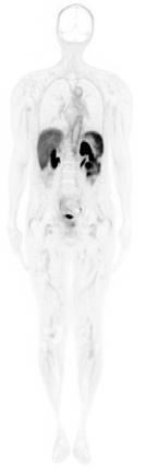
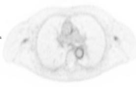
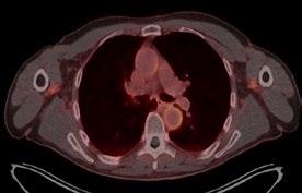
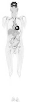
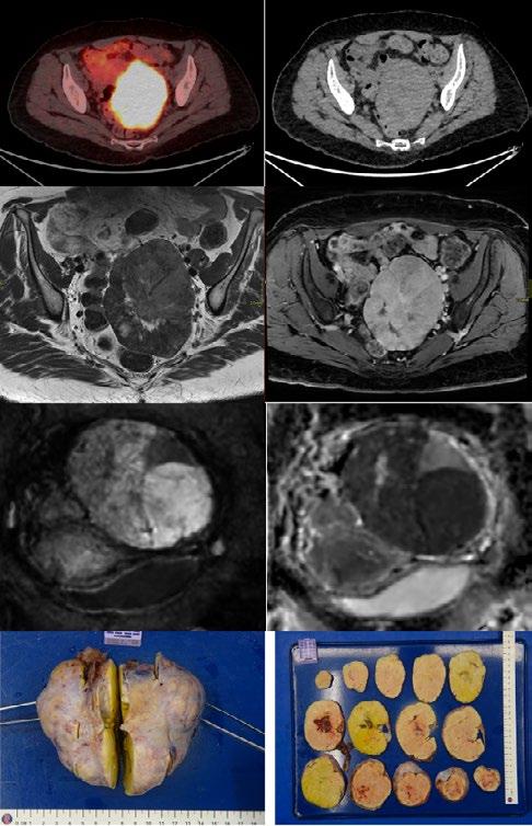
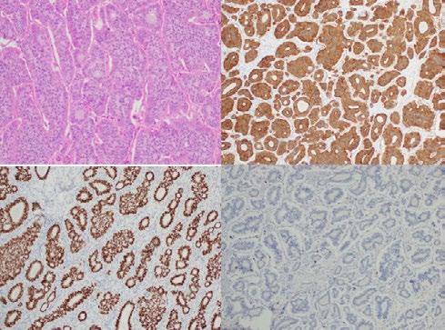
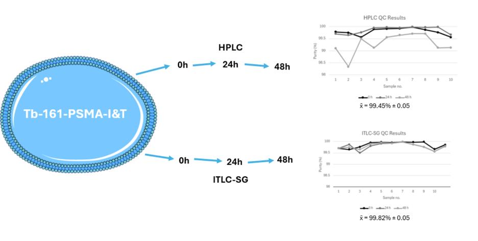
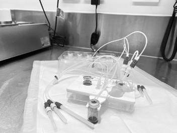
From Concept to Cure: Transforming Healthcare with Nuclear Medicine Case Study: Ga-68 Dotatate (Gallium-68) detected ovarian carcinoid presenting with carcinoid heart disease Creativity Vs Problem Solving In-vitro Radiochemical Stability of the Therapeutic Radiopharmaceutical [161Tb]Tb-PSMA-I&T ANZSNM ASM 2026 HIGHLIGHTS
ANZSNM
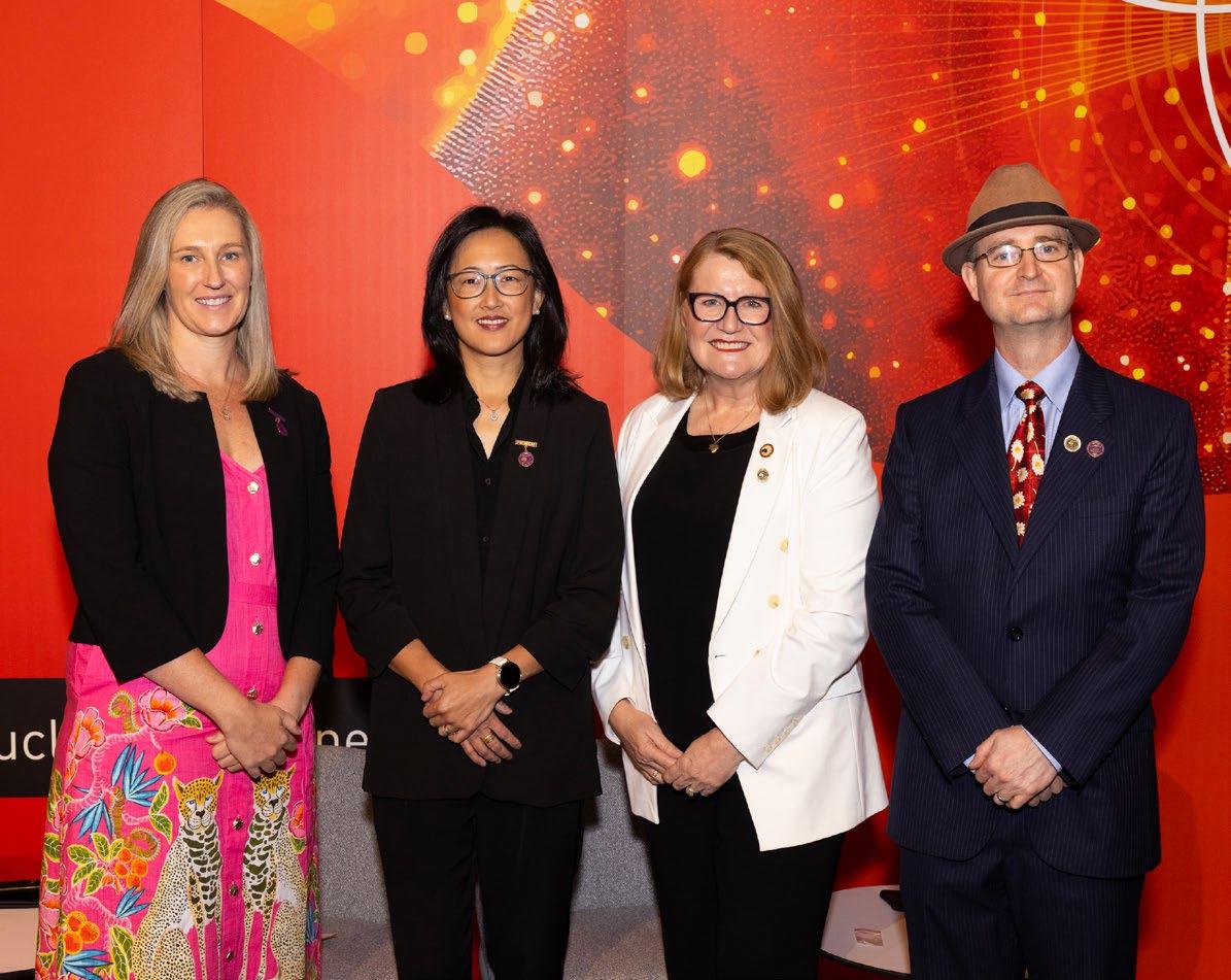
Suzanne McGavin (Vice President), A/Prof Grace Kong (President), Prof Karen Jones (Past President), Dr Daniel Badger (Treasurer)
Technologists
Medical
Physics
Interest Group (Physics SIG)
FROM THE PRESIDENT
Welcome to the 2026 WINTER EDITION of the Gamma Gazette
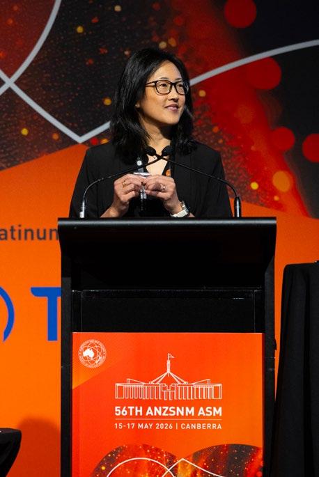
Grace Kong President
Dear members and friends,
Welcome to the 2026 Winter Edition of the Gamma Gazette from ANZSNM! It is a pleasure to contribute to the Gamma Gazette for the first time as the President of the Society. I am humbled and grateful to receive support from the other council members for this role. I must also again thank our past President Professor Karen Jones for her strong leadership, and appreciate the ongoing guidance and support. I look forward to working closely with our new Executive team, Federal Council members, Special Interest Groups & Committees, together with General Manager Rajeev Chandra and Secretariat Melissa Flannery, and I thank them sincerely for all their contributions to the ANZSNM and the members of the Society.
The Gamma Gazette is published two times per year, and it summarizes all the incredible work, contributions and upcoming activities from key members representing the multidisciplinary craft groups which form Nuclear Medicine and our Society. This edition is not to be missed!
The team has been incredibly busy and productive. You will hear about the important information and upcoming activities from all the Branches from Australia & New Zealand. You will receive exciting updates from the individual ANZSNM Special Interest Groups (TSIG and TSIG subcommittees, MSIG, and Physics & RPS groups, and ANSTO representative) highlighting multiple efforts including educational, student grant support, professional development, workforce advocacy, medical collaborations advocating for our Nuclear Medicine profession. It is important to emphasize the recent work on providing expert advice to multiple government / MSAC documents related to molecular imaging or theranostics in this rapidly growing field.
This edition provides the highlights of the recent successful 2026 annual scientific meeting held in Canberra from the Conference Convenors Justine Trpezanovski and Kevin London. A sincere congratulations on such a wonderful and stimulating meeting. You will find details of all the welldeserved winners and a summary of their excellent scientific work and prizes awarded at the 2026 ASM. I once again congratulate them on their contributions and wish them successes in the future. This edition includes several interesting scientific article contributions. We also congratulate Dr Alicia Corlett, Senior NM Technologist for winning 2 prestigious awards at the 2026 Society of Nuclear Medicine and Molecular Imaging (SNMMI) Annual Meeting! This edition is not to be missed!
Sincerely,
Grace
OUR CONTRIBUTORS
EDITORIAL COORDINATOR
Rajeev Chandra General Manager PO Box 6178, Vermont South, VIC 3133 T 1300 330 402 F (03) 8677 2970 gm@anzsnm.org.au
EVENTS & ADVERTISING ENQUIRIES
secretariat@anzsnm.org.au
secretariat@anzsnm.org.au
PUBLISHED IN
Summer and Winter
CONTENT SUBMISSIONS
Scientific submissions on all aspects of nuclear medicine are encouraged and should be forwarded to the Secretariat (instructions for authors published at https://www. anzsnm.org.au/activities/gamma-gazettecontent-submission-and-guidelines/).
Letters to the Editor or points of view for discussion are also welcome.
If original or public domain articles are found and considered to be of general interest to the membership, then they should be recommended to the Editor who may seek permission to reprint.
The ANZSNM Gamma Gazette is published two times per year. Deadlines for each issue of the journal can be found on our website anzsnm.org.au
SUBMISSIONS DISCLAIMER
The views expressed in any signed article in the journal do not necessarily represent those of the Society. The individual rights of all authors are acknowledged.
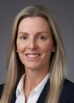
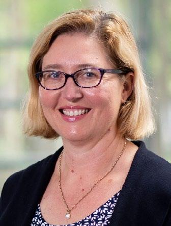
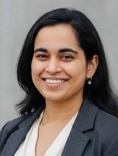
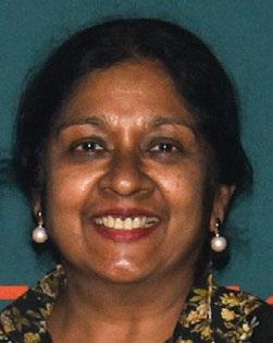
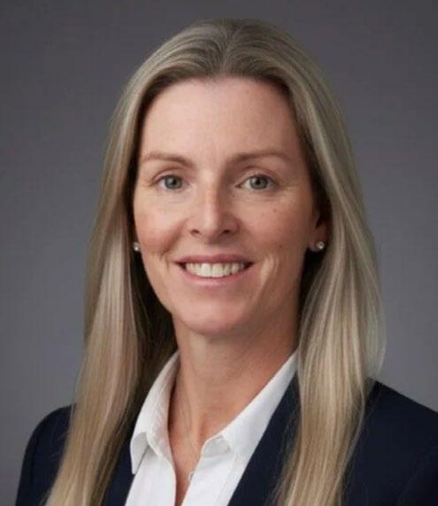
MEET THE NEW ANZSNM PRESIDENT, ASSOCIATE PROFESSOR GRACE KONG
The ANZSNM is delighted to welcome Associate Professor Grace Kong as the Society's new President. A highly respected leader in Nuclear Medicine, Grace brings a wealth of clinical, academic and leadership experience to the role. In this edition of the Gamma Gazette, we invite you to get to know Grace as she shares her professional journey, her vision for the Society, and her aspirations for advancing Nuclear Medicine across Australia and New Zealand.
Congratulations on your election as President of the ANZSNM. How does it feel to take on this role?
It is a great honour and privilege to be elected as the President of the ANZSNM. The ANZSNM is a society incorporating multiple collaborative disciplines, a true reflection of this profession.
There is incredible momentum in imaging innovation, theranostics, and multidisciplinary care. I feel a deep responsibility to advocate for our members and profession, and to strengthen collaborations across Australia, New Zealand, and our international partners. I’m grateful for the trust placed in me and look forward to working with the executive, committees, and the wider membership to build on the Society’s strong foundation.
Can you tell our members a little about your professional background and journey into nuclear medicine?
I graduated from Monash University in Victoria, Australia (MBBS), and embarked on internal medicine physician training. Nuclear Medicine was appealing given the broad aspects of advancing developments including cardiology, endocrinology and oncology fields which I enjoyed. During my training, I was drawn to the innovations and developments in nuclear oncology, with molecular imaging and theranostics transforming cancer management. My fellowship at the Royal Marsden Hospital in UK further paved my interest and passion in nuclear oncology as a clinician researcher, to further advance targeted precision oncology.
Looking back on your career, what achievements are you most proud of?
Joining a specialty which is growing, impactful and accelerating. Being the Co-Chair of the Neuroendocrine Tumour (NET) Centre of Excellence certified by the European NET Society (the first site accredited outside of Europe). Leading the high-volume theranostics service at our centre, changing and improving outcome for patients, which can only be achieved by working as a team with many multidisciplinary colleagues and collaborators. Leading and being co-investigator for diagnostic and therapeutic clinical trials (Phase 1-3). Continuing research
and collaborations nationally and internationally, speaker at many educational sessions at key oncologic and nuclear medicine societies, a recent highlight being an invited plenary speaker at oncology plenary session at EANM 2025.
The ANZSNM is a multidisciplinary society representing many craft groups. What does this multidisciplinary collaboration mean to you?
I enjoy working as a team and learning from different disciplines. The multidisciplinary craft groups form the backbone of our profession; the Society hence plays an important role in bringing all the key members together. This is the true representation of our profession and nuclear medicine.
What are your key priorities and goals for the Society during your term as President?
To build on prior successes and strong foundations of the Society, to further support our members from all disciplines, with a focus on education and workforce. To represent and advance nuclear medicine at national and international levels.
What opportunities do you see for nuclear medicine and molecular imaging over the next five years?
There will be continuing exponential growth in new radiotracer developments, advancements in technology, and expansion of theranostics to other tumour types and into clinical practice. There will be increasing developments and clinical trials creating opportunities for more national and international collaborations with the aim of transforming patient outcomes.
What challenges do you believe the profession will face, and how can the ANZSNM help address them?
Challenges are expected in any growing profession, particularly with multidisciplinary involvements, including addressing advocacy, educational support and workforce development issues. It will be important for the Society to focus on a collaborative approach, uniting disciplines to eliminate care fragmentation, elevate professional standards, and prioritize patient outcomes.
ANZSNM will work closely with members, other relevant societies, stakeholders, government, MSAC and regulatory bodies.
What role do education, research and workforce development play in the future of our profession?
In this rapidly growing field, new developments and clinical practice will continue to change significantly.
In parallel to this expected growth, education and workforce development are critical areas to focus on, across all disciplines.
What message would you like to share with ANZSNM members as you commence your presidency?
Please reach out if you have any comments, ideas or suggestions in regard to Society activities and needs. I look forward to working with you and meeting many of the members in the next 2 years.
What advice would you give to students, trainees and early-career professionals considering a career in nuclear medicine?
You have made a good career choice! It is a rapidly advancing and expanding field, with many new and impactful developments in progress bridging advanced molecular imaging with life-saving targeted therapies. You will work in a collaborative multidisciplinary environment.
Outside of work, what are some of your interests or hobbies?
I enjoy outdoor activities, my main current interest is running, and I am aiming to achieve some longer distance milestones.
Finally, what are you most looking forward to over the next two years as ANZSNM President?
I look forward to interacting and working with you all. I will do my best to uphold the incredible work from all the prior Presidents, to further support the members of our Society, to strengthen interdisciplinary collaborations and to advance our profession in this rapidly changing field.
ANZSNM AT THE BRITISH NUCLEAR MEDICINE
SOCIETY 60TH ANNIVERSARY
Professor Karen Jones
Immediate Past President
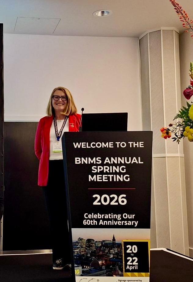
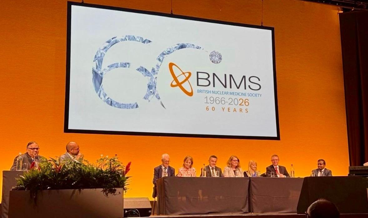
I had the privilege and pleasure of representing the Australian and New Zealand Society of Nuclear Medicine (ANZSNM) at the British Nuclear Medicine Society (BNMS) 60th Anniversary Spring Meeting, held at the Manchester Central Convention Centre in Manchester, UK, from April 20-22, 2026.
The conference brought together leaders, clinicians, scientists, technologists and researchers from across the nuclear medicine community to celebrate the 60th anniversary of the BNMS and explore current and emerging issues relevant to the profession internationally.
As President of the ANZSNM, I represented the Society on an international leadership panel comprising presidents and senior representatives from the Society of Nuclear Medicine and Molecular Imaging (SNMMI), the European Association of Nuclear Medicine (EANM), the British Nuclear Medicine Society (BNMS), the Asian Regional Cooperative Council for Nuclear Medicine (AOFNMB), the World Federation of Nuclear Medicine and Biology (WFNMB), and the Royal College of Radiologists (RCR). The discussion focussed on the challenges and opportunities facing nuclear medicine across different regions, including workforce development, education, training, regulation and future directions for the profession.
As well as participating in the international leadership panel, I was invited to deliver two presentations during the meeting. The first, Predicting the Future of the Nuclear Medicine Technologist Workforce: Perspectives from the Antipodes, highlighted the outcomes of the ANZSNM Nuclear Medicine Technologist
Workforce Summit and the initiatives implemented subsequently to address workforce capability, education and professional sustainability across Australia and New Zealand. The presentation generated considerable interest and highlighted the leadership role that ANZSNM has taken to address workforce challenges. I would like to
acknowledge and thank Suzanne (Suzi) McGavin, Melissa Shields, Mark Scalzo, Kunthi Pathmaraj, Katherine Guerrero and Pippa Bresser for sharing their invaluable insights and information relating to workforce issues and training programs for nuclear medicine technologists in Australia and New Zealand with me, in the lead up to the meeting.
My second presentation, The History of Gastric Emptying and Building a Career in Research, explored the evolution of research relating to gastric emptying research, complemented by my reflections on developing a research career in nuclear medicine. This presentation was also well received and stimulated productive discussions with delegates both during, and following, the session.
My attendance at the meeting allowed me to consolidate relationships between ANZSNM and our international colleagues and reinforced the importance of sharing experiences and collaborating on common challenges – workforce development, education, training pathways and professional practice. I am most grateful to the BNMS President, Professor Sabina Dizdarevic, and the BNMS organising committee for their warm hospitality and congratulate them on delivering an outstanding meeting to mark this important milestone.
Professor Karen Jones
Immediate Past President, ANZSNM
BRANCHES NEWS
As we move through another busy year, it's fantastic to see the continued enthusiasm and dedication across our ANZSNM Branches. From educational events and networking opportunities to student engagement and local professional development initiatives, our Branch Committees continue to provide valuable opportunities for members to connect, learn and support one another. I'd like to sincerely thank all of our Branch volunteers for the time and effort they invest in making these activities possible. I encourage all members to get involved in their local Branch and enjoy reading the updates below to see what's happening across Australia and New Zealand.
Rural/Regional Branch Meet n' Greet 2026
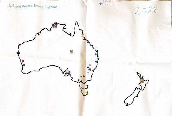
After another successful Annual Scientific Meeting in Canberra this May, we are happy to report the scintillating energy of the conference has expanded beyond the usual ‘who’s who’ of metropolitan guests to reach a new exciting breadth of engagement!
This year we held our inaugural ANZSNM Rural/ Regional Branch Meet n’ Greet lunch session to welcome binational society members from the ‘Back o’ Bourke’, one honouree-Aussie from Thunder Bay, Canada (thank you to SNMMI attendee, Jonathan Boekhoud!), and many city-based society members in support of strengthening nuclear medicine throughout country regions. With over fifty guests in attendance our first session was an invaluable opportunity from those of us based outside of the ‘big smoke’ to network and share ideas. While this session was held on the Sunday of the conference, we hope to present towards the middle of the program next year to allow greater access for our members who often have the greatest travel times of those attending our ASM.
We touched on the aims of this new branch—to improve the sense of collegiality and belonging for all society members in ‘extra-metropolitan’ settings, as well as promote initiatives specifically benefitting those working in regional and rural areas. These include increased access to CPD opportunities that are targeted to rural and regional practitioners, workforce advocacy to assist rural and regional specialists finding more locum work, and networking initiatives to share and strengthen our programs.
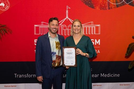
The session also included a great presentation from the winner of the first GE Healthcare ‘Miles of Care’ educational grant for rural and regional nuclear medicine professionals. Congratulations again to our inaugural
winner, Janelle Couch, from Cairns Hospital! Janelle shared a beautiful story of professional excellence and personal connection to her workplace and community at large.
The ‘Miles of Care’ educational grant will be awarded to a Rural/Regional society member in 2027, so please stay tuned for more information and application deadlines later this year.
Also, stay on the lookout for Rural/Regional committee members presenting at your state branches in the near future. We already covered SA/NT and Queensland, so we are now on our way south, breaking new ground as we move deeper into the heartland of nuclear medicine! Thank you to everyone in the wider ANZSNM community for your support.
Joshua James Morigi and Kerry Jewell on behalf of the Rural & Regional committee.
Jessica Fagan, MANZSNM Chair of Branches
Joshua James Morigi and Janelle Couch
New South Wales & Australian Capital Territory News Committee Members: Mei Yee Chan, Sameer Dave, Jeremy Hoang, Eleanor Kelliher, Nigel Lengkeek, Holly Spooner and Charlotte Yee
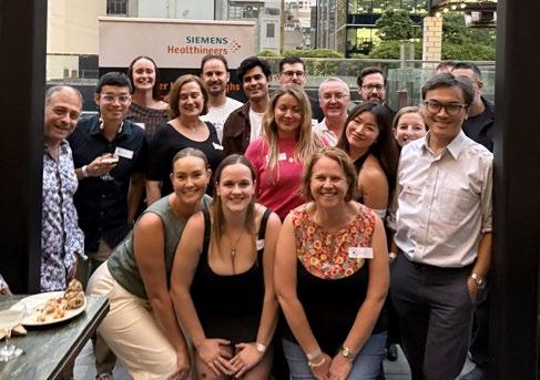
The NSW/ACT branch has had a solid start to the year. We opened the year with our annual Members' Networking Event at Babylon Rooftop Bar on 13 March, attended by 21 members. The evening provided a relaxed setting for members to reconnect, welcome new faces and exchange ideas across our various subspecialties.
We hosted an online Endocrine Nuclear Medicine CPD meeting on 19 March. We are grateful to A/Professor Geoff Schembri and Dr Veronica Wong for sharing their expertise. Geoff presented a state-of-the-art update on localisation of primary hyperparathyroidism using F-18 choline PET/CT, while Veronica provided valuable insights into the nuclear medicine implications of the recently published ATA Management Guidelines for Adults with Differentiated Thyroid Cancer. The excellent attendance reflected the strong interest in high-quality educational events within our branch.
Our next event is our inaugural Trivia Night, to be held at the Royal Albert Hotel on 24 July. A special thank you to Holly Spooner for coordinating the evening. We hope many members will be able to join us.
This year also marks a change in our committee. We extend our sincere thanks to Dr Karan Singh, who has stepped down from his role as Treasurer. Since joining the committee in 2024, Karan has made an outstanding contribution through his enthusiasm, calm leadership and thoughtful ideas. We wish him every success in the next stage of his career.
At the same time, we are delighted to welcome Dr Sameer Dave, Eleanor Kelliher, Dr Nigel Lengkeek and Charlotte Yee to the committee. It is particularly encouraging to see growing interest from members who are keen to contribute to our branch.
As we look ahead to the second half of the year, we encourage all members to participate in our educational and social events. Your involvement helps build a vibrant, supportive and connected community. We look forward to seeing many of you at our upcoming events.
Jeremy Hoang, NSW/ACT Chairperson
Victoria & Tasmania News
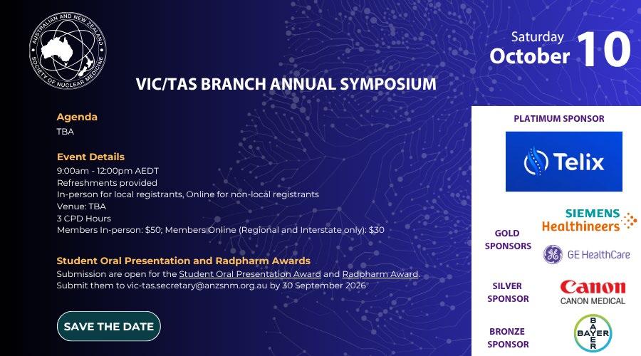
The VIC/TAS Branch Committee is busy preparing for the 2026 VIC/TAS Day Seminar, to be held in October. The program is shaping up well and will feature presentations on Digital SPECT/CT, Medical Radiation Practice Board (MRPBA) capabilities, and an awards session recognising excellence within the profession. We look forward to welcoming members for a day of education, networking and professional development. Myself and Branch Secretary My Linh Diep recently
NSW/ACT Branch Networking Event 13 March 2026
BRANCHES NEWS (CONTINUED)
attended the ASMIRT Early Careers Forum, engaging with students and early career professionals to promote nuclear medicine and the opportunities available through the ANZSNM.
The Branch is also supporting future members of the profession, with a branch representative attending the upcoming RMIT Open Day to speak with prospective students about careers in nuclear medicine. The Branch will also be reaching out to Federal Council members to explore opportunities for additional ANZSNM representation and support at the event.
Kim Jasper, VIC/TAS Chairperson
Western Australia News
We’ve had 2 branch meetings this year. The first hosted by Perth Rad Clinic in February and sponsored by Telix, and the second hosted by Sir Charles Gairdner Hospital and Perth Children’s Hospital sponsored by Canon who flew their team over to Perth to attend. This meeting included some MIBG paeds talks alongside some great physics presentations, delving into some very unique I-131 therapy cases.
Perth is grateful to have the new cyclotron at RAPID labs (at Sir Charles Gairdner Hospital) to coexist with the very aged 2003 Cyclotron. This brings us to 3 Cyclotrons in WA total, a good jump from when we only had the one >20 year old cyclotron only 18 months ago. Perth Children’s Hospital has been approved for a PET scanner to open early next year - this will be the first dedicated paediatric scanner in the state.
Rosemary Dallen, WA Secretary
New Zealand News
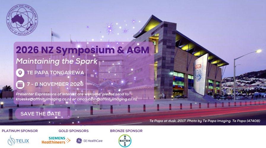
The ANZSNM New Zealand Branch has an exciting calendar of events coming up over the next few months. Join colleagues for the NZ Branch Meeting on 11 August and 13 October for excellent opportunities to connect with fellow professionals and hear the latest updates from across the profession. Looking ahead, members are encouraged to attend the Australasian Radiation Protection Society (ARPS) 2026 Conference, being held in Christchurch from 20–24 September, bringing together experts from across Australasia to explore advances in radiation protection under the theme From Rutherford to Tomorrow: Kaitiakitanga in Radiation Protection. Finally, don't miss the 2026 ANZSNM New Zealand Symposium – Maintaining the Spark, taking place 7–8 November at Te Papa Tongarewa in Wellington. This flagship event will feature a high-quality educational program, networking opportunities, and the chance to reconnect with colleagues from across New Zealand. Registrations for these events are now open via the ANZSNM website.
NZ Branch
Kim Jasper and My Linh Diep
BRANCHES NEWS (CONTINUED)
Queensland News
As we move through another busy year, I'd like to thank our members, volunteers and supporters for their ongoing commitment to the Nuclear Medicine profession across Queensland. While workforce pressures continue to challenge many departments, it has been encouraging to see the profession remain focused on collaboration, education and supporting the next generation of Nuclear Medicine Technologists.
Our first Queensland Branch meeting of the year featured three outstanding presentations focused on professional capability, clinical education and collaboration. Associate Professor Andrew Kilgour (RMIT University and Chair of the ASMIRT Professional Standards Committee) shared evidencebased approaches to best practice in the supervision and assessment of Medical Radiation students. Maddison Carroll, Queensland Health's State-wide Nuclear Medicine Clinical Educator and a member of the Medical Radiation Practice Board of Australia's Registration and Notifications Committee, provided members with an excellent overview of the Medical Radiation Practice Board of Australia's (MRPBA) updated Professional Capabilities for Medical Radiation Practitioners. The Queensland Branch was also pleased to join forces with the ANZSNM Rural and Regional Branch - Branch Chair, Josh, and Secretary, Lachlan, to introduce the Rural and Regional Branch and highlight several of its key initiatives and priorities. Their participation reflected our shared commitment to strengthening collaboration across ANZSNM branches, supporting practitioners working in rural and regional settings, and ensuring their perspectives continue to inform broader professional discussions.
Workforce shortages remain one of the greatest challenges facing Nuclear Medicine services throughout Queensland. Many departments continue to experience recruitment and retention difficulties, placing increasing pressure on existing staff while demand for services continues to grow. The QLD Branch Committee continues to advocate for sustainable workforce solutions and support initiatives that strengthen education, training and professional development. Advocacy has remained an important focus for the Committee this year. The Queensland Branch would also like to extend its sincere thanks to ANZSNM President, Karen Jones, for providing a formal letter of support to Queensland Health on behalf of the ANZSNM regarding the recent proposed changes to remuneration for Nuclear Medicine Technologists within the public sector. While the outcome was ultimately not in favour of our profession, Karen's advocacy demonstrated the Society's commitment to representing and supporting its members on issues that impact our workforce. We are grateful for her leadership and willingness to advocate for Nuclear Medicine Technologists across Queensland.
One of the strengths of the Queensland Branch has been the strong professional relationships we have built across the medical imaging community. Our longstanding collaboration
with the ACPSEM continues again this year, with ANZSNM members able to access discounted single-day registration to the EPSM 2026 Theranostics Symposium. Partnerships such as these provide valuable opportunities for multidisciplinary learning, strengthen relationships between our professions and reinforce the collaborative nature of modern Nuclear Medicine practice.
Education continues to be a major priority for the Branch and across the state. It has been pleasing to see the continued success of the RMIT Embedded Student Model (ESM) program, which has now been extended through to 2027, reflecting the ongoing interest and future retention of graduates to enter the Queensland workforce. It is particularly rewarding to see these students already contributing to the profession. Carley Chapman, one of the inaugural RMIT ESM students, has joined the TSIG Student Council and is leading initiatives to support students undertaking clinical placement. Carley will present at our upcoming Branch meeting on 11th August, where we will also welcome all students currently on placement to attend and participate in dedicated placement support workshops. Looking ahead, Queensland is excited to host the 2027 ANZSNM Annual Scientific Meeting on the Gold Coast. This is a fantastic opportunity to showcase the strength of Nuclear Medicine across Queensland, highlight the outstanding work being undertaken throughout our state and welcome colleagues from across Australia and New Zealand. Planning is already underway, and we look forward to sharing further updates over the coming year.
This year also marks an important period of transition for the Queensland Branch Committee. After many years of service, the current Committee intends to conclude its term at the end of the year. We encourage members who are interested in shaping the future of our Branch to consider nominating at our Annual General Meeting, to be held in conjunction with the Queensland RadPharm meeting later this year. Existing Committee members are committed to supporting a smooth transition and would be pleased to mentor incoming representatives or have new members join now to learn the role before stepping into committee positions.
Finally, the Queensland Branch congratulates Richard Boytar from Princess Alexandra Hospital, recipient of this year's Queensland RadPharm Award and National finalist. Richard recently retired after more than 40 years of dedicated service to Nuclear Medicine and Medical Imaging. His contribution to patient care, education and mentorship has had a lasting impact on our profession, and we thank him for his outstanding service while wishing him every happiness in retirement.
On behalf of the Queensland Branch Committee, thank you for your continued support of the ANZSNM. We look forward to seeing many of you at our upcoming Branch meeting on 11th August and at future events throughout the remainder of the year.
Anisa Kumari, QLD Chairperson
BRANCHES NEWS (CONTINUED)
South Australia and Northern Territory News
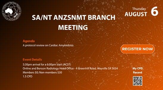
The SA/NT branch has had a great start to 2026!
Our March event was an in-person event at the Women’s and Children’s Hospital, and despite being on the same night, in the same suburb as the Ed Sheeran concert we had a great turnout, eager to learn and listen to all things paediatric-related. We heard from Catherine Baring, RN regarding the successful nurse-led vascular access service she and the team have built, followed by Dr Ashlesha Vaidya who provided some excellent paediatric case studies, Chloe Lowry who discussed the complexity of paediatric scanning, and finished with Meg Stirrat from Canon Medical System (our Silver Sponsor Partner) who showcased some impressive PET systems coming out Canon Medical Systems.
In April, the new committee of the ANZSNMT held a successful online event where Suzanne McGavin spoke about recognising and responding to anaphylaxis which was well supported and timely with the rollout of the new MRPBA capabilities for Nuclear Medicine professionals.
Our May event was an on-line event where Dr Rahul Solanki provided an interactive and informative talk showing some PET and general Nuclear Medicine interesting case studies. Also at this meeting we celebrated Liam Carey being awarded the University of South Australia prize for 2025, and we acknowledged the depth of South Australian and Northern Territory abstracts on display and
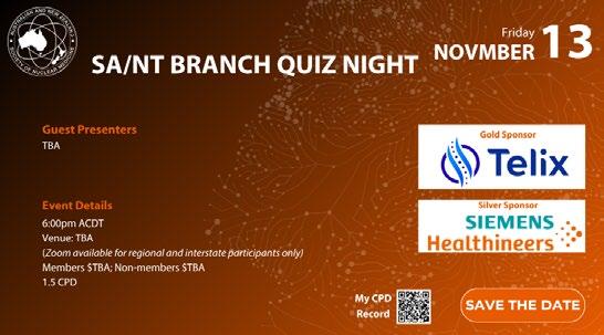
We look forward to welcoming members to the remaining
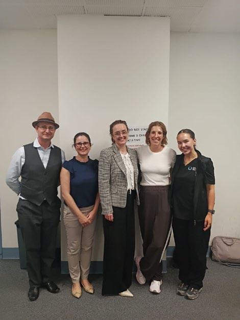
at our March in-person event at the Women’s and Children’s Hospital
Daniel Badger, Nerida Neumann, Kate Yeates, Meg Stirrat and Ruby Holman.
Canon Medical Systems representatives Kate Yeates and Meg Stirrat were in attendance
EDUTRACE EDUTRACE
By
The E-Learning Platform For ANZSNM Members
TECHNOLOGIST SPECIAL INTEREST GROUP (TSIG)
Dr Melissa Shields, MANZSNM TSIG Chair
As this is my first Gamma Gazette report as TSIG Chair, I reflected on the 2025 reports for inspiration and was struck by the breadth and impact of our contributions to the nuclear medicine community. I look forward to building on the strong foundation established by previous TSIG Chairs in continuing to support our profession.
I would like to sincerely thank the members of the CPD&E Committee, the Workforce Advocacy Committee, the Academic Reference Group, and the Student Representative Council for their ongoing commitment to the ANZSNM and the TSIG. Their contributions are entirely voluntary and undertaken alongside demanding professional and personal responsibilities, which makes their dedication all the more commendable.
Committee Updates
Following our AGM in late March, several committee leadership transitions have taken place within the TSIG. Shikha Sharma has been appointed Chair of the CPD&E Committee, and Karen Jones has assumed the role of Mentor Program Coordinator. Kunthi Pathmaraj and Erin Hemingway continue in their roles as Chair of the Workforce Advocacy Committee and University Liaison, respectively. Further detail on the valuable work undertaken by these committees is available in their respective reports.
I would like to extend my sincere appreciation to Suzi McGavin for her outstanding leadership and dedication during her two-year tenure as TSIG Chair. Suzi has been a tireless advocate for nuclear medicine technologists across Australia and New Zealand, representing the profession both nationally and internationally and strengthening the position of the TSIG. Congratulations to Suzi on her appointment as Vice-President of the ANZSNM. She will also continue on the ARPANSA Medical Radiation Safety Guides Working Group, collaborating with fellow ANZSNM members on this significant document upgrade.
Highlights
The introduction of the new MRPBA Professional Capabilities in early 2026 marked an important milestone for the profession. In response, the TSIG highlighted training pathways for anaphylaxis management, and we hope members have taken the opportunity to complete this essential requirement. Additionally, an introductory
leadership webinar was delivered to support Domain 6: Leader and Steward within the updated framework. This session, facilitated by Daniel Lonard—a radiographer and professional organisational coach with the Institute of Professional Coaching and Leadership—is available on Edutrace via the ANZSNM website.
In collaboration with AANMS, the TSIG allocated $10,000 for Semester 1 Student Clinical Placement Grants, helping to alleviate the financial pressures associated with essential clinical training. We also supported university orientation activities, welcoming a new cohort of students into the profession. Applications for Semester 2 grants will open shortly.
Preliminary findings from the Nuclear Medicine Technologists in Theranostics International Consortium (NMTTIC) survey were presented at both the ANZSNM Annual Scientific Meeting (ASM) and the SNMMI conference. These were delivered by Suzi McGavin and Karen Jones, representing the ANZSNM TSIG, alongside Kunthi Pathmaraj representing the IAEA.
Congratulations to all technologist award recipients at the ANZSNM ASM. The quality of presentations was exceptionally high, with the Radpharm Award particularly competitive. Thank you to the broader technologist community for their contributions as session chairs and judges. The TSIG Oversight Committee also met with representatives from the MRPBA and NZ MRTB during the ASM, reinforcing our collaborative relationships with regulatory bodies and ensuring alignment on current and emerging issues.
Dr Melissa Shields
Annual TSIG Symposium
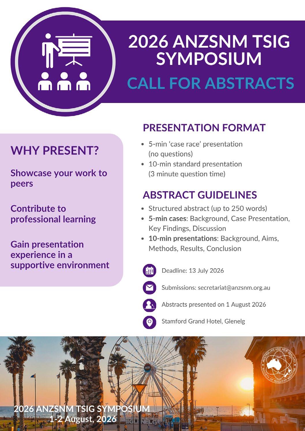
We are delighted to be hosting this year’s Annual TSIG Symposium in the seaside suburb of Glenelg, South Australia. The theme, “Seeing Beyond the Surface: A Day in Nuclear Medicine,” will be explored over an expanded program, with the event now extending across one and a half days.
The symposium will take place on 1–2 August and will include a half-day leadership workshop facilitated again by Daniel Lonard. Registrations are now open For the first time, the program will also feature short case presentations and free papers, offering delegates an opportunity to share their work in a more informal and collaborative environment. Abstract submissions are now invited.
The symposium will also feature the presentation of the 2026 Nuclear Medicine Technologist Award. Nominations are currently open and encouraged.
In Closing
I would like to once again thank all TSIG committee members for their ongoing contributions, as well as the many individuals outside the TSIG who generously assist with our initiatives and consistently go above and beyond. We welcome your feedback and encourage you to reach out with any suggestions or ideas for the TSIG.
VENUE Stamford Grand Adelaide Hotel, Glenelg
ANZSNM/AANMS Semester 1 2026 Student Clinical Placement Grant Recipients
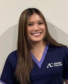
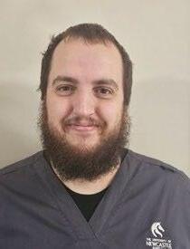
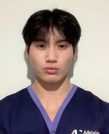
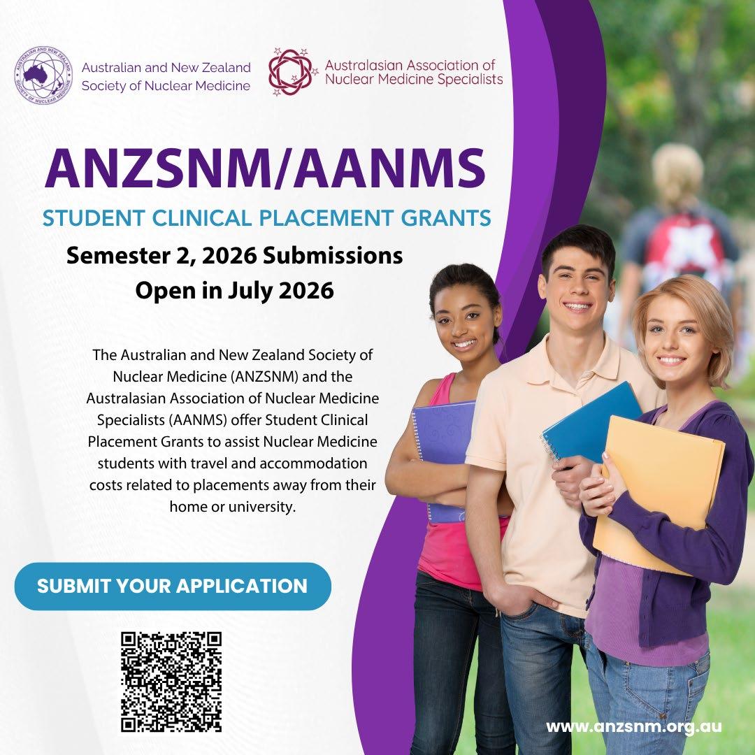
The ANZSNM and AANMS are delighted to announce the recipients of the Semester 1, 2026 Student Clinical Placement Grants. These grants support students undertaking clinical placements away from their home location, helping to reduce financial barriers and ensure equal access to high-quality training opportunities across the profession. The Placement Grants are an initiative of the ANZSNM and supported by the AANMS and commenced in 2024. We bring to you some of the recipients and future members of the profession.
Ashlee Brown University of Newcastle
Thomas Polhman University of Newcastle
Jake Phillip Greg University of Newcastle
Jacob Cobner University of Newcastle
Cates Borromeo Adelaide University
Emily Malcolm University of Newcastle
Bren Casipit Adelaide University
Catherine Juwan Morobe University of Newcastle
Elizabeth Hindson University of Newcastle
Hannah Fear University of Newcastle
Caitlin Bamford Adelaide University
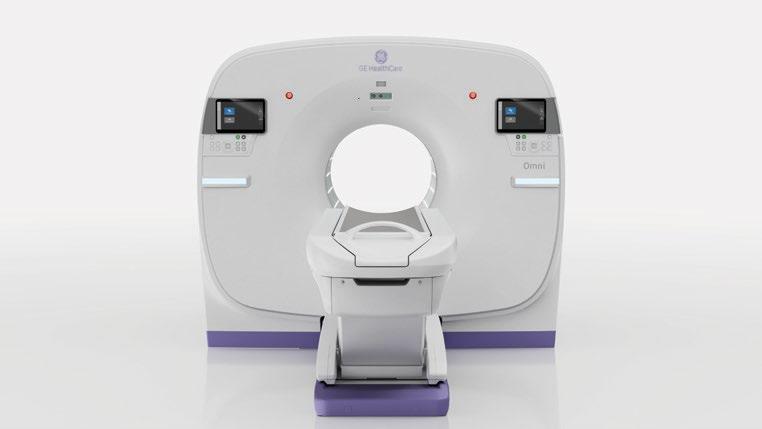
128 cm
Total Body PET/CT
128 cm total-body PET/CT imaging
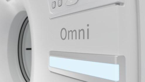
OMNI™ 128 is GE HealthCareʼs next-generation 128 cm total-body PET/CT system, enabling head-to-thigh imaging in a single bed position while supporting faster scans, streamlined workflow, and ultra-low dose imaging.
OMNI™ 128 is designed to support cancer diagnosis, staging, and treatment response monitoring, as well as theranostics, precision medicine, and research.
405 cps NEMA sensitivity
At a glace Omni 128 cm leaps ahead to a new dimension in PET/CT total body scanning.
Enable much lower dose
The high sensitivity enables a significant reduction in dose of up to 90%.
Total body acquisitions
Great 128 cm AFOV vertex-to-thigh images in one bed for virtually all patients.
Dynamic imaging research
New technology designed for ultra-fast dynamic reconstruction in under 5 minutes.
It is my pleasure to provide my first update as Chair of the CPD&E Committee.
We recently hosted a successful Men’s Health Webinar on 24 June, a topic that had been long overdue. I would like to extend my sincere thanks to my co-host, Elaine, our presenters Mr Rich, Mr Herzog and Dr Talmor, as well as everyone who attended and contributed to the event’s success. If you were unable to join us, the webinar recording is now available on Edutrace.
Our next major event is the annual TSIG Day Symposium, co-hosted with the ANZSNM SA Branch. The program is shaping up to be an engaging and informative event with a focus on “A day in Nuclear Medicine”. I encourage you to join us in Glenelg on 1st and 2nd August for what promises
to be an excellent opportunity for learning, networking and professional development.
We are also excited to be launching a Journal Club for ANZSNM members. Planning is well underway and we look forward to sharing more details and launching the program in the near future.
If you have any ideas on what you want us to focus on or would like to join the committee, please reach out to me directly.
The ANZSNM ASM held last month in Canberra provided a valuable opportunity to update ourselves on current practices and trends within our profession, explore emerging advancements, and connect with colleagues and industry partners. During the event, the TSIG oversight committee had productive discussions with representatives from the MRPBA and MRTB, addressing several important topics, including the workforce challenges facing Nuclear Medicine Technologists and Medical Radiation Scientists across Australia and New Zealand.
The TSIG Workforce Advocacy (WFA) committee is currently undertaking a significant workforce planning project to gain a clearer understanding of the Nuclear Medicine Technologists and Medical Radiation Scientists landscape in Australia. This initiative aims to help the Society identify the extent of workforce shortages in nuclear medicine, and to support future planning, advocacy, and engagement with both public and private healthcare providers, and education institutions. We will be reaching out to many of our colleagues in the NM community to gather accurate information about our workforce and look forward to working together to strengthen and stabilise our workforce pipeline.
Additionally, the WFA committee has been actively contributing feedback to consultations relevant to our profession, initiated by the Australian Government, MRPBA, MRTB, and other key organizations.
We are now seeking expressions of interest from our colleagues in Western Australia to join the WFA committee as the WA representative. Please watch for an official email from our secretariat inviting formal applications from interested ANZSNM members.
The WFA committee remains committed to supporting and nurturing the Nuclear Medicine Technologist workforce. We welcome your suggestions and feedback— please feel free to reach out via email at wfachair@anzsnm.org.au.
Hello and welcome to my University Liaison Officer update for the winter Gamma Gazette! I commenced in this role at the end of 2025 and look forward to fostering stronger connections between nuclear medicine students, universities and the ANZSNM. Education and workforce development are critical to the future of our profession, and I’m excited to contribute to initiatives to support future Nuclear Medicine Technologists across Australia and New Zealand.
Last month I had a great time attending the 2026 ANZSNM Annual Scientific Meeting in Canberra. As always, it was a fantastic opportunity to hear about the latest developments in nuclear medicine and catch up with colleagues and friends from across the region. With my household currently caught up in FIFA World Cup fever, it seems to me that professional conferences have a lot in common with major sporting tournaments: there are a lot of passionate people brought together, it fosters healthy competition and creates opportunities to learn from the best in the field. And of course, the ASM wouldn’t be complete without the Kevin London Fun Run Saga, which continues to keep us on the edge of our seats.
Congratulations also to Liam Carey, recipient of the ANZSNM University of Adelaide Student Prize for the 2025 academic year. Awarded by the ANZSNM Technologist Special Interest Group (TSIG), the prize recognises the most outstanding student in the Bachelor of Medical Radiation Science (Nuclear Medicine) program based on overall academic performance. Liam received a $500 prize in recognition of his achievement. We congratulate Liam on this well-deserved honour and wish him every success as he begins his career in Nuclear Medicine.
Congratulations also to Jemima Hodge, recipient of the ANZSNM University of Newcastle Student Prize for 2025. Awarded by the ANZSNM Technologist Special Interest Group (TSIG), the prize recognises the student
with the highest overall performance in the MRS-coded courses of the Bachelor of Nuclear Medicine program. Jemima received a $500 award in recognition of her outstanding academic achievement. We congratulate Jemima on this well-deserved recognition and wish her every success as she embarks on her career in Nuclear Medicine.
Just as sporting competitions depend on developing the next generation of talent, the TSIG Oversight Committee has awarded the first round of ANZSNM Student Placement Grants for 2026. A total of $10,000 has been distributed among successful applicants to help alleviate the financial burden associated with clinical placements. Placement poverty remains a significant challenge for many nuclear medicine students, and these grants represent a meaningful investment in the profession’s future.
Looking ahead, I’m excited to meet with both the ANZSNM Student Representative Council and the ANZSNM Academic Reference Group later this month. These discussions will generate valuable ideas around education, workforce development and student engagement.
I welcome any feedback, suggestions, or ideas that may be suitable for the University Liaison role. If you have any thoughts or would like to discuss opportunities for collaboration, please feel free to contact me at uniliaison@ anzsnm.org.au.
Erin Hemingway uniliaison@anzsnm.org.au
MEET THE ANSTO CUSTOMER SUPPLY CHAIN TEAM
The voices behind your orders, updates....and the occasional Sunday phone call
If you’ve ever picked up the phone to call ANSTO and heard a friendly voice on the other end, the chances are you’ve already met the Customer Supply Chain team, even if only briefly. They are the people answering your calls, processing your orders, tracking shipments and sometimes delivering the news no one wants to hear – a delay in supply. Most importantly, they are part of the integral team at ANSTO, working behind the scenes to keep nuclear medicine supply running smoothly for patients across Australia, New Zealand and beyond.
If you attended the Annual Society Meeting in Canberra this year, you may have popped into the ANSTO booth and had the chance to meet some of the team in person. I recently had the opportunity to catch up with the team to hear what their days really entail, and to help put some faces to the names you may already recognise.
"No two days are ever the same”
Sounds familiar, doesn't it? For Caity, one of ANSTO’s Customer Supply Chain Officers, variety is a part of the appeal. After five years of working in the Nuclear Medicine division at ANSTO, and a previous career in training and development; her role centres on supporting customers in real time. This means answering calls, responding to emails, processing orders and coordinating shipments. “The role can change from day to day, but its focus is always to support the customers”. And in our industry, this means staying sharp - “there is always something happening and it is forever changing, so you are always on your toes”.
Learning the language of Nuclear Medicine
Not everyone in the team started with a nuclear medicine background. “It’s a very niche area. Learning the products and decay rates takes time,” Caity explains. Michelle agrees. With nine years' experience in the role, she came in with a customer service background and “very little knowledge of radioisotope manufacture”.
So how do they get up to speed?
“ANSTO has an onboarding program which has developed strongly over the years covering the fundamentals of nuclear medicine, radiation safety, and the regulatory requirements, so you build a solid baseline fairly quickly.
This is also paired with online modules and on the job learning.” says Suzanne, Senior Manager - Planning and Customer Supply Chain. Michelle says, "I learned from an experienced and valued support team...but the most valued teaching is from listening and understanding what our customers need, and how we can best support them and their patients.”
Add in attendance at conferences and engagement with the broader nuclear medicine community at other events, much like the rest of us working in the Nuclear Medicine field, the learning never stops.
It's not just a help desk
It's easy to think of customer service as just answering phones, but the reality is far more complex. As a Nuclear Medicine Scientist, I see this daily with the crucial work our administration team does in keeping our department running.
Cindy has been a Customer Supply Chain Officer for four years and previously worked in the pharmaceutical manufacturing industry. She explains the role also involves regular meetings with planning, production, quality, engineering and dispatch teams. "It's a true coordination role in a highly specialised supply chain. There's always something going on in such a complex environment. I love seeing people work together to solve unique problems".
If you've ever wondered when the team are available – the answer is someone is always watching. Across the team, coverage is provided between 8am-5pm, Monday to Friday. There is a rotating Sunday roster to support manufacturing.
As Michelle puts it: “We rotate on call for Sunday manufacture day. We also monitor manufacturing and delivery 24 hours for any disruptions or changes". Cindy adds the team is always ready to jump on when needed
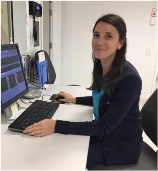
Annah Skillen Advanced NMT Gold Coast University Hospital
- "We're all pretty flexible and happy to help out with any supply disruptions”. And Suzanne who leads the team? “I live in the office!”.
The peaks and troughs
Despite the complexity, there is a clear common theme when the team talks about what they enjoy the most. For Michelle, its solving problems. “I enjoy the challenge of finding solutions when things go wrong, being able to resolve an issue so there is minimal impact to the patient is especially rewarding". For Cindy, it's the constant opportunity to learn. And for Caity, it’s simply being part of a unique environment that never stands still.
Of course, every role has hard moments, and not every phone call made to a customer is an easy one. Every member of the team acknowledges the same challenge – supply disruptions.
“It's always difficult to inform our customers of disappointing news", Michelle says. “We know the effect it has on them and their patients – we hate having to deliver bad news". Cindy echoes this- “We are aware of the impact disruptions can have...so we work hard to deliver the best possible outcome."
possible. Perhaps the most telling insight comes from a simple everyday moment. "One of the best things is when we answer a call and the customers simply say, "Hi, it's me" - and we know exactly who it is,” Michelle shares. It's a small moment, but it speaks volumes to the strong relationships forged through years of conversations, problem-solving and shared commitment to patient care.
The team behind every dose
To bring Nuclear Medicine services to patients, the team stretches far beyond our individual departments. From coordinating complex logistics to managing lastminute changes, the ANSTO Customer Service Team plays a critical role in the Nuclear Medicine ecosystem. As Suzanne puts it, their work sits within “a very unique, end-to-end nuclear medicine supply chain".
So next time you make a call or receive a (not-so-great) Sunday call, you'll know who's on the other end; and just how much is happening behind the scenes to keep everything moving.
I'd like to thank the ANSTO team for being available to chat
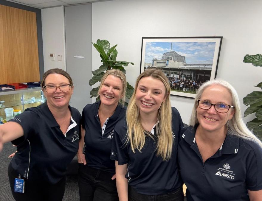
The ANSTO Customer Service Team, L-R is Michelle, Suzanne, Caity and Cindy.
MEDICAL SPECIAL INTEREST GROUP (MSIG)
Dr Jeremy Hoang, MANZSNM Chair, Medical SIG
Committee Members: Tahereh Erfani (John Hunter Hospital), Edward Hsiao (Royal North Shore Hospital), Jeremy Hoang (Royal North Shore Hospital), Grace Kong (Peter MacCallum Cancer Centre), Kevin London (The Children's Hospital at Westmead), Tam Ly (Wollongong Hospital) and Rachelle Steyn (Auckland City Hospital, New Zealand).
It has been an active first half of the year for MSIG. Our first AGM was held online on 25 March, providing an opportunity to reflect on the progress made during MSIG's formative year and to discuss priorities for the future. At the meeting, A/Professor Grace Kong concluded her term as our inaugural Chair as she commenced her role as President of the ANZSNM. Grace's leadership and commitment were instrumental in establishing MSIG as an effective voice within the Society. On behalf of the committee, I thank Grace and it is an honour to succeed her as Chair.
We also welcomed two new members to MSIG at the AGM, Dr Rachelle Steyn from Auckland City Hospital and Dr Tam Ly from Wollongong Hospital.
Much of MSIG's recent work has centred on providing expert advice to MSAC. Four of the eight applications to be considered by MSAC at its next meeting in August relate directly to molecular imaging or theranostics. This underscores both
the growing clinical and scientific prominence of Nuclear Medicine within Australia’s healthcare landscape, as well as the need for timely evidencebased funding decisions in our specialty.
On occasion, MSIG reaches out to our members for their expertise in providing advice and I would like to acknowledge them for their time and expertise to this work:
• Dr Samuel Wright, who authored MSIG’s response on the post-implementation review of MBS cardiac imaging items
Professor Andrew Scott, who authored MSIG's response on 18F-FDOPA PET/CT for the evaluation of Parkinsonism
• A/Professor Geoffrey Schembri, who led the response on amyloid PET/CT in support of PBS-subsidised lecanemab for people with mild cognitive impairment and mild dementia due to Alzheimer's disease
Dr Claire Mok, who at the time of writing is drafting our response to the MSAC application for 177LuPSMA-617 for PSMA-positive, taxane-naive patients with metastatic castration-resistant prostate cancer
On this last application, ANZSNM and AANMS are working closely in collaboration to present a unified and robust position on public funding for this clinically important theranostic therapy.
To ensure that MSIG's work remains grounded in the priorities of our members, we have recently distributed a short survey to better understand the issues most relevant to you and to guide our activities.
MSIG exists to serve the medical membership of ANZSNM. If you have questions, concerns or ideas you would like the group to consider, please do not hesitate to contact us.
Jeremy Hoang
Dr Rachelle Steyn, Dr Tam Ly & Dr Jeremy Hoang at the 2026 ANZSNM ASM in Canberra
This collaboration represents a major step towards harmonised, high-quality quantitative PET imaging worldwide.
It reflects ARTnet’ s ongoing commitment to advancing reliable imaging standards through global alignment and simplified expectations for imaging sites involved in clinical trials and patient care across Australia, New Zealand and beyond
As a core partner in this initiative, ARTnet has helped shape this internationally aligned framework designed to streamline accreditation and enhance reproducibility across internationally trace able radioactivity calibrations, PET and SPECT system validations and calibration and standardised dosimetry analysis protocols
Samantha Hawkins, ARTnet Project
Bailey DL, Dickson JC, Lapi SE, Uribe C, Vargas CS, Jackson P, et al. Pulling Together: A 5 -Year Plan to Improve Theranostic Outcomes by Improving the Accuracy of Dosimetry An FNIH Joint Academic, Clinical, and Industrial Collaboration. J Nucl Med. 2026:jn umed.125.271665.
HEAVY ION ACCELERATOR FACILITY (HIAF) TOUR, AUSTRALIAN NATIONAL UNIVERSITY
ANZSNM Physics SIG
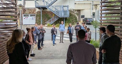
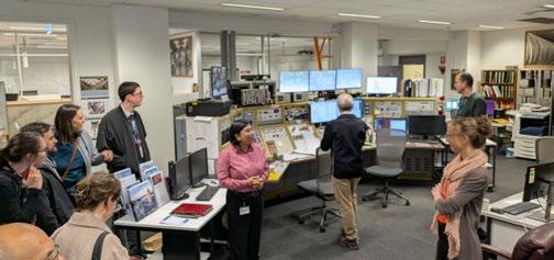
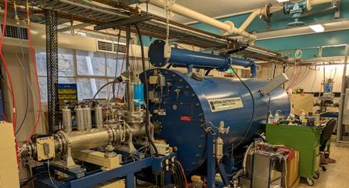
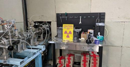
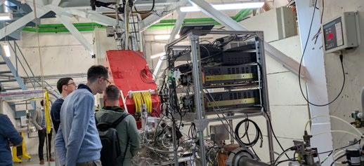
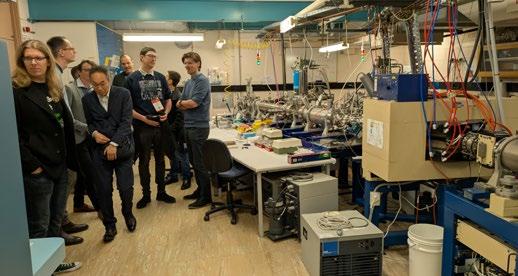
A group of physicists and radiopharmaceutical scientists attending the 56th ANZSNM ASM in Canberra were lucky enough to tour the Heavy ion Accelerator Facility (HIAF) at the Department of Nuclear Physics, Australian National University, on Friday 15th of May, during the ASM.
The HIAF comprises one of the world’s largest 14UD pelletron accelerators and a superconducting “booster” linear accelerator (LINAC). The 15 million volt pelletron is capable of accelerating ions to over 200 MeV(!!) and is used for a wide variety of nuclear and radiation research, including the search for new elements, testing equipment for resistance to space radiation, and studying the fundamental structure of matter with beams of high energy particles.
The ANZSNM members were amazed by the sheer size and complexity of the facility, but also are looking at possibilities of collaboration with the HAIF in research into new radionuclides for use in Nuclear Medicine.
The Physics and RPS SIGs would like to thank Dr Tom McGoram of the HIAF, for inviting the ANZSNM members for the tour.
Daniel Badger Physics SIG
ANZSNM 2026
FROM CONCEPT TO CURE: TRANSFORMING HEALTHCARE WITH NUCLEAR MEDICINE
Annual Scientific Meeting - Highlights
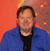
Justine Trpezanovski and Dr Kevin London Conference Conveners
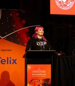
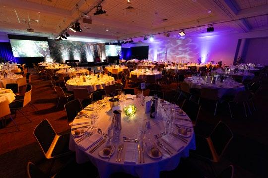
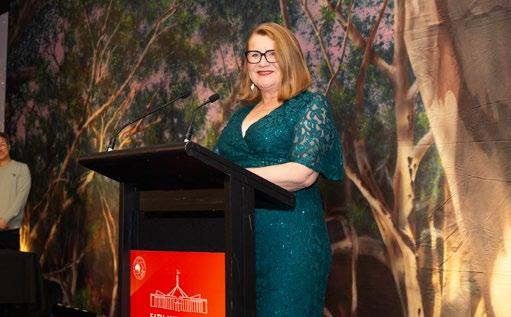
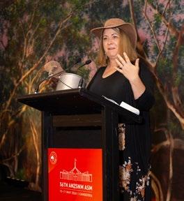
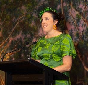
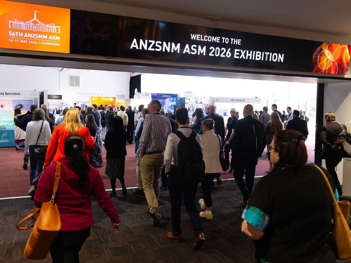
The 56th ANZSNM Annual Scientific Meeting, held in Canberra from 15–17 May 2026, brought together a vibrant and multidisciplinary community of nuclear medicine professionals from across Australia and New Zealand. With over 500 delegates in attendance, the meeting truly reflected the depth and diversity of our specialty—clinicians, technologists, physicists, radiopharmaceutical scientists, and trainees—all contributing to an atmosphere that was both highly engaged and genuinely collegial.
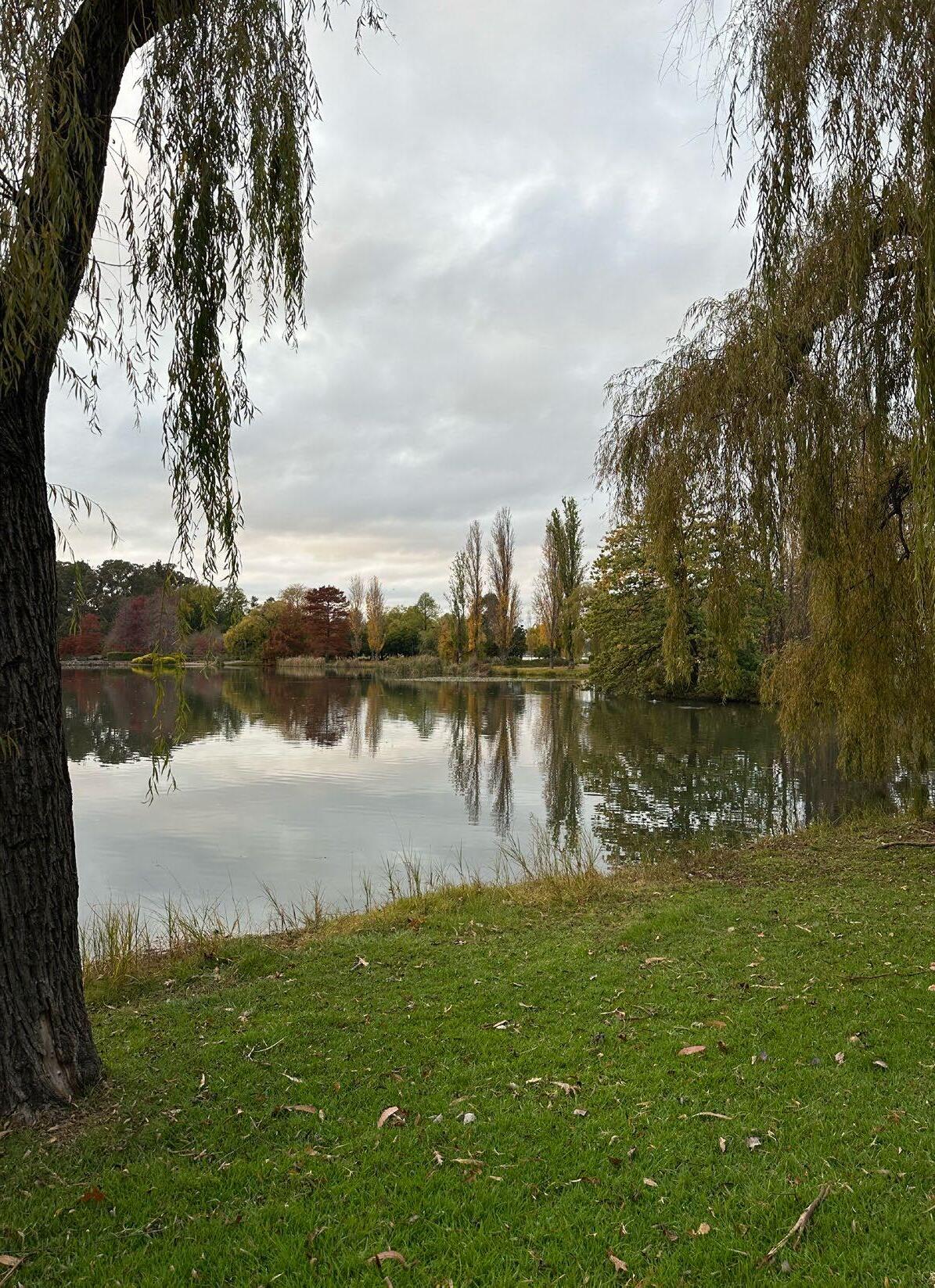
We were fortunate to enjoy Canberra at its best, with clear skies and crisp autumn weather providing a beautiful backdrop for the meeting. It created the perfect setting not only for the formal program but also for the many informal conversations and connections that are such an important part of the ASM experience. The meeting theme, “From Concept to Cure:
Transforming Healthcare with Nuclear Medicine,” was evident throughout, with content spanning early innovation through to real-world clinical impact.
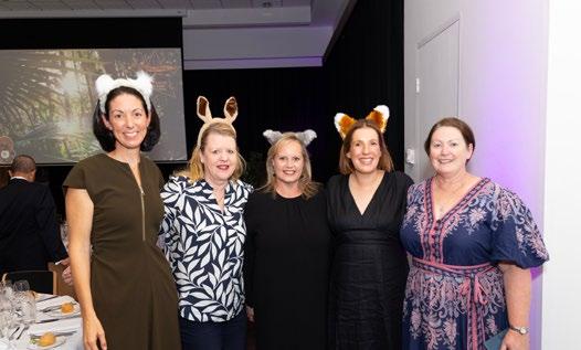
Of course, one of the most memorable features of this year’s ASM was Photon (“Pho”) the Platypus. Pho quickly became part of the personality of the meeting—popping up everywhere and adding a great sense of fun. It added a uniquely Australian sense of fun that resonated with delegates and gave this year’s ASM a distinct identity.
The scientific program was rich, practical, and strongly grounded in clinical reality. From the opening theranostics symposium, which explored both global perspectives and local implementation, through to the plenaries, concurrent sessions, proffered papers, and posters, there was a clear focus on how advances in nuclear medicine are being translated into patient care. Sessions were notably case-based
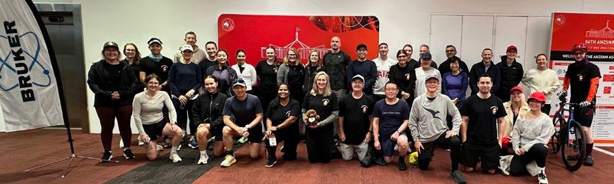
2026 ANZSNM ASM fun run team
ANZSNM 2026
FROM CONCEPT TO CURE: TRANSFORMING HEALTHCARE WITH NUCLEAR MEDICINE
and clinically oriented, covering a broad range of applications—from advanced oncologic imaging and therapy to paediatric, cardiac, and complex cases encountered in daily practice. It was particularly encouraging to see the strength of trainee and early career contributions, with award sessions showcasing the quality and enthusiasm of the next generation.
What stood out across the program was the genuine multidisciplinary nature of the field. Contributions from technologists, radiopharmaceutical scientists, and physicists sat seamlessly alongside physician-led sessions, reinforcing that progress in nuclear medicine relies on the combined expertise of the whole team.
Beyond the conference venue, the social program played a vital role in bringing the meeting to life. The fun run (or walk) around Lake Burley Griffin was a highlight for many, offering a relaxed and energising start to the day while making the most of Canberra’s scenery.
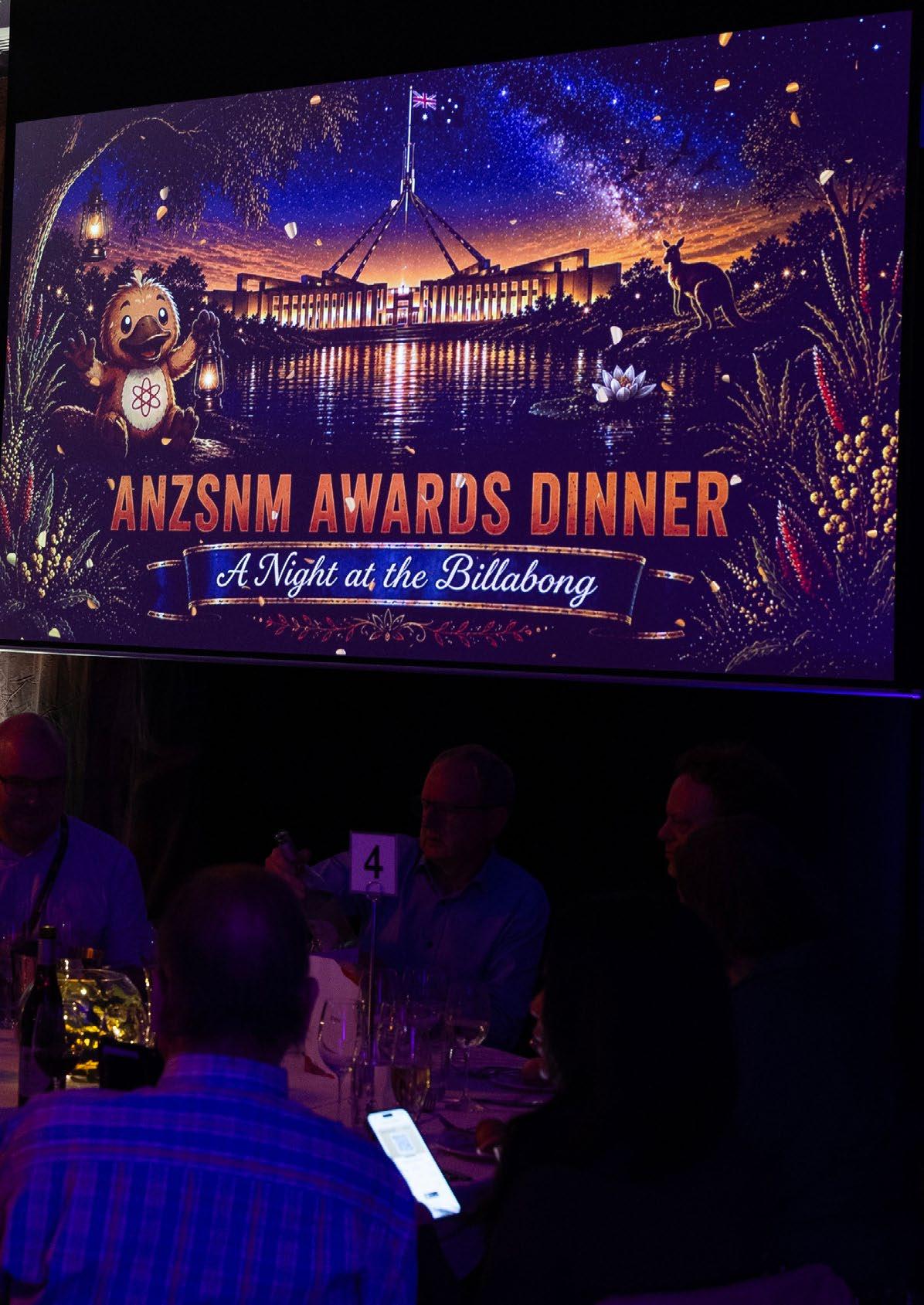
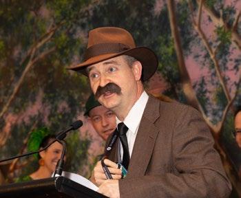
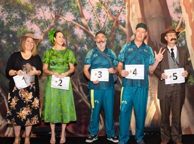
The gala dinner, themed “A Night at the Billabong,” was a standout evening—full of laughter, connection, and a distinctly Australian flavour. It was wonderful to see colleagues come together in such a relaxed setting, and particularly meaningful to celebrate the achievements of our peers as awards were presented across a range of categories, recognising excellence and contribution within our community.
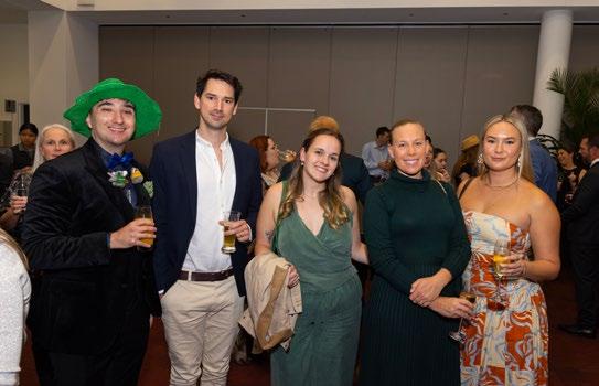
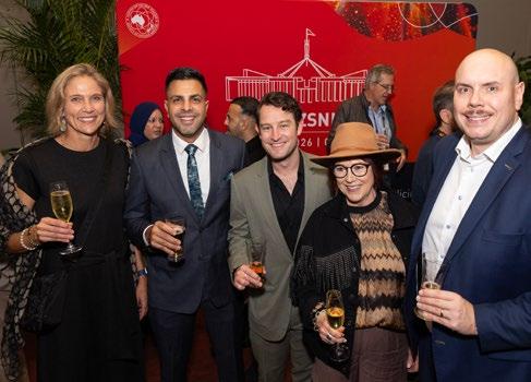
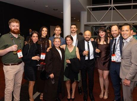
Alongside these key events, the welcome function and informal gatherings throughout the meeting created countless opportunities to reconnect with colleagues and build new collaborations—reminding us that the ASM is as much about people as it is about science.
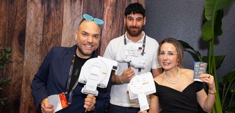
We would also like to acknowledge and sincerely thank our industry partners. Their support is essential in making an event of this scale and quality possible, and we are very grateful for their ongoing commitment to the nuclear medicine community.
Overall, the 2026 Canberra ASM was not only scientifically robust and clinically relevant, but also warm, inclusive, and genuinely enjoyable. It was a meeting that struck a thoughtful balance between innovation and practical application, while also celebrating the strength and spirit of our professional community.
As co-convenors, we would like to say how much we personally enjoyed hosting this year’s ASM. It has been a real privilege to welcome everyone to Canberra and to share in such a positive, energetic, and supportive gathering. The enthusiasm, generosity, and engagement of delegates made the experience both rewarding and memorable for us.
Thank you to all speakers, delegates, sponsors, and organisers who contributed to making this meeting such a success. We leave Canberra feeling proud of what has been achieved and optimistic about the future of nuclear medicine across our region.
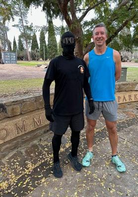
There is, however, still one question that remains unanswered—and may well become ASM folklore: did he really trip, or was it all part of the act?
We look forward to seeing you all again on the Gold Coast in 2027.
Telix ' Best of the Best ' Award
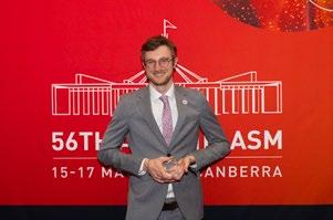
Stacey Rudd, University of Melbourne
Imaging PDL1 with Zirconium-89 Desferrioxamine B Squaramide: From Bench to Bedside
ORL060
Imaging PDL1 with Zirconium-89 Desferrioxamine B Squaramide: From Bench to Bedside
Dr Stacey Rudd
1, Dr Christian Wichmann2, Dr Peter Roselt3, Ms Jessica Van Zuylekom4, Dr Fiona Hegi-Johnson5, Dr Tim Akhurst6, Prof. Michael MacManus7, Dr Ben Blyth4, Prof. Rod Hicks8, Prof. Andrew Scott2, Prof. Paul Donnelly9
1School of Chemistry & Bio21 Institute, University of Melbourne, Melbourne, VIC, Australia. 2Olivia Newton John Cancer Research Institute & Department of Molecular Imaging and Therapy, Austin Health, Melbourne, Vic, Australia. 3Research Division, Peter MacCallum Cancer Centre & The Sir Peter MacCallum Department of Oncology, University of Melbourne, Melbourne, Vic, Australia.
4Research Division, Peter MacCallum Cancer Centre, Melbourne, Vic, Australia. 5Department of Radiation Oncology, Peter MacCallum Cancer Centre & The Sir Peter MacCallum Department of Oncology, University of Melbourne &, Melbourne, Vic, Australia. 6Department of Radiation Oncology & Cancer Imaging, Peter MacCallum Cancer Centre, Melbourne, Vic, Australia. 7Department of Radiation Oncology, Peter MacCallum Cancer Centre & The Sir Peter MacCallum Department of Oncology, University of Melbourne, Melbourne, Vic, Australia. 8Department of Medicine, St Vincent’s Medical School, University of Melbourne & Department of Medicine, Central Medical School, the Alfred Hospital, Monash University, Melbourne, Vic, Australia. 9School of Chemistry & Bio21 Institute, University of Melbourne, Melbourne, Vic, Australia
Theme.
Radiopharmaceutical Sciences, Oncology New tracers and targets
Abstract.
Aim: The aim of this study was to synthesise a novel bifunctional chelator and use this to prepare a PET imaging agent by labelling the antibody durvalumab with Zr-89. We then aimed to assess its distribution in both preclinical models of PDL1 positive disease and later in non-small cell lung cancer patients in a phase 0/1 trial.
Methods: Desferrioxamine squaramide was synthesised in a one-pot reaction between DFO mesylate and diethyl squarate. Bioconjugation to durvalumab was performed in borate buffer at pH 9.0 and the drug-to-antibody ratio (DAR) of the conjugate estimated by intact protein ESI -MS analysis. An automated radiolabelling procedure was optimised using a MultiSyn radiosyn thesizer module. PDL1-positive HCC827 tumour-bearing mice were imaged out to 144 h before a Phase 0/1 multicentre trial was undertaken to assess safety, optimum imaging timepoints, biodistribution, and changes in uptake following chemoradiation.
Stacey Rudd, University of Melbourne
Imaging PDL1 with Zirconium-89 Desferrioxamine B Squaramide: From Bench to Bedside (Continued)
Results: Desferrioxamine squaramide was synthesised in high yield (90%), and conjugation to durvalumab gave DFOSq-durvalumab with a DAR of 3-4, with no evidence of induced aggregation or compromised biological activity. Radiochemical yields in an automated synthese s of 75% with a radiochemical purity >99%, along with tumour SUVmax 8.32 ± 0.59 and tumour:background ratio of 17.17 ± 3.96 in PDL1-positive mice then led to a clinical imaging trial (Phase 0/1, ACTRN12621000171819). Phase 0 results demonstrated optimal imaging at day 5 post administration, with [89Zr]ZrDFOSq-durvalumab uptake observed in all known FDG-avid tumours, except for a single lesion that had progressed on pembrolizumab. One patient with unexpectedly high uptake was reclassified from low to high PDL1 on re-biopsy.
Conclusion: Desferrioxamine squaramide can be used to radiolabel durvalumab with zirconium-89 for PET imaging. [89Zr]ZrDFOSq-durvalumab provides optimum images at 5 days post administration, with initial patient results suggesting it may be a useful probe to assess durvalumab uptake in disease sites. In at least one patient, imaging results have been used to guide therapy. The tracer may also have the potential to predict a lack of response to immunotherapy as well as to provide insight into changes in PDL1 expression after chemoradiation therapy.
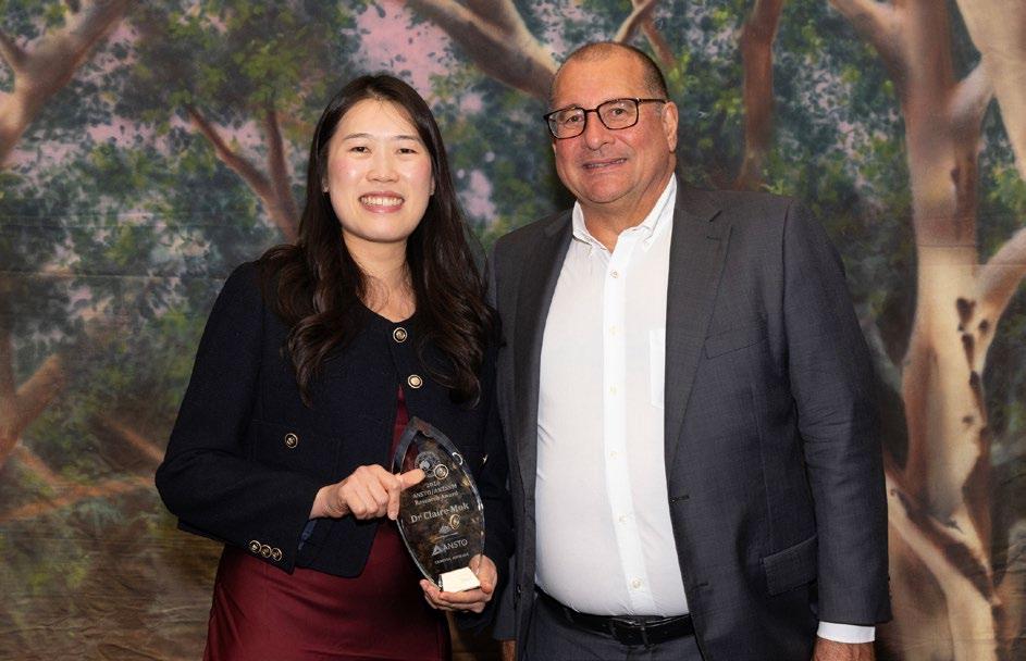
ANSTO/ANZSNM Research Grant
Dr Claire Mok, Royal North Shore Hospital
Establishing reference uptake values and patterns in the pancreas on Gallium-68 Exendin-4 PET/CT in a healthy population - a pilot study at an Australian quaternary referral centre
CLICK TO READ
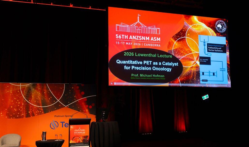
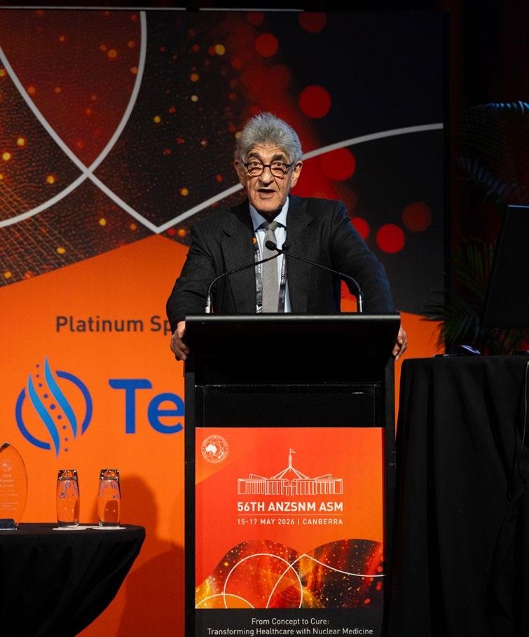
2026 Lowenthal Lecture Award
Professor Michael Hofman, Peter MacCallum Cancer Centre
Quantitative PET as a Catalyst for Precision Oncology
CLICK TO WATCH
2026 Pioneer Lecture Award
A/Professor Barry Elison, University of Wollongong
The development and evolution of a mature nuclear medicine service in a regional metropolitan hospital
AANMS Registrar Research Award
Dr Claire Mok, Royal North Shore Hospital
Efficacy of Peptide Receptor Radionuclide Therapy (PRRT) in Patients with Oligodiscordant Gastroenteropancreatic Neuroendocrine Tumours (NETs) –Outcomes from a Quaternary Neuroendocrine Referral Centre in NSW
Congratulations to Dr Claire Mok from Royal North Shore Hospital on the publication of her paper, Efficacy of Peptide Receptor Radionuclide Therapy (PRRT) in Patients with Oligodiscordant Gastroenteropancreatic Neuroendocrine Tumours (NETs) – Outcomes from a Quaternary Neuroendocrine Referral Centre in NSW, in the European Journal of Nuclear Medicine and Molecular Imaging.
This important research contributes to the growing evidence supporting the role of PRRT in the management of complex neuroendocrine tumours and highlights the valuable work being undertaken by Australian researchers to advance patient care. Claire was recognised for her outstanding research as the recipient of the 2026 AANMS Registrar Research Award, a well-deserved achievement that reflects her significant contribution to the field of Nuclear Medicine.
Read the full article here: https://www.anzsnm.org.au/public/28/files/Oligodiscordant%20PRRT%20 EJNMMI.pdf
Landauer Award
Jade Pearson-McKenzie, Adelaide University
The Role of 18F-FDG PET/CT in the Clinical Assessment of Sarcoidosis: A Retrospective, Single-Centre Review
ORL054
The Role of ¹⁸F-FDG PET/CT in the Clinical Assessment of Sarcoidosis: A Retrospective, Single-Centre Review
Miss Jade Pearson-McKenzie1, Dr Ashleigh Hull1,2, As Prof Dylan Bartholomeusz2,3, Ms Madison Bills2 , Ms Cristina Blefari1
1School of Allied Health and Human Performance, College of Health, Adelaide University, Adelaide, SA, Australia. 2Department of PET, Nuclear Medicine & Bone Densitometry, Royal Adelaide Hospital, Adelaide, SA, Australia. 3Adelaide Medical School, The University of Adelaide, Adelaide, SA, Australia
Theme.
Other
Abstract.
Aim: Sarcoidosis is a systemic disease characterised by granulomatous inflammation (GI) and clinical heterogeneity. Intervention requires prompt, accurate characterisation of disease, as untreated sarcoidosis can lead to cardiopulmonary death. Without standardised criteria, securing a definitive diagnosis is challenging. 18F-fluorodeoxyglucose Positron Emission Tomography Computed Tomography (18F-FDG PET/CT) demonstrates a high affinity for metabolically active GI, offering potential advantages in diagnosis and therapeutic decisionmaking. Despite advantages, 18F-FDG-PET/CT is not reimbursed for sarcoidosis by Australia’s Medicare program, and its clinical role remains poorly defined with a non-standardised diagnostic approach. This study aimed to assess the utility of 18F-FDG PET/CT in the clinical evaluation of sarcoidosis at an Australian public tertiary institution.
Methods: 18F-FDG PET/CT clinical reports and patient questionnaires from 1 January 2022 to 31 August 2024, including the term ‘sarcoid’, were retrospectively retrieved from the Radiology Information System (RIS). Complementary biopsy and Magnetic Resonance Imaging (MRI) reports were retrieved where available. Key data was extracted from reports to determine the effectiveness of 18F-FDG PET/CT through variables including clinical indications, patterns of uptake, cross-modal concordance, and therapeutic monitoring.
Results: 288 18F-FDG PET/CT reports were included, with 246 exhibiting evidence suggestive of sarcoidosis. The most common locations of suspected sarcoidosis lesions detected by 18F-FDG PET/CT were mediastinal (75.2%) and hilar (69.9%) lymph nodes, and a further 132 (53.7%) studies demonstrated extra-thoracic lesions. Dedicated cardiac imaging with dietary preparation was performed in 131 (43.5%) patients, with adequate myocardial suppression achieved in 104 of these studies (79.4%). MRI and biopsy reports were available for 25 and 107 patients, respectively. Among these, 20 (80.0%) MRI and 69 (64.4%) biopsies exhibited concordance with 18F-FDG PET/CT. Through analysing 60 pre- and post-treatment series, sarcoidosis-related activity improved in 19 (31.7%), progressed in 13 (21.7%), and was stable in 28 (46.7%) patients.
Conclusion: This study demonstrated the value of 18F-FDG PET/CT in assessing sarcoidosis. Future prospective investigations are justified to determine whether 18F-FDG PET/CT should be prioritised as a first-line investigation for sarcoidosis, and to evaluate further standardisation of clinical approach and protocols.
GMS Poster Award
Benjamin Nguyen, Austin Health
Radio-HPLC linearity: Minimising the uncertainty for low radioactivity measurements
PGMS03
Radio-HPLC linearity: Minimising the uncertainty for low radioactivity measurements
Mr Benjamin Nguyen1, A/Prof Graeme O'Keefe1, Dr Zhipeng Cao2, Dr Stuart McCluskey1, Prof Andrew Scott1,2,3,4
1Austin Health, Melbourne, Victoria, Australia. 2Olivia Newton-John Cancer Research Institute, Melbourne, Victoria, Australia. 3La Trobe University, Melbourne, Victoria, Australia. 4Melbourne University, Melbourne, Victoria, Australia
Theme.
Radiopharmaceutical Sciences
Abstract.
Aims: The quantities of radioactivity used to conduct radiopharmaceutical research and development is typically low, with focus on quantification of low-level radio-impurities. Our aim was to assess whether polynomial regression can minimise uncertainty in low radioactivity samples using radio-HPLC.
Methods: [68Ga]GaCl3 eluted from an Eckert & Ziegler 68Ge/68Ga generator was buffered using sodium acetate (0.25M), combined with diethylenetriaminepentaacetic acid (DTPA, 20mM sodium succinate, pH 6, containing 0.02% Tween 80) to give [68Ga]Ga-DTPA. Solutions of [68Ga]Ga-DTPA were repeatedly injected through a reverse phase HPLC column (Eclipse XDB C-18, 5µm, 4.6x150mm) equipped with flow scintillation analyser (Radiomatic Flo-One Beta 150TR) for radiometric quantification. Data analysis and polynomial regression was performed on 38 measurements using RStudio (2025.05.1, Build513). Regression parameters with the lowest residual error were selected to fit the data. Inverse regression using radioactive samples was performed to determine point estimates (with corresponding prediction intervals) for injected radioactivity.
Results: Determination of radio-detector linearity was undertaken between 12.3kBq and 151.7kBq of injected [68Ga]Ga-DTPA. For first order (linear) polynomial regression, diagnostic plots showed a tendency away from linearity, despite possessing an excellent coefficient of determination (r2 > 0.99); a second order (quadratic) polynomial provided better fit. At the low end of injected radioactivity (< 15kBq), comparison between the linear and quadratic regressions using a 99% prediction interval determined the approximate uncertainty of the point estimates to be ± 34.12% and ± 4.44%, respectively. Performing the same assessment at the high end of injected radioactivity (> 140kBq) yielded approximate uncertainties of ± 3.15% and ± 0.61%. We observed that the absolute error for low radioactivity measurements was significantly lower when using a quadratic fit, and this was consistent up to the point of saturation of the radio -detector.
Conclusion: For the equipment in this experiment, a second order polynomial was a better fit for the data, minimising uncertainty across the spectrum of measured radioactivity. The most pronounced effect was observed at low radioactivity concentrations. Further work is required to compare the regression to an independent dataset and explore if this method could be used to expand the quantifiable region of our instrumentation.
RAPHAEL Student Research Award
Philip Macilwraith, Gold Coast University Hospital
An audit of 18F-FDG PET timings to Lung MDT in non-metastatic lung cancer patients at a tertiary centre
ORL042
An audit of 18F-FDG PET timings to Lung MDT in non-metastatic lung cancer patients at a tertiary centre
Dr Philip Macilwraith
Gold Coast University Hospital, Gold Coast, QLD, Australia
Theme.
Oncology Clinical, Case Studies, Education and Learning
Abstract.
Aim: To audit the time intervals from PET referral through PET acquisition to lung multidisciplinary team (MDT) discussion in patients with stage I–III non-small cell lung cancer (NSCLC) at a large tertiary centre, and to benchmark these timelines against published guidelines.
Methods: A retrospective audit was conducted of 250 patients with stage I–III NSCLC who underwent 18F-FDG PET and were discussed in Lung MDT between January 2020 and January 2022. Demographic data, disease stage, referral source and key time intervals were collected. Outcomes included time from PET order to PET acquisition to MDT discussion. Median values were calculated and benchmarked against the Australian Optimal Care Pathway for Lung Cancer as well as relevant international guidelines.
Results: Of an initial 250 patients discussed at Lung MDT, 100 met the inclusion criteria based on staging, diagnosis and availability of PET imaging. The median age was 86 years, 40% were male and 60% female. Staging ranged from IA1 to IIIC with 50% being Stage I, 13% Stage II and 37% Stage III. 72 referrals were from outpatients and 28 from inpatients. Outpatient referrals were predominantly from respiratory medicine(n=68), with 2 from ENT and 1 from cardiothoracic surgery and hepatology.
The median time from PET order to PET scan was 5 days, consistent with guideline recommendations for PET completion within 7–10 days. The median time from PET scan to MDT discussion was 8 days, aligning with recommendations for MDT within 1–2 weeks of staging investigations. In 11 cases, PET was performed after MDT suggestion; the median time from MDT to PET was 7.7 days. Inpatients had shorter PET access times than outpatients (median 3 vs 7 days).
Conclusion: PET and MDT timelines for stage I–III NSCLC at our tertiary centre largely align with published benchmarks, with inpatients achieving faster PET access. These findings support earlier PET referral in outpatient pathways to improve MDT timeliness. Next steps include reviewing more recent data, as PET wait-times appear to have lengthened, to determine whether service pressures or workflow changes are contributing and whether further optimisation is needed.
SUMITOMO NMT Student Award
Mikayla Castle, University of Newcastle
Compassion satisfaction in nuclear medicine technologists working in Australia and New Zealand
ORL014
Compassion satisfaction in nuclear medicine technologists working in Australia and New Zealand
Miss Mikayla Castle, Dr Melissa Shields, Dr Daphne James
The University of Newcastle, Newcastle, NSW, Australia
Theme.
Other, Education and Learning
Abstract.
Aim: Compassion satisfaction describes the enjoyment and satisfaction found from caring for and helping others. In health professionals, compassion satisfaction has been positively related to resilience, enhanced coping abilities and the ability to learn from difficult experiences. High levels of compassion satisfaction in healthcare professionals play a large role in improving the quality of care they provide, their work satisfaction and personal wellbeing. The aim of this study was to investigate the levels of compassion satisfaction within nuclear medicine technologists (NMTs) working across Australia and New Zealand.
Methods: An online four-part questionnaire was distributed via QuestionPro. Included in this questionnaire was the Professional Quality of Life Scale (ProQOL) Version 5 (2009). Compassion satisfaction was measured within this scale.
Results: There were 162 survey responses. Overall, participants reported a moderate level of compassion satisfaction (mean = 35.0). There were 33 (20%) participants who scored within the high level of compassion satisfaction and 128 (79%) who scored within the moderate level of compassion satisfaction. One participant reported low levels of compassion satisfaction.
Conclusion: This study was the first investigation of compassion satisfaction of NMTs in Australia and New Zealand, as well as globally. This study found that overall, participants reported a moderate level of compassion satisfaction with no differences found between participant subgroups. The results of this study raise awareness to compassion satisfaction levels of NMTs in Australia and New Zealand.
Shimadzu Award
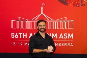
Harris Panopoulos, Austin Health
Optimisation in the Face of Limitations: Improving Solid Target Cu-64 Production Yields
ORL052
Optimisation in the Face of Limitations: Improving Solid Target Cu -64 Production Yields
Mr Harris Panopoulos1, Mr Artur Cichocki1, Mr Nathan Sonnberger1, Mx Talia Enright1, Mr Stan Poniger2, Dr Stuart McCluskey1, Prof Andrew Scott3,4,1,5
1Austin Health, Melbourne, VIC, Australia. 2Iphase Technologies, Melbourne, VIC, Australia. 3Oliva Newton-John Cancer Research Institute, Melbourne, VIC, Australia. 4La trobe University, Melbourne, VIC, Australia. 5Melbourne University, Melbourne, VIC, Australia
Theme.
Radiopharmaceutical Sciences, Other
Abstract.
Aim: Demand for high purity Cu-64 is increasing, and requires a reliable and high yield production process. Analysis of the last three years of cyclotron produced Cu-64 at our Solid Target Facility showed that while the amount of isotope produced had steadily increased, saturation yields (YSat) had been lower and with higher variation than anticipated when expressed as the percentage of theoretical maximum yield (Y% = 100 × YSat / YThMax). We aimed to establish how to stabilise and increase Cu-64 yields.
Methods: Initial investigations included optimisation of the solid targetry to increase the isotope production yield on target, as well as the dissolution and purification method to improve isolated yield. Beam-target interactions were investigated. Energy was tested using a double foil activation method, and beam target interaction via paper burns and foil section activation comparison. A new system to monitor and adjust beam position was implemented, and an updated purification method was developed on an IPhase Multisyn with Dissolution Module. The method retained the Triskem TK201 separation resin used before, while incorporating a Triskem TBP resin to remove common contaminates such as Fe and Ga. Cu-64 chemical purity was tested using ICP-OES (Agilent 5800) and radionuclidic purity with HPGe gamma spectroscopy (Canberra Be20 20).
Results: Improvements in beam-target interactions and purification process were achieved, resulting in reliable increases in YSat relative to theoretical maximum (Y%). Historical three year average Y% was 55.4% ± 12.76% (n=88) before optimisation. After optimization Y% was 73.3% ± 9.6% (n=7) corresponding to a ~25% increase in average yield, improving production efficiency. Repeatable yields in the range of 18 GBq at EOS were achieved, but due to limitations in site radiation licensing could not be increased any further
Radionuclidic purity of the produced Cu-64 remained high, at >99.9%, and complied with all QC testing.
Conclusion: Implemented strategies to improve saturation yields of Cu-64 have been successful, with noticeable increases in saturation yields resulting higher production capacity. This will allow for more efficient and reliability of supply of cyclotron produced Cu-64 to the broader Australian community.
RADPHARM Award
Sasha Aurisch, Royal Prince Alfred Hospital
PSMA and a Vascular Surprise: Detection of Giant Cell Arteritis on 68Ga-PSMA PET/CT
PSMA and a Vascular Surprise: Detection of Giant Cell Arteritis on 68GaPSMA PET/CT
Miss Sasha Aurisch, Professor Michael Fulham, Mrs Angela Brewer
Royal Prince Alfred Hospital, Camperdown, NSW, Australia
Introduction: Prostate-specific membrane antigen (PSMA) PET/CT has become an established and routine imaging modality for the staging and assessment of prostate cancer. Although PSMA uptake is primarily associated with the prostate, expression may also occur in non-prostatic diseases. Recognition of incidental findings on PSMA PET/CT is important, as they may have a significant impact on patient management and outcomes.
This case describes the incidental finding of Giant Cell Arteritis (GCA) on a staging 68Ga-PSMA PET/CT scan, ultimately leading to an early diagnosis and potentially life-saving treatment.
Case Presentation: A 67-year-old male with newly diagnosed intermediate-risk prostate cancer presented to the Royal Prince Alfred Molecular Imaging Department for a staging 68Ga-PSMA PET/CT scan.
The patient presented with increased urinary frequency and nocturia and had a PSA level of 6.2 ng/mL. A recent MRI demonstrated a PIRADS 4 lesion within the left prostatic lobe. Apart from his recent prostate cancer diagnosis, the patient had no significant medical history and reported feeling healthy and well.
The patient received 176 MBq of 68Ga-PSMA and scanned after a 48-minute uptake time using the Biograph Vision Quadra PET/CT scanner. Imaging was performed from vertex to toes with the patient's arms positioned by their sides, acquisition time was a total 12 minutes.
The 68Ga-PSMA PET/CT images demonstrated focal radiotracer uptake within the left prostatic lobe, corresponding to the suspected site of disease (Fig 1)
Fig.1 Shows the PET and fused images demonstrating uptake within the prostate lobe.
The 68Ga-PSMA PET/CT images also showed unusual prominence of tracer within the vessel walls of the thoracic and abdominal aorta with no dense calcification (Fig 2). There was also noticeable uptake both the ascending and descending aorta (Fig 3)
Sasha Aurisch, Royal Prince Alfred Hospital
PSMA and a Vascular Surprise: Detection of Giant Cell Arteritis on 68Ga-PSMA PET/CT (Continued)
Fig 2. The sagittal and coronal views of the PSMA PET/CT demonstrating tracer uptake in the aorta.
Fig 3. Transverse slices of the PSMA PET/CT showing the uptake within the aorta.
The 68Ga-PSMA PET/CT report suggested ESR and CRP testing and perhaps a FDG PET/CT to exclude the possibility of incidental vasculitis.
Follow-Up Investigation: Following a prostatectomy on 31 January 2025, the patient returned three months later for an18F-FDG PET/CT with raised inflammatory markers and reports of leg and muscle aches.
The patient received 230 MBq of 18F-FDG with an uptake time of 59 minutes. Imaging was again performed on the Biograph Vision Quadra with a total acquisition time of 10-minutes
The 18F-FDG PET/CT images reproduced and further expanded upon the abnormalities identified on the initial 68Ga-PSMA scan. The radiotracer uptake was much more extensive on the 18F-FDG PET/CT scan and included involvement of the carotid arteries, the subclavian arteries, thoracic aorta, abdominal aorta and the common iliac vessels (Fig 4)
Fig 4. The 18F FDG PET/CT images demonstrating extensive uptake within the vasculature.
Sasha Aurisch, Royal Prince Alfred Hospital
PSMA and a Vascular Surprise: Detection of Giant Cell Arteritis on 68Ga-PSMA PET/CT (Continued)
Subsequent clinical assessment and pathology findings confirmed the diagnosis of Giant Cell Arteritis.
What is Vasculitis? Vasculitis refers to a group of disorders characterized by the inflammation of blood vessel wall, with Giant Cell Arteritis (GCA) being the most common type. It has a wide variety of non-specific symptoms including:
- Anorexia
- Fever - Malaise
- Muscle Pain
- Night Sweats
- Weight Loss
What Causes Vasculitis? The exact aetiology of vasculitis remains unknown. It is primarily caused by a malfunction within the body’s immune system, caus ing the formation of multinucleated Giant Cells – hence the name Giant Cell Arteritis, which produce destructive enzymes that damage the vessel walls and causes inflammation.
The most significant risk factor for GCA is age. The mean age of onset is 75 years old. The prognosis of GCA depends on early diagnosis and treatment. Prompt corticosteroid therapy greatly reduces complications, though 40-60% of patients relapse and may need long-term immunosuppression.
Vascular Uptake on a PSMA scan: Uptake of PSMA is not exclusive to the prostate as PSMA expression can also be found within endothelial tissue. In conditions like vasculitis, the vessel walls are inflamed, and the body responds by forming new vessels and activating endothelial cells. These activated cells can express PSMA, causing radiotracer uptake along blood vessel walls.
Fig. 5. A direct whole-body comparison between the PSMA and FDG images.
Sasha Aurisch, Royal Prince Alfred Hospital
PSMA and a Vascular Surprise: Detection of Giant Cell Arteritis on 68Ga-PSMA PET/CT (Continued)
Fig 6. Trans axial slices comparison between PSMA and FDG images.
Comparison between the patient’s 68Ga-PSMA and 18F-FDG studies demonstrate notable vascular uptake. Both scans show involvement of the descending thoracic aorta, while the FDG demonstrated more extensive and intense uptake, particularly within the ascending aorta.
These findings illustrate that PSMA PET/CT images may identify vascular disease even though 18F-FDG PET/CT remains the established radiotracer for diagnosis and disease assessment.
Patient Outcome: Following confirmation of Giant Cell Arteritis, the patient was referred to an immunologist and commenced on high-dose corticosteroid therapy. Treatment was well tolerated, and a follow-up 18F-FDG PET/CT scan was suggested to assess response to therapy.
The prognosis for patients with untreated GCA vasculitis is extremely poor, as prolonged inflammation of the large vessels can lead to major complications such as irreversible blindness, or death caused by myocardial infarction, stroke or aortic aneurysm.
Incidental identification of vasculitis on 68Ga-PSMA PET/CT is unexpected, yet potentially lifesaving and has only been reported in one other case study (Sağer et al 2021). It is important to confirm a diagnosis of vasculitis if increased PSMA tracer is seen within otherwise normal vasculature so that appropriate treatment can be initiated.
Conclusion; This case study demonstrated the important of recognising incidental findings on PET/CT scans and illustrates that the uptake of PSMA is not exclusive to the prostate.
While 18F-FDG is the routine tracer for staging vasculitis on a PET/CT scan, this novel case highlights the usefulness of 68Ga-PSMA at visualising non-prostatic diseases.
Nuclear Medicine played a significant role in diagnosing GCA in this patient and will continue to be involved by monitoring treatment response with 18F-FDG PET/CT scans – and It all started with an incidental finding. This patient is now receiving life-saving treatment and will have a favourable outcome and improved quality of life.
Sasha Aurisch, Royal Prince Alfred Hospital
PSMA and a Vascular Surprise: Detection of Giant Cell Arteritis on 68Ga-PSMA PET/CT (Continued)
References:
• Healthdirect Australia. (2023, September). Vasculitis https://www.healthdirect.gov.au/vasculitisWojtylak, P., Ash, S., Tirumani, S., & Li, Q. (2024, June). Non-prostate diseases seen on PSMA PET imaging. Journal of Nuclear Medicine, 65(Suppl 2), 2412.
• Abukanna, A. M., Alanazi, Y. F., Alanazi, F. W. S., Alanazi, R. A., Alanazi, S. S., Alenezi, J. T., Alenezi, H. K., & Alanazi, A. W. S. (2023, December 10). Updates on the prognosis of giant cell arteritis: A systematic review. Cureus, 15(12), e50299. https://doi.org/10.7759/cureus.50299
• Sağer, M. S., Bilgiç, S., Uslu, L., Asa, S., Sağer, G., & Sönmezoğlu, K. (2021). Active giant cell vasculitis diagnosis with ⁶⁸Ga PSMA PET/CT imaging. Molecular Imaging and Radionuclide Therapy, 30(1), 57–59. https://doi.org/10.4274/mirt.galenos.2019.90532.
ILLUMINATING PROSTATE CANCER
Illuccix® is currenly the only ARTG listed PSMA diagnostic product. This means Illuccix® is the only PSMA diagnostic product that has been evaluated for its Quality, Safety and Efficacy in Australia.~
EDUCATION & CPD | Case study
GA-68 DOTATATE (GALLIUM-68) DETECTED OVARIAN CARCINOID PRESENTING WITH CARCINOID HEART DISEASE CASE STUDY
Dominic Ku, Hannah Son, Yangyi Ong, Patricia Guzman, King Tan and Unine Herbst.
Sydney Adventist Hospital, NSW Australia
Introduction
Carcinoid syndrome results from the systemic release of vasoactive substances from neuroendocrine tumours (NETs). Up to two-thirds of patients with carcinoid syndrome develop carcinoid heart disease, characterized by fibrous plaque deposition on cardiac valves leading to valvular dysfunction (Fox et al. 2004). The right-sided valves, particularly the tricuspid and pulmonary valves, are most commonly affected due to first-pass exposure of the heart to circulating serotonin and other vasoactive amines (Bhattacharyya et al., 2007).
New-onset or rapidly progressive tricuspid regurgitation, especially in the absence of left-sided heart disease or congenital shunts, should prompt investigation for an occult carcinoid tumor (Bernheim et al., 2020). Molecular imaging with Gallium-68 (68Ga)–DOTATATE PET/CT has emerged as a sensitive whole-body modality for localizing well-differentiated NETs through somatostatin receptor expression and plays a key role in this diagnostic context (Hope et al., 2019).
Primary ovarian carcinoid tumours are rare but represent a distinctive entity in which vasoactive substances drain directly into the systemic circulation via the ovarian veins and inferior vena cava (IVC), bypassing hepatic first-pass metabolism. Consequently, carcinoid syndrome and carcinoid heart disease may occur even in the absence of hepatic metastases, in contrast to more common gastrointestinal primaries (Robboy et al., 2009).
Case Description
A 64-year-old woman presented with an acute leftsided cerebellar infarct, initially attributed to presumed paroxysmal atrial fibrillation. As part of her stroke workup, transthoracic echocardiography was performed and unexpectedly demonstrated severe tricuspid regurgitation with marked failure of leaflet coaptation. There was no evidence of atrial septal defect.
The unexpected diagnosis of severe tricuspid regurgitation raised concern for carcinoid heart disease, prompting further evaluation. Whole-body 68Ga-DOTATATE PET/CT
demonstrated an intensely somatostatin receptor–avid lesion within the left ovary, with no additional sites of abnormal uptake (Figure 1). Further characterisation of the lesion was conducted with CT and MRI (Figure 1). Surgical excision of the ovarian mass was subsequently performed, and histopathologic examination confirmed carcinoid tumour (Figure 1).
The anatomic venous drainage of the left ovary indirectly to the IVC via the left renal vein and the right ovary directly into the IVC provides the mechanism for direct large volume systemic exposure to vasoactive tumour products, accounting for the development of carcinoid heart disease in this patient. This case highlights the importance of considering carcinoid syndrome in patients with new-onset isolated tricuspid regurgitation and illustrates the value of 68Ga-DOTATATE PET/CT as a whole-body screening tool for identifying occult NET primaries, particularly in atypical locations.
Disclosure statement
The authors have no conflict of interest to declare.
References
1. Fox DJ, Khattar RS. Carcinoid heart disease: presentation, diagnosis, and management. Heart. 2004;90(10):1224-8. doi: 10.1136/hrt.2004.040329.
4. Hope TA, Bergsland EK, Bozkurt MF, Graham M, Heaney AP, Herrmann K, et al. Appropriate use criteria for somatostatin receptor PET imaging in neuroendocrine tumors. J Nucl Med. 2019;59(1):66-74. doi: 10.2967/ jnumed.117.202275.
5. Robboy SJ, Scully RE, Norris HJ. Primary carcinoid tumors of the ovary. Cancer. 2009;36(2):404-16. doi: 10.1002/1097-0142(197508)36:2<404::AIDCNCR2820360212>3.0.CO;2-Q.
Case Study: GA-68 Dotatate (Gallium-68) Detected Ovarian
Carcinoid Presenting With Carcinoid Heart Disease (Continued)
Figure 1: Row 1: Ga-68 DOTATATE and noncontrast CT demonstrates a large left adnexal soft tissue lesion with intense avidity. Rows 2 and 3: Clockwise, T2, T1 fat saturated post gadolinium contrast, ADC map and DWI b800 images demonstrate left adnexal mass with mildly heterogenous enhancement and regions of restricted diffusion in the left lateral aspect. Row 4: Wellcircumscribed tumour completely contained in the ovarian stroma. Row 5: Histology demonstrates sheets and nests, cords and little cystic structures lined by epithelial cells with neuroendocrine features. There is less than 1 mitosis per 10 high-power field. There is no evidence of any associated in situ or invasive adenocarcinoma. Row: 6: Haematoxylin and Eosin (H&E stain), positive stain for Synaptophysin. Row 7: Positive stain for CDX2. Low Ki-67 (less than 1%).
TB-PSMA-I&T ARTICLE
IN-VITRO RADIOCHEMICAL STABILITY OF THE THERAPEUTIC RADIOPHARMACEUTICAL
[161TB]
Rebecca J. Mangelsen1*, Ruchi Rattan1, Madhusudan Vyas1,2, Jessica Fagan1, Remy Lim1, Andrew Henderson1
1Department of Nuclear Medicine and PET-CT, Allevia Radiology, Auckland, New Zealand; and 2School of Healthcare and Social Practice, Unitec Institute of Technology, Auckland, New Zealand
*Corresponding author:
Rebecca J. Mangelsen
Department of Nuclear Medicine and PET-CT Allevia Radiology
Auckland, New Zealand
Phone (or Mobile) No.: +64 29 045 07307
Email: rmangelsen@alleviaradiology.co.nz
Type of article: Original Research Article
Word count: 1453
Financial support disclosure: There was no financial support received for the current study from any organisation.
All compounds are >95% pure by HPLC analysis.
Abstract
Background: Prostate cancer is the most diagnosed cancer in New Zealand and the second leading cause of cancer-related death among men. Patients with metastatic castrate-resistant prostate cancer (mCRPC) face poor outcomes due to its incurable nature and significant disease burden. Radioligand therapy (RLT) targeting prostate-specific membrane antigen (PSMA), a transmembrane glycoprotein overexpressed in prostate cancer cells, has shown clinical efficacy in extending survival and preserving quality of life. Commonly used isotopes for PSMA imaging and therapy (PSMA-I&T) include yttrium-90 (90Y), and Lutetium-177 (177Lu); however, Terbium-161 (161Tb) is emerging as a promising alternative due to its emission of both medium-energy beta-minus (B-) particles and a high proportion of conversion and Auger electrons. This study aimed to evaluate the postformulation radiochemical stability and shelf life of [161Tb] Tb-PSMA-I&T.
Ten batches of [161Tb]Tb-PSMA-I&T. between July and August 2024, were produced using an automated synthesis
system. After each synthesis, the radiochemical purity (RCP) of [161Tb]Tb-PSMA-I&T was evaluated utilising highperformance liquid chromatography (HPLC), instant thinlayer chromatography (ITLC), and potential of hydrogen (pH) at three different time points: 0, 24 and 48 h.
Results: The average RCP of [161Tb]Tb-PSMA-I&T with HPLC was greater than 99 percent (99.65 ± 0.37 percent), and ITLC was greater than 99 percent (99.82 ± 0.15 percent) at all three time points. The pH was between 4.5- 5.
Conclusion: Our findings demonstrate that the synthesis of [161Tb]Tb-PSMA-I&T with an automated synthesis system demonstrates high in-vitro stability for up to 48 h after labelling.
ARTICLE: In-vitro Radiochemical Stability of the Therapeutic Radiopharmaceutical [161Tb]Tb-PSMA-I&T. (Continued)
Graphical Abstract
Introduction
Prostate cancer is the most diagnosed cancer in New Zealand and the second leading cause of cancer-related death among men (1). Patients with metastatic castrateresistant prostate cancer (mCRPC) face a poor prognosis, as the disease remains incurable and is associated with a significant disease burden (2). PSMA (prostatespecific membrane antigen) is a type II transmembrane glycoprotein overexpressed in the cell membrane of prostate epithelial cells (3). Radioligand therapy (RLT) that targets PSMA receptors is effectively utilised for the treatment of mCRPC to extend survival while preserving the patient's quality of life (2,3,4)
The most widely used isotopes for PSMA RLT are yttrium-90 (90Y), a high-energy beta minus (B )emitter and Lutetium-177 (177Lu), a medium-energy B emitter (5) PSMA analogues commonly include PSMA imaging and therapy (I&T), PSMA-617, and J591 (6). Terbium-161 (161Tb) is emerging as an advantageous isotope for PSMA I&T RLT, a medium-energy B radionuclide which exhibits similar in vivo and in vitro chemical properties and pharmacokinetic characteristics to 177Lu but emits a greater percentage of conversion electrons and Auger electrons per decay (5) Consequently, when 161Tb is bound to PSMA-I&T [161Tb] Tb-PSMA-I&T, it has a greater expected therapeutic effect when compared to 177Lu-PSMA-I&T, as it targets both single and clustered cancer cells due to the ultra-short tissue range (<500 nm) and high linear energy transfer (4026 keV/μm) of its Auger electrons (4). Consequently,
there is an increased probability of double stranded deoxyribonucleic acid (DNA) breaks (4). Post-treatment imaging utilising, single-photon emission computed tomography/computed tomography (SPECT/CT) with 161Tb is feasible due to its gamma emissions at energies of 48.9 keV and 74.5 keV (7). This permits quantitative dosimetry of active disease sites and norThe purpose of this study was to evaluate the post-formulation stability and shelf life of [161Tb]Tb-PSMA-I&T over a period of 48 h using HPLC, ITLC and pH quality control methods.s and qualitative evaluation of the patients' response to treatment (8)
Ensuring the radiochemical purity and stability of radiolabelled compounds, such as 161Tb PSMA-I&T, is essential prior to patient administration. In nuclear medicine, standard procedures involve labelling radiopharmaceuticals and conducting quality control tests to confirm their suitability for injection (9). This may include high-performance liquid chromatography (HPLC), instant thin-layer chromatography (ITLC) and potetial of hydrogen (pH) quality control methods (9). Each radiolabelled product has a defined shelf life, which indicates the duration it remains stable and effective in vitro. Beyond this period, the compound may degrade or lose its intended properties, requiring it to be discarded to ensure patient safety and therapeutic efficacy.
The purpose of this study was to evaluate the postformulation stability and shelf life of [161Tb]Tb-PSMA-I&T over a period of 48 h using HPLC, ITLC and pH quality control methods.
Labelling was performed using a computer-based automated system as observed in Figure 2 (Modular-Lab Eazy; Eckert & Ziegler). All production cassettes were supplied by vendors and were made for a single use only. [161Tb]TbCl3 was added to a reaction mixture containing PSMA-I&T, sodium acetate buffer (0.5 M), and hydrochloric acid (0.05 M). The mixture was incubated at 95 °C for 25 minutes at a reaction pH of 4.5, yielding [ 161Tb]TbPSMA-I&T with high specific activity, typically around 100 MBq/nmol. Radiolytic degradation was minimized by the addition of stabilizing agents, such as L-ascorbic acid, to prevent radiochemical decomposition.
The shelf life at room temperature of prepared [161Tb]TbPSMA-I&T was established based on radiochemical purity by HPLC, ITLC (ITLC) and pH measurements.
The final formulation included 200 μg PSMA-I&T per 5 GBq, reconstituted with 1.5 mL sodium ascorbate (0.57 M) to adjust pH to 4.5 ± 0.1.
HPLC was utilised for radiochemical purity (RCP) and stability analysis of 161Tb-labeled PSMA-I&T conjugates. A dual-pump HPLC unit with a Carbon-18 reversed-phase column (25 × 0.46 cm) (Knauer) purified the labelled conjugates with two mobile phases; Solvent A: 1% trifluoroacetic acid and Ultrapure (Sigma-Aldrich) water; Solvent B: 0.1% trifluoroacetic acid and acetonitrile. A gradient elution method was employed as follows: i.e., 0–3 min—A, 50% and B, 0%; 10–15 min—A, 0% and B, 100%.
The typical retention time of radiolabelled PSMA under above conditions was approximately 6-8 minutes.
To assess the RCP of [ 161Tb]Tb-PSMA-I&T, ITLC was employed. This analytical technique enables the separation of components based on their differential interactions with the stationary and mobile phases, primarily influenced by polarity. As a result, a radioactive sample applied to the stationary phase migrates at varying rates depending on its chemical form, allowing for the separation of impurities such as unbound 161TbCl3. For this study, 5 μL of the test solution was spotted at 1.5 cm from the lower end of the paper strips and was developed in 10% ammonium acetate in methanol as mobile phases (30:70 vol/vol).
The pH of [161Tb]Tb-PSMA-I&T was also measured to ensure stability and integrity of the radiopharmaceutical, and suitability for intravenous injection (pH 4.5-5.5) (10,11). 5 μL of the test solution was spotted onto a narrow-band pH litmus paper and matched to the reference colours.
After each synthesis, the given amount of radiolabelled complex (~10 MBq) was kept at room temperature for 48 h while being checked by HPLC, ITLC and pH at specified time intervals of 0, 24, and 48 h after preparation (Tables 1 and 2).
Results
Ten batches (n=10) of 161Tb labelled PSMA-I&T were completed between July and August. 2024. All compounds are >95% pure by HPLC analysis. The average RCP of [161Tb] Tb-PSMA-I&T with HPLC was greater than 99 percent (99.65 ± 0.37 percent), and ITLC-SG was greater than 99 percent (99.82 ± 0.15 percent) at room temperature at 0, 24 and 48 h post synthesis (Figs. 3-4). The pH obtained at 0, 24 and 48 h post synthesis was 4.5- 5, within the tolerance range of 4.5-5.5.
Discussion
RCP refers to the percentage of the total radiopharmaceutical that is bound to the intended radiolabelled compound, with a desirable RCP typically exceeding 95% (10) . The presence of radiochemical impurities or decomposition products can adversely affect the biodistribution of RLT, potentially compromising both therapeutic efficacy and the accuracy of quantitative analysis in post-treatment imaging (10) . Thus, it is imperative that RCP of [161Tb]Tb-PSMA-I&T is maintained and evaluated prior to intravenous administration in patients with mCRPC.
ARTICLE
ARTICLE: In-vitro Radiochemical Stability of the Therapeutic Radiopharmaceutical [161Tb]Tb-PSMA-I&T. (Continued)
The results of this study demonstrate that [161Tb]Tb-PSMAI&T, synthesised using an automated system, exhibits high in vitro stability for up to 48 hours post-labelling. This extended stability supports the feasibility of centralised production and distribution of the radiopharmaceutical, thereby enhancing access to patient treatment in regional areas as well as metropolitan centres with limited laboratory infrastructure. Given the similar in vivo and in vitro chemical properties and pharmacokinetic characteristics of 161Tb to those of 177Lu, [161Tb]Tb-PSMAI&T can be seamlessly incorporated into existing PSMA labelling and imaging protocols for staging and posttreatment scans with minimal adaptations 8iumk, Additionally, 161Tb offers a therapeutic advantage over 177Lu due to its greater percentage of conversion electrons and Auger electrons emitted per decay, which is resultantly beneficial for targeting micro metastatic disease in patients with mCRPC (5)
One of the key limitations of this study was that sterility testing could not be performed as the laboratory was not GMP-compliant. Terminal sterile filtration was performed during synthesis, but sterility, pyrogenicity, and elemental impurity data could not be included. A secondary limitation was that the study was conducted at room temperature, and temperature control for stability beyond room temperature (5–8 °C or 30–40 °C) was not performed. Refrigerated storage during transport or higher transport temperatures could influence the stability profile, prompting further investigations. In addition, ten production batches provided useful preliminary data; however, the small sample size is another limitation to the study. Future investigations could include GMP-compliant testing, sterility testing, temperature-dependent stability studies, and eventual clinical evaluation.
1. Centre for Health Outcome Measures New Zealand. PCOR-NZ – New Zealand Prostate Cancer Outcome Registry. https://www.chomnz.org.nz/pcor-nz-registry. Accessed 14 Jun 2025.
2. Chakrabarti A, Zhang T. Metastatic castration-resistant prostate cancer: advances in treatment and symptom management. Curr Treat Options Oncol. 2024;25(5):43144. doi:10.1007/s11864-024-01215-2.
4. Umbricht CA, Benešová M, Schmid RM, Türler A, Schibli R, Müller C. 161Tb-labeled PSMA ligands for radioligand therapy: preclinical investigations. J Nucl Med. 2019;60(12):1825-32.
5. Hindié E, Zanotti-Fregonara P, Quinto MA, Morgat C, Champion C. Dose deposits from 90Y, 177Lu, 111In, and 161Tb in micrometastases of various sizes: implications for radiopharmaceutical therapy. J Nucl Med. 2016;57(5):75964. doi:10.2967/jnumed.115.170423.
6. Von Eyben FE, Roviello G, Kiljunen T, et al. Thirdline treatment and 177Lu-PSMA radioligand therapy of metastatic castration-resistant prostate cancer: a systematic review. Eur J Nucl Med Mol Imaging. 2018;45(3):496-508. doi:10.1007/s00259-017-3895-x.
7. Al-Ibraheem A, Doudeen RM, Juaidi D, Abufara A, Maus S. 161Tb-PSMA radioligand therapy: first-in-humans SPECT/ CT imaging. J Nucl Med. 2023;64(8):1322-3. doi:10.2967/ jnumed.122.265291.
8. Yadav S, Lawhn-Heath C, Paciorek A, et al. The impact of posttreatment imaging in peptide receptor radionuclide therapy. J Nucl Med. 2024;65(3):409-15. doi:10.2967/ jnumed.123.266614.
9. Molavipordanjani S, Hosseinimehr SJ. Fundamental concepts of radiopharmaceuticals quality controls. Pharm Biomed Res. 2018;4(3):1-8. doi:10.18502/pbr.v4i3.538.
10. European Association of Nuclear Medicine. Radiopharmacy: an update – a technologist’s guide. https:// eanm.org/wp-content/uploads/2024/06/EANM_2019_ TechGuide.pdf. Accessed 14 Jun 2025.
11. Schaefer-Schuler A, Burgard C, Blickle A, et al. [161Tb] Tb-PSMA-617 radioligand therapy in patients with mCRPC: preliminary dosimetry results and intra-individual headto-head comparison to [177Lu]Lu-PSMA-617. Theranostics. 2024;14(5):1829-40. doi:10.7150/thno.92273.
ARTICLE: In-vitro Radiochemical Stability of the Therapeutic Radiopharmaceutical [161Tb]Tb-PSMA-I&T. (Continued)
Table 1.
HPLC Results at Various Times
Table 2.
ITLC-SG Results at Various Times Batch
Figure 1.
Modular-Lab Pharm cassette and reagent kit.
Figure 2.
ML-Eazy fully automated synthesis device, disposable cassette and schematic flow (a) [161Tb]] Tb vial, (b) Buffer and peptide vial, (c) Reaction vial and heater, (d) Saline vial, (e) Final product vial.
Figure 3.
A line graph showing HPLC quality control result comparison at 0, 24, and 48 h.
Figure 4.
A line graph showing ITLC-SG quality control result comparison at 0, 24, and 48 h.
CREATIVITY VS PROBLEM SOLVING
Dale L Bailey PhD
Department of Nuclear Medicine, Royal North Shore Hospital, Sydney, Australia & Faculty of Medicine & Health, University of Sydney, Australia
Contact for Correspondence: Dale.Bailey@sydney.edu.au
ORCID DB: 0000-0001-9154-7957
The Cambridge English Dictionary defines creativity as “the ability to produce original and unusual ideas, or to make something new or imaginative” and notes synonyms such as imagination, creative, vision, inventiveness, ingenuity and originality. The concept of creativity in science has always interested me as the lay person might regard the formality and structure of science as being designed to mitigate creativity, reducing existence and experience to rules that the physical world must obey, such as Newton’s Laws of Motion or Einstein’s equation for Special Relativity, E = mc2, upon which modern PET scanning is based. Surely, you might say, predicting the motion of the planets or identifying the constituent elements in an unknown chemical compound are examples of problem-solving rather than creativity, and you might be correct. Creativity is thought be more the domain of artists and designers, be they visual, audio, functional (e.g., architecture) or commercial in orientation. And, for them, problem-solving would not be considered as a major component or normal part of their “creative” lives. But the moment that the polymath Copernicus placed the sun at the centre of the solar system, removing the earth to an outer orbit, must rank as a moment of pure creativity given the definition above while, yes, solving a problem at the same time. Can we recognise that creativity and problem-solving can co-exist and can be used to enrich our social and working lives?
I have lived for over 25 years with a fine artist, a figurative painter. I cannot count the number of times I have heard the phrase “I have a problem with this painting that I can’t resolve...” which, if unable to be corrected, can result in the canvas being painted over with whitewash for a fresh start. Problem-solving is required to “fix” the problem with the painting. This, from an individual that most people would consider to be close to 100% creative all of the time, but here having to resolve an issue in an artwork of their own creation. And most would consider my own work as a physicist in medical imaging and radionuclide therapies is surely mostly concerned with problem-solving: writing software to correct for photon attenuation in a SPECT image, for example. A problem with the image that needs solving, simple. Not so simple, though, as there is no mathematically correct solution to the problem – the reason being that, in SPECT, we have two unknowns, the unknown distribution of the radiopharmaceutical AND the unknown shape and density of the body which attenuates the gamma photons, and only one measurement from which to derive the two unknowns. We were all taught in primary school that to solve for two unknowns (e.g., a and b in ax2 + b = 12) you need two equations (or measurements in the SPECT example) and we proceed to solve this by elimination. In SPECT we only have one “equation”. Therefore, we have a problem! In this case, the solution to the problem was known – we needed a second measurement, body density. The creativity came in how we could obtain this. When we faced this issue in the mid-1980s, soon after I had graduated from my undergraduate Physics degree, rotating single-detector SPECT cameras had only just been introduced into the clinic and there was little access to CT data. Our solution was to use a transmission flood source at the same time as the SPECT 99mTc measurements and measure the photon transmission through the body. We chose to use the long-lived radionuclide 153Gd (t½ = 9 months) for the transmission source which conveniently had imageable gamma emissions at 98 and 103 keV, well below the 140 keV PHA window of 99mTc photons. Thus, simultaneous measurement of radiopharmaceutical distribution and body density was introduced (1) for attenuation correction in SPECT. This is now regarded as one of the earliest attempts at “hybrid” multimodality imaging in nuclear medicine (2).
We need to be more creative in how we convey information as well. We produce wonderful images which are interpreted by specialists and often reduced to little more than a text file. And yet we know the old adage that “a picture
ARTICLE: Creativity Vs Problem Solving (Continued)
is worth a thousand words”. To think that the richness of our functional imagery is reduced to a page and a half of text. I think we can do better. My favourite presentation of data to communicate information is the chart of Napoleon’s Grand Armee campaign to Russia in 1812-1813 by Charles J Minard (1781 – 1870) (Fig. 1), a French civil engineer, which is regarded as possibly the greatest statistical chart of all time. The chart shows the army size by the width of the line, starting from France in the light brown colour (far left). As the army progresses, eventually becoming bogged down in the Russian winter, the width of the line decreases with time as soldiers die. Early in the march a group is sent via a different path and eventually reunites with the returning army remnants after they are defeated upon reaching the outskirts of Moscow (far right). The black line shows the returning army. The graph displays army size, time of year, temperature and certain geographical elements such as river crossings and subsequent loss of life. Note the point on the return to France when the temperature drops significantly at Dorogobuzh (“Dorogobouge”) in Smolensk Oblast, Russia (current-day Ukraine) and many soldiers die. In total, 144,000 soldiers made up the initial army headed for Russia and a mere 10,000 returned. Talk about creativity! This graph has it all: information, a story of human tragedy, miscalculation and overreaching ambition. Why could we not have something similar for a simple test like colon transit studies? We should be more imaginative and creative.
Another information-rich graph that I greatly admire is shown in Fig. 2. It is a timetable. For the Generation-Z’s reading this, a hard-copy timetable was a small leaflet or pamphlet that contained the timings of scheduled, regular transportation routes such as rail or bus at each stop. The graph shows the trains running between Paris and Lyon and the timing of the journey. You can see times where the train reaches a station and stops for a certain period of time before departing for the next station. The timetable shows both directions from Paris to Lyon and back. The vertical “graph” lines are at 10 min intervals. This single chart shows 24 hours of train operations. This is sheer genius and again attests to the creativity of the designer to incorporate so much information into a single graphical display1
We often battle today to recruit students to studying STEM subjects at both secondary and tertiary level. There are many reasons why this is the case including perceived lack of job security (e.g., recent job cuts at important agencies such as CSIRO and the Trump effect on scientific funding in the US), lack of financial incentive, and that it is all too hard. Some students are put off for other less well known reasons: at high school careers nights which I advise at one of the most frequent comments that I get is that the student was not aware that physics and maths could be used to “help people”. So many tell me that they love studying maths and physics but want to do something that is more useful to society, such as medicine or other therapy disciplines.
Figure 1. Figurative chart of the French Army campaign in Russia 1812-1813 by CJ Minard.
ARTICLE: Creativity Vs Problem Solving (Continued)
Figure 2. Train timetable for Paris-Lyon 1885. The chart is attributed to the French engineer Ibry.
Both of the graphs shown can be found in the fantastic book “The Visual Display of Quantitative Information” by the American psychologist Edward R Tufte. This is a book that I give to all of my doctoral students to have a look through as it is so rich in information about how to display information accurately and effectively.
I think we need to emphasise and explore the creative side of our discipline more. Try something different, look at things a different way, display your information in a more appealing and communicative way. Problem-solving skills are, rightly, highly praised but let’s not think that doing so excludes creativity, the spark that provides the extra insight. We are fortunate in nuclear medicine that we still have room to be creative as the field moves on in large leaps and bounds. Problem-solving and creativity are two sides to the same coin.
References
1. Bailey DL, Hutton BF, Walker PJ. Improved SPECT using simultaneous emission and transmission tomography. J Nucl Med. 1987;28:844-851.
2. Townsend DW. Multimodality imaging of structure and function. Phys Med Biol. 2008;53:R1-R39.
2026 SNMMI TECHNOLOGIST BEST PET ORAL PRESENTATION AWARD
DR
ALICIA CORLETT
The ANZSNM congratulates Nuclear Medicine Technologist Alicia Corlett from Peter MacCallum Cancer Centre on receiving international recognition at the Society of Nuclear Medicine and Molecular Imaging (SNMMI) Annual Meeting, where she was honoured with two prestigious awards.
Alicia received the 1st Place Technologist Best PET Oral Presentation Award from the SNMMI Technologist Section (SNMMI-TS) for her oral presentation titled, Results from a Novel Long-Axis Field-of-View "Total-Body" Digital BGO PET/ CT System: Visualizing Recurrent Malignancy Not Seen on Conventional PET/CT
In addition to this outstanding achievement, Alicia was also awarded the Education and Research Fund (ERF)–SNMMI Technologist Section (SNMMI-TS) Technologist Professional Development Grant Award, recognising her commitment to ongoing professional development and supporting her continued contribution to research and innovation in nuclear medicine.
These outstanding achievements recognise the high quality of Alicia's work and highlight the significant contribution Australian nuclear medicine technologists continue to make to advancing molecular imaging, research and patient care on the international stage. Congratulations, Alicia, on these well-deserved honours.
PAST PRESIDENT RECOGNITION
The ANZSNM was pleased to recognise the invaluable contributions of its Past Presidents during the 2026 Annual Scientific Meeting in Canberra, where they were presented with ANZSNM Past President badges. The badges acknowledge the dedication, leadership and lasting impact each Past President has made to the Society and the Nuclear Medicine profession. We thank our Past Presidents for their ongoing support and commitment to advancing the ANZSNM and look forward to their continued involvement in the Society.
PRESIDENT
A/Prof Grace Kong (VIC/TAS)
Vice President
Suzanne McGavin (SA/NT)
Immediate Past President
Prof Karen Jones (SA/NT)
Treasurer
Dr Daniel Badger (SA/NT)
FEDERAL
COUNCIL
A/Prof Grace Kong (President)
Prof Karen Jones (Immediate Past President)
Dr Daniel Badger (Treasurer)
Ms Suzanne McGavin (Vice President)
Dr Kevin London (Chair ARTnet)
Dr Melissa Latter (Chair RPS SIG)
Mr George McGill (Chair Physics SIG)
Dr Jeremy Hoang (Chair MSIG)
Dr Melissa Shields (Chair TSIG)
Ms Jessica Fagan (Chair of Branches/NZ Rep)
Prof Andrew Scott (IRC/Medical Rep)
SPECIAL INTEREST GROUPS/ COMMITTEES
Technologists
Chair: Dr Melissa Shields
Medical
Chair: Dr Jeremy Hoang
Radiopharmaceutical Science
Chair: Dr Melissa Latter
Physics
Chair: Mr George McGill
Quality and Technical Standards Committee
Chair: Dr Darin O’Keeffe
Scientific Advisory Panel
Chair: Prof Dale Bailey
International Relations Committee
Chair: Prof Andrew Scott
Conference Convening Committee
Chair: Prof Paul Roach
Scientific Education Committee
Chair: Prof Andrew Scott
ANSTO Representative
Annah Skillen
BRANCH
SECRETARIES
New South Wales/Australian Capital Territory
Holly Spooner
Queensland
Ms Neena Sunny and Ms Stephanie Schulz
South Australia/Northern Territory
Mr Jagi Sandhu and Ms Emily James
Victoria/Tasmania
Ms My Linh Diep
Western Australia
Ms Rosemary Dallen
New Zealand
Ms Rebecca Mangelsen
Rural/Regional Branch
Mr Lachlan Patterson
GENERAL
MANAGER & SECRETARIAT
All Correspondence
Mr Rajeev Chandra, General Manager
Ms Melissa Flannery, Secretariat
ANZSNM Secretariat, PO Box 6178, Vermont South, Victoria 3133
Tel: 1300 330 402 | Fax: (03) 8677 2970
AIMS AND OBJECTIVES OF THE AUSTRALIAN AND NEW ZEALAND SOCIETY OF NUCLEAR MEDICINE
1.
Promote:
• The advancement of clinical practice of nuclear medicine in Australia and New Zealand;
• Research in nuclear medicine;
• Public education regarding the principles and applications of nuclear medicine techniques in medicine and biology at national and regional levels;
• Co-operation between organisations and individuals interested in nuclear medicine; and
• The training of persons in all facets of nuclear medicine.
2. Provide opportunities for collective discussion on all or any aspect of nuclear medicine through standing committees and special groups:
• The Technical Standards Committee sets minimum standards and develops quality control procedures for nuclear medicine instrumentation in Australia and New Zealand.
• The TSIG Committee is the group overseeing the Technologist Special Interest Group (TSIG) and ensures that all projects, committees and activities of the TSIG align with the values and strategic plan of the ANZSNM. It reports directly to the ANZSNM Federal Council and oversees the two TSIG working groups: CPD & Education Working Group and Technologist Workforce Advocacy Working Group. The committee is able to form working groups to perform specific tasks as required to provide opportunities for the benefit of Technologist members of the ANZSNM after consultation with the ANZSNM Federal Council.
• The Radiopharmaceutical Science SIG, Medical Special Interest Groupand a Physics SIG that maintain standards of practice for their particular speciality and provide a forum for development in Australia and New Zealand.
Scan the QR code to stay informed about job offers in the field of Nuclear Medicine.