› In Brief › The Advantages
› The Applications › The System › Technology and Details
› Service
› In Brief › The Advantages
› The Applications › The System › Technology and Details
› Service
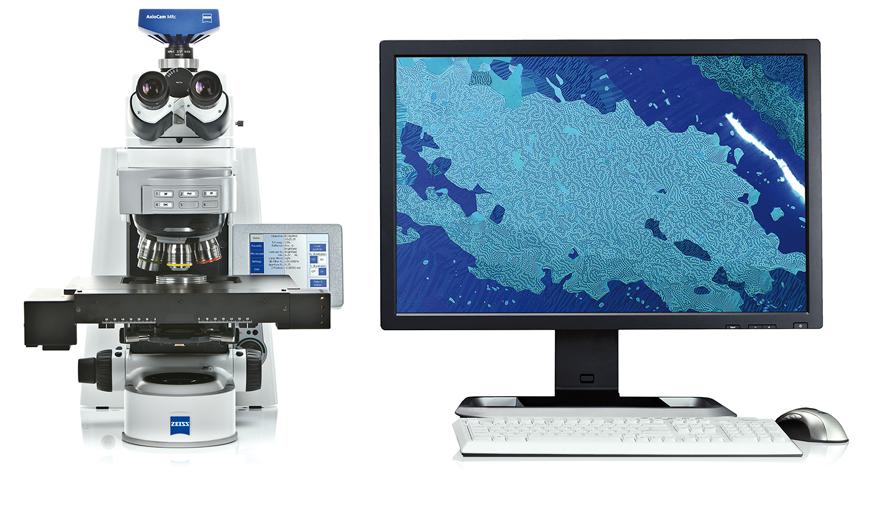
Axio Imager 2 from ZEISS is your system platform tailored to demanding materials analysis tasks, development of new materials as well as quality control.
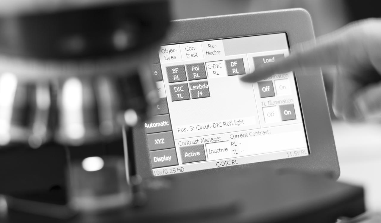
You always profit from crisp images and high optical performance. This applies in particular to sophisticated contrasting techniques, e.g. like the Circular Differential Interference Contrast (C-DIC) and polarization contrast.
Use the motorized stand to achieve reproducible illumination settings and, consequently, constant image quality. You always obtain comparable results and high productivity by automating your workflow. Axio Imager 2 offers a high degree of adaptability in line with your future requirements. The stands are open to expand and cover a wide range of applications.
In Brief
The Advantages
The Applications
The System
Technology and Details › Service
Whether in research, testing or failure analysis, materials microscopy faces quite various challenges. With Axio Imager 2 from ZEISS you will be able to meet and win these challenges. Attach application-specific components and perform e.g. particle analysis, investigate non-metallic inclusions (NMI), liquid crystals or semiconductorbased MEMs. Axio Imager 2 supports the correlative workflow to electron microscopic investigations, too.
Choose from a variety of contrasting techniques to achieve an optimum image quality for your dedicated applications. Examine your samples in reflected light in brightfield, darkfield, Differential Interference Contrast (DIC), Circular Differential Interference Contrast (C-DIC), polarization or fluorescence contrast. For transmitted light you can choose between brightfield, darkfield, Differential Interference Contrast (DIC), polarization or circular polarization. Minimized stray light enables homogenous illumination. You achieve outstanding image contrast, even at high magnifications.
Stability is essential if you want to obtain good results. You will appreciate the stable imaging conditions of Axio Imager 2, especially when working with high magnifications and performing time dependent studies. Due to the motorization of Axio Imager 2 you will achieve quick and reproducible results while you always work under constant conditions. For instance, the motorized apertures and the illumination control, which automatically adjusts the color temperature via filter wheels.
› In Brief
› The Advantages
› The Applications
› The System
› Technology and Details
› Service
Experience Competence in all Contrasting Techniques
Brightfield and Darkfield:
Maximum Homogeneity and a Stray Light Free Image Background
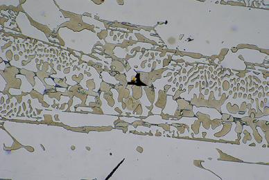
In brightfield Axio Imager 2 provides homogeneous illumination and exceptional contrast. By minimizing disturbing stray light and reducing the longitudinal color aberration of the illumination optics, the darkfield illumination contrast is suitable for the most challenging samples and impresses even when faced with finest structures. Switching between the techniques only requires a simple turn. The motorized stands allow you to work particularly quickly and conveniently.
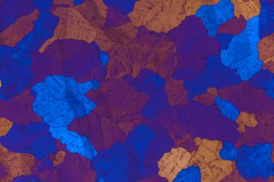
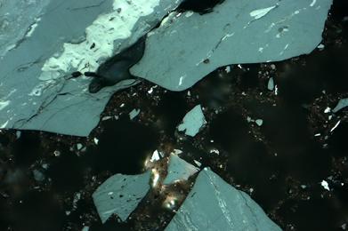
Perfect for All Structures
Circular Differential Interference Contrast (C-DIC) is a polarization-optical technique which, in contrast to ordinary Differential Interference Contrast (DIC), uses circularly polarized light. This technique has a number of decisive advantages for the contrasting of differently aligned object structures. The specimen no longer has to be rotated for best image
contrast and quality, as it is the case in basic DIC. With C-DIC it is simply enough to adjust the position of the C-DIC prism to achieve best image quality whether it is for contrast and/or resolution independent of sample orientation. And all this is possible using one C-DIC prism for a homogeneous unsurpassed quality image.
› In Brief
› The Advantages
› The Applications
› The System
› Technology and Details
› Service
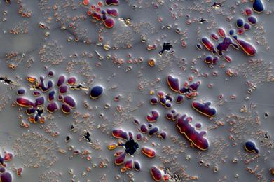
Experience Competence in All Contrasting Techniques

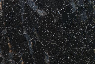


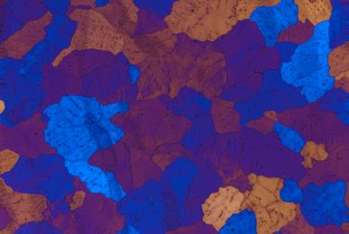
Sample:
› In Brief
› The Advantages
› The Applications
› The System
› Technology and Details
› Service
Industry, Typical Applications, Typical Samples Task
Automotive Industry
Aviation and Space Industry
Metal Producing and Processing Industry
Oil, Gas and Mining Industry
Particle Analysis
Correlative Microscopy
• Quality control and development of compound materials
• Quality control of welded joints
• Examination of inclusions and cracks
• Determination of grain sizes and non-metallic inclusions
• Particle Analysis
• Quality control and development of compound materials
• Quality control of welded joints
• Examination of inclusions and cracks
• Determination of grain sizes and microstructure phases
• Examination of inclusions and cracks
• Determination of grain sizes and non-metallic inclusions
• Analysis of anisotropic materials
• Analysis of texture and microstructure
• Analysis of pores
• Fluorescence analysis
• 2D- and 3D-Imaging
• Inspection of cleanliness acc. to ISO 16232, VDA 19
• Determination of residual contamination of oil and lubricants acc. to ISO 4406, ISO 4407, SAE AS 4059
• Combination of information from light- and electron microscope
• Fast relocation of Regions of Interest
ZEISS Axio Imager 2 Offers
• Hardware Auto Focus
• Correlative microscopy with ZEN module Shuttle & Find
• Polarization contrast and C-DIC
• ZEN core modules: Grains, Graphite, NMI, Multiphase
• AxioVision module: Particle Analyzer
• Hardware Auto Focus
• Correlative microscopy with ZEN core module Shuttle & Find
• Polarization Contrast and C-DIC
• ZEN core module: Grains, Graphite, Multiphase
• Hardware Auto Focus
• Correlative microscopy with ZEN core module Shuttle & Find
• Polarization Contrast and C-DIC
• ZEN core module: Grains, Graphite, Multiphase, NMI
• Laser Scanning Microscope LSM 900
• Correlative microscopy with ZEN core module Shuttle & Find
• AxioVision module Particle Analyzer
• Correlative microscopy with ZEN core module Shuttle & Find
› In Brief
› The Advantages
› The Applications
› The System › Technology and Details
› Service
Industry, Typical Applications, Typical Samples Task
Non-metallic Inclusions (NMI)
• Quantitative and qualitative analysis of microstructure of steel
• Determination of purity of steel
• Investigation of content and distribution of non-metallic inclusions based on color, brightness, shape and formation
• Evaluation of inclusions with comparative diagrams
• Precise identification of sulfides and oxides acc. to DIN 50602, EN 10247, ASTM E45, ISO 4967, GB/T 10561, SEP 1571 and JIS G 0555
ZEISS Axio Imager 2 Offers
core module NMI
3D-Topography
Temperature Microscopy
• Measurement of roughness
• Detection of height differences
• Measurement of thickness of transparent coatings, surface characteristics, color and gloss
• Examination of temperature influence on behavior of metals, crystals, ceramics and polymers
• Identification of phase transformations
• Determination of temperature for phase transition
• Determination of melting point
Laser Scanning Microscope LSM 900
Linkam heating stages and software module Linkam for ZEN core
Aviation and Space Industry
Industry

› In Brief
› The Advantages
› The Applications
› The System
› Technology and Details › Service


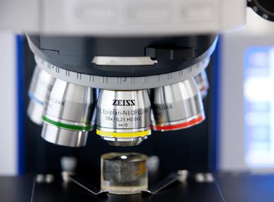
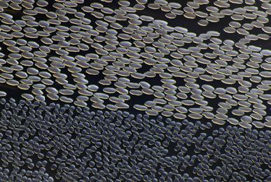
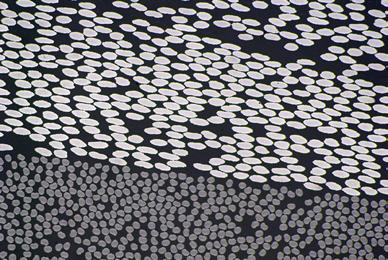
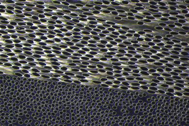
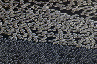
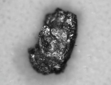
Particle analysis, brightfield, objective: EC Epiplan-NEOFLUAR
› In Brief
› The Advantages
› The Applications
› The System
› Technology and Details
› Service
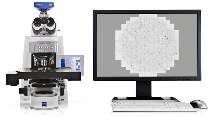
Analyze Tiny Particles: Accurately and Reproducibly
Particle Analyzer is a milestone for your quality control. With the fully motorized light microscope Axio Imager 2 you measure particles down to 2 µm.
Particle Analyzer software supports the standards for cleanliness testing ISO 16232, VDA 19, and oil analysis ISO 4406, ISO 4407, and SAE AS 4059. With the system solutions from ZEISS, you ensure that the required microscope settings are always selected correctly. You receive reliable, reproducible results nearly independent of the user carrying out the analysis. By carrying out correlative particle analyzes, you expand the depth of information contained within your findings to include the results of element and materials characterization.
› In Brief › The Advantages › The Applications › The System › Technology and Details › Service
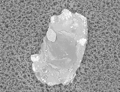
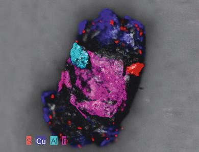
Correlative Automated Particle Analysis (CAPA): More Knowledge. Higher Quality.
Completely characterize residual dirt particles with Correlative Automated Particle Analysis from ZEISS. Detect particles with your Axio Imager 2 and relocate preselected particles automatically, using your SEM from ZEISS. Perform an EDX analyisis to reveal information of their elemental composition. Correlative Particle Analyzer automatically documents the results from both, the light microscopic and electron microscopic analysis. You receive a combined, informative report at the touch of a button.
As an experienced user, you can inspect the results of the combined light microscopic and electron microscopic analysis on an interactive overview screen. Retrieve particles at the touch of a button, automatically start new EDX analyzes, and automatically generate a report. With Correlative Particle Analyzer, your results will be available up to ten times faster than first conducting an analysis with a light microscope and then sub-sequently with an electron microscope. You can systematically focus on potentially process-critical particles. The complementary material characterization from both microscopic worlds gives you added security.
› In Brief
› The Advantages
› The Applications
› The System
› Technology and Details
› Service
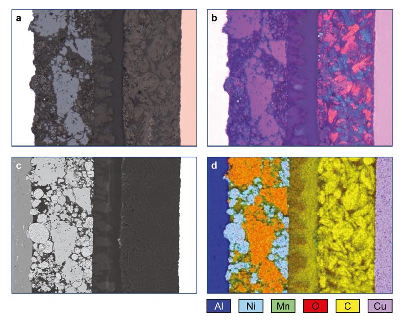
Correlative Microscopy with ZEISS Axio Imager 2: Bridging the Micro and Nano World
Are you looking for a way to combine imaging and analytical methods effectively?
Shuttle & Find offers precisely this: An easy-to-use, highly productive work fl ow from a light to an electron microscope – and vice versa. The work fl ow between the two systems has never been so easy. The precise recall of regions of interest enhances productivity. Instead of wasting valuable time searching, you now gain new insights into your samples with a few mouse clicks. Regions of interest, marked on one system, you can instantely relocate on the other system.
Open up new dimensions of information in numerous material analysis applications. Absolutely reproducible.
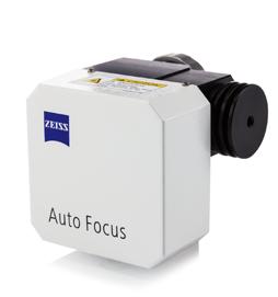
Examinations in the fields of research and industrial production (e.g. surface examinations of reflective, low-contrast specimens such as metallographic specimens and polished or textured wafers) require a fast focusing system that ensures high precision levels of max. 0.3 times the objective’s depth of field. This requirement can be easily met by combining your Axio Imager 2 with the Auto Focus system to benefit from fast and accurate focusing across a wide capture range of up to 12,000 µm. The Auto Focus system is designed to work with reflected light and transmitted light in brightfield, darkfield, polarized light and DIC.
The objective guides the structured light produced by an LED in the Auto Focus system onto the specimen, with the specimen’s surface reflecting it back. During this process, Auto Focus permanently analyses the signal and derives the appropriate control signals for the focus drive, to bring the surface into focus. The Auto Focus sensor detects changes and deviations in the focus position and compensates them automatically. The Auto Focus system comes with three different modes corresponding to different specimen characteristics (reflective/partially reflective/diffuse) and with three different precision levels (precision/balance/ speed).
› In Brief
› The Advantages
› The Applications
› The System
› Technology and Details
› Service
1 Microscope
• Axio Imager.A2m (encoded)
• Axio Imager.D2m (encoded, partly motorizable)
• Axio Imager.M2m (motorizable, TL manual)
• Axio Imager.Z2m (motorizable, TL motorized)
2 Objectives
Reflected Light
• EC EPIPLAN
• EC Epiplan-NEOFLUAR
• EC Epiplan-APOCHROMAT
Transmitted Light
• N-ACHROPLAN
• EC Plan-NEOFLUAR
• Plan-APOCHROMAT
• C-APOCHROMAT
• FLUAR
Long Working Distance
• LD EPIPLAN
• LD EC Epiplan-NEOFLUAR
3 Illumination Reflected Light
• MicroLED
• VisLED
• Halogen
• HBO / HXP
Transmitted Light
• MicroLED
• VisLED
• Halogen
4 Cameras
• Axiocam 105
• Axiocam 305
• Axiocam 506
• Axiocam 705
• Axiocam 712
5 Software
• ZEN core
• ZEN starter
6 Accessories
• Auto Focus
• Linkam heating- and cooling stages
• Focus Linear Sensor
• Correlative Microscopy
426702-0000-000
Condenser module DIC III/0.9 with polarizer 426703-0000-000 Polarizer D for condensers 0.8 and 0.9 427710-9050-000
Achromatic-aplanatic universal condenser 0.9 H D Ph DIC, mot. 424201-9902-000
Achromatic-aplanatic universal condenser 0.9 H D Ph DIC 424200-9901-000
Achromatic-aplanatic pathology condenser 0.9 H mot. 424220-9901-000
Achromatic-aplanatic pathology condenser 0.9 H 424219-9901-000
Condenser module DIC II/1.4 with polarizer
426708-0000-000
Condenser module DIC III/1.4 with polarizer 426709-0000-000
Achromatic-aplanatic condenser 1.4 H D Ph DIC 424208-0000-000
Condenser module DIC 0/0.8 with polarizer 426704-0000-000 Condenser module DIC I/0.8 with polarizer 426705-0000-000 Condenser module DIC II/0.8 with polarizer 426706-0000-000 Polarizer
Condenser, LD achromatic
H
424206-9901-000
Large-field DF slider for 2.5x-5x 424215-0000-000
Slider with phase stop Ph1 for condenser 0.9 H/0.4
Achromatic LD condenser 0.8 H D Ph DIC 424204-9901-000
424218-0000-000
Achromatic-aplanatic universal condenser 0.9 H Pol 424212-9901-000
Glass insert plate 432312-0000-000 Metal insert plate 432313-0000-000 Adapter plate for mounting frames 432310-0000-000 Mounting frame for slide 76x26 432315-0000-000 Rotary stage Pol 360°,
105x105/85 mm SCD 432018-9010-000 Display unit SCDplus 432035-9110-000 Mechanical stage, right transmitted/reflected light, 105x105/85 mm 432018-9020-000 Stage insert 160x116 mm with holder for 4" wafer, rotatable 432322-9010-000
Stage controller XY PIEZO; USB 432901-9903-000 Joystick XY for stage controller PIEZO/MCU 2008 432903-9902-000
432024-9903-000
Joystick XY for stage controller PIEZO/MCU
Technology and Details
Eyepiece insert plate on request
Eyepiece PL 10x/25 Br. foc.
444034-9000-000
Eyepiece E-PL 10x/25 Br. foc.
444234-9902-000
Eyepiece E-PL 10x/23 Br. foc. 444235-9901-000
Auxiliary microscope, d=30 444830-9902-000
Comfortable binocular Ergotube 8-33°/23, 50 mm high, reversed image 425518-9010-000
Binocular tube 30°/23, reversed image, Axio Imager 425520-9060-000
Eyepiece PL 10x/23 Br. foc. 444036-9000-000
Binocular phototube 30°/25 mot. with two camera ports 60N 425504-0000-000
Binocular tube 30°/25, reversed image 425500-0000-000
Tube carrier multidiscussion for 2 tubes, connect linear left/right
425145-9020-000
Tube carrier multidiscussion for 1 tube, arm left deflection, connect
425145-9030-000
Tube carrier multidiscussion for 1 tube, arm right deflection, connect
425145-9040-000
Tube carrier multidiscussion for 2 tubes, end panel linear, l/r
425145-9050-000
Tube carrier for 1 Co-observer, light-intensive, end panel, left 425145-9060-000
Note: For tube lens turrets the eyepieces PL 10x/25 Br. foc or PL 10x/23 Br. foc have to be used.
Note: The tubes 425500-0000-000, 425502-0000-000, 425503-9901-000, 425506-0000-000, 425515-0000-000, 425518-9010-000, can be combined with a tube lens turret or the center component for multidiscussion equipment.
Binocular phototube 30°/23 (50:50), reversed image, Axio Imager
425520-9070-000
Binocular phototube 30°/25 (100:0/30:70/0:100), reversed image 425506-0000-000
Binocular phototube 30°/25 (30vis:70doc), reversed image 425501-0000-000
Binocular Ergophototube 20°/23 MAT (100:0/0:100), upright image
425514-0000-000
Binocular phototube Pol 15°/23 (100:0/0:100), upright image 425517-0000-000
Quartz depolarizer with tube-lens for tubes Axio Scope.A1 428106-9000-000 (not in use with 425514 and 425517
Binocular phototube 15°/25 (100:0/0:100), upright image 425503-9901-000
Binocular phototube 30°/25 (100:0/30:70/0:100), reversed image 425502-0000-000
Analyzer slider fixed for transmitted light, 6x20 433605-0000-000
Comfortable binocular Ergophototube 15°/23 (50:50), upright image 425515-0000-000
Binocular phototube 6-25°/23 (100/100) 425518-9020-000
Quartz depolarizer with tube-lens for tubes Axio Imager 428106-9030-000
ACR P&C for reflected light 424929-9903-000 C-DIC prism for modulator turret 426921-0000-000 C-DIC prism for modulator turret 426922-0000-000 TIC prism for modulator turret for EC EPN 5x-100x 426923-9901-000 Antiglare screen 452163-0000-000
Center component for multidiscussion, for tube carrier left and right 425141-9901-000
We recommend the following components for easy cleaning of oil immersion on material samples: Immersol M, Oiler 20 ml 444965-0000-000 Immersol M, Bottle 100 ml 444966-0000-000 or Center comp. co-observation, for tube carrier light-intensive, left 425143-9000-000
Ob ect ves M27 ICS-Objectives on request
Diffusion disk for reflected-light illumination, switchable
423632-0000-000 (included in A2m, D2m, M2m, Z2m)
Switching mirror for 2 illuminators
447230-9903-000
Switching mirror mot.; CAN 447229-0000-000
Reflected-light illuminator for FL and HD for Axio Imager
423600-0000-000
Shutter, standard (included in A2m, D2m)
Reflected-light illuminator mot. for FL and HD for Axio Imager
423601-9901-000
Shutter, standard (included in M2m, Z2m)
Camera path deflection on the left side, interface 60N
425103-0000-000
Camera path deflection on the left side, mot., interface 60N
Shutter high speed, reflected-light for Axio Imager M and Z 423622-9901-000
Shutter, standard (included in reflected-light illumination)
for camera path deflection required: Beam splitter 50% for camera deflection, 34x46x2.2 mm
425110-9120-000 or Path deflecting mirror 100% for camera deflection, 34x46x4 mm 425110-9110-000 optional: Fine drive
Transmitted-light illumination for Axio Imager 2 423900-9901-000
Transmitted-light illumination mot. for Axio Imager 2 423901-9902-000 Shutter for transmitted-light illumination 423621-9901-000
› The Advantages › The Applications › The System
› Technology and Details › Service
Manager
Manager
Control
Included
Optional › In Brief
In Brief
The Advantages
The Applications
The System
Technology and Details
Service
Dimensions (width x depth x height)
Axio Imager stand, manual with HBO 100 approx. 300 mm × 721 mm × 505 mm
Axio Imager stand, motorized with HBO 100 and TFT display approx. 390 mm × 721 mm × 505 mm
Weight
Axio Imager, manual/motorized (dependent on equipment) approx. 18 to 40 kg
Ambient Conditions Transport (in packaging):
Permissible ambient temperature
Storage
Permissible ambient temperature
Permissible relative humidity (no condensation)
Operation
Permissible
Permissible
Operating data for coded Axio Imager, equipped with an integrated power supply or motorized Axio Imager using the VP232-2 external power supply
Operating environment
Protection Class I
Protection Type IP 20
room
Electrical safety in compliance with DIN EN 61010-1 (IEC 61010-1) including CSA and UL directives
Overvoltage category II
Radio interference suppression in accordance with EN 55011 Class B
Noise immunity in accordance with DIN EN 61326 -1
Line voltage for integrated power supply
Line voltage for external power supply VP232-2
Line
and
to 240 V ±10 % Change of line voltage setting is not required!
Power consumption of coded Axio Imager max. 260 VA
Power consumption of Axio Imager, motorized max. 190 VA
LED illuminator
Attachment lamp VIS-LED
Transformer HBO 100
Operating environment
to 700 nm, peak at 460 nm
to 700 nm, peak at 460 nm
Fuses in Accordance with IEC 127
Axio Imager microscope stand, manual
Power supply VP232-2 for Axio Imager, mot. T 4.0 A / 250V, 5×20 mm
Transformer HBO 100 T 2.0 A/H, 5×20 mm
The Advantages
The Applications › The System › Technology and Details
Service
Light Sources
Halogen lamp 12 V/100 W
Adjustment of light source continuous, approx. 0.7 to 12 V
Mercury vapor short-arc lamp HBO 103 W/2
Power consumption of HBO 103 W/2
Axio Imager, coded
Stand with manual stage focusing
Applicable for specimens weighing up to 5 kg › In Brief
Achromatic-aplanatic universal condenser 0.9 H D Ph DIC with swivel-type front lens, achromatic-aplanatic 0.9 DIC
Objective change
Change of method modules
Axio Imager, motorized
Stand with motorized stage focusing
W
Coarse drive approx. 2 mm/revolution
Fine drive approx. 1/10 gear transmission ratio
Lifting range max. 25 mm
Height stop mechanically adjustable
for objective magnifications <10× front lens 0.9 swiveled out for objective magnifications ≥10× front lens 0.9 swiveled in 8 position turret disc
Manually via 6-position or 7 position nosepiece, HD or HD DIC M27
Manually via 6-position reflector turret
Mean step size of stepper motor
Quick lowering/lifting of stage
Lifting range
Height stop
Focusing speed
Achromatic-aplanatic universal condenser 0.9 H D Ph DIC, mot. with swivel-type front lens, achromatic-aplanatic 0.9 DIC
Objective change
Change of method modules
nm (Axio Imager.M2)
nm ±10 (Axio Imager.Z2)
for objective magnifications <10× front lens 0.9 swiveled out for objective magnifications ≥10× front lens 0.9 swiveled in 8 position turret disc
Manually or motorized via 6 position or 7 position nosepiece
Manually via 6 position reflector turret
Motorized via 6 position or 10 position reflector turret
Manually/motorized via DIC or C-DIC modulator turret
High-performance focus for scanning stages
Because the ZEISS microscope system is one of your most important tools, we make sure it is always ready to perform. What’s more, we’ll see to it that you are employing all the options that get the best from your microscope. You can choose from a range of service products, each delivered by highly qualified ZEISS specialists who will support you long beyond the purchase of your system. Our aim is to enable you to experience those special moments that inspire your work.
Attain maximum uptime with your microscope. A ZEISS Protect Service Agreement lets you budget for operating costs, all the while reducing costly downtime and achieving the best results through the improved performance of your system. Choose from service agreements designed to give you a range of options and control levels. We’ll work with you to select the service program that addresses your system needs and usage requirements, in line with your organization’s standard practices.
Our service on-demand also brings you distinct advantages. ZEISS service staff will analyze issues at hand and resolve them – whether using remote maintenance software or working on site.
Enhance Your Microscope System.
Your ZEISS microscope system is designed for a variety of updates: open interfaces allow you to maintain a high technological level at all times. As a result you’ll work more efficiently now, while extending the productive lifetime of your microscope as new update possibilities come on stream.


Carl Zeiss Microscopy GmbH
07745 Jena, Germany microscopy@zeiss.com www.zeiss.com/axioimager-mat