1 minute read
NEWS NOTES


Is There a Safer Entrance to Caudal Pouches of the Lateral Femorotibial Joint?
Compared with other synovial compartments of the stifle, arthroscopic approaches to the caudal pouches of the lateral femorotibial joint are more challenging. The surgeon risks complications such as damaging the iatrogenic nerve and cartilage.
Advertisement
Surgeons found that an arthroscopic portal immediately cranial to the lateral collateral ligament permitted consistent entry into the popliteal tunnel and both pouches of the caudal lateral femorotibial joint, resulting in fewer complications
This cadaver and clinical study described and evaluated a novel arthroscopic approach to the caudal pouches of the lateral femorotibial joint as current approaches are considered challenging and risky.

Arthroscopic view of the right cranial compartment of the lateral femorotibial joint. The trajectory into the lateral recess is marked bythe green circle, and is located within the center of a triangle boundedby the lateral femoral condyle (LFC), cranial horn of the lateralmeniscus and associated meniscotibial ligament (LM) and finally thecombined body of the long digital extensor tendon and peroneus tertius(LDE/PT)
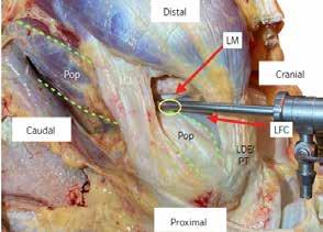
Anatomic dissection with the arthroscope and cannula in situ for the described lateral portal. The proposed skin portal (yellow circle) is located on the cranial aspect of the lateral collateral ligament (LCL)and caudal to the combined origin of the long digital extensor tendonand peroneus tertius (LDE/PT). The lateral femoral condyle (LFC) andlateral meniscus (LM) have been outlined, in addition to the tendinousand muscular portions of the popliteus (Pop; outlined by green dashedlines)
The surgical technique was developed initially using 19 cadaver limbs positioned to simulate dorsal recumbency and with the stifle held in 90° flexion. A portal was made immediately cranial to the lateral collateral ligament and the arthroscope advanced along the popliteal tunnel of the femorotibial joint in a cranial to caudal direction.
Following the cadaver study, 33 horses underwent inspection of 38 caudal lateral femorotibial joints using the alternative technique as part of routine joint inspection in 33 horses. Entry and examination of both pouches of the caudal lateral femorotibial joint were consistently achieved in both the cadaver and clinical limbs, with no intra- or post-operative complications in the latter.
For more information:
O’Neill HD, Bladon BM. An alternative arthroscopic approach to the caudal pouches of the equine lateral femorotibial joint. Equine Vet J. 2020 May 4 (Epub ahead of print). https://beva.onlinelibrary.wiley.com/doi/10.1111/ evj.13274