ORAL
and maxillofacial pathology case of the month
Clinical History
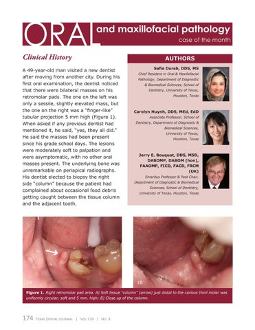
A 49-year-old man visited a new dentist after moving from another city. During his first oral examination, the dentist noticed that there were bilateral masses on his retromolar pads. The one on the left was only a sessile, slightly elevated mass, but the one on the right was a “finger-like” tubular projection 5 mm high (Figure 1). When asked if any previous dentist had mentioned it, he said, “yes, they all did.” He said the masses had been present since his grade school days. The lesions were moderately soft to palpation and were asymptomatic, with no other oral masses present. The underlying bone was unremarkable on periapical radiographs. His dentist elected to biopsy the right side “column” because the patient had complained about occasional food debris getting caught between the tissue column and the adjacent tooth.
1A
AUTHORS Safia Durab, DDS, MS Chief Resident in Oral & Maxillofacial Pathology, Department of Diagnostic & Biomedical Sciences, School of Dentistry, University of Texas, Houston, Texas
Carolyn Huynh, DDS, MEd, EdD Associate Professor, School of Dentistry, Department of Diagnostic & Biomedical Sciences, University of Texas, Houston, Texas
Jerry E. Bouquot, DDS, MSD, DABOMP, DABOM (hon), FAAOMP, FICD, FACD, FRCM (UK) Emeritus Professor & Past Chair, Department of Diagnostic & Biomedical Sciences, School of Dentistry, University of Texas, Houston, Texas
1B
Figure 1. Right retromolar pad area. A) Soft tissue “column” (arrow) just distal to the carious third molar was uniformly circular, soft and 5 mm. high; B) Close up of the column.
174
Texas Dental Journal | Vol 139 | No. 4


