2 minute read
International Journal for Research in Applied Science & Engineering Technology (IJRASET)


ISSN: 2321-9653; IC Value: 45.98; SJ Impact Factor: 7.538
Advertisement
Volume 11 Issue III Mar 2023- Available at www.ijraset.com
VII. MODULEDESCRIPTION
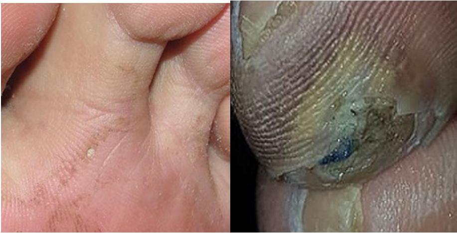
A. Image Acquisition Image Acquisition is the process of collection of images. We had performed a refined search on the dataset of many object images.
B. Image Pre-processing
Image pre-processing includes converting RBG (colour) images into Grayscale images using MATLAB. To improve the available dataset, RGB is converted to grayscale. Converting the images to grayscale helps in improving the accuracy of the result.
C. Image Segmentation
The conversion of segmented objects into representations that more accurately reflect their primary characteristics and qualities is the common objective of feature extraction and representation techniques. The goal is to simplify or change the representation into more meaningful image. It sets apart the things from the background or other objects that we wish to examine more closely.
D. Feature Extraction
Feature extraction is extracting or showing of the portion of the tumour so that classification becomes easy. To discriminate between the photos, features are retrieved. Nearly all machine vision techniques rely on features extraction. The conversion of segmented objects into representations that more accurately reflect their primary characteristics and qualities is the common objective of feature extraction and representation techniques. Here, shape of the wound is extracted.
E. Classification
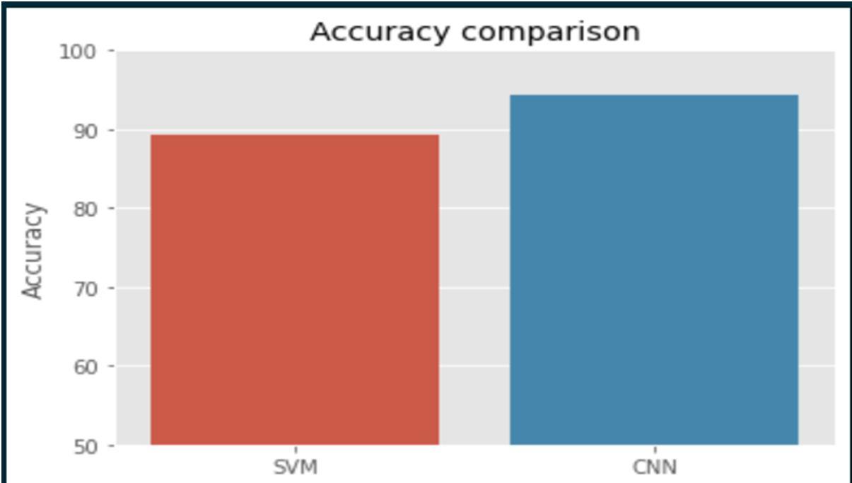
Based on the augmented images that are obtained so far, the machine learning algorithms like a SVM and CNN algorithms are used to train the images to classify them into normal and abnormal. The new input images are then tested.
VIII. RESULTANALYSIS
ISSN: 2321-9653; IC Value: 45.98; SJ Impact Factor: 7.538
Volume 11 Issue III Mar 2023- Available at www.ijraset.com
IX. CONCLUSION
The goal of proposed system is to provide the better wound image and healing status analysis. With these methods patients are actively involved in their own care and the wound analysis systems where by physicians can remotely access the image of wound and the result of wound healing. Also, it will reduce thepatients rare tensity and stress. Through the segmentation of wound photos, a doctor can quickly diagnose the issue. Mobile applications for ulcer recognition and user-friendly software tools are to be implemented. In future an automatic ulcer detection, recognition, and segmentation with the help of these classifiers can be developed to help patients receive better treatment and care.
References
[1] Reiber GE, Ledoux W R. Epidemiology of diabetic foot ulcers and amputations: Evidence for prevention. In: Williams R, Herman W, Kinmonth AL, Wareham NJ, editors. The evidence bases for diabetes care. Chichester; Hoboken (NJ): John Wiley & Sons, Ltd; 2003. pp. 641–665.
[2] Apelqvist J, Bakker K, van Houtum WH, Nabuurs- Franssen MH, Schaper NC. International consensus and practical guidelines on the management and the prevention of the diabetic foot. International Working Group on the Diabetic Foot. Diabetes Metab Res Rev. 2000;16Suppl 1: S84–S92.
[3] Khanolkar MP, Bain SC, Stephens JW. The diabetic foot.QJM. 2008; 101:685–695.
[4] UK Prospective Diabetes Study (UKPDS) Group. Intensive blood-glucose control with sulphonylureas or insulin compared with conventional treatment and risk of complications in patients with type 2 diabetes (UKPDS 33) Lancet. 1998; 352:837–853
[5] Williams DR, Airey M. The size of the problem: Epidemiological and economical aspects of the diabetic foot. In: Boulton AJM, Connor H and Cavanagh PR the foot in diabetes, editors. Chichester England; New York: Wiley; 2000. pp. 3–17.
