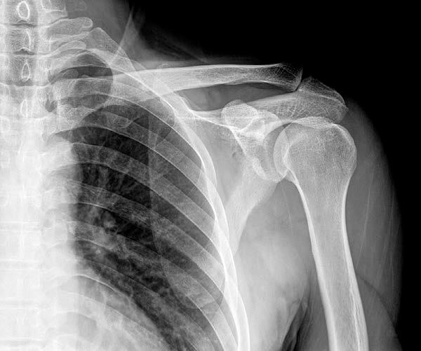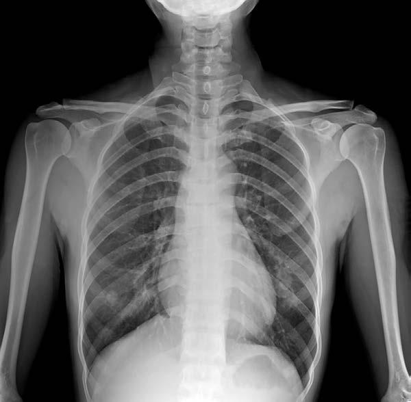
lesion. This is seen with posterior dislocations of the shoulder. They often need CT scan or MRI scanning in order to detect these. The AC joint dislocation is seen on the AC image of the shoulder joint. If there is a dislocation of more than eight millimeters, this is representative of a significant subluxation. You may need to get an image of the opposite side in order to have a representation of what the normal AC joint looks like. Osteoarthritis of the shoulder can be detected on x-ray. It can be a primary problem or secondary to a fracture or rotator cuff injury. There will be narrowing of the joint space from loss of cartilage, subchondral sclerosis with increased bone production, osteophyte formation, synovitis, and subchondral cysts. Rotator cuff impingement is seen when the rotator cuff tendons have been pinched between areas of bone. The pain is anterior and lateral with abduction or lifting of the arm being the most painful. The impingement usually occurs between the top of the acromion and the humeral head. This can lead to weakness and degeneration of the tendons. The supraspinatus tendon is the most susceptible to this type of impingement. The plain film can be helpful because it can show calcifications of the tendon.
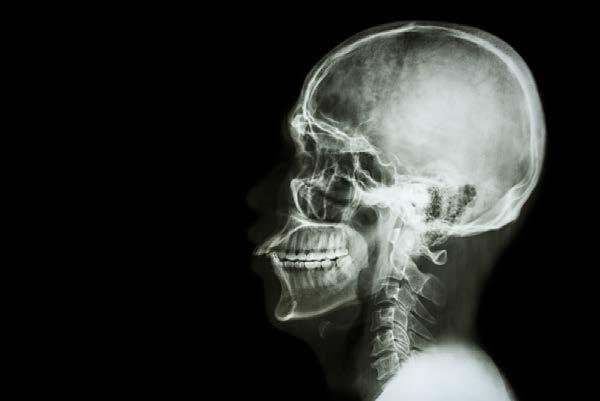
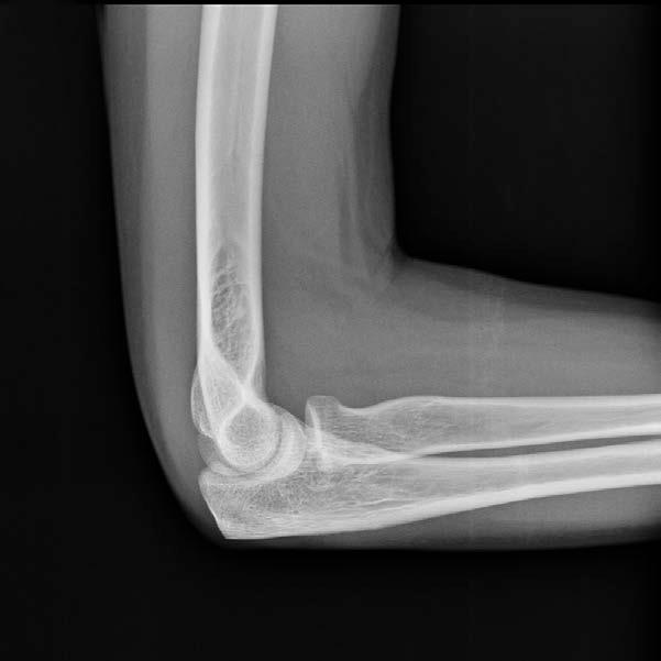
ELBOW PLAIN FILMS This generally results in obtaining a standard AP and lateral image of the elbow, although a capitellum image can be done, which views the radial head. The AP image is done with the palm facing up, which is external rotation of the elbow. A good film takes one of the joint and a third each of the distal humerus and the proximal radius and ulna. The lateral involves the shoulder at the same level as the elbow with the medial side of the patient’s arm in contact with the x-ray table. The palm faces the patient. The elbow is flexed to ninety degrees. The capitellum image is of the radial head for subtle fractures of this structure. The patient is positioned the same way as the lateral image with the image taken at a 45-degree angle to the x-ray table. There are actually three separate joints in the elbow. The humeroulnar joint involves the olecranon of the ulna articulating with the humerus through its trochlea. The
21